Embed Size (px)
Citation preview
Caldas, L. A., Lemgruber Soares, L., Seabra, S. H., Attias, M. and de
Souza, W. (2016) Monitoring of dynamin during the Toxoplasma gondii
cell cycle. Pathogens and Disease, 74(9), ftw108.
(doi:10.1093/femspd/ftw108)
This is the author’s final accepted version.
There may be differences between this version and the published version.
You are advised to consult the publisher’s version if you wish to cite from
it.
http://eprints.gla.ac.uk/132539/
Deposited on: 14 December 2016
Enlighten – Research publications by members of the University of Glasgow
http://eprints.gla.ac.uk
1
Monitoring of dynamin during the Toxoplasma gondii cell cycle
Lucio Ayres Caldas1,*
, Leandro Lemgruber Soares2,3
, Sergio Seabra4, Marcia Attias
1 and
Wanderley de Souza
1Instituto de Biofísica, Universidade Federal do Rio de Janeiro, Rio de Janeiro, 21941-902,
Brazil 2Instituto Nacional de Metrologia Normalizacao e Qualidade Industrial, Duqye de Caxias, RJ,
25250-020, Brazil 3University of Glasgow, Wellcome Trust Centre for Molecular Parasitology, 120 University
Place Glasgow, Glasgow, G12 8QQ, UK 4Microbiologia, UEZO, Av. Manuel Caldeira de Alvarenga, Rio de Janeiro, 23070-200, Brazil
*Corresponding author: E-mail: [email protected]
Abstract
The obligate intracellular protozoan parasite Toxoplasma gondii actively invades virtually all
warm-blooded nucleated cells. This process results in a non-fusogenic vacuole, inside which the
parasites replicate continuously until egress signaling is triggered. In this work, we investigated
the role of the large GTPase dynamin in the interaction of T. gondii with the host cell by using
laser and electron microscopy during three key stages: invasion, development and egress. The
detection of dynamin during invasion indicates the occurrence of endocytosis, while T. gondii
egress appeared to be independent of dynamin participation. However, the presence of dynamin
during T. gondii development suggests that this molecule plays undescribed roles in the
tachyzoite’s cell cycle.
1. Introduction
The apicomplexan parasite Toxoplasma gondii, which causes toxoplasmosis, is capable
of infecting virtually all nucleated cells of warm-blooded hosts and is estimated to infect one-
third of the world`s population. Host cell invasion relies on the active participation of the
parasite and the concomitant generation of a non-fusogenic parasitophorous vacuole (PV),
which is often translocated to the perinuclear region (Sibley 2004). After multiple replication
2
rounds, the parasites exit the PV, carrying remnants of the PV and ER membranes, before
eventually crossing the host cell plasma membrane (Caldas et al 2010).
Recent studies demonstrated that the inhibition of the large GTPase dynamin
successfully blocks internalization and/or infection for a variety of viruses (Abban et al 2008;
Mues et al 2015; Piccini et al 2015) and T. gondii invasion (Caldas et al 2013). This GTPase is
well known for pinching off endocytic vesicles from the plasma membrane and the trans-Golgi
network by polymerizing to form a helix around the neck of budding vesicles, leading to
membrane fission. The role of this GTPase has been extended to membrane remodeling,
cytoskeleton regulation via direct interactions with actin, and participation in exocytosis
(Arneson et al 2008; Mettlen et al 2009; Gu et al 2010; Ferguson and De Camilli 2012;
Williams and Kim 2014).
These roles of dynamin led us to hypothesize that this GTPase participates not only in
the disconnection of the PV from the host cell plasma membrane but also in the translocation of
the PV to the perinuclear region of the infected cell. To test this hypothesis, we labeled
dynamin during parasite invasion, development and egress, which are the three key steps of the
T. gondii cellular cycle, and used confocal microscopy, super-resolution microscopy and cryo-
immunomicroscopy techniques.
2. Materials and methods
2.1. Chemicals
The calcium ionophore A23817 was purchased from Sigma Chemical Company (St.
Louis, MO, USA) was employed during egress assay. For this compound, viability tests with
neutral red were performed, and no cytotoxic effect was observed with the concentrations and
incubation time used.
2.2. Parasites and host cell culture
T. gondii tachyzoites of the RH wild-type strain were maintained in mice via
intraperitoneal inoculation and harvested via peritoneal washing of mice infected for 2 to 3
3
days. The suspension was centrifuged at 1,000 × g for 10 min to remove cell debris and
peritoneal leukocytes, and the number of parasites in the supernatant was quantified in a
Neubauer chamber. The parasites were suspended in Dulbecco's modified Eagle's medium
(DMEM). Swiss mice were bred at the animal facility of the Federal University of Rio de
Janeiro. The experimental protocol was approved by the Instituto de Biofísica Carlos Chagas
Filho’s Ethics Committee for animal experimentation (Protocol n. IBCCF 096/097).
Macaca mulata monkey epithelial kidney cells (LLC-MK2) were maintained in vitro in
DMEM supplemented with 10% fetal bovine serum at 37 °C in 5% CO2. The cells were grown
in 25 cm2 plastic flasks or on round glass coverslips in 24-well plates.
2.3. In vitro infection
For the invasion assay, the tachyzoites of T. gondii were allowed to interact with LLC-
MK2 cells, at ratios of 5:1 (parasites-host cell), initially for 15 min at 4°C followed by
incubation at 37 ºC and 5% CO2 for 10 min, when the samples were fixed. The intracellular
development assay required 40–50 min of parasite-host cell incubation at 37 °C and 5% CO2.
After this time, the supernatant containing the free parasites was aspirated and replaced with
fresh medium. The infection was allowed to proceed for 24 h, and the cells were then rinsed
with DMEM and fixed. For the parasite egress assay, a 9 μM solution of the calcium ionophore
A23817, diluted in serum-free DMEM, was added at this time and the monolayers were fixed
after 5 min. Untreated infected monolayers of LLC-MK2 cells were used as negative controls.
2.4. Confocal microscopy
For immunofluorescence microscopy, the cells were seeded onto round coverslips and
fixed with 4% formaldehyde in phosphate buffered saline (PBS), pH 7.2, for 20 min. Half of the
samples were permeabilized with 0.1% Triton X-100 in PBS for 10 min at room temperature.
Pre-incubation was performed with 50 mM ammonium chloride and 3% BSA in PBS, pH 8.0,
for 45 min to block the free aldehyde groups. The samples were then incubated with a primary
anti-dynamin antibody (Invitrogen, Carlsbad, CA, USA) at a 1:100 dilution for 1 h, rinsed, and
4
incubated with a 1:400 dilution of the secondary goat anti-mouse IgG (H+L) antibody
conjugated to AlexaFluor 488 (Invitrogen) at room temperature for 1 h. Actin was stained with
phalloidin red (Sigma-Aldrich) at a dilution in PBS of 1:40 for 20 min in the dark. After rinsing
with phosphate buffered saline PBS and mounting with prolong antifade (Vector Labs,
Burlingame, CA, USA), the slides were visualized using a Zeiss 510 LSM microscope.
2.5. Super-resolution microscopy
For super-resolution microscopy, the infected cells were processed in a manner similar
to that described for confocal microscopy, except additional care was taken at washing steps,
gradually increasing in duration, as recently described by Whelan and Bell (2015). Images were
collected using a Zeiss Elyra PS.1 microscope in the structured illumination (SR-SIM) mode.
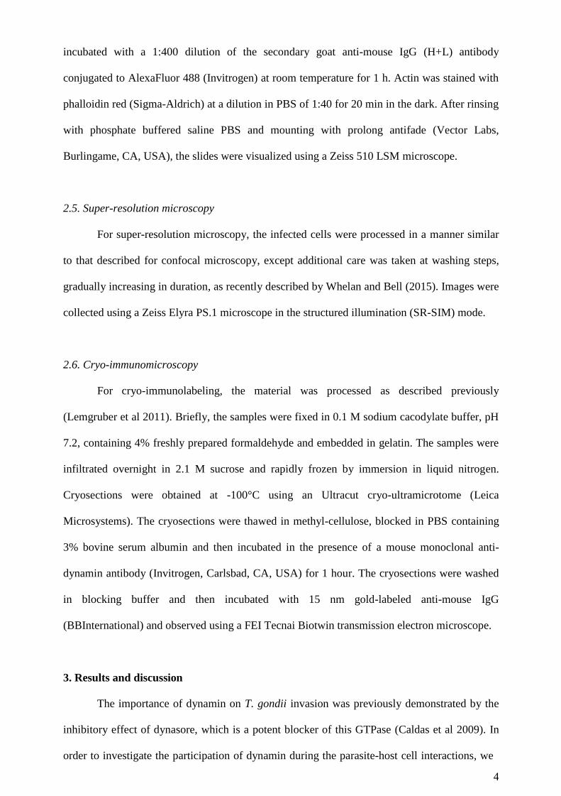
2.6. Cryo-immunomicroscopy
For cryo-immunolabeling, the material was processed as described previously
(Lemgruber et al 2011). Briefly, the samples were fixed in 0.1 M sodium cacodylate buffer, pH
7.2, containing 4% freshly prepared formaldehyde and embedded in gelatin. The samples were
infiltrated overnight in 2.1 M sucrose and rapidly frozen by immersion in liquid nitrogen.
Cryosections were obtained at -100°C using an Ultracut cryo-ultramicrotome (Leica
Microsystems). The cryosections were thawed in methyl-cellulose, blocked in PBS containing
3% bovine serum albumin and then incubated in the presence of a mouse monoclonal anti-
dynamin antibody (Invitrogen, Carlsbad, CA, USA) for 1 hour. The cryosections were washed
in blocking buffer and then incubated with 15 nm gold-labeled anti-mouse IgG
(BBInternational) and observed using a FEI Tecnai Biotwin transmission electron microscope.
3. Results and discussion
The importance of dynamin on T. gondii invasion was previously demonstrated by the
inhibitory effect of dynasore, which is a potent blocker of this GTPase (Caldas et al 2009). In
order to investigate the participation of dynamin during the parasite-host cell interactions, we
5
performed immunolabeling assays during three major steps of the T. gondii cellular cycle:
invasion, development and egress.
3.1. Invasion
The intricate signaling dynamics that characterize Apicomplexan invasion, from the
polarization of the parasites towards the host cell surface and the secretion of adhesion
molecules to PV formation, were carefully reviewed by Sharma & Chitnis (Sharma and Chitnis
2013). Molecular sieving performed at the moving junction during PV formation is determinant
to this vacuole’s non-fusogenic nature (Mordue et al 1999; Straub et al 2011). Nevertheless, this
vacuole must detach from the host cell plasma membrane, and dynamin is responsible for the
release of endocytic vesicles from the plasma membrane. Thus, this GTPase was localized by
immunofluorescence to track its position during T. gondii invasion.
In order to achieve this, the best scenario should rely on the synchronized T. gondii
invasion of host cells in culture. Then parasites interacted with the host cells for 15 min at 4°C
and were transferred to 37 °C for 10 min, after which the samples were fixed and processed for
the immunofluorescence assay. The inhibition of dynamin by dynasore was previously
demonstrated to block PV translocation to the perinuclear region (Caldas et al 2009). As
expected, dynamin is present at the site of parasite entry, forming a ring around the nascent
vacuole (Fig. 1A-B). However, dynamin appears to act at more than one site during the
pinching off of the forming PV, as shown in Fig. 1C. Dynamin labeling was intense in the area
corresponding to the moving junction, but a second area of labeling was seen at the front edge
of the nascent vacuole. This secondary site of dynamin labeling may be caused by the pinching
of vesicles containing ROP proteins, as reported by Bradley and Sibley (2007) and preceded by
seminal descriptions of the kinetics of T. gondii protein secretion during vertebrate host cell
invasion (Dubremetz et al 1993; Carruthers and Sibley 1997).
Immediately after invasion, the parasites were already internalized into a PV. Fig. 1D-E
shows an internalized parasite, for which labeling for dynamin was always negative.
6
Dynamin was also tracked during invasion via cryo-immunomicroscopy, and secondary
antibodies conjugated to 15 nm colloidal gold particles were observed at the interface between
the parasite and the host cell (Fig. 2A). This event occurred at the site where the secretion of the
rhoptry contents is believed to occur during the early stages of the interaction, which will lead
to the formation of the moving junction.
Labeling was intense at the sites of contact between the nascent PV and the apical
portion of the invading parasite, to which dynamin appears to be recruited (Fig. 2B).
Interestingly, strongly electron-dense loci also appear to correspond to the stable intracellular
clamp that is observed during the moving junction, where the parasites anchor and the
molecular sieving of PV proteins takes place (Straub et al 2011). This labeling of the interaction
sites between the tachyzoite and the host cell plasma membrane surface, after the initial contact
and/or the parasite’s reorientation of its apical end towards the host cell, also suggests the
recruitment of dynamin.
Freeze-fracture electron microscopy previously showed a pore-like structure in the
PVM, which is located in the same region as the parasite’s apical end (Dubremetz 2007). We
hypothesize that this structure results from the effect of a complex of solutes and vesicular
content that is secreted by the parasite (Bradley et al 2007; Ravindran and Boothroyd 2008),
which generates conditions for its own internalization into the cytosol. The dynamin detected at
the parasite-PVM interface could have been trapped there during the process of PV formation
and may contribute to the crossing of the PVM by secreted vesicles. Recent studies using
helium ion microscopy revealed pore-like openings in the intravacuolar face of the PV, varying
from 10 to 200 nm in diameter. These openings appear to increase in diameter relative to the
size of the PV (De Souza and Attias 2015). In addition to bacterial secretion systems (types i-
iv), secreted vesicles have been studied more recently as a keystone of pathogenic infection, in
spite of the difficulty caused by the fact that both the pathogen and the host are able to release
such vesicles to the extracellular medium, with consequences for disease pathogenesis (Schorey
et al 2015). Nevertheless, we cannot forget that in both the host cell and the parasite, a large
number of cellular processes depend on dynamin, such as membrane remodeling, endocytic
7
processes and cytoskeleton interactions (Williams and Kim 2014). For this reason, labeling was
distributed sparsely throughout the cytoplasm.
3.2. Development
Dynamin has long been known for its role during endocytosis (Van der Bliek and
Meyerowitz 1991; Chappie et al 2011), but its GTPase activity was later found to depend on an
oligomerization cycle. As this cycle is determined by the interacting molecule (i.e., lipids or
actin filaments) (Gu et al 2014), this discovery was important for improving our understanding
of additional roles of dynamin (e.g., direct and indirect actin-driven processes) (Orth and
McNiven 2003; Schlunck et al 2004; Gu et al 2010).
Because dynamin is known to be capable of nucleating actin tails via its proline-rich
domain (Lee and De Camilli 2002; Orth et al 2002), dynamin could participate in the process
of PV translocation to the perinuclear region. This translocation, which presupposes a
rearrangement of the host cell cytoskeleton, is present in pathogens, such as Chlamydia
trachomatis (Romano et al 2013).
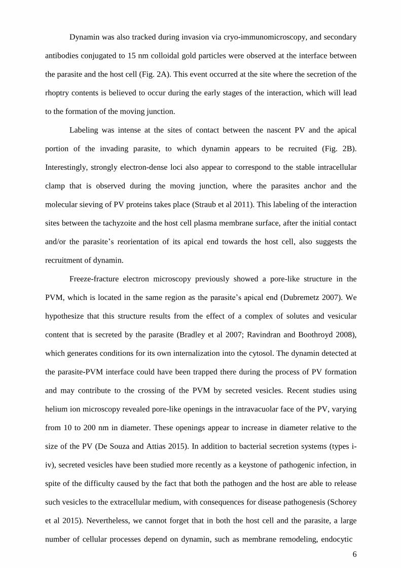
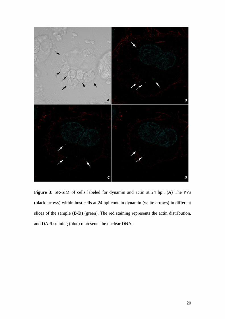
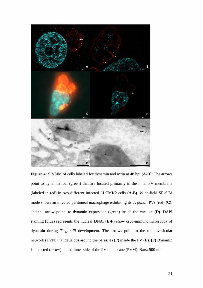
Cells were processed for SR-SIM at 24 hpi, and dynamin labeling revealed an
unexpected pattern. Punctual dynamin foci were observed inside the PVs (Fig. 3). SR-SIM
allowed an accurate localization (Fig. 3; renderization in the supplemental material), and higher
magnifications revealed that dynamin is located primarily at the inner border of the PVs, not
only in LLC-MK2 cells (Fig. 4A-B) but also in peritoneal macrophages (Fig. 4C-D and
supplemental material). However, cryo-immunomicroscopy indicated that this localization
might occur in both the tubulovesicular network (Fig. 4E) and the inner side of the PV
membrane (Fig. 4F).
It is worth noting that dynamin-like proteins appear to play a role in T. gondii apicoplast
fission (van Dooren et al 2009) and the generation of secretory pathway vesicles involved in the
formation of secretory organelles (Breinich et al 2009). However, in the present study, which is
focused on host cell dynamin, the data may indicate a distinct phenomenon. Although hard to
8
envision, the role of dynamin in parasite maturation must not be underestimated, considering
that dynamin may act to stabilize high-curvature membranes and/or mediate changes in the
physical properties of the PV membrane, similar to the changes that occur during endosomal
processes (Anitei and Hoflack 2012).
3.3. Egress
Once considered to be the reverse of invasion (Hoff and Carruthers 2012), T. gondii
egress is now understood as a distinct step of the cellular cycle. Toxoplasma egress is an active
process, which appears to result from mechanical stress caused by PV growth and subsequent
host cell ionic imbalances, leading to calcium signaling and parasite actin activation (Moudy
2001; Lourido et al 2012). During egress, no translocation of perinuclear PVs to the periphery
or plasma membrane fusion occurs, and the parasites escape by entering the host cell cytosol
and crossing the plasma membrane. When this process occurs collectively, cell lysis is observed
(Caldas et al 2010).
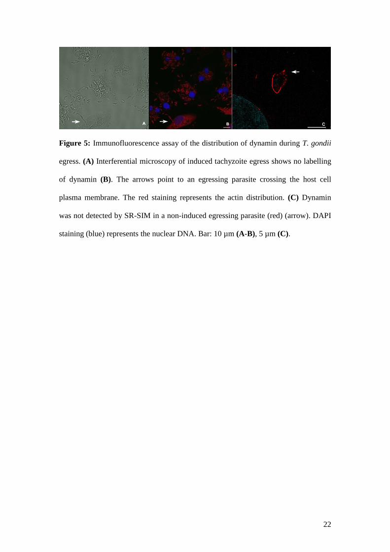
The dynamin inhibitor dynasore (Macia et al 2006) did not block T. gondii egress
(Caldas et al 2013), and this finding was expected, taking into account previous knowledge of
this large GTPase (Williams and Kim 2014). To corroborate these data, we performed an
immunofluorescence assay of T. gondii calcium ionophore-induced egress (Endo et al 1982),
and no dynamin was detected at this step of the parasite cell cycle (Fig. 5A-B). The same
outcome was shown for natural egress from the host cell, as observed by SR-SIM (Fig. 5C).
Because dynamin and dynamin-like proteins are involved in endocytosis, cellular
trafficking, and vacuole fission and fusion (Williams and Kim 2014), it is also necessary to take
into account the participation of this GTPase in the possible communication between different
PVs. It is also worth noting that the immediate association of dynamin with these endocytic
processes must be considered, as host cell endocytic structures and Golgi vesicles can be found
inside PVs (Romano et al 2013; Coppens 2014). In addition, the parasite secretes proteins
involved in the recruitment of host mitochondria, from which the tachyzoites are believed to
hijack nutrients (Sinai et al 1997; Pernas et al 2014; Mercier and Cesbron-Delauw 2015).
9
Additionally, parasite replication within PVs leads to an increase in PV size, resulting in
a mechanical stress to host cell (Moudy 2001; Magno et al 2005). However, this increase in PV
size causes a large amount of tension in the PV membrane, and host cell dynamin can
oligomerize during this high-curvature process, contributing to these changes in physical
membrane properties. Altogether, these data provide insights into the role of dynamin during
pathogen-host cell interactions.
Funding
This work was supported by grants from Conselho Nacional de Desenvolvimento
Científico e Tecnológico CNPq; Financiadora de Estudos e Projetos – FINEP and Fundação
Carlos Chagas Filho de Apoio à Pesquisa do Rio de Janeiro – FAPERJ to the authors. Lucio
Ayres Caldas and Leandro Lemgruber Soares received fellowships from Pronametro-
Inmetro.
Acknowledgements:
The authors thank Fernando Pereira de Almeida for technical assistance with the high
resolution optical microscopy.
Conflict of interest
We the authors declare no conflict of interest in writing this article.
10
References
Abban CY, Bradbury NA, Meneses PI. HPV16 and BPV1 infection can be blocked
by the dynamin inhibitor dynasore. Am J Ther 2008; 15(4):304-11.
Anitei M, Hoflack B. Bridging membrane and cytoskeleton dynamics in the secretory
and endocytic pathways. Nat Cell Biol 2012. 14(1):11-9.
Arneson LN, Segovis CM, Gomez TS et al. Dynamin 2 regulates granule exocytosis
during NK cell-mediated cytotoxicity. J Immunol 2008; 181(10):6995-7001.
Bradley PJ, Sibley LD. Rhoptries: an arsenal of secreted virulence factors. Curr Opin
Microbiol 2007; 10(6):582-7.
Breinich MS, Ferguson DJ, Foth BJ et al. A dynamin is required for the biogenesis of
secretory organelles in Toxoplasma gondii. Curr Biol 2009; 19(4):277-86.
Caldas LA, Attias M, de Souza W. Dynamin inhibitor impairs Toxoplasma gondii
invasion. FEMS Microbiol Lett 2009; 301(1):103-8.
Caldas LA, de Souza W, Attias M. Microscopic analysis of calcium ionophore
activated egress of Toxoplasma gondii from the host cell. Vet Parasitol 2010;
167(1):8-18.
11
Caldas LA, Seabra SH, Attias M et al. The effect of kinase, actin, myosin and
dynamin inhibitors on host cell egress by Toxoplasma gondii. Parasitol Int 2013;
62(5):475-82.
Carruthers VB, Sibley LD. Sequential protein secretion from three distinct organelles
of Toxoplasma gondii accompanies invasion of human fibroblasts. Eur J Cell Biol
1997; 73(2):114-23.
Chappie JS, Mears JÁ, Fang S et al. A pseudoatomic model of the dynamin polymer
identifies a hydrolysis-dependent powerstroke. Cell 2011; 147:209–22.
Coppens I. Toxoplasma, or the discovery of a heterophage. Trends Parasitol 2014;
30(10):467-9.
De Souza W, Attias M. New views of the Toxoplasma gondii parasitophorous vacuole
as revealed by Helium Ion Microscopy (HIM). J Struct Biol 2015; 191(1):76-85.
Dubremetz JF. Rhoptries are major players in Toxoplasma gondii invasion and host
cell interaction. Cell Microbiol 2007; 9(4):841-8.
Dubremetz JF, Achbarou A, Bermudes D et al. Kinetics and pattern of organelle
exocytosis during Toxoplasma gondii/host-cell interaction. Parasitol Res 1993;
79(5):402-8.
12
Endo T, Sethi KK, Piekarski G. Toxoplasma gondii: calcium ionophore A23187-
mediated exit of trophozoites from infected murine macrophages. Exp Parasitol 1982;
53(2):179-88.
Ferguson SM, De Camilli P. Dynamin, a membrane-remodelling GTPase. Nat Rev
Mol Cell Biol 2012; 13:75-88.
Gu C, Chang J, Shchedrina VA et al. Regulation of Dynamin Oligomerization in
Cells: The Role of Dynamin-Actin Interactions and Its GTPase Activity. Traffic 2014;
15(8):819-38.
Gu C, Yaddanapudi S, Weins A et al. Direct dynamin-actin interactions regulate the
actin cytoskeleton. MBO J 2010; 29(21):3593-606.
Hoff EF, Carruthers VB. Is Toxoplasma egress the first step in invasion? Trends
Parasitol 2002; 18(6):251-5.
Lee E, De Camilli P. Dynamin at actin tails. Proc Natl Acad Sci U S A 2002;
99(1):161-6.
Lemgruber L, Lupetti P, Martins-Duarte ES et al. The organization of the wall
filaments and characterization of the matrix structures of Toxoplasma gondii cyst
form. Cell Microbiol 2011; 13(12):1920-32.
13
Lourido S, Tang K, Sibley LD. Distinct signalling pathways control Toxoplasma
egress and host-cell invasion. EMBO J 2012; 31(24):4524-34.
Macia E, Ehrlich M, Massol R et al. Dynasore, a cell-permeable inhibitor of dynamin.
Dev Cell 2006; 10(6):839-50.
Magno RC, Lemgruber L, Vommaro RC et al. Intravacuolar network may act as a
mechanical support for Toxoplasma gondii inside the parasitophorous vacuole.
Microsc Res Tech 2005; 67(1):45-52.
Mercier C, Cesbron-Delauw MF. Toxoplasma secretory granules: one population or
more? Trends Parasitol 2015; 31(2):60-71.
Mettlen M, Pucadyil T, Ramachandran R et al. Dissecting dynamin's role in clathrin-
mediated endocytosis. Biochem Soc Trans 2009; 37(Pt 5):1022-26.
Mordue DG, Desai N, Dustin M et al. Invasion by Toxoplasma gondii establishes a
moving junction that selectively excludes host cell plasma membrane proteins on the
basis of their membrane anchoring. J Exp Med 1999; 190(12):1783-92.
Moudy R, Manning TJ, Beckers CJ. The loss of cytoplasmic potassium upon host cell
breakdown triggers egress of Toxoplasma gondii. J Biol Chem 2001; 276(44):41492-
501.
14
Mues MB, Cheshenko N, Wilson DW et al. Dynasore disrupts trafficking of herpes
simplex virus proteins. J Virol 2015; 89(13):6673-84.
Orth JD, Krueger EW, Cao H et al. The large GTPase dynamin regulates actin comet
formation and movement in living cells. Proc Natl Acad Sci U S A 2002; 99(1):167-
72.
Orth JD, McNiven MA. Dynamin at the actin–membrane interface. Curr Opin Cell
Biol 2003; 15(1):31-9.
Pernas L, Adomako-Ankomah Y, Shastri AJ et al. Toxoplasma effector MAF1
mediates recruitment of host mitochondria and impacts the host response. PLOS Biol
2014; DOI: doi: 10.1371/journal.pbio.1001845.
Piccini LE, Castilla V, Damonte EB. Dengue-3 Virus Entry into Vero Cells: Role of
Clathrin-Mediated Endocytosis in the Outcome of Infection. PLoS One 2015; 10(10)
e0140824.
Ravindran S, Boothroyd JC. Secretion of proteins into host cells by Apicomplexan
parasites. Traffic 2008; 9(5):647-56.
Romano JD, de Beaumont C, Carrasco JA et al. Fierce competition between
Toxoplasma and Chlamydia for host cell structures in dually infected cells. Eukaryot
Cell 2013; 12(2):265-77.
15
Romano JD, Sonda S, Bergbower E et al. Toxoplasma gondii salvages sphingolipids
from the host Golgi through the rerouting of selected Rab vesicles to the
parasitophorous vacuole. Mol Biol Cell 2013; 24(12):1974-95.
Schlunck G, Damke H, Kiosses WB et al. Modulation of Rac localization and
function by dynamin. Mol Biol Cell 2004; 15:256–67.
Schorey JS, Cheng Y, Singh PP et al. Exosomes and other extracellular vesicles in
host-pathogen interactions. EMBO Rep 2015;16(1):24-43
Sharma P, Chitnis CE. Key molecular events during host cell invasion by
Apicomplexan pathogens. Curr Opin Microbiol 2013; 16(4):432-7.
Sibley LD. Intracellular parasite invasion strategies. Science 2004; 304(5668):248-53.
Sinai A, Webster P, Joiner K. Association of host cell endoplasmic reticulum and
mitochondria with the Toxoplasma gondii parasitophorous vacuole membrane: a high
affinity interaction. J Cell Sci 1997; 110:2117–28.
Straub KW, Peng ED, Hajagos BE et al. The moving junction protein RON8
facilitates firm attachment and host cell invasion in Toxoplasma gondii. PLoS Pathog
2011; DOI: 10.1371/journal.ppat.1002007.
Van der Bliek AM, Meyerowitz EM. Dynamin-like protein encoded by the
Drosophila shibire gene associated with vesicular traffic. Nature 1991; 351:411–4.
16
van Dooren GG, Reiff SB, Tomova C et al. A novel dynamin-related protein has been
recruited for apicoplast fission in Toxoplasma gondii. Curr Biol 2009; 19(4):267-76.
Whelan DR, Bell TD. Image artifacts in single molecule localization microscopy:
why optimization of sample preparation protocols matters. Sci Rep 2015, DOI:
10.1038/srep07924.
Williams M, Kim K. From membranes to organelles: Emerging roles for dynamin-
like proteins in diverse cellular processes. Eur J Cell Biol 2014; 93(7):267-77.
17
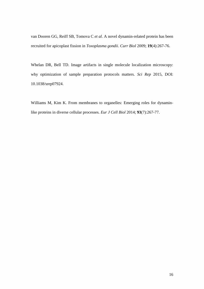
Figure 1: Immunofluorescence assay of the distribution of dynamin during T. gondii
invasion. (A) During the invasion of a single tachyzoite, the arrows point to loci near
the apical ends and backsides of the parasite. In distinct slices of the same sample,
18
dynamin labeling (green) occurs at the expected site of PV pinching off (arrow) (B),
and panel (C) shows the detection of dynamin at sites (arrows) that represent the
beginning and the end of T. gondii invasion and PV formation. (D-E) Parasites that
were completely internalized do not show dynamin labeling. The red staining
represents the actin distribution, and DAPI staining (blue) represents the nuclear
DNA. Bars: 10 µM.
19
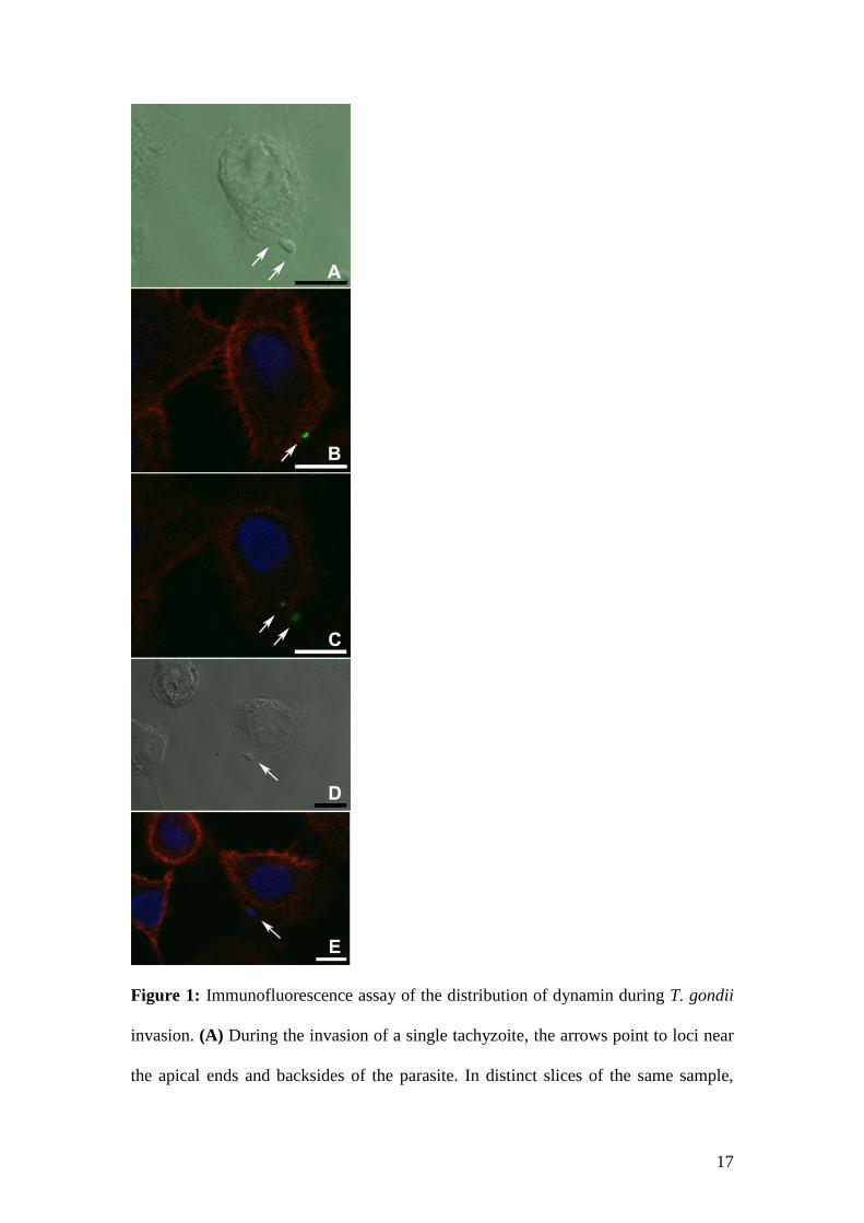
Figure 2: Cryo-immunomicroscopy of dynamin during T. gondii invasion. (A) 15 nm
immunogold particles accumulated at sites of rhoptry secretion (arrows) during
interactions of the tachyzoite with the host cell surface. (B) Labeling was observed at
loci where dynamin is recruited (arrows) during parasite (P) invasion. Bars: 500 nm.
20
Figure 3: SR-SIM of cells labeled for dynamin and actin at 24 hpi. (A) The PVs
(black arrows) within host cells at 24 hpi contain dynamin (white arrows) in different
slices of the sample (B-D) (green). The red staining represents the actin distribution,
and DAPI staining (blue) represents the nuclear DNA.
21
Figure 4: SR-SIM of cells labeled for dynamin and actin at 48 hpi (A-D): The arrows
point to dynamin foci (green) that are located primarily in the inner PV membrane
(labeled in red) in two different infected LLCMK2 cells (A-B). Wide-field SR-SIM
mode shows an infected peritoneal macrophage exhibiting its T. gondii PVs (red) (C),
and the arrow points to dynamin expression (green) inside the vacuole (D). DAPI
staining (blue) represents the nuclear DNA. (E-F) show cryo-immunomicroscopy of
dynamin during T. gondii development. The arrows point to the tubulovesicular
network (TVN) that develops around the parasites (P) inside the PV (E). (F) Dynamin
is detected (arrow) on the inner side of the PV membrane (PVM). Bars: 500 nm.
22
Figure 5: Immunofluorescence assay of the distribution of dynamin during T. gondii
egress. (A) Interferential microscopy of induced tachyzoite egress shows no labelling
of dynamin (B). The arrows point to an egressing parasite crossing the host cell
plasma membrane. The red staining represents the actin distribution. (C) Dynamin
was not detected by SR-SIM in a non-induced egressing parasite (red) (arrow). DAPI
staining (blue) represents the nuclear DNA. Bar: 10 µm (A-B), 5 µm (C).