Embed Size (px)
Citation preview
Author Pro
of
1ISSN 1758-2024xx.xxxx/XXX.xx.XXX © 2011 Future Medicine Ltd Neurodegen. Dis. Manage. (2011) 1(1), xxx–xxx
Summary Primary progressive aphasia (PPA) is a gradually progressive syndrome that robs patients of the ability to communicate. There are a variety of diagnostic challenges; international consensus has only recently been reached on the nomenclature for specific subtypes and there are a variety of underlying neurodegenerative pathologies. There are at present no established treatments, and efforts to develop treatments have been hampered by the lack of standardized methods to monitor progression of the illness. Although mea‑sures developed from work with stroke aphasia and with disorders such as Alzheimer’s dis‑ease have provided a valuable foundation for monitoring progression, PPA presents unique challenges to clinicians aiming to counsel patients and families on clinical status and progno‑sis, and to experts aiming to design clinical trials of potential interventions. Here we review some of the issues facing the field of PPA clinical research, summarize our current approach to monitoring progression of PPA and outline some ideas regarding goals for the develop‑ment of better tools for this purpose.
1MGH Frontotemporal Dementia Unit, Boston, MA, USA 2Department of Psychiatry, Massachusetts General Hospital and Harvard Medical School, Boston, MA, USA 3Department of Speech & Language Pathology, Massachusetts General Hospital and Harvard Medical School, Boston, MA, USA 4Department of Neurology, Massachusetts General Hospital and Harvard Medical School, Boston, MA, USA 5Division of Cognitive & Behavioral Neurology, Department of Neurology, Brigham & Women’s Hospital, Boston, MA, USA 6Massachusetts Alzheimer’s Disease Research Center, Massachusetts General Hospital, Boston, MA, USA 7Athinoula A Martinos Center for Biomedical Imaging, Massachusetts General Hospital and Harvard Medical School, Boston, MA, USA †Author for correspondence: Tel.: +1 617 726 5571; Fax: +1 617 726 5760; [email protected]
� Initial assessment of patients with suspected primary progressive aphasia should include: detailed history from patient and informant; neurologic examination; speech and language assessment; neuropsychiatric assessment; neuropsychological evaluation; brain imaging with MRI and possibly fluorodeoxyglucose (FDG)‑PET or single photon emission computed tomography; possibly other studies such as cerebrospinal fluid ana lysis.
� Longitudinal monitoring should include a subset of these assessments every 6 months to 1 year, with more frequent brief symptom‑focused check‑ups.
� Referral for enrollment in a research study at a specialized center should be considered.
� Treatment is symptomatic with both nonpharmacologic and pharmacologic approaches, including speech therapy, psychosocial therapy, and in some cases physical, occupational or cognitive therapy, and symptom‑based targeted pharmacologic interventions.
Prac
tice
Poi
nts
Monitoring progression of primary progressive aphasia: current approaches and future directions
Review
Daisy Sapolsky1,2,3, Kimiko Domoto‑Reilly1,4,5, Alyson Negreira1,2, Michael Brickhouse1,2, Scott McGinnis1,4,5 & Bradford C Dickerson†1,2,4,5,6,7
Author Pro
of
Neurodegen. Dis. Manage. (2011) 1(1) future science group2
review Sapolsky, Domoto‑Reilly, Negreira, Brickhouse, McGinnis & Dickerson
Primary progressive aphasia (PPA) is a gradu-ally progressive syndrome that was originally described in the late 1800s and has received growing attention since the 1980s, with a par-ticular surge in the past 5–10 years [1–5]. For the most part, it is considered to be a form of fronto-temporal dementia [6,7], although some forms of PPA may have other types of pathology. Although epidemiologic data on PPA are essentially nonex-istent, rough prevalence estimates from studies of frontotemporal dementia suggest that it may affect approximately 3–15/100,000 individuals [8,9], or approximately 10,000–45,000 people in the USA. Scientifically, these disorders are of great interest, in part because they strike the language networks of the brain with surprising specificity, which may shed light on the pathobiology of neu-rodegenerative disorders and on the neurobiology of language function. From a clinical perspective, these disorders are devastating to the patients and families who experience them. One patient’s hus-band recently told us, “it is heartbreaking to lose the ability to carry on dialog with one’s spouse.” Yet for some individuals, despite the gravity of language impairment, it may be possible to find hope in the relative sparing of other cognitive abilities and the potential for the development of alternative communication strategies.
Unfortunately, at present, despite substantial effort in several clinical trials of pharmacologic agents [10–12] and preliminary efforts with reha-bilitative techniques [13], we lack proof of efficacy of any form of treatment for PPA. Although there have been a number of important advances in clinical research related to PPA in the past few years, a critical barrier to the development of new treatments is the lack of standard assessment instruments that could be used in clinical trials. In this article, we will highlight some of the issues involved in the development of such instruments and initial efforts to develop such tools.
Clinical constructs of PPAThe core clinical phenotype of PPA is an insid-ious-onset, gradually progressive impairment of language production, object naming, syntax or word comprehension that is apparent during con-versation or through speech and language assess-ments. There are several excellent descriptions of the recommended diagnostic approach [4,14].
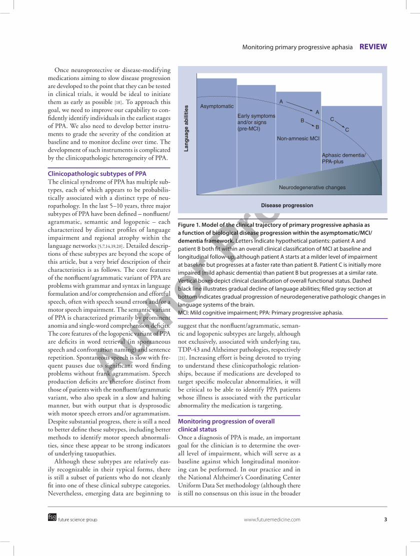
In our practice we find it helpful to concep-tualize PPA within the same general framework as Alzheimer’s disease (AD) and related neuro-degenerative disorders, which are thought of as
progressing in three clinical phases with respect to global function: asymptomatic/preclinical, mildly symptomatic/mild cognitive impairment and dementia (Figure 1). Because the pathophysi-ologic process of most neurodegenerative diseases is thought to take place over years prior to the development of symptoms, all of these conditions are thought to involve a phase in which neurode-generative changes are developing in the brain but are not yet producing symptoms. In the earliest clinical phase, patients may describe increasingly effortful speech but may be able to perform well on many tests of language function. For some patients, this phase may last for a number of years. We have worked with several individuals whose professions involved heavy use of speech/language skills (e.g., writers and teachers) who were aware of problems in these areas for years before deficits were apparent on clinical assessment. As the con-dition progresses, impairments become evident on psycholinguistic tests, such as measures of nam-ing, verbal fluency or picture description. At this clinical stage, many patients are still functioning relatively normally in complex activities of daily living and are capable of performing well on neu-ropsychological tests of cognitive domains other than language, such as memory, executive func-tion or visuospatial ability. Nevertheless, by this time, neuronal loss and neuropathologic change have damaged core brain regions subserving lan-guage and a diagnosis can often be supported by demonstrating the expected abnormalities of brain structure or function with neuroimaging tests. Finally, patients enter a dementia phase in which other cognitive, behavioral and/or motor domains outside of language are affected and the person develops the need for assistance by others in daily activities (this phase is sometimes called ‘aphasic dementia’ or ‘PPA-plus’ [15]). Terminal events are those typically associated with demen-tia, such as aspiration pneumonia. Although it is often said that the course of the illness progresses over approximately 7–10 years from diagnosis to death, recent studies suggest that some forms of PPA may be slowly progressive for 12 or more years [16], with reports of up to 20 years depending on how early a diagnosis is made. Although very little research has been performed on prognosis in PPA, one study suggests that patients may main-tain independence in activities of daily living for 6–7 years after the onset of symptoms, and that better functional outcomes may be associated with several baseline characteristics including fluent aphasia [17].
Author Pro
of
Monitoring primary progressive aphasia review
future science group www.futuremedicine.com 3
Once neuroprotective or disease-modifying medications aiming to slow disease progression are developed to the point that they can be tested in clinical trials, it would be ideal to initiate them as early as possible [18]. To approach this goal, we need to improve our capability to con-fidently identify individuals in the earliest stages of PPA. We also need to develop better instru-ments to grade the severity of the condition at baseline and to monitor decline over time. The development of such instruments is complicated by the clinicopathologic heterogeneity of PPA.
Clinicopathologic subtypes of PPAThe clinical syndrome of PPA has multiple sub-types, each of which appears to be probabilis-tically associated with a distinct type of neu-ropathology. In the last 5–10 years, three major subtypes of PPA have been defined – nonfluent/agrammatic, semantic and logopenic – each characterized by distinct profiles of language impairment and regional atrophy within the language networks [5,7,14,19,20]. Detailed descrip-tions of these subtypes are beyond the scope of this article, but a very brief description of their characteristics is as follows. The core features of the nonfluent/agrammatic variant of PPA are problems with grammar and syntax in language formulation and/or comprehension and effortfulspeech, often with speech sound errors and/or a motor speech impairment. The semantic variant of PPA is characterized primarily by prominent anomia and single-word comprehension deficits. The core features of the logopenic variant of PPA are deficits in word retrieval (in spontaneous speech and confrontation naming) and sentence repetition. Spontaneous speech is slow with fre-quent pauses due to significant word finding problems without frank agrammatism. Speech production deficits are therefore distinct from those of patients with the nonfluent/agrammatic variant, who also speak in a slow and halting manner, but with output that is dysprosodic with motor speech errors and/or agrammatism. Despite substantial progress, there is still a need to better define these subtypes, including better methods to identify motor speech abnormali-ties, since these appear to be strong indicators of underlying tauopathies.
Although these subtypes are relatively eas-ily recognizable in their typical forms, there is still a subset of patients who do not cleanly fit into one of these clinical subtype categories. Nevertheless, emerging data are beginning to
suggest that the nonfluent/agrammatic, seman-tic and logopenic subtypes are largely, although not exclusively, associated with underlying tau, TDP-43 and Alzheimer pathologies, respectively [21]. Increasing effort is being devoted to trying to understand these clinicopathologic relation-ships, because if medications are developed to target specific molecular abnormalities, it will be critical to be able to identify PPA patients whose illness is associated with the particular abnormality the medication is targeting.
Monitoring progression of overall clinical statusOnce a diagnosis of PPA is made, an important goal for the clinician is to determine the over-all level of impairment, which will serve as a baseline against which longitudinal monitor-ing can be performed. In our practice and in the National Alzheimer’s Coordinating Center Uniform Data Set methodology (although there is still no consensus on this issue in the broader
Lan
gu
age
abili
ties
Disease progression
Asymptomatic
Early symptomsand/or signs(pre-MCI)
Non-amnesic MCI
Aphasic dementia/PPA-plus
Neurodegenerative changes
A
A
BB
C
C
Figure 1. Model of the clinical trajectory of primary progressive aphasia as a function of biological disease progression within the asymptomatic/MCi/dementia framework. Letters indicate hypothetical patients: patient A and patient B both fit within an overall clinical classification of MCI at baseline and longitudinal follow‑up, although patient A starts at a milder level of impairment at baseline but progresses at a faster rate than patient B. Patient C is initially more impaired (mild aphasic dementia) than patient B but progresses at a similar rate. Vertical boxes depict clinical classification of overall functional status. Dashed black line illustrates gradual decline of language abilities; filled gray section at bottom indicates gradual progression of neurodegenerative pathologic changes in language systems of the brain. MCI: Mild cognitive impairment; PPA: Primary progressive aphasia.
Author Pro
of
Neurodegen. Dis. Manage. (2011) 1(1) future science group4
review Sapolsky, Domoto‑Reilly, Negreira, Brickhouse, McGinnis & Dickerson
PPA community), an initial objective is to deter-mine whether the patient would fit the designa-tion of mild cognitive impairment or dementia, optimally via a structured clinical interview with the patient and someone who knows the patient well, typically a spouse or adult child. Questionnaire-based instruments can be helpful for this purpose, particularly those that aim to ascertain the type and degree of impairments in instrumental and basic activities of daily living [22–25]. Overall level of impairment can be graded using a variety of clinician-rated scales, such as the Clinical Dementia Rating scale [26] with the supplemental Language and Behavior ratings, the Clinician Global Impression of Change [27] and related measures of global dementia severity.
It is more challenging to utilize perform-ance-based tests, such as the Mini-Mental State Exam, Addenbrooke’s Cognitive Exam (ACE), Alzheimer’s Disease Assessment Scale or neu-ropsychological test batteries, in part because of the effects of language deficits on nonlan-guage domains of performance [27]. While we believe these evaluations are useful for a variety of purposes, we always encourage the patient to use his/her most functional response modality, which may be writing. Even then, a language deficit can be as problematic in writing as in speech, and may impede the patient’s ability to display his knowledge. Therefore, we have added multiple-choice response options to tests for measuring orientation, memory and other abil-ities. The National Alzheimer’s Coordinating Center Uniform Data Set workgroup is devel-oping a Frontotemporal Dementia Module that should provide additional insights with respect to performance-based instruments. With these caveats in mind, however, a recent study dem-onstrated the value of the ACE in monitoring longitudinal change over time in patients with PPA [28].
Monitoring progression of speech & language impairmentPatients with PPA seek medical attention at different levels of severity – some patients seek neurologic/language evaluation at a very mild stage while others are surprisingly impaired by the first evaluation – and the rate at which symptoms progress varies considerably across patients (Figure 1). Therefore, the first step in monitoring speech/language is a comprehen-sive baseline assessment that goes beyond the language components of general cognitive
measures used in AD. The baseline speech/language assessment provides key information to determine clinical subtype, which will be increasingly important as clinical trials tar-geting specific pathologies become available. The baseline assessment, if sensitive enough, might reveal the earliest speech/language signs and symptoms and help us to identify those patients who meet inclusion criteria for a trial. Furthermore, knowing the baseline level of impairment is necessary to determine the effi-cacy of interventions including drug treatments or speech/language therapy.
Information from baseline and monitoring measures is also crucial for patient/family edu-cation regarding diagnosis, current level of sever-ity and prognosis. We are often asked questions such as, “How long until I won’t be able to speak at all?”, “Will I be able to understand even if I can’t speak?” or “Will my wife know how to use objects even if she can’t understand words?” As we learn more about the rate of change and evolution of language symptoms in PPA we will be better equipped to advise individuals as they plan for the future, as well as to help prepare them on an emotional/psychological level for future decline. Monitoring symptom severity and the emergence of new impairments is also critical for planning speech/language therapy and use of alternative communication strategies and devices.
Speech/language assessment can be carried out using a variety of performance-based tests from stroke aphasiology, such as the Western Aphasia Battery (WAB) [29] and Boston Diagnostic Aphasia Examination (BDAE) [30], pediatric speech–language pathology, such as the Curtiss–Yamada Comprehensive Language Evaluation–Receptive (CYCLE) [20], and tools recently developed for the PPA population, including the Northwestern Anagram Test (NAT) [31]. A motor speech exam is also typi-cally carried out to determine the presence of apraxia of speech, which can be the presenting symptom in the nonfluent/agrammatic subtype or may be a distinct subtype [32,33]. See Table 1 for examples of these instruments.
We have found that performance-based test-ing is only one component of the evaluation. There are often discrepancies between test per-formance and ability to communicate in daily life, which we observe directly or learn about through interviewing the partner. In addition, psycholinguistic measures typically do not
Author Pro
of
Monitoring primary progressive aphasia review
future science group www.futuremedicine.com 5
award credit for use of alternative communica-tion strategies and compensatory efforts, which can significantly improve a patient’s functional communication ability. We therefore consider the partner interview an important part of the assessment, and also obtain questionnaires about daily communication functioning from the partner (and patient when possible).
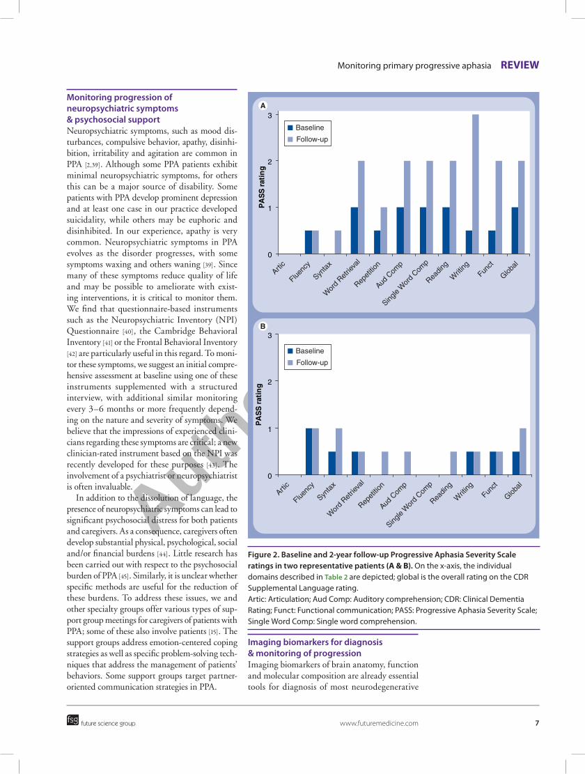
To integrate these sources of information, we recently developed a clinical rating scale that expanded upon the Clinical Dementia Rating (CDR) Supplemental Language rat-ing scale [27], the Progressive Aphasia Severity Scale (PASS) [34]. The scale is used to rate pres-ence and severity of impairment (0 = normal to 3 = severe impairment) in ten domains of language (Table 2). Ratings are made using the
clinician’s best judgment of the overall level of impairment in each domain, based on infor-mation from a structured interview with the patient and partner, and from the language assessment. The ratings represent a snapshot of the patient’s overall functioning in daily life in discrete domains of speech/language, and can be used at baseline and as a monitoring tool. There is also a functional communica-tion domain, which takes into account the patient’s ability to communicate despite the impairment (i.e., ability to use compensatory strategies). The scores in each domain can be summed to give a PASS Sum-of-Boxes measure, analogous to the CDR Sum-of-Boxes measure, representing a finer grading of overall level of impairment.
Table 1. examples of psycholinguistic test instruments, by domain of function, commonly used to assess and monitor primary progressive aphasia.
Domain Psycholinguistic test instruments
Articulation Apraxia Battery for Adults, motor speech examFluency BDAE seven‑point scale for phrase length, WAB Fluency, Grammatical Competence and Paraphasias Scale,
clinician impression of fluency from spontaneous speech and picture descriptions Syntax/grammar BDAE seven‑point scale for grammatical form, WAB Fluency, Grammatical Competence and Paraphasias Scale,
Northwestern Anagram Test, ana lysis of language samplesWord retrieval BNT, phonemic/category fluency tasks Repetition WAB and BDAE repetition tasks (words, phrases, sentences) Auditory comprehension WAB and BDAE following commands tasks, BDAE Complex Ideational Material, PAL Sentence Comprehension,
CYCLE Sentence–Picture MatchingSingle word comprehension BDAE Word Comprehension, WAB Auditory Word Recognition, CSB Category Comprehension, PALPA Spoken
Word–Picture Matching, Peabody Picture Vocabulary TestReading and writing WAB and BDAE written language tasksBDAE: Boston Diagnostic Aphasia Examination; BNT: Boston Naming Test; CSB: Cambridge Semantic Battery; CYCLE: Curtiss–Yamada Comprehensive Language Evaluation–Receptive; PAL: Psycholinguistic Assessment of Language; PALPA: Psycholinguistic Assessments of Language Processing in Aphasia; WAB: Western Aphasia Battery.
Table 2. Domains of the Progressive Aphasia Severity Scale.
PASS domain Description
Articulation Ability to say sounds and syllables accurately and effortlesslyFluency Degree to which speech flows easily or is interrupted by hesitations, fillers, pauses; reduced fluency is
associated with decreased phrase length and words per minute Syntax and grammar Use of word forms (run, ran), functor words (the, an), and word order when forming phrases and sentences
in most used modality (speech or writing)Word retrieval and expression Ability to express the intended word through primary modality (speech or writing)Repetition Ability to repeat words, phrases, sentences; difficulty should not be attributable to working memory
problem; do not penalize for sound distortions resulting from apraxia of speech or dysarthriaAuditory comprehension Ability to understand spoken phrases and sentences (e.g., conversation, commands) Single‑word comprehension Ability to understand spoken or written single wordsReading Ability to decode and understand written material; difficulty should not be attributable to an elementary
visual problemWriting Ability to write and spell; difficulty should not be attributable to an elementary motor problemFunctional communication Ability to communicate despite the speech/language impairment; ability to compensate for impairmentEach language domain is rated on a scale of 0 (normal), 0.5 (questionable or very mild impairment), 1 (mild impairment), 2 (moderate) or 3 (severe). PASS: Progressive Aphasia Severity Scale.
Author Pro
of
Neurodegen. Dis. Manage. (2011) 1(1) future science group6
review Sapolsky, Domoto‑Reilly, Negreira, Brickhouse, McGinnis & Dickerson
Because the PASS allows us to rate individual domains of speech/language, it captures the pattern of relative strengths and weaknesses at baseline and at each successive evaluation. We use it as a tool to monitor the emergence of prob-lems in initially unaffected areas, and the rate at which impaired domains worsen. We are cur-rently investigating whether baseline PASS assess-ment data are useful for predicting the domains of most prominent impairment (and relative pres-ervation) over the subsequent 2–4 years, which would be very valuable for discussions about prognosis. For example, we rated patient A over the course of 2 years (Figure 2). Ratings were rela-tively stable in articulation, syntax and fluency, whereas they declined in word retrieval, auditory comprehension and single-word comprehension, the areas that were initially most impaired. That is, the patient declined most significantly in the areas that were initially affected while preserved domains remained areas of strength. This is highly clinically relevant as patients and families often ask whether problems will develop in areas not initially affected. In the case of patient B, ratings were stable or changed only slightly (0.5) over 2 years, indicating a relatively slow rate of progression with many areas of relative strength (Figure 2). This information can be used to provide feedback to the patient about rate of progression and areas of stability, as well as to help clinicians plan speech therapy that targets the problem domains and capitalizes on the areas of strength.
More information about the PASS, including questionnaires and training materials, can be found on the MGH Frontotemporal Dementia Unit website [101]. We are currently working with collaborators to further assess the multi-center reliability of this instrument.
Monitoring progression of additional neurologic impairmentsAt initial assessment, a complete neurologic examination should be performed, in part to assess for evidence of an alternate etiology such as cerebrovascular or mass lesions. Such an evaluation can prove especially helpful if the patient is unable or unwilling to undergo brain imaging. In addition, the presence or absence of abnormalities on the neurologic exam can provide additional evidence in support of (limb apraxia) or against (cerebellar ataxia and chore-oathetosis) the PPA diagnosis [7]. Some patients exhibit subtle motor abnormalities very early in the course of PPA. The Frontal Assessment
Battery (FAB) can be particularly useful in the neurologist’s office for further assessing frontal systems functions [35].
As the disorder progresses, other neurologic findings may become evident; this stage of the illness has been termed PPA-plus [15]. In many cases, the disorder may evolve into an apha-sic dementia with other cognitive, behavioral and functional impairment. Neurologic and neuropsychiatric monitoring is particularly important in this phase of the illness because the family often needs to be counseled about these new sources of functional impairment and the possible need for additional treatments (e.g., pharmacologic interventions for behav-ioral symptoms or physical or occupational therapy for gait or eye–hand coordination abnormalities).
Executive dysfunction is common as PPA progresses, and often becomes obvious in daily life as patients stop using compensatory strate-gies effectively. Semantic dementia in particu-lar is often associated with later emergence of behavioral and attentional disturbances similar to those observed in behavioral variant fron-totemporal dementia. Assessments such as the Behavior Rating Inventory of Executive Function–Adult Version (BRIEF-A) [36] and the Frontal Systems Behavior Scale (FrSBe) [37] are useful scales that engage both patients and families in rating specific subcomponents of executive functions in daily life. These ques-tionnaires complement standard neurologic and neuropsychologic evaluations of attention and executive function (e.g., digit/spatial span, Trail-making test, Luria motor sequences and Tower of London test).
Motor abnormalities may contribute sub-stantially to overall functional disability in later stages of the illness. The nonfluent/agrammatic variant may be associated with the development of motor abnormalities consistent with progres-sive supranuclear palsy (gait impairment, axial rigidity and eye movement abnormalities) or corticobasal degeneration syndrome (asymmet-ric rigidity, alien limb and reflex myoclonus) [38]. Motor neuron disease findings (e.g., atrophy, weakness or fasciculations) may also arise and should be screened for in the neurologic exami-nation. Swallowing difficulties are common in middle and later stages of PPA; a formal swal-lowing evaluation and consultation regarding eating and nutrition may be indispensible in many patients.
Author Pro
of
Monitoring primary progressive aphasia review
future science group www.futuremedicine.com 7
Monitoring progression of neuropsychiatric symptoms & psychosocial support Neuropsychiatric symptoms, such as mood dis-turbances, compulsive behavior, apathy, disinhi-bition, irritability and agitation are common in PPA [2,39]. Although some PPA patients exhibit minimal neuropsychiatric symptoms, for others this can be a major source of disability. Some patients with PPA develop prominent depression and at least one case in our practice developed suicidality, while others may be euphoric and disinhibited. In our experience, apathy is very common. Neuropsychiatric symptoms in PPA evolves as the disorder progresses, with some symptoms waxing and others waning [39]. Since many of these symptoms reduce quality of life and may be possible to ameliorate with exist-ing interventions, it is critical to monitor them. We find that questionnaire-based instruments such as the Neuropsychiatric Inventory (NPI) Questionnaire [40], the Cambridge Behavioral Inventory [41] or the Frontal Behavioral Inventory [42] are particularly useful in this regard. To moni-tor these symptoms, we suggest an initial compre-hensive assessment at baseline using one of these instruments supplemented with a structured interview, with additional similar monitoring every 3–6 months or more frequently depend-ing on the nature and severity of symptoms. We believe that the impressions of experienced clini-cians regarding these symptoms are critical; a new clinician-rated instrument based on the NPI was recently developed for these purposes [43]. The involvement of a psychiatrist or neuropsychiatrist is often invaluable.
In addition to the dissolution of language, the presence of neuropsychiatric symptoms can lead to significant psychosocial distress for both patients and caregivers. As a consequence, caregivers often develop substantial physical, psychological, social and/or financial burdens [44]. Little research has been carried out with respect to the psychosocial burden of PPA [45]. Similarly, it is unclear whether specific methods are useful for the reduction of these burdens. To address these issues, we and other specialty groups offer various types of sup-port group meetings for caregivers of patients with PPA; some of these also involve patients [15]. The support groups address emotion-centered coping strategies as well as specific problem-solving tech-niques that address the management of patients’ behaviors. Some support groups target partner-oriented communication strategies in PPA.
imaging biomarkers for diagnosis & monitoring of progressionImaging biomarkers of brain anatomy, function and molecular composition are already essential tools for diagnosis of most neurodegenerative
0
1
2
3
PA
SS
rat
ing
0
1
2
3
PA
SS
rat
ing
Baseline
Follow-up
Baseline
Follow-up
Artic
Fluenc
y
Synta
x
Wor
d Ret
rieva
l
Repet
ition
Aud C
omp
Single
Wor
d Com
p
Readin
g
Writ
ingFun
ct
Global
Artic
Fluenc
y
Synta
x
Wor
d Ret
rieva
l
Repet
ition
Aud C
omp
Single
Wor
d Com
p
Readin
g
Writ
ingFun
ct
Global
Figure 2. Baseline and 2‑year follow‑up Progressive Aphasia Severity Scale ratings in two representative patients (a & B). On the x‑axis, the individual domains described in Table 2 are depicted; global is the overall rating on the CDR Supplemental Language rating. Artic: Articulation; Aud Comp: Auditory comprehension; CDR: Clinical Dementia Rating; Funct: Functional communication; PASS: Progressive Aphasia Severity Scale; Single Word Comp: Single word comprehension.
Author Pro
of
Neurodegen. Dis. Manage. (2011) 1(1) future science group8
review Sapolsky, Domoto‑Reilly, Negreira, Brickhouse, McGinnis & Dickerson
diseases and will almost certainly be critical for monitoring progression. For the purposes of clinical trials, these tools can be thought of as useful for identifying a relatively homogenous sample of participants (i.e., for the identification of disease-specific brain abnormalities and the exclusion of those lacking typical disease-asso-ciated abnormalities, much as is done in stroke or multiple sclerosis). They can also be thought of as potential outcome measures, although the clinical validation requirements called for by the US FDA make this a taller hurdle. Readers interested in a detailed discussion of some of the issues in the field of AD are referred to a previous publication [46].
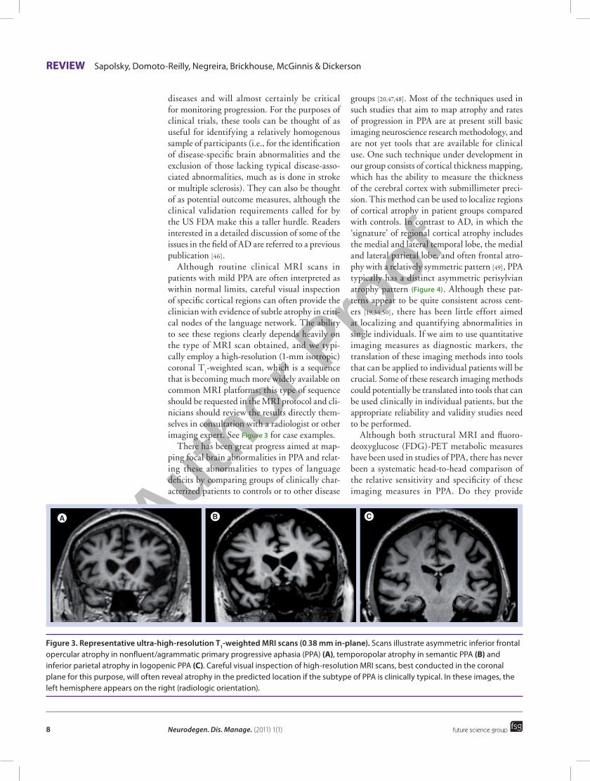
Although routine clinical MRI scans in patients with mild PPA are often interpreted as within normal limits, careful visual inspection of specific cortical regions can often provide the clinician with evidence of subtle atrophy in criti-cal nodes of the language network. The ability to see these regions clearly depends heavily on the type of MRI scan obtained, and we typi-cally employ a high-resolution (1-mm isotropic) coronal T
1-weighted scan, which is a sequence
that is becoming much more widely available on common MRI platforms; this type of sequence should be requested in the MRI protocol and cli-nicians should review the results directly them-selves in consultation with a radiologist or other imaging expert. See Figure 3 for case examples.
There has been great progress aimed at map-ping focal brain abnormalities in PPA and relat-ing these abnormalities to types of language deficits by comparing groups of clinically char-acterized patients to controls or to other disease
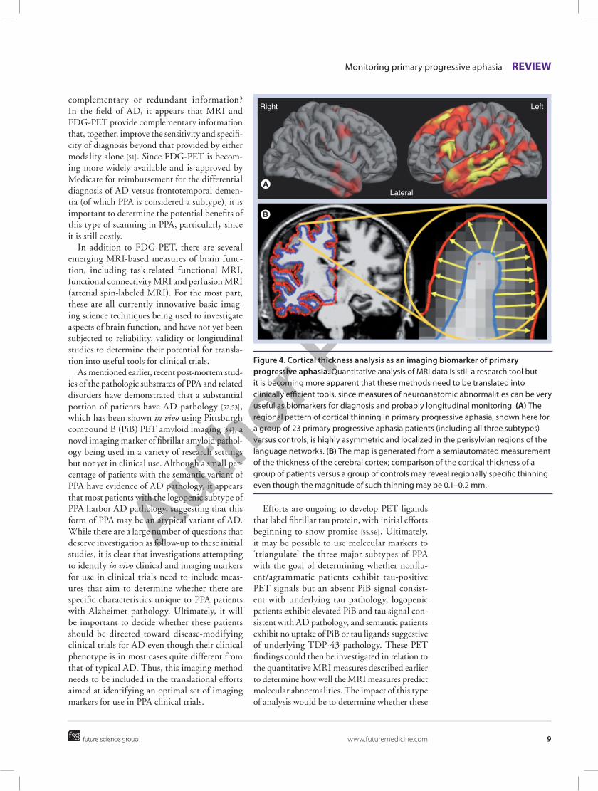
groups [20,47,48]. Most of the techniques used in such studies that aim to map atrophy and rates of progression in PPA are at present still basic imaging neuroscience research methodology, and are not yet tools that are available for clinical use. One such technique under development in our group consists of cortical thickness mapping, which has the ability to measure the thickness of the cerebral cortex with submillimeter preci-sion. This method can be used to localize regions of cortical atrophy in patient groups compared with controls. In contrast to AD, in which the ‘signature’ of regional cortical atrophy includes the medial and lateral temporal lobe, the medial and lateral parietal lobe, and often frontal atro-phy with a relatively symmetric pattern [49], PPA typically has a distinct asymmetric perisylvian atrophy pattern (Figure 4). Although these pat-terns appear to be quite consistent across cent-ers [19,34,50], there has been little effort aimed at localizing and quantifying abnormalities in single individuals. If we aim to use quantitative imaging measures as diagnostic markers, the translation of these imaging methods into tools that can be applied to individual patients will be crucial. Some of these research imaging methods could potentially be translated into tools that can be used clinically in individual patients, but the appropriate reliability and validity studies need to be performed.
Although both structural MRI and fluoro-deoxyglucose (FDG)-PET metabolic measures have been used in studies of PPA, there has never been a systematic head-to-head comparison of the relative sensitivity and specificity of these imaging measures in PPA. Do they provide
Figure 3. Representative ultra‑high‑resolution T1‑weighted MRi scans (0.38 mm in‑plane). Scans illustrate asymmetric inferior frontal opercular atrophy in nonfluent/agrammatic primary progressive aphasia (PPA) (a), temporopolar atrophy in semantic PPA (B) and inferior parietal atrophy in logopenic PPA (C). Careful visual inspection of high‑resolution MRI scans, best conducted in the coronal plane for this purpose, will often reveal atrophy in the predicted location if the subtype of PPA is clinically typical. In these images, the left hemisphere appears on the right (radiologic orientation).
Author Pro
of
Monitoring primary progressive aphasia review
future science group www.futuremedicine.com 9
complementary or redundant information? In the field of AD, it appears that MRI and FDG-PET provide complementary information that, together, improve the sensitivity and specifi-city of diagnosis beyond that provided by either modality alone [51]. Since FDG-PET is becom-ing more widely available and is approved by Medicare for reimbursement for the differential diagnosis of AD versus frontotemporal demen-tia (of which PPA is considered a subtype), it is important to determine the potential benefits of this type of scanning in PPA, particularly since it is still costly.
In addition to FDG-PET, there are several emerging MRI-based measures of brain func-tion, including task-related functional MRI, functional connectivity MRI and perfusion MRI (arterial spin-labeled MRI). For the most part, these are all currently innovative basic imag-ing science techniques being used to investigate aspects of brain function, and have not yet been subjected to reliability, validity or longitudinal studies to determine their potential for transla-tion into useful tools for clinical trials.
As mentioned earlier, recent post-mortem stud-ies of the pathologic substrates of PPA and related disorders have demonstrated that a substantial portion of patients have AD patho logy [52,53], which has been shown in vivo using Pittsburgh compound B (PiB) PET amyloid imaging [54], a novel imaging marker of fibrillar amyloid pathol-ogy being used in a variety of research settings but not yet in clinical use. Although a small per-centage of patients with the semantic variant of PPA have evidence of AD pathology, it appears that most patients with the logopenic subtype of PPA harbor AD pathology, suggesting that this form of PPA may be an atypical variant of AD. While there are a large number of questions that deserve investigation as follow-up to these initial studies, it is clear that investigations attempting to identify in vivo clinical and imaging markers for use in clinical trials need to include meas-ures that aim to determine whether there are specific characteristics unique to PPA patients with Alzheimer pathology. Ultimately, it will be important to decide whether these patients should be directed toward disease-modifying clinical trials for AD even though their clinical phenotype is in most cases quite different from that of typical AD. Thus, this imaging method needs to be included in the translational efforts aimed at identifying an optimal set of imaging markers for use in PPA clinical trials.
Efforts are ongoing to develop PET ligands that label fibrillar tau protein, with initial efforts beginning to show promise [55,56]. Ultimately, it may be possible to use molecular markers to ‘triangulate’ the three major subtypes of PPA with the goal of determining whether nonflu-ent/agrammatic patients exhibit tau-positive PET signals but an absent PiB signal consist-ent with underlying tau pathology, logopenic patients exhibit elevated PiB and tau signal con-sistent with AD pathology, and semantic patients exhibit no uptake of PiB or tau ligands suggestive of underlying TDP-43 pathology. These PET findings could then be investigated in relation to the quantitative MRI measures described earlier to determine how well the MRI measures predict molecular abnormalities. The impact of this type of ana lysis would be to determine whether these
Right Left
Lateral
Figure 4. Cortical thickness analysis as an imaging biomarker of primary progressive aphasia. Quantitative ana lysis of MRI data is still a research tool but it is becoming more apparent that these methods need to be translated into clinically efficient tools, since measures of neuroanatomic abnormalities can be very useful as biomarkers for diagnosis and probably longitudinal monitoring. (a) The regional pattern of cortical thinning in primary progressive aphasia, shown here for a group of 23 primary progressive aphasia patients (including all three subtypes) versus controls, is highly asymmetric and localized in the perisylvian regions of the language networks. (B) The map is generated from a semiautomated measurement of the thickness of the cerebral cortex; comparison of the cortical thickness of a group of patients versus a group of controls may reveal regionally specific thinning even though the magnitude of such thinning may be 0.1–0.2 mm.
Author Pro
of
Neurodegen. Dis. Manage. (2011) 1(1) future science group10
review Sapolsky, Domoto‑Reilly, Negreira, Brickhouse, McGinnis & Dickerson
additional but expensive molecular markers are needed to increase the specificity of diagnosis, or whether specificity is nearly as good using MRI (and possibly FDG-PET) measures alone. This will be essential information for the planning of treatment trials of compounds that are targeted toward specific molecular abnormalities, which is the case for a growing number of compounds.
If our long-term goal is to intervene in PPA using forms of disease-modifying therapy, or even to investigate potential plastic responses to forms of speech and language therapy, we need markers of brain structure and function that are sensitive to disease progression. We know that different types of PPA progress at different rates; new data indicate that the nonfluent/agrammatic variant progresses relatively rapidly and the semantic variant more slowly [16]. Furthermore, individual patients clearly vary with respect to each other in the level of impairment at initial presentation and in the rate of progression.
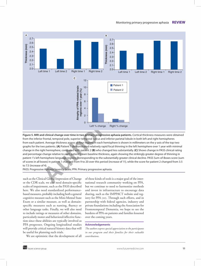
Several natural history studies are ongoing in which longitudinal MRI and in some cases PET data are being collected in patients with PPA and related disorders. One goal is to gener-ate data that would enable power calculations to be performed to estimate sample size for disease-modifying clinical trials, much as has been done in AD research; initial data in PPA look prom-ising [57,58]. Baseline imaging measures are also being investigated for their potential as predic-tive markers of clinical or functional–anatomic change, with the goal of determining whether some of these measures could be indicators of patients who may be ‘rapid progressors’, since this population would be an ideal target for clinical trials. See Figure 5 for an example of these types of longitudinal MRI measures.
Other biomarkersIn addition to imaging biomarkers, work is in progress to identify biofluid markers in blood or cerebrospinal fluid (CSF). At present, these are largely targeting CSF amyloid-b and tau abnor-malities that are consistent with AD pathology, and are primarily directed at early diagnosis and prognostication, as opposed to monitoring of dis-ease progression. A combination of elevated tau and phosphorylated tau accompanied by reduced amyloid-b is strongly suggestive of AD pathology [59]. In PPA patients exhibiting such a CSF profile, AD pathology would be strongly suspected and such patients might then be considered as poten-tial candidates for certain types of AD clinical
trials. This CSF molecular biomarker of AD is at present the only such approach that is routinely clinically available for this purpose and reimburs-able by many American third-party payors.
With regard to the detection of CSF markers of non-AD tau or TDP-43 pathology, few stud-ies have been conducted to date. Investigations of tau abnormalities are somewhat controversial, with some studies showing elevated tau and oth-ers showing reductions of tau. CSF assays for TDP-43 have recently been developed, and their preliminary use so far suggests that further work is needed. Finally, a variety of other proteins are being studied to investigate their potential use in understanding the pathobiology of the underly-ing diseases and differential diagnosis or moni-toring [5].
Conclusion & future perspectiveWe hope to see the development of efficacious interventions for PPA in the next decade. These treatments should improve function, slow pro-gression or both. Some of these treatments, such as specific forms of speech and language therapy, may be developed specifically for PPA and may target all individuals with PPA. Some drug treat-ments, such as neuroprotective medications, may also focus on this population or the broader popu-lation of patients with forms of frontotemporal dementia or kindred disorders. There is also increasing effort being devoted to the develop-ment of compounds targeting specific molecular abnormalities that contribute to the pathologic neurodegenerative process of these disorders, such as abnormalities of tau phosphorylation and aggregation.
For these latter compounds, we need to address several critical issues in clinical trial design. First, how do we identify patients with the specific biologic abnormality? This will likely involve a combination of clinical phenotype and one or more types of biomarkers. For example, a clini-cal trial of a tau agent in frontotemporal dementia could possibly enroll patients with a diagnosis of behavioral-variant or language-variant frontotem-poral dementia and biomarker evidence of a tau abnormality. Next, how do we monitor patients to determine whether the treatment is working? We envision the need for a multifaceted approach involving multiple types of clinical instruments as well as imaging or other biomarkers. The clini-cal instruments need to encompass the spectrum of abnormalities with reasonable sensitivity and specificity. Besides global functional measures,
Author Pro
of
Monitoring primary progressive aphasia review
future science group www.futuremedicine.com 11
such as the Clinical Global Impression of Change or the CDR scale, we also need domain-specific scales of impairment, such as the PASS described here. We also need standardized performance-based measures, probably including both a general cognitive measure such as the Mini-Mental State Exam or a similar measure, as well as domain-specific measures such as naming, fluency or other language tasks. Finally, we will also need to include ratings or measures of other domains, particularly motor and behavioral/affective func-tion since these abilities are typically involved as PPA progresses. Ongoing longitudinal studies will provide critical natural history data that will be useful for planning such trials.
We are optimistic that the development of all
of these kinds of tools is a major goal of the inter-national research community working on PPA, but we continue to need to harmonize methods and invest in infrastructure to encourage data sharing, such as the IMPPACT website and reg-istry for PPA [102]. Through such efforts, and in partnership with federal agencies, industry and private foundations including the Association for Frontotemporal Dementia, we hope to see the burdens of PPA on patients and families lessened over the coming years.
AcknowledgementsThe authors express special appreciation to the participants in our program and their families for their valuable contributions.
0
2
4
6
8
10
12
Left % change Right % change
Patient 1
Patient 2
2.7
2.6
2.5
2.4
2.3
2.2
2.1
2.0Left time 1 Left time 2 Right time 1 Right time 2
Th
ickn
ess
(mm
)
2.7
2.6
2.5
2.4
2.3
2.2
2.1
2.0Left time 1 Left time 2 Right time 1 Right time 2
Th
ickn
ess
(mm
)
Atr
op
hy o
f P
PA-r
elat
ed b
rain
reg
ion
s ov
er 1
yea
r (%
)
Figure 5. MRi and clinical change over time in two primary progressive aphasia patients. Cortical thickness measures were obtained from the inferior frontal, temporal pole, superior temporal sulcus and inferior parietal lobule in both left and right hemispheres from each patient. Average thickness across all four regions in each hemisphere is shown in millimeters on the y‑axis of the top two graphs for the two patients. (a) Patient 1 demonstrated relatively rapid focal thinning in the left hemisphere over 1 year with minimal change in the right hemisphere, compared with patient 2 (B) who changed less substantially. (C) Shows change in PASS clinical rating and percentage change relative to each patient’s own baseline thickness, again showing the strikingly greater degree of thinning in patient 1’s left hemisphere language regions, corresponding to the substantially greater clinical decline. PASS Sum‑of‑Boxes score (sum of scores in all boxes) in patient 1 changed from 9 to 20 over this period (increase of 11), while the score for patient 2 changed from 3.5 to 7.5 (increase of 4). PASS: Progressive Aphasia Severity Scale; PPA: Primary progressive aphasia.
Author Pro
of
Neurodegen. Dis. Manage. (2011) 1(1) future science group12
review Sapolsky, Domoto‑Reilly, Negreira, Brickhouse, McGinnis & Dickerson
Financial & competing interests disclosureThis work was supported by grants from the NIA (R01-AG29411, R21-AG29840 and P50-AG005134), and the Alzheimer’s Association. The authors have no other relevant affiliations or financial involvement with any
organization or entity with a financial interest in or finan-cial conflict with the subject matter or materials discussed in the manuscript apart from those disclosed.
No writing assistance was utilized in the production of this manuscript.
10 Johnson NA, Rademaker A, Weintraub S, Gitelman D, Wienecke C, Mesulam M: Pilot trial of memantine in primary progressive aphasia. Alzheimer Dis. Assoc. Disord. 24(3), 308 (2010).
11 Reed DA, Johnson NA, Thompson C, Weintraub S, Mesulam MM: A clinical trial of bromocriptine for treatment of primary progressive aphasia. Ann. Neurol. 56(5), 750 (2004).
12 Kertesz A, Morlog D, Light M et al.: Galantamine in frontotemporal dementia and primary progressive aphasia. Dement. Geriatr. Cogn. Disord. 25(2), 178–185 (2008).
13 Henry ML, Beeson PM, Rapcsak SZ: Treatment for lexical retrieval in progressive aphasia. Aphasiology 22(7–8), 826–838 (2008).
14 Gorno-Tempini ML, Hillis AE, Weintraub S et al.: Classification of primary progressive aphasia and its variants. Neurology (2010) (In Press).
� � Provides the new framework for the classification and diagnosis of PPA subtypes.
15 Rogalski EJ, Mesulam MM: Clinical trajectories and biological features of primary progressive aphasia (PPA). Curr. Alzheimer Res. 6(4), 331–336 (2009).
16 Hodges JR, Mitchell J, Dawson K et al.: Semantic dementia: demography, familial factors and survival in a consecutive series of 100 cases. Brain 133(Pt 1), 300–306 (2010).
17 Le Rhun E, Richard F, Pasquier F: Natural history of primary progressive aphasia. Neurology 65(6), 887–891 (2005).
18 DeKosky ST, Marek K: Looking backward to move forward: early detection of neurodegenerative disorders. Science 302(5646), 830–834 (2003).
19 Mesulam M, Wieneke C, Rogalski E, Cobia D, Thompson C, Weintraub S: Quantitative template for subtyping primary progressive aphasia. Arch. Neurol. 66(12), 1545–1551 (2009).
20 Gorno-Tempini ML, Dronkers NF, Rankin KP et al.: Cognition and anatomy in three variants of primary progressive aphasia. Ann. Neurol. 55(3), 335–346 (2004).
21 Grossman M: Primary progressive aphasia: clinicopathological correlations. Nat. Rev. Neurol. 6(2), 88–97 (2010).
22 Josephs KA: Frontotemporal dementia and related disorders: deciphering the enigma. Ann. Neurol. 64(1), 4–14 (2008).
23 Wicklund AH, Johnson N, Rademaker A, Weitner BB, Weintraub S: Profiles of decline in activities of daily living in non-Alzheimer dementia. Alzheimer Dis. Assoc. Disord. 21(1), 8–13 (2007).
24 Johnson N, Barion A, Rademaker A, Rehkemper G, Weintraub S: The Activities of Daily Living Questionnaire: a validation study in patients with dementia. Alzheimer Dis. Assoc. Disord. 18(4), 223–230 (2004).
25 Mioshi E, Kipps CM, Dawson K, Mitchell J, Graham A, Hodges JR: Activities of daily living in frontotemporal dementia and Alzheimer disease. Neurology 68(24), 2077–2084 (2007).
26 Mioshi E, Hodges JR: Rate of change of functional abilities in frontotemporal dementia. Dement. Geriatr. Cogn. Disord. 28(5), 419–426 (2009).
27 Morris JC: The Clinical Dementia Rating (CDR): current version and scoring rules. Neurology 43(11), 2412–2414 (1993).
28 Knopman DS, Kramer JH, Boeve BF et al.: Development of methodology for conducting clinical trials in frontotemporal lobar degeneration. Brain 131(Pt 11), 2957–2968 (2008).
� Demonstrates an approach to the design of longitudinal clinical research in PPA and behavioral variant frontotemporal dementia with an emphasis on the possibilities and limitations for translating these methods for use in clinical trials.
29 Leyton CE, Hornberger M, Mioshi E, Hodges JR: Application of Addenbrooke’s cognitive examination to diagnosis and monitoring of progressive primary aphasia. Dement. Geriatr. Cogn. Disord. 29(6), 504–509 (2010).
� Provides evidence that performance-based testing of multiple cognitive functions can detect longitudinal decline in PPA.
30 Kertesz A: Western Aphasia Battery. Grune and Stratton, NY, USA (1982).
31 Goodglass H, Kaplan E, Barresi B: Boston Diagnostic Aphasia Examination (3rd Edition). Pro-Ed, Austin, TX, USA (2001).
BibliographyPapers of special note have been highlighted as:� of interest� � of considerable interest
1 Kertesz A, Hudson L, Mackenzie IR, Munoz DG: The pathology and nosology of primary progressive aphasia. Neurology 44(11), 2065–2072 (1994).
2 Hodges JR: Frontotemporal dementia (Pick’s disease): clinical features and assessment. Neurology 56(11 Suppl. 4), S6–S10 (2001).
3 Mesulam MM: Slowly progressive aphasia without generalized dementia. Ann. Neurol. 11(6), 592–598 (1982).
4 Mesulam M, Weintraub S: Primary progressive aphasia and kindred disorders. Handb. Clin. Neurol. 89, 573–587 (2008).
� � Contains a practical guide to the approach to diagnosis of primary progressive aphasia (PPA), as well as reviewing other clinical and biological aspects of these disorders.
5 Grossman M: Primary progressive aphasia: clinicopathological correlations. Nat. Rev. Neurol. 6(2), 88–97 (2010).
� � Summarizes current knowledge of the relationships between clinical symptoms and pathobiology in PPA, with an emphasis on biomarkers.
6 McKhann GM, Albert MS, Grossman M, Miller B, Dickson D, Trojanowski JQ: Clinical and pathological diagnosis of frontotemporal dementia: report of the Work Group on Frontotemporal Dementia and Pick’s Disease. Arch. Neurol. 58(11), 1803–1809 (2001).
7 Neary D, Snowden JS, Gustafson L et al.: Frontotemporal lobar degeneration: a consensus on clinical diagnostic criteria. Neurology 51(6), 1546–1554 (1998).
8 Ratnavalli E, Brayne C, Dawson K, Hodges JR: The prevalence of frontotemporal dementia. Neurology 58(11), 1615–1621 (2002).
9 Harvey RJ, Skelton-Robinson M, Rossor MN: The prevalence and causes of dementia in people under the age of 65 years. J. Neurol. Neurosurg. Psychiatry 74(9), 1206–1209 (2003).
Author Pro
of
future science group www.futuremedicine.com 13
Monitoring progression of primary progressive aphasia: current approaches and future directions review
32 Weintraub S, Mesulam MM, Wieneke C, Rademaker A, Rogalski EJ, Thompson CK: The northwestern anagram test: measuring sentence production in primary progressive aphasia. Am. J. Alzheimers Dis. Other Demen. 24(5), 408–416 (2009).
33 Josephs KA, Duffy JR, Strand EA et al.: Clinicopathological and imaging correlates of progressive aphasia and apraxia of speech. Brain 129(Pt 6), 1385–1398 (2006).
� Analyzes clinical, imaging and pathological relationships in patients with PPA.
34 Ogar J, Slama H, Dronkers N, Amici S, Gorno-Tempini ML: Apraxia of speech: an overview. Neurocase 11(6), 427–432 (2005).
35 Sapolsky D, Bakkour A, Negreira A et al.: Cortical neuroanatomic correlates of symptom severity in primary progressive aphasia. Neurology 75(4), 358–366 (2010).
36 Dubois B, Slachevsky A, Litvan I, Pillon B: The FAB: a Frontal Assessment Battery at bedside. Neurology 55(11), 1621–1626 (2000).
37 Roth RM, Isquith PK, Gioia GA: BRIEF-A: Behavior Rating Inventory of Executive Functions–Adult Version. Psychological Assessment Resources, Odessa, FL, USA (2005).
38 Grace J, Malloy PF: Frontal Systems Behavior Scale. Psychological Assessment Resources, Lutz, FL, USA (2001).
39 Kertesz A, Davidson W, Munoz DG: Clinical and pathological overlap between frontotemporal dementia, primary progressive aphasia and corticobasal degeneration: the Pick complex. Dement. Geriatr. Cogn. Disord. 10(Suppl. 1), 46–49 (1999).
40 Banks SJ, Weintraub S: Neuropsychiatric symptoms in behavioral variant frontotemporal dementia and primary progressive aphasia. J. Geriatr. Psychiatry Neurol. 21(2), 133–141 (2008).
41 Kaufer DI, Cummings JL, Ketchel P et al.: Validation of the NPI-Q, a brief clinical form of the Neuropsychiatric Inventory. J. Neuropsychiatry Clin. Neurosci. 12(2), 233–239 (2000).
42 Nagahama Y, Okina T, Suzuki N, Matsuda M: The Cambridge Behavioral Inventory: validation and application in a memory clinic. J. Geriatr. Psychiatry Neurol. 19(4), 220–225 (2006).
43 Kertesz A, Davidson W, Fox H: Frontal behavioral inventory: diagnostic criteria for frontal lobe dementia. Can. J. Neurol. Sci. 24(1), 29–36 (1997).
44 de Medeiros K, Robert P, Gauthier S et al.: The Neuropsychiatric Inventory–Clinician rating scale (NPI-C): reliability and validity of a revised assessment of neuropsychiatric symptoms in dementia. Int. Psychogeriatr. 22(6), 984–994 (2010).
45 Brodaty H: Meaning and measurement of caregiver outcomes. Int. Psychogeriatr. 19(3), 363–381 (2007).
46 Mioshi E, Bristow M, Cook R, Hodges JR: Factors underlying caregiver stress in frontotemporal dementia and Alzheimer’s disease. Dement. Geriatr. Cogn. Disord. 27(1), 76–81 (2009).
47 Dickerson BC, Sperling RA: Neuroimaging biomarkers for clinical trials of disease-modifying therapies in Alzheimer’s disease. NeuroRx 2(2), 348–360 (2005).
48 Rohrer JD, Ridgway GR, Crutch SJ et al.: Progressive logopenic/phonological aphasia: erosion of the language network. Neuroimage 49(1), 984–993 (2010).
49 Peelle JE, Troiani V, Gee J et al.: Sentence comprehension and voxel-based morphometry in progressive nonfluent aphasia, semantic dementia, and nonaphasic frontotemporal dementia. J. Neurolinguistics 21(5), 418–432 (2008).
50 Dickerson BC, Bakkour A, Salat DH et al.: The cortical signature of Alzheimer’s disease: regionally specific cortical thinning relates to symptom severity in very mild to mild AD dementia and is detectable in asymptomatic amyloid-positive individuals. Cereb Cortex 19(3), 497–510 (2009).
51 Rohrer JD, Warren JD, Modat M et al.: Patterns of cortical thinning in the language variants of frontotemporal lobar degeneration. Neurology 72(18), 1562–1569 (2009).
52 Mosconi L, Brys M, Glodzik-Sobanska L, De Santi S, Rusinek H, de Leon MJ: Early detection of Alzheimer’s disease using neuroimaging. Exp. Gerontol. 42(1–2), 129–138 (2007).
53 Alladi S, Xuereb J, Bak T et al.: Focal cortical presentations of Alzheimer’s disease. Brain 130(Pt 10), 2636–2645 (2007).
54 Mesulam M, Wicklund A, Johnson N et al.: Alzheimer and frontotemporal pathology in subsets of primary progressive aphasia. Ann. Neurol. 63(6), 709–719 (2008).
55 Rabinovici GD, Jagust WJ, Furst AJ et al.: Ab amyloid and glucose metabolism in three variants of primary progressive aphasia. Ann. Neurol. 64(4), 388–401 (2008).
56 Small GW, Kepe V, Ercoli LM et al.: PET of brain amyloid and tau in mild cognitive impairment. N. Engl. J. Med. 355(25), 2652–2663 (2006).
57 Okamura N, Suemoto T, Furumoto S et al.: Quinoline and benzimidazole derivatives: candidate probes for in vivo imaging of tau pathology in Alzheimer’s disease. J. Neurosci. 25(47), 10857–10862 (2005).
58 Gordon E, Rohrer JD, Kim LG et al.: Measuring disease progression in frontotemporal lobar degeneration: a clinical and MRI study. Neurology 74(8), 666–673 (2010).
59 Knopman DS, Jack CR Jr, Kramer JH et al.: Brain and ventricular volumetric changes in frontotemporal lobar degeneration over 1 year. Neurology 72(21), 1843–1849 (2009).
� � Provides evidence regarding longitudinal change over time in clinical and brain imaging measures in PPA and behavioral variant frontotemporal dementia, including sample size estimates to assist in the design of clinical trials.
60 Shaw LM, Vanderstichele H, Knapik-Czajka M et al.: Cerebrospinal fluid biomarker signature in Alzheimer’s disease neuroimaging initiative subjects. Ann. Neurol. 65(4), 403–413 (2009).
� websites101 MGH Frontotemporal Disorders (FTD) Unit
www.ftd-boston.org
102 IMPPACT website and registry for PPA www.ppaconnection.org