Embed Size (px)
Citation preview
Monitoring the function of membrane transportproteins in detergent-solubilized formMatthias Quick* and Jonathan A. Javitch*†‡
*Center for Molecular Recognition and ‡Departments of Psychiatry and Pharmacology, Columbia University College of Physicians and Surgeons,New York, NY 10032
Edited by H. Ronald Kaback, University of California, Los Angeles, CA, and approved January 4, 2007 (received for review October 30, 2006)
Transport proteins constitute �10% of most proteomes and playvital roles in the translocation of solutes across membranes of allorganisms. Their (dys)function is implicated in many disorders,making them frequent targets for pharmacotherapy. The identifi-cation of substrates for members of this large protein family, stillreplete with many orphans of unknown function, has provendifficult, in part because high-throughput screening is greatlycomplicated by endogenous transporters present in many expres-sion systems. In addition, direct structural studies require thattransporters be extracted from the membrane with detergent,thereby precluding transport measurements because of the lack ofa vectorial environment and necessitating reconstitution into pro-teoliposomes for activity measurements. Here, we describe a directscintillation proximity-based radioligand-binding assay for deter-mining transport protein function in crude cell extracts and inpurified form. This rapid and universally applicable assay withadvantages over cell-based platforms will greatly facilitate theidentification of substrates for many orphan transporters andallows monitoring the function of transport proteins in a nonmem-branous environment.
membrane protein � neurotransmitter:sodium symporter � scintillationproximity � substrate binding
Membrane transport proteins fulfill an essential function inevery living cell by catalyzing the translocation of solutes,
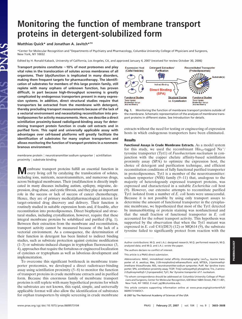
including ions, nutrients, neurotransmitters, and numerous drugs,across biological membranes. Their (mal)function is directly impli-cated in many diseases including autism, epilepsy, migraine, de-pression, drug abuse, and cystic fibrosis, and they play an importantrole in the success or lack of success of cancer chemotherapy.Hence, they are of primary medical/pharmacological interest fortarget-oriented drug discovery and delivery. Their function isroutinely studied in suitable expression hosts and, if feasible, afterreconstitution into proteoliposomes. Direct biophysical and struc-tural studies, including crystallization, however, require that theseintegral membrane proteins be solubilized and purified (Fig. 1).Between their extraction from the membrane and reconstitution,transport activity cannot be measured because of the lack of avectorial environment. As a consequence, the determination oftheir function in detergent has been limited to indirect bindingstudies, such as substrate protection against cysteine modification(1–3) or substrate-induced changes in tryptophan fluorescence (3,4), approaches that require the fortuitous or engineered localizationof cysteines or tryptophans as well as laborious development andimplementation.
To overcome this significant bottleneck in membrane trans-porter proteomics, we developed a direct radiotracer-bindingassay using scintillation proximity (5–8) to monitor the functionof transport proteins in crude membrane extracts and in purifiedform. Because this enormous family of membrane transportproteins is still replete with many hypothetical proteins for whichthe substrates are not known, this rapid, simple, and universallyapplicable format will also allow the identification of substratesfor orphan transporters by simple screening in crude membrane
extracts without the need for testing or engineering of expressionhosts in which endogenous transporters have been eliminated.
ResultsFunctional Assays in Crude Membrane Extracts. As a model systemfor this study, we used the recombinant His10-tagged Na�/tyrosine transporter (Tyt1) of Fusobacterium nucleatum in con-junction with the copper chelate affinity-based scintillationproximity assay (SPA) to optimize the expression host, thechoice of detergent and purification technique, and efficientreconstitution conditions of fully functional purified transporterin proteoliposomes. Tyt1 is a member of the neurotransmitter:sodium symporter (NSS) family (9–11) that, analogous to themajority of heterologously expressed transport proteins, wasexpressed and characterized in a suitable Escherichia coli host(9). However, our extensive attempts to reconstitute purifiedTyt1 isolated from a number of E. coli hosts were unsuccessful.Because it is not possible by using only transport assays todetermine the amount of functional transporter in the cytoplas-mic membrane, we hypothesized that most of the Tyt1 detectedby immunoblotting or protein staining was nonfunctional andthat the small fraction of functional transporter in E. coliaccounted for the robust transport activity. This hypothesis wassupported by the observation that when the Tyt1 mutant wasexpressed in E. coli C41(DE3) (12) or MQ614 (9), the substratetyrosine failed to significantly protect from reaction with the
Author contributions: M.Q. and J.A.J. designed research; M.Q. performed research; M.Q.analyzed data; and M.Q. and J.A.J. wrote the paper.
The authors declare no conflict of interest.
This article is a PNAS direct submission.
Abbreviations: IMAC, immobilized metal affinity chromatography; LeuTAa, leucine trans-porter of A. aeolicus; Mes, 2-(N-morpholino)-ethanesulfonic acid; MTSEA, 2-(aminoethyl-)methane thiosulfonate; NSS, neurotransmitter:sodium symporter; PutP, Na�/proline trans-porter; SPA, scintillation proximity assay; TCEP, Tris(2-carboxyethyl) phosphine; Tris, 2-amino-2-(hydroxymethyl)-1,3-propanediol; Tyt1, Na�/tyrosine transporter of F. nucleatum.
†To whom correspondence should be addressed at: Columbia University College of Physi-cians and Surgeons, Center for Molecular Recognition, 630 West 168th Street, P&S 11-401,New York, NY 10032. E-mail: [email protected].
This article contains supporting information online at www.pnas.org/cgi/content/full/0609573104/DC1.
© 2007 by The National Academy of Sciences of the USA
Fig. 1. Monitoring the function of membrane transport proteins outside ofthe membrane. Schematic representation of the analyses of membrane trans-port proteins in different states. See Introduction for details.
www.pnas.org�cgi�doi�10.1073�pnas.0609573104 PNAS � February 27, 2007 � vol. 104 � no. 9 � 3603–3608
PHA
RMA
COLO
GY
thiol reagent 2-(aminoethyl)methane thiosulfonate (MTSEA)[see supporting information (SI) Text] a single cysteine substi-tuted for Ile-104 (index position 3.46 (see refs. 13 and 14 forindexing scheme), which lines the substrate-binding site of thehomologous tryptophan transporter TnaT (N. R. Goldberg andJ.A.J., unpublished work) and of LeuTAa (11). Thus, the pre-dominant species of Tyt1 expressed in the E. coli inner mem-brane appears to be nonfunctional, suggesting that the expressedprotein is not suitable for biochemical studies of the detergent-solubilized and/or purified protein.
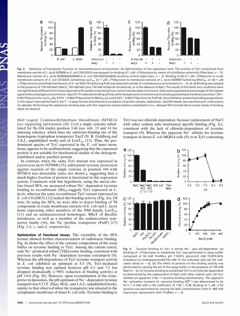
In contrast, when the same Tyt1 mutant was expressed inLactococus lactis NZ9000 (15), substantial tyrosine protectionagainst reaction of the single cysteine at position 104 withMTSEA was detectable (data not shown.), suggesting that amuch higher fraction of protein is functional in this expressionsystem. Consistent with this hypothesis, using the metal che-late-based SPA, we measured robust Na�-dependent tyrosinebinding to recombinant (His10-tagged) Tyt1 expressed in L.lactis, whereas the same recombinant Tyt1 variant expressed inE. coli C41(DE3) (12) lacked this binding activity (Fig. 2a). Ofnote, by using the SPA, we were able to detect binding of 3Hcompounds in crude membrane extracts of E. coli and L. lactishosts expressing other members of the NSS family, LeuTAa(11) and an uncharacterized homologue, MhsT of Bacillushalodurans, as well as a member of the sodium:solute sym-porter family (16), the Na�/proline transporter (PutP) (17)(Fig. 2 b, c, and d, respectively).
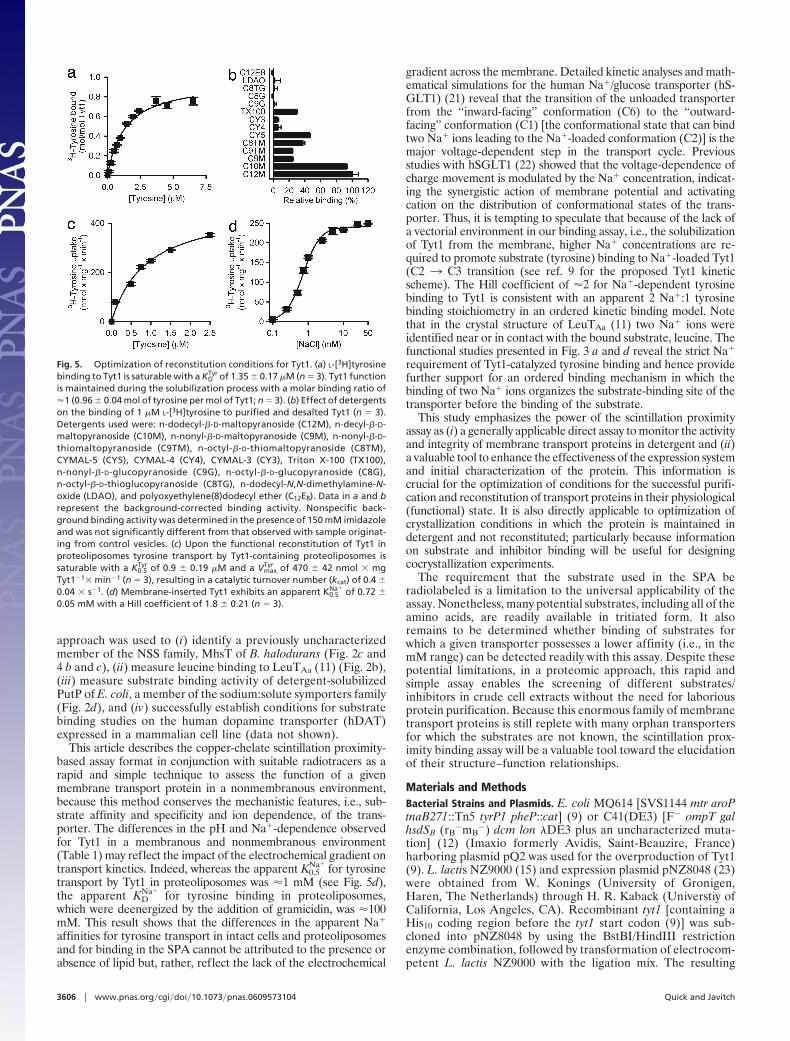
Optimization of Functional Assays. The versatility of the SPAformat allowed further characterization of radiotracer binding.Fig. 3a shows the effect of the cationic composition of the assaybuffer on tyrosine binding to Tyt1. Among the cations tested,only Na� promoted robust [3H]tyrosine binding, consistent withprevious results with Na�-dependent tyrosine cotransport (9).Whereas the pH-dependence of Tyt1 tyrosine transport activityin E. coli exhibited an optimum at 8.5 (9), Tyt1-mediatedtyrosine binding was optimal between pH 6.5 and 7.5 butdropped dramatically (�90% reduction of binding activity) atpH �8.0 (Fig. 3b). However, upon reconstitution of the trans-porter in liposomes, the pH optimum of Na�-dependent tyrosinetransport was 8.5 (Y. Zhao, M.Q., and J.A.J., unpublished work),similar to that observed when the transporter was situated in thecytoplasmic membrane of intact E. coli cells. Tyrosine binding to
Tyt1 was not chloride-dependent, because replacement of NaClwith other sodium salts maintained specific binding (Fig. 3c),consistent with the lack of chloride-dependence of tyrosinetransport (9). Whereas the apparent Na� affinity for tyrosinetransport in intact E. coli MQ614 cells (9) or in Tyt1-containing
Fig. 2. Detection of transporter function in nonmembranous environment. (a) Optimization of the expression host. The activity of Tyt1 solubilized frommembrane vesicles of L. lactis NZ9000 or E. coli CD41(DE3) was assayed for binding of 1 �M L-[3H]tyrosine by means of scintillation proximity (filled bars; n � 3).Membrane vesicles of L. lactis NZ9000/pNZ8048 or E. coli CD41(DE3)/pQE60 served as control (open bars; n � 3). Binding of (b) 0.1 �M L-[3H]leucine to crudemembranes extracts of E. coli C41(DE3) containing LeuTAa, (c) 1 �M L-[3H]tyrosine to membrane extracts of L. lactis NZ9000 harboring MhsTBh, or (d) 1 �ML-[3H]proline to solubilized membranes of E. coli WG170 harboring PutP was detected outside of a membranous environment (n � 3). (b–d) Binding was assayedin the presence of 150 mM NaCl (NaCl), 150 mM NaCl plus 150 mM imidazole (Imidazole), or in the absence of NaCl. The results of the latter two conditions werenot significantly different from those observed with sample originating from control vesicles (data not shown). Data were expressed as percentage of the highestsignal without background correction. Specific 3H-substrate binding activity (after background correction) was (in pmol/�g solubilized membrane protein) 1.04 �0.09 [3H]leucine for LeuTAa (b), 0.074 � 0.004 [3H]tyrosine for MhsTBh (c), and 0.033 � 0.002 [3H]proline for PutP (d). All scintillation proximity binding assays shownin this report were performed in the T � 0 assay format (simultaneous incubation of protein sample, radiotracer, and SPA beads; see manufacturer’s instructionsfor details). Performing the radiotracer binding step with the respective vesicles before solubilization (i.e., delayed SPA format) led to similar levels of binding(data not shown).
Fig. 3. Tyrosine binding to Tyt1 is strictly Na�- and pH-dependent. (a)Binding of L-[3H]tyrosine to solubilized Tyt1 was performed in assay buffercomposed of 50 mM Tris/Mes, pH 7.5/20% glycerol/2 mM TCEP/0.05%N-dodecyl-�-D-maltopyranoside/150 mM of the indicated salts (at the indi-cated ratios) (n � 6). (b) The effect of protons on the binding activity wasperformed by varying the pH of the assay buffer in the presence of 150 mMNaCl (n � 6). (c) Tyrosine binding to solubilized Tyt1 is not chloride-dependentas determined by the replacement of NaCl with other sodium salts. (d) Tyt1exhibits an apparent 2 Na�:1 tyrosine binding stoichiometry. The apparentNa�-activation constant of L-tyrosine binding (KD
Na�) was determined to be91.5 � 4 mM with a Hill coefficient of 1.94 � 0.28. Binding of 1 �M L-[3H]tyrosine was performed by varying the NaCl concentration from 0–400 mM(equimolar replacement with Tris/Mes; n � 3).
3604 � www.pnas.org�cgi�doi�10.1073�pnas.0609573104 Quick and Javitch

proteoliposomes (Fig. 5d) was �1 mM, Na� activation ofTyt1-mediated tyrosine binding was half-maximal at �92 mMNaCl, with a Hill coefficient of 1.94 � 0.28 (Fig. 3d).
The substrate specificity of Tyt1 was tested by a competitionSPA assay (Fig. 4a). Among the naturally occurring 20 aminoacids, 10 �M L-tyrosine inhibited binding of 0.1 �M L-[3H]tyrosine to Tyt1 almost completely. Of the other amino acids andtyrosine analogues tested, only �-methyl-L-tyrosine, phenylala-nine, 5-diiodo-L-tyrosine, or 3-(3,4-dihydrophenyl)-L-alanine(DOPA) inhibited L-[3H]tyrosine binding �25% at a concen-tration of 10 �M, demonstrating the high substrate specificity ofTyt1 for tyrosine. In contrast, a similar application of the SPAidentified MhsTBh as a multisubstrate transporter (Fig. 4 b andc; M.Q., H. Yano, and J.A.J., unpublished work).
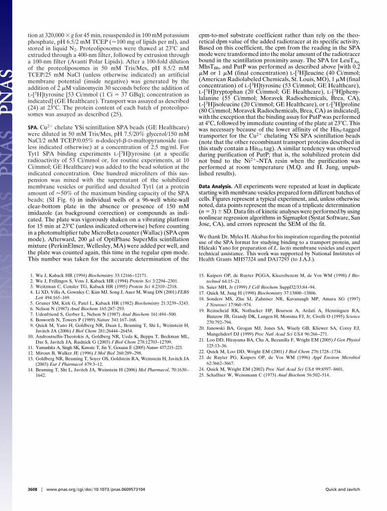
Monitoring the Purification and Reconstitution Conditions. To mon-itor the (potential) activity loss of Tyt1 during the purificationprocess, an SPA was performed with detergent-solubilized Tyt1 ata Na� concentration of 150 mM NaCl in which the [3H]tyrosineconcentration was increased from 0.1 to 6.5 �M (Fig. 5a). L-[3H]tyrosine binding is saturable, with a half-maximum saturationconstant (KD
Tyr) of 1.35 � 0.17 �M, a value comparable withthe apparent K0.5
Tyr for tyrosine transport in intact E. coli cells (9)(Table 1). Determination of the molar binding ratio of Tyt1revealed a binding coefficient of �1, indicating (i) a 1 tyrosine:1Tyt1 binding stoichiometry and (ii) �100% activity (i.e., no signif-icant activity loss) of Tyt1 upon solubilization with n-dodecyl-�-D-maltopyranoside. The same binding assay was performed after thepurification of Tyt1 by immobilized metal chelate affinity chroma-tography (IMAC) (Y. Zhao, M.Q., and J.A.J., unpublished work),and similar results were obtained (Table 1). This result indicatesthat Tyt1 retained its functionality during the purification proce-dure when n-dodecyl-�-D-maltopyranoside was used as detergent.
The effect of different detergents on the binding activity ofpurified Tyt1 was assayed (Fig. 5b). In the presence of detergents
with maltopyranoside head groups, Tyt1 exhibited the highestactivity with a reduction of the activity pattern generally reflect-ing the chain length of the detergent. However, detergents witha glucopyranoside moiety or n-dodecyl-N,N-dimethylamine-N-oxide- or polyoxyethylene(8)dodecyl ether-impaired Tyt1 bind-ing activity. Triton X-100 was the only nonmaltoside detergenttested that preserved �30% residual binding activity of Tyt1compared with that in n-dodecyl-�-D-maltopyranoside.
Tyt1-containing proteoliposomes exhibited saturable tyrosinetransport, which depended strictly on the Na� concentration(Fig. 5 c and d). The (apparent) K0.5 for tyrosine and Na� werecomparable with those reported for Na�-dependent tyrosinecotransport measured in intact E. coli cells expressing Tyt1 (9)(Table 1). After the determination of the actual Tyt1 amountused per transport assay, the turnover number (kcat) of Na�/tyrosine cotransport was determined to be 0.4 � 0.04 � s�1. Thisvalue is in perfect agreement with the turnover number calcu-lated for the human dopamine transporter (18), consistent withthe functional homology of Tyt1 and other NSS family members.
DiscussionThis study introduces an approach to transporter proteomics bydirectly monitoring the function of recombinant membranetransport proteins in detergent-solubilized crude membraneextracts and in purified form by using the scintillation proximityassay format. Although SPA approaches have been widely usedto study ligand binding to receptors (19, 20), to our knowledge,they have not been used previously to study substrate binding totransporters, despite the many advantages that we demonstratehere, including the suitability of the assay for relatively low-affinity substrates, the binding of which is difficult or impossibleto study by using standard filtration methods and has necessi-tated the development of complex indirect binding assays.
By using Tyt1, we were able to evaluate this method on arecently characterized NSS transporter (9). In addition, this
Fig. 4. Determination of the substrate specificity of Tyt1 and MhsT. (a) Tyt1 binding is highly specific. The substrate specificity of solubilized Tyt1 was measuredby a competition assay. The binding of 0.1 �M (final concentration) L-[3H]tyrosine was assayed in the presence or absence of 10 �M nonradioactive amino acid(or tyrosine analogue) as indicated (a–c, in the one-letter amino acid code used; the tyrosine analogues shown in a are: tyramine, D-tyrosine (D-Tyr), dopamine,�-methyl-L-tyrosine (a-Met-Tyr), 5-diiodo-L-tyrosine (Diiodo-Tyr), and 3-(3,4-dihydrophenyl)-L-alanine (DOPA). (b) MshT of B. halodurans is a multisubstratetransporter. Binding of 0.1 �M L-[3H]tyrosine was tested in the presence or absence of 10 �M nonradioactive amino acid as indicated (n � 3). L-[3H]tyrosine bindingto MshTBh was inhibited �50% by 10 �M L-tyrosine, L-phenylalanine, L-tryptophan, L-valine, L-isoleucine, or L-threonine. (c) This specificity pattern was confirmedby assessing the binding activity of a 1 �M concentration of the indicated L-[3H]amino acids (n � 3) as well as by transport assays (data not shown; M.Q., HideakiYano, and J.A.J., manuscript in preparation). Data represent the background-corrected binding activity measured in the presence of 150 mM imidazole, whichwas not significantly different from that observed with sample originating from control vesicles.
Quick and Javitch PNAS � February 27, 2007 � vol. 104 � no. 9 � 3605
PHA
RMA
COLO
GY
approach was used to (i) identify a previously uncharacterizedmember of the NSS family, MhsT of B. halodurans (Fig. 2c and4 b and c), (ii) measure leucine binding to LeuTAa (11) (Fig. 2b),(iii) measure substrate binding activity of detergent-solubilizedPutP of E. coli, a member of the sodium:solute symporters family(Fig. 2d), and (iv) successfully establish conditions for substratebinding studies on the human dopamine transporter (hDAT)expressed in a mammalian cell line (data not shown).
This article describes the copper-chelate scintillation proximity-based assay format in conjunction with suitable radiotracers as arapid and simple technique to assess the function of a givenmembrane transport protein in a nonmembranous environment,because this method conserves the mechanistic features, i.e., sub-strate affinity and specificity and ion dependence, of the trans-porter. The differences in the pH and Na�-dependence observedfor Tyt1 in a membranous and nonmembranous environment(Table 1) may reflect the impact of the electrochemical gradient ontransport kinetics. Indeed, whereas the apparent K0.5
Na�for tyrosine
transport by Tyt1 in proteoliposomes was �1 mM (see Fig. 5d),the apparent KD
Na�for tyrosine binding in proteoliposomes,
which were deenergized by the addition of gramicidin, was �100mM. This result shows that the differences in the apparent Na�
affinities for tyrosine transport in intact cells and proteoliposomesand for binding in the SPA cannot be attributed to the presence orabsence of lipid but, rather, reflect the lack of the electrochemical
gradient across the membrane. Detailed kinetic analyses and math-ematical simulations for the human Na�/glucose transporter (hS-GLT1) (21) reveal that the transition of the unloaded transporterfrom the ‘‘inward-facing’’ conformation (C6) to the ‘‘outward-facing’’ conformation (C1) [the conformational state that can bindtwo Na� ions leading to the Na�-loaded conformation (C2)] is themajor voltage-dependent step in the transport cycle. Previousstudies with hSGLT1 (22) showed that the voltage-dependence ofcharge movement is modulated by the Na� concentration, indicat-ing the synergistic action of membrane potential and activatingcation on the distribution of conformational states of the trans-porter. Thus, it is tempting to speculate that because of the lack ofa vectorial environment in our binding assay, i.e., the solubilizationof Tyt1 from the membrane, higher Na� concentrations are re-quired to promote substrate (tyrosine) binding to Na�-loaded Tyt1(C2 3 C3 transition (see ref. 9 for the proposed Tyt1 kineticscheme). The Hill coefficient of �2 for Na�-dependent tyrosinebinding to Tyt1 is consistent with an apparent 2 Na�:1 tyrosinebinding stoichiometry in an ordered kinetic binding model. Notethat in the crystal structure of LeuTAa (11) two Na� ions wereidentified near or in contact with the bound substrate, leucine. Thefunctional studies presented in Fig. 3 a and d reveal the strict Na�
requirement of Tyt1-catalyzed tyrosine binding and hence providefurther support for an ordered binding mechanism in which thebinding of two Na� ions organizes the substrate-binding site of thetransporter before the binding of the substrate.
This study emphasizes the power of the scintillation proximityassay as (i) a generally applicable direct assay to monitor the activityand integrity of membrane transport proteins in detergent and (ii)a valuable tool to enhance the effectiveness of the expression systemand initial characterization of the protein. This information iscrucial for the optimization of conditions for the successful purifi-cation and reconstitution of transport proteins in their physiological(functional) state. It is also directly applicable to optimization ofcrystallization conditions in which the protein is maintained indetergent and not reconstituted; particularly because informationon substrate and inhibitor binding will be useful for designingcocrystallization experiments.
The requirement that the substrate used in the SPA beradiolabeled is a limitation to the universal applicability of theassay. Nonetheless, many potential substrates, including all of theamino acids, are readily available in tritiated form. It alsoremains to be determined whether binding of substrates forwhich a given transporter possesses a lower affinity (i.e., in themM range) can be detected readily with this assay. Despite thesepotential limitations, in a proteomic approach, this rapid andsimple assay enables the screening of different substrates/inhibitors in crude cell extracts without the need for laboriousprotein purification. Because this enormous family of membranetransport proteins is still replete with many orphan transportersfor which the substrates are not known, the scintillation prox-imity binding assay will be a valuable tool toward the elucidationof their structure–function relationships.
Materials and MethodsBacterial Strains and Plasmids. E. coli MQ614 [SVS1144 mtr aroPtnaB271::Tn5 tyrP1 pheP::cat] (9) or C41(DE3) [F� ompT galhsdSB (rB
�mB�) dcm lon �DE3 plus an uncharacterized muta-
tion] (12) (Imaxio formerly Avidis, Saint-Beauzire, France)harboring plasmid pQ2 was used for the overproduction of Tyt1(9). L. lactis NZ9000 (15) and expression plasmid pNZ8048 (23)were obtained from W. Konings (University of Gronigen,Haren, The Netherlands) through H. R. Kaback (Universtiy ofCalifornia, Los Angeles, CA). Recombinant tyt1 [containing aHis10 coding region before the tyt1 start codon (9)] was sub-cloned into pNZ8048 by using the BstBI/HindIII restrictionenzyme combination, followed by transformation of electrocom-petent L. lactis NZ9000 with the ligation mix. The resulting
Fig. 5. Optimization of reconstitution conditions for Tyt1. (a) L-[3H]tyrosinebinding to Tyt1 is saturable with a KD
Tyr of 1.35 � 0.17 �M (n � 3). Tyt1 functionis maintained during the solubilization process with a molar binding ratio of�1 (0.96 � 0.04 mol of tyrosine per mol of Tyt1; n � 3). (b) Effect of detergentson the binding of 1 �M L-[3H]tyrosine to purified and desalted Tyt1 (n � 3).Detergents used were: n-dodecyl-�-D-maltopyranoside (C12M), n-decyl-�-D-maltopyranoside (C10M), n-nonyl-�-D-maltopyranoside (C9M), n-nonyl-�-D-thiomaltopyranoside (C9TM), n-octyl-�-D-thiomaltopyranoside (C8TM),CYMAL-5 (CY5), CYMAL-4 (CY4), CYMAL-3 (CY3), Triton X-100 (TX100),n-nonyl-�-D-glucopyranoside (C9G), n-octyl-�-D-glucopyranoside (C8G),n-octyl-�-D-thioglucopyranoside (C8TG), n-dodecyl-N,N-dimethylamine-N-oxide (LDAO), and polyoxyethylene(8)dodecyl ether (C12E8). Data in a and brepresent the background-corrected binding activity. Nonspecific back-ground binding activity was determined in the presence of 150 mM imidazoleand was not significantly different from that observed with sample originat-ing from control vesicles. (c) Upon the functional reconstitution of Tyt1 inproteoliposomes tyrosine transport by Tyt1-containing proteoliposomes issaturable with a K0.5
Tyr of 0.9 � 0.19 �M and a VmaxTyr of 470 � 42 nmol � mg
Tyt1�1� min�1 (n � 3), resulting in a catalytic turnover number (kcat) of 0.4 �0.04 � s�1. (d) Membrane-inserted Tyt1 exhibits an apparent K0.5
Na� of 0.72 �0.05 mM with a Hill coefficient of 1.8 � 0.21 (n � 3).
3606 � www.pnas.org�cgi�doi�10.1073�pnas.0609573104 Quick and Javitch
plasmid, designated pNZ2, was isolated by plasmid miniprep(Qiagen, Valencia, CA) after lysozyme (10 mg/ml) treatment at55°C for 10 min. The gene of an uncharacterized NSS homologueof B. halodurans (accession no. NP�241994), designated mhsT(mhsTBh) hereafter, was PCR-amplified from B. haloduransgenomic DNA (BAA-125D; American Type Culture Collection,Manassas, VA). Unique NcoI and NheI sites were introduced atthe haaT 5� and 3� termini, respectively. mhsTBh was subclonedinto pQ2 by means of NcoI/NheI restriction digest, replacing theentire tyt1 gene. To generate an N-terminally His10-taggedmhsTBh gene product, NcoI/NheI-digested mhsTBh was ligatedinto similarly cut pNZ2N (in pNZ2N the HindIII site flankingthe tyt1 stop codon was replaced with an unique NheI site afterHindIII-digestion of pNZ2, followed by Klenow treatment andrelegation of the vector) generating pNZmhsT. Similarly, theleuT gene of Aquifex aeolicus was PCR-amplified with genomicDNA of A. aeolicus (A. aeolicus cells were a generous gift fromH. Huber, University of Regensburg, Germany) as template.Unique NcoI and HindIII sites were introduced at the 5� and 3�termini, and the resulting gene product was cloned into pQ2 byusing this enzyme combination. The fidelity of all plasmids wasconfirmed by DNA sequencing (Columbia University Sequenc-ing Facility).
Gene Expression. L. lactis NZ9000 harboring pNZ8048 or itsderivatives were cultivated in M17 medium supplemented with0.5% glucose and 5 �g of chloramphenicol per ml at 30°C. Geneexpression in exponentially growing cells was induced for 4 h bythe addition of 5 ng of nisin per ml. Cells were harvested bycentrifugation at 7,500 � g for 10 min at 4°C, followed by a 1-htreatment with 10 mg of lysozyme per ml in 100 mM potassiumphosphate, pH 7.0, at 30°C. L. lactis protoplasts (�1 g wet weightper ml) were disrupted by a 3-fold passage through an Emulsi-Flex-C5 cell homogenizer (Avestin, Ottawa, Canada) at 30,000psi. Unbroken cells and cell debris were removed by centrifu-gation (10,000 � g for 10 min at 4°C) before membrane vesicleswere collected at 262,000 � g for 45 min at 4°C. Expression ofthe tyt1 gene in E. coli MQ614 or C41(DE3) was performed asdescribed (9). Alternatively, cells were incubated in Terrificbroth, supplemented with 100 �g of ampicillin per ml at 37°Cuntil the cultures reached an absorbance at 600 nm of 0.6. Tyt1expression was induced by the addition of 0.15 mM isopropyl-�-D-thiogalactopyranoside for 16 h at 20°C. E. coli MQ614 orC41(DE3) harboring pQE60 (9) served as control. LeuT andPutP were expressed in E. coli C41(DE3) or WG170 as described(11, 17). E. coli membrane vesicles were prepared by passing thecell suspension through an Avestin EmulsiFlex-C5 cell homog-
enizer at 15,000 psi. For SPA-based substrate-binding experi-ments, membrane vesicles of E. coli C41(DE3) harboring pQE60(9), or WG170 transformed with pTrc99a (17) served as control.
Solubilization and Purification. Membrane vesicles were resus-pended in ice-cold 50 mM 2-amino-2-(hydroxymethyl)-1,3-propanediol (Tris)/2-(N-morpholino)-ethanesulfonic acid(Mes), pH 7.5/20% glycerol/150 mM NaCl/2 mM Tris(2-carboxyethyl) phosphine (TCEP) (unless otherwise noted) at aprotein concentration of 10 mg/ml and solubilized by the addi-tion of 1% (wt/vol) n-dodecyl-�-D-maltopyranoside for 1 h at4°C. Solubilized protein and insoluble matter were separated bycentrifugation at 262,000 � g for 45 min at 4°C. The supernatantwas either used for binding assays (see below) or incubated withpreequilibrated Ni2�-Sepahrose Fastf low (GE Healthcare, Pis-cataway, NJ) (1 ml of resin/200 mg of membrane protein) for1.5 h with gentle shaking at 4°C in the presence of 20 mMimidazole. The protein–resin complex was then loaded into acolumn. Unbound protein was removed by washing with 50 mMTris/Mes, pH 7.5/20% glycerol/150 mM NaCl/2 mM TCEP/60mM imidazole/0.05% (wt/vol) n-dodecyl-�-D-maltopyranosideuntil the absorbance at 280 nm returned to the baseline. Tyt1 waseluted by a step increase of the imidazole concentration in thebuffer to 250 mM. Removal of imidazole or desalting/bufferexchange of the eluted fraction before the SPA was achievedwith Zeba Desalt Spin Columns (Pierce, Rockford, IL). To assessthe effect of various detergents on Tyt1 activity, the purified anddesalted sample was incubated in the presence of various deter-gents as indicated at a concentration of �2.5 times their criticalmicellar concentration (CMC) for 16 h at 4°C. The binding stepwas performed by diluting the samples in assay buffer in which0.05% n-dodecyl-�-D-maltopyranoside was replaced with therespective detergent at �1 times their CMC in the presence of1 �M (final concentration) L-[3H]tyrosine.
Reconstitution/Transport Assay. Preformed liposomes were pre-pared as described (24) by using E. coli total lipid extract (AvantiPolar Lipids, Alabaster, AL). The liposome suspension wassuspended in 100 mM potassium phosphate, pH 7.5/2 mM TCEPand frozen in liquid N2. Before reconstitution, thawed liposomeswere extruded through a 400-nm membrane filter (Whatman,Clifton, NJ). Liposomes (5 mg lipid per ml) were destabilized bythe addition of 0.12% (wt/vol) Triton X-100 (onset of solubili-zation) and mixed with purified Tyt1 in a 250:1 ratio (wt/wt) for10 min at 23°C. Detergent was removed by adding Bio-BeadsSM-2 (Bio-Rad, Hercules, CA). Tyt1-containing proteolipo-somes (or control liposomes) were concentrated by centrifuga-
Table 1. Kinetic parameters of Tyt1 in different states
E. coli (9) Solubilized IMAC Proteoliposomes
Tyrosine affinity (K0.5Tyr,KD
Tyr) (�M) 0.34 � 0.08 1.35 � 0.06 1.33 � 0.24 0.9 � 0.19Maximum velocity (Vmax
Tyr )(nmol � mg�1 � min�1)*
6.9 � 0.3 – – 470 � 42
Molar binding ratio – 0.93 � 0.04 0.98 � 0.03 –App. Na� affinity (K0.5
Na�,KD
Na�) (mM) 0.75 � 0.03 95.2 � 6.7 86.4 � 12.3 0.72 � 0.05
Hill coefficient 2 � 0.2 2.01 � 0.08 1.93 � 0.16 1.8 � 0.21Turnover no. (kcat) – – – 0.4 � 0.04 s�1
Kinetic parameters were determined as described in the legend to Figs. 3 or 5 or taken from ref. 9. Binding data for Tyt1 purified byimmobilized metal affinity chromatography (IMAC) were determined as described for the detergent-solubilized fraction of Tyt1containing membrane vesicles with �0.5 �g Tyt1 per mg of YSi beads (see SI Fig. 6 and SI Text). Kinetic parameters for solubilized orIMAC-purified Tyt1 were determined in three individual experiments, and the constants represent the average � SD of the individualdeterminations. The kinetic parameters for Tyt1 reconstituted into proteoliposomes are based on the data shown in Fig. 5 c and d, witherrors representing the SEM of the fit. Note that the variation of these parameters is ltequ20% when compared with independentexperiments (Y. Zhao, M.Q., and J.A.J., unpublished work. App., apparent.*Note that the maximum velocity refers to the total amount of membrane protein in E. coli cells or the amount of purified Tyt1reconstituted in proteoliposomes.
Quick and Javitch PNAS � February 27, 2007 � vol. 104 � no. 9 � 3607
PHA
RMA
COLO
GY
tion at 320,000 � g for 45 min, resuspended in 100 mM potassiumphosphate, pH 6.5/2 mM TCEP (�100 mg of lipids per ml), andstored in liquid N2. Proteoliposomes were thawed at 23°C andextruded through a 400-nm filter, followed by extrusion througha 100-nm filter (Avanti Polar Lipids). After a 100-fold dilutionof the proteoliposomes in 50 mM Tris/Mes, pH 8.5/2 mMTCEP/25 mM NaCl (unless otherwise indicated) an artificialmembrane potential (inside negative) was generated by theaddition of 2 �M valinomycin 30 seconds before the addition ofL-[3H]tyrosine [53 Ci/mmol (1 Ci � 37 GBq); concentration asindicated] (GE Healthcare). Transport was assayed as described(24) at 23°C. The protein content of each batch of proteolipo-somes was assayed as described (25).
SPA. Cu2� chelate YSi scintillation SPA beads (GE Healthcare)were diluted in 50 mM Tris/Mes, pH 7.5/20% glycerol/150 mMNaCl/2 mM TCEP/0.05% n-dodecyl-�-D-maltopyranoside (un-less indicated otherwise) at a concentration of 2.5 mg/ml. ForTyt1 SPA binding experiments L-[3H]tyrosine (at a specificradioactivity of 53 Ci/mmol or, for routine experiments, at 10Ci/mmol; GE Healthcare) was added to the bead solution at theindicated concentration. One hundred microliters of this sus-pension was mixed with the supernatant of the solubilizedmembrane vesicles or purified and desalted Tyt1 (at a proteinamount of �50% of the maximum binding capacity of the SPAbeads; (SI Fig. 6) in individual wells of a 96-well white-wallclear-bottom plate in the absence or presence of 150 mMimidazole (as background correction) or compounds as indi-cated. The plate was vigorously shaken on a vibrating platformfor 15 min at 23°C (unless indicated otherwise) before countingin a photomultiplier tube MicroBeta counter (Wallac) (SPA cpmmode). Afterward, 200 �l of OptiPhase SuperMix scintillationmixture (PerkinElmer, Wellesley, MA) were added per well, andthe plate was counted again, this time in the regular cpm mode.This number was taken for the accurate determination of the
cpm-to-mol substrate coefficient rather than rely on the theo-retical dpm value of the added radiotracer at its specific activity.Based on this coefficient, the cpm from the reading in the SPAmode were transformed into the molar amount of the radiotracerbound in the scintillation proximity assay. The SPA for LeuTAa,MhsTBh, and PutP was performed as described above [with 0.2�M or 1 �M (final concentration) L-[3H]leucine (40 Ci/mmol;(American Radiolabeled Chemicals, St. Louis, MO), 1 �M (finalconcentration) of L-[3H]tyrosine (53 Ci/mmol; GE Healthcare),L-[3H]tryptophan (20 Ci/mmol; GE Healthcare), L-[3H]pheny-lalanine (55 Ci/mmol; Moravek Radiochemicals, Brea, CA),L-[3H]isoleucine (20 Ci/mmol; GE Healthcare), or L-[3H]proline(80 Ci/mmol; Moravek Radiochemicals, Brea, CA) as indicated],with the exception that the binding assay for PutP was performedat 4°C, followed by immediate counting of the plate at 23°C. Thiswas necessary because of the lower affinity of the His6-taggedtransporter for the Cu2� chelating YSi SPA scintillation beads(note that the other recombinant transport proteins described inthis study contain a His10 tag). A similar tendency was observedduring purification of PutP; that is, the solubilized protein didnot bind to the Ni2�–NTA resin when the purification wasperformed at room temperature (M.Q. and H. Jung, unpub-lished results).
Data Analysis. All experiments were repeated at least in duplicatestarting with membrane vesicles prepared form different batches ofcells. Figures represent a typical experiment, and, unless otherwisenoted, data points represent the mean of a triplicate determination(n � 3) � SD. Data fits of kinetic analyses were performed by usingnonlinear regression algorithms in Sigmaplot (Systat Software, SanJose, CA), and errors represent the SEM of the fit.
We thank Dr. Myles H. Akabas for his inspiration regarding the potentialuse of the SPA format for studying binding to a transport protein, andHideaki Yano for preparation of L. lactis membrane vesicles and experttechnical assistance. This work was supported by National Institutes ofHealth Grants MH57324 and DA17293 (to J.A.J.).
1. Wu J, Kaback HR (1994) Biochemistry 33:12166–12171.2. Wu J, Frillingos S, Voss J, Kaback HR (1994) Protein Sci 3:2294–2301.3. Weitzman C, Consler TG, Kaback HR (1995) Protein Sci 4:2310–2318.4. Li XD, Villa A, Gownley C, Kim MJ, Song J, Auer M, Wang DN (2001) FEBS
Lett 494:165–169.5. Gruner SM, Kirk G, Patel L, Kaback HR (1982) Biochemistry 21:3239–3243.6. Nelson N (1987) Anal Biochem 165:287–293.7. Udenfriend S, Gerber L, Nelson N (1987) Anal Biochem 161:494–500.8. Bosworth N, Towers P (1989) Nature 341:167–168.9. Quick M, Yano H, Goldberg NR, Duan L, Beuming T, Shi L, Weinstein H,
Javitch JA (2006) J Biol Chem 281:26444–26454.10. Androutsellis-Theotokis A, Goldberg NR, Ueda K, Beppu T, Beckman ML,
Das S, Javitch JA, Rudnick G (2003) J Biol Chem 278:12703–12709.11. Yamashita A, Singh SK, Kawate T, Jin Y, Gouaux E (2005) Nature 437:215–223.12. Miroux B, Walker JE (1996) J Mol Biol 260:289–298.13. Goldberg NR, Beuming T, Soyer OS, Goldstein RA, Weinstein H, Javitch JA
(2003) Eur J Pharmacol 479:3–12.14. Beuming T, Shi L, Javitch JA, Weinstein H (2006) Mol Pharmacol, 70:1630–
1642.
15. Kuipers OP, de Ruyter PGGA, Kleerebezem M, de Vos WM (1998) J Bio-technol 64:15–21.
16. Saier MH, Jr (1999) J Cell Biochem Suppl32/33:84–94.17. Quick M, Jung H (1998) Biochemistry 37:13800–13806.18. Sonders MS, Zhu SJ, Zahniser NR, Kavanaugh MP, Amara SG (1997)
J Neurosci 17:960–974.19. Reinscheid RK, Nothacker HP, Bourson A, Ardati A, Henningsen RA,
Bunzow JR, Grandy DK, Langen H, Monsma FJ, Jr, Civelli O (1995) Science270:792–794.
20. Janowski BA, Grogan MJ, Jones SA, Wisely GB, Kliewer SA, Corey EJ,Mangelsdorf DJ (1999) Proc Natl Acad Sci USA 96:266–271.
21. Loo DD, Hirayama BA, Cha A, Bezanilla F, Wright EM (2005) J Gen Physiol125:13–36.
22. Quick M, Loo DD, Wright EM (2001) J Biol Chem 276:1728–1734.23. de Ruyter PG, Kuipers OP, de Vos WM (1996) Appl Environ Microbiol
62:3662–3667.24. Quick M, Wright EM (2002) Proc Natl Acad Sci USA 99:8597–8601.25. Schaffner W, Weissmann C (1973) Anal Biochem 56:502–514.
3608 � www.pnas.org�cgi�doi�10.1073�pnas.0609573104 Quick and Javitch