Embed Size (px)
Citation preview

JOURNAL OF CLINICAL MICROBIOLOGY,0095-1137/00/$04.0010
June 2000, p. 2065–2075 Vol. 38, No. 6
Copyright © 2000, American Society for Microbiology. All Rights Reserved.
Monoclonal Antibodies Directed against Conserved Epitopeson the Nucleocapsid Protein and the Major Envelope
Glycoprotein of Equine Arteritis VirusEMILIE WEILAND,1* SYLVIA BOLZ,1 FRANK WEILAND,1 WERNER HERBST,2
MARTIN J. B. RAAMSMAN,3 PETER J. M. ROTTIER,3
AND ANTOINE A. F. DE VRIES3†
Federal Research Centre for Virus Diseases of Animals, D-72076 Tubingen,1 and Institute for Hygiene and InfectiousDiseases of Animals, D-35392 Giessen,2 Germany, and Virology Unit, Department of Infectious Diseases
and Immunology, Veterinary Faculty, Utrecht University, 3584 CL Utrecht, The Netherlands3
Received 7 September 1999/Returned for modification 29 December 1999/Accepted 25 March 2000
We recently developed a highly effective immunization procedure for the generation of monoclonal antibodies(MAbs) directed against the porcine reproductive and respiratory syndrome virus (E. Weiland, M. Wieczorek-Krohmer, D. Kohl, K. K. Conzelmann, and F. Weiland, Vet. Microbiol. 66:171–186, 1999). The same methodwas used to produce a panel of 16 MAbs specific for the equine arteritis virus (EAV). Ten MAbs were directedagainst the EAV nucleocapsid (N) protein, and five MAbs recognized the major viral envelope glycoprotein(GL). Two of the EAV GL-specific MAbs and one antibody of unknown specificity neutralized virus infectivity.A comparison of the reactivities of the MAbs with 1 U.S. and 22 newly obtained European field isolates of EAVdemonstrated that all N-specific MAbs, the three nonneutralizing anti-GL MAbs, and the weakest neutralizingMAb (MAb E7/d15-c9) recognized conserved epitopes. In contrast, the two MAbs with the highest neutraliza-tion titers bound to 17 of 23 (MAb E6/A3) and 10 of 23 (MAb E7/d15-c1) of the field isolates. Ten of the virusisolates reacted with only one of these two MAbs, indicating that they recognized different epitopes. TheGL-specific MAbs and the strongly neutralizing MAb of unknown specificity (MAb E6/A3) were used for theselection of neutralization-resistant (NR) virus variants. The observation that the E6/A3-specific NR virusvariants were neutralized by MAb E7/d15-c1 and that MAb E6/A3 blocked the infectivity of the E7/d15-c1-specific NR escape mutant confirmed that these antibodies reacted with distinct antigenic sites. Immunoelec-tron microscopy revealed for the first time that the antigenic determinants recognized by the anti-GL MAbswere localized on the virion surface. Surprisingly, although the immunofluorescence signal obtained with theneutralizing antibodies was relatively weak, they mediated binding of about three times as much gold granulesto the viral envelope than the nonneutralizing anti-GL MAbs.
Equine arteritis virus (EAV) is the etiological agent ofequine viral arteritis (EVA), a respiratory and reproductivedisease that affects horses throughout the world (18). EAVusually causes subclinical infections but may also produce clin-ical illness with symptoms resembling those of equine influenza(45). Infection of pregnant mares frequently results in abor-tion, and the virus has occasionally been associated with foaldeath (10). EAV can establish a persistent infection in thegenital tracts of peripubertal colts and stallions (25). Althoughonly one serotype of the virus has been recognized, EAVisolates differ in genomic sequence, antigenic properties, andpathogenic qualities (18). Virus-neutralizing (VN) antibodiesare first detected 1 to 2 weeks after natural or artificial infec-tion with EAV. These antibodies generally persist for manyyears and help to protect horses against EVA but rarely pre-vent reinfection (32). Accordingly, VN antibody titers haveproven to be reliable indicators of the effectiveness of EAVvaccination protocols. The observation that colostrum fromimmune mares but not colostrum from nonimmune dams mod-
erates or prevents EVA in young foals (33) emphasizes theimportance of VN antibodies in the protection against EAV.
EAV is a spherical, enveloped, positive-stranded RNA virusthat belongs to the genus Arterivirus within the monogenericfamily Arteriviridae. Other members of this virus group arelactate dehydrogenase-elevating virus, porcine reproductiveand respiratory syndrome virus (PRRSV), and simian hemor-rhagic fever virus. Arteriviruses are the smallest represen-tatives of the order Nidovirales, which further includes thebigeneric family Coronaviridae (15, 40). Negatively stainedarterivirus particles have diameters of 55 to 75 nm (51) andcontain a relatively smooth surface without prominent spikes.The nucleocapsids of the arterivirus possess an icosahedralstructure and accommodate single copies of the viral genomicRNA. The genome of EAV has a length of 12.7 kb and con-tains eight genes (11, 41). The largest gene consists of two openreading frames (ORFs; ORFs 1a and 1b) which occupy nearlythree-quarters of the viral genome. ORFs 1a and 1b are di-rectly translated from the viral genomic RNA and direct thesynthesis of nonstructural proteins. The other genes (ORFs 2a,2b, and 3 through 7) are expressed from a 39 coterminal nestedset of six leader-containing subgenomic mRNAs (13) and codefor the structural proteins of the virus with the possible excep-tion of EAV ORF 3. EAV particles contain three major virionproteins: (i) a phosphorylated nucleocapsid protein (N) of 14kDa encoded by ORF 7, (ii) a 16-kDa unglycosylated mem-brane protein (M) specified by ORF 6, and (iii) a heteroge-
* Corresponding author. Mailing address: Federal Research Centrefor Virus Diseases of Animals, Paul-Ehrlich-Strasse 28, D-72076 Tu-bingen, Germany. Phone: 49-7071-967204. Fax: 49-7071-967105. E-mail: [email protected].
† Present address: Gene Therapy Section, Department of MolecularCell Biology, Leiden University Medical Center, 2333 AL Leiden, TheNetherlands.
2065
on April 13, 2019 by guest
http://jcm.asm
.org/D
ownloaded from

neously N-glycosylated membrane protein (GL) of 30 to 42kDa encoded by ORF 5 (14, 26, 55). The GL and M proteinsare present in virus particles as covalently linked heterodimers(16). In addition, three minor structural proteins have beenidentified in EAV particles: (i) the poorly characterized trans-lation product of ORF 4, (ii) an N-glycosylated membraneprotein (GS) of 25 kDa encoded by ORF 2b, and (iii) an 8-kDaunglycosylated envelope protein (E) specified by ORF 2a (14,17, 41).
The humoral immune response of horses to EAV is mainlydirected against the three major protein components of thevirus (7, 8, 9, 24, 28, 31). By pepscan analysis, an immunodom-inant epitope was localized between amino acids 70 and 99of the EAV GL protein (30). Immunization of mice withEAV particles yielded both VN-positive (VN1) and VN-neg-ative (VN2) monoclonal antibodies (MAbs). The neutralizingMAbs whose antigen specificities could be determined were alldirected against the EAV GL protein of the virus, whereas thenonneutralizing MAbs recognized either the EAV N or GLprotein (2, 3, 4, 6, 12, 23, 29, 52). Consistently, peptides derivedfrom the GL ectodomain induced VN antibodies in mice, rab-bits, and/or horses (2, 7). Different MAbs directed against thesame virus displayed differential reactivities with laboratoryisolates and field strains of EAV, indicating that the GL proteincontains multiple antigenic determinants (2, 3, 23, 29, 52, 54).Nucleotide sequence analysis of MAb neutralization-resistant(NR) escape mutants and field isolates of EAV showed thatthe GL protein is composed of constant and variable domainsand led to the identification of at least four neutralization siteswithin the second half of the hydrophilic amino-terminal ecto-domain (2, 3, 5, 23, 42, 43, 54). Furthermore, the observationthat only 4 of 11 MAbs that showed strong competition forbinding to virions in an enzyme-linked immunosorbent assay(ELISA) reacted with the GL protein in an immunoblot assay(3) implied that some GL-specific MAbs are directed againstlinear epitopes, whereas others recognize conformational epi-topes.
In the study described here, we have established and char-acterized a new panel of 16 EAV-specific MAbs. The proteinspecificities of 15 of these MAbs were determined, and thebinding of each of the antibodies to a unique collection of 23field isolates of EAV was investigated. Our experiments re-vealed that 14 of 16 of the MAbs recognized each of the virusisolates that were tested. These MAbs may thus be good can-didates for the development of novel diagnostic assays for thedetection of EAV-specific antigens or antibodies. We also in-vestigated the usefulness of the anti-EAV MAbs for immuno-fluorescence studies and tested their ability to neutralize viralinfectivity and to interact with the virus exterior. Unexpectedly,three of the GL-specific MAbs bound to the surfaces of thevirions but could not prevent virus entry.
MATERIALS AND METHODS
Cells and viruses. OST-7.1 (21), rabbit kidney (RK-13), and Vero cells weremaintained in Dulbecco’s modified Eagle’s medium (DMEM; Gibco-BRL LifeTechnologies) supplemented with 10% fetal calf serum (FCS), 100 IU of peni-cillin per ml, and 100 mg of streptomycin per ml (DMEM–10% FCS). BHK-21cells were cultured in Glasgow minimal essential medium (Gibco-BRL LifeTechnologies) containing 10% FCS, and Sp2/0-Ag14 cells (39) and hybridomaswere propagated in RPMI 1640 medium supplemented with 12% FCS. Virusstocks of the Bucyrus strain of EAV (19) and a plaque derivative thereof (EAVB15) were grown in Vero cells, whereas the Utrecht variant of the Bucyrus strainof EAV (EAV Utr) was propagated in BHK-21 cells (14). The virions wereconcentrated by centrifugation through a 20% (wt/wt) sucrose cushion in Tris-buffered saline (TBS) for 5 h at 25,000 rpm and 4°C in an SW27 rotor (Beck-man). The virus pellet was resuspended in TBS and was further purified in a 10to 50% (wt/wt) continuous sucrose gradient prepared in TBS by spinning for 24 hat 32,000 rpm and 4°C in an SW41 Ti rotor (Beckman). Virus particles were
harvested by puncturing the bottom of the centrifuge tube and were directly usedfor immunoelectron microscopy. Alternatively, the virus suspension was dilutedwith TBS and was centrifuged for 1 h at 45,000 rpm and 4°C in an SW55 Ti rotor(Beckman) to produce a highly concentrated virus preparation for immuniza-tions and Western blotting.
The field isolates of EAV were collected in Germany between 1991 and 1997from aborted foals or horse semen; S1112 virus, however, was isolated in theUnited States (see Table 3). The viruses were passaged two to three times inVero cells before preparation of stocks in these cells.
The recombinant vaccinia viruses vAVE07, vAVE16, and vAVE25, whichexpress the EAV N, M, and GL proteins, respectively (2, 14), were propagated inRK-13 cells and their titers were determined. Transient expression experimentswere carried out in OST-7.1 cells as described by Balasuriya et al. (2).
Plasmid construction. Recombinant DNA techniques were adopted fromSambrook et al. (37) and Ausubel et al. (1). To produce a fusion product of theEAV N protein and the amino terminus of the bacteriophage MS2 polymerase,the 0.5-kb HindIII fragment from pAVI07 (14) was inserted into the HindIII siteof the bacterial expression vector pEX30A (44). The resulting construct carryingthe EAV-specific part in the proper orientation was denoted pAVP07. ORF 4was cloned behind the T7 promoter of pBluescript KS(1) by insertion of afilled-in 0.6-kb DdeI fragment from cDNA clone PB106 (14) into the SmaI siteof the vector to generate plasmid pAVI04. Subsequently, pAVI04 was digestedwith SpeI and BglII, and the large vector-containing fragment was ligated to anadapter molecule consisting of oligonucleotides 830 (59-CTAGGATCCACCACCATGAA-39) and 831 (59-GATCTTCATGGTGGTGGATC-39). The resultingconstruct was designated pMRI14. The cloning results were verified by restric-tion enzyme digestions and nucleotide sequence analysis of alkali-denaturedplasmid DNA with a T7 DNA polymerase sequencing kit (Amersham PharmaciaBiotech) and [a-33P]dATP ($2,500 Ci per mmol; 10 mCi per ml; AmershamPharmacia Biotech). Recombinant plasmids were transfected into Escherichiacoli strain PC2495 (Phabagen). In the case of construct pAVP07, the bacterialacceptor strain already contained plasmid pcI857 (36) which encodes a temper-ature-sensitive mutant of the bacteriophage lambda cI repressor protein.
Inclusion body purification. A fresh 20-ml culture of pAVP07- and pcI857-transfected PC2495 cells was mixed with 180 ml of prewarmed Luria-Bertanimedium containing 50 mg of ampicillin and kanamycin per ml, and the mixturewas incubated overnight at 30°C. Next, 800 ml of similar medium heated to 42°Cwas added to the bacterial culture. After incubation for 4 h at 42°C, the bacteriawere pelleted by centrifugation for 10 min at 5,000 rpm and 4°C in a JA10 rotor(Beckman). The cell pellet was resuspended in 60 ml of ice-cold lysis buffer (50mM Tris-HCl [pH 8.0], 100 mM NaCl, 1 mM EDTA [TNE]) and frozen at270°C. The suspension was then thawed and supplemented with the proteaseinhibitors leupeptin, pepstatin A, and phenylmethylsulfonyl fluoride to finalconcentrations of 1, 1, and 50 mg per ml, respectively. After stirring for 5 min at4°C, 60 mg of lysozyme was added and the sample was incubated for 15 min atroom temperature (RT). The sample was then squeezed three times through a21-gauge needle, supplemented with 120 ml of Triton X-100, 600 ml of 1 MMgCl2, 60 ml of 1 M MnCl2, and 600 mg of DNase I (grade II; BoehringerMannheim), and incubated for another quarter of an hour at RT. The cell lysatewas centrifuged for 15 min at 10,000 rpm and 4°C in a JA20 rotor (Beckman),and the resulting pellet was resuspended in 30 ml of ice-cold TNE–1% NonidetP-40–1% sodium deoxycholate. The inclusion bodies were pelleted from thesuspension by another round of centrifugation and were washed with 50 mMTris-HCl (pH 8.0) containing 1, 3, and 5 M urea, respectively. The final proteinpreparation (named FP07) was taken up in 3 ml of phosphate-buffered saline(PBS), (partially) dissolved by sonication, and analyzed by sodium dodecyl sul-fate (SDS)-polyacrylamide (PAA) gel electrophoresis (PAGE) prior to injectioninto rabbits.
Polyclonal antisera. The production and characterization of the EAV Mprotein-specific antipeptide serum has been reported previously (14). To gener-ate an antiserum that recognizes the whole EAV N protein, a rabbit was injectedsubcutaneously with 2 ml of antigen emulsion containing 1 ml of Freund’scomplete adjuvant and 1 mg of FP07 in 1 ml of PBS. At 4, 8, and 12 weeks afterprimary immunization, the animals were boosted with 500 mg of antigen inincomplete Freund’s adjuvant. Two weeks later the animal was bled and theserum was stored in aliquots at 220°C.
MAbs. The properties of EAV GL protein-specific MAb 93B have been doc-umented elsewhere (23). The new panel of anti-EAV MAbs was established byfusion of Sp2/0-Ag14 myeloma cells with spleen cells of STU mice (38) immu-nized by the intraperitoneal route with the Bucyrus strain of EAV. The virus usedfor immunization was propagated in Vero cells, purified by density gradientcentrifugation as described above, and mixed with an equal volume of aluminumhydroxide adjuvant (Pierce). To diminish the production of antibodies directedagainst host cell components, immunotolerance against uninfected Vero cellswas induced in the mice prior to antigen administration (49). For the primaryimmunization 109 50% tissue culture infective doses (TCID50s) of virus con-tained in a total volume of 350 ml was used per mouse. The mice were boostedwith 250 ml of the same antigen preparation at 26 days after the first immuni-zation. A final injection with 3 3 108 TCID50s of virus in 200 ml of TBS withoutadjuvant was given 3 weeks after the second immunization and 3 days before thecell fusion. Splenocytes were fused with myeloma cells essentially as describedpreviously (49). An indirect immunofluorescence test (see below) carried out
2066 WEILAND ET AL. J. CLIN. MICROBIOL.
on April 13, 2019 by guest
http://jcm.asm
.org/D
ownloaded from

with EAV- and mock-infected Vero cells was used to screen the hybridoma-spentmedia for anti-EAV MAbs. Hybridomas that secreted EAV-specific MAbs werecloned by the limiting dilution method. The isotypes of the MAbs were deter-mined by using an IsoStrip Mouse Monoclonal Antibody Isotyping Kit (Boehr-inger Mannheim). To measure the antibody concentration in the hybridomaculture supernatants, a commercial ELISA for the quantification of murineimmunoglobulin G (IgG) (Boehringer Mannheim) was used.
Indirect immunoperoxidase monolayer assay (IPMA). Mock- and EAV-in-fected Vero cells in 96-well microtiter plates (Greiner) were fixed for 25 min withice-cold 96% ethanol at the indicated time points after infection. Next, the cellswere washed two times for 3 min each time with PBS and incubated overnight atRT with EAV-specific MAb diluted in PBS containing 5% FCS (PBS–5% FCS)and 0.1% Tween 20. After several 3-min washes with PBS, the cells were incu-bated for 45 min with a 1:1,000 dilution of a horseradish peroxidase (HRPO)-conjugated goat-anti mouse IgG (heavy and light chains) antibody (Bio-Rad) inPBS–5% FCS. Following three additional washes with PBS, 100 ml of substratesolution (200 mM sodium acetate [pH 5.0] containing 200 mg of aminoethylcar-bazole [AEC] per ml and 0.03% H2O2) was added to the cells. The enzymaticreaction was terminated after 20 min by replacing the substrate with water.
ELISA. The ELISA was identical to the IPMA except that 2,29-azino-bis(3-ethylbenzthiazoline-6-sulfonic acid) (ABTS) was used as a substrate. After ad-dition of 100 ml of a ready-to-use ABTS solution (Hoechst) to each well, theplates were incubated for 20 min at RT. The HRPO reaction was stopped byadding 50 ml of 1% SDS to each well, and the optical density was measured at 405nm in an SLT Spectra plate reader.
Metabolic labeling experiments. For the EAV- and mock-infected BHK-21cells, the radiolabeling was started at 8 to 10 h postinfection (p.i.), while theOST-7.1 cells infected with recombinant vaccinia viruses were labeled at 5 h afteraddition of the inoculum. In each case, the radiolabeling was preceded by astarvation period of 30 min. The labeling medium consisted of DMEM withoutL-cystine, L-glutamine, and L-methionine (BioWhittaker) which was supple-mented with 5% dialyzed FCS, 10 mM HEPES (pH 7.4), 2 mM L-alanyl–L-glutamine, and 20 ml of Redivue Pro-mix L-[35S]-in vitro cell labeling mix([35S]Met plus [35S]Cys; .1,000 Ci per mmol; 10 mCi per mmol; AmershamPharmacia Biotech) per ml. After labeling for 30 min to 2 h, the culture disheswere placed on ice. True cell lysates or total lysates of cells and media were thenprepared by established procedures (17). Whenever indicated the SDS concen-tration in the postnuclear supernatant was increased to 0.5%.
Immunoprecipitation, gel electrophoresis, and fluorography. Immunoprecipi-tations were performed as described by Balasuriya et al. (2). When lysates ofEAV-infected cells were used as the antigen, the immunoprecipitations wereeither carried out in the absence of a reducing agent or in the presence of 1 mMdithiothreitol to disrupt disulfide-linked GL-M heterodimers. Prior to analysis inSDS–15% PAA gels, the immunoprecipitates were resuspended in 30 ml ofsample buffer and were either incubated for 5 min at 96°C (N protein) or left atRT for $15 min (M and GL proteins). After electrophoresis the gels were fixed,impregnated with scintillant, and dried on filter paper as described previously(41). The gels were exposed at 270°C to Kodak X-OMAT LS films, and molec-ular weight estimations were based on digital images of the fluorographs ob-tained with a DC40 camera (Eastman Kodak Company) and were analyzed withKodak Digital Science 1D image analysis software, version 1.6.
Immunoblotting. Sucrose gradient-purified virus particles were dissolved inSDS-PAGE sample buffer containing 5% 2-mercaptoethanol and were appliedto a discontinuous SDS-PAA gel containing 12% acrylamide. After electro-phoresis the proteins were transferred to a nitrocellulose membrane (46) whichwas cut in strips and blocked with 5% nonfat dry milk powder (Heirler) in TBScontaining 0.1% Tween 20 (TBST) for 1 h at RT. Next, the filter pieces wereincubated with the culture medium (diluted 1:10 in the case of the N-specificMAbs and 1:2 in the case of the anti-GL antibodies) of the EAV-specific hybrid-omas for 1 h at ambient temperature, followed by incubation for 16 h at 4°C, andwere washed three times for 5 min each time in TBST. The nitrocellulose stripswere probed with a biotin-labeled anti-mouse IgG antibody (MaxTag Immuno-blotting Kit; Biotrend) diluted 1:200 in TBST for 30 min at RT. After three10-min washings, HRPO-conjugated streptavidin was added to the filter paperpieces at a dilution of 1:200 in TBST. Following a 30-min incubation, the nitro-cellulose strips were washed again, and AEC-containing substrate solution wasadded (see above). The enzymatic reaction was stopped after 15 min.
VN assays. To find out whether an MAb displayed any VN activity, 10-foldserial dilutions of EAV in DMEM–10% FCS were mixed with equal volumes ofundiluted hybridoma culture supernatant or with fresh culture medium, and themixtures were incubated for 1 h at 37°C. Next, 100 ml of the virus-antibodymixture was added to 3 wells of a 96-well microtiter plate containing confluentmonolayers of Vero cells. Three days later the wells were screened for thedevelopment of cytopathic effects (CPEs) and were stained for viral antigen in anIPMA carried out with the N-specific MAb E1/b5. The neutralization titer of theVN MAbs was subsequently determined by incubation of twofold serial dilutionsof the MAb-containing culture medium with 100 TCID50s of EAV B15 for 1 h at37°C. It was expressed as the reciprocal of the maximum dilution at which 100%of the cell monolayers were protected from a CPE and/or stained negative forviral antigen in an IPMA. Finally, a complement-dependent neutralization testwas performed to analyze whether the nonneutralizing GL-specific MAbs ren-dered EAV sensitive to inactivation by complement. To this end, serial 10-fold
dilutions of EAV B15 were mixed with an equal volume of a 1:2 dilution of eachMAb in DMEM–10% FCS or with an equal volume of culture medium devoidof antibodies. Following incubation for 1 h at 37°C, complement-containingguinea pig serum (Sigma) diluted in DMEM–10% FCS was added to the virusmixtures to final concentrations of between 5 and 20%. After another 1-h incu-bation at 37°C, the samples were added to 1-day-old monolayers of Vero cells in96-well microtiter plates. Following an incubation period of 3 days, the cells weresubjected to IPMA as described above.
Generation of NR virus variants. To select NR viral escape mutants, 5 3 104
TCID50s of EAV B15 in 5 ml were incubated with 1 ml of undiluted culturesupernatant of hybridomas E3/c16, E3/d10, E6/A3, E6/B8, E7/d15-c1, and E7/d15-c9 for 1 h at 37°C. The virus-antibody mixtures were then added to confluentmonolayers of Vero cells and the mixtures were incubated at 37°C until cyto-pathic changes occurred. A 5-ml portion of the supernatant of the infected cellswas incubated for 1 h at 37°C with 1 ml of the selecting MAb and was used toinoculate fresh monolayers of Vero cells. After another round of selection, aplaque assay was performed and the cell cultures were covered with a 0.5%agarose overlay. Individual plaques were collected by aspiration at 96 h p.i. andwere used for the preparation of large virus stocks in Vero cells. NR virusvariants received the name of the MAb used for their selection followed by theplaque number.
Immunofluorescence assay (IFA). Vero cells in suspension were either mockinfected or infected with EAV B15 at a multiplicity of infection of 0.2 TCID50sper cell and were seeded onto multitest slides. At 26 h after plating the cells werebriefly immersed twice in PBS, fixed for 25 min with ice-cold 96% ethanol, airdried, and stored at 270°C until use. After thawing, the cells were incubated for90 min with EAV-specific MAbs at various dilutions in PBS–5% FCS. Followingthree 2-min washes with PBS, the cells were stained with a fluorescein-conju-gated horse-anti mouse IgG (heavy and light chain) antibody (Camon) diluted1:150 in PBS–5% FCS for 45 min at ambient temperature. After washing thecells were embedded in glycerol buffer containing 25 mg of 1,4-diazobicyclo-(2,2,2)-octane and 2 mg of propidium iodide per ml. The samples were examinedwith a Zeiss Axioskop microscope, and pictures were taken with a Zeiss 340objective by using Ilford 400 Delta Professional black and white film.
Immunoelectron microscopy. Sucrose-gradient-purified virus was adsorbed tocopper grids (400 mesh) for 10 min at RT. The grids were subsequently washedfour times for 3 min each time with PBS–0.5% bovine serum albumin (BSA) andincubated for 45 min with undiluted hybridoma culture supernatant. After fur-ther washing steps with PBS–0.5% BSA, the grids were immersed for 45 min indrops of goat anti-mouse IgG plus IgM (heavy and light chains)–5-nm goldconjugate (Biocell) diluted 1:15 in PBS–0.5% BSA. Thereafter, the grids werewashed three times with PBS–0.5% BSA, three times with PBS, and once withdistilled water. They were then subjected to negative staining with 1% unbuffereduranyl acetate solution for 45 s at ambient temperature. The specimens wereexamined and photographed with a Zeiss 910 electron microscope.
RESULTS
Production of EAV-specific MAbs. Immunization of micewith PRRSV particles has yielded MAbs directed against fivedifferent structural proteins (20, 48, 50, 53). In contrast, allEAV-specific murine MAbs that have been obtained thus farrecognize either the N or the GL protein of the virus (2, 3, 4,6, 12, 23, 29, 52). A possible explanation for this discrepancyis the fact that only BALB/c mice have been immunized forthe production of EAV-specific MAbs, while different mousestrains were used to generate PRRSV-specific hybridomas. Inan attempt to expand the existing repertoire of EAV-specificMAbs, STU mice were intraperitoneally injected with a su-crose gradient-purified preparation of the B15 isolate of EAV.To increase the likelihood of recovering hybridomas that se-crete virus-specific antibodies, the mice were made immuno-tolerant for the Vero cells used to propagate EAV prior toimmunization. Three fusion experiments yielded a total of 780hybridomas after plating of 20% of the fusion products of eachspleen into four 96-well and two 24-well plates. Twenty-eighthybridomas recognized EAV-specific antigen in an indirectIFA carried out with EAV- and mock-infected Vero cells, and16 of them remained stable after cloning. Interestingly, differ-ent staining patterns were observed for the EAV-specificMAbs when applied as undiluted hybridoma culture superna-tants. Ten of the MAbs (MAbs E1/A19, E1/b5, E1/c4, E3/a9,E3/a61, E3/d17, E3/d52, E6/A7, E6/a27, and E6/d24) gave avery intense cytoplasmic fluorescence that required only veryshort exposure times for recording (Fig. 1A). Furthermore, all
VOL. 38, 2000 NONNEUTRALIZING EAV GL PROTEIN-SPECIFIC MAbs 2067
on April 13, 2019 by guest
http://jcm.asm
.org/D
ownloaded from

of these MAbs stained distinct areas in some of the nuclei ofEAV-infected cells but not the nuclei of mock-infected cells. Avery similar immunofluorescence pattern was previously re-ported for a panel of six MAbs directed against the N proteinof the vaccine strain of EAV (29). Three other MAbs (MAbsE3/c16, E3/d10, and E6/B8) caused a bright labeling of thecytoplasms of EAV-infected Vero cells (Fig. 1B). When usedat a higher dilution the distal parts of the cells were no longerlabeled by these antibodies. Instead, the immunofluorescencesignal was concentrated in juxtanuclear granular structures.The sizes of these granules appeared to decrease with increas-ing distance from the nucleus. The labeling pattern of MAb
E7/d15-c1 was also characterized by fine and coarse dots in theperinuclear region (Fig. 1C). MAb E6/A3 produced the weak-est immunofluorescence signal but showed an intracellular dis-tribution comparable to that of MAb E7/d15-c1 (Fig. 1D). Thestaining pattern of MAb E7/d15-c9 could not be easily de-duced, as this antibody cross-reacted with an unidentified hostcell component (data not shown).
Isotyping of the anti-EAV MAbs revealed that they be-longed to the IgG1, IgG2a, or IgG2b subclass (Table 1). Quan-tification of the immunoglobulins secreted in the culture su-pernatants indicated that the antibody concentration variedfrom 3 mg per ml for hybridoma E1/A19 to 85 mg per ml for
FIG. 1. Immunofluorescence staining of EAV B15-infected Vero cells with MAbs E1/A19 (A), E6/B8 (B), E7/d15-c1 (C), and E6/A3 (D). The cells were fixed at26 h p.i. with ice-cold 96% ethanol. Incubation of mock-infected Vero cells with the same antibodies did not yield a detectable immunofluorescence signal (data notshown).
2068 WEILAND ET AL. J. CLIN. MICROBIOL.
on April 13, 2019 by guest
http://jcm.asm
.org/D
ownloaded from
hybridoma E6/A7 (Table 2). These hybridoma culture mediawere subsequently used for an ELISA in which fixed EAV-infected Vero cells served as the specific antigen and fixedmock-infected Vero cells were used as the negative control. Asis evident from Table 1, the immunoglobulin concentration inthe hybridoma culture media did not correlate with the opticaldensities (ODs) obtained in the ELISA. For instance, MAbsE3/a9, E3/a61, and E3/d17 produced high net absorbance val-ues in the ELISA but were of low concentration. Likewise, thehighly concentrated E7/d15-c1 and E7/d15-c9 antibody prepa-rations gave rise to rather low ELISA titers. Furthermore,MAb E7/d15-c9 showed an exceptionally high background ab-sorbance which is consistent with the IFA results that showedcross-reactivity with a cellular protein. The group of six MAbsthat produced a granular intracellular immunofluorescencepattern yielded ODs that were at the lower end of the spec-trum. The ELISA readings for these MAbs generally corre-lated with their IFA signals; e.g., MAb E6/A3 produced theweakest immunofluorescence staining and gave the lowest spe-cific absorbance value in the ELISA. Finally, an IPMA wasused to establish the maximum dilution at which the MAbs stillgave a clearly detectable coloration of EAV-infected Verocells. As is shown in Table 1, the highest dilution of the MAbsat which a specific signal was observed was not merely deter-mined by the initial antibody concentration.
EAV-specific MAbs recognize either the EAV N or GL pro-tein. To get a first impression of the protein specificity of theEAV-specific MAbs, EAV Utr- and mock-infected BHK-21cells were metabolically labeled for 2 h starting at 8 h p.i. Totallysates of cells and media were prepared, aliquots of whichwere incubated with culture supernatants of each of the EAV-specific hybridomas or with monospecific antisera that recog-nize the three major virion proteins (N, M, and GL) of EAV.The resulting immunoprecipitates were analyzed in an SDS–15% PAA gel. This experiment indicated that nine MAbs(MAbs E1/A19, E1/b5, E1/c4, E3/a9, E3/a61, E3/d17, E3/d52,E6/A7, and E6/a27) recognized the EAV N protein, whereastwo MAbs (MAbs E7/d15-c1 and E7/d15-c9) precipitated sub-
stantial amounts of both the EAV GL and M proteins. Thelatter finding can be explained by the fact that the EAV GL andM proteins form disulfide-linked heterodimers in virus-in-fected cells (16). For MAbs E3/c16, E3/d10, E6/A3, E6/B8, andE6/d24, no decisive conclusion could be drawn as to the EAVproteins that they recognized (data not shown).
To unequivocally determine the protein specificity of theMAbs directed against EAV, we used vaccinia virus recombi-nants that express the genes for the GL, M, or N protein ofEAV Utr. Immunoprecipitations were performed with each ofthe MAbs with lysates from vAVE07-, vAVE16-, or vAVE25-infected OST-7.1 cells as the antigen source. As is clear fromFig. 2A and B, MAbs E1/A19, E1/b5, E1/c4, E3/a9, E3/a61,E3/d17, E3/d52, E6/A7, and E6/a27 specifically recognized a15-kDa protein in lysates of vAVE07-infected cells but not inextracts from cells infected with vAVE06. As the same proteinspecies was recognized by an antiserum directed against anN-specific bacterial fusion protein, we concluded that theseMAbs indeed react with the core protein of EAV. To excludethe possibility that the precipitation of the independently ex-pressed EAV N protein was the result of direct binding to theimmunosorbent and/or the formation of aggregates, we alsoincubated the lysate of vAVE07-infected cells with two GL-specific MAbs (MAbs E6/B8 and E7/d15-c9; see below). Witheither of these MAbs, only minute amounts of the EAV Nprotein were brought down, which confirms our previous in-terpretation that MAbs E1/A19, E1/b5, E1/c4, E3/a9, E3/a61,E3/d17, E3/d52, E6/A7, and E6/a27 are N specific. Of theremaining seven MAbs, five reacted with proteins of 29 and 25kDa in the lysate of the vAVE25-infected OST-7.1 cells (Fig.2C). Since the same two protein species were recognized byGL-specific MAb 93B (23) and none of these MAbs specificallyimmunoprecipitated a polypeptide from the lysate of vAVE16-infected cells, our data showed that MAbs E3/c16, E3/d10,E6/B8, E7/d15-c1, and E7/d15-9 are GL specific.
MAbs E6/A3 and E6/d24 did not recognize the indepen-dently expressed N, M, or GL protein of EAV Utr, nor did theyreact with the expression products of ORFs 2b and 4 at an SDSconcentration in the undiluted cell lysate of 0.5% (data notshown). The reactivities of these antibodies with the transla-tion products of ORFs 2a and 3 have not been tested.
The failure to determine the protein specificities of MAbsE6/A3 and E6/d24 in immunoprecipitation assays may be ex-plained in three ways: (i) MAbs E6/A3 and E6/d24 are directedagainst conformational epitopes which are destroyed duringthe analytical procedures, (ii) MAbs E6/A3 and E6/d24 are oflow avidity and/or recognize poorly expressed proteins, and(iii) MAbs E6/A3 and E6/d24 react with epitopes that differ
TABLE 1. Characteristics of EAV-specific MAbs
MAba IsotypeIgG
concn(mg/ml)
ELISAODb
Antibodytiterc
Proteinspecificity
Neutrali-zation(titer)d
E1/A19 2a 3 2.6 (0.21) 40 N 2E1/b5 1 20 3.3 (0.18) 80 N 2E1/c4 1 5 2.0 (0.19) 20 N 2E3/a9 1 5 3.2 (0.18) 20 N 2E3/a61 1 9 3.2 (0.24) 20 N 2E3/c16 2a 42 1.9 (0.32) 40 GL 2E3/d10 2b 30 1.4 (0.31) 10 GL 2E3/d17 2b 9 3.3 (0.21) 20 N 2E3/d52 2b 12 2.4 (0.22) 20 N 2E6/A3 2a 22 0.6 (0.21) 3 GL? 1(80)E6/A7 1 85 2.6 (0.21) 160 N 2E6/a27 1 5 1.8 (0.18) 10 N 2E6/B8 1 12 1.0 (0.28) 10 GL 2E6/d24 1 4 2.8 (0.17) 40 N 2E7/d15-c1 2b 50 1.2 (0.32) 20 GL 1(40)E7/d15-c9 2a 42 1.7 (0.71) 40 GL 1(5)
a Hybridoma culture supernatant.b OD reading for EAV-infected (mock-infected) Vero cells that were fixed to
the ELISA plate.c By IPMA as the reciprocal of the highest dilution of the culture supernatant
at which specific cytoplasmic reactivity was clearly observed.d Reciprocal of the highest dilution of the culture supernatant that completely
inhibited the CPE of 100 TCID50s of EAV.
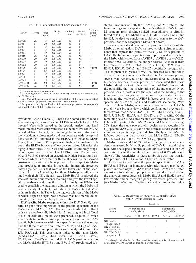
TABLE 2. Reactivities of (putative) GL-specific MAbswith NR virus variants in IPMA
Escapemutant
Reactivity
Nonneutralizinganti-GL MAbs
Neutralizinganti-GL MAbs
E3/c16 E3/d10 E6/B8 E6/A3 E7/d15-c1 E7/d15-c9
NR E6/A3,plaque no. 5
1 1 1 2 1 1
NR E7/d15-c1,plaque no. 7
1 1 1 1 2 1
NR E7/d15-c9,a
plaque no. 41 1 1 1 1 1
a Although stainable by the MAb used for selection, this NR was less wellneutralized by MAb E7/d15-c9 than the parental virus.
VOL. 38, 2000 NONNEUTRALIZING EAV GL PROTEIN-SPECIFIC MAbs 2069
on April 13, 2019 by guest
http://jcm.asm
.org/D
ownloaded from

between EAV B15 and EAV Utr. Although Table 1 indicatesthat MAb E6/A3 may indeed possess poor binding character-istics, this is definitely not the case for MAb E6/d24, which hadrelatively high IPMA and ELISA titers, despite a low antibodyconcentration (Table 1). Moreover, the observation that each
of the EAV-specific MAbs including E6/A3 and E6/d24 didrecognize Vero cells infected with the Utr strain of EAV in anIPMA (data not shown) militates against the third possibility.In addition, MAb E6/A3 efficiently neutralized EAV Utr (datanot shown).
FIG. 2. (A and B) Immunoprecipitation analysis of N-specific MAbs. OST-7.1 cells were infected with the vaccinia virus recombinants vAVE07 or vAVE16, whichexpress the EAV N and M proteins, respectively, and were labeled for 1 h starting at 5 h p.i. with [35S]Met plus [35S]Cys. Total lysates of cells and media were preparedand incubated with 0.5 to 3 ml of N-specific (FP07) or M-specific (SP06) rabbit antiserum or 100 ml of culture supernatant from mouse hybridoma E1/A19, E1/b5, E1/c4,E3/a9, E3/a61, E3/d17, E3/d52, E6/A7, E6/a27, E6/B8, or E7/d15-c9. (C) Immunoprecipitation analysis of GL-specific MAbs. OST-7.1 cells were infected with thevaccinia virus recombinants vAVE16 (lanes 6) and vAVE25 (lanes 5), which express the EAV M and GL proteins, respectively (12), and were labeled as described above.Total lysates of cells and media were prepared and incubated with 0.5 ml of anti-M rabbit serum (SP06) or ascitic fluid containing the GL-specific MAb 93B (IgG2asubtype) (23) or 100 ml of culture supernatant from mouse hybridoma E3/c16, E3/d10, E6/B8, E7/d15-c1, E7/d15-c9, or E1/A19. The numbers at the right indicate thesizes (in kilodaltons) and positions of marker proteins analyzed in the same SDS–15% PAA gels. The positions of the N and M proteins and the full-length and atruncated form (GL*) (12) of the GL glycoprotein are displayed at the left.
2070 WEILAND ET AL. J. CLIN. MICROBIOL.
on April 13, 2019 by guest
http://jcm.asm
.org/D
ownloaded from

As an alternative approach to establishing the protein spec-ificities of MAbs E6/A3 and E6/d24, we performed immuno-blotting experiments with EAV B15 virions that serve as theantigen. Remarkably, in these assays MAb E6/d24 clearly rec-ognized the EAV N protein (Fig. 3, lanes 10 and 11), whereasMAbs E6/A3, E7/d15-c1, and E7/d15-c9 did not specificallybind to any viral protein on the Western blot (data not shown).In contrast, MAbs E3/c16, E3/d10, and E6/B8 weakly bound toa polypeptide of between 30 and 42 kDa (Fig. 3, lane 12)which, on the basis of its size and appearance, is believed tocorrespond to the EAV GL protein. In agreement with ourearlier findings, MAbs E1/A19, E1/b5, E1/c4, E3/a9, E3/a61,E3/d17, E3/d52, E6/A7, and E6/a27 strongly recognized theEAV N protein (Fig. 3, lanes 1 through 9).
Detection of neutralizing and nonneutralizing anti-EAV GLMAbs. Experiments in which constant amounts of antibodywere mixed with different doses of virus indicated that onlyMAbs E6/A3, E7/d15-c1, and E7/d15-c9 displayed VN activity(data not shown). To compare the neutralizing capacities ofthese MAbs, a constant number of infectious virus particleswas mixed with different amounts of each antibody and addedto Vero cells. Complete inhibition of the CPE was accom-plished by the culture supernatant of hybridoma E6/A3 atdilutions of up to 1:80, by the E7/d15-c1 culture supernatant ata maximum dilution of 1:10, and by the E7/d15-c9 culturesupernatant at dilutions of up to 1:5 (Table 1). Protection ofhalf of the cell monolayers was observed after treatment of thevirus with 1:320, 1:160, and 1:20 dilutions of MAbs E6/A3,E7/d15-c1, and E7/d15-c9, respectively. However, despite theabsence of a visible CPE, single cells and small foci expressingthe EAV N protein were detected by IPMA in the cell culturesinoculated with the virus-antibody complexes at 3 days afterinfection. In concordance with the neutralization titers, theleast residual infectivity was observed in the monolayers in-fected with the E6/A3-treated virus, whereas E7/d15-c9-treated EAV particles yielded the largest number of infectiouscenters. Thus, only three MAbs showed VN activity.
To investigate whether the nonneutralizing GL-specificMAbs could sensitize EAV for neutralization by comple-ment (see references 27 and 35 and references therein) amicroneutralization test in the presence of guinea pig serumwas performed. None of the VN2 anti-GL MAbs was capableof blocking EAV infectivity in the presence of complement.
Furthermore, addition of complement did not affect the neu-tralization indices of MAbs E6/A3, E7/d15-c1, and E7/d15-c9(data not shown).
Immunoelectron microscopy. To investigate whether the dif-ferent anti-GL MAbs recognized surface components of viri-ons, immunoelectron microscopy was performed. For this pur-pose, the virus particles were first incubated with dilutedhybridoma culture supernatant and were then immersed in asolution of goat anti-mouse antibodies conjugated to 5-nmgold particles. As expected, none of the N-specific MAbs in-teracted with the viral surface (Fig. 4C). In contrast, all VN1
and GL-specific MAbs mediated binding of immunogold par-ticles to the viral envelope. Interestingly, labeling was morepronounced with the neutralizing MAbs than with the nonneu-tralizing anti-GL MAbs (compare Fig. 4A and B). Statisticalanalyses of 100 virions per antibody indicated that the neutral-izing MAb E7/d15-c1 on average mediated the binding of 8.8gold granules per virus particle, whereas the average numberof gold particles attached to E3/c16-coated virions was 2.7. Asimilar threefold difference in the recruitment of gold particlesat the viral surface was observed when the binding mediated byVN1 antibodies E6/A3 and E7/d15-c9 and the binding medi-ated by VN2 MAbs E3/d10 and E6/B8 were compared.
Selection of NR variant viruses. To find out whether the VNantibodies recognized related or independent epitopes, EAVB15 was repeatedly passaged in the presence of MAbs E6/A3,E7/d15-c1, and E7/d15-c9 in an attempt to generate NR escapemutants of the virus. Individual virus clones were then selectedin plaque assays, amplified in Vero cells, and subjected tomicroneutralization tests. For comparison, the selection pro-cedure was also performed with the nonneutralizing GL-spe-cific MAbs E3/c16, E3/d10, and E6/B8. Eleven NR virus vari-ants of MAb E6/A3 were obtained, whereas treatment of EAVB15 with MAb E7/d15-c1 yielded one NR escape mutant. TheE6/A3 NR viruses were recognized by all five GL-specific an-tibodies in IPMAs (Table 2) and were neutralized with thesame efficiency as the parental virus by MAbs E7/d15-c1 andE7/d15-c9 (data not shown). Similarly, the E7/d15-c1-specificviral escape mutant was no longer labeled or neutralized by theselecting MAb, but cells infected with this virus were stained asintensively with the other four anti-GL MAbs and the stronglyneutralizing MAb E6/A3 as EAV B15-infected cells. Further-more, MAbs E6/A3 and E7/d15-c9 were still able to neutralizeE7/d15-c1 NR virus infectivity. In contrast, the plaque deriva-tives of EAV B15 that were obtained after treatment of thevirus for three consecutive passages with MAb E7/d15-c9 werestill recognized by the selecting MAb in IPMAs (Table 2).However, these virus clones were less well neutralized by MAbE7/d15-c9 than the parental virus. Finally, the properties ofEAV B15 were not detectably altered after selection with thenonneutralizing GL-specific MAbs E3/c16, E3/d10, and E6/B8.Taken together these data indicate that the three VN1 MAbsrecognize distinct epitopes.
Interaction of MAbs with field isolates of EAV. To discrim-inate MAbs directed against conserved epitopes from antibod-ies that recognize variable protein domains, we compared thebinding of each of the 16 EAV-specific MAbs to a panel of 23field isolates of EAV in IPMAs of virus-infected Vero cells.The 10 N-specific MAbs as well as the 3 nonneutralizing an-ti-GL MAbs and weakly neutralizing MAb E7/d15-c9 recog-nized all virus isolates. In contrast, the other two VN1 MAbsreacted only with a subset of the field viruses. Eight EAVisolates were recognized by both of these MAbs, two fieldviruses reacted with MAb E7/d15-c1 but not with MAb E6/A3,and eight other EAV isolates were recognized by MAb E6/A3but not by MAb E7/d15-c1. Four field viruses did not react with
FIG. 3. Immunoblot analysis of EAV-specific MAbs. Nitrocellulose stripscontaining electrophoretically separated virion proteins of EAV were incubatedwith MAbs E1/b5 (lane 1), E1/c4 (lane 2), E1/A19 (lane 3), E3/d17 (lane 4),E3/d52 (lane 5), E3/a61 (lane 6), E3/a9 (lane 7), E6/a27 (lane 8), E6/A7 (lane 9),and E6/d24 (lane 10, diluted 1:3; lane 11, diluted 1:8), with a mixture of MAbsE3/c16, E3/d10, and E6/B8 (lane 12), and with the anti-PRRSV MAb P11/d72(lane 13) (50). The positions of the (putative) N and GL proteins of EAV areshown at the right side; the numbers at the left side indicate positions and sizes(in kilodaltons) of marker proteins analyzed in the same gel.
VOL. 38, 2000 NONNEUTRALIZING EAV GL PROTEIN-SPECIFIC MAbs 2071
on April 13, 2019 by guest
http://jcm.asm
.org/D
ownloaded from

FIG. 4. Immunoelectron microscopy of EAV particles. Sucrose gradient-purified virions were first incubated with MAb E7/d5-c1 (A), E3/c16 (B), or E1/A19 (C)and were then labeled with gold-conjugated secondary antibodies. Bar, 100 nm.
2072 WEILAND ET AL. J. CLIN. MICROBIOL.
on April 13, 2019 by guest
http://jcm.asm
.org/D
ownloaded from

either of the two strongly neutralizing MAbs. Although theIPMAs were always carried out with monolayers infected withdifferent amounts of virus to compensate for differences in titerand/or growth rate between EAV isolates, it was sometimeshard to decide whether a specific MAb recognized the entirevirus population. In particular, MAb E6/A3 showed a very faintstaining of infected cells in IPMAs that corresponded to itsweak signal in IFA and ELISA, and it seemed to recognizeonly a fraction of the EAV-infected cells for the virus isolatesV200, V979, V1791, V2416, V2716, and V8908 (Table 3). Thedifferent reactivities of the VN1 MAbs with the EAV fieldisolates once again illustrated that they recognize distinct epi-topes.
DISCUSSION
In this paper, we have described the production and char-acterization of a panel of 16 EAV-specific MAbs. Immuno-blotting and immunoprecipitation analyses revealed that 10MAbs were directed against the EAV ORF 7-encoded N pro-tein and five MAbs reacted with the EAV ORF 5-encoded GLprotein. Two of the GL-specific MAbs (E7/d15-c1 and E7/d15-c9) displayed VN activity, while the other three anti-GL MAbsdid not inhibit the infectivity of EAV. The protein specificity ofone MAb (MAb E6/A3) could not be determined, but on thebasis of its VN capacity we speculate that it is also directedagainst the EAV GL protein. The observation that MAb E6/A3produces similar labeling patterns by immunofluorescence mi-croscopy of infected cells and immunoelectron microscopy ofvirus particles as MAbs E7/d15-c1 and E7/d15-c9 would be
consistent with this notion. Moreover, as mentioned above, theGL protein is the only target of EAV-neutralizing antibodiesidentified until now (2, 3, 4, 7, 12, 23, 29, 52, 54).
Although the immunization method and mouse strain thatwe used to produce EAV-specific MAbs differed from thoseused by others, like in previous studies (2, 3, 4, 6, 12, 23, 29,52), only antibodies directed against the N and GL proteins ofEAV were obtained. This result was unexpected since the sameimmunization procedure yielded MAbs specific for five differ-ent PRRSV polypeptides (50, 53). Moreover, the EAV Mprotein was recently shown to be more consistently recognizedin immunoblots by serum antibodies obtained from naturallyor experimentally infected horses than either the N or the GLprotein (24, 31). Collectively, these findings indicate that dif-ferences exist between the murine and equine humoral im-mune responses to EAV and between the immunogenicities ofthe structural proteins of EAV and PRRSV in mice. If oneassumes that arteriviruses have similar virion architectures, theantigenic differences between the structural proteins of EAVand PRRSV most likely result from subtle changes in theirtertiary or quaternary structures or are a direct consequence ofdifferences in their primary amino acid sequences. In addition,differences in the purity and integrity of the virus preparationsused for immunization may have contributed to the observeddifferences in antibody response following the inoculation ofmice with EAV or PRRSV. If indeed the stability of the virushas an effect on the number of structural proteins for whichmurine antibodies can be obtained, chemical and/or physicaldisruption of EAV particles may help to expand the existingrepertoire of EAV-specific MAbs.
If MAb E6/A3 is indeed directed against the EAV GL pro-tein, it probably recognizes a conformational epitope which isdestructed during the preparation of protein samples for im-munoblotting or immunoprecipitation. In contrast, the otherVN1 GL-specific MAbs seem to be directed against epitopesthat are preserved during cell lysis and immunoprecipitationanalysis but which do not survive the denaturing conditionsthat apply to SDS-PAGE and Western blotting. The nonneu-tralizing GL-specific MAbs most likely interact with (partially)linear epitopes as they weakly bound to the GL protein in animmunoblot. The same may hold true for the N-specific MAbswhich strongly recognized the N protein after Western blot-ting. Interestingly, the N-specific MAb E6/d24 reacted with theEAV N protein in IFA and IPMA and on an immunoblot butnot in an immunoprecipitation analysis. A possible explanationfor this peculiar result may be that MAb E6/d24 recognizes alinear epitope that is exposed on the Western blot and afterfixation of virus-infected cells with 96% ethanol but is notaccessible for antibody binding at the low detergent concen-trations in the immunoprecipitation buffer. Alternatively, MAbE6/d24 recognizes a conformational epitope that is denaturedby the detergents present in the cell lysis and immunoprecipi-tation buffer but refolds after transfer of the N protein tonitrocellulose and is preserved during ethanol fixation of EAV-infected cells. It thus seems that we have raised N-specificMAbs directed against at least two different epitopes. Thesebinding sites are highly conserved since all 10 N-specific MAbsreacted with each of the EAV isolates tested. Ultimately, com-petitive binding assays will have to be performed to determinethe number of independent epitopes recognized by the anti-NMAbs.
The GL-specific MAbs E3/c16, E3/d10, E6/B8, and E7/d15-c9 also reacted with all 23 field isolates of EAV in IPMAs.Since MAbs E3/c16, E3/d10, and E6/B8 exhibited similar prop-erties in antigen binding assays and VN tests, they presumablyrecognize identical or related epitopes. MAb E7/d15-c9 most
TABLE 3. Reactivities of EAV-specific MAbs withEAV field isolates by IPMA
Virus isolate/yr of isolation/passage no.a
Source
Reaction with neutralizinganti-GL MAbs
E6/A3 E7/d15-c1 E7/d15-c9
V2416/1991/4 Aborted foal 1b 1 1V2716/1991/4 Aborted foal 1b 1 1S1112/1992/4 Semen 1 2 1V8633/1993/3 Semen 1 2 1V4987/1994/3 Semen 1 2 1V979/1994/4 Semen 1b 2 1V8062/1995/4 Semen 1 2 1V2592/1995/3 Aborted foal 2 1 1V2715/1995/3 Aborted foal 2 1 1V4202/1995/3 Semen 1 1 1V4675/1995/3 Semen 1 2 1V8908/1995/3 Semen 1b 1 1V1790/1995/4 Semen 1 1 1V1791/1995/3 Semen 1b 2 1V1792/1995/3 Semen 1 2c 1V273/1996/4 Semen 2 2 1V1763/1996/4 Semen 2 2 1V1764/1996/4 Semend 2 2 1V4137/1996/4 Semen 2 2 1V1206/1996/4 Semen 1 2 1V5915/1996/3 Semen 1 1 1V152/1997/3 Semene 1 1 1V200/1997/4 Aborted foal 1b 1 1
a Passage in Vero cells.b A major part of the infected cells were reactive.c A minor part of the infected cells were reactive.d Obtained 3 months later from the same stallion that provided isolate V273/
1996/4.e Obtained 1 month later from the same stallion that provided isolate V5915/
1996/3.
VOL. 38, 2000 NONNEUTRALIZING EAV GL PROTEIN-SPECIFIC MAbs 2073
on April 13, 2019 by guest
http://jcm.asm
.org/D
ownloaded from

likely binds to a different part of the GL protein as it displayedweak VN activity, in contrast to the other three antibodies. Thestrongly neutralizing GL-specific MAb E7/d15-c1 reacted withonly 10 of the 23 EAV field isolates and therefore recognizesyet another epitope on the major envelope glycoprotein of thevirus. Consistently, MAb E7/d15-c1 NR escape mutants werestill recognized in IPMAs by the four other GL-specific MAbsand by MAb E6/A3. Moreover, loss of the E7/d15-c1-specificepitope did not influence virus neutralization by MAbs E7/d15-c9 and E6/A3. A possible fourth epitope on the EAV GLprotein may be defined by the strongly neutralizing MAbE6/A3 as it reacted with a different subset of the 23 EAV fieldisolates than MAb E7/d15-c1. In addition, NR virus variants ofMAb E6/A3 were still recognized by the GL-specific MAbsE3/c16, E3/d10, E6/B8, E7/d15-c1, and E7/d15-c9. Competitivebinding assays between MAb E6/A3 and the GL-specific MAbsproduced by others (2, 3, 4, 12, 23, 29, 52, 54) may providedefinitive evidence for its protein specificity. The same type ofexperiments and nucleotide sequence analyses of the E7/d15-c1- and E6/A3-specific NR escape mutants will also be helpfulin defining the epitopes recognized by MAbs E3/c16, E3/d10,E6/A3, E6/B8, E7/d15-c1, and E7/d15-c9. Furthermore, it maybe of interest to find out whether the viruses selected in thepresence of MAbs E3/c16, E3/d10, E6/B8, and E7/d15-c9 haveundergone sequence changes in the EAV GL protein.
A general picture that emerged from our studies on theGL-specific antibodies was that the VN1 MAbs weakly reactedwith the intracellular GL protein in IFAs but strongly bound toextracellular virus particles, whereas the VN2 MAbs poorlylabeled secreted virions but produced a bright immunofluores-cence in EAV-infected cells. The most plausible explanationfor these findings is that there are differences in the accessi-bilities of the epitopes recognized by VN1 and VN2 GL-specific MAbs between intra- and extracellular antigens. Theobservation that after dilution the immunofluorescence pat-terns obtained with MAbs E3/c16, E3/d10, and E6/B8 resem-bled those of undiluted MAbs E6/A3 and E7/d15-c1 may thenbe a consequence of the fact that the GL protein mainly accu-mulates at the intracellular side of virus assembly. In this sce-nario, the heavily labeled perinuclear granules would bothcontain GL molecules (being) incorporated in virus particlesand GL molecules that are not engaged in virus assembly, whilethe more peripheral cytoplasmic areas which were only stainedby the VN2 MAbs would mainly contain spillover of the GLprotein. The virus-associated GL protein would be most effec-tively recognized by the VN1 and the VN2 MAbs, whereas thefree GL molecules would be preferentially bound by the VN2
MAbs. Such an interpretation would also fit the results ob-tained by van der Meer et al. (47), who found that at earlytimes p.i. the GL protein is exclusively localized in the Golgicomplex but that at later stages of infection vesicles elsewherein the cytoplasm are labeled as well.
It may seem surprising that GL-specific MAbs E3/c16, E3/d10, and E6/B8 bound to the surfaces of virus particles butwere unable to reduce EAV infectivity or to sensitize thevirions for neutralization by complement. The amount of goldgranules that decorated the viral surface after immunogoldlabeling of virions with these MAbs was, however, fairly low.Individual virus particles could thus still contain enough un-shielded GL protein to mediate virus entry. In addition, Mc-Collum and Swerczek (34) and Fukunaga et al. (22) previouslynoticed that complement enhancement of virus neutralizationis a strain-dependent phenomenon. The fact that the neutral-ization titers of the VN1 MAbs were not affected by the pres-ence of complement may indicate that EAV B15 indeed be-
longs to the virus isolates whose neutralization is insensitive tocomplement addition.
The relevance of VN2 GL-specific antibodies in naturalEAV infections remains to be elucidated. The absence of anabsolute correlation between the virus neutralization titer ofconvalescent-phase equine sera and their reactivity with a GL-specific bacterial expression product in an ELISA (7) mayindicate that horses do produce nonneutralizing anti-GL anti-bodies in response to an EAV infection, although this resultcould also be explained in a number of other ways. Moreover,Chirnside et al. (8) reported induction of nonneutralizing GL-specific antibodies in 9 of 25 horses immunized with a chemi-cally inactivated whole-virus vaccine. The situation in naturallyinfected horses may, however, differ from that in vaccinatedanimals. The observation that the VN2 anti-GL MAbs recog-nized each of the EAV isolates tested indicates that theirepitopes are not subject to immune selection in vivo. Theseconstant epitopes which have not been previously identifiedcould be potentially useful for the development of a serologicaltest that is less sensitive to the antigenic variation than the VNassay. Furthermore, the N- and GL-specific MAbs that recog-nize each of the EAV isolates tested may find application insandwich ELISAs for the detection of anti-EAV antibodies orcapture ELISAs for the detection of EAV-specific antigens.
ACKNOWLEDGMENTS
We thank Amy Glaser for supplying us with the EAV GL-specificMAb 93B. We are indebted to Bernard Moss and to Peter Timoney forproviding us with the OST-7.1 cell line and EAV field isolate S1112,respectively. We gratefully acknowledge the excellent technical assis-tance of Britta Kanzok and Karina Mildner and thank Henrik Garofffor general support.
REFERENCES
1. Ausubel, F. M., R. Brent, R. E. Kingston, D. D. Moore, J. G. Seidman, J. A.Smith, and K. Struhl. 1987. Current protocols in molecular biology. JohnWiley & Sons, Inc., New York, N.Y.
2. Balasuriya, U. B. R., N. J. MacLachlan, A. A. F. de Vries, P. V. Rossitto, andP. J. M. Rottier. 1995. Identification of a neutralization site in the majorenvelope glycoprotein (GL) of equine arteritis virus. Virology 207:518–527.
3. Balasuriya, U. B. R., J. F. Patton, P. V. Rossitto, P. J. Timoney, W. H.McCollum, and N. J. MacLachlan. 1997. Neutralization determinants oflaboratory strains and field isolates of equine arteritis virus: identification offour neutralization sites in the amino-terminal ectodomain of the GL enve-lope glycoprotein. Virology 232:114–128.
4. Balasuriya, U. B. R., P. V. Rossitto, C. D. DeMaula, and N. J. MacLachlan.1993. A 29K envelope glycoprotein of equine arteritis virus expresses neu-tralization determinants recognized by murine monoclonal antibodies.J. Gen. Virol. 74:2525–2529.
5. Balasuriya, U. B. R., P. J. Timoney, W. H. McCollum, and N. J. MacLachlan.1995. Phylogenetic analysis of open reading frame 5 of field isolates ofequine arteritis virus and identification of conserved and nonconserved re-gions in the GL envelope glycoprotein. Virology 214:690–697.
6. Chirnside, E. D., R. F. Cook, M. W. Lock, and J. A. Mumford. 1988. Mono-clonal antibodies to equine arteritis virus, p. 262–267. In D. G. Powell (ed.),Proceedings of the 5th International Conference on Equine Infectious Dis-eases, Lexington, Ky., 1987. The University Press of Kentucky, Lexington.
7. Chirnside, E. D., A. A. F. de Vries, J. A. Mumford, and P. J. M. Rottier. 1995.Equine arteritis virus-neutralizing antibody in the horse is induced by adeterminant on the large envelope glycoprotein GL. J. Gen. Virol. 76:1989–1998.
8. Chirnside, E. D., P. M. Francis, A. A. F. de Vries, R. Sinclair, and J. A.Mumford. 1995. Development and evaluation of an ELISA using recombi-nant fusion protein to detect the presence of host antibody to equine arteritisvirus. J. Virol. Methods 54:1–13.
9. Chirnside, E. D., P. M. Francis, and J. A. Mumford. 1995. Expressioncloning and antigenic analysis of the nucleocapsid protein of equine arteritisvirus. J. Virol. Methods 54:277–288.
10. del Piero, F., P. A. Wilkins, J. W. Lopez, A. L. Glaser, E. J. Dubovi, D. H.Schlafer, and D. H. Lein. 1997. Equine viral arteritis in newborn foals:clinical, pathological, serological, microbiological and immunohistochemicalobservations. Equine Vet. J. 29:178–185.
11. den Boon, J. A., E. J. Snijder, E. D. Chirnside, A. A. F. de Vries, M. C.Horzinek, and W. J. M. Spaan. 1991. Equine arteritis virus is not a togavirus
2074 WEILAND ET AL. J. CLIN. MICROBIOL.
on April 13, 2019 by guest
http://jcm.asm
.org/D
ownloaded from

but belongs to the coronaviruslike superfamily. J. Virol. 65:2910–2920.12. Deregt, D., A. A. F. de Vries, M. J. B. Raamsman, L. D. Elmgren, and P. J. M.
Rottier. 1994. Monoclonal antibodies to equine arteritis virus proteins iden-tify the GL protein as a target for virus neutralization. J. Gen. Virol. 75:2439–2444.
13. de Vries, A. A. F., E. D. Chirnside, P. J. Bredenbeek, L. A. Gravestein, M. C.Horzinek, and W. J. M. Spaan. 1990. All subgenomic mRNAs of equinearteritis virus contain a common leader sequence. Nucleic Acids Res. 18:3241–3247.
14. de Vries, A. A. F., E. D. Chirnside, M. C. Horzinek, and P. J. M. Rottier.1992. Structural proteins of equine arteritis virus. J. Virol. 66:6294–6303.
15. de Vries, A. A. F., M. C. Horzinek, P. J. M. Rottier, and R. J. de Groot. 1997.The genome organization of the Nidovirales: similarities and differencesbetween arteri-, toro-, and coronaviruses. Semin. Virol. 8:33–47.
16. de Vries, A. A. F., S. M. Post, M. J. B. Raamsman, M. C. Horzinek, andP. J. M. Rottier. 1995. The two major envelope proteins of equine arteritisvirus associate into disulfide-linked heterodimers. J. Virol. 69:4668–4674.
17. de Vries, A. A. F., M. J. B. Raamsman, H. A. van Dijk, M. C. Horzinek, andP. J. M. Rottier. 1995. The small envelope glycoprotein (GS) of equinearteritis virus folds into three distinct monomers and a disulfide-linkeddimer. J. Virol. 69:3441–3448.
18. de Vries, A. A. F., P. J. M. Rottier, A. L. Glaser, and M. C. Horzinek. 1996.Equine viral arteritis, p. 171–200. In M. J. Studdert (ed.), Virus infections ofequines. Elsevier Science Publishers, Amsterdam, The Netherlands.
19. Doll, E. R., J. T. Bryans, W. H. McCollum, and M. Crowe. 1957. Isolation ofa filterable agent causing arteritis of horses and abortion by mares. Itsdifferentiation from the equine abortion (influenza) virus. Cornell Vet. 47:3–41.
20. Drew, T. W., J. J. M. Meulenberg, J. J. Sands, and D. J. Paton. 1995.Production, characterization and reactivity of monoclonal antibodies to por-cine reproductive and respiratory syndrome virus. J. Gen. Virol. 76:1361–1369.
21. Elroy-Stein, O., and B. Moss. 1990. Cytoplasmic expression system based onconstitutive synthesis of bacteriophage T7 RNA polymerase. Proc. Natl.Acad. Sci. USA 87:6743–6747.
22. Fukunaga, Y., H. Imagawa, T. Kanemaru, and M. Kamada. 1993. Comple-ment-dependent serum neutralization with virulent and avirulent Bucyrusstrains of equine arteritis virus. Vet. Microbiol. 36:379–383.
23. Glaser, A. L., A. A. F. de Vries, and E. J. Dubovi. 1995. Comparison of equinearteritis virus isolates using neutralizing monoclonal antibodies and identi-fication of sequence changes in GL associated with neutralization resistance.J. Gen. Virol. 76:2223–2233.
24. Hedges, J. F., U. B. R. Balasuriya, S. Ahmad, P. J. Timoney, W. H. McCol-lum, T. Yilma, and N. J. MacLachlan. 1998. Detection of antibodies toequine arteritis virus by enzyme linked immunosorbant assays utilizing GL,M and N proteins expressed from recombinant baculoviruses. J. Virol. Meth-ods 76:127–137.
25. Holyoak, G. R., T. V. Little, W. H. McCollum, and P. J. Timoney. 1993.Relationship between onset of puberty and establishment of persistent in-fection with equine arteritis virus in the experimentally infected colt.J. Comp. Pathol. 109:26–46.
26. Hyllseth, B. 1973. Structural proteins of equine arteritis virus. Arch. Gesa-mte Virusforsch. 40:177–188.
27. Hyllseth, B., and U. Pettersson. 1970. Neutralization of equine arteritis virus:enhancing effect of guinea pig serum. Arch. Gesamte Virusforsch. 32:337–347.
28. Kheyar, A., S. Martin, G. St-Laurent, P. J. Timoney, W. H. McCollum, andD. Archambault. 1997. Expression cloning and humoral immune response tothe nucleocapsid and membrane proteins of equine arteritis virus. Clin.Diagn. Lab. Immunol. 4:648–652.
29. Kondo, T., H. Akashi, Y. Fukunaga, S. Sugita, K. Sekiguchi, R. Wada, andM. Kamada. 1994. Production and characterisation of monoclonal antibod-ies against structural proteins of equine arteritis virus, p. 21–26. In H. Na-kajima and W. Plowright (ed.), Proceedings of the 7th International Con-ference on Equine Infectious Diseases, Tokyo, Japan, 1994. R & WPublications, Ltd., Newmarket, England.
30. Kondo, T., S. Sugita, Y. Fukunaga, and H. Imagawa. 1998. Identification ofthe major epitope in the GL protein of equine arteritis virus (EAV) recog-nized by antibody in EAV-infected horses using synthetic peptides. J. EquineSci. 9:19–23.
31. MacLachlan, N. J., U. B. R. Balasuriya, J. F. Hedges, T. M. Schweidler,W. H. McCollum, P. J. Timoney, P. J. Hullinger, and J. F. Patton. 1998.Serologic response of horses to the structural proteins of equine arteritisvirus. J. Vet. Diagn. Invest. 10:229–236.
32. McCollum, W. H. 1969. Development of a modified virus strain and vaccinefor equine viral arteritis. J. Am. Vet. Med. Assoc. 155:318–322.
33. McCollum, W. H. 1976. Studies of passive immunity in foals to equine viralarteritis. Vet. Microbiol. 1:45–54.
34. McCollum, W. H., and T. W. Swerczek. 1978. Studies of an epizootic ofequine viral arteritis in racehorses. Equine Vet. J. 2:293–299.
35. Radwan, A. I., and D. Burger. 1974. The mechanisms of neutralization ofsensitized equine arteritis virus by complement components. J. Gen. Virol.25:229–237.
36. Remaut, E., H. Tsao, and W. Fiers. 1983. Improved plasmid vectors with athermoinducible expression and temperature-regulated runaway replication.Gene 22:103–113.
37. Sambrook, J., E. F. Fritsch, and T. Maniatis. 1989. Molecular cloning: alaboratory manual, 2nd ed. Cold Spring Harbor Laboratory, Cold SpringHarbor, N.Y.
38. Schafer, W. 1979. Der Mause-Inzuchtstamm STU. Entwicklung und Eigen-schaften. Z. Naturforsch. Teil C 34:306–309.
39. Shulman, M., C. D. Wilde, and G. Kohler. 1978. A better cell line for makinghybridomas secreting specific antibodies. Nature 276:269–270.
40. Snijder, E. J., and J. J. M. Meulenberg. 1998. The molecular biology ofarteriviruses. J. Gen. Virol. 79:961–979.
41. Snijder, E. J., H. van Tol, K. W. Pedersen, M. J. B. Raamsman, and A. A. F.de Vries. 1999. Identification of a novel structural protein of arteriviruses.J. Virol. 73:6335–6345.
42. Stadejek, T., H. Bjorklund, C. Ros Bascunana, I. M. Ciabatti, M. T. Sci-cluna, D. Amaddeo, W. H. McCollum, G. L. Autorino, P. J. Timoney, D. J.Paton, B. Klingeborn, and S. Belak. 1999. Genetic diversity of equine ar-teritis virus. J. Gen. Virol. 80:691–699.
43. St-Laurent, G., N. Lepage, S. Carman, and D. Archambault. 1997. Geneticand amino acid analysis of the GL protein of Canadian, American andEuropean equine arteritis virus isolates. Can. J. Vet. Res. 61:72–76.
44. Strebel, K., E. Beck, K. Strohmaier, and H. Schaller. 1986. Characterizationof foot-and-mouth disease virus gene products with antisera against bacte-rially synthesized fusion proteins. J. Virol. 57:983–991.
45. Timoney, P. J., and W. H. McCollum. 1993. Equine viral arteritis. Vet. Clin.N. Am. Equine Pract. 9:295–309.
46. Towbin, H., T. Staehelin, and J. Gordon. 1979. Electrophoretic transfer ofproteins from polyacrylamide gels to nitrocellulose sheets: procedure andsome applications. Proc. Natl. Acad. Sci. USA 76:4350–4354.
47. van der Meer, Y., H. van Tol, J. Krijnse Locker, and E. J. Snijder. 1998.ORF1a-encoded replicase subunits are involved in the membrane associa-tion of the arterivirus replication complex. J. Virol. 72:6689–6698.
48. van Nieuwstadt, A. P., J. J. M. Meulenberg, A. van Essen-Zandbergen, A.Petersen-den Besten, R. J. Bende, R. J. M. Moormann, and G. Wensvoort.1996. Proteins encoded by open reading frames 3 and 4 of the genome ofLelystad virus (Arteriviridae) are structural proteins of the virion. J. Virol.70:4767–4772.
49. Weiland, E., H.-J. Thiel, G. Hess, and F. Weiland. 1989. Development ofmonoclonal neutralizing antibodies against bovine viral diarrhea virus afterpretreatment of mice with normal bovine cells and cyclophosphamide. J. Vi-rol. Methods 24:237–244.
50. Weiland, E., M. Wieczorek-Krohmer, D. Kohl, K. K. Conzelmann, and F.Weiland. 1999. Monoclonal antibodies to the GP5 of porcine reproductiveand respiratory syndrome virus are more effective in virus neutralization thanmonoclonal antibodies to the GP4. Vet. Microbiol. 66:171–186.
51. Weiland, F., H. Granzow, M. Wieczorek-Krohmer, and E. Weiland. 1995.Electron microscopic studies on the morphogenesis of PRRSV in infectedcells—comparative studies, p. 499–502. In M. Schwyzer, M. Ackermann, G.Bertoni, R. Kocherhans, K. McCullough, M. Engels, R. Wittek, and R.Zanoni (ed.), Immunobiology of viral infections, Proceedings of the 3rdCongress of the European Society for Veterinary Virology, Interlaken, Swit-zerland. Fondation Marcel Merieux, Lyon, France.
52. Westcott, D., M. Lucas, and D. Paton. 1995. Equine arteritis virus: antigenicanalysis of strain variation, p. 479–483. In M. Schwyzer, M. Ackermann, G.Bertoni, R. Kocherhans, K. McCullough, M. Engels, R. Wittek, and R.Zanoni (ed.), Immunobiology of viral infections, Proceedings of the 3rdCongress of the European Society for Veterinary Virology, Interlaken, Swit-zerland. Fondation Marcel Merieux, Lyon, France.
53. Wieczorek-Krohmer, M., F. Weiland, K. Conzelmann, D. Kohl, N. Visser, P.van Woensel, H.-J. Thiel, and E. Weiland. 1996. Porcine reproductive andrespiratory syndrome virus (PRRSV): monoclonal antibodies detect com-mon epitopes on two viral proteins of European and U.S. isolates. Vet.Microbiol. 51:257–266.
54. Yamaguchi, S., T. Kanno, H. Akashi, and T. Kondo. 1997. Identification oftwo neutralization sites in GL protein of equine arteritis virus by means ofmonoclonal antibodies. J. Equine Sci. 8:7–11.
55. Zeegers, J. J. W., B. A. M. van der Zeijst, and M. C. Horzinek. 1976. Thestructural proteins of equine arteritis virus. Virology 73:200–205.
VOL. 38, 2000 NONNEUTRALIZING EAV GL PROTEIN-SPECIFIC MAbs 2075
on April 13, 2019 by guest
http://jcm.asm
.org/D
ownloaded from