Embed Size (px)
Citation preview
8/3/2019 More Fun in the Philippines - P&P
http://slidepdf.com/reader/full/more-fun-in-the-philippines-pp 1/9
Studying Pelvis & Perineum. More fun in the
Philippines! >:)
Pelvic Girdle A basin-shaped ring of bones that
connects the vertebral column to the twofemurs
The bony structure that surrounds thepelvis1. Hip/Pelvic bones (2)2. Sacrum3. Coccyx
Functions of Pelvic Girdle1. Bear the weight of the upper body when
sitting and standing
2. Transfer weight from axial to the lowerappendicular
3. Attachments of locomotor, abdominaland postural muscles
4. Protect the lower abdominal and pelvicviscera
5. Support the gravid uterus6. Attachments of erectile tissues of the
external genitalia7. Attachment of the pelvic floor
1. Pelvic Bone♥ a.k.a. Coxal Bones♥ Large irregularly shaped bones♥ Composed of 3 bones
1. Ilium2. Ischium3. Pubis
♥ Pubic Symphysis (Anteriorly)♥ Sacroiliac Joint (Posteriorly)♥ In children, the hip bones are separated
♥ United by the Triradiate Cartilage in theacetabulum
♥ After puberty, fuse to form the hip bone
1. Ilium∴ Superior, fan-shaped part of the hip bone⋅ Ala⋅ Body
∴ Iliac Crest∴ Iliac Spines⋅ ASIS⋅ PSIS⋅ AIIS⋅ PIIS
∴ Iliac Fossa∴ Iliac Tuberosity∴ Auricular Surface∴ Arcuate Line *
2. Ischium∴ Forms the lower and back part of the hip
bone∴ Body∴ Ischial Ramus **⋅ Superior⋅ Inferior
∴ Ischial Tuberosity∴ Ischial Spine
1. Greater Sciatic Notch2. Lesser Sciatic Notch
3. Pubis∴ Forms the lower and anterior part of thehip bone
∴ Body⋅ Pubic Crest⋅ Pubic Tubercle
∴ Pubic Ramus **⋅ Superior⋅ Inferior
∴ Pectineal Line / Pecten Pubis *⋅ Pubic Arch
⋅ Subpubic Angle
2. Sacrum♥ Wedge-shaped vertebra♥ 5 bones fused into 1♥ Concave anteriorly♥ Articulations:
٭ Lumbosacral Joint
xoxo: Gorj.
8/3/2019 More Fun in the Philippines - P&P
http://slidepdf.com/reader/full/more-fun-in-the-philippines-pp 2/9
Studying Pelvis & Perineum. More fun in the
Philippines! >:)
٭ Sacroiliac Joint٭ Sacrococcygeal Joint
♥ Sacral Hiatus♥ Anterior and Posterior Sacral Foramina♥ Sacral Ala♥ Sacral Canal♥ Sacral Promontory
3. Coccyx♥ Tail bone♥ 4 vertebrae fused into 1
Clinics
1. Pelvic Fractures2. Coccydynia
Divisions of Pelvis1. Greater Pelvis∞ a.k.a. False Pelvis∞ Superior to the Pelvic Inlet∞ Bounded by the Iliac Alae and S1
vertebrae∞ Occupied by the abdominal viscera
(Ileum and Sigmoid)∝ Pelvic Inlet• Superior Pelvic Aperture• Pelvic Brim
Promontory and Ala ofSacrum
Linea Terminalis… Arcuate Line … Pecten Pubis … Pubic Crest
Symphysis Pubis∝ Pelvic Outlet• Inferior Pelvic Aperture
Tip of Coccyx Sacrotuberous Ligament Ischial Tuberosities Pubic Arch
2. Lesser Pelvis∞ a.k.a. True Pelvis∞ Between the Pelvic Inlet and Pelvic
Outlet∞ Bounded by the pelvic surfaces of hip
bone, sacrum and coccyx∞ Division (Pelvic Diaphragm)
a. True Pelvic Cavityb. Perineum
Clinical SignificancePelvic Diameters and Conjugates
1. Pelvic Inlet
2. Pelvic Cavity / Midpelvis Plane of Greatest Dimension Plane of Least Dimension
3. Pelvic Outlet
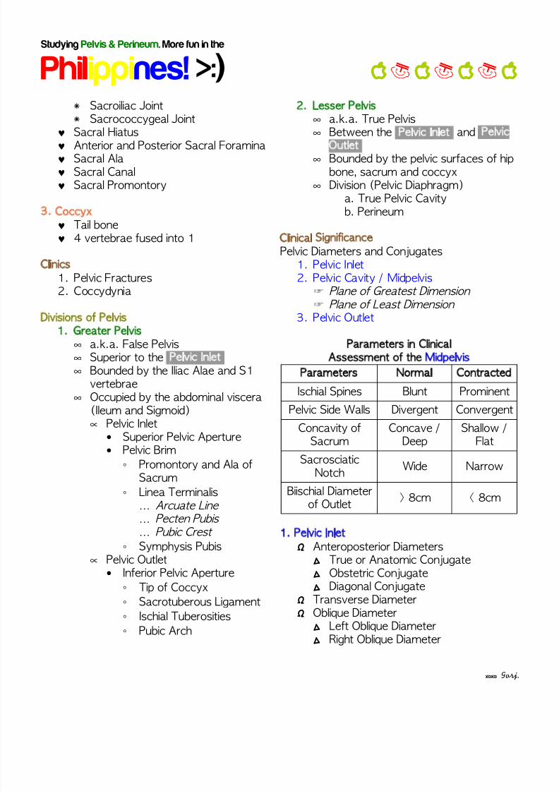
Parameters in ClinicalAssessment of the Midpelvis
Parameters Normal Contracted
Ischial Spines Blunt Prominent
Pelvic Side Walls Divergent Convergent
Concavity ofSacrum
Concave /Deep
Shallow /Flat
SacrosciaticNotch
Wide Narrow
Biischial Diameterof Outlet
> 8cm < 8cm
1. Pelvic InletΩ Anteroposterior Diameters∆ True or Anatomic Conjugate∆ Obstetric Conjugate∆ Diagonal Conjugate
Ω Transverse DiameterΩ Oblique Diameter∆ Left Oblique Diameter∆ Right Oblique Diameter
xoxo: Gorj.
8/3/2019 More Fun in the Philippines - P&P
http://slidepdf.com/reader/full/more-fun-in-the-philippines-pp 3/9
Studying Pelvis & Perineum. More fun in the
Philippines! >:)
2. Pelvic Cavity / MidpelvisΩ Plane of Greatest Dimension∆ Anteroposterior Diameter∆ Transverse Diameter
Ω Plane of Least Dimension∆ Anteroposterior Diameter∆ Transverse / Bispinous Diameter
3. Pelvic OutletΩ Anteroposterior Diameter∆ Anatomic∆ Obstetric
Ω Transverse / Biischial Diameter
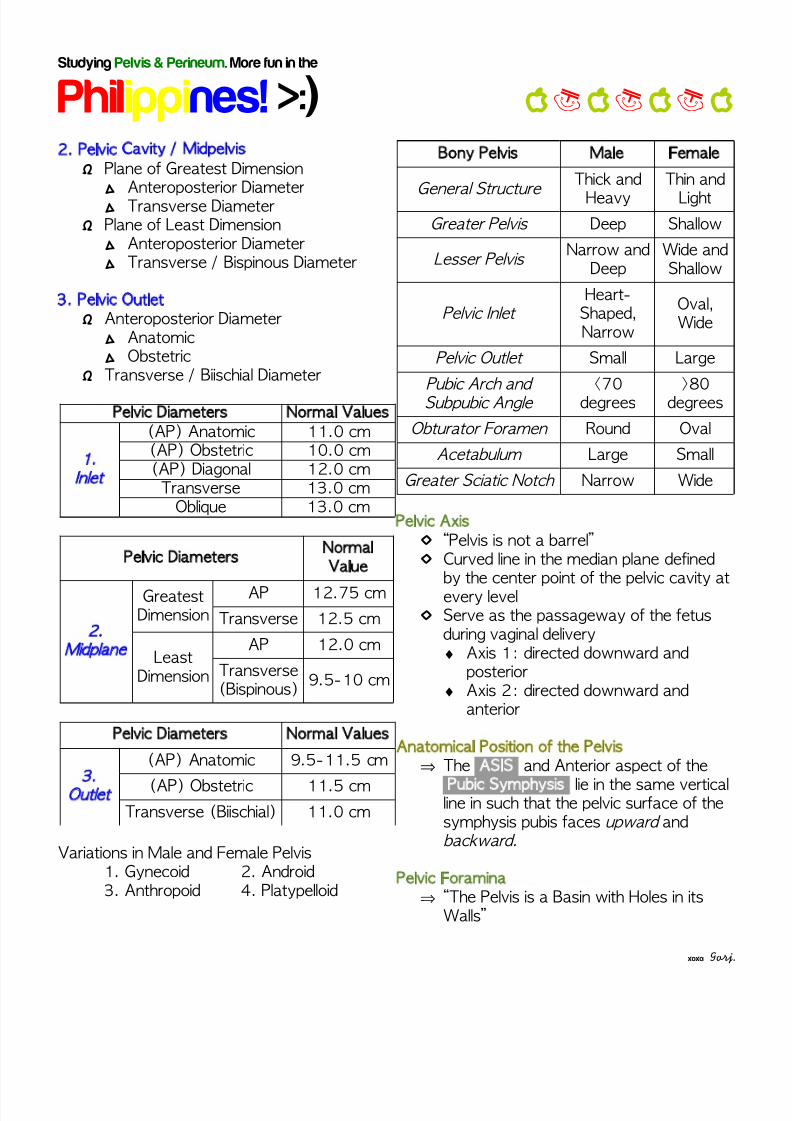
Pelvic Diameters Normal Values
1.
Inlet
(AP) Anatomic 11.0 cm(AP) Obstetric 10.0 cm(AP) Diagonal 12.0 cm
Transverse 13.0 cmOblique 13.0 cm
Pelvic DiametersNormalValue
2.
Midplane
GreatestDimension
AP 12.75 cm
Transverse 12.5 cm
LeastDimension
AP 12.0 cm
Transverse(Bispinous)
9.5-10 cm
Pelvic Diameters Normal Values
3.
Outlet
(AP) Anatomic 9.5-11.5 cm
(AP) Obstetric 11.5 cmTransverse (Biischial) 11.0 cm
Variations in Male and Female Pelvis1. Gynecoid 2. Android3. Anthropoid 4. Platypelloid
Bony Pelvis Male Female
General Structure
Thick and
Heavy
Thin and
LightGreater Pelvis Deep Shallow
Lesser Pelvis Narrow and
DeepWide andShallow
Pelvic Inlet Heart-Shaped,Narrow
Oval,Wide
Pelvic Outlet Small Large
Pubic Arch and Subpubic Angle
<70degrees
>80degrees
Obturator Foramen Round Oval
Acetabulum Large Small
Greater Sciatic Notch Narrow Wide
Pelvic Axis◊ “Pelvis is not a barrel”◊ Curved line in the median plane defined
by the center point of the pelvic cavity atevery level
◊ Serve as the passageway of the fetusduring vaginal delivery♦ Axis 1: directed downward and
posterior♦ Axis 2: directed downward and
anterior
Anatomical Position of the Pelvis⇒ The ASIS and Anterior aspect of the
Pubic Symphysis lie in the same verticalline in such that the pelvic surface of thesymphysis pubis faces upward andbackward.
Pelvic Foramina⇒ “The Pelvis is a Basin with Holes in its
Walls”
xoxo: Gorj.
8/3/2019 More Fun in the Philippines - P&P
http://slidepdf.com/reader/full/more-fun-in-the-philippines-pp 4/9
Studying Pelvis & Perineum. More fun in the
Philippines! >:)
→ Anterior Sacral Foramina→ Greater Sciatic Foramen→ Lesser Sciatic Foramen→ Obturator Foramen
⇒ “For the wires (Pudendal Nerve) to gainentrance to the apartment below(Perineum), without going through thefloor (Pelvic Floor), they have to piercethe wall (Greater Sciatic Foramen) to getoutside the building and then returnthrough a second hole (Lesser SciaticForamen).”
Pelvic Joints1. Sacroiliac Joint2. Sacrococcygeal Joint3. Pubic Symphysis
1. Sacroiliac Joint♣ Strong, weight bearing compound joint♣ Both Synovial and Syndesmois type of
joint♣ Synovial: Auricular surfaces of the
sacrum and ilium↣ Irregular but congruent elevations and depressions that interlock
♣ Syndesmosis: Tuberosities of sacrumand ilium
♣ Ligaments:↣ Anterior Sacroiliac Ligaments↣ Interosseous Sacroiliac Ligaments↣ Posterior Sacroiliac Ligaments↣ Sacrotuberous Ligament↣ Sacrospinous Ligament
2. Sacrococcygeal Joint♣ S5 and Coccyx♣ Ligaments:
↣ Anterior Sacrococcygeal Ligament↣ Lateral Sacrococcygeal Ligament↣ Posterior Sacrococcygeal Ligament
3. Pubic Symphysis Joint♣ Joint between the two pubic bone♣ Cartilaginous joint consisting of
fibrocartilage interpubic disc♣ Ligaments:
↣ Superior Interpubic Ligament↣ Inferior (Arcuate) Interpubic Ligament
Pelvic Wall Muscles1. Obturator Internus
enters lesser sciatic notch2. Piriformis
enters greater sciatic notch
Pelvic Floor Muscles‣ Funnel-shaped pelvic diaphragm‣ Incomplete anteriorly to allow passage of
urethra and vagina‣ Inferior limit of the pelvic cavity proper
1. Levator AniĬ Anterior Fibers
ī Levator Prostatae/Sphincter
VaginaeĬ Intermediate Fibersī Puborectalisī Pubococcygeus
Ĭ Posterior Fibersī Iliococcygeus
Ĭ Origin : linear thickening of theobturator fasciaī Body of Pubis ī Tendinous Arch ī Ischial Spine
2. Coccygeus
Pelvic Cavity⁙ A funnel-shaped space containing:
⁘ Distal parts of the urinary system(distal ureter and urinary bladder )
⁘ Rectum
xoxo: Gorj.
8/3/2019 More Fun in the Philippines - P&P
http://slidepdf.com/reader/full/more-fun-in-the-philippines-pp 5/9
Studying Pelvis & Perineum. More fun in the
Philippines! >:)
⁘ Pelvic Genital⁘ Organs
⁘Pelvic Blood Vessels, Lymphatics andNerves
Borders
Superior Pelvic Inlet
Inferior Pelvic Floor / Diaphragm
Lateral Wall Hip Bones & ObturatorForamen closed by Obturator
Mebrane
Posterior Wall Saccrum, Coccyx, & PiriformisAnterior Wall Pubic Bone & Pubic Symphisis
Pelvic Fascia⊛ Connective tissue that covers the pelvic
cavity⊛ 2 types:1. Membranous Pelvic Fascia
Continuous with the TransversalisFascia
Layers: Parietal Pelvic Fascia Visceral Pelvic Fascia
2. Endopelvic Fascia
Connective tissue that fills the spacebetween the Parietal and VisceralPelvic Fascia
Pelvic Peritoneum Parietal peritoneum lining the abdominal
cavity continues inferiorly into the pelviccavity but does not reach the pelvic floor. Covers the superior and superolateral
surfaces of the pelvic viscera Variations:
Ovaries – not covered withperitoneum
Uterine Tubes / Fallopian Tubes
Peritoneal Reflections and Recess Supravesical Fossa
Pararectal Fossa
Male: Rectovesical Pouch
Female: Uterovesical Pouch Rectouterine Pouch (Pouch/Cul-
de-sac of Douglas) Rectovesical Pouch (s/p
Hysterectomy)
Pelvic Viscera
1. Urinary Bladder2. Rectum3. Neurovascular Structures
a. Pelvic Arteriesb. Veinsc. Pelvic Nervesd. Pelvic Lymphatics
1. Urinary Bladder Temporary reservoir for urine
A hollow, distensible viscus with strongmuscular wall Posterior to the pubic bones separated
by a potential space Retropubic Spaceof Retzius
External Parts: Apex, Fundus, Body, Neck
Walls: Detrusor Muscle
Orifice: Ureteral Orifices & Urethral Orifice
Bladder Trigone 2 ureteric and internal urethral orifices
Sphincters: Internal Urethral Sphincter
(Involuntary) External Urethral Sphincter
(Voluntary)
xoxo: Gorj.
8/3/2019 More Fun in the Philippines - P&P
http://slidepdf.com/reader/full/more-fun-in-the-philippines-pp 6/9
Studying Pelvis & Perineum. More fun in the
Philippines! >:)
Innervation:
Pudendal Nerve (S2-S4) Motor to External Urethral
Sphincter
Sympathetic Supply T10-T12 via Pelvic Plexus and
Hypogastric Plexus Motor to Internal Urethral
Sphincter Inhibitory to Detrusor Muscle
Parasympathetic Supply S2-S4 via Pelvic Splanchnic
Nerves and Inferior HypogastricPlexus
Motor to Detrusor Muscle Inhibitory to Internal Urethral
Sphincter
2. Rectum Pelvic part of the digestive tract
Continuous proximally with the sigmoidcolon Superior third is covered by peritoneum
on its anterolateral surface Related to Prostate in males, Cervix and
Vagina in females Rectosigmoid junction
S3 vertebrae Taenia Coli spread to form a
continuous outer longitudinal layer ofsmooth muscle
No fatty omental appendices
FLEXURES Anterior Flexures Sacral Flexure Anorectal Flexure (80º)
Lateral Flexures (Valve of Houston)
Superior (Left) Intermediate (Right)
Inferior (Left)
DILATATION Rectal Ampulla Terminal portion of the rectum Stores fecal material
Innervation: Parasympathetic (S2-S4) Pelvic Splanchnic Nerves Hypogastric Plexus
Rectal Plexus
Sympathetic (Lumbar Spinal Cord) Lumbar Splanchnic Nerves Hypogastric Pelvic Plexus
3. Neurovascular Structuresa. Pelvic Arteries⊗ 6 Main Arteries1. (2) Internal Iliac
2. (2) Ovarian Artery*Female Reproductive Module3. Median Sacral Artery4. Superior Rectal Artery
1. Internal Iliac Artery Arises from the common iliac artery (L4-
L5 IV Disc) Usually crossed by the ureter Principal Artery of the Pelvis Divisions:
Anterior Division (8 branches)ت Umbilical Artery
* Superior Vesical Artery ت Obturator Arteryت Inferior Vesical Arteryت Uterine Artery / Ductus Deferens
Artery
xoxo: Gorj.
8/3/2019 More Fun in the Philippines - P&P
http://slidepdf.com/reader/full/more-fun-in-the-philippines-pp 7/9
Studying Pelvis & Perineum. More fun in the
Philippines! >:)
ت Vaginal Arteryت Middle Rectal Arteryت
Internal Pudendal Artery* Inferior Rectal Artery ت Inferior Gluteal Artery
Posterior Division (3 branches)ت Iliolumbar Arteryت Lateral Sacral Arteryت Superior Gluteal Artery
Blood Supply of the Rectum Inferior Mesenteric Artery Superior Rectal Artery
Internal Iliac Artery Middle Rectal Artery
Internal Pudendal Artery Inferior Rectal Artery
Abdominal Aorta Median Sacral Artery
Blood Supply of the Urinary Bladder Umbilical Artery Superior Vesical Artery
Internal Iliac Artery Inferior Vesical Artery
b. Pelvic Veins Pelvic Venous Plexus Usually drain in one of the following:
Internal Iliac Vein* Caval Circulation
Superior Rectal Vein* Portal Circulation
Lateral Sacral Vein* Internal Vertebral Venous Plexus
Veins Draining the Rectum 2 Groups of Rectal Venous Plexus Internal Rectal Venous Plexus
(Submucosa) External Rectal Venous Plexus
(External Muscularis) Drainage:
Superior Rectal Vein* Inferior Mesenteric Vein
Middle Rectal Vein* Internal Iliac Vein
Inferior Rectal Vein* Internal Pudendal Vein
Veins Draining the Urinary Bladder Drains via Vesical Venous Plexus
* Internal Iliac Vein
c. Pelvic Nerves Lumbo-Sacral and Coccygeal Spinal
Nerves Pelvic Part of Autonomic Nervous
System Somatosensory (Skin and Skeletal
Muscles) Lumbo-Sacral and Coccygeal Spinal
Nerves Visceral (Smooth Muscles)
Pelvic Part of Autonomic NervousSystem Lumbo-Sacral and Coccygeal Spinal
Nerves Iliohypogastric Ilioinguinal Genitofemoral Obturator Nerve Femoral Nerve Sciatic Nerve (Common Peroneal,
Tibial)
Superior Gluteal Nerve Inferior Gluteal Nerve Nerve to Piriformis Nerve to Obturator Internus and
Gemellus Superior Nerve to Quadratus Femoris and
Gemellus Inferior
xoxo: Gorj.
8/3/2019 More Fun in the Philippines - P&P
http://slidepdf.com/reader/full/more-fun-in-the-philippines-pp 8/9
Studying Pelvis & Perineum. More fun in the
Philippines! >:)
Pudendal Nerve Nerve to Levator Ani and Coccygeus
Lateral Cutaneous Nerve of the Thigh
Posterior Cutaneous Nerve of theThigh
Pelvic Part of Autonomic NervousSystem Sacral Sympathetic Trunk Periarterial Plexuses Hypogastric Plexuses Superior Hypogastric Plexus Inferior Hypogastric Plexus
Pelvic Splanchnic Nerves
d. Pelvic Lymphatics External Iliac Nodes Internal Iliac Nodes Sacral Lymph Nodes Common Iliac Nodes
Perineum A shallow compartment of the body
bounded by the pelvic outlet and the
inferior surface of the pelvic diaphragm In anatomical position, it is the narrowregion between the proximal part of thethighs.
In abducted position, it is diamond inshape
Symphysis Pubis Ischial Tuberosities Tip of Coccyx
Division of the Perineum Via a transverse line that joins the
two ischial tuberosities The center of the line marks the
position of the Perineal Body Converts the diamond into two
triangles
Triangles of Perineum 1. Urogenital Triangle
Forms the anterior of perineum
Bounded by: Pubic symphysis Ischial Tuberosities Transverse line connecting the
tuberosities Male / Female Repro. System
2. Anal Triangle Forms the posterior of perineum Bounded by:
Transverse line connecting thetuberosities
Sacrotuberous Ligament andGluteus Maximus
Tip of the coccyx Contents:
Ischiorectal Fossa Anal Canal
ISCHIOANAL FOSSA
Wedge-shaped space located oneach side of the anal canal Walls:
Base: skin
Medial Wall: sloping levator ani and anal
canal Lateral Wall: Obturator Internus and
Pelvic Fascia Roof: Levator Ani
Contents: Pudendal Nerve (Alcock Canal) Internal Pudendal Artery and
Vein (Alcock Canal) Fat
xoxo: Gorj.
8/3/2019 More Fun in the Philippines - P&P
http://slidepdf.com/reader/full/more-fun-in-the-philippines-pp 9/9
Studying Pelvis & Perineum. More fun in the
Philippines! >:)
ANAL CANAL Terminal Part of the GIT
Rectal Ampulla
* Anus Surrounded by 2 Sphincters
Internal Anal Sphincter External Anal Sphincter
Support: Perineal Body Anococcygeal Ligament
1. Internal Anal Sphincter Involuntary sphincter
Thickening of the circular muscle layer(Upper 2/3)
Sphincter Relaxes when the rectalampulla dilates
Innervation: Sympathetic (Superior rectal and
Hypogastric Plexus) Stimulatory
Parasympathetic (PelvicSplanchnic Nerves) Inhibitory
2. External Anal Sphincter Voluntary sphincter (Inferior 2/3) Attached to Perineal Body and
Anococcygeal Ligament Blends with puborectalis 3 types:
Deep
Superficial Subcutaneous
Innervation: Deep – Nerve to Levator Ani and
Inferior Rectal nerve Superficial and Subcutaneous –
Inferior Rectal Nerve
Internal Features Columns of Morgagni Contains the terminal portion of
the superior rectal artery and vein Proximal End
* Anorectal Line Distal End
* Pectinate Line/ Dentate Line
Pectinate Line / Dentate Line Marks the area of transitional zone Above : Simple Columnar Epithelium
Below : Stratified Squamous Non-
Keratinizing Epithelium Superior Hindgut Superior Rectal Artery Superior Rectal Vein Inferior Hypogastric Plexus Internal Iliac Lymph Nodes
Inferior
Proctodeum Inferior Rectal Artery Inferior Rectal Vein Superficial Inguinal Lymph
Nodes Inferior Rectal Nerve
Anal Valve and Anal Sinuses Valves connects the columns of
Morgagni Sinuses are small recesses
Anoderm, Pecten, Anocutaneous Line
Corrugator Cutis Ani Muscle
Surgical Anal Canal vs AnatomicalAnal Canal
xoxo: Gorj.