Embed Size (px)
Citation preview
40 ACTA ORTOP BRAS 10(4) - OUT/DEZ, 2002
AUGUSTIN MALZAC1, TARCÍSIO ELOY PESSOA DE BARROS FILHO2
ARTIGO ORIGINAL
SUMMARY
Spinal canal measurements obtainned from radiographic ima-ging studies are an integral part of diagnostic evaluation of cervicalspine stenosis. Before abnormal spinal morphometry can be deter-mined, it is first necessary to establish normal values for the specificpatient population being evaluated. Cervical spinal canal stenosisincrease risk of quadriplegia after "minor trauma" in the head orneck, mainly in athletes who participate in contact or collision sports.Prospective and random selection of 500 plain film of the lateralcervical spine in young militaries population in age group 18-20years old. Those were performed a hundred set of film were for eachgeographic region, including Manaus, Recife, São Paulo, PortoAlegre and Campo Grande. The first part of this study establishednormal values for cervical morphometry. The second part determi-ned the most accurate screenning method for detecting cervicalspinal stenosis. Normal spinal canal mean value for C3 was18,27mm, C4 17,98mm, C5 18,33mm and 18,76mm in C6. TheTORG ratio was evaluated as a method to detect significant cervicalspinal stenosis and was shown to have sensitivity and high positivepredictive value. It was observed TORG's ratio of 0,80 or less in14,4% of the X-rays.
Key Words: Spinal stenosis; cervical vertebrae
INTRODUCTION
Sports cervical medullary injuries have growing interestfrom researchers, particularly for involving healthy young pe-ople during a variety of physical activities, mostly when invol-ving collision or physical contact, who once have a trauma-tism present an extremely dramatic clinical picture of transi-ent tetraplegia or medullary neuro-apraxia(43,44,45,47,49,50). In thissituation, cervical spine radiograph generally doesn’t haveany sign of fracture or dislocation(2,3,4,5,6,8,18,19,20,22,24,29, 37,38,42).
Trabalho recebido em 29/07/2002. Aprovado em 31/07/2002
Trabalho realizado no Instituto de Ortopedia e Traumatologia da Faculdade deMedicina da Universidade de São Paulo - São Paulo - SP
1- Pós-Graduando2- Professor Associado e Diretor do Serviço de Coluna Vertebral
Endereço para correspondência: Rua Marte, 300 - BairroVila PlanaltoCampo Grande - MS - CEP 79110-222 - E-mail: [email protected]
Morfometria do canal vertebral no segmento cervicalem militares jovens assintomáticos
Morphometry of the spinal canal atcervical region in asymptomatic military young men
RESUMO
A medição do diâmetro sagital do canal vertebral é parte inte-grante da avaliação diagnóstica de estenose da coluna cervical. An-tes de identificar a morfometria anormal é necessário estabelecervalores normais para uma população específica. A estenose do canalcervical pode se converter em fator agravante na vigência de um"trauma menor" na cabeça ou no pescoço, aumentando o risco dedano medular especialmente naqueles que praticam esporte de con-tato. Foram selecionadas de maneira prospectiva e randomizadaquinhentas radiografias em perfil da coluna cervical de militares jo-vens assintomáticos na faixa etária dos 18 aos 20 anos, agrupadosem cem em cada região geográfica, nas cidades de Manaus, Recife,São Paulo, Porto Alegre e Campo Grande. A primeira parte desteestudo estabelece valores normais para a morfometria da colunacervical. A segunda parte identifica a precisão do índice de TORGpara detectar um canal estreito. O valor médio normal encontradopara o diâmetro sagital de C3 foi 18,27mm, C4 17,98mm, C5 18,33mme 18,76mm em C6. O índice de TORG mostrou ser um métodosensível e possui alto valor preditivo positivo, tendo sido observadopresença de índice de TORG anormal (menor ou igual a 0,8) em14,4% das radiografias analisadas.
Descritores: Estenose espinhal; vértebras cervicais
INTRODUÇÃO
As lesões medulares da coluna cervical decorrentes do es-porte, têm despertado crescente atenção dos pesquisadores,especialmente porque acometem jovens saudáveis durante a prá-tica das mais variadas atividades físicas, sobretudo quando en-volvem colisão ou contato corporal que ao sofrerem o traumatis-mo, apresentam quadro clínico extremamente dramático, a tetra-plegia transitória ou neurapraxia medular(43,44,45,47,49,50). Em geral,nesta situação a radiografia da coluna cervical não apresenta qual-quer sinal de fratura ou luxação(2,3,4,5,6,8,18,19,20,22,24,29, 37,38,42).
Work performed at Instituto de Ortopedia e Traumatologia da Faculdade deMedicina da Universidade de São Paulo - São Paulo - SP
1- Post-graduate student2- Associate Professor and Director of Spine Surgery Service
Address: Rua Marte, 300 - Bairro Vila PlanaltoCampo Grande - MS - CEP 79110-222 - E-mail: [email protected]
ACTA ORTOP BRAS 10(4) - OUT/DEZ, 2002 41
Nos Estados Unidos da América a preocupação, inicialmente, foimaior, em razão do crescente número de vítimas durante a prática deseu futebol, de tal forma que foi criada uma organização para estudaros tipos, a incidência e as causas de tais lesões. Assim, surgiu o"Comitê para Estudo das Lesões e Fatalidades da Associação dosÁrbitros de Futebol Americano"(33), cuja equipe médica estabeleceunovos regulamentos, regras para seleção dos atletas, modificaçõesnos equipamentos individuais de proteção, condutas para prontoatendimento e diagnóstico precoce. A conseqüência destas medi-das foi um declínio importante no número desse tipo de lesão.
Posteriormente, o "Centro Nacional para Estudos das LesõesCatastróficas no Esporte"(7,39,40,41,54) passou a coletar dados demaneira mais global, abrangendo as mais diversas modalidadesde atividades esportivas, nas quais ocorriam tais lesões neuroló-gicas, uma vez que esta entidade nosológica passou a ser identi-ficada em outros esportes, como: salto em altura, salto ornamen-tal, equitação, ginástica olímpica, hóquei, futebol, lutas, etc.
As referências epidemiológicas nos Estados Unidos da Amé-rica mostram que a incidência de lesões da coluna vertebral du-rante atividade física sem supervisão, pode chegar a 10% ematividades classificadas como recreação, como é o caso do sur-fe, mergulho, etc. Nas situações em que há orientação técnica aincidência gira entre 0,6 a 1,0%(7,22).
A relação entre o fenômeno clínico da tetraplegia transitória e apresença de estenose do canal vertebral cervical ocorreu inicialmentenos anos setenta com o trabalho de PENNING(30, 31) pela associaçãocom o que ocorre nas mielopatias por espondilose. A diferença esta-va exatamente no fato de que, neste último, os fatores compressivoseram decorrentes das estruturas degeneradas, como hipertrofia dosligamentos amarelo e longitudinal posterior e facetas articulares, bemcomo osteofitose, enquanto que na tetraplegia transitória que ocorreem jovens, o estreitamento deve-se à alteração congênita da vérte-bra, com conseqüente redução das dimensões do canal.
Vários autores tentaram estabelecer medidas para a normalidade dodiâmetro sagital do canal vertebral na região cervical, em radiografiassimples na incidência de perfil, com a finalidade de caracterizar valoresnormais deste segmento, ignorando a magnificação radiográfica queocorre neste tipo de exame. Pela distância entre o foco-filme e o indivíduo-filme, houve discrepância nos parâmetros, que variou de treze milímetros,preconizado por WOLF(56), quinze por EPSTEIN(13,14) até dezoito e meiopor COUNTEE et al. (apud PAVLOV et al.)(29). Isto levou os autores, a partirdos anos oitenta, a investigar métodos diagnósticos radiográficos quefossem confiáveis e facilmente reprodutíveis por diferentes examinado-res, sem a influência da distorção.
TORG et al., em 1986, estabeleceram um método que consistena razão canal-corpo, pela medida em radiografia simples em perfilda região cervical, do diâmetro sagital do canal vertebral pelo com-primento antero-posterior do corpo, no seu ponto médio. Destamaneira não há relevância do fator magnificante, uma vez que a dis-torção é proporcional.
No Brasil, as considerações epidemiológicas para as lesões da co-luna cervical no esporte ainda estão restritas ao estado de São Paulo,onde há casos publicados na literatura especializada(3) e estudo populaci-onal com investigação da incidência de estenose do canal vertebral(2).
Os objetivos desta pesquisa são:
1. Estabelecer valores normais para o diâmetro sagital do
In USA the initial concerning was higher because of thegrowing number of victims during football practice, so lea-ding to the creation of an organization to study the incidenceand causes of these injuries. This way it was founded the“Injuries and Fatalities Study American Football Referees As-sociation Committee”(33). Their medical team established newregulations for athlete selection, changes in individual pro-tection equipments, conduct for emergency attention, andearly diagnosis. The consequence of these measures was animportant reduction in the incidence of this kind of injury.
Later the “National Center for Study of Catastrophic SportsInjuries”(7,39,40,41,54) started to collect data more globally, rea-ching different sport activities once this problem started tobe identified in other sports.
The epidemiological references in USA demonstrate thatspinal injuries during physical activity without supervision canreach 10% in activities considered as recreational as surf,diving, and others. Under technical orientation, the incidenceis between 0.6 and 1.0% (7,22).
The relationship between the transient tetraplegia clinicalpicture and the presence of spine canal stenosis was initiallyestablished by PENNING (30,31) in the seventies for the relati-onship it occurs in myelopathies due to spondilosis. Thedifference resided exactly in that in this case, compressivefactors due to degenerated structures such as yellow andposterior longitudinal ligament hypertrophy, articular facetsas well as osteophytes while in transient tetraplegia in youngthe narrowing is due to a congenital vertebral deformity, witha consequent spine canal diameter reduction.
Several authors tried to establish normal measurementsof the sagital diameter of the spinal canal at cervical region inplain radiographs in lateral view aiming to define normal valu-es in this segment, ignoring the radiographic enlargementoccurring in this kind of test. Due to the distance between thefocus-film-individual, there were parameters discrepancy ran-ging from 13 mm, as defined by WOLF(56), 15 mm (EPS-TEIN(13,14) up to 18.5 mm by COUNTEE et al. (apud PAVLOVet al.)(29). This led the authors, starting from the eighties, toinvestigate radiographic diagnostic methods, which could beeasily reproducible by different examiners, without influenceof distortion.
TORG et al, in 1986, established a method consisting in aproportion canal-body, by measurement in plain lateral cervi-cal spine radiograph, the sagital diameter of the spine canalby the anterior-posterior length of the body, at its mean point.In this way there is no relevance for the enlarging factor, oncethe distortion is proportional.
In Brazil, epidemiological considerations for sport cervi-cal spine injuries are still restricted to Sao Paulo State, wherethere are published cases (3) and a populational study evalu-ating the incidence of spinal canal stenosis (2).
The aims of this study are:
1. Establish normal values for the sagital diameter of the cer-vical spine canal, measured in plain radiographs in five hundredyoung asymptomatic Brazilian military men who regularly practi-ced sports involving collision and physical contact.
42 ACTA ORTOP BRAS 10(4) - OUT/DEZ, 2002
canal cervical da coluna vertebral, cuja medição foi realizada emradiografia simples, em quinhentos jovens militares brasileirosassintomáticos, os quais praticavam regularmente esportes queenvolvem colisão ou contato corporal.
2. Identificar a precisão do índice de TORG para detectar canalestreito da coluna cervical.
3. Avaliar as regiões brasileiras quanto à incidência de esteno-se vertebral.
CASUÌSTICA E MÉTODOS
O estudo de 500 radiografias da coluna cervical na incidência emperfil foi realizado de maneira prospectiva e a seleção randomizada,pelo voluntariado de soldados, do sexo masculino, nativos de ondehaviam sido incorporados para servir ao Exército Brasileiro.
Agrupados por cem em cada região geográfica, nas cidades deManaus, Recife, São Paulo, Porto Alegre e Campo Grande, capitais esco-lhidas aleatoriamente para representar respectivamente o Norte, Nordes-te, Sudeste, Sul e Centro-Oeste, durante o decorrer do ano 2000.
A preleção geral sobre o tema da pesquisa foi ministrada nosquartéis onde habitualmente os exercícios que envolvem colisão oucontato corporal eram rotina diária. Casualmente os voluntários eramidentificados e aqueles que referiam algum sintoma neurológico, do-ença e/ou cirurgia prévia relacionadas com a coluna cervical, foramexcluídos. Os que fizeram parte do estudopreencheram e assinaram o termo de con-sentimento pós-informação, conforme re-solução do Conselho Nacional de Saúde nº196 de 10 de outubro de 1996.
Os voluntários eram todos do sexo mas-culino, na faixa etária dos 18 aos 20 anos, bra-sileiros natos, sem distinção de cor ou raça.
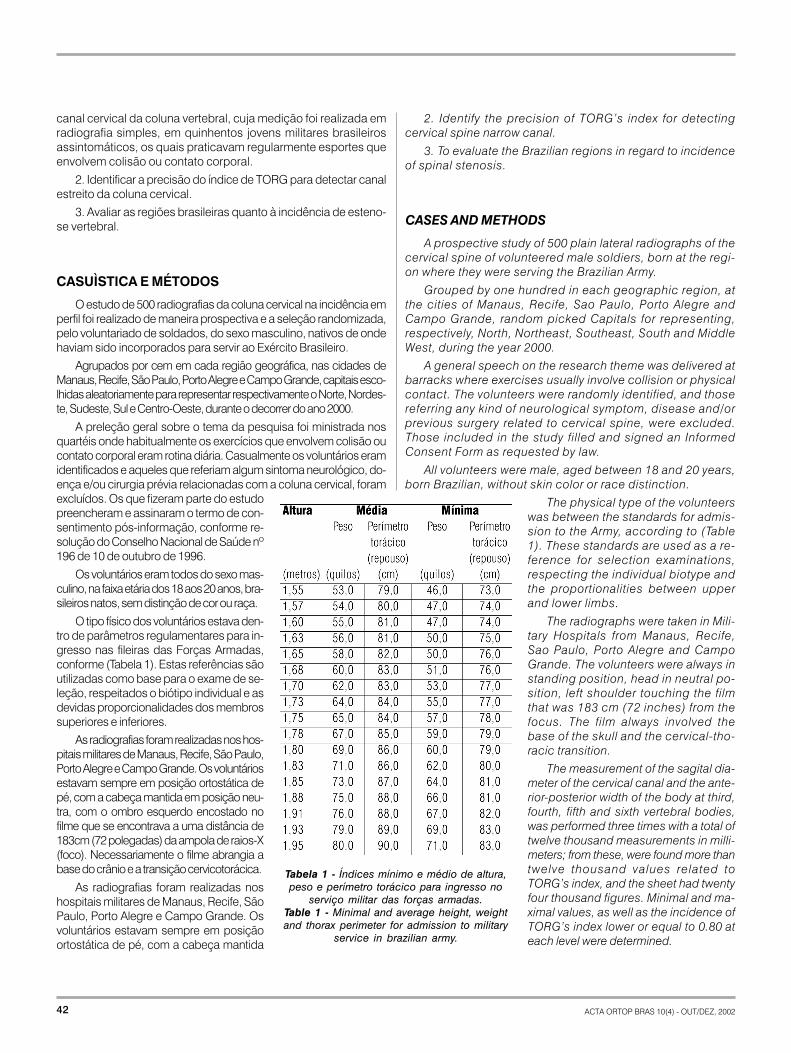
O tipo físico dos voluntários estava den-tro de parâmetros regulamentares para in-gresso nas fileiras das Forças Armadas,conforme (Tabela 1). Estas referências sãoutilizadas como base para o exame de se-leção, respeitados o biótipo individual e asdevidas proporcionalidades dos membrossuperiores e inferiores.
As radiografias foram realizadas nos hos-pitais militares de Manaus, Recife, São Paulo,Porto Alegre e Campo Grande. Os voluntáriosestavam sempre em posição ortostática depé, com a cabeça mantida em posição neu-tra, com o ombro esquerdo encostado nofilme que se encontrava a uma distância de183cm (72 polegadas) da ampola de raios-X(foco). Necessariamente o filme abrangia abase do crânio e a transição cervicotorácica.
As radiografias foram realizadas noshospitais militares de Manaus, Recife, SãoPaulo, Porto Alegre e Campo Grande. Osvoluntários estavam sempre em posiçãoortostática de pé, com a cabeça mantida
Tabela 1 - Índices mínimo e médio de altura,peso e perímetro torácico para ingresso no
serviço militar das forças armadas.Table 1 - Minimal and average height, weightand thorax perimeter for admission to military
service in brazilian army.
2. Identify the precision of TORG’s index for detectingcervical spine narrow canal.
3. To evaluate the Brazilian regions in regard to incidenceof spinal stenosis.
CASES AND METHODS
A prospective study of 500 plain lateral radiographs of thecervical spine of volunteered male soldiers, born at the regi-on where they were serving the Brazilian Army.
Grouped by one hundred in each geographic region, atthe cities of Manaus, Recife, Sao Paulo, Porto Alegre andCampo Grande, random picked Capitals for representing,respectively, North, Northeast, Southeast, South and MiddleWest, during the year 2000.
A general speech on the research theme was delivered atbarracks where exercises usually involve collision or physicalcontact. The volunteers were randomly identified, and thosereferring any kind of neurological symptom, disease and/orprevious surgery related to cervical spine, were excluded.Those included in the study filled and signed an InformedConsent Form as requested by law.
All volunteers were male, aged between 18 and 20 years,born Brazilian, without skin color or race distinction.
The physical type of the volunteerswas between the standards for admis-sion to the Army, according to (Table1). These standards are used as a re-ference for selection examinations,respecting the individual biotype andthe proportionalities between upperand lower limbs.
The radiographs were taken in Mili-tary Hospitals from Manaus, Recife,Sao Paulo, Porto Alegre and CampoGrande. The volunteers were always instanding position, head in neutral po-sition, left shoulder touching the filmthat was 183 cm (72 inches) from thefocus. The film always involved thebase of the skull and the cervical-tho-racic transition.
The measurement of the sagital dia-meter of the cervical canal and the ante-rior-posterior width of the body at third,fourth, fifth and sixth vertebral bodies,was performed three times with a total oftwelve thousand measurements in milli-meters; from these, were found more thantwelve thousand values related toTORG’s index, and the sheet had twentyfour thousand figures. Minimal and ma-ximal values, as well as the incidence ofTORG’s index lower or equal to 0.80 ateach level were determined.
ACTA ORTOP BRAS 10(4) - OUT/DEZ, 2002 43
em posição neutra, com o ombro esquerdo en-costado no filme que se encontrava a uma distânciade 183cm (72 polegadas) da ampola de raios-X(foco). Necessariamente o filme abrangia a base docrânio e a transição cervicotorácica.
A medição de cada diâmetro sagital do canalcervical e da largura ântero-posterior do corpo, daterceira, quarta, quinta e sexta vértebra, foi realizadapor três vezes, perfazendo um total de doze milmedidas em milímetro; a partir destes, foram en-contrados mais doze mil valores, referentes a índicede TORG e médias compondo uma planilha comvinte e quatro mil dados. Determinados os valoresmínimos e máximos, bem como a incidência de ín-dices de TORG menor ou igual a 0,80 em cada nível.
O instrumento utilizado para aferir foi um paquí-metro digital Mitutoyo código 500-143B, com preci-são de aproximadamente 0,02mm.
A radiografia em perfil da coluna cervical eraexcluída da série e substituída nas seguintes condi-ções: má qualidade técnica, rotação da coluna, de-formidade ou desvio ósseo, doença prévia comodefeito de formação ou segmentação vertebral, in-versão da lordose cervical, alteração congênita dajunção craniocervical e vértebra em "limbus".
Para que não houvesse qualquer distorçãoque interferisse nas dimensões do canal e corpo,foi necessário calcular de maneira aleatória umamajoração de 10% da amostragem final.
Todas as medidas foram feitas três vezes,pelo mesmo observador(11), de maneira que depois de traçadas emensuradas as linhas, estas eram apagadas e novamente repeti-do o procedimento, sem referência da marcação anterior, nosquatro níveis das quinhentas radiografias.
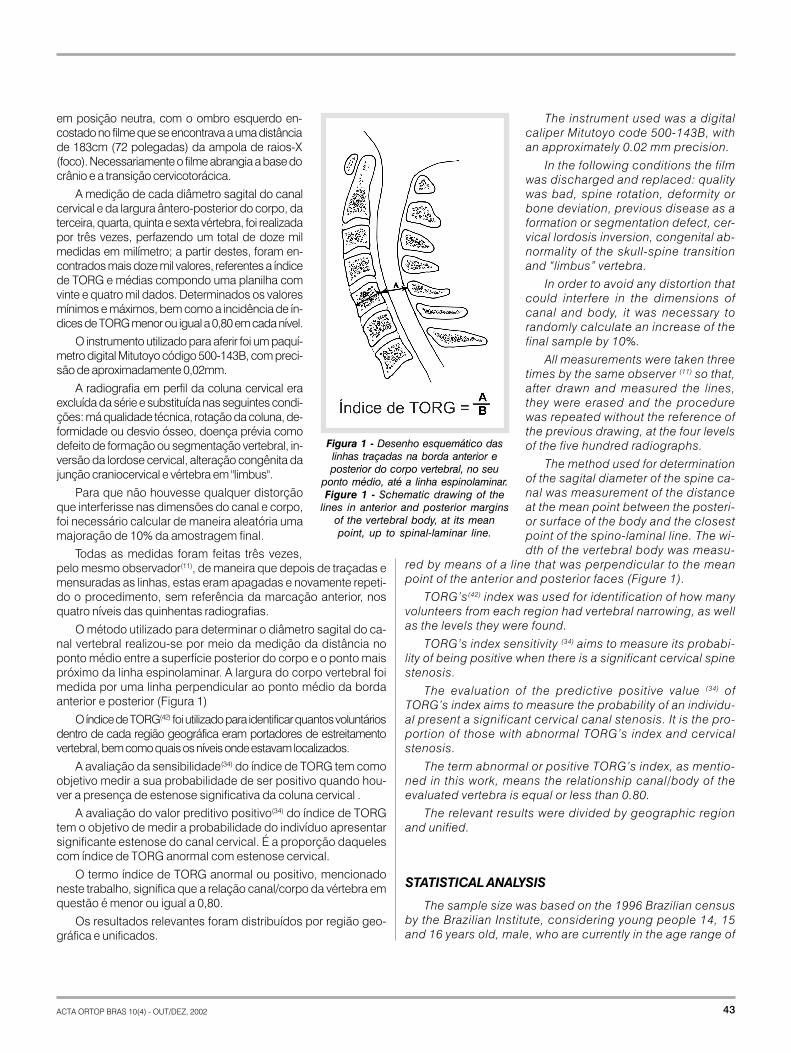
O método utilizado para determinar o diâmetro sagital do ca-nal vertebral realizou-se por meio da medição da distância noponto médio entre a superfície posterior do corpo e o ponto maispróximo da linha espinolaminar. A largura do corpo vertebral foimedida por uma linha perpendicular ao ponto médio da bordaanterior e posterior (Figura 1)
O índice de TORG(42) foi utilizado para identificar quantos voluntáriosdentro de cada região geográfica eram portadores de estreitamentovertebral, bem como quais os níveis onde estavam localizados.
A avaliação da sensibilidade(34) do índice de TORG tem comoobjetivo medir a sua probabilidade de ser positivo quando hou-ver a presença de estenose significativa da coluna cervical .
A avaliação do valor preditivo positivo(34) do índice de TORGtem o objetivo de medir a probabilidade do indivíduo apresentarsignificante estenose do canal cervical. É a proporção daquelescom índice de TORG anormal com estenose cervical.
O termo índice de TORG anormal ou positivo, mencionadoneste trabalho, significa que a relação canal/corpo da vértebra emquestão é menor ou igual a 0,80.
Os resultados relevantes foram distribuídos por região geo-gráfica e unificados.
Figura 1 - Desenho esquemático daslinhas traçadas na borda anterior eposterior do corpo vertebral, no seu
ponto médio, até a linha espinolaminar.Figure 1 - Schematic drawing of the
lines in anterior and posterior marginsof the vertebral body, at its meanpoint, up to spinal-laminar line.
The instrument used was a digitalcaliper Mitutoyo code 500-143B, withan approximately 0.02 mm precision.
In the following conditions the filmwas discharged and replaced: qualitywas bad, spine rotation, deformity orbone deviation, previous disease as aformation or segmentation defect, cer-vical lordosis inversion, congenital ab-normality of the skull-spine transitionand “limbus” vertebra.
In order to avoid any distortion thatcould interfere in the dimensions ofcanal and body, it was necessary torandomly calculate an increase of thefinal sample by 10%.
All measurements were taken threetimes by the same observer (11) so that,after drawn and measured the lines,they were erased and the procedurewas repeated without the reference ofthe previous drawing, at the four levelsof the five hundred radiographs.
The method used for determinationof the sagital diameter of the spine ca-nal was measurement of the distanceat the mean point between the posteri-or surface of the body and the closestpoint of the spino-laminal line. The wi-dth of the vertebral body was measu-
red by means of a line that was perpendicular to the meanpoint of the anterior and posterior faces (Figure 1).
TORG’s(42) index was used for identification of how manyvolunteers from each region had vertebral narrowing, as wellas the levels they were found.
TORG’s index sensitivity (34) aims to measure its probabi-lity of being positive when there is a significant cervical spinestenosis.
The evaluation of the predictive positive value (34) ofTORG’s index aims to measure the probability of an individu-al present a significant cervical canal stenosis. It is the pro-portion of those with abnormal TORG’s index and cervicalstenosis.
The term abnormal or positive TORG’s index, as mentio-ned in this work, means the relationship canal/body of theevaluated vertebra is equal or less than 0.80.
The relevant results were divided by geographic regionand unified.
STATISTICAL ANALYSIS
The sample size was based on the 1996 Brazilian censusby the Brazilian Institute, considering young people 14, 15and 16 years old, male, who are currently in the age range of
44 ACTA ORTOP BRAS 10(4) - OUT/DEZ, 2002
ANÁLISE ESTATÍSTICA
O tamanho da amostra foi baseado no censo de 1996 doInstituto Brasileiro de Geografia e Estatística, considerando jo-vens com 14, 15 e 16 anos de idade, do sexo masculino, os quaisatualmente estão na faixa etária que foi escolhida para estudo. Nãoforam levadas em conta as mortes existentes, imigração e emi-gração durante o referido período no Brasil.
O cálculo para o tamanho da amostra foi feito baseado nafórmula(1), mostrando que o número mínimo para representativi-dade estatística seria 387 pessoas, este número foi ampliado par-ticularmente para equilibrar a série estudada entre as regiões efacilitar a confrontação matemática, utilizada para população fini-ta, descrita por FONSECA(15):
n = Z . p . q . N
e2 (N-1) + Z2 .p.q
Onde:N = tamanho da amostra
Z = abcissa da curva normal padrão com a probabilidade de 0,045
p = estimativa da população
q = 1-p
e = erro amostral.
A probabilidade p (0,50) e q (0,50) foi adotada porque nessascondições a variação é máxima para o cálculo da amostra e destaforma oferece maior segurança para essa faixa.
A análise estatística foi realizada usando o programa (softwa-re) NTIA (Núcleo de Tecnologia e Informática). As correlaçõesentre as médias dos dados foram determinadas usando o mode-lo linear de regressão. A comparação entre os valores médiosdos dados foi feita utilizando a análise de variância (ANOVA) comdelineamento de dois critérios e teste de TUKEY (paramétrico).
A análise de variância (ANOVA) conforme BANZATTO(1), foi ado-tada visando à comparação de duas ou mais médias, utilizado ométodo de duplo critério. O delineamento experimental utilizado foide blocos ao acaso com três repetições. A seguir realizou-se o testede TUKEY a 1% e 5% de probabilidade. Os resultados foram consi-derados significantes estatisticamente ao nível de p<0,01.
A estatística descritiva foi utilizada para análise dos dadosmais relevantes como média, mediana, desvio padrão, intervalode confiança, valores mínimos e máximos.
RESULTADOS
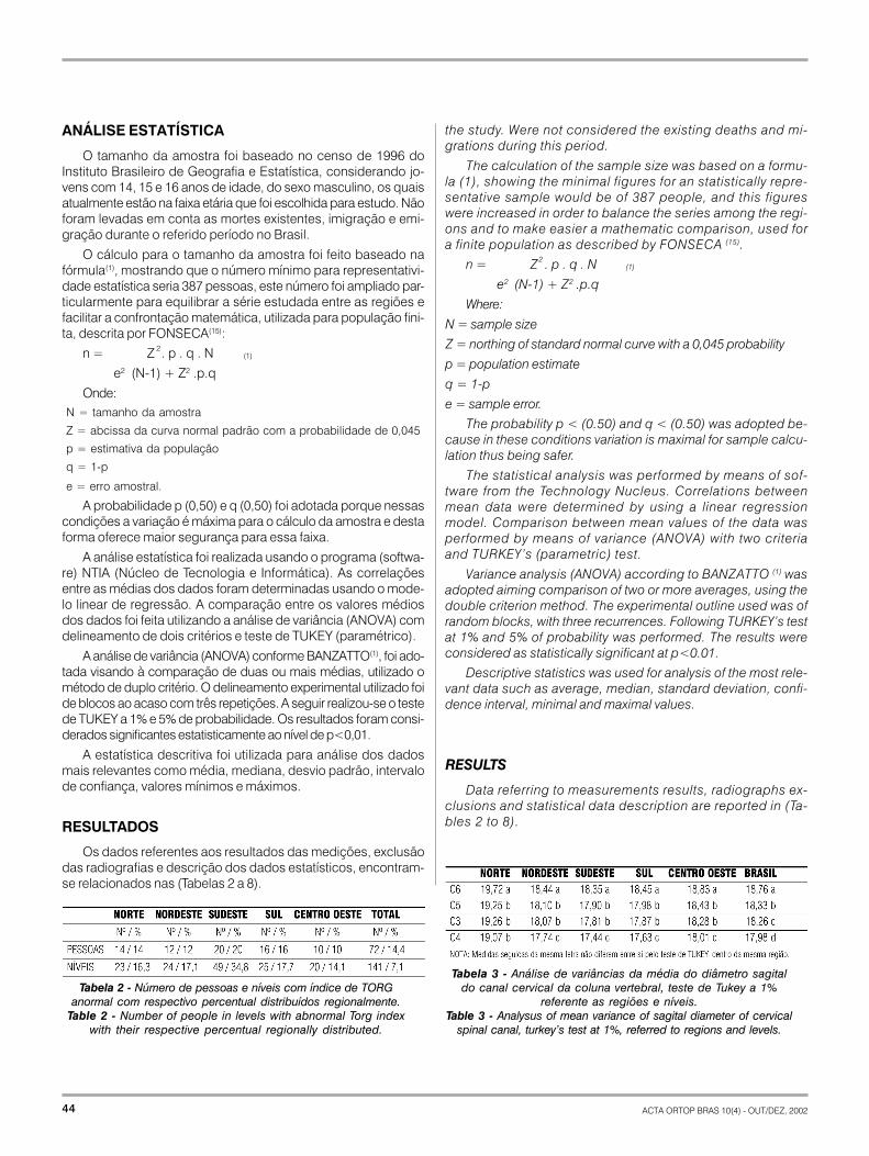
Os dados referentes aos resultados das medições, exclusãodas radiografias e descrição dos dados estatísticos, encontram-se relacionados nas (Tabelas 2 a 8).
(1)2
Tabela 3 - Análise de variâncias da média do diâmetro sagitaldo canal cervical da coluna vertebral, teste de Tukey a 1%
referente as regiões e níveis.Table 3 - Analysus of mean variance of sagital diameter of cervical
spinal canal, turkey’s test at 1%, referred to regions and levels.
the study. Were not considered the existing deaths and mi-grations during this period.
The calculation of the sample size was based on a formu-la (1), showing the minimal figures for an statistically repre-sentative sample would be of 387 people, and this figureswere increased in order to balance the series among the regi-ons and to make easier a mathematic comparison, used fora finite population as described by FONSECA (15).
n = Z . p . q . N
e2 (N-1) + Z2 .p.q
Where:
N = sample size
Z = northing of standard normal curve with a 0,045 probability
p = population estimate
q = 1-p
e = sample error.
The probability p < (0.50) and q < (0.50) was adopted be-cause in these conditions variation is maximal for sample calcu-lation thus being safer.
The statistical analysis was performed by means of sof-tware from the Technology Nucleus. Correlations betweenmean data were determined by using a linear regressionmodel. Comparison between mean values of the data wasperformed by means of variance (ANOVA) with two criteriaand TURKEY’s (parametric) test.
Variance analysis (ANOVA) according to BANZATTO (1) wasadopted aiming comparison of two or more averages, using thedouble criterion method. The experimental outline used was ofrandom blocks, with three recurrences. Following TURKEY’s testat 1% and 5% of probability was performed. The results wereconsidered as statistically significant at p<0.01.
Descriptive statistics was used for analysis of the most rele-vant data such as average, median, standard deviation, confi-dence interval, minimal and maximal values.
RESULTS
Data referring to measurements results, radiographs ex-clusions and statistical data description are reported in (Ta-bles 2 to 8).
(1)2
Tabela 2 - Número de pessoas e níveis com índice de TORGanormal com respectivo percentual distribuídos regionalmente.
Table 2 - Number of people in levels with abnormal Torg indexwith their respective percentual regionally distributed.
ACTA ORTOP BRAS 10(4) - OUT/DEZ, 2002 45
DISCUSSÃO
A medição obtida da imagem radiográfica é parte integrante na ava-liação diagnóstica da coluna cervical, entretanto, para que este procedi-mento tenha utilização clínica é necessário estabelecer parâmetros nor-mais. A presente pesquisa estabelece valores normais para o canal cervi-cal em jovens militares brasileiros natos, na faixa etária dos 18 aos 20anos, oriundos de todas as regiões geográficas.
O estudo em questão foi elaborado dentro dos mais rígidos critérioscientíficos, ordenando e classificando os resultados, comparando comos já descritos na literatura, obedecendo a princípios básicos da experi-mentação: casualidade, repetição e controle local(1).
O interesse pelas dimensões do canal cervical tem relação particularcom a compressão medular e/ou a mielopatia, que podem ocorrer maiscomumente por espondilose e/ou estreitamento congênito das estrutu-ras ósseas da vértebra. As manifestações clínicas variam de incapacida-de progressivamente lenta que acontece espontaneamente até tetraple-gia abrupta após maior ou menor trauma.
A mielopatia aguda após trauma menor na coluna cervical podeocorrer sem fratura ou luxação. Este fenômeno está associado à espon-dilose, que produz estreitamento gradual do canal, presente nos idosos,associado ou não à estenose congênita que notadamente acomete pes-soas mais jovens, as quais praticam esporte que envolve colisão ou
DISCUSSION
Measurements obtained from radiographic images is partof diagnostic evaluation of cervical spine, however for thisprocedure to be clinically useful it is necessary to establishnormal parameters. This study establishes normal values forcervical canal in young Brazilian military males aged between18 and 20 years, from all geographic regions of the Country.
This study was performed according to the best scientificcriteria, ordering the results and comparing to the existing inliterature sticking to basic experimentation principles: random,repetition and local control (1).
The interest for cervical canal measures has a particularrelationship with medullary compression and/or myelopathy,which can more frequently occur due to spondilosis and/orcongenital narrowing of spine bony elements. Clinical mani-festations range from a progressively slow incapacity occur-ring spontaneously, to a sudden tetraplegia after a major orminor trauma.
Acute myelopathy after minor trauma of cervical spine canoccur without any fracture or dislocation. This phenomenonis linked to spondilosis, which leads to a gradual canal narro-wing, present in elder, whether or not linked to congenitalstenosis which occurs in younger people, who practice sportswith collision or physical contact.
Cervical canal diameter evaluation started to gain impor-tance by the middle of the past century with the works byBOIJSEN apud WOLF(56) and later by WOLF et al.(56). Starting
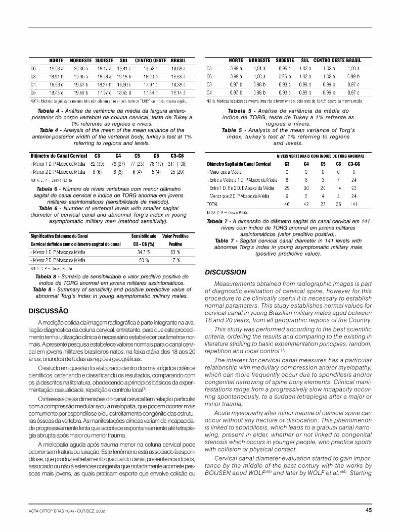
Tabela 4 - Análise de variância da média da largura antero-posterior do corpo vertebral da coluna cervical, teste de Tukey a
1% referente as regiões e níveis.Table 4 - Analysis of the mean of the mean variance of the
anterior-posterior widith of the vertebral body, turkey’s test at 1%referring to regions and levels.
Tabela 5 - Análise de variância da média doíndice de TORG, teste de Tukey a 1% refrente as
regiões e níveis.Table 5 - Analysis of the mean variance of Torg’s
index, turkey’s test at 1% referring to regionsand levels.
Tabela 8 - Sumário de sensibilidade e valor preditivo positivo doíndice de TORG anormal em jovens militares assintomáticos.
Table 8 - Summary of sensitivity and positive predictive value ofabnormal Torg’s index in young asymptomatic milirary males.
Tabela 6 - Número de níveis vertebrais com menor diâmetrosagital do canal cervical e índice de TORG anormal em jovens
militares assintomáticos (sensibilidade de método).Table 6 - Number of vertebral levels with smaller sagital
diameter of cervical canal and abnormal Torg’s index in youngasymptomatic military men (method sensitivity). Tabela 7 - A dimensão do diâmetro sagital do canal cervical em 141
níveis com índice de TORG anormal em jovens militaresassintomáticos (valor preditivo positivo).
Table 7 - Sagital cervical canal diameter in 141 levels withabnormal Torg’s index in young asymptomatic military male
(positive predictive value).
46 ACTA ORTOP BRAS 10(4) - OUT/DEZ, 2002
contato corporal.
As investigações do diâmetro do canal cervical começaram a tergrande impulso em meados do século passado com os trabalhosde BOIJSEN apud WOLF(56) e posteriormente WOLF et al.(56). A partirdaí passou a ser realizada por vários autores(2,3,5,12,16,25,28,42,56), a me-dição em radiografias de perfil para estabelecer valores mínimosnormais, mas a diferença entre eles gerou uma grande preocupaçãocom as medidas absolutas, em virtude da variabilidade da magnifica-ção. Algumas destas pesquisas não têm registrado claramente se amedição nas radiografias é feita exatamente na linha média sagital davértebra, o que é crucial para determinar estenose cervical(12,16,30,56).
TORG et al.(42) com a finalidade de eliminar os efeitos da variabilidadeda magnificação, descrevem método que ignora a distorção causadapela distância foco-filme e objeto-filme, que consiste na relação entre doisvalores medidos em uma radiografia de perfil, por meio do ponto médioda vértebra, o diâmetro sagital do canal cervical partindo da cortical pos-terior do corpo vertebral até o ponto mais próximo que encontra a linha deunião do processo espinhoso com as lâminas, pelo comprimento dalargura antero-posterior do corpo, conforme exposto na (Figura 1).
O método de TORG propõe que o índice, menor ou igual a 0,80,identifique um estreitamento significante do diâmetro do canal cervi-cal. Em seu trabalho clássico, TORG et al.(42) avaliam atletas comtetraplegia transitória e demonstram que muitos deles eram portado-res de estenose.
A tetraplegia transitória é um fenômeno de disfunção medular tempo-rário, caracterizado por TORG et al.(42) como uma entidade clínica distinta,que acomete o atleta vítima de trauma no pescoço em hiperflexão ouhiperextensão ou na cabeça(28,30,33,42,46,54), com início abrupto dos sinto-mas que podem acometer os membros superiores, inferiores, quatromembros, ou ipsilateral, membro superior e inferior, alteração de sensibi-lidade como dor em queimação ou em choque, podendo chegar àanestesia. O déficit motor consiste de fraqueza ou paralisia. O episódiotípico persiste por menos que 15 minutos(9, 24, 42,48), entretanto, em algunscasos, a regressão gradual pode atingir 48 horas. Há completo retornoda função motora, mobilidade cervical e remissão do quadro doloroso.Entretanto, a partir do segundo episódio as chances de remissão com-pleta do quadro neurológico são menores.
Na literatura médica não há unanimidade em torno de valores nor-mais para as dimensões do canal vertebral. Alguns autores(8,9,26,36,42,56)
tentaram determinar estenose por valores abaixo de 13mm. WOLF(56)
que repetiu o trabalho de BOIJSEN apud WOLF(56) realizado dois anosantes, examinou 200 adultos assintomáticos, modificando a distância en-tre a fonte de raios-X e o filme para 183cm (72 polegadas), mostrou queo valor mais freqüente para a normalidade foi de 17,00mm de C3 a C7, oque está de acordo com os achados da presente pesquisa.
O coeficiente de variação calculado para os valores encontradosneste estudo demonstra a baixa diferença entre os dados. Esta ho-mogeneidade traduz confiabilidade, que foi observada na análiseestatística tanto para o índice de TORG quanto para as medidas docanal e corpo vertebral.
A análise de variância das médias do diâmetro sagital do canal verte-bral, largura do corpo e índice de TORG, foi realizada segundo doiscritérios com três repetições. Mostrou haver diferença estatisticamentesignificativa (p<0,01) entre as vértebras em todas as regiões, em seguidacalculado pelo teste de TUKEY à 1% de probabilidade, mostrou quais asvariáveis semelhantes, conforme (Tabelas 21, 22, 23).
from there, several other authors(2,3,5,12,16,25,28,42,56) started me-asuring radiographs aiming to establish the minimal normalvalues, however the differences between them brought greatconcert in relation to absolute measurements, due to the en-largement variability. Some of these works did not clearly re-cord if the radiographic measurements were performed atthe mean sagital line of the vertebra, essential for cervicalstenosis identification (12,16,30,56).
Aiming to eliminate enlargement variability effects, TORGet al. (42) describe a method that ignores the distortion cau-sed by the distance between focus-object-film, consisting inthe relationship between two measured values in a plain late-ral radiograph, through the mean point of a vertebra, the sa-gital diameter of the cervical canal starting from the posteriorcortical of the vertebral body to the closest point meeting theline linking the spinous process with the laminae, by thelength of the anterior-posterior width of the body, accordingto (Figure 1).
TORG’s method proposes that the index, smaller or equalto 0.80 identify a significant narrowing of the cervical canaldiameter. In his classic paper, TORG et al.(42) evaluate athle-tes with transient tetraplegia and demonstrate that many ofthem had stenosis.
Transient tetraplegia is a temporary medullary dysfuncti-on, characterized by TORG et al. (42) as a distinctive clinicalentity involving athletes with cervical trauma in hyperflexion orhyperextension, or head (28,30,33,42,46,54), with a sudden symp-tom start that may involve upper limbs, lower limbs, the fourlimbs, or upper and lower limbs at same side, with sensitivitychanges as parestesias and eventually reaching anesthesia.Motor deficit consists in weakness or palsy. The typical epi-sode persists for at least 15 minutes (9, 24, 42,48) however insome cases, gradual regression can take 48 hours. There isa complete recovery of motor picture, cervical mobility andremission of the painful picture. However, from the secondepisode there is less chance of a complete recovery.
In medical literature there is no unanimity in regard of thenormal values of spinal canal dimensions. Some authors(8,9,26,36,42,56) tried to determine the stenosis by values below13 mm. WOLF (56), who repeated the work by BOIJSEN apudWOLF (56) performed two years before, examined 200 asymp-tomatic adults, changing the distance between the X-Raysource and the film to 183 cm (72 inches), showing that themost frequent value for the normality was 17,00 mm from C3to C7, and this agrees with the findings in this trial.
The variation coefficient calculated for the values found inthis study shows a small difference between the data. Thishomogeneity translates that these were trustworthy, as ob-served by statistical analysis for both TORG’s index and ca-nal and vertebral body measurements.
The variance analysis of spinal canal sagital diameter measure-ments, body width and TORG’s index, was performed accordingto the three recurrence criteria. It was shown to have a statisticallysignificant difference (p< 0.01) between vertebras in all regions,following calculated by TURKEY’s test at 1% probability, showingsimilar parameters as shown in Tables 21, 22 and 23.
ACTA ORTOP BRAS 10(4) - OUT/DEZ, 2002 47
A atual pesquisa mostrou, por meio do teste de TUKEY paraanálise de variância que os menores valores para o diâmetro sagitaldo canal vertebral estavam em C3 e C4, enquanto que os menorescomprimentos antero-posteriores do corpo em C4 e C5. Para arelação canal/corpo os mais baixos índices estavam em C3/C4 semdiferença estatística seguida de C6. Estes achados estão equivalen-tes com outros estudos(12,13,14,16,25,26,58).
A análise de variância da média de todas as regiões conjuntasmostrou que existe diferença significativa (p<0,01) entre as vértebrasda coluna cervical, isto é, não há qualquer semelhança estatísticaentre C3, C4, C5 e C6, comprovada quando aplicado o teste deTUKEY, tanto para o canal como para o corpo.
Os menores valores para o canal vertebral foram encontra-dos naqueles níveis em que o índice de TORG foi menor ouigual a 0,80; estas dimensões estavam abaixo de 17,00mm em47,8% na região Norte, 91,7% Nordeste, 87,7% Sudeste, 100%Sul e 90% na região Centro-Oeste.
Os valores mínimos para o canal vertebral encontrado em cadaregião foram, em ordem crescente: Nordeste 10,68mm, Sudeste13,80mm, Centro-Oeste 13,94mm, Sul 14,37mm e Norte 14,48mm.
O índice de TORG, em todo o Brasil, menor ou igual a 0,80, foiencontrado em um ou mais níveis em 72 pessoas (14,4%) e 141níveis (7,1%).
A região Sudeste foi a que apresentou maior incidência de este-nose do canal cervical com 49 (34,8%) níveis, seguida do Sul com 25(17,7%), Nordeste 24 (17,1%), Norte 23 (16,3%) e Centro-Oeste 20(14,1%). O nível mais freqüente foi C3 (32,6%), seguido de C4 (29,8%),C5 (19,2%) e C6 (18,4%), conforme exposto na tabela 2. A regiãoSudeste foi também a que mostrou maior número de pessoas commenores dimensões do canal cervical.
O valor médio do diâmetro sagital do canal cervical no seg-mento subaxial (C3-C6) foi acima de 17,00mm em todos os níveisde todas as regiões.
Há uma extensa série de pesquisas atuais que registram a rela-ção entre estenose do canal cervical e disfunção medular secundáriaao trauma da coluna cervical(5,7,9,10,16,18,20,22,25,36,49,52,55,56), particularmen-te durante a prática de atividade esportiva que envolva colisão corpo-ral, sobretudo porque um canal estreito predispõe o atleta a um piorprognóstico neurológico diante de um menor trauma vertebral(7,53).
A avaliação radiográfica no presente estudo não sofreu qualquerinfluência intrínseca de origem degenerativa, em virtude da faixa etáriado contingente estudado, 18 a 20 anos, o que favoreceu a homoge-neidade dos dados, especialmente a investigação da incidência deestenose do canal cervical.
Observando a análise dos dados no presente estudo, a região Su-deste mostrou que em 12,3% dos níveis, o índice de TORG foi menor ouigual a 0,80, enquanto que no Sul foram 6,3%, no Nordeste em 6%, noNorte em 5,8% e no Centro-Oeste em 5% dos níveis. A incidência énotadamente maior no Sudeste, provavelmente em razão das váriascorrentes migratórias oriundas não só do Brasil, mas também de váriasnações, causando grande miscigenação em uma cidade cosmopolitacomo São Paulo, onde foram selecionados os voluntários.
A baixa incidência de estenose significativa da coluna cervical permitiuno atual estudo, que fossem determinados a sensibilidade e o valor pre-ditivo positivo do índice de TORG anormal para detectar canal estreito.Para isso foi confrontado o diâmetro sagital do canal vertebral e o valor da
This research showed, by means of TURKEY’s test forvariance analysis that the lower values for sagital diameter ofthe spine canal were in C3 and C4, while the lower anterior-posterior lengths of the body was in C4 and C5. For the rela-tionship canal/body the lowest index were in C3/C4 withoutstatistical difference, followed by C6. These finding are equi-valent to other studies (12,13,14,16,25,26,58).
The analysis of the mean variance of all regions togethershowed that there are statistically significant differences(p<0.01) between cervical spine vertebras, that is, there is nostatistical similarity between C3, C4, C5 and C6, proven byTURKEY’s test, both for canal and body.
The lowest values for spine canal were found in the levelswhere TORG’s index was lesser or equal to 0.80; these mea-surements were below 17.00 mm in 47.8% in North region,91.7% in Northeast; 87.7% in Southeast, 100% in South and90% in Middle-West regions.
The minimal value for spine canal found in each regionwere: Northeast: 10.68 mm; Southeast 13.80 mm; West-West13.94 mm; South 14.37 mm; and North 14.48 mm.
TORG’s index in Brazil as a whole, smaller or equal to0,80 was found in one or more levels in 72 people (14.4%)and 141 levels (7.1%).
Southeast region was the one with higher incidence ofspine canal stenosis, with 49 (34.8%) levels, followed by Southwith 25 (17.7%). Northeast 24 (17.1%); North 23 (16.3%) andMiddle West 20 (14.1%). The most frequently affected levelwas C3 (32.6%), followed by C4 (29.8%), C5 (19.2%) and C6(18.4%) according to Table 2. Southwest region was also theone with larger number of people with smaller dimensions ofcervical canal.
The mean value of sagital diameter of cervical canal insub-axial segment (C3 to C6) was above 17.00 mm in alllevels of all regions.
There is a large series of current researches registeringthe relationship between cervical canal stenosis and medu-l lary dysfunct ion secondary to cerv ical spine t rauma(5,7,9,10,16,18,20,22,25,36,49,52,55,56), particularly during sports practi-ce involving physical contact, mostly because a narrow canalpredisposes the athlete to a worse neurological prognosisafter a minor spine trauma (7, 53).
Radiographic evaluation in this study had no intrinsic de-generative influence, due to the low age of the series, 18 to20 years, favoring data homogeneity especially in investigati-on of cervical canal stenosis incidence.
Observation of data analysis of this study, Southeast regionhad in 12.3% of the levels a TORG’s index lesser or equal to0.80, while in South it was 6.3%, Northeast 6%, North, 5.8% andMiddle West 5%. This incidence in clearly higher in Southeastprobably because intensive migration not only from Brazil butalso from several nations, in such a cosmopolitan city as SaoPaulo, where the volunteers were selected.
The low incidence of significant cervical spine stenosis inthe current study allowed determining the sensitivity and po-sitive predictive value of abnormal TORG’s index for detecti-on of narrow canal. For this, the sagital diameter of the canal
48 ACTA ORTOP BRAS 10(4) - OUT/DEZ, 2002
relação canal/corpo menor ou igual a 0,80. Estes testes são utilizados emlevantamento populacional para estudar pessoas doentes e sãs e osseus resultados são mais relevantes para a prática clínica.
A sensibilidade(34) do índice de TORG avalia a probabilidade deser anormal ou positivo, em virtude da presença de estenose docanal cervical; já o valor preditivo positivo(34) do índice avalia a proba-bilidade de um canal com medida reduzida resultar em um índice deTORG abaixo do normal.
Se estenose significativa do canal cervical(16) for definida pelo seudiâmetro sagital menor do que um desvio padrão abaixo da média, asensibilidade do índice de TORG para detectá-la é 34,7%. Considerandoque o parâmetro estabelecido seja o menor que dois desvios-padrãoabaixo da média, a sensibilidade do índice de TORG aumenta para 80%.
O valor preditivo positivo do índice de TORG anormal foi determi-nado em 83% dos níveis, os quais tinham o diâmetro sagital do canalcervical menor que um desvio-padrão abaixo da média, enquantosomente 17% apresentavam o diâmetro sagital do canal menor quedois desvios-padrão abaixo da média.
Se estenose significativa do canal cervical for definida pelo diâme-tro sagital do canal menor do que um desvio-padrão abaixo da mé-dia, o valor preditivo positivo do índice de TORG anormal é de 83%.Caso o parâmetro estabelecido seja o menor que dois desvios-padrão abaixo da média, o valor preditivo positivo é de 17%.
O índice de TORG menor ou igual a 0,80 foi sensível para esteno-se cervical e apresentou alto valor preditivo positivo no levantamentode jovens militares assintomáticos, dos quais 14,4% tinham a relaçãocanal/corpo anormal em um ou mais níveis.
Comparativamente ao estudo de HERZOG et al.(16) há diferençano que se refere ao valor preditivo positivo, particularmente porquena atual pesquisa houve uma maior concentração de níveis comíndice de TORG anormal com diâmetro sagital do canal cervical entreum desvio-padrão e dois desvios-padrão abaixo da média, enquan-to que no primeiro a convergência foi maior entre a média e umdesvio-padrão abaixo da média.
A sensibilidade do índice de TORG para detectar estenose signi-ficativa do canal cervical foi relativamente menor no atual estudo,porque a quantidade de níveis com diâmetro sagital menor do queum desvio padrão abaixo da média foi muito superior àquele queapresentava a relação canal/corpo anormal.
A diferença na sensibilidade e valor preditivo positivo de outrosestudos(16,43,48) pode ser uma característica da população brasileira,especialmente porque as seis medições de cada nível foram rigoro-samente tomadas no ponto médio de cada vértebra.
HERZOG et al.(16) descrevem em sua série que em 41% dosatletas assintomáticos o índice de TORG foi menor ou igual a 0,80 emum ou mais níveis. Preocupados com este alto percentual, passarama estudar as causas que poderiam ser responsáveis por tal resulta-do. Consideram que não foi a medida do canal que determinou alte-ração significante no índice, mas a diferença no diâmetro do corpo,julgando que TORG et al.(42),no seu trabalho clássico, não valoriza-ram estas dimensões no grupo controle, o qual não era compostopor atletas. Supõem a possibilidade que em um estudo epidemioló-gico usando a relação canal/corpo para determinar estenose signifi-cativa, possa ocorrer que um atleta seja classificado como estenóti-co, sendo normal; orientando, para este caso, que sejam investiga-das as dimensões do canal cervical por meio de ressonância magné-
was compared to the relationship canal/body lesser or equalto 0.80. These tests as used in populational researches tostudy ill and healthy people, and their results are more rele-vant for clinical practice.
TORG’s index sensitivity (34) evaluates the probability ofbeing abnormal or positive due to cervical canal stenosis;positive predictive value (34) of this index evaluates the proba-bility that a reduced measurements canal result in a belownormal TORG’s index.
If significant cervical canal stenosis (16) is defined by itssagital diameter lesser than one standard deviation belowthe average, the sensitivity of TORG’s index to detect it is34.7%. If the parameter considered is to be lesser than twostandard deviation below the average, TORG’s index sensiti-vity raises to 80%.
Positive predictive value of abnormal TORG’s index wasdetermined in 83% of the levels, that had their sagital cervicalcanal diameter lesser than one standard deviation below theaverage, while only 17% had a sagital cervical canal diameterlesser than two standard deviation below the average.
TORG’s index lesser or equal to 0.80 was sensitive forcervical stenosis and presented a high positive predictive valuein screening asymptomatic young military, from who 14.4%had their ratio canal/body abnormal in one or more levels.
In comparison to HERZOG’s et al.(16) study there is a di-fference in regard of the positive predictive value, particularlybecause in the current study there was a higher concentrationof levels with abnormal TORG’s index with the sagital diame-ter of the cervical canal between one and two standard devi-ation below the average, while in the first the agreement washigher between the average and one standard deviation be-low the average.
TORG’s index sensitivity to detect a significant cervicalcanal stenosis was relatively lesser in the current study, be-cause the amount of levels with a sagital diameter lesser thanon standard deviation below the average was much higherthan those presenting a normal canal/body relationship.
The sensitivity difference and positive predictive value fromother studies (16,43,48) can be a feature of Brazilian population,specially because the six measurements of each level wererigorously taken at the mean point of each vertebra.
HERZOG et al. (16) describe in their series that in 41% of theasymptomatic athletes TORG’s index was lesser or equal to0.80 in one or more levels. Concerned about this high percenta-ge, they started studying causes that could be responsible forthe results. They understand that it was not the measurement ofthe canal that determined the significant change in the index, butthe difference in the body diameter, judging that TORG et al.(42),in their classical paper, did not value this dimension in the controlgroup, which was not formed by athletes. They suppose thepossibility that in an epidemiological study using the ratio canal/body for determine significant stenosis, it may occur that oneathlete is rated as having stenosis, being normal; and instructthat in these cases, canal dimensions are investigated by MRI,for evaluating a possible “functional cervical canal stenosis”.
The so called “real functional spine canal stenosis” (6) is
ACTA ORTOP BRAS 10(4) - OUT/DEZ, 2002 49
tica, a fim de afastar a possibilidade de "estenose vertebral funcional".
A estenose cervical "verdadeira" chamada de "estenose vertebralfuncional"(6) está relacionada com ausência de "reserva funcional"(15)
da medula espinal, que é um sinal visto na imagem pesada em T2 daressonância magnética, mielotomografia ou mielografia, representa-da por um halo claro que a envolve, referindo-se ao líquido cefalora-quidiano, significando que existe um espaço que a separa da estrutu-ra óssea vertebral adjacente, o qual também representa proteção.
Os atletas com índice de TORG abaixo ou igual a 0,80 requeremuma avaliação pela imagem da ressonância magnética(16,25) para es-tudo da reserva funcional prévia, antes do diagnóstico de estenosesignificativa do canal cervical, especialmente naqueles que praticamesporte de contato ou colisão corporal em razão da natural predis-posição ao trauma da coluna vertebral. Isto é extremamente impor-tante porque há um risco maior de ocorrer uma das mais dramáticaslesões neurológicas, a tetraplegia(7,8,53).
Na atual pesquisa, o tipo físico dos voluntários foi semelhanteparticularmente porque o jovem para ingressar nas Forças Armadasprecisa ser aprovado no exame de saúde, o qual possui rígidoscritérios médicos, especialmente para relação peso/altura.
TORG et al.(35,51) em artigo editorial concluíram que não há asso-ciação entre tetraplegia e o porte físico do atleta. A correlação entre acircunferência do pescoço e tamanho da vértebra não foi comprova-da por HERZOG et al.(16), que, em seu trabalho, mostraram não haverqualquer equivalência.
Outros estudos(12, 56) têm concluído que a estenose do canal ver-tebral cervical aumenta o risco de dano medular também em não-atletas de maneira semelhante (9) quando expostos ao trauma nacabeça ou no pescoço, particularmente em idosos com alteraçõesdegenerativas importantes, as quais reduzem ainda mais as dimen-sões pela espondilose. Há uma tendência maior de melhora nospacientes com lesão neurológica incompleta com canal largo, doque naqueles com estreitamento(12).
A estenose do canal cervical é conhecida como agravante no"menor trauma", aumentando o risco de dano neurológico perma-nente. Uma série de registros traz consistência a esta afirma-ção(3,5,7,9,10,12,16,17,19,22,23,24,26,29,37,42,49).
A mielopatia associada à estenose do canal cervical, especial-mente em jovens atletas, tem provavelmente o mecanismo de lesãomedular diferente daquele visto em pacientes idosos(13,18,27,57). Asbases fisiopatológicas da tetraplegia transitória não são completa-mente entendidas, apesar da preocupação cada vez maior que su-pera os interesses acadêmicos(42).
A estenose cervical também pode trazer repercussões neuro-lógicas exclusivamente ao plexo braquial, pois o canal estreitoneste caso faz um fulcro na saída da raiz e produz um estiramentona sua emergência causando uma disfunção neurológica comsintomatologia transitória(10,17,20,32,55). Atletas com estenose do fo-rame intervertebral apresentaram índice de TORG significativa-mente menores do que o grupo controle(54), os quais estão sujei-tos à neurapraxia radicular conseqüente a trauma esportivo, es-pecialmente quando envolve colisão corporal(10).
A estabilidade da coluna vertebral foi definida por WHITE etal.(55) como a capacidade que esta possui de manter seus limitesde mobilidade sob cargas fisiológicas sem causar dano ou irrita-ção medular ou radicular.
related to the absence of a “functional reserve” (15) of spinecord, that is a sign seen in T2 weighted images in MRI, mye-lotomography or myelography, represented by a clear haloinvolving it, that is, cephalorrhachidian liquid, meaning thatthere is a space to the adjacent bony spine structure, thatalso represents protection.
Athletes with TORG’s index lesser or equal to 0.80 requirean image evaluation by MRI (16,25) for a study of the previousfunctional reserve before a diagnosis of significant canal ste-nosis, mostly in those practicing sports with collision andphysical contacts due to the natural exposition to spinal trau-ma. This is extremely important, because there is a higher riskof one of the most dramatic nerve injuries, tetraplegia (7,8,53).
In the current research the biotype of the volunteers wassimilar, particularly because for a young man to be admittedto Army has to be approved in a health test with rigid criteriaparticularly for weight and height.
In an editorial paper, TORG et al. (35,51) concluded thatthere is no relationship between tetraplegia and the physicalbuild of an athlete. The correlation with neck circumferenceand vertebral size were not proven by HERZOG et al. (16) who,in their work, showed to have no equivalence.
Other studies (12, 56) concluded that cervical spinal canal ste-nosis increases the risk of medullary damage similarly also innon-athletes (9) when exposed to head and neck traumas, parti-cularly in aged people with important degenerative changes, whichadditionally reduce the dimensions, due to spondilosis. There isa higher trend to improvement in patients with incomplete nerveinjury with a wide canal than in patients with narrowing (12).
Cervical canal stenosis is known as aggravating in “minor trau-ma” increasing the risk of permanent nerve injury. There is a series ofreports backing this statement(3,5,7,9,10,12,16,17,19,22,23,24,26,29,37,42,49).
Myelopathy associated to cervical canal stenosis, mostlyin young athletes, has probably a different medullary injurymechanism than the observed in aged patients (13, 18,27,57).The phatophysiologic bases of transient tetraplegia are notcompletely understood, even though a growing concerningovercoming academic interest(42).
Cervical stenosis can as well have neurological effects only inbrachial plexus once a narrow canal in this case plays a fulcrumby the exit of the root producing stretching at its exit causing aneurological dysfunction with transient symptoms(10,17,20,32,55).Athletes with intervertebral foramen stenosis presented signifi-cantly lesser TORG index than the control group(54) who are sub-ject to radicular neuro apraxia in consequence of sport trauma,mostly when involving physical contact(10).
Spine stability was defined by WHITE et al (55) as the abilityit has to keep its mobility limits under physiological burdenswithout causing damage or irritation to medulla or roots.
Cervical instability, spine canal stenosis and exposure totrauma are factors that are part of a current consensus forpredisposing an individual to a catastrophic neurological in-jury(49) during sports practice involving collision or physicalcontact.
The guideline to exclude an athlete from taking part in physical
50 ACTA ORTOP BRAS 10(4) - OUT/DEZ, 2002
Instabilidade cervical, estenose do canal vertebral e exposiçãoao trauma são fatores que fazem parte de um consenso atual parapredispor um indivíduo a uma lesão neurológica catastrófica(49) du-rante a prática de esporte que envolve colisão ou contato corporal.
A diretriz para excluir um atleta da participação em esportes decontato corporal é multifatorial(8,52,53). Há necessidade de avaliar acoluna cervical sob três aspectos: alteração congênita da vértebra(defeito de segmentação, anomalia do odontóide, fusão atlanto-oc-cipital), presença de estenose congênita do canal ou seqüela pós-traumática (fratura ou luxação). Cada condição pode apresentar umacontra-indicação relativa, absoluta ou não haver contra-indicação,com base na variedade dos parâmetros(50,52).
Como se pode notar pelos itens supracitados, é crucial o estudoda estabilidade da coluna cervical, de tal forma que TORG et al.(49)
afirmaram que sem instabilidade vertebral não há risco aumentadopara dano neurológico permanente. Estas informações são impres-cindíveis para a tomada de decisão que implique interromper oupermitir a continuidade da vida esportiva.
Recentemente Watkins(52) passou a revisar esta difícil questãodecisória quanto ao retorno à vida esportiva daquele atleta que so-freu uma lesão cervical. A decisão deve estar baseada em três fato-res: mecanismo que causou o dano, objetiva lesão anatômica, erecuperação do atleta. Devido ao risco potencial de haver uma se-qüela catastrófica diante de retorno inapropriado ou prematuro, acompreensão da história natural de uma específica síndrome clínicadeve ser familiar ao médico especialista.
Esta pesquisa não tem como ponto central focalizar a tetraplegiatransitória, mesmo porque todas as pessoas que foram examinadaseram completamente assintomáticas, mas estabelecer valores nor-mais para a morfometria do canal cervical e identificar aqueles comestenose significante.
A grande vantagem deste trabalho é seguramente poder chamara atenção para a possibilidade de prevenir uma lesão medular con-seqüente de um menor trauma naqueles que praticam esporte decolisão ou contato corporal.
Os resultados desta pesquisa vão contribuir para que o Esta-do-Maior das Forças Armadas possa rever os critérios para in-gresso nas suas fileiras.
A importância da correta medição do canal cervical fica muitoclara quando se consideram as conseqüências clínicas da estenoseneste segmento da coluna vertebral, que é raramente reconhecida,mas potencialmente trágica.
CONCLUSÕES
O estudo da morfometria do canal vertebral no segmen-to cervical em militares jovens assintomáticos possibilitouconcluir que:
1. A incidência de estenose do canal vertebral da coluna cervi-cal é relevante nos jovens que ingressam nas Forças Armadas.
2. O índice de TORG possui sensibilidade e alto valor preditivopositivo.
3. A morfometria do canal vertebral no segmento cervical variade acordo com a região do país estudada.
contact sports is multifactorial(8, 52, 53). It is necessary to evaluate thecervical spine under three different aspects: vertebra congenital ab-normality (segmentation defect, odontoid’s defect, atlanto-occiputfusion), presence of congenital canal stenosis or post-traumaticsequela (fracture or dislocation). Each condition may present arelative, absolute or no contra-indication, based in the variety of theparameters(50,52).
As it can be noticed by the above mentioned items, thestudy of cervical spine stability is crucial, so that TORG et al.(49) state that without spine instability there is no increasedrisk of a permanent neurological damage. This information isvital for decision making that results in stopping or allowingcontinuity of sports life.
Recently Watkins(52) started analyzing this difficult decisi-on making of allowing a cervical injured athlete to return tosport activity. The decision should be based in three factors:the mechanism causing the injury; objective anatomical in-jury; and the recovery of the athlete. Due to a potential risk ofcatastrophic sequela in case of an inappropriate or too earlyreturn, the understanding of the natural history of an specificclinical syndrome should be familiar to the specialist.
This research doesn’t have as central point to focus ontransient tetraplegia, even though all examined people werecompletely asymptomatic, but to establish normal values forcervical canal morphometry, and identify those with signifi-cant stenosis.
The major advantage of this work is, for sure, to draw theattention to the possibility of preventing a medullary injurydue to a minor trauma in those who practice sports with co-llision or physical contact.
The results from this research will contribute for the Major-State of the Army to review their criteria for admission.
The importance of a correct measurement of the cervicalcanal is much clearer when are considered the clinical conse-quences of stenosis in this spinal segment, rarely recogni-zed, however potentially tragic.
CONCLUSIONS
The study of morphometry of spinal canal at cervical regi-on in young asymptomatic military male, allowed us to con-clude that:
1. The incidence of spinal canal stenosis at cervical spineis relevant in young men admitted to the Army.
2. TORG’s index has sensitivity and a high positive pre-dictive value.
3. Morphometry of spinal canal at cervical region has avariation according to the geographic region studied.
ACTA ORTOP BRAS 10(4) - OUT/DEZ, 2002 51
REFERÊNCIAS BIBLIOGRÁFICAS
1. BANZATTO; D.A., KRONKA, S.N. Experimentação agrícola. 3.ed, Jaboticabal,FUNEP, 1995. Cap.4, p.91-119: Análise de variância.
2. BARROS FILHO, T.E.P.; OLIVEIRA, R. P.; RODRIGUES, N.R.; PRADA, F.S.; KALIL,E. M. Estudo radiográfico do canal vertebral no segmento cervical. Acta Ortop.Bras. , v.2, n.2, p. 70-2, 1994.
3. BARROS FILHO, T. E. P.; OLIVEIRA, R.P.; RODRIGUES, N.R.; UHLENDORFF, E. F.V. Tetraparesia transitória durante prática esportiva. Rev. Bras. Ortop. , v.29, n.10,p. 711-3, 1994
4. BARROS FILHO, T. E. P.; BASILE JUNIOR, R.; OLIVEIRA, R. P. Lesões da colunavertebral nos esportes. Rev. Bras. Ortop. , v.2, n.2, p.741-3, 1995.
5. BEY, T.; WAER, A.; WALTER, F.G.; FORTUNE, J.; SEEGER, J.; FRYBURG, K.; SMITH,W.; Spinal cord injury with a narrow spinal canal: Utilizing Torg's Ratio method of analy-zing cervical spine radiographs. J. Emerg. Med. , v.16, n.1, p.79-82, 1998.
6. BLACKLEY, H. R.; PLANK, L.D.; ROBERTSON, P.A. Determining the sagittal dimen-sions of the canal of the cervical spine. J. Bone Joint Surg.[Br], v.81, n.1, p. 110-12, 1999.
7. CANTU, R. C. The cervical spinal stenosis controversy. Clin. Sports Med. , v.17, n.1,p.121-126, 1998.
8. CANTU, R.V.; CANTU, R. C. Guidelines for return to contact sports after transientquadriplegia. J. Neurosurg. , v.80, n.3, p.592-94, 1994.
9. CORCORAN, T. A.; CANTU, R.C. Transient quadriplegia. SpineLine, v.1, n.2, p.11-12, 2000.
10. CASTRO, F.P.; RICCIARDI, J.; BRUNET, M.E.; BUSCH, M.T.; WHITECLOUD, T.S.Stingers, the Torg ratio, and the cervical spine. Am. J. Sports Med. , v.25, n.5, p.603-8, 1997.
11. COWELL, H. Radiographic measurements and clinical decisions. J. Bone JointSurg.[Am] , v. 72, n.3, p.321, 1990.
12. EISMONT, F.J.; CLIFFORD, S.; GOLDBERG, M.; GREE, B. Cervical sagittal spinalcanal size in spine injury. Spine, v.9, n.7, p.663-6, 1984.
13. EPSTEIN, J.A.; CARRAS, R.; HYMAN, R.A.; COSTA, S. Cervical myelopathycaused by developmental stenosis of the spinal canal. J. Neurosurg. , v.51, n.6,p.362-7, 1979.
14. EPSTEIN, N.; EPSTEIN, J.A.; BENJAMIN, V.; RANSOHOFF, J. Traumatic mye-lopathy in patients with cervical spinal stenosis without fracture or dislocation.Methods of diagnosis, Management, and Prognosis. Spine, v.5, n.6, p.489-96,1980.
15. FONSECA, J.S., MARTINS, G.A. Curso de estatística. 6.ed. São Paulo, Atlas,1996. Cap.7, p.177-185: Amostragem.
16. HERZOG, R.J.; WIENS, J.J.; DILLINGHAM, M.F.; SONTAG, M.J. Normal cervicalmorfometry and cervical spinal stenosis in asymptomatic professional footballplayers: plain film radiographic, multiplanar computed tomography, and mag-netic resonance imaging. Spine, v.16, p.178-86, 1991. Supplement.
17. KELLY, J.D.; ALIQUO, D.; SITLER, M.R.; ODGERS, C.; MOYER, R.A. Associationof burners with cervical canal and foraminal stenosis. Am. J. Sports Med., v. 28, n.2,p.214-17, 2000.
18. KOTHARI, P.; FREEMAN, B.; GREVITT, M.; KERSLAKE, R. Injury to the Spinalcord without radiological abnormality (SCIWORA) in adults. J. Bone Joint Surg.[Br],v. 82, n.7, p.1034-37, 2000.
19. LADD, A.L.; SCRANTON, P.E. Congenital cervical stenosis presenting as transientquadriplegia in athletes. J. Bone Joint Surg. [Am], v.68, n.9, p.1371-74, 1986.
20. LEVITZ, C.L.; REILLY, P.J.; TORG, J.S. The pathomechanics of chronicrecurrent nerve root neuropraxia: the chronic burner syndrome. Am. J. SportsMed., v.25, n.1, p.73-6, 1997.
21. LINTNER, D.M.; KNIGHT, R.Q.; CULLEN, J.P. The neurologic sequelae of cervicalspine facet injuries: the role of canal diameter. Spine, v.18, n.6, p.725-9, 1993.
22. MAROON, J.C.; BAILES, J.E. Athletes with cervical injury. Spine, v.21, n.19,p.2294-9, 1996.
23. MEYER, S.A.; SCHULTE, K.L.; CALLAGHAM, J.J.; ALBRIGHT, J.P.; POWELL,J.W.; CROWLEY, E.T.; EL-KHOURY, G.Y. Cervical spinal Stenosis and stingers incollegiate football players. Am. J. Sports Med. , v.2, n.2, p.158-66, 1994.
24. MOIEL, R.H.; RASO, E.; WALTZ, T.A. Central cord syndrome resulting fromCongenital narrowness of the cervical spinal canal. J. Trauma. , v.10, p.502-510,1970.
25. MOSKOVICH, R.; SHOTT, S.; ZHANG, Z. Does the cervical canal to body ratiopredict spinal stenosis ? Bull Hosp Joint Diseases. , v.55, n.2, p.61-70, 1996.
26. ODOR, J.M.; WATKINS, R.G.; DILLIN, W.H.; DENNIS, S.; SABERI, M. Incidenceof cervical spinal stenosis in professional and rookie football players. Am. J.Sports Med. , v.18, n.5, p.507-9, 1990.
27. OGINO, H.; TADA, K.; OKADA, K.; YONENOBU, K.; YAMAMOTO, T.; ONO, K.;NAMIKI, H. Canal diameter, anteroposterior compression ratio, and spondyloticmyelopathy of the cervical spine. Spine , v.8, n.1, p.1-15, 1983.
28. PALEY, D.; GILLESPIE, R. Chronic repetitive unrecognized flexion injury ofcervical spine (high jumper's neck). Am. J. Sports Med. , v.14, n.1, p.92-5, 1986.
29. PAVLOV, H.; TORG, J.S.; ROBIE, B.; JAHRE, C. Cervical spinalstenosis: determination with vertebral body ratio method. Radiology,v.164, n.3, p.771-5, 1987.
30. PENNING, L. Some aspects of plain radiography of the cervical spine inchronic myelopathy. Neurology , v.12, p.513-9, 1962.
31. PENNING, L. Normal movements of the cervical spine. Am. J. Roentgenol.v.130, p.317-26, 1978.
32. ROBERTSON, W.C.; EICHMAN, P.L.; CLANCY, W.G. Upper trunk brachialplexopathy in football players. J.A.M.A. , v.241, n.14, p.1480-2, 1979.
33. SCHNEIDER, R.C.; REIFEL, E.; CRISLER, H.O.; OOSTERBAAN, B.G. Serious andfatal football injuries involving the head and spinal cord. J.A.M.A. v. 177, p.106-111,1961.
34. SPRATT, K.F. Statistical and methodological issues in Evaluating clinical rese-arch. In: Annual Meeting of the North American Spine Society, 14th. , Chicago,1999.
35. SUMMERFIELD, S.L. Correspondence. J. Bone Joint Surg. [Am], v.80, n.10,p.1554-55, 1998.
36. TALL, R.L.; DEVAULT, W. Spinal injury in sport: epidemiologic considerations.Clin. Sports Med. , v.12, n.3, p.441-8, 1995.
37. TAYLOR, A.R.; BLACKWOOD, W. Paraplegia in hyperextension cervicalinjuries with normal radiographic appearances. J. Bone Joint Surg.[Br], v.30,n.2, p.245-8, 1948.
38. TORG, J.S.; TRUEX, R.C.; MARSHALL, J.; HODGSON, V.R.; QUEDENFELD, T.C.;SPEALMAN, A.D.; NICHOLS, C.E. Spinal injury at the level of third and fourth cervicalvertebrae from football. J. Bone Joint Surg. [Am], v.59, n.8, p.1015-19, 1977.
39. TORG, J.S.; TRUEX, R.C.; QUEDENFELD, T.C.; BURSTEIN, A.; SPELMAN, A.;NICHOLS, C. The national football head and neck injury registry. Report andConclusions 1978. J.A.M.A., v.241, n.14, p.1477-9, 1979.
40. TORG, J.S.; DAS, M. Trampoline - related quadriplegia: Review of the literatureand reflections on the american academy of Peditrics' position statement.Pediatrics, v.74, n.5, p.804-12, 1984.
41. TORG, J.S.; VEGSON, J.J.; SENNETT, B.; DAS, M. The national football headand neck injury registry. 14-year report on cervical quadriplegia, 1971 through1984. J.A.M.A., v.254, n.24, p.3439-43, 1985.
42. TORG, J.S.; PAVLOV, H.; GENUARIO, S.E.; SENNETT, B.; WISNESKI, R.J.;ROBIE, B.H.; JAHRE, C. Neupraxia of the cervical spinal cord with transientQuadriplegia. J. Bone Joint Surg.[Am] , v.68, n.9, p.1354-70, 1986.
43. TORG, J.S.; VEGSO, J.J.; O'NEILL, M.J.; SENNETT, B. The epidemiologic,pathologic, biomechanical, and cinematographic analysis of football - inducedcervical spine trauma. Am. J. Sports Med. , v.18, n.1, p.50-7, 1990.
44. TORG, J.S.; SENNETT, B.; VEGSO, J.J.; PAVLOV, H. Axial loading injuries tothe middle cervical spine segment: An analysis and classification of twenty -five cases. Am. J. Sports Med. , v.19, n.1, p.6-20, 1991.
45. TORG, J.S.; PAVLOV, H.; O'NEILL, M.J.; NICHOLS III, C.E.; SENNETT, B. Theaxial load teardrop fracture: A biomechanical, clinical and roentgenographicanalysis. Am. J. Sports Med. , v.19, n.4, p.355-64, 1991.
46. TORG, J.S.; SENNETT, B.; PAVLOV, H.; LEVENTHAL, M.R.; GLASGOW, S.G.Spear tackler's spine: An entity precluding participation in tackle football andCollision activities that expose the cervical spine to axial energy inputs. Am.J. Sports Med., v.21, n.5, p.640-9, 1993.
47. TORG, J.S.; THIBAULT, L.; SENNETT, B.; PAVLOV, H. The pathomechanics andpathophysilogy of cervical spinal cord injury. Clin. Orthop. , n.321, p.259-69, 1995.
48. TORG, J.S.; NARANJA, J.; PAVLOV, H.; GALINAT, B.J.; WARREN, R.; STINE,R.A. The relationship of developmental narrowing of the cervical spinal canalto reversible and irreversible injury of the cervical spinal cord in football players.J. Bone joint Surg. [Am] , v.78, n.9, p.1308-14, 1996.
49. TORG, J.S.; RAMSEY-EMRHEIN, J.A. Management guidelines for participationin collision activities with congenital, developmental, or post- injury lesionsinvolving the cervical spine. Clin. Sports Med. , v.16, n.3, p.501-31, 1997.
50. TORG, J.S.; CORCORAN, T.A.; THIBAULT, L.E.; PAVLOV, H.; SENNETT,. B.J;NARANJA, J.; PRIANO, S. Cervical cord neuropraxia: classification, pathome-chanics, morbidity, and management guidelines. J. Neurosurg., v.87, n.6, p.843-50, 1997.
51. TORG, J.S., NARANJA, R.J., PAVLOV, H., GALINAT, B.J:, STINE, R.A. Cor-respondence to the Editor. J. Bone Joint Surg. [Am], v.80, n.10, p.1554-55, 1998.
52. WATKINS, R.G. Criteria for return to athletic play after cervical spine injury.SpineLine, v.2, n.4, p.17-8, 2001.
53. WEINSTEIN, S.M. Assessment and rehabilitation of the athlete with a "stinger":A model for the management of noncatastrophic athletic cervical spine injury.Clin. Sports Med., v.17, n.1, p.127-35, 1998.
54. WEINSTEIN, S.M. Stinger: clinical pearls and misconceptions. SpineLine. , v.1,n.2, p.13-15, 2000.
55. WHITE III, A.A.; JOHNSON, R.M.; PANJABI, M. M.; SOUTHWICK, W.O. Biome-chanical analysis of clinical stability in the cervical spine. Clin. Orthop. , n.109,p.86-96, 1975.
56. WOLF, B.S.; KHILNANI, M.; MALIS, L. The sagittal diameter of the bony cervicalspinal canal and its significance in cervical spondylosis. J. Mt. Sinai Hosp., v.23,p.283-92, 1956.
57. YUE, W.M.; TAN, S.S.; TAN, M.H.; KOH, D.C.S.; TAN, C.T. The Torg-Pavlov ratioin cervical spondylotic myelopathy. Spine., v.26, n.16, p.1760-64, 2001.