Embed Size (px)
Citation preview
IN VITRO PROPAGATION AND PRODUCTION OF SOME
NATURAL ANTIMICROBIAL COMPOUNDS FROM
MORINGA OLEIFERA TREES USING TISSUE CULTURE
TECHNIQUES
Submitted By
Fawzia Ghareeb Abd El-Naby Sayed Ahmed
B.Sc. of Agricultural Science (General), Faculty of Agriculture, Cairo University, 1988
Diploma of Environmental Sciences, Institute of Environmental Studies & Research,
Ain Shams University, 1998
M. Sc. of Environmental Science, Institute of Environmental Studies & Research,
Ain Shams University, 2006
A Thesis Submitted in Partial Fulfillment
Of
The Requirement for the Doctor of Philosophy Degree
In
Environmental Science
Department of Environmental Agricultural Science
Institute of Environmental Studies and Research
Ain Shams University
2014

APPROVAL SHEET
IN VITRO PROPAGATION AND PRODUCTION OF SOME
NATURAL ANTIMICROBIAL COMPOUNDS FROM
MORINGA OLEIFERA TREES USING TISSUE CULTURE
TECHNIQUES
Submitted By
Fawzia Ghareeb Abd El-Naby Sayed Ahmed
B.Sc. of Agricultural Science (General), Faculty of Agriculture, Cairo University, 1988
Diploma of Environmental Sciences, Institute of Environmental Studies & Research,
Ain Shams University, 1998
M. Sc. of Environmental Science, Institute of Environmental Studies & Research,
Ain Shams University, 2006
This Thesis Towards a Doctor of Philosophy Degree in
Environmental Science Has been Approved by: Name Signature
1-Prof. Dr. Seham Salah El-Din EL-Hawary ………………
Emeritus Prof. of Pharmacognosy
Faculty of Pharmacy - Cairo University
2-Prof. Dr. Hassan Mohamed Fadel ……………….
Prof. of Pomology - Department of Horticulture
Faculty of Agriculture - Ain Shams University
3-Prof. Dr. Nadia Mohamed Sokkar ………………
Prof. of Pharmacognosy - Faculty of Pharmacy
Cairo University
4-Dr. Ahmed Abd El-Hamid Awad ……………….
Associate Prof. of Pomology - Department of Horticulture
Faculty of Agriculture - Ain Shams University
2014

IN VITRO PROPAGATION AND PRODUCTION OF SOME
NATURAL ANTIMICROBIAL COMPOUNDS FROM
MORINGA OLEIFERA TREES USING TISSUE CULTURE
TECHNIQUES
Submitted By
Fawzia Ghareeb Abd El-Naby Sayed Ahmed
B.Sc. of Agricultural Science (General), Faculty of Agriculture, Cairo University, 1988
Diploma of Environmental Sciences, Institute of Environmental Studies & Research,
Ain Shams University, 1998
M. Sc. of Environmental Science, Institute of Environmental Studies & Research,
Ain Shams University, 2006
A Thesis Submitted in Partial Fulfillment
Of
The Requirement for the Doctor of Philosophy Degree
In
Environmental Science
Department of Environmental Agricultural Science
Under The Supervision of: 1-Prof. Dr. Wafaa Hasanien Wanas
Prof. of Pomology - Department of Horticulture
Faculty of Agriculture - Ain Shams University
2-Dr. Nadia Mohamed Sokkar
Associate Prof. of Pharmacognosy
Faculty of Pharmacy - Cairo University
3-Dr. Ahmed Abd El-Hamid Awad
Lecturer of Pomolgy - Department of Horticulture
Faculty of Agriculture - Ain Shams University
4-Dr. Essam Mohamed A. Youssef Emeritus Prof. Researcher in Department of Woody Plants
Horticultue Research Institute - Agriculture Research Center
2014

ACKNOWLEDGEMENT
First and foremost, I feel always indebted to
Allah, the beneficent and merciful
I wish to express my gratitude to Prof. Dr. Wafaa Hassanien
Wanas Prof. of Pomology, Dept. of Horticulture, Fac. of Agric., Ain Shams
Univ. for her supervision, her valuable guidance, useful suggesions,
continuous, sincere help and advice with no limits during the different
phases of this investigation.
Great thanks to Prof. Dr. Nadia Moh amed Sokkar professor of
cognize, Faculty of Pharmacy, Cairo University for here supervision, efforts
and helps.
I'm extremely grateful to Dr. Ahmed Abd El-Hamid Awad
Associated Prof. of pomology, Dept. of Horticulture, Fac. of Agric., Ain
Shams Univ. for his supervision, stimulating criticism and continuous efforts
in writing and reviewing the manuscript.
Deep thanks are offered to Prof. Dr. Essam Mohamed A. Youssef
Professor and Senior Researcher at Tissue Culture and Germplasm
Conservation Researches Lab. Horticulture Researched Institute, Agriculture
Research Center, Giza, for his supervision, suitable suggestion, following up
the stages of laboratory work.
Also I wish to express my deep thanks to Dr. Mai
Mohamed Raslan, Lecturer of Biotechnology, Faculty of Post Graduate
Studies, Bani-Suef University, for here kind help especially in HPLC
analysis.
I`m extremely grateful to the staff of Tissue Culture Research
Laboratory, Hort. Res. Institute. Agric., Res., Cent., Giza for offering all
facilities and helping.
My sincere thanks are, also, due to the member staff of the
Department of Drugs, Fac. of Pharma., Cairo Univ. for their valuable
guidance during the chemical determination of the biocompounds.
i
ABSTRACT
The experiments were carried out during the years from 2010 to 2014 at
Tissue Culture and Germplasm Conservation Research Lab. Hort. Res. Inst.,
Agric. Res. Center, Giza, Egypt, to investigate some factors that affecting
propagation and production of biocompounds such as quercetin and
kaempferol from different explant types of Moringa oleifera Lam. trees
using tissue culture techniques. The results obtained from micropropagation
of moringa experiments showed that Clorox at 10% for 15 min and mercuric
chloride (MC) at 0.1 % for 15 min significantly gave the lowest
contamination and the highest survival percentages of stem nodal explants.
The lowest contamination and survival percentages were observed with
shoot tip explants exposed to 0.1 % mercuric chloride for 7 min. MS
medium gave the highest significant number and length of proliferated
shoots/explant compared with WPM and B5 media. The highest
multiplication rates were achieved using modified MS medium
supplemented with 30 g/L sugar, 1.0 mg/L BAP. Repeating subculture up to
3 times increased average number and length of proliferated shoots. The
highest rooting percentage and average root number and length were
achieved on 3/4 strength MS medium. As for the effect of PGR on rooting
stage, using NAA at 2.0 mg/L with 3/4 MS gave the highest rooting
percentage. Rooted plantlets were successfully acclimated on mixture of peat
moss: sand: vermiculite at 1:1:1 v/v.
As for callus induction, data proved that the highest callus formation
percentage was recorded for leaf explants taken from in vitro grown
microshoots cultured on MS medium provided with 2,4-D at 2.0 mg/L. The
callus growth (accumulative fresh weight) increased with increasing period
ii
of incubation and the most favorable conditions were MS medium
supplemented with 2.0 mg/L 2,4-D plus 2.0 mg/L kin followed by4.0 mg/L
2,4-D plus 2.0 mg/L kin. Results on biocompounds produced from in vitro
callus cultures cleared that the most favorable conditions for callus growth
(fresh and dry weight) was that callus cultured on MS medium supplemented
with 2.0 mg/L 2,4-D plus 2.0 mg/L kin and incubated under 35°C compared
with the other treatments under investigation (UV, microwave and
incubation temperature). HPLC analysis indicated that quercetin was
detected in a very small percent in leaves of mother tree and increased in
callus cultured into medium containing 2.0 mg/L 2,4-D plus 2.0 mg/L kin
incubated at 35 and 25°C. While, kaempferol could not be detected in all
treatments under study or mother tree except callus culture of 4.0 mg/L 2,4-
D plus 2.0 mg/L kin exposed to 100 and 200 w microwave for 10 and 20 sec.
Also 30 w of UV-C for 30 min induced callus culture of 2.0 mg/L 2,4-D plus
2.0 mg/L kin to produce kampferol. Antimicrobial activity was studied for
all extracts of callus exposed to plant growth regulators, incubation
temperature, UV and microwave treatments as well as extracts of leaves and
seeds of mother tree, greenhouse seedlings and in vitro shoots. The extracts
were tested against two fungi and three bacterial species.
Erwinia amylovora and Staphylococcus aureus were more sensitive
to leaves and seeds extracts of mother tree, respectively. While, Salmonella
typhis was strongly affected by extract of callus cultured on medium
contained 2.0 mg/L kin plus 4.0 mg/L 2,4-D and microwaved at 200 W for
10 sec. Microwave induced callus cultured on medium contained 2.0 mg/L
2,4-D plus 2.0 mg/L kin to produce antifungal substances, using extract of
callus exposed to microwave irradiation at 100 W for 10 sec. recorded the
largest inhibition zone of Aspergillus niger. Whereas, applying the extract of
callus exposed to microwave irradiation at 200 W for 20 sec. recorded the
largest inhibition zone for Penicellium digitatum.
LIST OF CONTENTS
Page 1. Introduction 1
2. Review of literature 5
3. Material and methods 59
4. Results and discussions 78
4.1. In vitro propagation 78
4.1.1. Establishment stage 78
4.1.2. Multiplication stage 85
4.1.3. Rooting stage 120
4.1.4. Acclimatization stage 128
4.2. In vitro calls formation 133
4.2.1. Effect of plant growth regulators 133
4.2.2. Effect of plant growth regulators, temperature degree,
ultraviolet and microwave irradiation.
143
4.3. Secondary metabolites production of phenolic
compounds
159
4.3.1. Effect of plant growth regulators, temperature degrees,
ultraviolet and microwave irradiation
159
4.4. Antimicrobial activity 179
4.4.1. Antibacterial activity 179
4.4.2. Antifungal activity 184
Summary 200
References 208
Arabic summary
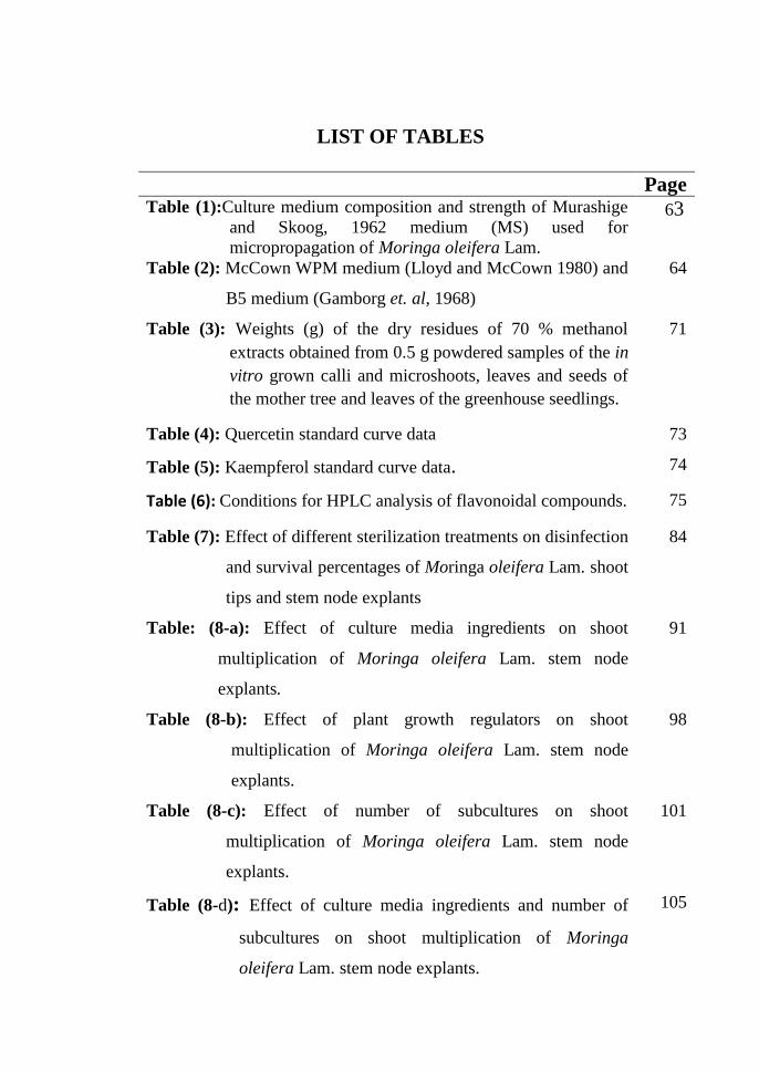
LIST OF TABLES
Page Table (1):Culture medium composition and strength of Murashige
and Skoog, 1962 medium (MS) used for
micropropagation of Moringa oleifera Lam.
63
Table (2): McCown WPM medium (Lloyd and McCown 1980) and
B5 medium (Gamborg et. al, 1968)
64
Table (3): Weights (g) of the dry residues of 70 % methanol
extracts obtained from 0.5 g powdered samples of the in
vitro grown calli and microshoots, leaves and seeds of
the mother tree and leaves of the greenhouse seedlings.
71
Table (4): Quercetin standard curve data 73
Table (5): Kaempferol standard curve data. 74
Table (6): Conditions for HPLC analysis of flavonoidal compounds. 75
Table (7): Effect of different sterilization treatments on disinfection
and survival percentages of Moringa oleifera Lam. shoot
tips and stem node explants
84
Table: (8-a): Effect of culture media ingredients on shoot
multiplication of Moringa oleifera Lam. stem node
explants.
91
Table (8-b): Effect of plant growth regulators on shoot
multiplication of Moringa oleifera Lam. stem node
explants.
98
Table (8-c): Effect of number of subcultures on shoot
multiplication of Moringa oleifera Lam. stem node
explants.
101
Table (8-d): Effect of culture media ingredients and number of
subcultures on shoot multiplication of Moringa
oleifera Lam. stem node explants.
105
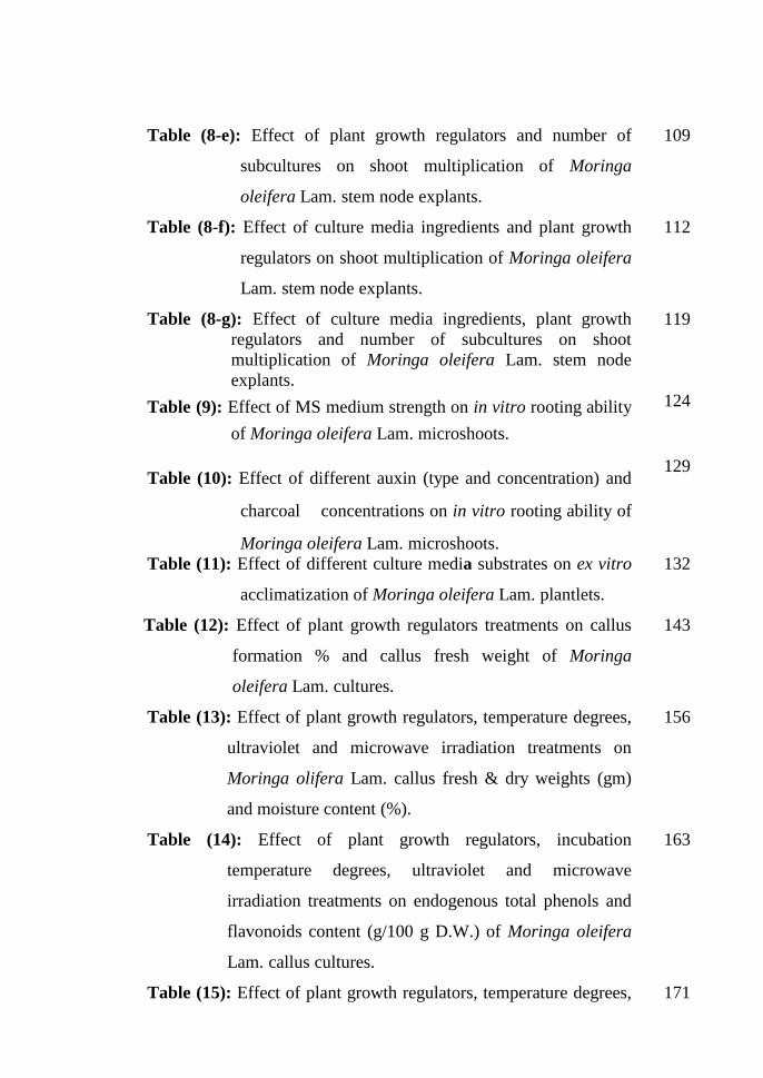
Table (8-e): Effect of plant growth regulators and number of
subcultures on shoot multiplication of Moringa
oleifera Lam. stem node explants.
109
Table (8-f): Effect of culture media ingredients and plant growth
regulators on shoot multiplication of Moringa oleifera
Lam. stem node explants.
112
Table (8-g): Effect of culture media ingredients, plant growth
regulators and number of subcultures on shoot
multiplication of Moringa oleifera Lam. stem node
explants.
119
Table (9): Effect of MS medium strength on in vitro rooting ability
of Moringa oleifera Lam. microshoots.
124
Table (10): Effect of different auxin (type and concentration) and
charcoal concentrations on in vitro rooting ability of
Moringa oleifera Lam. microshoots.
129
Table (11): Effect of different culture media substrates on ex vitro
acclimatization of Moringa oleifera Lam. plantlets.
132
Table (12): Effect of plant growth regulators treatments on callus
formation % and callus fresh weight of Moringa
oleifera Lam. cultures.
143
Table (13): Effect of plant growth regulators, temperature degrees,
ultraviolet and microwave irradiation treatments on
Moringa olifera Lam. callus fresh & dry weights (gm)
and moisture content (%).
156
Table (14): Effect of plant growth regulators, incubation
temperature degrees, ultraviolet and microwave
irradiation treatments on endogenous total phenols and
flavonoids content (g/100 g D.W.) of Moringa oleifera
Lam. callus cultures.
163
Table (15): Effect of plant growth regulators, temperature degrees, 171
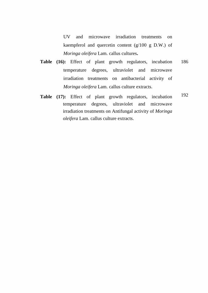
UV and microwave irradiation treatments on
kaempferol and quercetin content (g/100 g D.W.) of
Moringa oleifera Lam. callus cultures.
Table (16): Effect of plant growth regulators, incubation
temperature degrees, ultraviolet and microwave
irradiation treatments on antibacterial activity of
Moringa oleifera Lam. callus culture extracts.
186
Table (17): Effect of plant growth regulators, incubation
temperature degrees, ultraviolet and microwave
irradiation treatments on Antifungal activity of Moringa
oleifera Lam. callus culture extracts.
192
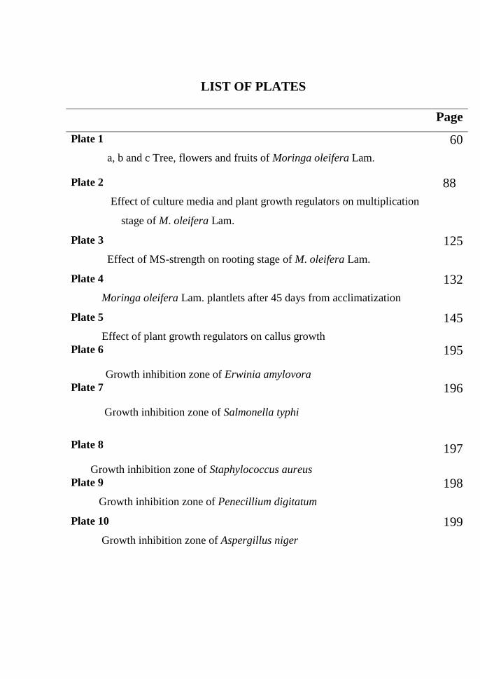
LIST OF PLATES
Page
Plate 1
a, b and c Tree, flowers and fruits of Moringa oleifera Lam.
60
Plate 2
Effect of culture media and plant growth regulators on multiplication
stage of M. oleifera Lam.
88
Plate 3
Effect of MS-strength on rooting stage of M. oleifera Lam.
125
Plate 4
Moringa oleifera Lam. plantlets after 45 days from acclimatization
132
Plate 5
Effect of plant growth regulators on callus growth
145
Plate 6
Growth inhibition zone of Erwinia amylovora
195
Plate 7
Growth inhibition zone of Salmonella typhi
196
Plate 8
Growth inhibition zone of Staphylococcus aureus
197
Plate 9
Growth inhibition zone of Penecillium digitatum
198
Plate 10
Growth inhibition zone of Aspergillus niger
199