Embed Size (px)
Citation preview
5/14/2013
1
Morning Report: An Interactive Case Presentation
Mark Wurth MD/PhD
May 18, 2013
Clinical Presentation
14 y/o male presents with 3 day history of i l b k i li iti hiprogressive low back pain, now limiting his
ability to walk.
5/14/2013
2
HPI details
• Right sided• Progressively worsening• Progressively worsening• No preceding injury, remembered, but child plays multiple contact sports
• Some knee pain initially, but now resolved• No fevers at home, though mom has checked repeatedly because she was concerned about him
• Denies numbness, tingling, weakness, or incontinence
• ROS: negative other than per above.
More History
• PMHx: – Vaccines UTDVaccines UTD– No chronic medical problems/No medications
• PSH:– Freshman– Participant in contact sports– Denies sex/drugs/rock’n roll
• FHx:– Hashimoto’s thyroiditis in parent– No hx of other rheumatologic disease
5/14/2013
3
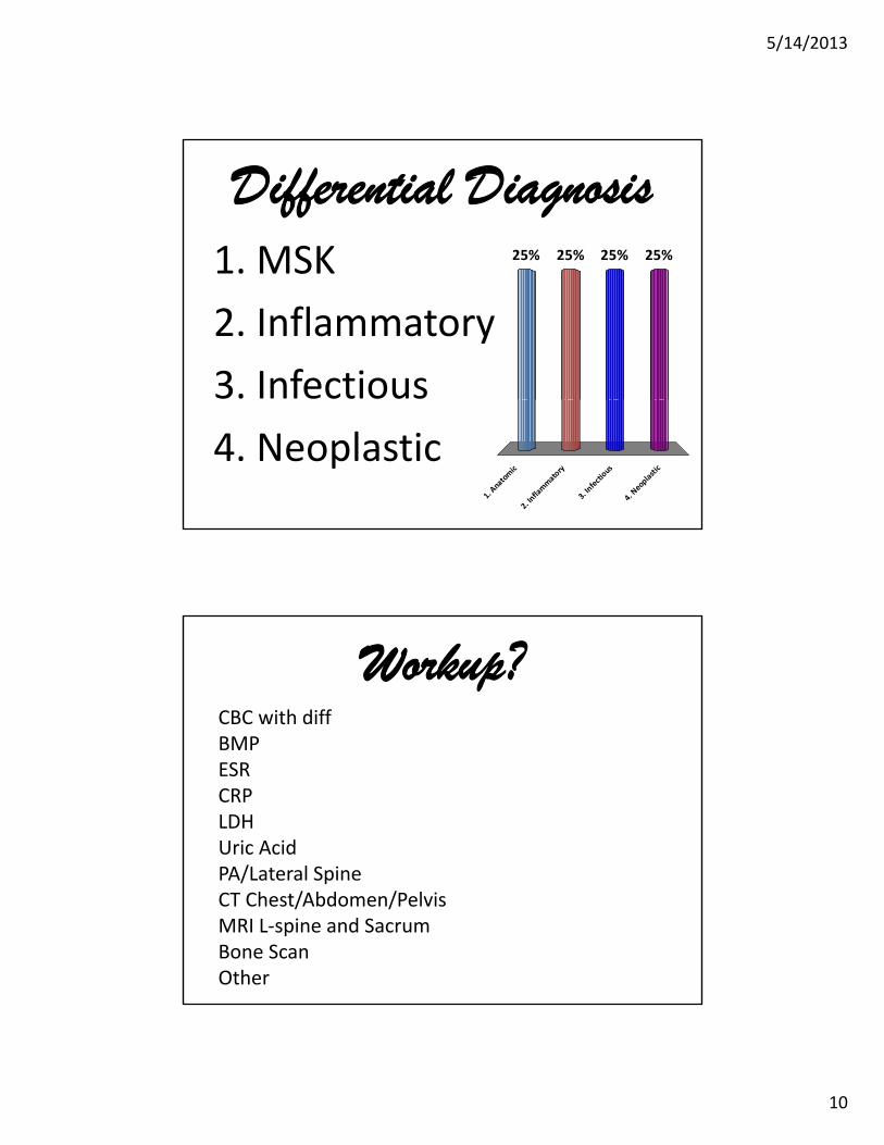
Differential Diagnosis
Differential Diagnosis1. MSK 25% 25%25%25%1. MSK
2. Inflammatory
3. Infectious
4. Neoplastic1. Anatomic
2. Inflammatory
3. Infectious
4. Neoplastic
5/14/2013
4
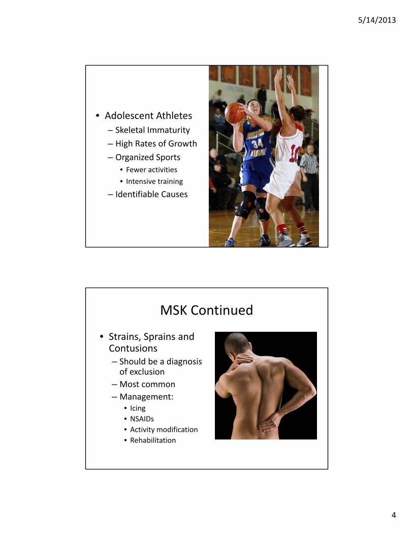
• Adolescent Athletes
– Skeletal Immaturity
– High Rates of Growth
– Organized Sports
• Fewer activities
• Intensive training
– Identifiable Causes
MSK Continued
• Strains, Sprains and ContusionsContusions– Should be a diagnosis of exclusion
– Most common
– Management:• IcingIcing
• NSAIDs
• Activity modification
• Rehabilitation
5/14/2013
5
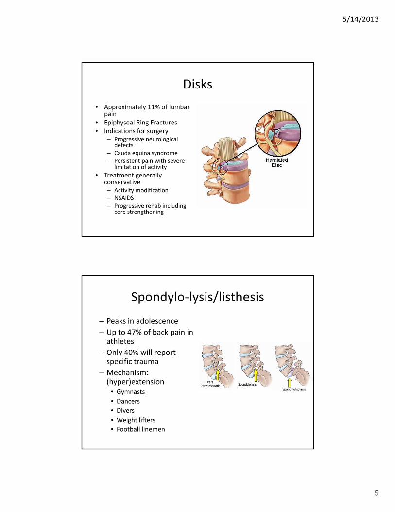
Disks
• Approximately 11% of lumbar painE i h l Ri F• Epiphyseal Ring Fractures
• Indications for surgery– Progressive neurological
defects– Cauda equina syndrome– Persistent pain with severe
limitation of activity
• Treatment generally g yconservative– Activity modification– NSAIDS– Progressive rehab including
core strengthening
Spondylo‐lysis/listhesis
– Peaks in adolescence
– Up to 47% of back pain inUp to 47% of back pain in athletes
– Only 40% will report specific trauma
– Mechanism: (hyper)extension
• Gymnasts• Gymnasts
• Dancers
• Divers
• Weight lifters
• Football linemen
5/14/2013
6
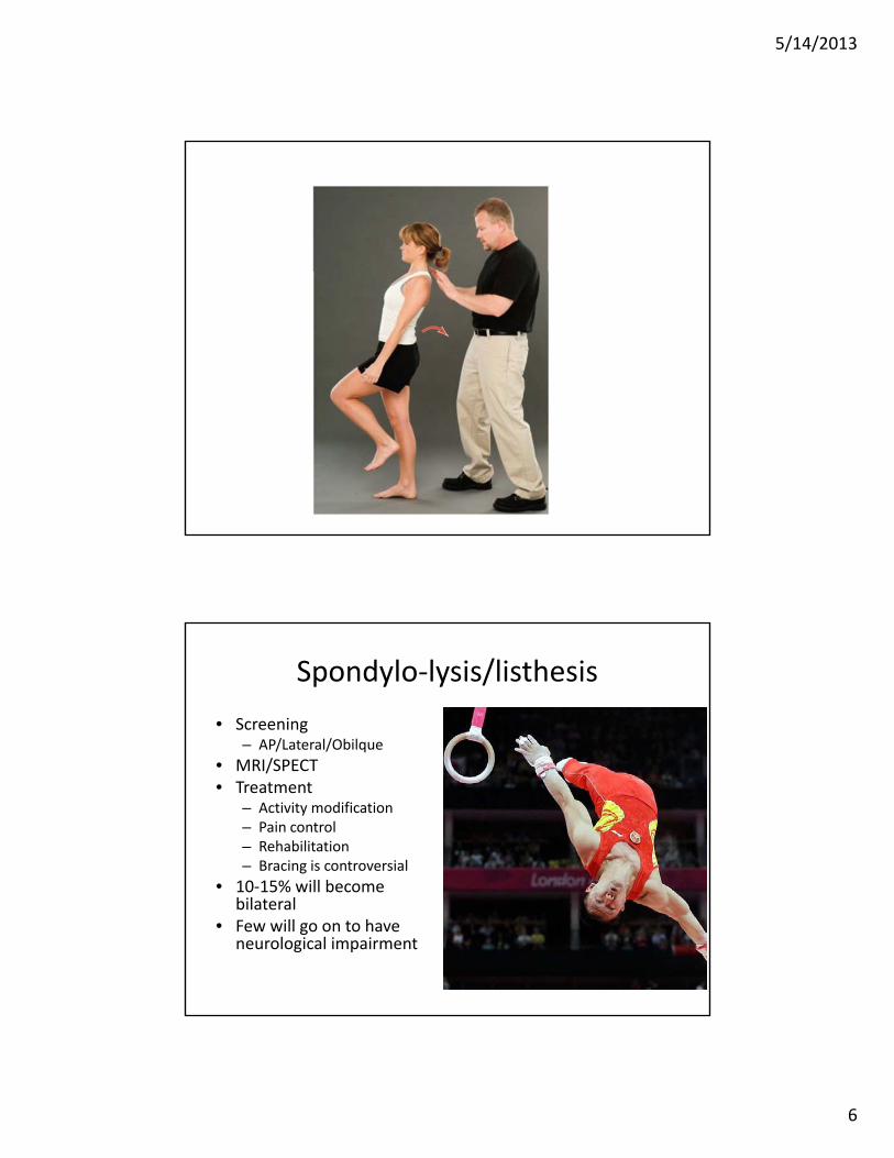
Spondylo‐lysis/listhesis
• Screening– AP/Lateral/Obilque
• MRI/SPECT• Treatment
– Activity modification– Pain control – Rehabilitation– Bracing is controversial
• 10 15% will become• 10‐15% will become bilateral
• Few will go on to have neurological impairment
5/14/2013
7
Anatomic‐ Skeletal
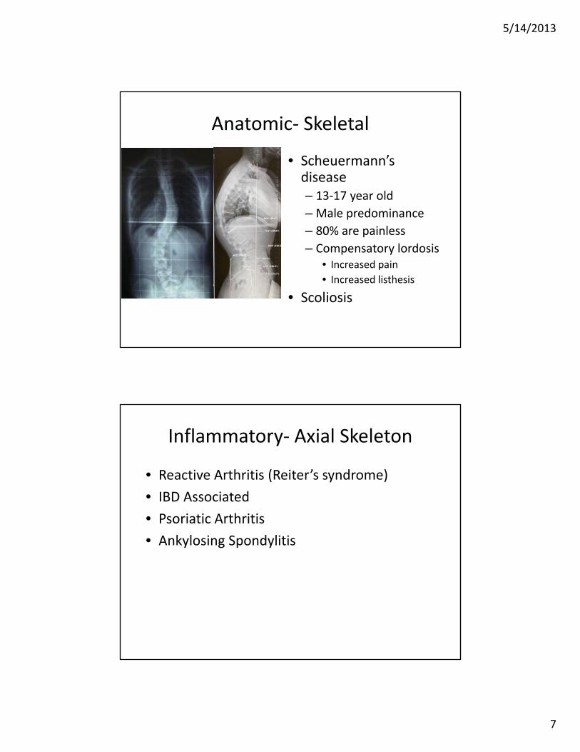
• Scheuermann’sdiseasedisease– 13‐17 year old
– Male predominance
– 80% are painless
– Compensatory lordosisI d i• Increased pain
• Increased listhesis
• Scoliosis
Inflammatory‐ Axial Skeleton
• Reactive Arthritis (Reiter’s syndrome)
• IBD Associated
• Psoriatic Arthritis
• Ankylosing Spondylitis
5/14/2013
8
Commonalities
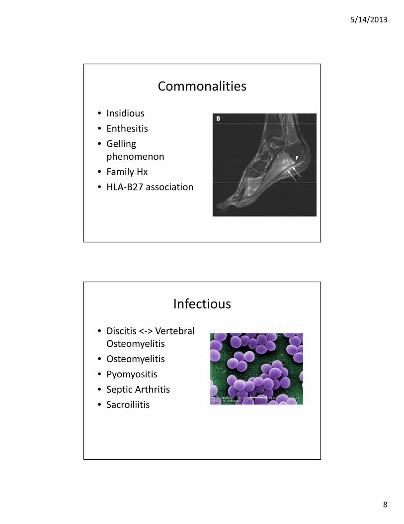
• Insidious
• Enthesitis
• Gelling phenomenon
• Family Hx
• HLA‐B27 association
Infectious
• Discitis <‐> Vertebral O t litiOsteomyelitis
• Osteomyelitis
• Pyomyositis
• Septic Arthritis
• Sacroiliitis
5/14/2013
9
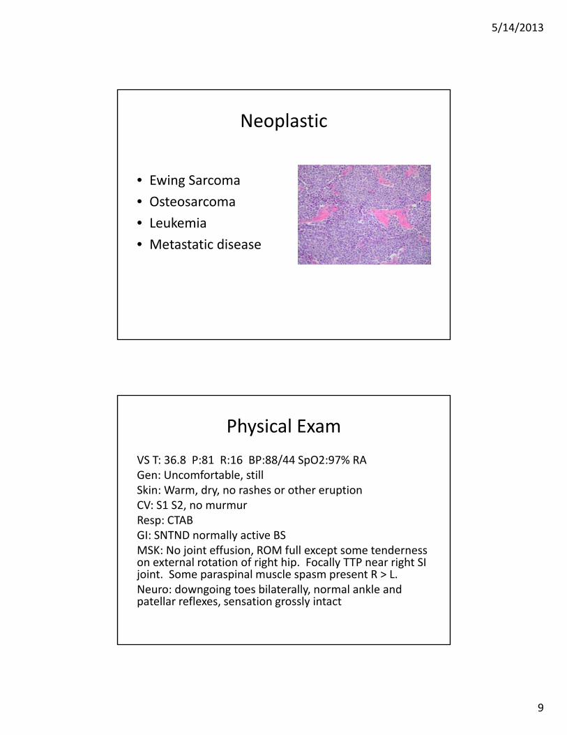
Neoplastic
• Ewing Sarcoma
• Osteosarcoma
• Leukemia
• Metastatic disease
Physical Exam
VS T: 36.8 P:81 R:16 BP:88/44 SpO2:97% RAGen: Uncomfortable, stillGen: Uncomfortable, stillSkin: Warm, dry, no rashes or other eruptionCV: S1 S2, no murmurResp: CTABGI: SNTND normally active BSMSK: No joint effusion, ROM full except some tenderness on external rotation of right hip Focally TTP near right SIon external rotation of right hip. Focally TTP near right SI joint. Some paraspinal muscle spasm present R > L.Neuro: downgoing toes bilaterally, normal ankle and patellar reflexes, sensation grossly intact
5/14/2013
10
Differential Diagnosis1. MSK 25% 25%25%25%1. MSK
2. Inflammatory
3. Infectious
4. Neoplastic1. Anatomic
2. Inflammatory
3. Infectious
4. Neoplastic
Workup?CBC with diffBMPBMPESRCRPLDHUric AcidPA/Lateral SpinePA/Lateral SpineCT Chest/Abdomen/PelvisMRI L‐spine and SacrumBone ScanOther
5/14/2013
11
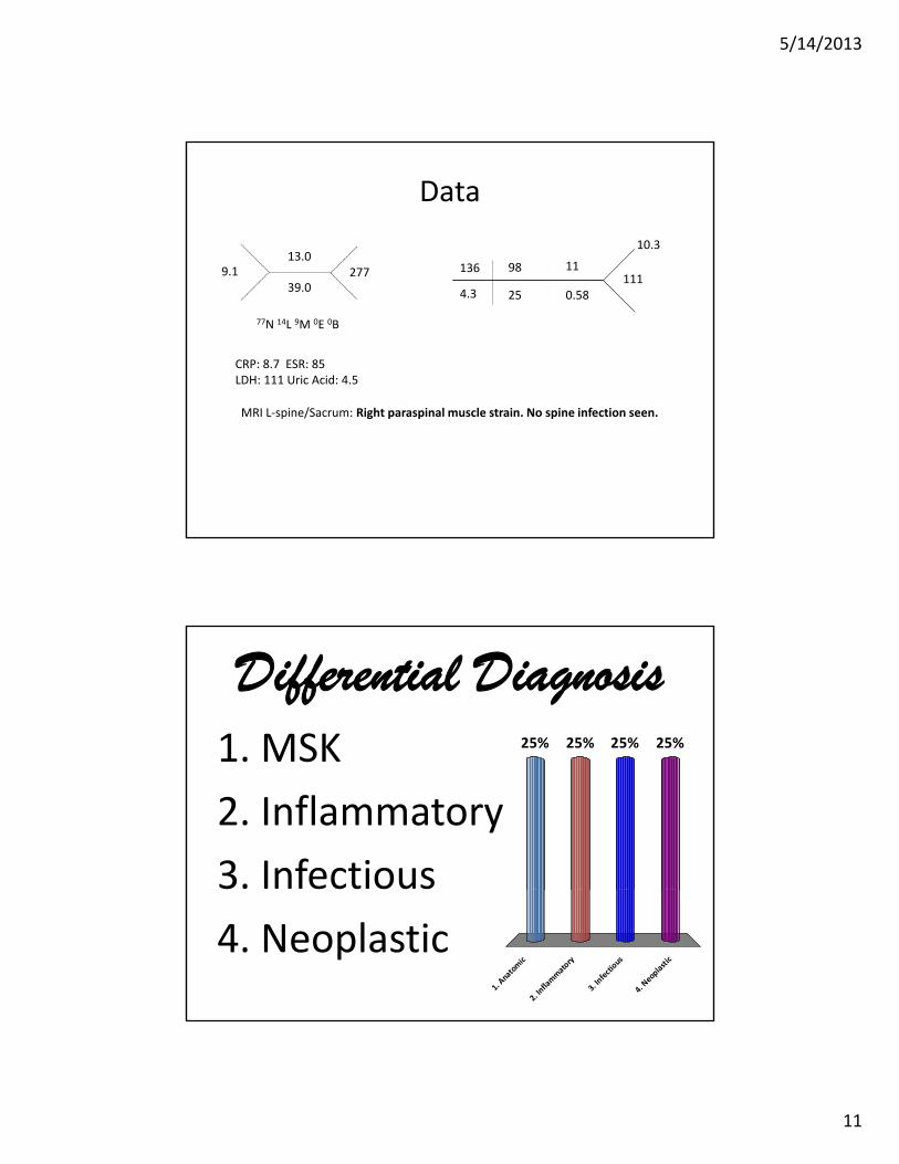
Data
13.09 1 277 136 98 11
10.3
9.139.0
277
77N 14L 9M 0E 0B
4.3 25 0.58111
CRP: 8.7 ESR: 85 LDH: 111 Uric Acid: 4.5
MRI L‐spine/Sacrum: Right paraspinal muscle strain. No spine infection seen.
Differential Diagnosis1. MSK 25% 25%25%25%1. MSK
2. Inflammatory
3. Infectious
4. Neoplastic1. Anatomic
2. Inflammatory
3. Infectious
4. Neoplastic

5/14/2013
12
Data
13.09 1 277 136 98 11
10.3
9.139.0
277
77N 14L 9M 0E 0B
4.3 25 0.58111
CRP: 8.7 ESR: 85 LDH: 111 Uric Acid: 4.5
MRI L‐spine/Sacrum: Right paraspinal muscle strain. No spine infection seen.
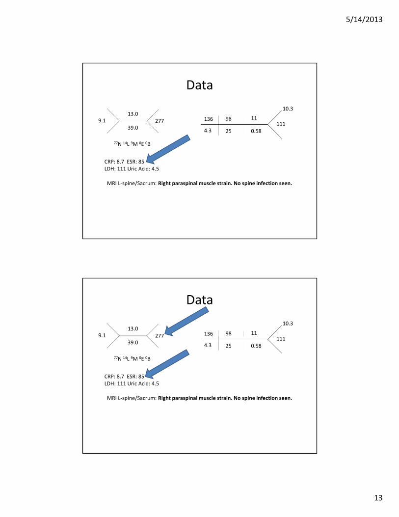
5/14/2013
13
Data
13.09 1 277 136 98 11
10.3
9.139.0
277
77N 14L 9M 0E 0B
4.3 25 0.58111
CRP: 8.7 ESR: 85 LDH: 111 Uric Acid: 4.5
MRI L‐spine/Sacrum: Right paraspinal muscle strain. No spine infection seen.
Data
13.09 1 277 136 98 11
10.3
9.139.0
277
77N 14L 9M 0E 0B
4.3 25 0.58111
CRP: 8.7 ESR: 85 LDH: 111 Uric Acid: 4.5
MRI L‐spine/Sacrum: Right paraspinal muscle strain. No spine infection seen.
5/14/2013
14
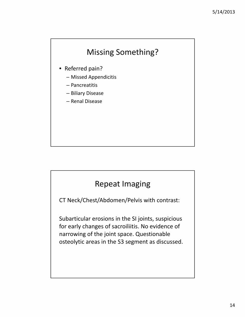
Missing Something?
• Referred pain?
– Missed Appendicitis
– Pancreatitis
– Biliary Disease
– Renal Disease
Repeat Imaging
CT Neck/Chest/Abdomen/Pelvis with contrast:
Subarticular erosions in the SI joints, suspicious for early changes of sacroiliitis. No evidence of narrowing of the joint space. Questionable osteolytic areas in the S3 segment as discussedosteolytic areas in the S3 segment as discussed.
5/14/2013
15
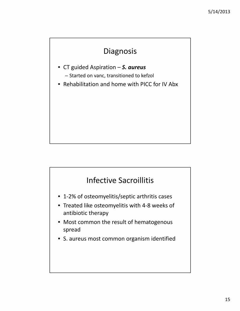
Diagnosis
• CT guided Aspiration – S. aureus
– Started on vanc, transitioned to kefzol
• Rehabilitation and home with PICC for IV Abx
Infective Sacroillitis
• 1‐2% of osteomyelitis/septic arthritis cases
• Treated like osteomyelitis with 4‐8 weeks of antibiotic therapy
• Most common the result of hematogenousspread
• S aureus most common organism identified• S. aureus most common organism identified
5/14/2013
16
Questions?