Embed Size (px)
Citation preview
MORPHOLOGICAL ALTERATIONS AND THE
EFFECT ON TUMOUR SUPRESSOR GENE
EXPRESSION INDUCED BY HIGH DOSE RATE
INTRALUMINAL BRACHYTHERAPY IN
OESOPHAGEAL CARCINOMA
MONAUSA SUR
M.B., B.S., D.C.Path (Clin Path), F.C.Path.S.A. (Anat Path)
A thesis submitted to the Faculty of Health Sciences, University of the
Witwatersrand, Johannesburg, in fulfillment of the requirements for the
degree of Master of Medicine (Anatomical Pathology)
Johannesburg, February, 1999
2.1
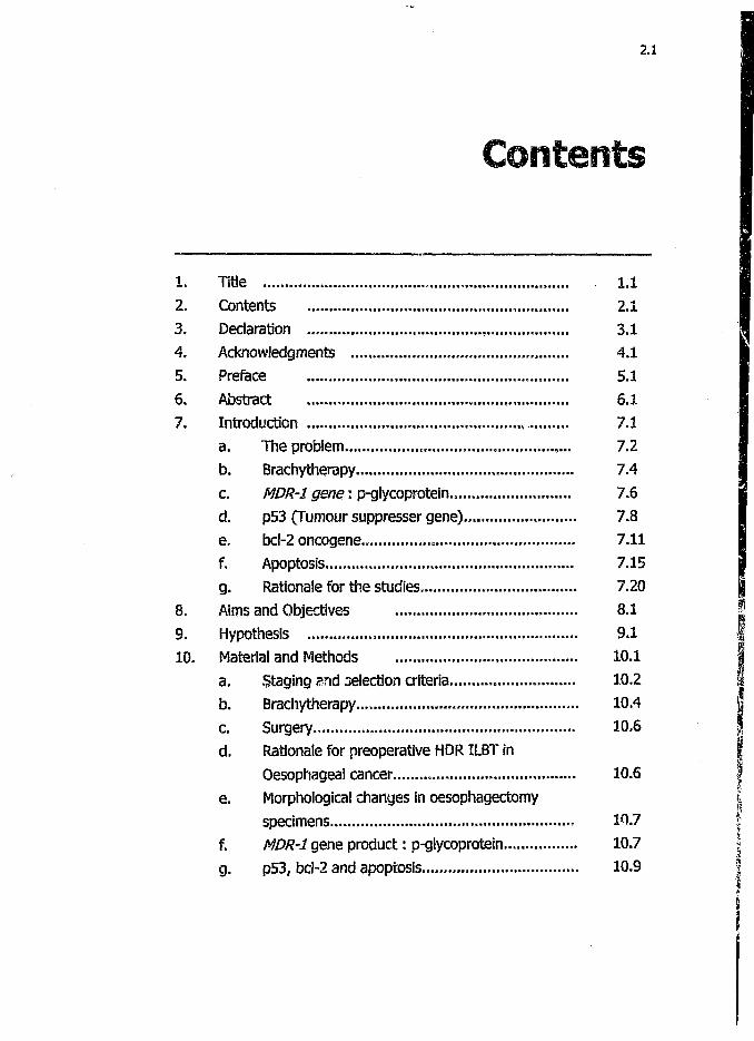
Contents
1. Title .................................................................... 1.12. Contents .......................................... 2.13. Declaration ..................................................................... 3.14. Acknowledgments .......... 4.15. Preface ..................................................................... 5.16. Abstract ..................................................................... 6.17. Introduction ............................................................... 7.1
a. The problem..................... ..................................... 7.2b. Brachytherapy......................................................... 7.4c. MDR-1 gene : p-glycoprotein................................ 7.6d. p53 (Tumour suppressor gene)............................. 7.8e. bcl-2 oncogene.... ....... 7.11f. Apoptosis. ......... 7,15g. Rationale for the studies................................ 7.20
8. Aims and Objectives ......... 8.19. Hypothesis .......................................................... 9.110. Material and Methods ................................................ 10.1
a. Staging and selection criteria................................. 10.2b. Brachytherapy............................... 10.4c. Surgery.................................................................... 10.6d. Rationale for preoperative HDRILBT in
Oesophageal cancer............................................... 10.6e. Morphological changes in oesophagectomy
specimens.............................................................. 10.7f. MDR-1 gene product: p-glycoprotein.... 10.7g. p53, bcl-2 and apoptosis......................................... 10.9
Contents
1. Title ....... 1.12. Contents............ ................................................................ 2.13. Declaration ....... 3.14. Acknowledgments ............... 4.15. Preface........................................................................ 5.16. Abstract ................................................................ 6.17. Introduction ......................... 7.1
a. The problem........................................................ 7.2b. Brachytherapy......................................................... 7.4c. MDR-1 gene : p-glycoprotein......................... ...... 7.6d. p53 (Tumour suppressor gene).............................. 7.8e. bcl-2 oncogene................... 7.11f. Apoptosis ......................................................... 7.15g. Rationale for the studies......................................... 7.20
8. Aims and Objectives ................................................ 8.19. Hypothesis................................ 9.110. Material and Methods .................................................. 10.1
a. Staging and selection criteria.................... 10.2b. Brachytherapy.............................................. 10.4c. Surgery..................................................................... 10.6d. Rationale for preoperative HDRILBT in
Oesophageal cancer............................................... 10.6e. Morphological changes in oesophagectomy
specimens.............................. 10.7f. MDR-1 gene product: p-glycoprotein.................... 10.7g. p53, bcl-2 and apoptosis......................................... 10.9
2.1
Contents
1. Title ................................................................................ l . l2. Contente ................................................................. 2.13. Declaration ..................................................................... 3.14. Acknowledgments ......................................................... 4.15. Preface ....... ........................ ............................... . 5.16. Abstract .................................................................... 6.17. Introduction .................................................................... 7.1
a. The problem........................... 7.2b. Brachytherapy........... ....... 7.4c. MDR-1 gene : p-glycoprotein...... 7,6d. p53 (Tumour suppresser gene).....,......... 7.8e. bcl-2 oncogene ........ 7.11f. Apoptosis.......................................................... 7.15g. Rationale for the studies......................................... 7.20
8. Aims and Objectives ................................................ 8.19. Hypothesis ............................................. .......................... $ 110. Material and Methods ..................................... ........... 1C. '
a. Staging and selection criteria..................... 10.2b. Brachytherapy.................. 10.4c. Surgery........................................-.......................... lO.f;d. Rationale for preoperative HDRILBT in
Oesophageal cancer.................... 10.6e. Morphological changes in oesophagectomy
specimens............................................................... 10.7f. MDR-1 gene product: p-glycoprotein................... 10.7g. p53, bcl-2 and apoptosis......................................... 10,9
2.2
11. Review of Literature ............... ................................ 11.1I. Morphological changes induced by Radiotherapy
'■i oesophageal carcinoma............. ........................ 11.2a. In vivo studies................................................ 11,2b. In vitro/animal studies.................................. 11.7
II. MDR-1 gene product: p-glycoprctein...................... 11.8III. p53, bcl-2 and apotosis............................................ 11.15
a. Effect of chemo/radiotherapy on theexpression of p53 in oesophageal cancers.. 11.15
b. Effect of chemo/radiotherapy on theexpression of bcl-2 in oesophageal cancers. 11.22
c. Role of apoptosis in treated oesophagealcancer ................................................... 11.26
12. Results ........................................................................ 12.1a. Morphological changes................................ ............ . 12.2b. Effect on expression of MDR-1 gene product:
p-glycoprotein............................................. 12.4c. Effect on expression of p53, bcl-2 and apoptosis... 12.5
13. Discussion .................................... .................................... 13.114. Conclusions ........................................................................ 14.115. References .............. ..................... ...... ........................... 15.1

3.1
Declaration
I declare that this thesis is my own unaided work, except where otherwise
acknowledged. This thesis is being submitted for the degree of Master of
Medicine (Anatomical Pathology) at the University of the Witwatersrand,
Johannesburg. This thesis has not been submitted before for any degree or
examination at any other university.
Signed this 27th day of January, 1999
MONALISA SUR

4.1
Acknowledgments
I would like to acknowledge the following without whom this work could not
have been possible:
I would like to express my gratitude for the support and encouragement that I
received from Prof. Kum Cooper, who gave me the opportunity to carry on with
my research and provided the help, support and encouragement that one looks
for in a Head of department. It was my immense pleasure to have worked under
his supervision.
I express gratitude for the financial support for the conducting of my research
that I received from the South African Institute of Medical Research (SAIMR). I
thank the photographic unit of the SAIMR for taking the photomicrographs
presented in this thesis.
I express my thanks to Louise Tayler and Zenobia Haffajee who helped me with
the material ard methods in the research laboratory. I also thank my
departmental colleagues and staff for their consistent encouragement and
support which pnabled me to complete this study.
I would like to thank the Department of Surgery, and Dr. Damon Bizos in
particular for making sure that the esophagectomy specimens were sent to the
department in my name. I express gratitude to the department of Radiation
Oncology for entering patients into this study and for providing the photographs
of the procedure involved in HDRILBT.

I would like to make a mention of the tremendous amount of encouragement,
patience and support that I received from my husband, Dr. Ranjan K. Sur. I
would like to thank him for initiating the spark to complete writing this report,
and for his encouragement and valuable suggestions which enabled me to
complete the work in good time.
I would like to thank my father-in-law for his constant support in furthering my
career, and his appreciation of my achievements and efforts.
I acknowledge with thanks the efforts of Ms. Katerina Chronias of the
department of English, WITS University for going through the manuscript and
giving valuable suggestions in the text and language.
I would like to acknowledge the comments that I received from the reviewers of
all the peer reviewed refereed journals in which my articles from these studies
have been published, which enabled me to write up the discussion with ease.
I would like to dedicate this work to my father. Prof. K.C.Das, and mother, Mrs.
M.Das, who have always encouraged me to achieve my ambitions in life.
Monalisa SurJohannesburg, South Africa

5.1
Preface
The following papers have been published/accepted for publication
and are based directly o r indirectly on th e work presen ted in th is
thesis:
1. Sur M., Sur R.K., Cooper K., Levitt V., Bizos D., Dubazana N.
Morphologic Alterations in Esophageal Squamous Cell Carcinoma after
Preoperative High Dose Rate Intraluminal Brachytherapy.
Cancer 1996; 77:2200 - 2205
2. Sur M., Taylor L., Cooper Kv Sur R.K.
Lack of correlation of P glycoprotein expression with response to MIC
chemotherapy in oesophageal cancer.
Journal ofCLiica! Pathology1997; 50:534
3. Sur M,f Cooper K
bclZ, p53 immunophenotypes and apoptosis in squamous cell carcinoma
of the esophagus,
Histopathology 1998; 33: 87- 88
4 Sur M., Cooper K.f Sur R.K., Taylor L., Bizos D
Effect of Brachytherapy on expression of p53, bcl2 and apoptosis in
squamous cell carcinoma of the esophagus
Molecular Pathology 1999: submitted for publication
5.2
The following conference presentations are done from work directly based on the thesis presented:
1. Biannual Cardiothoracic Forum, Pilansberg, South Africa, October, 1997:
Morphological changes following brachytherapy in esophageal cancer and
effect of brachytherapy on the expression of various oncogenes
2. 37* Annual Congress of the Federation of the South Afiican Societies of
Pathologists, Cape Town, September, 1997:
Effect of Brachytherapy on expression of p53 and bcl2 and apoptosis in
squamous cell carcinoma of the esophagus
3. Biannual Congress of the South African Society of Radiation Oncology,
Durban, March, 1997:
P glycoprotein (MDR1 gene) is of no value in assessing response to
brachytherapy / chemotherapy in squamous cell esophageal cancer
4. Biannual Congress of the South African Society of Radiation Oncology,
Thtianchu Sun, South Africa, September 1995:
Morphologic Alterations in esophageal squamous cell carcinoma after
preoperative high dose rate intraluminal brachytherapy.

Chapter 6
Abstract
6.2
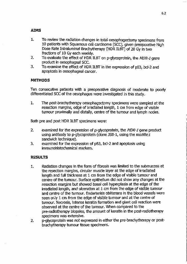
AIMS
1. To review the radiation changes in total oesophagectomy specimens from 10 patients with Squamous cell carcinoma [SCC], given preoperative High Dose Rate Intraluminal Brachytherapy [HDRILBT] of 20 Gy in two fractions of 10 Gy each weekly.
2. To evaluate the effect of HDR ILBT on p-glycoprotein, the MDR-1 gene product in oesophageal SCC.
3. To examine the effect of HDR ILBT in the expression of p53, bcl-2 and apoptosis in oesophageal cancer.
METHODS
Ten consecutive patients with a preoperative diagnosis of moderate to poorlydifferentiated SCC of the oesophagus were investigated in this study.
1. The post-brachytherapy oesophagectomy specimens were sampled at the resection margins, edge of irradiated length, 1 cm from edge of visible tumour proximally and distally, centre of the tumour and lymph nodes.
Both pre and post HDR ILBT specimens were:
2. examined for the expression of p-glycoprotein, the MDR-1 gene product using antibody to p-glycoprotein (clone JSB-1, using the modified sandwich technique).
3. examined for the expression of p53, bcl-2 and apoptosis using immunohistochemical markers.
RESULTS
1. Radiation changes in the form of fibrosis was limited to the submucosa at the resection margins, circular muscle layer at the edge of irradiated length and full thickness at 1 cm from the edge of visible tumour and centre of the tumour. Surface epithelium did not show any changes at the resection margins but showed basal cell hyperplasia at the edge of the irradiated length, and ulceration at 1 cm from the edge of visible tumour and centre of the tumour. Endarteritis obliterans in the blood vessels were seen only 1 cm from the edge of visible tumour and at the centre of tumour. Necrosis, intense keratin formation and giant cell reaction were observed at the centre of the tumour. When compared to the pre-radiotherapy biopsies, the amount of keratin in the post-radiotherapy specimens was extensive.
2. p-glycoprotein was not expressed in either the pre-brachytherapy or post- brachytherapy tumour tissue specimens.
6.3
3. In one patient there was no expression of p53 in either pre orpost-brachytherapy specimens. In 8 patients, p53 staining was strongly positive (3+), with approximately 50% or more cells showing a diffuse pattern in the pre-brachytherapy biopsies. The tumour areas of the post- brachytherapy specimens of these patients showed strong 3+ positivity with p53 (10-50% positive cell count), with the pattern being focal and peripheral in the tumour islands. The centre of the tumour islands showed necrosis and/or keratinization. In one patient, the pre- brachytherapy biopsy showed expression of p53, whilst the post-brachytherapy specimen was negative, bcl-2 expression was equivocal in both pre and post-brachytherapy. Apoptosis was not demonstrated in either the pre or post-brachytherapy tissue sections in the presence of positive control.
CONCLUSION
1. HDRILBT may induce keratin gene in the irradiated cells to differentiate towards better differentiated cells. Preopa?,tive HDR ILBT may have a role in improving the prognosis of early oe sophageal cancer treated with a combination of radiotherapy and surgery.
2. p-glycoprotein expression is of no value in predicting the responsiveness of the tumour to radiotherapy in SCC of the oesophagus.
3. Brachytherapy does not cause cell death by apoptosis but by necrosis and maturation of the cells into better differentiated cells which is caused by OH" free radical and induction of the keratin gene respectively. It is possible that brachytherapy may cause destruction of cells producing wild type p53, while mutant p53 in cells located at the tumour periphery escapes the effect of brachytherapy. This maybe responsible for the high incidence of local recurrence and distant metastasis in oesophageal cancer treated with radiotherapy. Brachytherapy does not affect bcl-2 expression in oesophageal cancer.

Chapter 7
Introduction
7.2
The Problem
Seventy years ago, squamous carcinoma of the oesophagus was a rare disease
in black South Africans ( Rose, 1978). The incidence has risen since then, and it
is currently the most common cancer amongst black men in South Africa, and
the second most common cancer amongst black women (Sitas et al., 1997). In
the 1950's, a large number of cases were diagnosed in the former Transkei and
Ciskei, and in predominantly rural areas of South Africa. The prevalence in the
urban population was considerably less (McGlashen, 1988). Cancer of the
oesophagus remains the most frequently reported cancer in the Transkei,
representing 45.8% of malignant disease (Sitas et al., 1997). The incidence of
oesophageal cancer in Soweto, South Africa's largest urban black community,
has risen and is estimated to be between four to five times greater than that in
the Transkei. In 1985, the age standardised incidence of oesophageal cancer in
Soweto was 125/100,000 in men, and 37/100,000 in women (Kneebone &
Mannell, 1985).
In South Africa, it is a common perception that squamous cancer of the
oesophagus in black South Africans is more advanced by the time of diagnosis,
and that the patients are more debilitated, when compared with patients with
the same disease in Western countries (Hunt, 1978). In 1989, the national study
group for oesophageal cancer in South Africa collected and centralized data on
1926 new cases of squamous carcinoma of the oesophagus, seen in the Ciskei,
7.3
and in major provincial centers of South Africa from 1985 to 1988 (Mannell &
Murray, 1989). There were 1438 men and 488 women (M: F-3: 1) 55% of
whom were between 50 and 69 years of age (mean age, 56 years). 22% of the
patients were in their fifth decade, 14.2% were 70 years of age or older, and
8.8% were younger than forty years of age. 62% of patients complained of
dysphagia to solid food, 12% had difficulty swallowing liquids, and 24% suffered
from total dysphagia. Although 2% of the patients had no dysphagia, they did
have evidence of metastatic disease.
The National Cancer Registry, based a t the South African Institute of Medical
Research, recently reported the incidence of oesophageal cancer in South Africa
(Sitas et al., 1997): cancer of the oesophagus is the second most common
cancer in all males (8.1% of all cancers). It is the most common cancer in black
males (17.9%) (L. R. = 1 in 39). Among white males, by contrast, oesophageal
cancer ranked eighth (1.5%) (L. R. 1 in 119), with a risk factor three times
lower. Among coloured and Asian males, oesophageal cancer ranked fourth and
seventh respectively.
When the South African incidence is compared with that of the rest of Africa, the
age standardised rates amongst black males are comparable to rates from
Zimbabwe, but are higher when compared to West African countries like Mali or
7.4
Gambia. It has been estimated that in Mali, for example, 95% of the patients
have advanced stage disease (Sitas et al., 1997).
It has been shown that the length of the primary tumour is inversely related to
its curability, and is directly related to its stage of advancement. For tumors
smaller than 5 cm in length, 40% are localized, 25% are locally advanced, and
35% have distant metastasis or are unresectable for cure. For tumors larger
than 5 cm, only 10% are localized, 15% are locally advanced, and 75% have
distant metastasis or are unresectable for cure (Clayton, 1928, Fleming, 1943,
Merendiono & Maerk, 1952). The average tumour length seen in South Africa is
about 10 cm (Sur et al, 1998). More than 95% of cancers are squamous cell
carcinomas (SCC) followed by adenocarcinomas. Rare cancers include
undifferentiated carcinoma, carcinoid, malignant melanoma, small cell carcinoma,
and adenocarcinoma arising from submucosal glands.
Brachytherapy
Traditionally oesophageal cancer has been treated with external beam
radiotherapy (EBRT) wherein the radiation is delivered externally. With the
advent of remote afterloading units, it is now possible to treat a tumour with a
radioactive isotope placed within, or in close proximity, to the tumour. This form
of radiation treatment is called Brachytherapy. In the oesophagus, this is done
7.5
by placing a catheter in the oesophageal lumen. A radioactive isotope, such as
Iridium-192 or Cobalt-60, is then passed through the catheter by means of a
remote after loading system, which delivers a high dose to the luminal aspect of
the tumour. This part of the tumour is thought to be more hypoxic and
therefore relatively radioresistant. Due to the close proximity of the radiation
source, a very high dose of radiation is delivered to the luminal aspect of the
tumour, which then undergoes rapid necrosis, thus rapidly restoring lumen
patency. This allows the patient to rapidly restore swallowing which is
maintained for a prolonged period of time. Further, high specific activity radiation
sources, for example, small Ir-192 sources with an activity of 10 Ci (370 GBq)
allows a dose rate of > 2 Gy / minute to be delivered in a very short time - so
called High Dose Rate Intraluminal Brachytherapy (HDRILBT), are used.
Further advantages include:
1. Short treatment time - the whole treatment including the procedure
takes about 20 minutes
2. No anesthesia is required - The procedure is done under Pethidine
analgesia
3. The procedure can be performed on an outpatient basis - Therefore no
admission is required, which saves hospital beds for optimal utilization of
resources.
7.6
4. There is minimal patient discomfort and morbidity of the treatment
and procedure.
5. Risk of radiation to personnel is minimal.
6. Radiation dose to surrounding normal tissue structures due to rapid dose
fall off, is minimal.
7. Many patients can be treated in one day on one HDR unit,
ti. It is a cost effective and safe method of treatment.
There are various genes which may show altered expression prior to and after
therapeutic procedures implemented in the treatment of oesophageal 5CC.
MDR-1 gene
The curative potential of chemotherapy for a number of tumour types has been
obscured by the fact that many patients manifest resistance to a wide array of
structurally unrelated anti-neoplastic agents. One of the most perplexing
problems encountered in chemotherapy is the resistance of certain tumors to all
chemotherapeutic regimes, while other tumors which are initially chemosensitive
to a particular agent, show resistance to treatment over time and disease
progression. Furthermore, tumour cells which are resistant to one drug often
show cross resistance to a wide variety of other, structurally unrelated drugs.
Resistance may be inherent in a tumour cell or may evolve under the selection
pressure of drug administration. This is known as the multidrug drug resistance
7.7
(MDR) phenomenon. The MDR phenomenon includes cross resistance amongst
anthracyclines, Vinca alkaloids, Taxol, and other compounds. The normal role of
MDR is to protect cells from environmental carcinogens, and the tissues that are
most at risk, and richly supplied with MDR, will produce drug resistant neoplasms
(Spiers A.S., 1994). A number of possible molecular explanations for MDR exist.
These include exclusion of the drug from the cells; failure to activate the pro
drug to its active form; increased detoxification; alterations in drug target;
enhanced repair capability of the cell after injury and failure to engage an
appropriate response leading to apoptosis in the damaged cells ( Harrison D.X,
1995). The MDR is due to overexpression of p-glycoprotein, the MDR-1 gene
product. The MDR-1 gene is located on chromosome 13. The family of so-called
MDR genes encodes the p-glycoprotein, apparently the only protein encoded by
the MDR-1 gene, which induces the MDR phenotype. Only 2 of the MDR genes
are known to be present in man. The MDR-1 and MDR-3 genes; but only the
MDR-1 gene product confers the MDR phenotype. The MDR-1 gene codes for
the expression of a cell surface protein (p glycoprotein), which is an integral
plasma membrane protein of 170 kd molecular weight, p-glycoprotein contains
12 putative transmembrane regions and 2 ATP binding sites, p-glycoprotein
expression serves as an excretory pathway for xenobiotic drugs and toxins.
Expression of p-glycoprotein is tissue specific and is found in a number of normal
tissues e.g. colon, small intestine, kidney, liver, adrenal gland, capillary of brain
and testis (Chin K.V. and Liu B., 1994). p-glycoprotein functions as an ATP-
7.8
dependent drug efflux pump with broad specificity for chemically unrelated
hydrophobic compounds (Kartner N. and Ling V., 1989; Gottesman M.M.et al.,
1991). There is extensive evidence from in-vitro studies, especially with non
human cell lines, that overexpression of p-glycoprotein results in reduced
accumulation of the drug within the cell. Recently, mice have been generated
with knock out of MDR-1. These animals showed abnormalities of transport at
the blood brain barrier and were more sensitive to drugs. The MDR-1 gene has
some value in predicting responsiveness to treatment in adenocarcinomas,
p-glycoprotein is overexpressed in a number of intrinsically resistant tumors, and
appears to vary according to the differentiation of the cell. This has been
demonstrated clinically in breast cancer (Glazer R.L and Rohlff C , 1994), renal
cell carcinomas (Mickishc G.H., 1994), cancer of the urinary bladder (Scher H.I.,
1992), colo-rectal malignancies (Linn S.C. and Glaccone G., 1995), gastric
(Wallner J e t al., 1993) and ovarian carcinomas (Sekiya S. et al., 1992). Patients
who express the gene have poor survival.
p53 protein (Tum or suppresser gene)
The p53 gene is a tumour suppresser gene located on the short arm of
chromosome 17 (17pl3.1) and is the single most common target for genetic
alterations in human cancer (Zenbetti G.P. and Levine A.J., 1993, Chang F.,
1993). Homozygous loss of the p53 gene is found in 70% of colon cancers,
30-50% of breast cancers, 50% of lung cancers, oesophageal cancers, stomach

7.9
cancers and others. The p53 gene is a nuclear phosphoprotein that regulates
DNA replication, cell proliferation and cell death. The p53 protein inhibits DNA
replication in response to DNA damage and serves as a checkpoint gene product
that induces cell cycle arrest in G1 by activating growth suppresser genes (Harris
C.C. and Holistein M., 1993). Under physiological conditions, p53 has a very
short half life measured in minutes, and there is no evidence that this protein is
required for normal cell division. A combination of genetic events that affect
both alleles of the gene, results in the loss of expression of the wild type gene.
Mutations of the p53 leads to the loss of DNA binding and transcriptional
regulatory activities of the p53 phosphoprotein, with the corresponding loss of its
growth suppressive activity and its role as "the guardian of the genome". The
mutated protein has abnormal conformation, impaired DNA binding, and a
prolonged or stabilized half life, the latter resulting in immunohistochemicaliy
stainabie levels within nuclei in nearly all tumors showing p53 gene mutation
(Batsakis J.G., L-Nagger A.K., 1995).
When cells are exposed to mutagenic agents such as chemicals or radiation,
there are dramatic changes in wild type p53 cycle (Zenbetti G.P. and Levine A.J.,
1993). Through post translational modifications, p53 is stabilized, and it
accumulates within the nucleus. The accumulated wild type p53 binds to DNA
and causes cells to arrest in the G1 phase of the cell. This reversible p ,;ise in
the cell cycle is welcome, because it allows the cells time to repair the DNA

7.10
damage inflicted by the mutagenic agent. If, for some reason, ihe repair
mechanisms fail, p53 triggers cell death by apoptosis. With loss of normal p53
(common in many tumors), cells exposed to mutagenic agents replicate the
damaged DNA, and thus mutations become fixed in the genome. Although no
single mutation is sufficient to transform cells, the loss of p53 pathway
predisposes cells to additional mutations and to ultimately malignant
transformation. Radiation and chemotherapy rely in part on their ability to alter
the DNA of rapidly dividing cells, leading to their death and elimination by the
p53 mechanism. If p53 is absent, the genetic damage goes unnoticed and these
mutations may result in the lesion being more aggressive. Inactivation of the p53
gene occurs most commonly in missense mutations. These abnormal proteins
fail to bind to DNA but have a remarkably long half life and are detected in
tumour cells by immunohistochemical staining (Kaklamanis L. et al., 1993). Loss
of wild type p53 activity induces a release from Gl-S cell cycle checkpoint control
following DNA damage, increasing genomic instability and promoting gene
amplification. In addition, certain mutant forms of p53 not only lose their normal
function, but also gain the ability to bind and inactivate normal p53 protein.
p53 tumour suppressor gene normally stimulates apoptosis, but, when mutated
or absent (as seen in certain cancers), favors cell survival. It appears that p53 is
required for the apoptosis following DNA damage by irradiation ( Lane D.P.,
1993). Immunohistochemical detection of the nuclear p53 protein is based on

7.11
the increase in concentration of the protein to detectable levels, secondary to an
increased synthesis and a lower degradation with a longer half-life. Although
there is good agreement between the frequency of positive immunostaining and
the frequency of tumors with mutations, there are discrepancies between these
findings and analysis a t the protein level. p53 protein can be stabilized by other
means, such as sequestration of normal nuclear protein in the cytoplasm with
inactivation of its tumour suppressor function or by binding with other cellular
proteins. Therefore, positive staining may not always be an indication of an
underlying mutations (Hall P.A. and Lane D.P., 1994). The analysis of p53 in
neoplastic and pre-neoplastic states and the ability to stain for the abnormal
forms of the protein in tissue sections is a powerful tool which provides molecular
information on the oncogenic process. Furthermore, there is evidence to suggest
that the expression of abnormal p53 may be a prognostic parameter in some
neoplasms (Batsakis J.G., L-Nagger A.K., 1995).
oncogene
The bcl-2 gene was identified more than a decade ago with the discovery and
analysis of the t(14; 18) (q32;q21) translocation (Bakhshi A. et al, 1985). This
translocation is the most common specific recurrent chromosomal aberration in
non-Hodgkins lymphomas, occurring in 70-80% of cases of follicular lymphoma
(Cleary M.L. et al. 1986 ; Landanyi M.and Wang S., 1992). As a result of this
translocation, bcl-2 sequences are juxtaposed with the immunoglobulin heavy

7.12
chain gene on chromosomal 14q32 (Chen-Levy Z. et ak, 1989). This results in
an overexpression of the translocated bcl-2 allele induced by enhancers in the
immunoglobulin heavy chain region (Pezzella F, et alv 1990). However, this
translocation is not a prerequisite for bcl-2 protein expression and does not result
in an interruption of the coding region of bcl-2. The bcl-2 polypeptide is a 26 kD
protein that is found on intracellular (mitochondrial and nuclear) membranes and
in the cytosol of the smooth endoplasmic reticulum (Krajewski K. et al., 1993).
The association of bcl-2 with lymphomas immediately led to the inference that it
was an oncogene. However, bcl-2 protein was found to have no effect on cell
replication (Baer R., 1994). It acts as a survival factor that prevents cells from
undergoing apopiosis, and confers a survival advantage on cells harboring the
t(14;18) translocation. By extending cell survival, over expression of bcl-2 allows
other mutations that affect proto-oncogene and cancer suppresser genes to
supervene. Recent studies suggest that bcl-2 inhibits apoptosis by regulating an
anti- oxidant pathway (Hockenberry D.M., 1993).
Several genes that have sequence homology with the bcl-2 gene have been
identified and are classified as members of the bcl-2 family of genes (Lu Q.L. et
al., 1996). They can be divided into two groups on the b: sis of their effects on
the life span of cells. Those that are anti-apoptotic are t d-2, bcl-XL and MCL1.
Those that promote cell death are Bax, bcl-XS and Bak. bcl-2 protein is
produced early in the process of embryogenesis (Veis-Novack D.and Korsmeyer
7.13
SJ., 1994). bcl-2 is present in all three germ layers and once tissues have
matured, expression of bcl-2 is restricted, bcl-2 may be required during
embryogenesis and development to ensure cell viability and survival so that
differentiation and maturation can occur. In normal lymphoid tissue bcl-2
antibody reacts with B-lymphocytes in the mantle zone and with many cells
within T-cell areas. In the thymus many cells in the medulla are stained, with
weak/negative reaction in the cortex (Chetty R. e t al. 1997).
Expression of bcl-2 has been studied in various tumors (Lu Q.L. et al., 1993;
Pezzella F. et al., 1993; Colombel M. et al., 1993, Mcdonald T.J., 1992). In
general, bcl-2 positive neoplasms have a better prognosis than negative ones,
with certain prostatic cancers being the exception to the rule.
The frequency of bcl-2 positivity in the more common malignancies studied are
as follows:Chronic lymphocytic leukemia- almost 100%; chronic myeloid
leukemia- almost 0%; chronic myeloid leukemia with blastic crisis- almost 100%;
acute myeloid leukemia- almost 100%; follicular lymphoma- 80%; T and B cell
non-Hodgkins lymphoma- 65%; Hodgkins disease- 40%; breast cancer- 80%;
neuroblastoma- 40%; nasopharyngeal carcinoma- 80%; prostatic cancer 75%;
lung SCC- 25%; lung adenocarcinoma 10-15%.
7.14
Transcription of bcl-2 could affect transport across membranes, influence
intracellular ion distribution, or initiate pathways that prevent oxidative damage
to sub cellular constituents as lipid-containing membranes (Craig R.W., 1995).
Both bcl-2 and calcium are localized in the mitochondrion, so bcl-2 may affect
calcium regulation, bcl-2 may redistribute or sequester calcium, thereby
decreasing calcium dependent nuclease activity and inhibiting apoptosis (Craig
R.W., 1995). It is therefore clear that the ability of bcl-2 to inhibit apoptosis
depends on 3 factors: regulation of expression of the bcl-2 gene, regulation of
expression of other members of the family, and the species of dimers then
formed.
bcl-2 can block the apoptosis that usually results from treatment with radiation
(Strasser A. et al., 1994), or chemotherapeutic agents (Miyashita T. and Reed
J.C., 1993). Wild type p53 downreguiates bcl-2 and leads to an increase in Bax,
thereby promoting apoptotic death. However, mutant p53 may have the
opposite effect and function as a substitute for bcl-2, thereby promoting cell
survival. These proteins also have an inverse relationship in some malignancies.
A reciprocal relationship has been demonstrated between bcl-2 reactivity and
p53 overexpression in 65% of colorectal neoplasia, and in follicular lymphoma
(Cooper K and Haffejee Z, 1997). bcl-2 positive/ p53 negative subgroup shows
• Tong correlation with negative lymph node status, implying a less aggressive
pa'dr, ., vf of neoplastic transformation (Kaklamanis L. et al, 1996). bcl-2 protein
7.15
has been detected in all grades of cervical intraepithelial neoplasia (CIN), with a
striking increase in the number of positive cells within increasing severity of CIN,
in combination with a mild increase in cytoplasmic staining intensity. Beyond the
application of bcl-2 immunostaining, to distinguish a reactive follicular l\ uphold
hyperplasia from follicular lymphoma , bcl-2 staining offers little diagnostic value
(Cooper K and Haffajee Z., 1996). The use of bcl-2 expression in various
ma'ignancies has been shown to be of prognostic merit. The results of bcl-2
staining in initial diagnostic biopsies may point to therapeutic options and be of
prognostic importance, bcl-2 is an important player in the cell cycle. It has a
crucial impact on several other genes and proteins that dictate the path taken by
cells, i.e., survival or death.
Apoptosis
It has been shown that a decrease in tumour volume often occurs after
treatment with radiation or chemotherapy, due to cell death by necrosis or
apoptosis. Necrosis results from direct physical or chemical damage to the
plasma membrane, or disturbances in the osmotic balance of the cell. With the
entrance of extra cellular fluid into the cell, resultant cell swelling and lysis
precedes a subsequent inflammatory response. Furthermore, necrosis affects
groups of ceils, with consequent disruption of normal tissue architecture. In
contrast to necrosis, apoptotic cell death (programmed cell death) is a highly
regulated physiological process. The balance between apoptosis and cell
7.16
proliferation results in the maintenance of cell homeostasis (Ken J.F.R. et al.,
1972). Apoptotic bodies are rapidly engulfed by the neighboring cells or
macrophages without an inflammatory response being elicited. The nuclear
structure alteration in apoptotic cells is induced by endonuclease DNA cleavage
that results in the generation of large 50-300 Kb fragments. This produces the
characteristic DNA "ladders" of apoptosis, as viewed on agarose gel
electrophoreses (Oberhammer F. et.al., 1993). The histologic features of
apoptosis include cell shrinkage and loss of junctional contact, resulting in a
"halo" around the cell. The nucleus shows condensation and margination of the
chromatin. This is followed by fragmentation or "pinching" of pieces of nuclear
material, which are surrounded by cytoplasm with intact cytoplasmic organelles
as shown at ultra structural level. These apoptotic fragments of pyknotic nuclear
material and cytoplasm are phagocytosed by adjacent cells or macrophages.
Apoptotic cells are also called by other names which include Councilman bodies,
Civatte bodies, necrobiotic cells and nuclear dust.
Apoptosis is thought to be responsible for numerous physiological and pathologic
events which include the following (Kerr J.F.R. and Harmon B.V., 1991):
1. The programmed destruction of cells during embryogenesis and
metamorphosis,
2. Hormone dependent involution in the adults,
3. Cell deletion in a proliferating cell population.
7.17
4. Cell death in tumors, most frequently during regression, but also in
tumors with active cell growth,
5. Death of (immune cells) both B and T lymphocytes after cytokine
depletion,
6. Pathologic atrophy of hormone dependent tissues,
7. Pathologic atrophy in parenchymal organs after duct obstruction,
8. Cell death induced by cytotoxic T cells,
9. Cell injury in certain viral diseases,
10. Cell death produced by a variety of injurious stimuli that are capable of
producing necrosis, but when given in low doses-including mild thermal
injury, radiation, cytotoxic anticancer drugs and possibly hypoxia- induced
apoptosis.
One important feature of apoptosis is its dependence, in many instances, on
gene activation and new protein synthesis (Vaux D., 1993). A number of genes
can be induced by stimuli causing apoptosis, such as heat shock proteins and
proto- oncogenes, but these genes are not directly related to the triggering of
apoptosis. Apoptosis specific genes that stimulate (ced-3, 4) or inhibit (Ced- 9)
cell death have been identified in the development of the nematode C. elegans
(Hengartner M.O., 1992), and mammalian homologs have been described.
Certain genes involved in growth and in the causation of cancer play a regulatory
role in the induction of apoptosis. bcl-2 oncogene inhibits apoptosis. The c-myc
7.18
oncogene, whose protein product can stimulate either apoptosis, or, in the
presence of bcl-2, cell growth. Alterations in the ratio of bcl-2:Bax are implicated
in shifting the fine balance between cell proliferation and apoptotic cell death.
Since Bax antagonizes the inhibitory effect of bcl-2 on apoptosis, p53 appears to
provide a key regulatory step in the cellular decision to die by apoptosis
(Bissonette R.P., 1992), p53 normally stimulates apoptosis but when mutated or
absent, favors cell survival. p53 is required for the apoptosis following DNA
damage by irradiation. Restoration of wild type p53 protein, which acts
upstream of bcl-2 and Bax, is predicted to restore the ability of many tumour
ceils to respond to genotoxic therapy by undergoing apoptosis. bd-2 gene
protects tumour cells from apoptosis induced by a variety of agents, including
ionizing radiation, and is thus related to resistance to DNA damaging therapeutic
agents. The p53 tumour suppresser gene, however, has been related with
growth arrest, apoptosis, and thus with selective sensitivity to the killing effects
of ionizing radiation and DNA damaging drugs. This functional antagonism
between the two genes occurs because of reciprocal downregulation, due to the
presence of a p53- transcription silencer in the untranslated region of the bcl-2
gene. Growth arrest in the G1 phase of the cell cycle, and induction of
apoptosis, are two distinct and dissectable functions of p53. bcl-2 is able to
antagonize the induction of apoptosis by p53 but not the growth arrest in Gl.
However, coexpression of bcl-2 and of the oncogene c-myc efficiently
antagonizes effects of p53 on Gl arrest and apoptosis. Therefore, knowledge of

7.19
interaction between these three genes involved in human cancer is a
fundamental prerequisite for the improvement of knowledge on prognosis, and
to design innovative therapeutic approaches (Chiarugi V. and Ruggiero M.,
1996).
Radiation induced apoptosis is prevalent in thymocytes, lymphocytes and
haemopoietic and embryonal cells, but is expressed at low levels in other
mammalian cell types (Allan D.J., 1992). Dewey W.C. et al. (1995) compared
published data on the extent of radiation induced apoptosis versus that of
clonogenic cell death for a variety of cell systems. At single doses that results in
90% clonogenic cell kill, less than 50% and in most instances, only 10%-25%
could be attributed to apoptosis. In fau, several cell types, such as Chinese
hamster ovary cells, do not undergo apoptosis after exposure to high radiation
doses (Dewey W.C. et al., 1995, Olive P.L et al., 1993, McKenna W.G., 1996).
To date, the morphological changes that occur in oesophageal cancer following
brachytherapy have not been described. Further, no studies have examined the
effect of brachytherapy on the MDR-X gene or the effect on bcl-2, p53 and
apoptosis expression.

7.20
In view of the above, the rationale for these studies were formulated:
1. The effect of brachytherapy may extend into all the layers of the
oesophagus, causing radiation changes which can be observed
histologically.
2. At the tissue level, it is likely that brachytherapy may alter the expression
of the MDR-1 gene, p53 and bcl-2 genes.
3. The above alterations and interaction between the various genes may lead
to apoptotic cell death.
Although there are several studies on the effect of chemotherapy on the
expression of MDR-1 gene, p53, bcl-2 and apoptosis in oesophageal cancer,
there are no studies in literature that prospectively examine the effect of either
EBRT or ILBT on oncogene expression. Furthermore, there are no studies in
literature that described morphological changes in tire oesophagus after a course
of brachytherapy.
In this report, morphological changes following brachytherapy are examined for
the first time. The report also examines the effect of brachytherapy on the
expression of MDR-1 gene product, p53, bcl-2 and apoptosis in oesophageal
cancer for the first time.

8 . 1
Chapter 8
Aims and Objectives
8 , 2
The following are the aims and objectives of the study:
1. To evaluate the morphological changes that occur in the oesophagus
following a course of preoperative brachytherapy.
2. To examine the effect of brachytherapy on p-glycoprotein, the MDR-1
gene product.
3. To examine the effect of brachytherapy on the protein expression of p53,
bcl-2 and apoptosis in oesophageal cancer treated with preoperative ILBT.

Chapter 9
Hypothesis

9.2
MORPHOLOGICAL CHANGES
Hi Brachytherapy causes characteristic changes in the oesophageal mucosa,
submucosa, muscularis propria , serosa and in the tumour within the
oesophageal wall and lymph nodes in the vicinity.
MDR1 GENE EXPRESSION
Ho Brachytherapy has no effect on the MDR1 gene expression in SCC
oesophageal cancer.
Hi Brachytherapy has an effect on the MDR1 gene in SCC oesophageal
cancer
p53, bc!2 AND APOPTOSIS
Ho Brachytherapy has no effect on the expression of p53, bc!2 and apoptosis
in SCC of the esophagus
Hi Brachytherapy has an effect on the expression of p53, bcl2 and apoptosis
in SCC of the esophagus

10.1
Chapter 10
Material and Methods
10.2
STAGING AND SELECTION CRITERIA
All patients in this study were staged by the computed tomography (CT) staging
system described by Halvorsen and Thompson (1984). Most patients were
worked up by the surgical units which referred the patients. They were deemed
to be operable by the surgical unit if they fulfilled the following selection criteria :
a. Good performance score (Karnofsky performance score > 70)
b. Tumour length < 8 cm length as seen on upper G.I., endoscopy and
barium swallow
c. CT stage II after a CT scan of the chest and abdomen
d. Good lung function test and pulmonary reserve
e. Tumour in the thoracic oesophagus
f. No evidence of distant metastasis on sonar of the abdomen and bone
scan
g. Normal haematologica! and biochemical parameters including liver
function tests.
h. Initial biopsy of the tumour after upper G.I. endoscopy showing features
of infiltrating squamous carcinoma on histopathologic assessment.
A total of 10 patients were entered into the study.
10.3
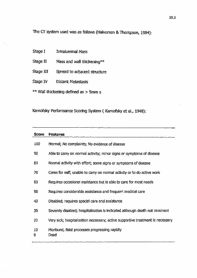
The CT system used was as follows (Halvorsen & Thompson, 1984):
Stage I Intraluminal Mass
Stage II Mass and wall thickening**
Stage III Spread to adfacent structure
Stage IV Distant Metastasis
** Wall thickening defined as > 5mm s
Kamofsky Performance Scoring System ( Karnofsky e t ai., 1948):
Score Features
100 Normal; No complaints; No evidence of disease
90 Able to carry on normal activity; minor signs or symptoms of disease
80 Normal activity with effort; some signs or symptoms of disease
70 Cares for self; unable to carry on normal activity or to do active work
60 Requires occasional assistance but is able to care for most needs
50 Requires considerable assistance and frequent, medical care
40 Disabled; requires special care and assistance
30 Severely disabled; hospitalization is indicated although death not imminent
20 Very sick; hospitalization necessary; active supportive treatment is necessary
10 Moribund, fatal processes progressing rapidly0 Dead
BRACHYTHERAPY (as performed by the Radiation Oncologist)
R equirement :
A nalgesia :
Equipment :
P rocedure
The patient must fast at least 12 hours before the procedure
with Pethidine 50 mg + Buscopan 1 amp given deep I.m,
30-45 minutes before the procedure
Flexible fiberoptic upper G.I., Stainless steel guide wire
(Figirp, I), Nucletron 6 and 4 mm intra oesophageal
catheters, Marker wire (Figure II), Fluoroscopy control
(C-arm) (Figure III), Brachytherapy Treatment Planning
system (Figure IV), and afterloading microsource HDR unit
(Figure V)
1. Oral Gastrograffin is given to the patient, the tumour is
localised and tracheo-oesophageal fistula is ruled out prior
to the procedure (Figure VI).
2. Fiberoptic upper g.i. endoscopy is performed in all
patients and the tumour is visualised (Figure VII, VIII).
3. Stainless-steel guide wire is passed across the tumour
length under Endoscopic vision (Figure IX, X).
4. The Nucletron Intra Oesophageal Catheter with an outer
diameter of 6 mm is passed across the tumour length over
the guide wire (Figure X I).
5. Positioning of the Catheter is performed under
fluoroscopic vision using a C Arm (Phillips BV29) after giving
10.5
the patient oral Gastrograffin (Figure XII). A margin of 4 cm
is prescribed above and below the visible tumour, including
the tumour (Figure XIII).
6. The catheter is connected to a remote after loading HDR
unit (Microselectron HDR, Nucletron, The Netherlands) which
has an Iridium-192 source of 10 Ci (370GBq) initial activity
(Figure XIV, XV).
7. Treatment is given following dose optimization on the
treatment planning system for brachytherapy treatment
(Plato, Nucletron, The Netherlands) - see Figure XVI, XVII.
The dose is prescribed at 10 mm from the centre of the
source axis, and includes the tumour and a 4 cm margin
proximally and distally to the tumour.
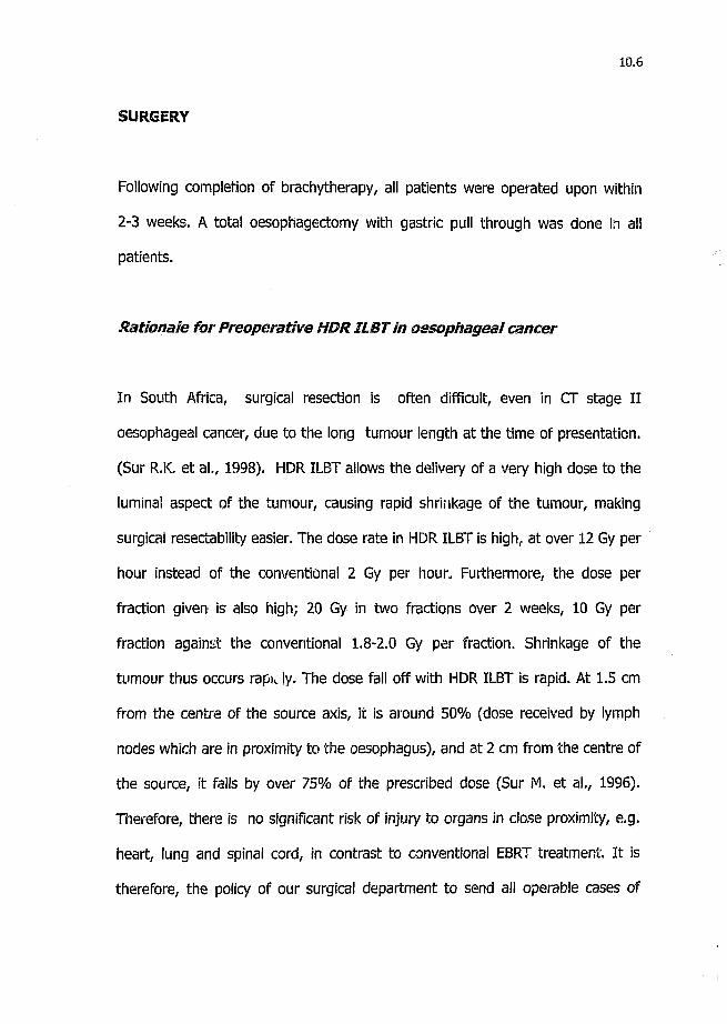
8. Treatment delivery is controlled from the remote console
of the HDR unit (Figure XVHI, XIX).
Treatm ent Timc: The entire procedure depending on source activity, takes
20-30 minutes, and is done on an out patient basis
D ose and fra ctio n : A total of 20 Gy was given in 2 fractions over 2 weeks. 10 Gy
was given per fraction at 1 cm from the centre of the source
axis. One week interval was given between the 2 fractions.
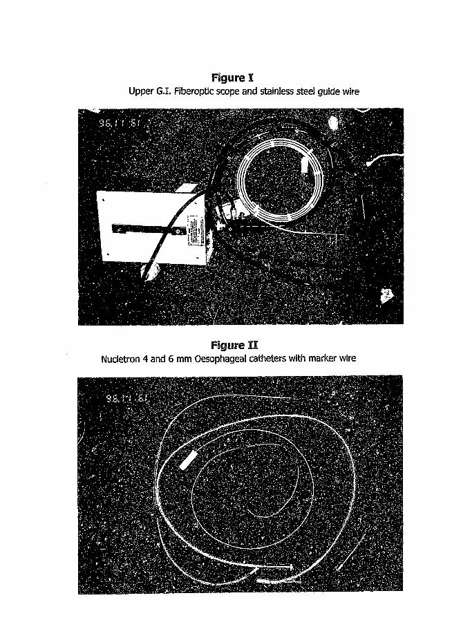
Figure IUpper G.I. Fiberoptic scope and stainless steel guide wire
Figure IINucletron 4 and 6 mm Oesophageal catheters with marker wire
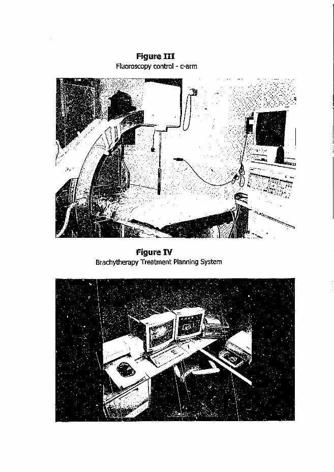
Figure IIIFluoroscopy control - c-arm
Figure IVBrachytherapy Treatment Planning System

Figure VNudetron - Microselectron HDR - Remote After loading HDR unit

Figure VIVisualization of the tumor after giving the patient 10 ml of Oral Gastrograffin. This also rules out presence of a tracheo-oesophaaeal fistula
Figure VIITumor as visualized on fiberoptic endoscopy
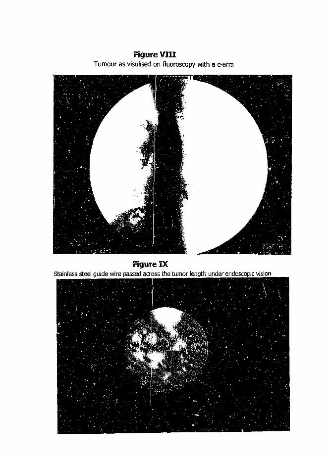
Figures VIIITumour as visuiised on fluoroscopy with a c-arm
Figure IXStainless steel guide wire passed across the tumor length under endoscopic vision

Figure XStainless steel guide wire passed across the tumor length as seen on fluoroscopy with a c-arm
Figure XINucletron Intra-oesophageal catheter passes across the tumor length through the guide wire as
seen on fluoroscopy with a c-arm
omu
, ONS .9S9&/9#
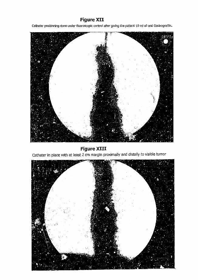
Figure XIICatheter positioning done under fluoroscopic control after giving the patient 10 ml of oral Gastrograffin.
Figure XIIICatheter in place with at least 2 cm margin proximally and distally to visible tumor

Figure XIVMicroselectron HDR with 18 treatment channels
Figure XV
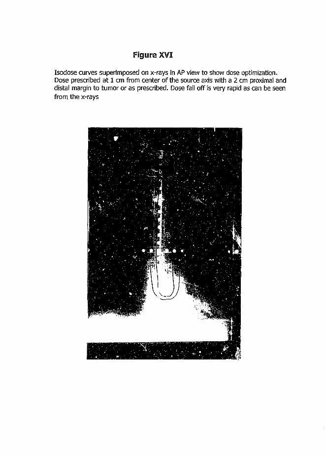
Figure XVI
Isodose curves superimposed on x-rays in AP view to show dose optimization. Dose prescribed at 1 cm from center of the source axis with a 2 cm proximal and distal margin to tumor or as prescribed. Dose fall off is very rapid as can be seen from the x-rays

Figure XVII
Isodose curves superimposed on x-rays in lateral view to show dose optimization. Dose prescribed at 1 cm from center of the source axis with a 2 cm proximal and distal margin to tumor or as prescribed.
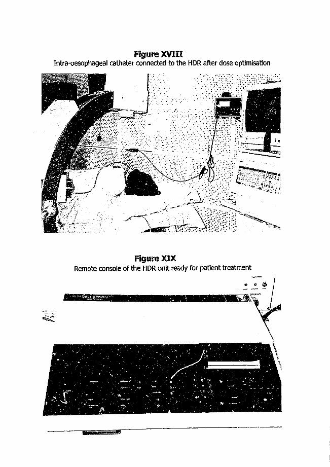
Figure XVIIIIntra-oesophageal catheter connected to the HDR after dose optimisation
Figure XIXRemote console of the HDR unit ready for patient treatment
10.6
SURGERY
Following completion of brachytherapy, all patients were operated upon within
2-3 weeks. A total oesophagectomy with gastric pull through was done in all
patients.
Rationale for Preoperative HDRILBT in oesophageal cancer
In South Africa, surgical resection is often difficult, even in CT stage II
oesophageal cancer, due to the long tumour length at the time of presentation.
(Sur R.K. et al., 1998). HDR ILBT allows the delivery of a very high dose to the
luminal aspect of the tumour, causing rapid shrinkage of the tumour, making
surgical resectability easier. The dose rate in HDR ILBT is high, at over 12 Gy per
hour instead of the conventional 2 Gy per hour. Furthermore, the dose per
fraction given is also high; 20 Gy in two fractions over 2 weeks, 10 Gy per
fraction against the conventional 1.8-2.0 Gy per fraction. Shrinkage of the
tumour thus occurs rapk ly. The dose fall off with HDR ILBT is rapid. At 1.5 cm
from the centre of the source axis, it is around 50% (dose received by lymph
nodes which are in proximity to the oesophagus), and at 2 cm from the centre of
the source, it falls by over 75% of the prescribed dose (Sur M. et al., 1996).
Therefore, there is no significant risk of injury to organs in close proximity, e.g.
heart, lung and spinal cord, in contrast to conventional EBRT treatment. It is
therefore, the policy of our surgical department to send all operable cases of

10.7
oesophageal cancer for preoperative HDRILBT and operate upon the patients in
2-3 weeks time to allow for rapid shrinkage of the tumour that makes surgical
resection easier and more complete.
MORPHOLOGICAL CHANGES IN OESOPHAGECTOMY SPECIMENS
The specimens were fixed in formalin for a period of 12-14 hours. The following
sections were then sampled:
1. Proximal' and distal resection margins
2. Edge of irradiated length (i.e., 4 cm proximaliy and distally to the tumour)
3. 1 cm from the margin of the visible tumour proximaliy and distally
4. Centre of the tumour
5. Palpable lymph nodes in proximity to the oesophagus were also sampled.
Paraffin embedded sections were processed and stained with hematoxylin and
eosin (H & E) and examined under light microscopy.
MDR-1 GENE PRODUCT - p-gEyccprotem
Paraffin wax embedded tissue blocks of formali fixed pretreatment biopsies and
blocks from the centre of tumour (from surgically resected specimens) following
brachytherapy were assessed for expression for p-glycoprotein. All sections were

10.8
immunostained for p-gtycoprotein using the modified sandwich technique.
Formalin fixed paraffin embedded sections were deparaffinized in Xylene and
rehydrated with graded Ethanol.
Prior to quenching, and application of primary antibody, sections were subjected
to heat induced epitope retrieval (HIER) in citrate buffer (using microwave
pretreatment) for antigen retrieval. A three step method for quenching
endogenous peroxide was used which is as follows:
1. The slides were treated with 20% methanol + 80% ethanol + 2%
Hydrogen peroxide for 10 minutes
2. The slides were then treated with 0.2% periodic acid + 3% Hydrogen
peroxide for 20 minutes
3. Lastly, the slides were treated with TBS pH 7.5 for 5 minutes
Normal rabbit serum was used to block nonspecific protein binding. Slides were
incubated with primary antibody to p-glycoprotein (clone JSB-1 from Novocastra
Labs Ltd., Newcastle upon Tyne, U.K.) at dilution 1:20, overnight at 4 degrees
centigrade. The primary complex was amplified using a modified sandwich
technique which included incubation in the following:
1. Rabbit anti-mouse IgG 1:20 + peroxide labeled rabbit anti-mouse 1:100
at 4 degrees centigrade for 45 minutes
10.9
2. Mouse PAP complex 1:50 dilution at 4 degrees centigrade for 45 minutes
3. Peroxidase labeled rabbit anti-mouse 1:100 dilution at 4 degrees
centigrade for 45 minutes
The complex was then detected with diaminobenzidine and counterstained in
Mayer's Hematoxilin for 30 seconds. The sections were then dehydrated, cleared
and mounted with entellan (Merck, Germany). Both negative and positive
controls were included in each run in order to determine if the technique was
optimal. Negative controls were processed in a similar manner except that the
primary antiserum was omitted. Positive controls comprised normal liver which
showed bile canaliculi staining in paraffin wax embedded blocks. All sections
were examined under light microscopy.
p53, bd-2 AND APOPTOSIS
Paraffin wax embedded tissue blocks of formalin fixed pretreatment biopsies and
blocks from the centre of the tumour (from surgically resected specimen)
following brachytherapy, were assessed for expression of p53, bcl-2 and
apoptosis.
p53 and bcl-2 immunodetection : All sections were immunostained for p53 and
bcl-2 using the Avidin-Biotin complex (ABC) method. Formalin fixed paraffin
10.10
embedded sections were deparaffinized in Xylene and rehydrated with graded
ethanol. Prior to quenching and application of primary antibody, sections were
subjected to heat induced epitope retrieval (HIER) in citrate buffer (using
microwave pretreatment) for antigen retrieval. Endogenous peroxide was
blocked with 3% Hydrogen peroxide for 30 minutes. After washing with PBS,
slides were incubated with normal goat serum 1:50 in PSA (PBS -f- 3% BSA) for
30 minutes. Sections were then incubated with p53 primary antiserum DO-7
(Dako, Denmark) 1:50 and with bcl-2 oncoprotein, clone 124 ( Dako, Denmark)
1 : 100 for 1 hour at room temperature (antibody DO-7 detects both wild type
and mutant p53). This was followed by respective incubation with biotinylated
goat antiserum and streptavidin HRP (Duet strep HRP kit, Dako, Denmark) for 30
minutes each. A diffuse brown signal was detected using diamino-benzidine
tetrahydrochloride (DAB) (Sigma Chemical Co., St.Louis, MO). Sections were
counterstained with Mayer's haematoxylin, dehydrated, cleared and mounted
with enteilan (Merck, Germany). Negative controls were processed in a similar
manner except that the primary anti sera was omitted. Positive controls
comprised known p53 protein positive breast carcinoma in paraffin wax
embedded blocks. For bcl-2, positive controls comprised known bcl-2 protein
positive follicular centre cell lymphoma in paraffin wax embedded blocks.
Apootosis detection : For detection of apoptosis at single cell level, based on
labeling of DNA strand breaks, In Situ Cell Death Detection Kit, POD (Boehringer
10.11
Mannheim, Germany) was used. Formalin fixed paraffin embedded parallel
ctions were deparaffinized in xylene and rehydrated with graded ethanol to
water. Sections were quenched in 0.3% HzOz for 30 minutes. Digestion was
performed using Proteinase K (Boehringer Mannheim) 20 ug / ml, for 15 minutes
at room temperature. Sections were rinsed in PBS and subjected to the Timel
reaction mixture. The DNA strand breaks can be identified by labeling free 3' -
OH termini with modified nucleotides in an enzymatic reaction. In this kit,
terminal deoxy nucleotidyl transferase (TdT) which catalyzes polymerization of
nucleotides to free 3' - OH DNA ends in a template - independent manner, is
used to label DNA strand breaks. Incorporated fluorescein is detected by
anti-fluorescein antibody FAB fragments and conjugated with horse radish
peroxidase (POD). After substrate reaction, stained cells can be analyzed under
light microscopy. Diamino-benzidine tetrahydrochloride DAB, (Sigma) was used
as chromagen. Sections were then counterstained with methyl green, cleared
and mounted in DPX. Negative and positive controls were used respectively. For
the negative control, the step using TdT was omitted and only distilled water was
applied. For the positive control, DNA strand breaks were induced using D'nase
1=0.001 gm / ml, PBS at room temperature for 10 minutes prior to labeling.
10.12
Expression of p53, bcl-2 and apoptosis in pre and post-brachytherapy tumour
sections were studied based on :
2. Intensity of brown signals - nuclear (p53) or cytoplasmic (bcl-2) or single
cell (apoptosis) was graded as negative, +, ++ or +++
2. Number of positive cells - type A < 10%, type B 10-50% and type C >
50%
3. Pattern o f distribution, whether focal or diffuse
All sections were examined under light microscopy.

11.1
Chapter 11
Review of literature
This review of literature deals with the morphology! changes induced by
radiotherapy in oesophageal cancer. It also reviews the effect of radiation on the
expression of tumour supressor genes (p53 and bcl-2) and apoptosis and the
effect of radiotherapy on the expression of p-glycoprotein, a product of MDR-1
gene.
11.2
Brachytherapy is a form of radiation treatment in which the radioactive isotope is
in and around the tumour. In the oesophagus this is done by placing the
catheter in the lumen of the oesophagus and subsequently passing a radioactive
isotope sue-, as Iridium -192 remotely by computers. The methodology involved
is described under Material and methods (Sur et al., 1998)
I. MORPHOLOGICAL CHANGES INDUCED BY RADIOTHERAPY IN
OESOPHAGEAL CARCINOMA
a. In vivo Studies
Radiation changes induced by brachytherapy have previously been reported.
Hishikawa et al (1984) demonstrated that a boost of High dose rate Intraluminal
Brachytherapy ( HDRILBT) following External Beam Radiotherapy (EBRT) caused
ulceration of the mucosa in all patients. These ulcers developed in the field of
ILBT. The ulcers were predominantly circumferential (12/22) and occurred 108
months following completion of ILBT. Linear ulcers were also seen (10/22) and
occurred 3-12 months following ILBT. All ulcers had a thick crater wall and
margins. Hishikawa e t al also noted that deep ulceration formation after EBRT,
but before ILBT, was usually associated with a tracheo-oesophageal fistula.
Based on this observation, Hishikawa et al recommended that ILBT should be
avoided in patients who develop deep ulceration following EBRT and that the
ILBT dose should not exceed 20 Gy when used with EBRT.
11.3
In a subsequent report, Hishikawa and others (1985) treated 53 patients with
oesophageal squamous cell carcinoma (SCC) with EBRT and HDR ILBT. EBRT
ranged from 40 -70 Gy and HDR ILBT ranged from 6-24 Gy in one to four
fractions. Autopsy was performed in 10 cases. Only 2 cases had good
microscopic specimens of the irradiated oesophagus. In the first case,
microscopic sections of the specimen taken at 1 cm from the superior margin of
the tumour showed a difference between the segment treated with EBRT alone
and that treated with combined radiation. The histological changes were most
serious in the segment that underwent intracavitary radiation. The major
epithelial injury was ulceration. Degenerative changes of the small vessels and
fibrinous exudates were found in the submucosa. In the second case the
mucosa and submucosa were preserved in the portion of external irradiation,
however, regenerative epithelium and fibrinous exudates of the sub mucosa
were recognized in the area of intraluminal radiation. Capillary vessels of the
submucosa had been destroyed, and fibrosis and fibrinous exudates were found
in the muscle layers in the portion of ILBT. Fibrosis of the muscle layers was
also found. Hishikawa and others therefore concluded that characteristic changes
were found in the oesophagus irradiated by ILBT following EBRT.
In a subsequent report, Hishikawa et al. (1988) described the autopsy findings in
35 cases treated with EBRT of various doses- alone, and in combination with
EBRT and ILBT. Residual tumour was seen a t autopsy in only 7 of 16 patients
treated with HDR ILBT following EBRT. In contrast, 13 of 14 patients treated
11.4
with EBRT of 50 Gy or more and all 5 patients treated with EBRT of less than 50
Gy had residual tumour. They showed that the addition of ILBT improved local
control in treatment of oesophageal cancer with radiation.
The use of ILBT alone in the treatment of small superficial SCC of the
oesophagus was subsequently reported by Hishikawa et al. in 1989. Six patients
with small, superficial carcinoma of the oesophagus were treated with 18-24 Gy
of HDR ILBT alone. Alterations in the oesophagus were examined with an
endoscope within one month after therapy, and it was observed that the tumour
had disappeared in all 6 patients. Erosion induced by HDR ILBT was seen in five
patients. Five of the patients experienced no local recurrence, and endoscopic
biopsy showed local recurrence in the remaining patient seven months after
radiotherapy. All patients survived 6-16 months. Oesophageal ulceration induced
by ILBT occurred in 3 of the 6 patients. The ulcers, however, healed on
conservative management. Thus HDR ILBT could be used in the treatment of
patients with small, superficial carcinoma of the oesophagus
Hishikawa's studies therefore demonstrated:
1. Severe radiation changes are induced by the addition of ILBT Mowing
EBRT in the radiation treatment of oesophageal cancer. This may improve
local control.
2. ILBT alone could be used to effectively to control local disease in
superficial carcinoma of the oesophagus.
11.5
Berry et a!. (1989) reported pathologic findings in 21 oesophagectomy specimens
from patients having pre-operative combined ILBT and EBRT. Eleven patients
received 15 G/ ILBT and 40 Gy EBRT (group 1) and 10 patients received 15 Gy
ILBT and 20-30 Gy EBRT (group 2). Effectiveness of radiotherapy was expressed
as the ratio between depth of radiation effect and depth of tumour invasion.
This was expressed as one of four levels:
Level 1 : not deeper than the muscularis mucosa;
Level 2 : involving but not deeper than sub mucosa;
Level 3 : involving but not deeper than muscularis propria;
Level 4 : involving peri oesophageal soft tissue.
The depth of radiation damage to tumour cells was comparable between the two
groups. Light microscopic evaluation included cell type, maximal depth in the
oesophageal wall reached by viable tumour cells, predominant depth reached by
tumour cells with radiation damage, and the presence and viability of lymph
node metastasis. Criteria for diagnosis of radiation damage to tumour included
tumour giant cells with pyknotic multilobated nuclei, rare and abnormal mitotic
figures and condensation or vacuolization of the cytoplasm. The ratio of depth
of radiation damage to depth of identifiable tumour was proportional to the local
effectiveness of the pre-operative treatment. When assessing depth of radiation
effect (numerator), the level at which all tumour cells showed evidence of

11.6
radiation damage was assigned. When assessing depth of recognizable tumour
(denominator), the level of the point of deepest invasion was assigned.
The microscopic appearance was one of surface erosion or shallow ulceration
with variable degrees of fibrous thickening of the oesophageal wall. There was
variable degree of surface epithelial denudation and replacement with cellular
granulation tissue. Variable degrees of mucosal atypia in areas adjacent to
surface erosion were present. This atypia was characterized by architectural
disorder, nuclear enlargement and hyperchromasia, and cellular enlargement.
These changes were believed to be radiation induced particularly when involving
mucosa of different type from the existing malignancy, although this could not
be proven. There was moderate to marked mural fibrosis which was responsible
for the gross mural thickening. There was no perforation. The depth of radiation
damage to tumour cells was comparable between groups 1 and 2. However,
only 1 of 11 patients in group 1 had involvement of peri oesophageal soft tissue
as compared to 8 of 10 in group 2. Similarly, although six patients in group 1
had no viable looking residual tumour cells in the oesophageal wall after
treatment, this was the case for only one patient in group 2. Lymph node
metastasis were identified in 12 patients: 4 in group 1 and 8 in group 2.
A ratio of 1 between radiation effect and depth of tumour invasion was present
in 6 patients receiving high dose EBRT and 1 patient receiving lower dose EBRT.
The study demonstrated that ILBT combined with EBRT gave good local tumour

11.7
control in the majority of patients. Higher doses of EBRT give a better radiation
effect in the deeper layers of the oesophageal wall. The ratio between depth of
radiation affect and tumour invasion provides a simple and objective approach to
the pathologic analysis of oesophagectomy specimens. ILBT is intended to
provide high doses to the luminal aspects of the tumour which maybe more
hypoxic and relatively radioresistant.
b. In vitro/ animal studies
Freund e t al. (1989) examined the tolerance of sound oesophageal mucosa to
HDR ILBT with doses of 6Gy and 12Gy in 15 pigs. No macroscopic or
microscopic alterations of the mucosa were found after 15 Gy. An application of
12 Gy produced severe side effects in the form of vascular occlusion due to
fibrosis of the intima, formation of fistulas, and perforation of oesophageal wall.
The authors recommended elaborate schemes for Dose Fractionation in HDR
ILBT of oesophageal cancer.
Soejima (1992) investigated the histopathological responses of the rabbit
oesophagus to HDR ILBT. Oesophageal ulceration was observed in the specimen
that received a dose of 15 Gy, 7-28 days after ILBT. Before the mucosal
changes were observed, edema and cell infiltration were found in the lamina
propria. Chronic injury such as necrosis and degeneration of epithelium, and
degeneration of the wall of blood vessels was seen at six-months. No marked
changes were found in specimens that received 5 and 10 Gy HDR ILBT. In
11.8
conclusion, it was strongly suggested that a single dose of HDR ILBT should be
less than 10 Gy to prevent the oesophagus from severe injury.
The pathologic sequence of events following a single large fraction of radiation
have been described by various authors. Phillips et at. (1974) reported the
following sequence of events following a single large traction of radiation. On
day 3, vacuolization and absence of mitosis of the basal layer along with the
thinning of the keratinized squamous cell layer occurred. Between days 7 and
14, foci of proliferating basal cells and regenerating epithelium with zones of
complete denudation occurred simultaneously. The rapidity of repopulation
determined the survival or death of the animal. After 21 days, there was usually
complete regeneration of the oesophageal lining with increased basal cell layer
proliferation and increased thickening of the squamous layer. Reports of acute
pathologic response of the oesophagus to radiation in the humans include those
of Seaman et al. (1957) and Mascarenhas et al. (1989). The acute pathologic
changes described parallel those observed in the mouse.
II. MDR-1 GENE PRODUCT - P glycoprotein
Darnton (1995), examined the effect of multidrug drug gene product-
p-glycoprotein in biopsy specimens from 27 oesophageal SCC and 10
adenocarcinomas before and after treatment with Mitomycin, Ifosphamide and
cis-Platin (MIC), p-glycoprotein was assessed immunohistochemically with
11.9
antibody JSB-1. Formalin fixed, paraffin wax embedded tissue sections of the
biopsy specimens and post treatment resected tumours were subjected to
immunohistochemical stains. Of the SCC, 74% (20/27) responded to MIC but
only one expressed p-glycoprotein before and after treatment. Of the
adenocarcinomas 30% (3/10) responded. Seven of the 10 adenocarcinomas
expressed p-glycoprotein before treatment. But all 10 were p-glycoprotein
positive after the chemotherapy. As the difference in prevalence and induction
of p-glycoprotein between the histologic types was highly significant, the authors
felt that this could correlate with the greater response to MIC chemotherapy
seen in squamous carcinomas compared with adenocarcinomas, p-glycoprotein
could not be used as the predictive marker of response as tumours expressed it
inconsistently. Resistance to MIC chemotherapy could be due to other
mechanisms.
Soini et al. (1996) investigated the immunohistochemical expression of the
MDR-1 gene product p-glycoprotein in histological samples in 31 hepatocellular
carcinomas. They also examined the correlation of expression of the protein
with cell proliferation, p53 expression, disease free interval and cumulative
patient survival. C219,CM-1 were used to detect expression of P-glycoprotein, by
means of the Avidin-Biotin peroxidase method. Membrane bound positivity for
p-glycoprotein was observed in 20/31 (65%) hepatocellular carcinomas. In
addition, there were no significant associations between expression of
p-glycoprotein and cell proliferation or p53 expression. Patients with
11.10
p-glycoprotein positive tumours had a shorter disease free interval and survival
time than those with p-glycoprotein negative tumours.
Chou and others (1995) reviewed the results of 29 children treated for
medulloblastoma. 13 patients with high grade medulloblastoma characterized by
incomplete resection, diploid tumour or subarachnoid dissemination received
chemotherapy following radiation therapy. Three patients received post
operative chemotherapy. 8 patients who had been treated with post-operative
radiation therapy also received Chemotherapy for recurrent tumours. A’ter a
minimum three-year follow up period, 16 were alive but 13 had died from
recurrent tumours. In order to evaluate the possible participation of
p-glycoprotein mediated multidrug resistance in medulloblastoma therapy and its
correlation with prognosis, archival specimens were examined by
immunohistochemistry utilizing three monoclonal antibodies against
p-glycoprotein and six cases by reverse-transcriptase polymerase chain reaction
(RT-PCR) utilizing MDR-1 specific primers. Sixteen patients (55%) had MDR-1
expression detected either by one of the three antibodies or RT-PCR. DNA ploidy
study was also performed on 18 specimens. Patient outcome was correlated
with extent of surgical resection, chemotherapy, DNA ploidy and MDR-1
expression. A statistically significant association was found between MDR-1
expression and outcome (p= 0.007). Among the patients who received
chemotherapy, positive MDR-1 expression significantly correlated with poor
11.11
outcome (p= 0.036). The results showed that p-glycoprotein mediated intrinsic
MDR-1 in medulioblastomas correlated with an adverse outcome.
Ng 1.0. et ai. (1998) investigated the effects of radiation on the expression of
p-glycoprotein in 56 patients with primary oral cancer. No patient received prior
or concurrent Chemotherapy. The patient cohort consisted of three groups.
Group 1: 20 patients with pre-radiation specimens only.
Group 2 :18 patients with both pre and post-radiation specimens.
Group 3 :18 patients with post-rsdiation specimens only,
p-glycoprotein expression was determined by immunohistochemistry with two
monoclonal antibodies, C 219 and C494. Amongst patients in groups 1 and 2,
only 1 (2.6%) and 2 (5.3%) patients had p-glycoprotein expression in their
tumours before treatment with C219 and C494 respectively. For group 2
patients, 66.7% and 72.2% had tumours that expressed p-glycoprotein with the
two antibodies respectively, only after and not prior to radiation. When patients
in groups 2 and 3 were combined, 63.9% and 72.2% had p-glycoprotein
expression with the two antibodies respectively after radiation, p-glycoprotein
m sr.y,7!on was significantly induced after radiation compared with expression
before treatment (pcO.OOl). The study showed that p-glycoprotein expression
was significantly induced by radiation in human oral cancers. This induction of
p-glycoprotein expression conferred multi-drug resistance to the cancer cells and
could affect the efficacy of subsequent or concurrent chemotherapy. This may
also explained the poor rate of response to chemotherapy among patients who
11.12
have previously received radiation. This was the first study in literature that
looked at the expression of the MDR-1 gene product after radiotherapy. Prior to
this, no studies have confirmed this observation at other sites or the same site.
Drug resistance remains a major problem in gastric carcinomas. To evaluate the
mechanisms involved in this resistance, Wallner J and others (1993) examined
the expression of the MDR-1 gene, in primary gastric carcinomas. MDR-1 RNA
levels of gastric carcinoma specimens were determined by Slot Blot analysis.
MDR-1 cDNA (Probe 5 A) was used for the hybridization. MDR-1 RNA was
detected in 41% of the gastric carcinomas, with high levels in 18% of the
specimens. MDR-1 gene expression was independent of patient age, tumour
localization, and lymph node involvement. However, MDR-1 RNA expression was
less frequent in locally advanced tumours and was absent in the primary tumours
of all six patients who had distant metastasis. The study showed a multi-drug
resistant cells were present in primary gastric carcinomas and this suggests that
multi-drug resistance might contribute to the clinical drug resistance of these
tumours.
The human p-glycoprotein gene family contains the MDR-1 and the MDR-3 gene.
The MDR-1 p-glycoprotein is over expressed in multi drug resistant tumour cells
and is believed to play a role in the elimination of certain cytotoxic drugs used in
the chemotherapy of cancer. The MDR-3 gene has not been found to be
amplified or over expressed in MDR cells. Chao C.C. I et al. (1991) studied the
11.13
effect of p-glycoprotein gene expression in human colon cancer cell line.
Gene-specific MDR-1 gene probes were developed for the detection of the gene
and the total m-RNA level. Southern and Northern hybridization analysis showed
that the MDR gene and the m-RNA levels were increased 30-40 fold in a MDR
human colon cancer cell line.
Benson M.C. et al. (1991) used a monoclonal antibody to detect the cell surface
P-glycoprotein product of the multi drug resistance gene (MDR-1) in the human
bladder. A total of 32 patients had 44 different specimens analyzed. The
samples consisted of 8 normal bladders, 21 transitional cell carcinomas, 1
mucinous adenocarcinomas, 3 pediatric bladder wall specimens and 10
nonmalignant samples from cystectomies, p-glycoprotein was not detected in the
normal adult or pediatric bladders. Transitional cell carcinomas demonstrated
low expression a t diagnosis (<6%), although 3 patients had enhanced initial
expression (11%, 12% and 31% respectively). Three patients treated with
chemotherapy demonstrated 56%, 76% and 50% expression of p-glycoprotein.
Nonmalignant tissue from cystectomy specimens had low expression of
p-glycoprotein. The specificity of this system was confirmed with human bladder
cell lines. The study also demonstrated the ability of flow cytometry to detect
and quantify the expression of the MDR-1 gene product. This may allow for the
early detection of chemotherapy resistance in patients with transitional cell
carcinoma treated with systemic and intravesical therapy.
X .r war .1 n
11.14
Sekiya S. et al. (1992) studied the effect of p-glycoprotein expression in
multi-drug resistant human ovarian carcinoma cel! lines. An increase in
p-glycoprotein level, specific to multi-drug resistant tumour cells was not
apparently associated with the increase in resistance to Vincrisitine or
Adriamycin. MDR-1 transcripts (4.5 kilobases) were observed in the RNA
preparations t >tained from only one cell line (Shin-3) that showed the highest
resistance to both drugs in vitro and in vivo. No cell lines showed MDR-1 DNA
amplification. These results suggested that the insensitivity of human ovarian
carcinoma to chemotherapy could be partially explained by the expression of
MDR-1 gene.
Glazer R.I. and Rohlff C. (1994) reviewed the development of cross resistance to
many natural product anticancer cancer drugs, termed multidrug resistance
(MDR) and attributed this to be a major reason for the failure of cancer
chemotherapy . This type of MDR is often associated with over expression of the
MDR-1 gene product, p-glycoprotein, a multifunctional drug transporter. The
expression of MDR in breast tumours is related to their origin from tissue that
constitutively expresses p-glycoprotein as well as to the development of
resistance during successive courses of chemotherapy. Therefore understanding
the mechanisms that regulate the transcriptional activation of MDR-1 may afford
a means of reducing or eliminating MDR. The authors found that MDR-1 gene
expression could be motivated by type I cAMP dependent protein kinases. This
opened up a possibility of modulating MDR by selectively down regulating the
11.15
activity of PKA dependent transcriptional factors which up regulate MDR-1
expression. High levels of type I PKA occurs in primary breast carcinomas and
patients exhibiting this phenotype showed decreased survival. This selective
type I cAMP dependent protein kinase inhibitors, 8-CI-cAMP and Rp8-CI-cAMP (S)
may be particularly useful for down regulating PKA dependent MDR associated
transcription factors. These compounds down regulate transient expression of
reporter genes under control of several MDR-1 promoter elements. Further
studies are needed to understand the mechanisms involved in the expression of
the multidrug resistance and also focus on policy intervention by a new class of
inhibitors.
III. p53, bcl-2 AND APOPTOSIS
(a) Effect o f chem o/rad io therapy on th e expression o f p53 in
oesophageal cancers
Puglisi e t al. (1996) investigated the immunohistochemical expression of p53 and
bcl-2 proteins in SCC of oesophagus to assess whether expression of these
oncoproteins could be used to stratify patients into groups with favorable or
unfavorable response to preoperative chemo/radiotherapy. The initial diagnostic
biopsy and the corresponding resected samples were obtained from 22
consecutive patients with SCC. All patients underwent preoperative
chemo/radiotherapy. Tumour sections were incubated with a monoclonal
antibody directed against p53 (DO-7). 24 non-neoplastic oesophageal biopsy
11.16
specimens immuno stained for p53 served as controls, 12 randomly chosen
sections from the 22 SCC samples were immunostained to test for bcl-2 protein
expression. After chemo-radiotherapy, 12 (55%) of the 22 patients had no
evidence of tumour in the resected oesophagus. Before chemo-radiotherapy,
however, 17 (77%) patients were p53 positive. After treatment, residual
carcinoma was detected in 7 (41%) of the 17 p53 positive patients. All
non-responsive cases had the same p53 immunopattern as before treatment
bcl-2 immunoexpression was detected in 6 (50%) of 12 patients. Residual
tumour was detected in the residual oesophagus in two (33%) of the six bcl-2
positive patients. After treatment, bcl-2 expression was no longer detected in
the residual neoplastic cells of a previously bcl-2 positive tumour. Using Fisher's
exact test no significant association was found between oncoprotein expression
and response to preoperative treatment. The study confirmed the observation
that p53 protein is frequently expressed in SCC of the oesophagus probably as a
result of a mutation of the TP53 gene. No significant association was found
between Once protein expression and response to chemo/radiotherapy.
Anticancer agents did not seem to modify the expression of p53 and bcl-2
proteins.
Resistance to chemotherapy remains a serious problem in the treatment of
gastric and oesophageal cancer. DNA-damaging agents alter levels of p53
proteins in several cell types and it has been speculated that regulation of p53
can be involved in the resistance or sensitivity of cancer cells to some
11.17
chemotherapeutic drugs, depending on whether cells have mutant /or wild type
p53; however, little is known about the relationship of p53 to drug sensitivity in
gastric/esophageal cancers. Nabeya Y. et al. (1995) examined human
gastric/esophageal adenocarcinoma cell lines for p53 mutational status,
chemosensitization to 5 FU, Mitomycin C and cis-Platin, and alteration in p53
levels following exposure of cells to these drugs. The mechanisms involved in
regulating p53 levels were also studied. Their results indicated that wild-type
p53 protein levels increased after treatment to each of these drugs through
either post-translational and/or translational mechanisms. This increase in
wild-type p53 appeared to be required for effective chemotherapeutic growth
control of gastric/esophageal adenocarcinoma cells. In contrast,
gastric/esophageal cancer cells expressing either mutated p53 protein or no p53
protein were resistant to the growth inhibitory effects of these drugs, despite the
fact that drug exposure can also increase mutant p53 levels by a translational
mechanism. Their data indicated that the mutational status of p53 was
predictive of chemosensitivity of gastric and oesophageal adenocarcinomas.
Muro K and others (1996) determined the clinical role of p53 mutations with
locally advanced oesophageal carcinoma treated with concurrent chemotherapy
by immunohistochemical analysis. 20 patients with previously untreated
oesophageal carcinomas with T4 disease and/ or distant node metastasis were
selected. Treatment comprised of a protracted 5FU and 2Hr cis-Platin effusions
along with radiation treatment with a total radiation dose of 60 Gy. Tumour

11.18
specimens from 18 of the 20 patients were analyzed histochemically. Mutant p53
protein expression was analyzed by immunohistochemical staining using a
polyclonal antibody, RSP53. Expression of p53 was detected
immunohistochemically in 10 of the 18 oesophageal tumours (56%), the cancer
cell nuclei of which were diffusely stained. There were no significant differences
between the patient backgrounds of the p53- "positive" and "negative" groups.
Four of the 10 patients (40%) with p53 expression achieved overall complete
remission and 7 of the 10 (70%) achieved complete remission of their primary
tumours. In contrast, none of the 8 p53 negative patients achieved overall
complete remission (CR) and two (25%) achieved complete remission of their
primary tumour. The overall CR rates in the primary tumours tended to be higher
in the p53 positive than negative groups, but the differences were not
significant. The survival rate for the 10 patients with p53 expression was better
than that for the 8 negative ones (p>0.01): the median survival times being 12
and 4.5 months respectively. The study concluded that the expression of p53
protein maybe an indicator of a favorable prognosis in patients with locally
advanced oesophageal carcinomas treated with concurrent chemoradiotherapy.
The p53 gene product is known to regulate cell growth and proliferation.
Whereas the wild type p53 protein suppresses cell growth, mutant p53 protein
act as an oncogene. Mutations in the p53 gene usually results in p53 protein
stabilization and accumulation; so that the gene product can be detected by
immunohistochemistry. Recently, the immunohistochemical detection of the p53

11.19
protein was associated with breast, colorectal cancer and other types of cancer.
However, its prognostic role in oesophageal cancer remains to be elucidated.
Sarbia M. and others (1994) examined p53 expression in formalin fixed, paraffin
embedded samples of 204 patients with SCC of the oesophagus, who underwent
oesophageal resection. DO-1, a monoclonal antibody that detects wild- type and
mutant form of p53 was used. The relationship between p53 immunoreactivity
and prognostic factors were determined by the chi-square test, and the
prognostic impact of p53 protein expression was analyzed using univariate and
multivariate survival analysis. In 204 tumours (67.2 percent), nuclear
immunoreactivity for p53 protein was detected. There was no correlation with
sex, age, pathologic tumour categories, pathologic lymph node category,
metastasis category, residual cancer category, histologic grade, or pre-operative
radiation therapy. In addition, p53 expression did not correlate with prognosis
using univariate and multivariate survival analysis. The study demonstrated that
p53 protein could be detected by immunohistochemistry in a high percentage of
SCC of the oesophagus. However, the overexpression of p53 gene product had
no impact on the prognosis.
Jung M. et al. (1992) examined alterations of the p53 gene in five of 6 human
SCC cell lines characterized as radiation sensitive (SQ-38, SCC-9,SQ-9G) or
radiation resistant (SQ-38, SCC-35, JSQ.-3). The point mutations and a deletion
were detected by using single stranded conformational polymorphism analysis
(SSCP) and polymerase chain reaction (PCR) data sequencing. Three of three
11.20
radiation sensitive and two of three radiation resistant cel! lines revealed
mutations in the p53 gene. Point mutations were located in exons 4, 6, and 8
(at codons 72 and 298 in JSQ-3; 273 in SCC-35; 196 in SQ-38), and deletion
consisted of 32 base pairs between codons 274 and 285 in SCC-9 and 1 base
pair at codon 271 in SQ-9G. Three mutations resulted in substitutions for an
arginine residue. Immunocytochemical analysis, confirmed p53 protein over
expression in SCC-35 cells which contain a missense mutation a t codon 273. In
contrast to previous studies which linked alterations in the ras, myc and raf
expression with radiation resistance, this study indicated that mutations in the
tumour supressor gene p53 did not correlate with such resistance.
Wild type p53 protein plays an important role in the cellular response to ionizing
radiation and other DNA damaging agents and is mutated in many human
tumours. Pomp J. e t al. (1998) evaluated the relationship of the
immunohistochemically determined p53 protein status and the disease control
with radiotherapy alone for carcinoma of the oesophagus. Immunostaining for
the p53 protein was performed on paraffin embedded specimens with 69
patients with adenocarcinoma and SCC of the oesophagus. All patients were
treated with radiotherapy consisting of combinations of EBRT and ILBT using
two different dose levels. 54% (37/69) of the tumours showed over expression
of the p53 protein. No difference in pretreatment parameters for p53 positive
and p53 negative cases was detected. In multivariate analysis p53 was
significantly associated with overall survival next to weight loss, tumour stage

11.21
and N stage. For metastasis free survival p53 status proved to be the sole
independent prognostic factor. The influence of p53 on local recurrence free
survival, however, was not as strong as on overall survival and metastasis free
survival. Immunohistochemically detected over-expression of mutated p53
protein in oesophagus carcinoma was an independent prognostic factor in a
group of patients treated with radiation therapy alone.
Coggi G. (1997) investigated the occurrence of p53 alterations in oesophageal
carcinoma, the correlation between the analysis a t the gene and protein level
and a prognostic significance. A series of 74 oesophageal carcinomas (46 SCC,
21 adenocarcinomas, 7 undifferentiated carcinomas) were studied by SSCP
analysis and immunohistochemistry to detect p53 mutations and accumulation,
respectively. p53 mutations in exons 5-8 were detected in 53% of the
carcinomas whereas p53 accumulation was observed in 57% of the cases.
Comparing SSCP and immunohistochemistry, there were 27 discordant (38%)
cases. Overall, only 20 tumours (27%) did not display p53 mutations and/or p53
accumulation. No associations were found between p53 aberrations and clinico
pathologic parameters which included patients age and gender, tumour type,
stage and grade. p53 protein accumulation and p53 gene mutation were not
related to patient survival by univariate or multivariate analysis in oesophageal
carcinomas. This study concluded that p53 aberrations were very common in
oesophageal carcinomas. However, p53 gene mutation and p53 protein
accumulation had a significant discordance suggesting that p53 function maybe

11.22
inactivated by mechanisms other than mutations. p53 aberrations did not
independently predict prognosis in oesophageal tumours.
(b) Effect of chemo/radiotherapy on the expression of bcl-2 in
oesophageal cancers
Sarbia M. e t al. (1996) examined bcl-2 expression by immunohistochemistry and
correlated it with prognosis in a series of 150 potentially curatively resected SCC
of the oesophagus. For comparison, bcl-2 protein expression was analyzed in
normal oesophageal mucosa, severe squamous dysplasia, and carcinoma in-situ.
bcl-2 immunoreactivity was found in 40 / 150 invasive SCC; the remaining
carcinomas were completely negative, bcl-2 protein expression was found more
frequently among poorly differentiated than among well differentiated tumours
(p<0.001). No correlation was found between bcl-2 protein expression and the
parameters tumour size, depth of invasion and nodal status, bcl-2 protein
expression had no significant influence on overall survival. Whereas in normal
mucosa bcl-2 immunoreactivity was restricted to the basal cell layer, in 9/15
severe squamous dysplasia and 7/14 carcinomas in-situ, bcl-2 staining was
detected in all epithelial layers. Therefore bcl-2 protein was found to be
frequently expressed in invasive SCC of the oesophagus and precursor lesions of
oesophageal cancer, but had no significant impact on the outcome of
oesophageal cancer.
11.23
Ohbu M. e t al. (1996) attempted to determine a correlation between bcl-2
expression and oesophageal SCC histopathology, lymph node metastasis, and
clinical variables, and assessed its applicability as a parameter for prognosis.
Immunohistochemical staining for bcl-2 (clone 124) was performed on archival
material from 105 oesophageal SCC's. bcl-2 was expressed in 58% of
oesophageal carcinomas, demonstrating positive correlations with a lack of
keratinization and early stage of disease. Lymph node metastasis was
significantly less frequently observed in the subset of early stage carcinomas with
bcl-2 staining. Univariate analysis revealed significantly longer disease free
survival in patients with bcl-2 positive carcinomas than in those which were bcl-2
negative, bcl-2 expression did not have independent prognostic value in a
multivariate survival analysis. The study showed that bcl-2 was frequently
expressed in oesophageal carcinomas and this expression was positively
associated with non-keratinization and an early stage, bcl-2 expression was
associated with reduced metastasis to the lymph nodes and may indicate a
favorable prognosis.
Goldblum J.R. and Rice T.W.(1995) undertook a study to evaluate the role of
bcl-2 in the Barrett's metaplasia- dysplasia- carcinoma sequence. 36 oesophageal
resection specimens were studied, using a monoclonal antibody to the bcl-2
protein on fixed paraffin embedded specimens. Barrett's mucosa was present in
each specimen: low grade dysplasia in 35, high-grade dysplasia in 34, intra
mucosal carcinoma in 23 and submucosal carcinoma in 13. In addition, a section

11.24
of the gastric resection margin was evaluated for bcl-2 immunoreactivity in each
case. In all cases, the regenerative compartment of the gastric mucosa in the
resection margins stained for bcl-2; however, no immunorescavity was seen in
any of the cases of Barrett's mucosa with or without dysplasia or carcinoma.
This study shows that although, bcl-2 protein is normally expressed in the
regenerative crypt compartment of the colon, small intestine and stomach and
has been found to be abnormally over expressed as an early event in the
dyplasia - carcinoma sequences of both ulcerative colitis-related and c i tr ic
neoplasia, bcl-2 alterations are not an important molecular marker in the
neoplastic progression of Barrett's mucosa.
Parenti A.R. et al. (1997) examined 64 cases of SCC of the oesophagus
coexisting with squamous intra-epithelial lesion (SIL) in 18 cases for any
overexpression of bcl-2 and p53 proteins. Any association of bcl-2 and p53
protein expression with patient survival was also analyzed, bcl-2 and p53
expression was immunohistochemically analyzed using clone 100 monoclonal
antibody and Pab 1801 monoclonal antibody respectively, bcl-2 positive immuno
staining decreased significantly during the progression of oesophageal
carcinogenesis. A decreasing frequency in the expression of bcl-2 in advanced
oesophageal SCC coincided with frequent p53 over expression, bcl-2 expression
was correlated with patient survival by univariate analysis. This association
disappeared after adjusting for tumour stage. p53 over expression showed no
association with patient survival by either univariate or multivariate analysis.
11.25
bd-2 positive immunostaining was always detected in the basal layer of the
normal epithelium adjacent to the pre-cancerous lesion or oesophageal SCC in
the squamous intra-epithelial lesions, bcl-2 positive cells were detected
throughout the thickness of the dysplastic squamous epithelium whereas in the
infiltrative cancer component, bcl-2 over expression was detected in scattered
clusters of neoplastic -pithelia located in the submucosa and/ or in the
muscularis propria. A bcl-2 positive immuno reaction was detected in 100% of
low and high-grade SIL, as well as in the pTl cancers. pT2, pT3 and pT4
oesophageal SCC showed positive immunostaining 20%, 23% and 20% of cases
respectively. A significant inverse correlation was detected between bcl-2
expression and pathological stage of the cancer, the bcl-2 immunoreactivity
being detected more frequently in the early (SIL and pTl) than in the advanced
(pT2-4) stages of the neoplastic disease ( Spaearmans rank correlation, p<0.01).
p53 positive immunostaining was focally detected in the basal layer of the
normal epithelium adjacent to SIL lesions or oesophageal SCC. In the SIL areas,
p53 expression was detected throughout the thickness of the dysplastic
epithelium. In the infiltrative component p53 over expression was detected in
50%, 50%, 60%, 54% and 80% of SILs, pTl, pT2, pT3 and pT4 neoplastic
lesions respectively. No significant correlation was observed between p53
overexpression and the local invasiveness of the tumour. T? e study showed that
down-regulation of bcl-2 and up-regulation of p53 in advanced oesophageal SCC
suggested a role for bcl-2 and p53 proteins interaction in the progression of
oesophageal SCC.

11.26
(c) Roie of Apoptosis in treated oesophageal cancers
Graeber T.G. (1996) reported that hypoxia induced apoptosis in oncogenically
transformed cells. However, genetic alterations, such as loss of p53 human
supressor gene or over expression of the apoptosis inhibitor protein bcl-2,
substantially reduced hypoxia induced cell death. Highly apoptotic regions
strongly correlated with hypoxic regions in transplanted tumours expressing wild
type p53 whereas little apoptosis occurred in hypoxic regions of p53 deficient
tumours. Tne authors proposed that hypoxia provided a physiological selective
pressure in tumours for the expansion of variants that had lost their apoptotic
potential, and in particular in cells acquiring p53 mutations. Kinzler K.W. and
Vogelstein B (1996) reported that the low levels of oxygen found in those parts
of tumours with poor blood supply resulted in p53 dependent programmed cell
death. Mutations of p53 reduced the cell death, giving the mutant cells a survival
advantage over cells with intact p53. As a tumour grows to a critical size, the
central portion suffers from hypoxia. This induces expression of the p53 gene
which then leads to growth arrest or apoptosis. Cells that have acquired p53
gene mutation can escape the growth arrest or apoptosis. These clones will
eventually outgrow the rest to become the dominant clone. Therefore,
emergence of clones with p53 gene mutation may be driven by hypoxia.
Moreira L.F. et al. (1995) examined apoptosis immunohistochemicaliy using the
Bml Mab in 55 samples oesophageal SCC. Patients not treated (group I, n=12),
preoperatively treated by Chemotherapy (group II, n = ll) , radiation (group III, n

11.27
= 13) or both (group IV, n = 8), and 11 additional cases of high grade dysplasia-
early cancer were examined. Most of the apoptotic cells were BM1 positive, and
when checked by Tunel, also proved to be positive. They accounted for 7 (11%),
19 (29%), 21 (32%), 26 (38%) cells per field in those groups respectively.
Chemotherapy and/or radiation significantly increased the number of apoptotic
cells as compared to controls (p = 0.029 and p = 0.029 respectively). Additional
sections stained with bcl-2 and p53 were negative for bcl-2 and were positive for
p53 in 16 samples (37%). Overall, positive cases for p53 mutations showed a
significantly decreased incidence of apoptotic cells (p=0.03). These results
suggested that in-situ assessment of apoptotic response correlated better to the
apoptosis induced by radiation than that by chemotherapy. Further,
abnormalities of the p53 protein decreased the apoptotic response in
oesophageal carcinoma and the former may help to determine the sensitivity of
these anti-cancer agents.
Ohbu M. et al. (1995) assessed apoptotic index, the Ki-67 proliferative index and
overexpression of p53 protein in 69 oesophageal 5CC surgically resected from
Japanese patients. Apoptosis was examined by Gabrieli's method in histological
sections, and proved to be significantly related to keratinization and oesophageal
SCC progression. Overall labeling indices were 15.68 + 4.04 (positive/ 1000
uclei) and 6.79 ± 0.64 respectively, in keratinizing and non-keratinizing types.
The ripoptosis labeling index increased especially in keratinizing lesions, from
4.50 ± 0.59 with cancer invasion to mucosa through 11.46+ 2.70 with

11.28
involvement of the submucosa up to 21.18 ± 3.72 in cases of penetration to the
muscularis propria or adventitia. The relationship between apoptosis, Ki-67
scores and p53 expression was determined in identical cancer nests on serial
sections. An inverse correlation was shown between the apoptotic score and the
Ki-67 score in both keratinizing and non-keratinizing types. There was no
significant correlation between apoptosis score and p53 expression, either overall
or separately in keratinizing or non-keratinizing types of oesophageal SCC. The
study suggests that a mechanism of induction of apoptosis similar to that
operating in normal epidermis may be implicated in keratinizing oesophageal
SCC, and that as tumour volume increases, single cell death becomes more
frequent.
Hamada M. et al. (1996) examined formalin fixed, paraffin wax embedded
sections taken from 25 patients who underwent oesophagectomy for SCC. 14
patients had no preoperative therapy (control group), while the other 11 patients
received preoperative radiotherapy (radiation group). There was no difference in
pathological TNM classification between the two groups. These sections were
examined by immunostaining with monoclonal antibody PAblSOl to determine
the accumulation of p53 protein. Apoptotic frequency was determined by Tdt
mediated dUTP-biotin nick end- labeling (Tunel). In the control group, well to
moderately differentiated cases showed a significantly higher apoptotic index
(which was the number of apoptotic cells amongst 1,000 cancer cells), 51.7
+/-S3.4. In the poorly differentiated cases, apoptotic index was 1.3 +/-1.0

11.29
(p<0.05). Similar results were obtained in the radiation group. The group that
received no pre-operative therapy (control group) included 4 cases of p53 grade
4 (p53 protein detected in over 70% of the tumour cells). The group that
received pre-operative radiotherapy (radiation group) included 2 cases of p53
grade 4. Few apoptotic cells were observed in any of six tumour tissues. Tumour
cells with accumulated p53 protein rarely showed apoptosis. In contrast,
apoptosis was observed in tumour cells without p53 protein accumulation.
Spontaneous apoptosis in oesophageal SCC can be induced more easily in
differentiated than in poorly differentiated cases. This tendency may be
enhanced by preoperative radiotherapy. Extensive p53 protein may suppress
apoptotic induction in oesophageal SCC.
Shears L.L. et ak (1998) examined whether residual tumour cells in
oesophagectomy specimens following induction chemo-radiotherapy were viable,
in order to provide putative evidence for appropriateness of oesophagectomy.
Forty-six patients were entered into an induction chemoradiotherapy trial
consisting of 5FU, cisplatin, alpha-interferon and concurrent EBRT followed by
oesophagectomy. Response was determined histologically and apoptosis
assessed with Tunel method. p53 status was determined by
immunohistochemistry and mutational analysis. 38 patients underwent
oesophagectomy, 33 of whom had either a complete (n=10) or partial (n=23)
response. None of the 28 patients with residual tumour in the resected
specimen had'100% apoptotic cells and a vast majority of specimens had <10%
11.30
apoptotic rate. The percentage of apoptotic cells correlated with tumour
differentiation but not with the presence of p53 mutations. Tissue sections
following upfront chemo-radiotherapy was a necessary component of a
multi-modality approach to oesophageal cancer. This would ultimately provide
superior local regional control to a non-surgical approach.
Katada N. et al. (1997) evaluated the alteration of apoptosis in the oesophageal
epithelium in the oesophagitis- Barrett's oesophagus- adenocarcinoma sequence.
Archival tissue samples of 85 lesions in 58 cases were used. The lesions
represented seven groups: normal oesophagus (n=10), reflux oesophagitis
(n=12), Barrett metaplasia (n=21), Barrett low grade dysplasia (n=17), Barrett
high-grade dysplasia (n=5), well or moderately differentiated adenocarcinoma
(n=10), and poorly differentiated adenocarcinoma (n=10). Apoptotic cells with
fragmented DNA were detected by Tunel method. Monoclonal antibodies against
bcl-2 protein were applied using the avidin-biotin complex immuno peroxidase
method. The oesophagitis group showed many apoptotic cells on the epithelial
surface; whilst few apoptotic cells were seen in the other groups. Weak bcl-2
expression was seen in the basal cells in normal subjects and those with
oesophagitis. There was over expression of bcl-2 in 72% of Barrett's metaplasia,
100% of Barrett's low grade dysplasia, 25% of Barrett's high-grade dysplasia,
40% of were well differentiated or moderately differentiated adenocarcinoma,
and 20% of poorly differentiated adenocarcinoma. The authors concluded that
increased apoptosis in reflux oesophagitis may be a protective mechanism
11.31
counteracting increased proliferation. Inhibition of apoptosis by over expression
of bcl-2 protein occurred early in the dysplasia- carcinoma sequence of Barrett's
oesophagus. The resulting prolongation of cell survival could promote neoplastic
progression. Despite the absence of apoptosis, bcl-2 was not widely over
expressed in Barrett's high-grade dysplasia and adenocarcinoma suggesting that
cells acquire other ways of avoiding apoptosis as malignancy appears. The
findings of this study are similar to those prev iously reported by Goldblum J.R.
and Rice T.W.(1995) who also observed that bcl-2 alterations were not an
important molecular marker in the neoplastic progression of Barret's mucosa.
Ohashi K. e t al. (1997) assessed p53 protein accumulation, apoptosis, and
proliferating activity (Ki-67 labeling index) using oesophageal SCC samples of
both intramucosa and advanced types. The relationship of these values to
progression or differentiation grade of tumours was also analyzed. In terms of
proliferating activity and the proportion of positive cases with p53 accumulation,
a statistically significant difference was demonstrated between intra epithelial
carcinomas and intramucosal carcinomas with stromal invasion (17.2% vs.
31.7% for the Ki-67 labeling index, and 23.5% vs. 67.4% for the proportion of
positive cases of p53 accumulation). Values for the latter were almost
comparable to those of advanced carcinoma. Immunohistologically, Ki-67
positive, proliferating cells were distributed preferentially in the peripheral fronts
of invasion. Apoptotic cells were observed in the areas of the invasion of the
intramucosa carcinomas with stromal invasion and in more advanced lesions, but
11.32
were rarely observed in the normal epithelium or intraepithelial carcinomas.
Apoptotic cells were seen mainly around areas of keratinization, and the
apoptotic cell index was higher in well and moderately differentiated types of
advanced carcinomas than in poorly differentiated type (2.59% vs. 1.09%). The
study showed that an increase in proliferative activity and an accumulation of
p53 protein was associated with the onset of early carcinomatous invasion, while
apoptosis is closely linked with the differentiation grade of carcinoma cells.

12.1
Chapter 12
Results

12.2
MORPHOLOGICAL CHANGES
MACROSCOPIC EXAMINATION :
In all patients, the entire oesophagus was resected. Tumour length ranged from
25-55 mm in length and 10-20 mm in width. Six patients had an ulcerative
tumour whilst 4 patients had circumferential iumoui.j. The patient clinical data is
shown in Table I.
LIGHT MICROSCOPY
Resection margins; In the 7 patients with tumour in the middle-third of the
oesophagus, only the proximal resection margin showed characteristic radiation
changes. The proximal resection margin was located 5.5, 5.0, 5.5., 5.0, 4.5, 4.5,
and 5.5. cm, respectively, from the edge of the visible tumour, in the 7 patients.
Similarly, changes were seen in 3 patients with tumour in the lower third, with
radiation changes a ' / in the distal resection margin. The distal resection margin
was located 5.0, 5.0, and 5.5 cm from the edge of the visible tumour in these 3
patients. Radiation changes were limited to the submucosa with sparing of the
muscularis propria and adventitia. There was no ulceration of the squamous
mucosa. The lamina propria showed collections of lymphocytes and plasma cells.
The muscularis mucosae and the submucosa showed minimal fibrosis. There
were no morphological alterations in the blood vessels. These features are
demonstrated in Figures XX, XXI.
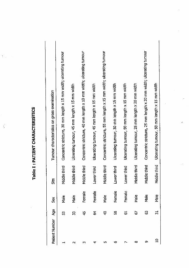
Tabl
e I
: PAT
IENT
CH
ARAC
TERI
STIC
S
gC
I8
§s
I1sE
I&
<D
<
S.
30E3cn
13
I5EEinv4X
II0 coE
1U
Iu
OJro
TDiEEin1-4x
IEE
3OE5CDC
s
%
8
S3
iIEE01—IX
%SEE?E
1U
!I E E5 3 3<D (U <u
g ■a■as S
0)fDESi
$
T33EEint H
x
IEE5i_r3OE3cnc
s
"Es
IjyraE
.OJ
8E3g
y3
|3EEinvHx%sEEsEti
u
as"E6
%
ID
■a3£Eint H
x
IoCO
8E3g
y
iI
i3EEint H
X
!EEg3
1g
y
1
<u
IID
EEsx
I£EinCM
8£3g
y
E5OJ
5
to
8E3cnc
y
cnsEEinC\l
Eti
o
§S
■a*a
2
i3EEintH
X
1EEg
oE3O)c
_a>•a■a
0) (U tt)ID ID IDs : z z
OJ vo co
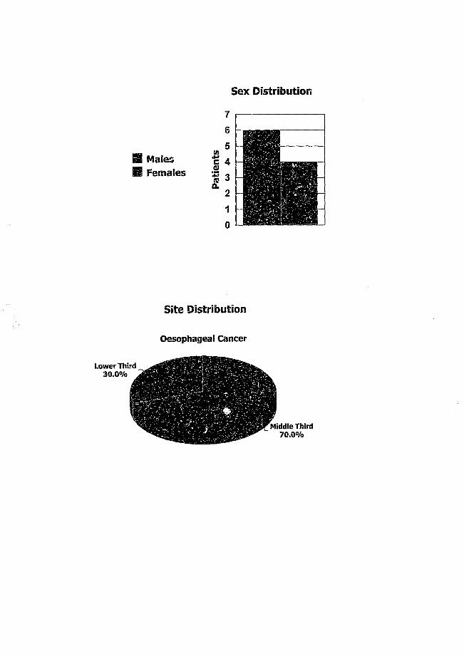
Sex Distribution
Site Distribution
Oesophageal Cancer
Lower Third 3 0 .0% ,
Middle Third 70 .0%
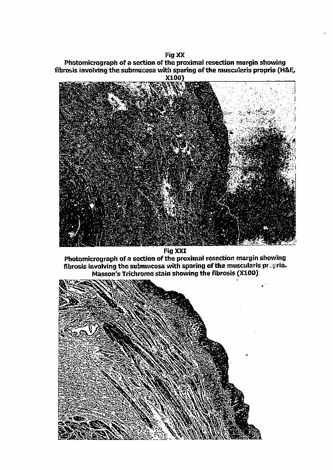
Fig XXPhotomicrograph of a section of the proximal resection margin showing
fibrosis involving the submucosa with sparing of the muscuiaris propria (H&E,X1Q0) _______________
Fig XXIPhotomicrograph of a section of the proximal resection margin showing fibrosis involving the submucosa with sparing of the muscuiaris propria.
Masson's Trichrome stain showing the fibrosis (X100)
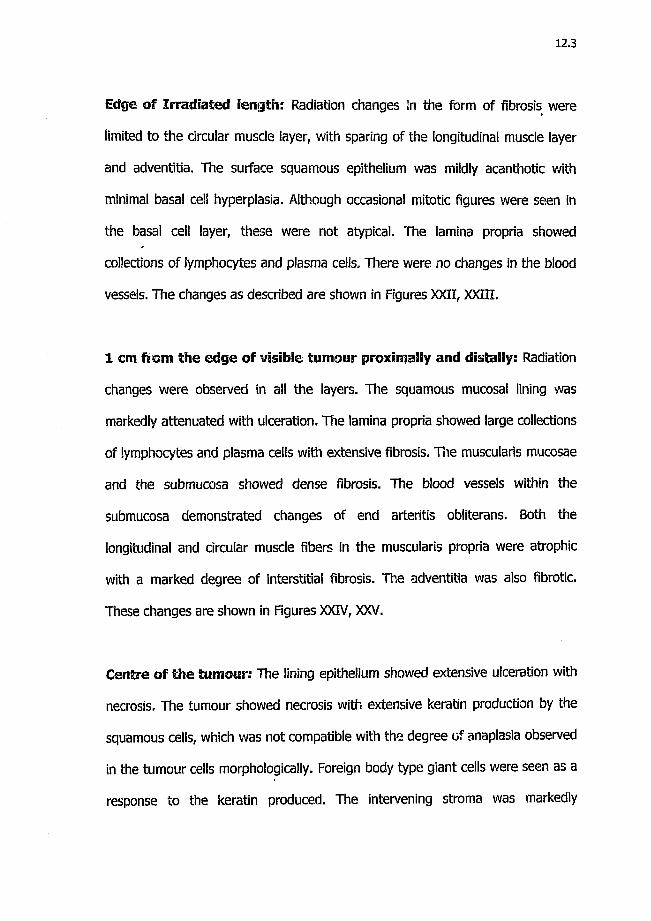
12.3
Edge of Irradiated length: Radiation changes in the form of fibrosis were
limited to the circular muscle layer, with sparing of the longitudinal muscle layer
and adventitia. The surface squamous epithelium was mildly acanthotic with
minimal basal cell hyperplasia. Although occasional mitotic figures were seen in
the basal cell layer, these were not atypical. The lamina propria showed
collections of lymphocytes and plasma cells. There were no changes in the blood
vessels. The changes as described are shown in Figures XXII, XXIII.
1 cm ftom the edge of visible tumour proximally and distally: Radiation
changes were observed in all the layers. The squamous mucosal lining was
markedly attenuated with ulceration. The lamina propria showed large collections
of lymphocytes and plasma cells with extensive fibrosis. The muscularis mucosae
and the submucosa showed dense fibrosis. The blood vessels within the
submucosa demonstrated changes of end arteritis obliterans. Both the
longitudinal and circular muscle fibers in the muscularis propria were atrophic
with a marked degree of interstitial fibrosis. The adventitia was also fibrotic.
These changes are shown in Figures XXIV, XXV.
Centre of the tumour: The lining epithelium showed extensive ulceration with
necrosis. The tumour showed necrosis with extensive keratin production by the
squamous cells, which was not compatible with the degree of anaplasia observed
in the tumour cells morphologically. Foreign body type giant cells were seen as a
response to the keratin produced. The intervening stroma was markedly

Fig XXIIPhotomicrograph «t'a section of the edge of irradiated length demonstrating acanthotic epithelium and basal cell hyperplasia with fibrosis limited to the
circular muscle layer (H&E, X200)
Fig XXIIIPhotomicrograph of a section of the edge of irradiated length demonstrating
acanthotic epithelium and basal cell hyperplasia (H&E, X400)
d
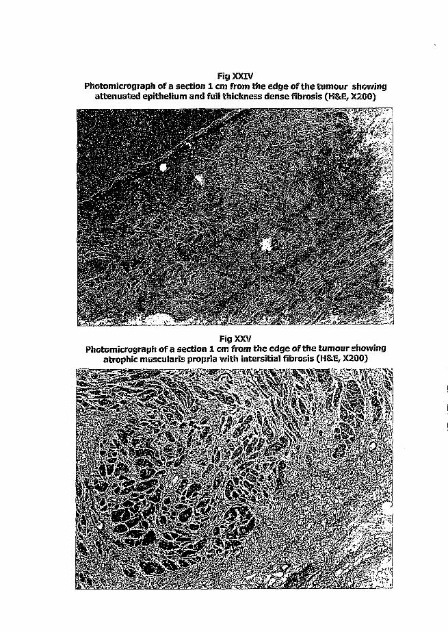
Fig XXIVPhotomicrograph of a section 1 cm from the edge of the tumour showing
attenuated epithelium and full thickness dense fibrosis (H&E, X200)
Fig XXVPhotomicrograph of a section 1 cm from the edge of the tumour showing
atrophic muscularis propria with intersitial fibrosis (H&E, X200)
12.4
desmoplastic, with a keloid type of collagenization that extended to the serosa.
The blood vessels in this area also showed changes of endarteritis obliterans.
These are shown in Figures XXVI, XXVII, XXVIII, XXIX.
Lymph Nodes: A cluster of lymph nodes with metastatic tumour was present in
3 patients with a tumour in the mid one-third (one patient) and lower third (two
patients). The lymph nodes were located in the adventitial tissue around the
oesophagus, 2mm away from the oesophageal wall. The entire lymph node
parenchyma was replaced by tumour with a rim of normal lymphoid tissue at the
periphery. Similar radiation changes present in the tumour in the oesophagus
were also noted in the lymph node (Figures XXX, XXXI).
All changes described above have been shown in Table II.
EFFECT ON EXPRESSION OF MRD-1 GENE PRODUCT: a-qivcoprotein
In all 10 cases, p-glycoprotein was not expressed in either pre-brachtherapy
(Figure XXXII) nor post-brachytherapy (Figure XXXIII) tumour tissue specimens
(after brachytherapy of 20 Gy). Figure XXXIV shows a positive control of
p-glycoprotein staining in normal liver bile canaliculi.

Fig XXVIPhotomicrograph of a section from the centre of the tumour demonstrating profuse keratin production by anapiastic squamous ceils with foreign body
giant cell reaction (H&E, X400)
m m
Fig XXVIIPhotomicrograph of a section from the centre of the tumour showing a keloid
type of desmoplastic fibrosis of the stroma. (H&E, X200)

Fig XXVIIIPhotomicrograph of a section from the centre of the tumour showing changes
of endarteritis obliterans (H&E, X400)
Fig XXIXPhotomicrograph of a 1 cm from the edge of the tumour showing atrophic
muscularis [propria with interstitial fibrosis (H&E, X200)

Fig XXXPhotomicrograph of a section from a lymph node with metastatic squamous ceil carcinoma showing profuse keratin production and dense desmoplastic
fibrosis in the lymph node (H&E, X100)
880Fig XXXI
Photomicrograph of a section from a lymph node with metastatic squamous cell carcinoma showing profuse keratin production and dense desmoplastic fibrosis in the lymph node. Masson's Trichrome stain showing the fibrosis
(X100)
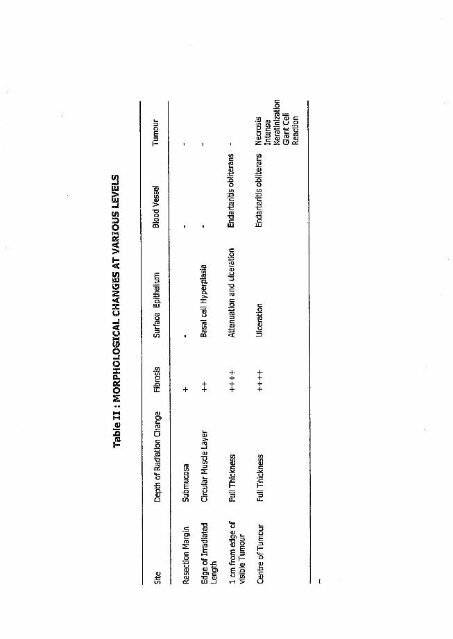
1 t i i f iQ) ro
iO I
I
■8tn
roT3
SeS-8
I0
y
1u-j
ssoz&o
I
&
-Ea
s
I1
++
Ig
s
++++
++++
«J2|2
(U
g6
. 51%
I
I0)
3
5i -
I
.Ee?S
i
I13
S 3
O
I !E 5u "EntH >
1H
^ 6<u
3

FlgXXXnPhotomicrograph showing preradiation biopsy of oesophageal squamous cell
carcinoma. No MDR-1 gene expression (X200)
Fig XXXIIIPhotomicrograph showing post brachytherapy section of oesophageal
squamous cell carcinoma. No MDR-1 gene expression (X200)
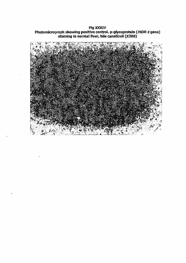
Fig XXXIVPhotomicrograph showing positive control, p-glycoprotein (MDR-1 gene)
staining in normal liver, bile canaliculi (X200)

12.5
EFFECT ON EXPRESSION OF p53f bcl-2 AND APOPTOSIS
The pre and post-brachytherapy findings for expression of p53 are shown in
Table III. There was no expression of p53 in one patient (no.l) in both pre and
post-brachytherapy specimens (group 1) . In 8 patients (group 2), p53 staining
was strongly positive (3+) with approximately 50% or more cells, and with
diffuse and no specific pattern in the pre-brachytherapy biopsies (Figure XXXV).
The post-brachytherapy specimens of group 2 showed strong 3+ positivity with
p53 (10-50% positive cell count) with the pattern being focal and peripheral
(Figure XXXVIa) in the tumour islands. The centre uf the tumour islands showed
necrosis (Figure XXXVIb) and/or keratinization (Figure XXXVIc) (group 2). In one
patient (case 3), the pre-brachytherapy biopsy showed expression of p53 (Figure
XXXVII), whilst the post-brachytherapy specimen was negative (group 3) (Figure
XXXVIII), (Table IV).
bcl-2 expression in both pre and post-brachytherapy is shown in table V. bcl-2
staining was equivocal and inconclusive in both the pre and post-brachytherapy
specimens, in that specific patterns of bcl-2 expression could not be established.
Apoptosis was negative in all the pre (Figure XXXIX) and post-brachytherapy
(Figure XXXX) tissue sections in the presence of positive control (Figure XXXXI).
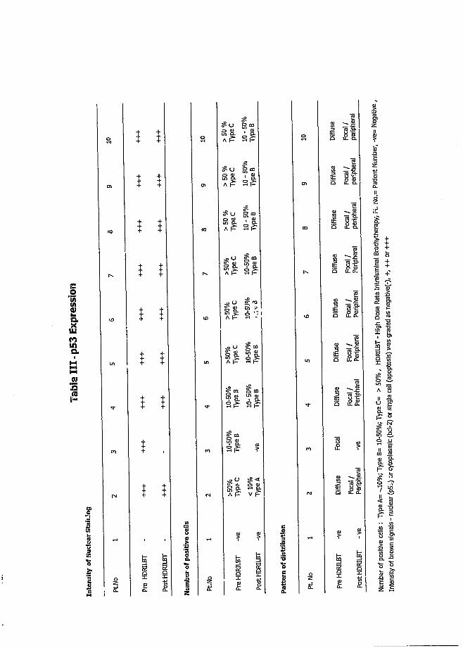
<w3
i
i
t+
£
I
I $
Ii
s > 50
%
Typ
eC
10 -
50%
T
ypeB
S
S
1 I t
> 50
%
Typ
eC
10 -
50%
T
ypeB
Diff
use
Foca
l /
perip
hera
i
> 50
%
Typ
eC
10 -
50%
T
ypeB
00
E
1 It
r>. >50%
Typ
eC
10-5
0%T
ypeB
P's Diff
use
Foca
i/Pe
riphe
rai
VO >50
%T
ypeC
10-5
'.!%
,4^
3
lO Diff
use
Foca
l/pe
riphe
ral
i n >50%
Typ
eC
10-5
0%T
ypeB
m
E
l i t
10-5
0%T
ypeB
10-
50%
T
ypeB
Diff
use
Foca
l/
Perip
hera
l
m 10-5
0%T
ypeB
-ve
m Foca
l
-ve
N >50%
Typ
eC
< 1
0%
Type
A
Diff
use
Foca
l/Pe
riphe
ral
tH
1 Pre
HDRI
LBT
-ve
PostH
DRIL
BT
-ve 1
|0
1 Pt No
1
Pre
HDRI
LBT
-ve
PostH
DRI
LBT
-ve
I
u
I I
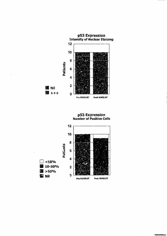
Nil+ + +
p53 ExpressionIntensity of Nuclear Staining
P r e HDRILBT P o s t HDRILBT
p53 Expression Number of Positive Cells
n < 10%H 10-50% M >50% M Nil
P r s HDRILBT P o s t HDRILBT

p53 ExpressionPattern o f Distribution
H Focal H Diffuse IB Nil
P re HDRILBT P o s t HDRXLBT
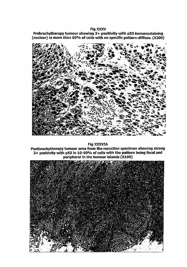
Fig XXXVPrebrachytherapy tumour showing 3+ positivity with p53 immunostaining
(nuclear) in more than 50% of cells with no specific pattern-diffuse. (X200)
m s s
Fig XXXVIAPostbrachytherapy tumour area from the resection specimen showing strong
3+ positivity with p53 in 10-50% of cells with the pattern being focal and peripheral in the tumour islands (X100)
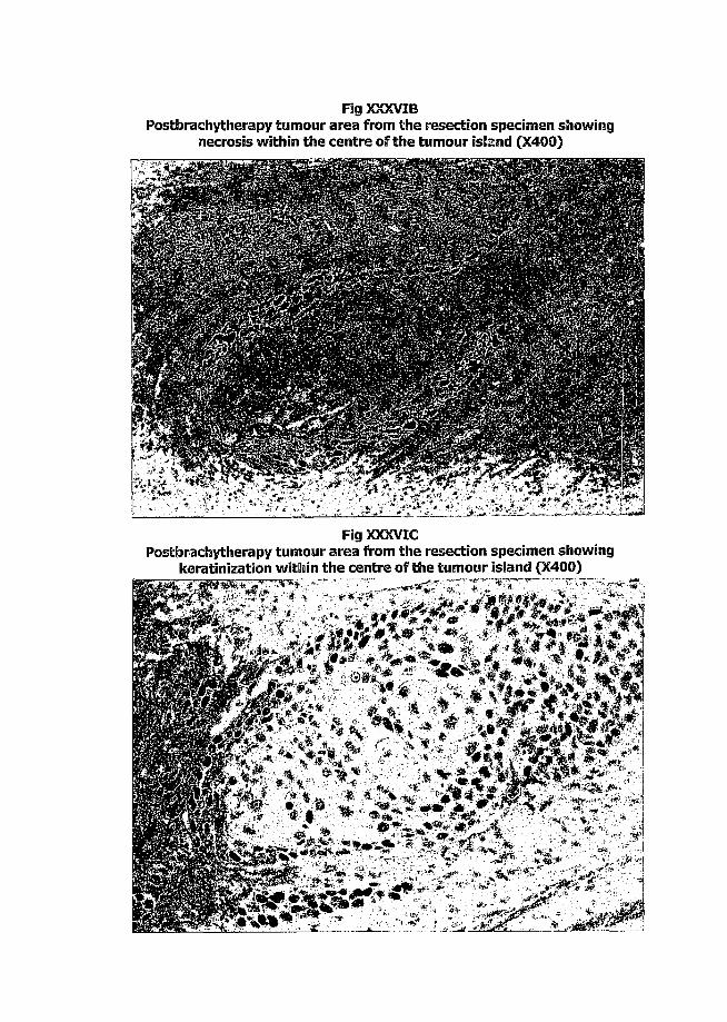
Fig XXXVIBPostbrachytherapy tumour area from the resection specimen showing
necrosis within the centre of the tumour island (X400)
■ 1
Fig XXXVICPostbrachytherapy tumour area from the resection specimen showing
keratinization within the centre of the tumour island (X400)
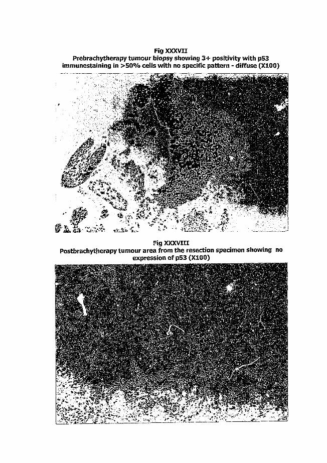
Fig XXXVIIPrebrachytherapy tumour biopsy showing 3+ positivity with p53
immunostaining in >50% cells with no specific pattern - diffuse (X100)
/W-UF. •yX-
Fig XXXVIIIPostbrachytherapy tumour area from the resection specimen showing no
expression of p53 (X100)

Tabi
e IV
: Pa
ttern
s of
p53
Expr
essio
n pre
an
d po
st br
achy
ther
apy
I
a
1Oc
£
1II£
i lIICTI1 1
IfD> <U w
III§ t i
■F.E 8
°CD " 5
1 rot jd ra
J D
K wCL CL
in
§
Ioz
a:
Ira£KCL
I
2
1
CD
•sis t 00
I (N m

i
i<N
i>
I
gx
iIts
00
r*>
VO
in
m
<N
c
0)>£tilQaooSi oEs £2
O'* CQ CO
S.
I S
II
II
n
gx
o
cn
CO
rs
VD
m
tr
m
IN
cow35£,52XoE oa z
% siCL
n<2 - =
£
I
Iti
Num
ber
of po
sitiv
e ce
ils
: Ty
pe
A=
<10%
; Ty
pe
B= 10
-50%
; Ty
pe
C= >
50%
, HD
FULB
T -
High
Do
se
Rate
Intra
lum
inal
Bra
chyt
hera
py,
PL No
.= Pa
tient
N
umbe
r, -ve
= N
egat
ive,
In
tens
ity
of br
own
signa
ls -
nucl
ear
(p53
) or
cyto
plas
mic
(b
d-2)
or
singl
e ce
ll (a
popt
osis
) wa
s gr
aded
as
nega
tive(
-),
+, +
+ or
++
+
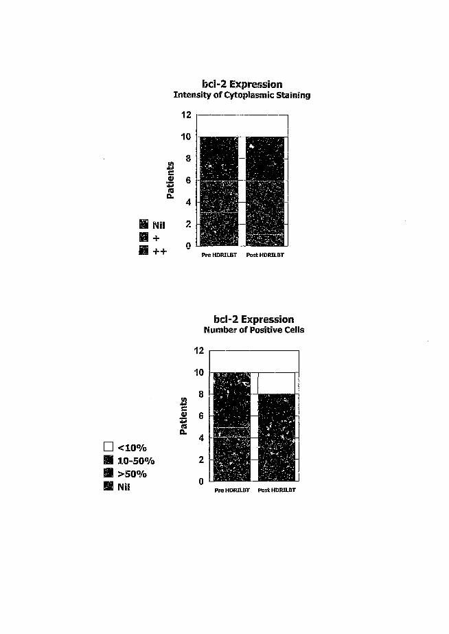
bcl-2 ExpressionIntensity o f Cytoplasmic Staining
12
10
8sti«5 6
4
H Nil 2H +H ++ 0
I"'-.P re HDRILBT P o s t HDRILBT
bcl-2 Expression Number of Positive Ceils
n < 10%B 10-50% B >50% H Nil
0)I
12
10
8
Lr—
P re HDRILBT P o s t HDRILBT
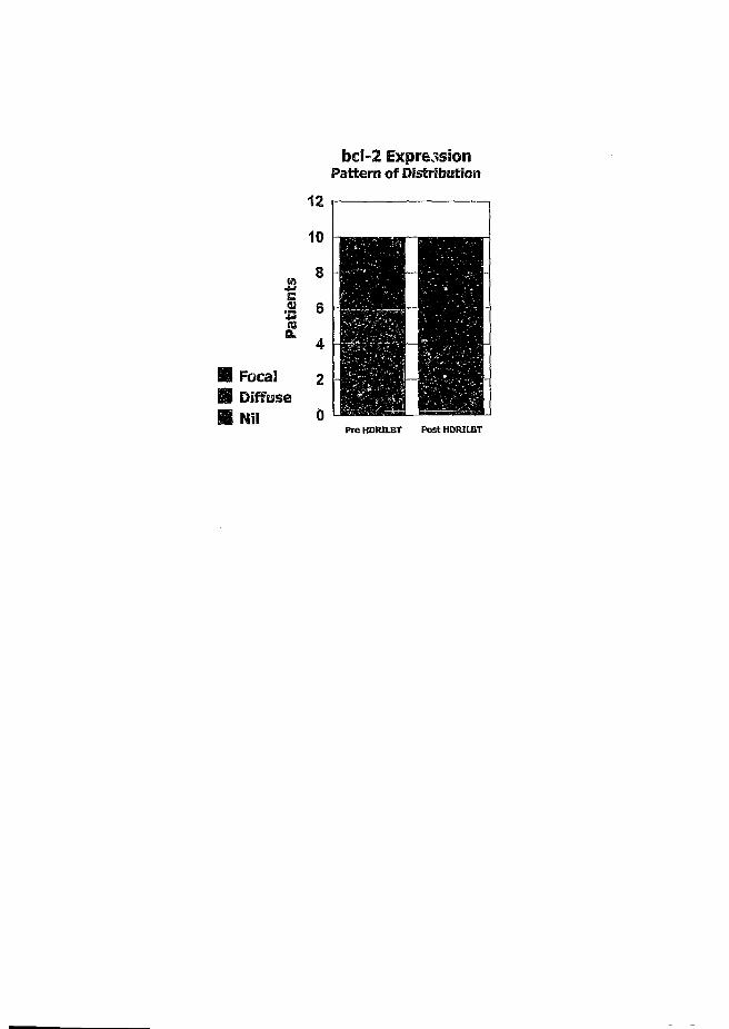
bcl-2 ExpressionPattern o f Distribution
12
10
e.2 6
B Focal 2S Diffuse H Nil 0
P r e HDRILBT P o s t HDRILBT

Fig XXXIXPrebrachytherapy tumour biopsy showing no expression of apoptosis (XiOO)
FigXXXXPostbrachytherapy tumour area from the resection specimen showing no
expression of apoptosis (X200)
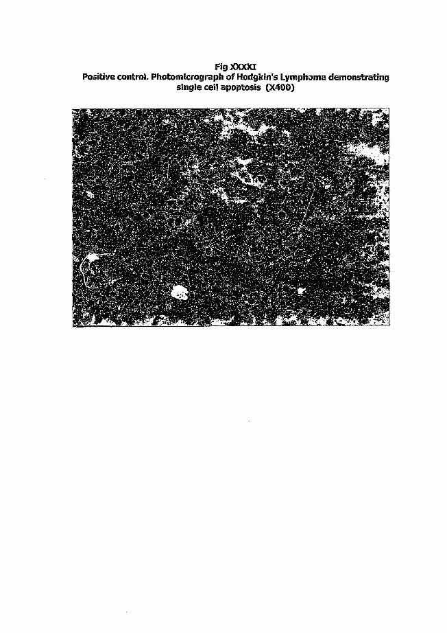
FigXXXXIPositive control. Photomicrograph of Hodgkin's Lymphoma demonstrating
single ceil apoptosis (X400)

13.1
Chapter 13
Discussion
13.2
Morphological Changes
The pathological sequence of events occurring in the oesophagus following EBRT
have been well described (Berthrong M., 1986, Northway N.G. et al, 1979,
Novack J.M. et al., 1979, Phillips T.L. and Margolis L, 1972, Seaman W.B.,
1957).
The pathologic sequence of events after a single large EBRT fraction of radiation
in mice are as follows (Phillips T.L. and Ross G., 1974):
Day 3 : Vacuolization and absence of mitosis of the basal cell
layer along with thinning of the keratinized squamous
cell layer occurs.
Between day 7 and 14: Foci of proliferating basal cells and regenerating
epithelium with zones of complete denudation occur
simultaneously.
After 21 days: There is usually complete regeneration of the
oesophageal lining with increased basal cell layer
proliferation and increased thickness of the squamous
layer.
Similar changes have been reported in humans using EBRT (Coia L.R. et al.,
1995). However, there are no reports on radiation changes after ILBT alone,
although, this technique has a higher therapeutic effect because there is minimal
penetration of radiation beyond the local area (Berry B. et al, 1989).
Hishikawa et al reported radiation changes in autopsy specimens from patients
treated with preoperative E8RT and HDR ILBT(1985, 1988). They reported
remarkable changes in the tumour, but not in the normal oesophagus following
EBRT. However, radiation changes were seen in the norma! oesophagus and
tumour following EBRT and HDR ILBT. Denudation and regeneration of the
epithelium were recognized. Blood vessel alterations and fibrosis with fibrinous
exudate of the submucosa were severe. Fibrosis of the muscle layers was also
found. Berry et al (1989) correlated the ratio of the depth of radiation damage
to the depth of identifiable tumour to the local effectiveness of preoperative
treatment after EBRT and ILBT. Surgery was performed 6 weeks after EBRT and
HDR ILBT. Ratios above 2:4 (involving but not deeper than submucosa) were
considered to indicate a good response. In our study, all patients had radiation
effects extending beyond the adventitia all along the irradiated length.
Therefore, lymph nodes located in the adventitial tissue around the oesophagus
also showed radiation changes, as was seen in our patients. Based on the
isodose curves generated from the Treatment Planning System [TPS] (Plato;
Nucletron, Amsterdam, Netherlands), approximately 50% of the dose prescribed
(10 Gy at 1.0 cm from the source axis) reached the adventitial tissues around the
oesophagus (within 0.5 cm from the oesophageal wall or 1.5 cm from the centra
of the source axis) per application of HDRILBT. In our patients, the lymph noda
were located 2 mm from the oesophageal wall in the adventitial tissue. Thus,
radiation changes were seen as the lymph node received at least 50% of lli-a
dose given (20Gy in 2 fraction). Actual measurements can only be performed at

13.4
autopsy in which thermoluminescent dosimeters are placed in the adventitial
tissue and the oesophagus irradiated. Thus, it is likely, based on the isodose
curves generated from the TPS, that lymph nodes that are located beyond the
adventitial tissue (more than 0.5 cm from the oesophageal wall) will show
minimal changes due to radiation, if the dose is prescribed at 1.0 cm. Although
Hishikawa et al. have suggested that the maximum dose to the oesophagus
should be limited to 20 Gy by ILBT (1988), this is following a course of radical
EBRT of 50 Gy and is therefore not relevant if HDR ILBT is used alone.
The proximal resection margins in 7 patients with tumour in the mid one-third of
the oesophagus and the distal resection margins in 3 patients with tumour in the
lower one-third of the oesophagus, showed radiation changes limited to the
submucosa with minimal fibrosis. This could be explained by the fall off of
radiation dose at the resection margins, which were 1-1.5 cm Worn the edge of
the irradiated length and therefore received only 10-25% of the dc.v» prescribed.
Thus radiation effects were not observed beyond the submucosa (Table II).
Pathologic analysis of oesophagectomy specimens after EBRT for SCG have been
reported. Akakura et al (1970) noted the gross appearance of such case? to
usually consist of a small re-epitheiia!ized ulcer with mural fibrosis.
Microscopically, approximately 65% of their cases showed a definite cytologic
radiation effect, as defined by mitotic incompetence, nuclear pyknosis.
13.5
cytoplasmic vacuolation, and/or tumour cell disappearance. We did not find any
report in the literature on the acute radiation effects after HDRILBT.
In all our cases, surgery was performed within 2 - 3 weeks of brachytherapy.
Thus, typical acute radiation changes described were seen. Interestingly, the
irradiated tumours in all 10 cases showed extensive keratin production as
opposed to the previous biopsies, which showed minimal to negligible keratin
production due to poor differentiation. This could be attributed to the induction
of the keratin gene in tumour cells to differentiate towards better differentiated
cells. This has been previously reported in solid tumours with the use of
anti-neoplastic drugs (Melguizo G., 1995). In our patients, blood vessel walls
were found to be intact with no evidence of vessel wall necrosis. This contrasts
to earlier reports with EBRT (Coia L.R. et al, 1995) in which necrosis of the vessel
walls is a frequent occurrence. The biological basis is inexplicable, but may
possibly lead to increased tumour oxygenation and better radiation effects, as
observed in the present study. Radiation effects were also observed in the
adjacent peri-oesophageal lymph nodes containing metastatic tumour and even
at the resection margins (which were not as extensive). Changes described
previously, such as oedema and fibrinous exudate (Coia L.R. et al., 1995), were
not seen in any of the patients. HDR ILBT given to our patients in the dose and
fractionation of 20 Gy in 2 weekly fractions of 10 Gy each, adequately covered
the local disease and peri-oesophageal lymph nodes, all of ’ ' 'wed
radiation changes. It is not possible to comment on whethe < . Jith
13.6
tumour sterilization because the oesophagectomies were performed within 2 - 3
weeks. Tumour sterilization can only be established from autopsy after long term
follow up. Thus, typical radiation changes were observed in the irradiated length
of the oesophagus after HDRILBT that were not observed after EBRT.
Expression of MDR-1 gene product (p- glycoprotein)
Multidrug resistance phenotype, first described in 1970, is now known to be due
to the presence of the transmembrane glycoprotein (p-glycoprotein) product of
the MDR-1 gene, p-glycoprotein has been associated with both intrinsic and
acquired resistance to certain chemotherapeutic agents, and cells selected for
resistance against one cytotoxic drug simultaneously, acquire cross resistance to
a range of other cytotoxins (Yusa K. e t al., 1991, Ferry D.R. and Kerr D.J., 1994,
Twentyman P.R., 1986). p-glycoprotein appears to be a highly conserved
membrane transport protein, and may be a common route for the export of
environmentally occurring toxins. Tumours responsive to chemotherapy generally
show low levels of p-glycoprotein expression, and solid tumours that are most
responsive to systemic chemotherapy, such as seminomas and embryonal
carcinomas, rarely display detectable levels of the protein. Tumours from
patients previously treated with chemotherapy show frequent elevation of
p-glycoprotein, suggesting that the MDR phenotype is induced by exposure to
chemotherapy. Tumours that initially respond to chemotherapy maybe resistant
on relapse, and commonly show increase in the expression of p-glycoprotein
(Yuen A.Rand Sikia B.I., 1994).
13.7
Damton e t al. (1995) have reported the lack of p-glycoprotein expression with
response to MIC (Mitomycin C, Ifosphamide and C/>Platin) chemotherapy in
oesophageal SCC. These cancers responded to chemotherapy better than
oesophageal adenocarcinomas; the latter showing expression of the MDR-l
gene. However, p- glycoprotein could not be used as a predictive marker of
response as tumours expressed it inconsistently with response to MIC. Saito et
al. (1992) studied three cell lines from SCC, and found a pleiotropic mechanism
of resistance without expression of p-glycoprotein. Robey-Cafferty et al (1990)
suggested that clinical response correlated with expression of p-glycoprotein in
adenocarcinomas. These authors did not match pre-treatment biopsy specimens
with treated tumours.
There have been no studies on expression of p-glycoprotein in oesophageal SCC
prior to and after radiotherapy. In ail our 10 cases, p-glycoprotein was not
expressed in either pre or post-brachytherapy tissue specimens. These findings
together with those of Darnton e t al (1995) indicate that p-glycoprotein
expression is of no value in predicting the responsiveness of the tumour to
radiotherapy and chemotherapy in SCC of the oesophagus. MDR-l gene does
however have some value in predicting the response to treatment in
adenocarcinomas. Clinically this has been demonstrated in breast carcinomas
(where the expressors of this gene have been shown to have poor survival),
renal cell carcinoma, cancer of the urinary bladder, colo-rectai malignancies,
gastric and ovarian carcinomas (Glazer R.I. and Rohlff C., 1994, Mickisch G.H.,

13.8
1994, Scher HI., 1992, Linn S.C. and Glaccone G., 1995, Wallner J. e t at., 1993,
Sekiya S. et al., 1993). Unlike chemotherapy, radiotherapy effects cell death
through the formation of free radicals. This involves interaction with other atomu
or molecules, particularly water, to produce free radicals that are able to diffuse
and damage the critical targets. These include the DNA in the chromosome
(which is the most critical target), and the nuclear membrane. Studies have
shown that about 2/3rds of X-ray damage by high voltage ionizing radiation is
caused to the DNA by the OH free radical (Hall EJ., 1988). The p-glycoprotein,
on the other hand, acts as an ATP dependent drug efflux pump which transports
drugs associated with multidrug resistance out of the cell before cytotoxic effects
can occur (Kartner N. and Ling V., 1989, Gottesman M.M. e t al., 1991). It is
therefore unlikely that the expression of p-glycoprotein could have any bearing
on responsiveness of the tumours to radiation. It is likely for this reason that
alternate factors maybe associated with resistance in SCC of the oesophagus.
Expression of p53, bel-2 and apoptosis
The effects of ILBT on oncogene expression and apoptosis have not been
previously reported in clinical studies.
In the present study, most patients showed expression of p53 (8/10)
immunoreactivity in the pre-operative biopsies. This was not altered following
brachytherapy which was evaluated in the resected specimens, with the intensity
of p53 nuclear staining being the same (3+). However, the distribution In pre
13.9
IlBT biopsies was mostly type C (>50%) whilst the post ILBT specimens showed
Type B distribution (10 - 50%). The pattern of distribution was diffuse in the
biopsies, and focal and peripheral in the post ILBT specime is. The centre of the
tumour islands showed no expression of p53. These areas showed either
necrosis or keratinization. It is suggested that ILBT induces destruction of cells
expressing wild type p53, which subsequently undergoes necrosis. The surviving
p53 positive cells in the periphery of tumour islands, post ILBT, may represent
mutant protein, representing cells with p53 gene mutation permitting escape
from growth arrest. It is further suggested that these cells may eventually
outgrow the tumour population to become dominant clones. I t is conceivable
that these latter cells may be responsible for the high incidence of local failure
and distant metastasis, seen in oesophageal cancer following radiation
treatment. In the single patient without p53 expression following brachytherapy,
(although the pre ILBT specimen stained p53 positive) it is probable that the
tumour may have only expressed wild type of p53 which was obliterated by
brachytherapy.
A high degree of p53 mutations is seen in oesophageal squamous cell
carcinomas (Coggi G. et a!., 1997, Wang L.D. et al., 1996). Muro et al (1996)
evaluated 20 patients to determine the clinical role of p53 mutation in locally
advanced oesophageal cancer treated with chemoradiotherapy. They observed
that patients who did not express p53 protein did not achieve complete
remission and response to the tumour. They suggested that expression of p53
13.10
may be an indicator for favorable prognosis in patients with locally advanced
oesophageal cancer, treated with concurrent chemoradiotherapy. On the other
hand, Puglisi et al (1996) investigated (i) the relation between
immunohistochemical expression of p53 and bcl-2 protein; (ii) the rate of tumour
response after neo-adjuvant chemo-radiotherapy in locally advanced squamous
cell carcinoma of the oesophagus ;and (iii) whether antibiastic treatment could
modify the immunohistochemical expression of these oncoproteins . They
reported that p53 positive immunoreaction was detected in biopsy specimens of
most patients. Following preoperative chemoradiotherapy there was no
difference in p53 expression in the oesophagectomy specimens. These authors
suggested that anticancer agents do not seem to modify the expression of p53
and bcl-2 proteins.
Few studies have attempted to establish a relationship between p53 expression
and outcome following radiotherapy. Pomp et al. (1998) evaluated the
relationship of the immunohistochemically determined p53 protein status and the
disease control with radiotherapy alone, for carcinoma of the oesophagus.
Immunostaining for p53 protein was performed on paraffin embedded specimens
in 69 patients with adenocarcinoma and SCC of the oesophagus. All patients
were treated by radiotherapy exclusively, consisting of all combinations of EBRT
and ILBT using two different dose levels. 54% (37/69) of the tumours showed
overexpression of the p53 protein. No difference in pretreatment parameters for
p53 positive and p53 negative cases was detected. In multivariate analysis p53
13.11
was significantly associated with overall survival next to weight loss, tumour
stage and nodal stage. For metastasis free survival, p53 status proved to be the
sole independent prognostic factor. The influence of p53 on local recurrence free
survival, however, was not as strong as on overall survival and metastasis free
survival. Immunohistochemicaily detected overexpression of mutated p53 protein
in (.esophagus carcinoi.a was thus shown to be an independent prognostic
factor in a group of patients treated with radiation therapy alone.
Nabeya et al. (1995) reported on the mutational status of the p53 protein in
gastric and oesophageal adenocarcinoma cell lines following chemotherapy. They
observed that the wild type p53 increases after treatment with chemotherapy.
They suggested that wild type p53 appears to be essential for effective
chemotherapeutic response. Cell lines exhibiting mutated p53 were more
resistant to the effect of chemotherapeutic drugs. A similar situation may exist in
the clinical setting with brachytherapy.
In the present study, one patient did not express p53 protein after
brad'ev therapy, although, there was p53 positive staining in the pre ILBT biopsy
specimen. It is likely that the p53 may have been of the wild type, and had
therefore responded to brachytherapy. The other 8 patients may have expressed
mutant p53 that may have escaped the effect of brachytherapy, especially at the
periphery of the tumour islands. The single patient who did not express any p53
13.12
before or after brachytherapy did not show any response and has subsequently
developed recurrent disease.
Our initial findings are in concurrence with Pomp et al (1998), who observed that
p53 expression was related to the outcome of the disease to treatment.
However, long term followup is required before a definite association can be
established on p53 expression and outcome. This is beyond the scope of this
study which examines the expression of p53 following HDRILBT.
The recent emergence of apoptosis as a mechanism of cell death has been
reviewed by various authors (Haimovitz F.A. et al., 1996). Apoptosis is a death
pathway of sequential biochemical events that can be induced and is
constitutively expressed in many cells, even in an inactive form. The apoptosis
cascade can be triggered by a variety of physiological and stress stimuli. Graeber
T.G. (1996) reported that hypoxia induced apoptosis in oncogenically
transformed cells and that further genetic alterations, such as loss of p53 tumour
suppressor gene or overexpression of the apoptosis inhibitor protein bcl-2,
substantially reduced hypoxia induced cell death. Hypoxia also selectively
affected cells with defects in apoptosis, because small numbers of transformed
cells lacking p53 overtook similar cells expressing wild type p53 when treated
with hypoxia. Highly apoptotic regions strongly correlated with hypoxic regions in
transplanted tumours expressing wild type p53, whereas little apoptosis occurred
in hypoxic regions of p53 deficient tumours. Hypoxia therefore provided a
13.13
physiological selective pressure in tumours for the expansion of variants that had
lost their apoptotic potential, and in particular in cells acquiring p53 mutations.
Kinzler K.W. and Vogelstein B (1996) reported that the low levels of oxygen
found in those ports of tumours with poor blood supply resulted in p53
dependent programmed cell death. Mutations of p53 reduced the cell death,
giving the mutant cells a survival advantage over cells with intact p53. As a
tumour grows to a critical size, the central portion suffers from hypoxia. This
induces expression of the p53 gene which then leads to growth arrest or
apoptosis. Cells that have acquired p53 gene mutation can escape the growth
arrest or apoptosis. These clones will v1 /entually outgrow the rest to become the
dominant clone. Therefore, emergence of clones with p53 gene mutation may be
driven by hypoxia. The molecular events that regulate the process of apoptosis
have been closely linked to the identification of oncogenes and tumour
suppressor genes in various neoplasias.
Expression of bcl-2 oncoprotein blocks many instances of apoptotic cell death in
developing neural (Allsop T.E. et al., 1993) and lymphoid tissues (Vaux D.L et
al., 1988). It has been reported that bcl-2 can block apoptosis that usually
results from treatment with ionizing radiation (Strasser A. et al., 1994) or
chemotherapeutic agents (Miyashita T. and Reed J.C., 1993). Expression of p53
gene has been shown to be a positive regulator of apoptosis (Puglisi F. et al.,
1996, Kernohana N.M. and Cox L.S., 1996). It has been suggested that loss of

13.14
function of p53 gene and gain in function of bci-2 gene can produce tumours
resistant to chemo- radiotherapy.
bcl-2 expression was equivocal in this series. Brachytherapy does not appear to
alter the bcl-2 expression in oesophageal squamous cell carcinomas. Further,
apoptosis was not present in the pre and post-brachytherapy tissue specimens.
This study therefore demonstrates that oesophageal squamous cell carcinomas
do not undergo apoptosis following brachytherapy. Following brachytherapy, the
decrease in tumour volume is due to necrosis and maturation of the cells by
induction of the keratin gene ( Sur M. et al., 1996).
Thus, brachytherapy does not cause cell death by apoptosis, but by necrosis and
mat ration of the cells into better differentiated cells, which is caused by OH" free
radical, and the induction of the keratin gene respectively. It is possible that
brachytherapy may cause destruction of cells containing wild type p53, while
mutant p53 in cells located at the tumour periphery escapes the effect of
brachytherapy. This maybe responsible for the high incidence of local recurrence
and distant metastasis in oesophageal cancer treated with radiotherapy. There is
no effect of brachytherapy on bcl-2 expression in oesophageal cancer.

14.1
Chapter 14
Conclusions
14.2
Morphological Changes
1. Typical acute radiation changes were observed in the irradiated length of
the oesophagus after preoperative HDRILBT. These are not observed
following EBRT.
2. Changes were seen at various levels of the oesophagus, with full thickness
changes observed at the center of the tumour, and 1 cm from the edge of
visible tumour.
3. HDR ILBT may cause the keratin gene in the irradiated cells to induce
differentiation towards better differentiated cells.
Expression of MDR-1 gene product (p-glycoprotein)
1. p-glycoprotein was not expressed in either pre or post- brachytherapy
tissue specimens in this study.
2. These findings indicate that p-glycoprotein expression is of no value in
predicting the responsiveness of the tumour to radiotherapy in SCC of the
oesophagus.
Expression of p53, be!-2 and apoptosis
1. Prior to HDR ILBT, there is expression of both wild and mutant p53
protein in oesophageal cancers, (antibody DO -7 detects both wild type
and mutant p53)
2. Following HDR ILBT, there is destruction of cells. It is postulated that this
may involve destruction of cells expressing wild type p53, whilst residual
14.3
surviving cells contain mutant p53. These cells are located at the tumour
periphery and escape the effect of brachytherapy. These cells survive and
may eventually become the dominant clone, responsible for the high
incidence of local recurrence and distant metastasis in oesophageal cancer
treated with radiotherapy.
3. There is no effect of brachytherapy on bcl-2 expression in oesophageal
SC C.
4. Oesophageal SCC, do not undergo apoptosis following brachytherapy.
5. Following brachytherapy, the decrease in tumour volume is due to
necrosis and maturation of the cells by induction of the keratin gene.

15.1
Chapter 15
References
15.2
Allan DJ. Radiation induced apoptosis: its role in a MADCaT (mitosis-apoptosis-
differentiation-calcium toxicity) scheme of cytotoxicity mechanisms. Int J Radiat
Oncol Biol Phys 1992: 62: 145-52.
Allsopp T.E., Wyatt S., Paterson H.F., Davis A.M. The proto- oncogene bcl2 can
selectively rescue neurotrophic factor-dependent neurons from apoptosis. Cell
1993: 73: 295-307
Baer R. bcl2 breathes life into embryogenesis. Am J Pathol 1994; 145: 7-10.
Bakhshi A., Jensen J.P., Goldman P. Cloning the chromosomal breakpoint of t
(14; 18) human lymphomas: clustering around JH on chromosome 14 near a
transcriptional unit on chromosome 18. Cell 1985; 41: 899- 906
Batsakis J.G., L-Nagger A.K.p53: 15 years after discovery. Advances Anatomic
Pathol 1995; 2: 71- 88
Benson M.C., Gielia J., Whang I.S., Buttyan R., Hensle T.W., Karp F., Olsson C.A.
Flow cytometric determination of the muitidrug resistant phenotype in
transitional cell cancer of the bladder: Implications and applications. J Urol 1991;
146: 982- 6
15.3
Berry B., Miller R., Luoma A., Nelems B., Hay J., Flores A.D. Pathologic findings
in total esophagectomy specimens after intracavitary and external beam
radiotherapy. Cancer 1989; 64:1833- 7
Bissonette R.P. Apoptotic cell death induced by c-myc is inhibited by bcl2. Nature
1992; 359: 552
Chang F. The p53 tumour supressor gene as a common cellular target in human
carcinogenesis. Am J Gastroenterol 1993; 88:174
Chao C.C. the, Ma C.M., Lin- Chao S. Co-amplification and over expression of two
MDR genes in a multidrug resistant human colon carcinoma cell line. Febs Lett
(Netherlands) 1991; 291: 214-8
Chen- Levy Z., Nourse J., Cleary M.L. The bcl2 candidate proto-oncogene is a 24
kD integral- membrane protein highly expressed in lymphoid cell lines and
lymphomas carrying the t(14; 18) translocation. Mol Cell Biol 1989; 9: 701-10.
Chetty R., Dada M.A., Gatter K.C. bcl2: longevity personified. Advances Anatomic
Pathol 1997; 4:134- 8.
Chiarugi V., Ruggiero M. Role of three cancer "mastergenes" p53, bcl2 and
c-myc on the apoptotic process. Tumori 1996; 82: 205- 9
15.4
Chin K.V., Liu B. Regulation of the multidrug resistance (MDR1) gene expression.
In vivo 1994; 8: 835- 41
Chou P.M., Reyes-Mugica M., Barquin N., Yasuda T., Tan X., Tomita T. Multidrug
resistance gene expression in childhood medulloblastoma: correlation with clinical
outcome and DNA ploidy in 29 patients. Pediatr Neurosurg 1995; 23: 283-91
Clayton E.S. Carcinoma of the Oesophagus. Surg Gynecol Obstet 1928; 46: 52
Cleary M.L., Smith S.D., Sklar J. Cloning and structural analysis of c-DNAs for
bc!2 and a hybrid bcl-2 immunoglobulin transcript resulting from the t(14; 18)
translocation. Cell 1986; 47: 19- 28.
Coggi G., Bosari S., Roncalli M., Graziani D., Bossi P., Viale G., Buffa R., Ferrero
S., Piazza M., Blandamura S., Segalin A., Bonavina L., Peracchia A. p53 protein
accumulation and p53 gene mutation in esophageal carcinoma. Cancer 1997;
79: 425-32
Colombel M., Symmans F., Gill S. Detection of the apoptosis suppressing
oncoprotein bc!2 in hormone refractory human prostrate cancer. Am J Pathol
1993; 143: 390- 400.
15.5
Cooper K., Haffejee Z. bcl2 immunohistochemistry distinguishes follicular
lymphoma from follicular hyperplasia in formalin-fixed tissue with microwave
antigen retrieval. J Cellular Pathol 1996; 1: 52- 6
Cooper K., Haffejee Z. bcl-2 and p53 protein expression in follicular lymphoma. J
Pathol 1997; 182: 307-10
Craig R.W. The bc!2 gene family. Semin Cancer Biol 1995; 6: 85- 43.
Damton S J ., Jenner K.; Steyn R.S., Ferry D.R., Mathews H.R. Lack of correlation
of p-glycoprotein expression with response to MIC chemotherapy in esophageal
cancer. J Clin Pathol 1995; 48: 1064-6
Dewey W.C., Ling C.C., Meyn R.E. Radiation induced apoptosis: Relevance to
radiotherapy. In tJ Radiat Oncol Biol Phys 1995; 33: 781-96.
Ferry D.R., Kerr DJ. Multidrug resistance in cancer. BMJ 1994; 308:148- 9
Flemeing J.A.C. Carcinoma of thoracic esophagus: Some notes on its pathology
and spread in relation to treatment. Br J Radiol 1943; 16: 212
15.6
Freund U., Brambs HJ., Laaff H., Roth R., Bruggmoser G., Wannenmacher M.
Intracavitary High Dose Rate afterloading radiotherapy in the esophagus of the
pig. Strahlenther Onkol 1989; 165: 852- 4.
Glazer R.I., Rohiff C. Transcription regulation of multidrug resistance in breast
cancer. Breast Res Treat 1994; 31; 263- 71
Goldblum 3.R., Rice T.W. bc!2 protein expression in the Barretts metaplasia-
dysplasia- carcinoma sequence. Mod Pathol 1995; 8 : 866-9
Gottesman M.M., Goldstein L.3., Fojo A. Expression of the multidrug resistance
gene in human cancer, in : Ronison LB. (ed). Molecular Cellular Biology of
Multidrug Resistance in Tumour cell. New York; Plenum Press, 1991; 291- 301
Graeber T.G., Osmanian C., Jacks T., Houseman D.E., Cameron J.K. Scott W.L,
Amato J.G. Hypoxia mediated selection of cells with diminished apoptotic
potential in solid tumors. Nature 1996; 379: 88- 91
Hall EJ. The Oxygen Effect and re-oxygenation, in Radiobiology for the
Radiologist. 3rd ed. J.B.Lippincott Co. Philadelphia, 1988:137- 59
Hall P.A. and Lane D.P. p53 in tumor pathology: Can we trust
immunohistochemistry. 3 Pathol 1994; 172:1-5

15.7
Hamada M., Naomoto Y., Fujiwara T., Kamikawa Y., Tanaka N. Suppressed
apoptotic induction in esophageal squamous cell carcinomas expressing
extensive p53 protein. Jpn J Clin Oncol 1996; 6: 398-404.
Harmsel B.T., Smedts F. Kruijpers J., Jeunink M., Trimbos B., Ramaekers F. bc!2
immunoreactivity increases with severity of CIN: A study of normal cervical
epithelium, CIN, and cervical carcinoma. J Pathol 1996; 179: 26- 30.
Harris C.C., Hollstein M. Clinical implications of the p53 tumour supressor gene.
New Eng J Med 1993; 329:1318
Harrison DJ. Molecular mechanism of drug resistance in tumours. J Pathol 1995;
175: 7-12
Hengartner M.O., Horvitz H.R. C. elegans gene ced-9 protects cells from
programmed cell death. Nature 1992; 356: 494
Hishikawa Y., Tanaka S., Miura T. Esophageal Ulceration induced by intracavitary
irradiation for esophageal carcinoma. AJR1984; 143: 269-73
Hishikawa Y., Mitsunobu M., Uematsu K.f Miura T. Histological findings of
esophageal injury induced by intracavitary irradiation, Radiat Med 1985; 3:112-7
15.8
Hishikawa Y., Taniguchi M., Kamikonya N., Tanaka S., Miura T. External Beam
Radiotherapy Alone or combined with High Dose Rate Intracavitary irradiation in
the treatment of cancer of the esophagus: Autopsy findings in 35 cases.
Radiother Oncol 1988; 11: 223-7.
Hishikawa Y., Kurisu K., Taniguchi M., Kanikonya N.r Miura T. Small superficial
esophageal carcinoma treated with high dose rate intracavitary irradiation only.
Radiology 1989; 172: 267-70
Hockenberry D.M. bcl2 functions in an antioxidant pathway to prevent apoptosis.
Cell 1993; 75: 241
Hunt J.A. Squamous cancer of the oesophagus in urban South African blacks: A
preliminary report of baseline studies, in: Silber W.f ed. Carcinoma of the
Oesophagus. Cape Town, South Africa: AA Balkema, 1978: 287- 311
Jung M., Notario V., Dritschilo A. Mutations in the p53 gene in radiation sensitive
and radiation resistant human squamous carcinoma cells. Cancer Res 1992; 52:
6690-3.
Kaklamanis L. p53 expression in colorectal adenomas. Am 3 Pathol 1993; 142:
87
15.9
Kaklamanis L, Savage A., Mortensen N. Early expression of bcl2 protein in the
adenoma- carcinoma sequence of colorectal neoplasia. J Pathol 1996; 179:10- 4
Karnofsky D.A., Abelmann W.H., Graver L.F. The use of nitrogen mustards in the
palliative treatment of carcinoma with particular reference to bronchogenic
cancer. Cancer 1948; 1: 634- 69
Kartner N., Ling V. Multidrug resistance in cancer. Science 1989; 260: 44- 51
Kerr J.F.R., Wyllie A.H., Currie A.R. Apoptosis: A basic biological phenomenon
with wider ranging implications in tissue kinetics. Br J Cancer 1972; 26: 239- 257
Katada N., Hinder R.A., Smyrk T.C., Hirabayashi N., Perdikis G., Lund RJ.,
Woodward T,, Kiingler PJ. Apoptosis is inhibited early in the dysplasia-carcinoma
sequence of Barrett esophagus. Arch Surg 1997; 132: 728-33.
Kerr J.F.R., Harmon B.V. Definition and incidence of apoptosis: A historical
perspective, in: Tomey L.D. and Cope P.O. (eds): Apoptosis: The Molecular basis
of cell death. Coldspring Harbor, NY, Coidspring Harbor Laboratory Press, 1991
Kinder K.W., Vogelstein B. Life and death in a malignant tumor. Nature 1996;
379: 19-20
15.10
Kneebone R.L., Mannell A. Cancer of the Oesophagus in Soweto. S. Afr Med J
1985; 67: 839- 42
Krajewski K.f Tanaka S., Takayama S, Schibler M.J., Fenton W., Reed J.C.
Investigation of the subcellular distribution of the bci2 oncoprotein : residence in
the nuclear envelope, endoplasmic reticulum and outer mitochondrial
membranes. Cancer Res 1993; 53: 4701- 14
Landanyi M., Wang S. Detection of rearrangements of the bcl2 major breakpoint
region in follicular lymphomas. Correlation of polymerase chain reaction results
with Southern blot analysis. Diagn Mol Pathol 1992; 1: 31- 5.
Lane D.P. A death in the life of p53. Nature 1993; 362: 786
Linn S.C., Glaccone G. MDR 1 / p glycoprotein expression in colorectal
malignancies. Eur J Cancer 1995; 31:1291-4
Lu Q-L, Abel P., Foster C.S., Lalani E.N. bcl2: role in epithelial differentiation and
oncogenesis. Human Pathol 1996; 27:102- 10.
Mannell A., Murray W. Oesophageal cancer in South Africa- A Review of 1926
cases. Cancer 1989; 64: 2604- 8
15.11
Mascarenhas F., Silvestre M.E., Small da Costa M., Grima N., Campost C., Chaves
P. Acute secondary effects in the esophagus in patients undergoing radiotherapy
for carcinoma of the lung. Am J Clin Oncol 1989; 12: 34- 40.
McDonnel T.J., Troncoso P., Brisbay S.M, Expression of the protooncogene bcl2
in the prostrate and its association with emergence of androgen independent
prostate cancer. Cancer Res 1992; 52: 6940- 4.
McKenna W.G., Bernhard EJ., Markiewicz D.A. Regulation of radiation induced
apoptosis in oncogene-transfected fibroblasts: Influence of H-ras on the G2
delay. Oncogene 1996; 12: 237- 45
Merendiono K.A., Maerck VJ. An analysis of 100 cases of squamous cell
carcinoma II with special references to its theoretical curability. Surg Gynecol
Obstet 1952; 94:110
Mickisch G.H. Chemoresistance of renal cell carcinoma. World J Urol 1994; 12:
214- 23.
Miyashita T., Reed J.C. bcl2 oncoprotein blocks Chemotherapy-induced apoptosis
in a human leukemia cell line. Blood 19993; 81:151-7
15.12
Moreira L.F., Naomoto Y., Hamada M.f Kamikawa Y., Orita K. Assessment of
apoptosis in esophageal carcinoma preoperatively treated by Chemotherapy and
radiotherapy. Anticancer Res 1995; 15: 639-44
Muro K., Ohtsu A., Baku N., Chin K., Oda Y., Fujii T., Hosokawa K., Yoshida S.,
Hasebe T. Association of p53 protein expression with responses and survival of
patients with locally advanced esophageal carcinoma treated with chemo
radiotherapy. Jpn J Clin Oncol 1996; 26: 65-9
Nabeya Y.f Loganzo F., Maslak P., Lai L., Oliveria A.R., Schwartz G.K., Blundell
M.L., Altorki N.K., Kelsen D.P., Albino A.P. The mutational status of p53 protein
in gastric and esophageal adenocarcinoma cell lines predicts sensitivity to
chemotherapeutic agents. Int. 3 Cancer 1995; 64: 37-46
Ng. I.O., Lam K.Y., Ng ML, Kwong D.L., Sham J.S. Expression of P glycoprotein, a
multi-drug resistance gene product, these induced by radio therapy in patients
with oral squamous cell carcinoma. Cancer 1998; 83: 851-7
Oberhammer F., Wilson 3.W., Dive C. Apoptotic death in epithelial cells: cleavage
of DNA to 300 and/ or kb fragments prior to or in the absence of
internucleosomal fragmentation. EMBO J 1993; 12: 679- 84

15.13
Ohashi K., Nemoto T.; Eishi Y., Matsuno A., Nakamura K., Hirokawa K.
Proliferative activity and p53 protein accumulation correlate with early invasive
trend, and apoptosis correlates with differentiation grade in esophageal
squamous cell carcinomas Virchows Arch 1997; 430:107-15
Ohbu M., Saegusa M., Okayasu I. Apoptosis and cellular proliferation in
esophageal squamous cell carcinomas: differences between keratinizing and
non-keratinizing types. Virchows Arch 1995; 427: 271-6
Ohbu M., Saegusa M., Kobayashi N,, Tsukamoto H., Mieno H., Kakita A.,
Okayasu I. Expression of bc!2 protein in esophageal squamous cell carcinomas
and its association with lymph node metastasis. Cancer 1997; 79:1287-93
Olive P.L., Frazer G., Banath J.P. Radiation induced apoptosis measured in TK6
human B lymphoblast cells using the comet assay. Radiat Res 1993; 136:130- 6.
Parenti A.R., Rugge M., Horng Shiao Y,, Ruol A., Bozzola L, Ninfo V. bc!2 and
p53 immunophenotypes in pre-invasive, early and advanced esophageal
squamous cancer. Histopathology 1997; 31: 430-5
Pezzella F., Ralfkiaer E., Gatter K.C., Mason D.Y. The t(14:18) translocation in
European cases of follicular lymphoma: Comparison of Southern blotting and
polymerase chain reaction. Br. 3 Haematol 1990; 76: 58- 64.
15.14
Pezzella F., Turley H., Kuzu I. bcl2 protein expression in non- small cell lung
carcinoma. Immunohistochemical evidence for abnormal expression and
correlation with survival. New Eng J Med 1993; 329: 690 - 4
Pezzella F., Gatter K. What is the value of bc!2 protein for histopathologists.
Histopathol 1995; 26: 89- 93.
Phillips T.L., Ross G. Time dose relationships in the mouse esophagus. Radiology
1974; 113: 435-40
Pomp 3., Daveliar 3., Blom 3., van Krimpen C , Zwinderman A., Quint W.,
Immerzeel 3. Radiotherapy for esophagus carcinoma: the impact of p53 on
treatment outcome. Radiother Oncol 1998; 46:179-84
Puglisi F., Di Loreto C., Panizzo R., Avellini C , Fongione S., Cacitti V,, Beltrami
C.A„ Expression of p53 and bcl2 and response to preoperative Chemotherapy and
radiotherapy for locally advanced squamous cell carcinoma of the esophagus .3
Clin Pathol 1996; 49: 456-9.
Robey-Cafferty S.S., Rutledge M.L., Bruner 3.M. Expression of a multidrug
resistance gene in oesophageal adenocarcinoma. Correlation with response to
chemotherapy and comparison with gastric adenocarcinoma. Am 3 Clin Pathol
1990; 93: 1-7
15.15
Rose E.F. Cancer of Oesophagus. S Afr J Hosp Med 1978; 4:110- 2
Saito T., Hikita M., Kohno K.# Sato S., Takano H., Kobayashi M. Different
sensitivities of human oesophageal cancer cells to multiple anti- cancer agents
and related mechanisms. Cancer 1992; 70: 2402- 9.
Sarbia M., Porschen R., Borchard F., Horstmann 0., Willers R., Gabbert H.E. p53
protein expression and prognosis in squamous cell carcinoma of the esophagus.
Cancer 1994; 74: 2218-23
Sarbia M., Bittinger F., Porschen R., Verreet P., Dutkowski P., Willers R., Gabbert
H.E. bc!2 expression and prognosis in squamous celt carcinomas of the
esophagus. Int J Cancer 1996; 69: 324-8
Scher H.I. Systemic chemotherapy in regionally advanced bladder cancer.
Theoretical considerations and results. Urol Clin North Am 1992; 19: 747- 59.
Seaman W.B. The effect of radiation of the esophagus. Radiology 1957; 68: 534-
40

15.16
Sekiya S., Nunoyama T., Shirasawa H., Kimura H., Kawata M., lijima N.,
Sugimoto Y., Tsuruo T., Takamizawa H. Expression of a human multi drug
resistance gene in human ovarian carcinoma cell lines. Arch Gynecol Obstet
1992; 251: 79- 86
Shears L.L., Ribeiro U., Kane J., Safotle- Ribeiro A. Watkins S., Posner N.
Apoptosis in esophageal cancer following induction chemo radiotherapy. 3 Surg
Res 1998; 79: 20-4
Sitas F. Blaauw D., Terblanche M., Madhoo J., Carrara H. Incidence of
histologically diagnosed cancer in South Africa. 1992. National Cancer Registry of
South Africa pb.. South African Institute of Medical Research, Johannesburg,
1997: 22
Soejima T. Histopathological study of the esophageal injury induced by high dose
rate intracavitary irradiation. Experimental study on the rabbits. Nippon Igaku
Hoshasen Gakkai Zasshi 1992; 52:1168-76
Soini Y., Virkajarvi N.f Raunio H., Paakko P. Expression of p-glycoprotein in
hepatocellular carcinoma: a potential marker of prognosis. J Clin Pathol 1996;
49: 470-3

15.17
Spiers A.S. Multidrug resistance, the MDR gene, and the law of maximum
perversity as it applies to Oncology: an Hypothesis. Hematol Oncol 1994; 12:
155- 61
Strasser A., Harris a .W., Jacks T., Cory S. DNA damage can induce apoptosis in
proliferating lymphoid cells via p53- independent mechanisms inhibitable by bclZ.
Cell 1994; 79: 329-39
Sur M., Sur R.K., Cooper K., Levin V., Bizos D., Bubazana N. Morphologic
alterations in oesophageal squamous cell carcinoma after preoperative high dose
rate intraluminal brachytherapy. Cancer 1996; 77: 2200-5
Sur R.K., Donde B., Levin C.V., Manneil A. Fractionated High Dose rate
intraluminal brachytherapy in palliation of advanced oesophageal cancer. Int J
Radiat Oncol Biol Phys 1998; 40:447-53
Twentyman P.R., Fox N.E., Wright K.A., Bleehen N.M. Derivation and preliminary
characterization of Adriamycin resistant lines of human lung cancer cells. Br. 3
Cancer 1986; 53: 529- 37
Vaux D. Towards and understanding of the molecular mechanisms of
physiological cell death. Proc Nat Acad Sci. 1993; 90: 786

15,18
Vaux D.L., Cory S., Admas J.M, bc!2 gene promotes haemopoetic cell survival
and cooperates with c-tnyc to immortalize pre- B cells. Nature 1988;335: 440-2
Wallner J., Depisch D., Gsur A., Gotzl M., Haider K., Pirker R. MDR1 gene
expression and its clinical relevance in primary gastric carcinomas. Cancer 1993;
71: 667- 71
Weis- Novack D., Korsmeyer FJ. bcl2 protein expression during murine
development. Am J Pathol 1994; 145: 61- 73.
Yuen E.R., Sikia B.I. Multidrug resistance in lymphomas. 3 Clin Oncol 1994; 12:
2453- 9.
Yusa K., Sato W., Yamazaki A., Tsukahara S., Tsuruo T. Cross- resistance of
human multidrug- resistant cells to mitomycin-c. Anticancer Res 1991; 11:
1301-4
Zenbetti G.P., Levine A.J. A comparison of the biologic activities of wild type and
mutant p53. Faseb J 1993; 7: 855

Author Sur M
Name of thesis Morphological Alterations And The Effect On Tumour Suppressor Gene Expression Induced By High Dose
Rate Intraluminal Brachytherapy In Oesophageal Carcinoma Sur M 1999
PUBLISHER: University of the Witwatersrand, Johannesburg
©2013
LEGAL NOTICES:
Copyright Notice: All materials on the Un i ve r s i t y o f the Wi twa te r s rand , Johannesbu rg L ib ra ry website are protected by South African copyright law and may not be distributed, transmitted, displayed, or otherwise published in any format, without the prior written permission of the copyright owner.
Disclaimer and Terms of Use: Provided that you maintain all copyright and other notices contained therein, you may download material (one machine readable copy and one print copy per page) for your personal and/or educational non-commercial use only.
The University of the Witwatersrand, Johannesburg, is not responsible for any errors or omissions and excludes any and all liability for any errors in or omissions from the information on the Library website.