Embed Size (px)
Citation preview

Morphological and Histological Study of The Tongue in Rock Pigeon Columba livia gaddi Gemlin, 1789
Iman S. Al-Jumaily; Entidhar .M.Mnati; Baydaa .H. Mutlak and Hussain A.M. Dauod
Department of Biology, College of Education (Ib-Al-Haitham), Al Adhamia , Baghdad ,Iraq
Abstract
The present study aimed to recognize the morphological description and histological structure of the tongue in rock pigeon Columba livia gaddi using light microscope. Ten adult rock pigeon of both sexes were used in this study. Results of the present study showed that the tongue is characterized by an elongated triangular format. Three parts are distinguished in the dorsal surface: the apex the body and the root. On the dorsal surface of the apex and body a median groove is found. Large conical papillae are located symmetrically in the form of the letter V at the median line between the body and the root, also there is one large papillae at each half was observed behind the main row of papillae. The mucosa of the tongue is covered by a thick stratified squamous epithelium which is cornified only on the ventral surface. The desquamate cells were observed on the dorsal surface in the lingual apex and body. The tongue was supported by a hyaline cartilage , which extended from the lingual root to the apex. The lingual glands (branched tubulo- alveolar gland) were embedded in the connective tissue of lamina propria of the dorsal surface and extended laterally from the apex to the laryngeal clefts, while the ventral surface devoid of any glandular structure. Key words: Morphology, Histological structure, Tongue, Rock pigeon

دراسة مظهرية ونسجية لمسان في الحمام الطوراني
Columba livia gaddi Gemlin 1789
ايمان سامي الجميمي و انتظار محمد مناتي وبيداء حسين مطمك وحسين عبد المنعم داود
قسم عموم الحياة ,كمية التربية )ابن الهيثم (, الاعظمية ,بغداد, العراق
Introduction The cross anatomy and histology of the
adult tongue of domestic animals and its papillae is described in numerous text books of histology [1,2]. All birds are adapted to their different environments with respect to food sources, reflecting their different life styles; birds have different feeding habits, leading differences in the structures of their tongues. Anatomy of the tongue, revealed
that are three distinguished parts: apex, body and root [3,4]. The tongue is a highly muscular organ covered with squamous epithelium and situated at oropharyngeal region [5].
Studies on the morphology of the tongue, especially the structure of the dorsal surface have been conducted on a small number of avian species such as golden eagle [6], wood pecker [7], cormorants [8],
الخلاصة Colomda liviaهددت الدراسدة الحاليدة التعدرى عمدص الومدى المظهدرك والتركيد النسدجي لمسدان تدي الحمدام الطدوراني
gaddi .بأستخدام المجهر الضوئي اظهدر نتدائا الدراسدة الحاليدة ان المسدان تدي الحمدام ة الحالية عشرة طيور بالغة من كلا الجنسين .استخدم تي الدراس
الطوراني البالغ يتميز بشكمه المثمد المتطداول والممسدم الدص ثلاثدة اجدزاء متميدزة هدي الممدة والجسدم والجدور , ويلاحدظ وجدود عمدص طدول V الحميمدا المخروطيدة الكبيدرة بشدكل حدرىتترتد المسدان اخدود وسطي عمدص السدطا الظهدرك لممدمدة وجسدم
الواقع بين جسم وجور المسان, كولك يلاحظ وجود الحميما الجانبية التي تكون كبيرة وتمع ا خمى الظهرك الخط الوسطي يغطددص السددطا الظهددرك والبطنددي لممددة المسددان بنسدديا ظهددارك حرشدد ي مطبددق, المددى الرئيسددي مددن الحميمددا المخروطيددة.
ون متمرناً تي السطا البطني منه كما يمكن ملاحظة خلايا غير حرش ية تي السطا الظهرك لممة وجسم المسدان ويددعم ويكتكدون الغددد المعابيدة المسدانية )غددد .المسان بتركي مكون من الغضروى الزجاجي والوك يمتد من الجور الص قمدة المسدان
ام لممد يحة الامديمة لمسدطا الظهدرك والتدي تمتدد جانبيداً مدن الممدة الدص سنخية مت رعدة ( وتظهدر تدي النسديا الضد –نبيبية الشق الحنجرك البمعومي ,بينما لم يلاحظ وجود اك تركي غدك عمص السطا البطني.
الكممات المفتاحية :الشكل المظهري,التركيب النسجي, المسان , الحمام الطوراني

ostrich [9], owl [10], domestic goose [11] and little tern [12]. In the literature, almost the morphological data characterizing the surface structure of the tongue in pigeon are very scanty. Thus, this study described the morphological and histological features of the tongue of rock pigeon. Material and Methods
Ten adult pigeons (5 males and 5 females) donated from a local abattoir were used to study the morphology and the histology of the tongue.
Samples were fixed in the 10% neutral formaldehyde for 48 hour or Bouin,s fluid for 24 hours at room temperature and later submitted to the dehydration process in a series of ethanol at increasing concentration (70-100%) and embedded in paraffin wax. Histological slides of thickness of about 5 µm were stained routinely with haematoxylin- eosin in order to determination the type of the lingual epithelium [13].
Selected sections were examined with light microscope (Kruss-Germany) and photographed with digital camera (Sony 14.1 MP). Results
Tongue of the rock pigeon is characterized by an elongated triangular format for both sex. Neither the morphology nor the dimensions of the tongue show sex-
specific differences. Three parts were distinguished in the dorsal surface of the tongue: apex, body and root. On the dorsal surface of the apex and the body of the tongue a median groove is found, this groove divides the apex and body into two symmetrical halves .Large conical papillae are located symmetrically in the form of the letter V in the median line between the body and the root of the tongue .An additional row, composed only one large papillae in each half was observed behind the main row of papillae (Figure 1).
The mucosa of the dorsal surface of the lingual apex is covered with a thick stratified squamous epithelium which is cornified only on the ventral surface of the tongue, whereas lingual body and root are covered with non keratinized stratified squamous epithelium, on the dorsal surface of the epithelium of the lingual apex the desquamate cells are present in the lingual mucosa (Figure 2). Penetration of many capillaries was observed on the superficial part of the epithelium .The connective tissue of the lamina propria penetrated deeply into the epithelium, forming connective tissue papillae (Figure 3), the lamina propria and submucosa are dense irregular connective tissue, which contain collagen fibers, adipose cells and many blood vessels.
In the multilayered epithelium of the lingual mucosa, a basal, intermediate and superficial layer could be distinguished .In all parts of the tongue, the cells of the basal layer were found to be round or elliptical,

their nuclei occupying two third parts of the cell size and showing 1 or 2 nucleoli (Figure 4), the cells were flatted towards the surface of the epithelium and created the intermediated layer, the cells of this layer type appeared polygonal with round nuclei (Figure 5). The structure of the superficial epithelial layer was very diverse containing highly condensed nuclei (Figure 6).
The lingual salivary glands of the rock pigeon composed of two laterally situated single strands, are located in the lamina propria of the dorsal lingual surface and extended from the apex to both sides of the laryngeal cleft (Figure 7). The glands are branched tubulo-acinar type and consist of mucous secretory units composed of tall columnar cells with extensive vesicular cytoplasm. The ventral surface of the tongue is devoid of any glandular structure (Figure 8). There are muscles in the lamina propria of the dorsal surface of the tongue. The muscles are arranged thin and striated in the form of circular in the apex, but oriented in the form of circular and longitudinal in different direction in the body and root of the tongue (Figure 9). Gustatory papillae are not found in the epithelium covering the tongue in the rock pigeon .The tongue is supported by cartilage hyoid apparatus revealed on entoglossal bone as skeletal element of the tongue which extending from the lingual root to lingual apex (Figure 2).
Discussion Previous studies on the avian tongue
has shown that the shape, mucosal epithelium, supportive elements and papillary localization are closely correlated with the type of food, method of feeding as well as the habitat [14]. In many avian species, the tongue has a triangular shape and fully fits the shape of the lower part of the beak [15]. In the case in rock pigeon which is the subject of the present study. In some other species including emu, the tongue body occupied the middle third of the floor of the oropharynx and was appeared as a triangular structure with the apex pointing rostral [16], while the tongue in male ostrich is semicircular, short and quite thick, and it contains the unpaired broad intra glossal bone which articulates with the basihyoid bone, a blunt round apex, base and body [9]. Elongated flat tongue can be found in bean goose [17].
Results obtained from the present study showed that the tongue of rock pigeon is a well developed triangular organ with three distinct anatomical parts: apex, body and root. These morphological features resemble those of common quail, domestic pigeon and chuker partridge [13, 14, 15].
Data obtained from the present study also showed that distinct median groove divides the apex and body of the tongue into two symmetrical halves, this result resemble of those found on the tongue of white tailed eagle and domestic goose [18,11], whereas it is absent on the tongue of chickens [19].

Results of the present study showed that a main row of large conical papillae are located symmetrically in the form of the letter V in the marginal region between the body and the root of the tongue. This results is similar to those the documented by Parchami et al.,[15] and Parchami and Dehkordi, [13] in common quail and domestic pigeon. In the chucker partridge and common quail, the caudal region of the tongue bears conical papillae with the pointed a pieces directed posteriorly arranged in the letter V, behind this row there is additional row composed of laterally located large papillae[14, 15], while in the Middendroff,s bean goose there are giant conical papillae located between the anterior and posterior region ,on both of the lateral sides of the anterior region .There are lingual hairs are compactly distributed and small number of large cylindrical papillae are arranged between these lingual hairs [17], that doesn’t note in the present study. On the contrary, Pasand et al., [9] reported the large conical papillae in ostrich were not observed between the lingual body and root.
Distribution of these lingual papillae have been considered to be related to species feeding habits of birds, the conical papillae found in the lingual body was sits aiding in the transfer of swallowed food towards the esophagus and at the same time preventing its regurgitation [18]. Based on the findings of this study, we showed that the mucosa of the dorsal surface of the lingual apex is covered with a thick stratified squamous epithelium which is
keratinized only on the ventral surface, whereas the lingual body and root are covered with non- keratinized stratified squamous epithelium, this finding is similar to that described by Jacckowiak and Godyniki, [18] in the white tailed eagle and by Parchami and Dehkcrdi, [13] in the domestic pigeon. Contrary to reports in the chucker partridge that the dorsal lingual surface was covered by keratinized stratified squamous epithelium with layer of keratin being rather thick [14], whereas in the ostrich , the dorsal and ventral surfaces of the tongue are covered by non keratinized stratified squamous epithelium [9]. The differences in the degree of keratinization of the lingual epithelium between different species seem to be related to the differences in habitat, this differences clearly appear in chicken live in habitat much drier than that of the water Middendroff,s bean goose and the little tern. Even clearer example are provided by reptiles, for example the lingual epithelium of snakes which are adapted to dry terrestrial life is strongly keratinized [20], whereas that of fresh water turtles which are adapted to aquatic life ,is non- keratinized [21]. In birds, the degree of keratinization of lingual epithelium seems to be a certain extent to reflect differences in life style [17].
Results of the present study showed that the lamina propria is dense irregular connective tissue, which contain adipose cells and many blood vessels. This connective tissue is supported by the strong

layer of striated muscle fibers which are oriented in longitudinal and circular direction in the body and base of the tongue, also it has been observed that the tongue contains hyaline cartilage, which extending from the lingual apex to the lingual root and enclosed by lingual muscle fibers. These observations are similar to that of Pasand et al., [9] in male ostrich and by Parchami and Dehkrochi, [13] in domestic pigeon. In the rock pigeon which is the subject of the present study we showed that the lingual glands are simply branched tublo-acinar glands with mucous secretion, there is no any serous cells, the morphology of the lingual salivary glands and the kind of secretion in the white tailed eagle [18]. In the domestic pigeon [13] were similar to the rock pigeon. In the ostrich, the lingual glands in
which the lamina propria of the lingual mucosa is filled with mucous glands whose openings were found on both the dorsal and ventral surfaces of the tongue [22], but in the Japanese quail ,salivary lingual glands are in pairs and are located in the right and left side of the entoglossal cartilage [15]. In the common vampire, the salivary glands are elongated tubular with both mucous and sero mucous secretions [23]. Contrary to report in the cormorant phalacrocorax, there is no any salivary gland [24].
In conclusion, results of the present study showed that the unique features of the tongue in the rock pigeon were the presence of conical papillae with V-shaped arrangement an also presence of one large papillae in each half behind the main papillae.
Figure 1. Morphological feature of the tongue showed: three parts of the dorsal surface: Apex(A) Body (B) Root(R) ,Conical papillae (arrows), large lateral papillae (head of arrows).

Figure 2. Cross section of the lingual apex in the rock pigeon showed: dorsal (D) and ventral surface (V), lingual salivary glands (SG), desquamate cells (Dc), cartilage (C), H&E (40 X).
Figure 3. The penetration of the blood capillaries in the epithelium of the dorsal surface of the tongue (arrows) and lamina propria (LP). H&E (100X).
Figure 4. Higher magnification of the basal layer of the epithelium of the Lingual body showed: a rounded cells nucleus with nucleoli (arrows). H&E (400X).

Figure 5. Higher magnification of the intermediate layer of the lingual body showed: the polygonal cell with rounded and flatted nuclei (arrows and head of arrows) H&E (1000 X).
Figure 6. Higher magnification of the superficial layer of the lingual body showed: the cells have light cytoplasm and flat nucleolus (arrows), H&E (1000X).
Figure 7. Cross section of the lingual root showed: the salivary glands (SG) on the both side of the dorsal surface (D).H&E (40 X).
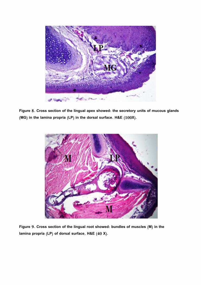
Figure 8. Cross section of the lingual apex showed: the secretory units of mucous glands (MG) in the lamina propria (LP) in the dorsal surface. H&E (100X).
Figure 9. Cross section of the lingual root showed: bundles of muscles (M) in the lamina propria (LP) of dorsal surface, H&E (40 X).

References 1. Gali, M.A. and Dauod, H.A.M. (2002).
Comparative anatomy of chordates. Baghdad University Press: 799pp (in Arabic).
2. Kent, G. C. and Carr, R.K. (2001). Comparative anatomy of the vertebrates, 9th ed.McGraw-Hill Co.,New York:XV11+524 PP.
3. Komarek, K.V., Malinovesky, L. and Lemez,L. (1986). Anatomia avium domesticarum et embryology galli. Priroda vedavatel,stvo Knihacasposov ,Bratyslava. (Abstract).
4. Iwasaki ,S.I. (2002). Evolution of the structure and function of the vertebrate tongue.J.Anat.201:1-13.
5. Stevens,A. and Lowe, J.S. (2005). Human histology (3rd ed.). Elsevier limited, USA: 190-194.
6. Parchami, A., Dehkordio, F.R.A. and Bahadoran,S. (2010). Scanning electron microscopy of the tongue in the golden eagle Aquila chrysaetos (Aves: Falconiformes Accipitridae). World. J. Zool.,5(4): 257-263.
7. Emura,S., Okumura, T. and Chen, H. (2009). Scanning electron microscopic study of the tongue in the Japanese pygmis woodpecker (Dendrocopos kizuki). okajimas Folia Anat. Jpn., 86(1): 31-35.
8. Jackowiak, H., Andrzejewski, W. and Codynicki, S. (2006). Light and scanning
electron microscopic study of the tongue in the cormorant phalacrocorax carbo
(phalacroracidae,aves). Zool. Sci., 23: 161-167.
9. Pasand, A. P., Tadjalli, M. and Mansouri, H. (2010). Microscopic study on the tongue of male ostrich. Eur. J. Bio.Sci., 2(2): 24-31.
10. Emura,S. and Chen,H. (2008). Scanning electron microscopic study of the tongue in the owl (Strix uralensis). Anat. Histol. Emberyol., 37: 475-478.
11. Jackowiak,H., Skieresz-szwczyk, K., Iwasaki, S.I. and Meyer,W. (2O11). Functional morphology of the tongue in the domestic goose (Anser anser domestica). Anat. Rec., 294: 1574-1584.
12. Iwasaki, S.I. (1992). Fine structure of the dorsal lingual epithelium of the little tern, sterna albifrons (Aves, Lari). J. Morphol., 212: 13-26.
13. Parchami, A. and Dehkordio, F.R.A. (2011). Lingual structure of the domestic pigeon (Columba livia domestica). A light and scanning electron microscopic studies. Mid. Eas. J. Sci, Res., 7(1): 81-86.
14. Erdogan, S., Sagsoz, H. and Akbalik, M.E. (2012). Anatomical and histological structure of the tongue and histochemical characteristics of the lingual salivary glands in the chukar partridge (Alectoris chukar, Gray 1830). Brit. Poul. Sci., 53(3): 307-315.
15. Parchami, A., Fatahian, R. A. and Bahadoran,S. (2010). Fine structure of the dorsal lingual epithelium of the

common quail (Coturinx coturnix) Worl. Appl. Sci. J. 10(10): 1185-1189.
16. Crole, M. R. and Soley, J.T. (2009). Morphology of the tongue of the emu (Dromaius novaehollandae). Histological features .Onderst. J.Veter. Res. 76: 347-361.
17. Iwasaki, S.I., Asami, T. and Chiba, A. (1997). Ultra structural study of the keratinization of the dorsal epithelium of the tongue of Middendorff,s bean goose ,Anser fabalis middendorffi (Anseres,Antidae) .Anat.Rec. , 247:149-163.
18. Jackowiak,H. and Godyicki, S. (2005). Light and scanning electron microscopic study study of the tongue in the white tailed eagle(Haeliaeetus albicilla, Accitripidae, Aves). Anna. Anat.,1 87: 197-222.
19. Grant, V. and Temeles, E.J. (1992). Faraging ability of Rufus humming birds on humming bird flowers and how kmoth flowers. Proc. Noit. Acad. Sci. USA, 89: 9400-9404.
20. Iwasaki, S.I. and Kumakura, M. (1994). An ultrastructural study of the dorsal lingual epithelium of the rat snake Elaphe quadrivirgata.Acta. Anat.,176:455-462.
21. Mutlak, B.H., Dauod, H.A.M. and Al-Dori,T.Y. (2000). Morphological and Histological study of fresh water turtle Cleymms caspica caspica. J. Dyalla, 8(1): 115-126.
22. Jackowiak, H. and Ludwing, M. (2008). Light and scanning microscopic study of
the structure of the ostrich (Strutio camelus) tongue. Zool. Sci., 25(2): 188-194.
23. Bernard, T., Kuniaki, T., Yuji, S., and Carleton, J. (1997). Ultrastructure of the salivary glands in the mid tongue of the common vampire bat,Domodus rotundus. Anat. Rec.,246: 196-205.
24. Jackowiak, H. and Andrz, W. (2006). Light and scanning electron microscopic study of the tongue in the cormorant phacrocorax carbo (Phalacoracidae, Aves). Zool. Sci., 23(2): 161-167.


![Morphological, histological, and molecular evidence of ......Mugil cephalus from the Ago Bay in the city of Shima, Mie Prefecture, Japan by Maeno et al. [7]. Subsequently, M. spinacurvatura](https://img.pdfslide.net/doc/110x75/60d4fa383641a61d9627fafe/morphological-histological-and-molecular-evidence-of-mugil-cephalus-from.jpg)