Embed Size (px)
Citation preview
135
ENDODONTOLOGYENDODONTOLOGYENDODONTOLOGYENDODONTOLOGYENDODONTOLOGY
135
Case Report
Morphological variations in the root canal system ofmandibular second molar : A case series
Shraddha Chokshi #Jahnvi Mehta #Pallav Chokshi #Rupal Vaidya #
ABSTRACT
Consistently high levels of success in endodontic treatment require an understanding of root canal anatomy and
morphology. To achieve endodontic success, the entire root canal system must be three dimensionally cleaned,
shaped and obturated. The clinician must have a thorough understanding of normal anatomy and of its variations from
the norm. As with most of posterior teeth, the mandibular second molar has several variants in its canal configuration.
This includes single canal, two canals, three canals, four canals and five canals and; C-shaped canal system. All these
variations represent a challenge to its thorough debridement and obturation. This has led to the proposal of many
modified techniques to optimize the technical quality and hence the prognosis of endodontic therapy.
Key words: root canal morphology, diagnosis, radiography, endodontic treatment.
IntroductionThe result of successful endodontics revolves
around knowledge, respect, and appreciation for
root canal anatomy and careful, thoughtful,
meticulously performed cleaning and shaping
procedures. Knowledge of pulpal anatomy, its usual
and unusual configurations and possible variations
is critical for success in endodontics and lack of
such knowledge may lead to treatment failure.1
A clinician is required to have an insight of the
morphology of tooth related to its shape, form and
structure before commencing treatment. This can
be achieved by routine periapical radiographs to
assess the number, length, curvature and aberrations
of the canal system of the tooth.
Mandibular second molars usually have two
roots and three root canals but variations in the
number of roots as well as canal morphology are
not uncommon. Which includes single canal, two
canals, three and four canals, five canals and the C-
shaped canal system.2,3 Because proper cleaning,
shaping, and three dimensional obturation of the
entire root canal system is regarded as an important
determinant to good prognosis, the variations in root
canal system, thus, represents a challenge to its
proper diagnosis, debridement and obturation.4
Case SeriesAlthough, root canal therapy has been a
practice trend to save teeth since ages, nature till
date does not stop mystifying the dentist with the
various root canal morphologies. In all the presented
cases, teeth had been planned for routine root canal
treatment followed by full coverage restoration after
their detailed history and clinical as well as
# Dept. of Conservative Dentistry and Endodontics, Ahmedabad Dental College and Hospital, Ahmedabad
136
ENDODONTOLOGYENDODONTOLOGYENDODONTOLOGYENDODONTOLOGYENDODONTOLOGY
136
radiographic examination. The access cavity
preparation had been done under 2.5x
magnification loupes and with rubber dam isolation.
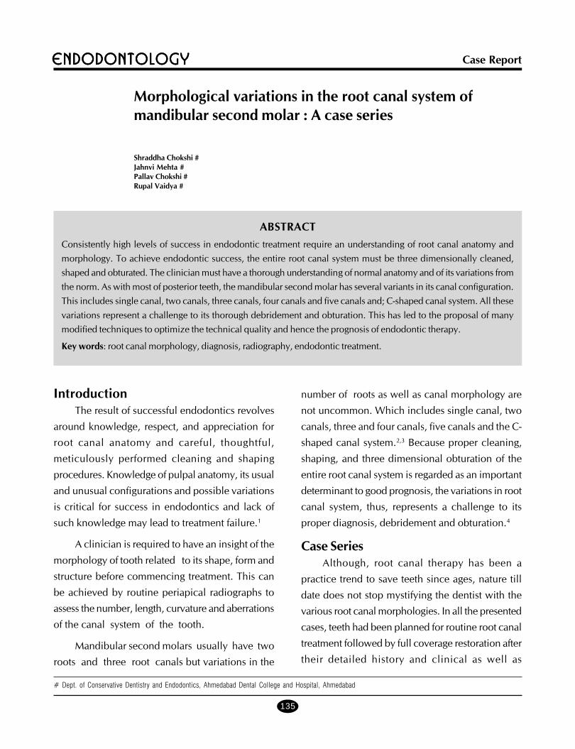
Case 1: SINGLE CANAL
After thorough debridement of the pulp tissue
from the pulp chamber, a single but considerably
large opening was found in the center of the pulp
chamber floor, which was confirming the presence
of a single root and single canal which was
suspected from the radiograph (Fig.1a). Working
length was measured and cleaning and shaping of
the canal was done with K- file of No. 45-80 followed
by obturation with corresponding gutta purcha
cones in sectional obturation manner (Fig.2a).
Case 2: TWO CANALS
While getting access to the pulp chamber, two
canal orifices representing one mesial and one distal
canal connected by a developmental fusion line at
the chamber floor was found (Fig.1band their
presence were confirmed with radiograph. After
determination of correct working length, cleaning
and shaping was done with the help of K-files
followed by obturation in lateral condensation
manner. (Fig.2b)
Case 3, Case 4: THREE CANALS and FOUR CANALS
Of all the above mentioned variations;
Mandibular 2nd molar with 3 canals and 4 canals
has got the maximum share (Fig.1c & Fig.1d). After
the access cavity preparation and determination of
correct working length, cleaning and shaping was
done with the help of Rotary ProTaper files, followed
by obturation with corresponding gutta purcha
cones (Fig.2c & Fig.2d).
SHRADDHA CHOKSHI, JAHNVI MEHTA, PALLAV CHOKSHI, RUPAL VAIDYA
Figure-1: (a) to (f): showing clinical picture of the access cavity after cleaning and shaping representing the number of canals present.
(a) (b) (c)
(d) (e) (f)
137
ENDODONTOLOGYENDODONTOLOGYENDODONTOLOGYENDODONTOLOGYENDODONTOLOGY
137
Case 5: FIVE CANALS
While gaining access to the canal system,
mesiobuccal and mesiolingual canal were found
to be located very far from each other. While
examination of access cavity using magnification
loupes a third tiny orifice was located in between
mesiobuccal and mesiolingual canals (Fig.1e). 6 No.
C+ file along with EDTA was used to negotiate the
canal initially. Two other canals were located
distobuccally and distolingually. The presence of
five canals was then confirmed with angulated
radiography. Cleaning and shaping of the middle
mesial canal was performed with hand K file and
rest of the canals was prepared using Rotary
ProTaper files followed by obturation with gutta
purcha cone (Fig.2e).
Case 6: C - SHAPED CANAL
After the preparation of access cavity its
thorough debridement with 5.25% sodium
hypochlorite, the pulpal floor showed a single C-
shaped canal (Fig.1f). Working length was
determined using apex locator and angulated
radiographs. Cleaning and shaping was done with
KSS files. In between the instrumentation, the canal
was irrigated with 5.25% sodium hypochlorite
thoroughly for maximum debridement of the
complex anatomy of the root. Calcium hydroxide
intracanal medicament was placed for one week.
At the next appointment, the medicament was
flushed and smear layer was removed with 17%
EDTA and 5.25% sodium hypochlorite. The canal
was obturated with thermoplasticized gutta-percha
technique and AH-Plus sealer (Fig.2f).
Discussion :When anatomic variations are detected
clinically, treatment can be performed with
conventional or rotary instrumentation and
obturation techniques respecting technical and
biological principles. Endodontic success in teeth
with variations in the number and morphology of
canals requires a correct diagnosis and careful
clinical and radiographic inspection.5,6
MORPHOLOGICAL VARIATIONS IN THE ROOT CANAL SYSTEM OF MANDIBULAR SECOND MOLAR : A CASE SERIES
Figure-2 : (a) to (f): radiographic images after root canal obturation.
(a) (b) (c)
(d) (e) (f)
138
ENDODONTOLOGYENDODONTOLOGYENDODONTOLOGYENDODONTOLOGYENDODONTOLOGY
138
SHRADDHA CHOKSHI, JAHNVI MEHTA, PALLAV CHOKSHI, RUPAL VAIDYA
While preparation of access cavity, dental
operating microscope and dental loupes, offer
magnification and illumination of the operating
field and substantially improve the visualization of
root canal orifices which enhance the quality of
vision and make the correct identification of the root
canal system easier. The use of apex locator can
be important to determine the working length.
Additional anatomic information about the root
canals can be obtained by angulated radiography,
R.V.G, CT-scan and 3D reconstruction.7
Mandibular second molars usually have two
roots and three root canals. Two root canals are
located in mesial root and another one in distal
root. Hess reported that the prevalence of three root
canals in mandibular molars was 78%.8,9,10 There
is an abundant amount of reports that relate the
anatomic variations of mandibular second molars,
which includes presence of single canal- ~2 to7%,
two canal- ~30%, four canals- ~20%, five canals-
~2% and C shaped system - ~8.3 to 8.5%.9,11
All these variations represent a challenge to
its thorough debridement and obturation. This has
led to the proposal of many modified techniques to
optimize the technical quality and hence the
prognosis of endodontic therapy.
Conclusions :When root canal treatment is to be performed
the clinician should be aware that both external and
internal anatomy may be abnormal. Knowledge of
possible variations in internal anatomy of human
teeth is important for successful endodontic
treatment. The early recognition of these
configurations facilitates cleaning, shaping, and
obturation of the root-canal system. Every attempt
should be made to find and treat all root
canals to ensure successful endodontic treatment.
The importance of an accurate clinical evaluation
of root canal number and morphology in
mandibular second molars cannot be
overemphasized.
References :
1. Rahimi S, Shahi S, Lotfi M, Zand V, AbdolrahimiM,Es’haghi R. Root canal configuration and the prevalence ofC-shaped canals in mandibular second molars in an Iranianpopulation. J Oral Sci. 2008;50(1):9-13.
2. Franklin S., Weine, Richard A., Pasiewicz,R. Ted R. Canalconfiguration of the Mandibular second molar using aclinically oriented in vitro method.J Endod 1988;14:207-213.
3. FabraCampos H. Unusual root anatomy of mandibularfirst molars. J Endod 1985;11:568-72.
4. C. Maniglia et. al, A Case of Unusual Anatomy in SecondMandibular Molar with Four Canals, Eur J Dent 2008;2:217-219
5. Frank j., Vertucci, Root canal morphology and itsrelationship to endodontic procedures; Endodontic Topics2005, 10, 3–29
6. Kala M., Chandki R., Lodha E., Shaktidar P., c shaped canalconfiguration: a diagnostic dilemma ; AOSR 2011;1(2):79-83.
7. Ruwan J., BDS, MS Thomas Ka-Lun Li; C-shaped canals inmandibular second molars in the Hong Kong population: acomputed tomographic study; Hong Kong Dental Journal2008;5:27-30.
8. Cleghorn BM, Goodacre CJ, Christie WH: Morphologyof teeth and their root canal system. In: Ingle JI, Backland LK,Baumgarthner JC: ENDODONTICS, 6th Edition. BC Decker:Inc, 2008: pp. 151-210.
9. S. A. MANNl’NG; Root canal anatomy of mandibularsecond molars. Part I, International Endedmticjminuil (1990)23,34-39.
10. S.A.MANNl’NG; Root canal anatomy of mandibularsecond molars. Part II, International Endedmticjminuil (1990)23,40-45.
11.Skidmore AE, Bjorndal AM. Root canal morphologyof the human mandibular first molar. Oral Surg Oral MedOral Pathol 1971;32:778-84.