Embed Size (px)
Citation preview

Br. J. exp. Path. (1969) 50, 600.
MORPHOLOGY OF EARLY LARGE VESSEL LESIONS INEXPERIMENTAL HYPERTENSION
B. VERESS, A. KOCZI2 AND H. JELLINEKFrom the IInd Department of Pathology, University Medical School, Budapest, Hungary
Received for publication July 7, 1969
SUMMARY.-Lesions of the aorta and large muscular arteries were examinedin experimental hypertension ofthe albino rat. In contradiction to the publisheddata the earliest hypertensive lesion was a small artery type subendothelialfibrinoid and necrosis of single smooth muscle cells in the media and not anintimal thickening. In the large vessels such changes developed only afterlong periods of hypertension. No lesion corresponding to the media fibrinoidof small vessels was found in the large muscular vessels or in the aorta, owingapparently to inhibition of a greater penetration of plasma by the elastic fibres.This barrier role may in addition account for the absence of perivasculargranulation around the aorta.
In a later stage of the process, macrophages appeared in the subendothelialfibrinoid, phagocytosed it, and gave rise to intimal proliferation analogous tothe intimal thickening in small vessels.
As reported previously, in small and medium arteries morphologically identicallesions, fibrinoid necrosis, were produced by various aetiological factors, such asnoradrenaline treatment (Jellinek, Hiittner, Kerenyi and Gabor, 1966a), malignanthypertension (Kerenyi, Jellinek, Hittner, Gora6cz and Konyar, 1966a), sensitizationby horse serum and painting with acid or alkali (Hiittner, Jellinek, Kereny; andSzemenyei, 1966, Jellinek, Szemenyei and Hittner, 1966b). Subsequently it wasfound that the fibrinoid necrosis consisted of two morphologically distinguishableparts with dissimilar electron microscopic properties: (1) subendothelial fibrinoid,composed of plasma condensed in the space between the endothelium andinternal elastic lamina, and (2) media fibrinoid, composed of necrotic smoothmuscle cells admixing with the plasma which had penetrated the media.In a later stage of the process, either a perivascular granulation developed as aresult of penetration of plasma through the vessel wall, or macrophages appearedwhich phagocytized the fibrinoid and gave rise to proliferation of the intima,while in the media either fibrosis or hyalinosis developed.
The hypertensive lesions of the aorta have been described (Fishberg, 1925;Dill and Isenhour, 1942; McGill, Frank and Geer, 1961) as arteriosclerosis-likechanges manifesting by intimal thickening of partly foamy cell nature. Incon-sistently, we have found that the hypertensive changes of the aorta and largemuscular vessels develop by an essentially similar mechanism to those seen insmall arteries, thus the intimal proliferation represents only the final stage of theprocess. The present experiments were undertaken to clarify the precise nature ofthe preceding acute lesions.
MATERIALS AND METHODS
Forty Wistar rats of both sexes were used. They were rendered hypertensive by Lorinczand GorAcz's (1954) kidney compression method. From the second day on, the animals'blood pressure rose from the normal 90-110 mm. Hg. to 150-200 mm. Hg. The rats were

EARLY LARGE VESSEL LESIONS IN HYPERTENSION
killed in succession on the 2nd, 5th, 9th, 14th, 28th, 30th, 35th, 40th, 44th and 70th days ofthe experiment. The abdominal and thoracic segments of the aorta, the iliac and femoralarteries and small mesenteric vessels were fixed in formalin and embedded in paraffin.Sections were stained with haematoxylin and eosin, Azan, Mallory's phosphotungstic acidhematoxylin (PTAH), Krutsay's trichrome stain and PAS; unstained sections were examinedby phase contrast microscopy and, after aniline reaction, by polarization microscopy.
RESULTS
In the large muscular arteries the initial lesions developed by the 28-30th dayof experimental hypertension. By that time the vascular fibrinoid had alreadybeen replaced by intimal proliferation in the small arteries. In the large arteries,the first sign of damage was necrosis of single smooth muscle cells. The necroticvascular smooth muscle cells stained vivid red with Azan and trichrome stain,had a marked affinity for phosphotungstic acid, were PAS positive and showedan increased birefringence. The internal elastic lamina had elongated and plasmamaterials had accumulated between it and the detached endothelium (Fig. 1).The subendothelial fibrinoid, which developed by the 35-40th day, considerablyconstricted the vascular lumen. Its staining, polarization and phase opticalproperties were similar to those of similar small arterial lesions (Ashworth andHaynes, 1948; Lendrum, 1955; Kerenyi et al., 1966a). Simultaneously, aperivascular granulation developed, as seen also in the case of small arteries(Fig. 2). By the 44th day slightly elongated cells with round nuclei appeared inthe subendothelial fibrinoid with, however, no evidence of fibrinoid in theirenvironment (Fig. 3). In some vessels these cells occupied the entire subendo-thelial space, but clumps of fibrin were seen exclusively above the internal elasticlamina (Fig. 4).
In the abdominal segment of the aorta, the first sign of damage was .smoothmuscle cell necrosis which appeared by the 40th day. Some cellular nucleibecame pyknotic, some cells appeared swollen. The staining properties of theimpaired muscle cells resembled those of similarly affected cells in the musculararteries. With the progression of the process, the necrosis involved many adjacentcells, but the homogenous media fibrinoid involving the full width of the vesselwall as observed in small vessels, was not seen either in the aorta or in the largearteries (Fig. 5). Simultaneously with the development of muscle cell necrosis,some endothelial cells detached from the internal elastic lamina and either palestaining, or eosinophilic dilute plasma (Fig. 6) had appeared beneath them, withstaining and submicroscopic properties corresponding to the reactions of sub-endothelial fibrinoid in other types of vessels (Fig. 7-8). In none of the casesstudied did a perivascular granulation develop around the aorta. In the aorta'sthoracic segment no subendothelial fibrinoid was found; in that area exclusivelysmooth muscle cell necrosis was seen.
In animals with a milder degree of hypertension, the vascular lesions developedat a later time; e.g. in the aorta of an animal killed on the 70th day only moderatecondensation of fibrinoid was seen in the subendothelial space. But in everycase, lesions representing different degrees of severity occurred simultaneouslyin the various types of vessels. Fig. 9 shows the aorta and a large muscularartery of a rat killed on the 44th day. While in the muscular artery an intimalthickening resulting from earlier damage is seen, in the aorta subendothelialfibrinoid is present, indicating acute impairment.
601

B. VERESS, A. KOCZE' AND H. JELLINEK
DISCUSSION
As compared to the wide interest in arteriosclerosis, relatively few authors havedealt with the morphology of hypertensive vascular changes. Of the investigatorsexamining autopsy material, Ashworth and Haynes (1948) have described theearliest sign of hypertension as smooth muscle hypertrophy in the vasa vasorum.Analysing 20 cases, Zlateva (1960) has noted that in hypertension, the firstvascular lesion was hyperplasia of medial smooth muscle cells, viz. the fibrinoidnecrosis of the vasa vasorum. Later on fibrosis or hyalinosis developed in thevasa vasorum and the consequent reduction of oxygen supply gave rise to ischaemicsmooth muscle cell necrosis in the media. Inconsistently, Smirnova (1958),and Lange (1924) have believed that impairment of the adventitial vessels gaverise to intimal hyperplasia rather than to medial lesions. Fishberg (1925) hasregarded the intimal proliferation an early hypertensive lesion. Simultaneously,Fishberg (1925) and Dietrich (1930) have reported a pronounced hyperplasia ofthe aortic elastic membrane in hypertension.
Authors generally agree that the hypertensive changes of the large vessels areof the same nature as the arteriosclerotic lesions. Examining the aortas of ratsrendered hypertensive, and killed between 4-9 month periods of hypertension,McGill et al. (1961) found intimal thickening consisting in part of connective tissueand a few foamy cells, in part of many lipid containing foamy cells. The grossstructure of the changes resembled the arteriosclerotic lesions. Hypertensivevascular changes manifesting by intimal thickening were observed also in rabbits(Dill and Isenhour, 1942; Bronte-Stewart and Heptinstall, 1954) and dogs (Moser,1954; Wakerlin, Moss and Kiely, 1957). Recently Still (1967) has observed byelectron microscopy that in early hypertension, monocytes and fibrin containingplasma materials appeared in the enlarged subendothelial space of the rat aorta.None of the above mentioned authors has described necrosis of the medial smooth
EXPLANATION OF PLATESFIG. 1.-Subendothelial fibrinoid between the elongated internal elastic lamina and the
detached endothelial cells of a large muscular artery. The lesion stains bluish-black; thenecrotic medial smooth muscle cells show an increased affinity for the stain and have fallenapart in places (arrow). Hypertension 30 days. PTAH. x 250.
FIG. 2.-The vascular lumen is constricted by the broad subendothelial fibrinoid. Note thebroad zone of granulation tissue around the artery. Hypertension 40 days. PTAH. x 340.
FIG. 3.-Cells with round nuclei along the luminal side of the subendothelial fibrinoid. Noevidence of fibrinoid between the cells. Hypertension 44 days. PTAH. x 340.
FIG. 4.-In the broad intimal proliferation residues of fibrinoid are evident exclusively abovethe internal elastic lamina (arrow). Hypertension 44 days. PTAH. x 340.
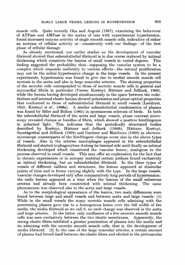
FIG. 5.-Single muscle cell necrosis in the aorta. The necrotic cells are separated by elasticfibres (arrow). Hypertension 40 days. PTAH. x 250.
FIG. 6.-Some endothelial cells have been lifted from the internal elastic lamina by the penetra-ting plasma (arrow). Note vacuolar degeneration of medial smooth muscle cells. Hyper-tension 40 days. H. and E. x 380.
FIG. 7.-Plasma substances deposited in the subendothelial space of the aorta give the colourreaction typical of the subendothelial fibrinoid, viz. stain red with Krutsay's trichrome stain.Hypertension 44 days. x 340.
FIG. 8.-In polarized light, clumps showing positive birefringence are apparent in the fibrinoid.Hypertension 44 days. Anilin reaction. x 250.
FIG. 9.-In the muscular artery the subendothelial fibrinoid (SF) has been partly replaced byintimal proliferation (P). In the aorta the narrow subendothelial fibrinoid (arrow) signifiesacute damage. Hypertension 44 days. PTAH. x 70.
602

BRITISH JOURNAL OF EXPERIMENTAL PATHOLOGY. Vol. L, No. 6.
I
0 *:
...ISE
4..
- ,
I
ik .%. .,*,.?
V
NIk., vi 7
Veress, K6cze' and Jellinek.
4rpt
ki,..k
.w ...
A,:..-t,F"
...-P.
Pc
NO
iL

BRITISH JOURNAL OF EXPERIMENTAL PATHOLOGY. Vol. L, No. 6.
..
$ W~e< A
It
Veress, Kocze and Jellinek.
if-ii,-A
e*I
:u
.1
EARLY LARGE VESSEL LESIONS IN HYPERTENSION
muscle cells. Quite recently Oka and Angrist (1967), examining the behaviourof ATPase and AMPase in the aortas of rats with experimental hypertension,found increased enzyme activity of single smooth muscle cells, indicative of eitheran increase of cellular activity or consistently with our findings of the firstphase of cellular damage.
As already mentioned, our earlier studies on the development of vascularfibrinoid showed that subendothelial fibrinoid is in due course replaced by intimalthickening which constricts the lumina of small vessels in varied degrees. Thisfinding suggested the probability that-supposing the vascular system to be acomplex which responds uniformly to various effects the intimal proliferationmay not be the initial hypertensive change in the large vessels. In the presentexperiments, hypertension was found to give rise to medial smooth muscle cellnecrosis in the aorta and also in large muscular arteries. The staining propertiesof the necrotic cells corresponded to those of necrotic muscle cells in general andmyocardial fibrils in particular (Veress, Kerenyi, Huttner and Jellinek, 1966),while the lesions having developed simultaneously in the space between the endo-thelium and internal elastic lamina showed polarization and phase optical propertiesthat conformed to those of subendothelial fibrinoid in small vessels (Lendrum,1955; Ker6nyi et al., 1966a). A similar subendothelial condensation of plasmawas found by Siller and Bolton (1961) in spontaneous sclerosis of birds. In boththe subendothelial fibrinoid of the aorta and large vessels, phase contrast micro-scopy revealed clumps or bundles of fibrin, which showed a positive birefringencein polarized light. This indicates that the periodically structured bodiesdescribed by Kerenyi, Huttner and Jellinek (1 966b), Htittner, Kere'nyi,Szenta6gothai and Jellinek (1968) and Gardner and Matthews (1969) as electron-microscope counterparts of the birefringent clumps occur also in the above typesof vessels. Also in the arteries macrophages appeared in the subendothelialfibrinoid and started to phagocytose it along its luminal side until finally an intimalthickening developed which constricted the vascular lumen; analogous to theprocess observed in small vessels. This may offer an explanation for the fact thatin chronic experiments or in autopsy material certain authors found exclusivelyan intimal thickening, but no subendothelial fibrinoid. In the three types ofvessels of different calibres and structures, the lesions appeared at dissimilarpoints of time and in forms varying slightly with the type. In the large vessels,vascular changes developed only after comparatively long periods of hypertension:the early lesions appeared at a time when the lumina of the small subserousarteries had already been constricted with intimal thickening. The samephenomenon was observed also in the aorta and large vessels.
As to the morphological appearance of the lesions, two main differences werefound between large and small vessels and between aorta and large vessels: (1)While in the small vessels the many necrotic muscle cells admixing with thepenetrating plasma gave rise to a homogenous lesion over the full width of themedia, the media fibrinoid as we call it, no such change was observed in the aortaand large arteries. In the latter, only confluence of a few necrotic smooth musclecells was seen exclusively between the two elastic membranes. Apparently, thestrong elastic fibres inhibited a greater penetration of plasma into the media andits admixing with the necrotic smooth muscle cells, that is, the development ofmedia fibrinoid. (2) In the case of the large muscular arteries, a certain amountof plasma had forced itself between the elastic fibres and elicited in the perivascular
603

604 B. VERESS, A. KOCZE AND H. JELLINEK
space a granulation which was, nevertheless, of a lesser degree than in thecase of the small arteries. But in no case was a perivascular granulation foundaround the aorta, suggesting that the elastic fibres had actually played a barrierrole.
REFERENCESASHWORTH, C. T. AND HAYNES, D. M.-(1948) Am. J. Path., 24, 195.BRONTE-STEWART, B. AND HEPTINSTALL, R. H.-(1954) J. Path. Bact., 68, 407.DIETRICH, K.-(1930) Virch. Arch. Path. Anat., 274, 452.DILL, L. V. AND ISENHOUR, C. E.-(1942) Arch. Path., 33, 655.FISHBERG, A. M. (1925) Arch. Int. Med. Chicago, 35, 650.GARDNER, D. L. AND MATTHEWS, MARGARET, A. (1969) J. Path., 97, 51.HUTTNER, I., JELLINEK, H., KERENYI, T. AND SZEMENYEI, KLARA.-(1966) Acta mnorph.
Hung., 14, 169.HUTTNER, I., KERIENYI, T., SZENTAGOTHAI, KLARA AND JELLINEK, H.-(1968) Exp.
rmol. Path., 7, 187.JELLINEK, H.-(1967) Angiology, 18, 547.JELLINEK, H., HUTTNER, I., KERiENYI, T., GABOR, GY. AND POGAiTSA, G.-(1966a)
Acta morph. Hung., 14, 183.JELLINEK, H., SZEMENYEI, KLARA, KEREINYI, T. AND HUTTNER, I.-(1966b) Acta morph.
Hung., 14, 165.KER1ENYI, T., HUTTNER, I. AND JELLINEK, H.-(1966b) Ztschr. mikr. Forsch., 74, 121.KERE1NYI, T., JELLINEK, H., HPTTNER, I., GORAICZ, GY. AND KONYAR, 1RVA.-(1966a)
Acta morph. Hung., 14, 175.LANGE, F.-(1924) Virch. Arch. Path. Anat., 248, 463.LENDRUM, A. C.-(1955) J. clin. Path., 8, 180.L6RINCZ, GY. AND GORAcz, GY.-(1954) Acta physiol. flung., 5, 489.MCGILL, H. C., FRANK, M. H. AND GEER, J. C. (1961) Arch. Path. Chicago, 71, 96.MOSER, C. (1954) Circ. Res., 2, 243.OKA, M. AND ANGRIST, A.-(1967) Lab. Invest., 16, 25.SILLER, W. G. AND BOLTON, W.-(1961) Nutr. Soc. Proc., 20, 163.SMIRNOVA.-(1958) cited by ZLATEVA, M. D. (1960) Ark. Patol., 22, 23.STILL, W. I. S.-(1967) Am. J. Path., 51, 721.VERESS, B., KERE'NYI, T., HUTTNER, I. AND JELLINEK, H. (1966) J. Path. Bact., 92, 511.WAKERLIN, G. E., Moss, W. G. AND KIELY, J. P. (1957) Circ. Res., 426.ZLATEVA, M. D.-(1960) Ark. Patol., 22, 23.