Embed Size (px)
Citation preview

STUDY OF RESPIRATION IN HEALTHY AND MOSAIC-INFECTED TOBACCO PLANTS
VIOLETTE F. C.-GLASSTONE
(WITH FIVE FIGURES)
Introduction
Comparatively little investigatioii has been made into the fundamentaliiietabolic processes of plant viruses. BRONFENBRENNER and REICHERT (2,3, 4) were unable to detect any respiration in either viruses or bacterio-phages. The isolation of virus, free from plant cells, and in a conditionsuitable for such experiments, is obviously very difficult. The effect of virusinfections on the metabolism of host plants is also a matter of great interestand may help to elucidate the action of the virus if sufficient data are col-lected. Siniee most viruses affect the chlorophvll eontent of the plant, it isnot surprisincg that the work that has been done so far has been directedprimarily on respirationi. TIIUNG (20) observed that the respirationi rate ofpotato leaves infected with leafroll was higher than that of the correspond-ing healthy leaves. Also working with potatoes infected by leafroll, WHITE-HEAD (21) studied the course of respiration from the immiiature tuber up tothe development of the plant. In miaturing tubers and in leaves, or shootswith expanded leaves, the respiration rate of the diseased planits was higherthani that of the healthy plants; but for a period lasting from dormancy ofthe tuber until the expansion of the leaves, the respiration rate of the healthymaterial was the greater. This was true both for aerobic and anaerobic con-ditions, and also for various conditions of light, temperature, anid carbondioxide supplyl. He concluded that the virus affects the respiration ratenot directly, but only by initerfering with the translocation of the respirablesiubstrate.
In a number of species of plants naturally infected by miiosaic or yellowsvirus diseases it was found (9) that the respiration rate was increasedin young leaves affected by virus but was decreased in older leavesthus affected. This was not apparently connieeted with the variationis incarbohydrate or niitrogen contents of the leaves. On the other han-d,DUFRE'NOY (8) noted that the high rate of respiration in healthy tobaccobuds was decreased by tobacco-mosaic virus, whereas the lower respiratoryactivity of the discolored leaves was increased by the presence of the virus.
CALDWELL (6) inoculated tomato plants with aucuba mosaic as seedlingsand at the five-leaf stage and measured the respiration of the diseased tops.In all his experiments there was an initial high output of carbon dioxidewhich fell off to a steady level. Tops of plants inoculated at the five-leafstage evolved more carbon dioxide thani the healthy controls throughout the
2a,7
www.plantphysiol.orgon April 21, 2020 - Published by Downloaded from Copyright © 1942 American Society of Plant Biologists. All rights reserved.

PLANT PHYSIOLOGY
experimnental period; but those inoculated as seedlinigs showed a lower out-put at the beginning, followed by a higher output than the healthy topsonce the stable level had been reached. He considered his experiments indi-cated that the virus affects. the enzymes which "activate" the substratebefore it is broken down to carbon dioxide in the respiration mechanism.
In a series of experiments on discs of healthy and tobacco-mosaic-infectedtobacco leaves carried out by LEMMON (17), the respiration rate of thehealthy leaves was always higher than that of the diseased. KEMPNER (15)was unable to find any change in the respiration of tobacco leaves infectedby tobacco-mosaic virus, unless necrosis had set in.
There appears therefore to be a certain amount of discrepancy amongthe published results of the various workers in this field. All the-experi-ments quoted above were made with tissue that was alreadv diseased, and itseemed possible that a study eovering the entire development of the diseasewould be profitable.
During the summer of 1940 the writer made some preliminary observa-tions on the respiration of plants grown in soil. A respiration chamber wassealed over the plant top by means of a wax mixture. This seal was per-fectly airtight, and it is usual to assume that the respiration of the top canbe measured independently from that of the root by such a device. It wasdiscovered, however, that air can pass easily through plants, especially to-bacco, from root to leaf and vice versa (10). Hence it was possible that theair in the soil or roots below the surface of the wax seal was in some wayinterfering with the measurements.
For this reason it was decided to experiment with entire plants in nutri-ent solution to avoid effects due to the passage of air from other organs orto cutting, handling, drying out, or partial starvation of isolated material,all of which are known to have some influence on respiration. The presentpaper therefore is a report of experiments designed to follow the course ofthe respiration in entire plants from the time of their inoculation up to theappearance of the mosaic mottling, using the same plants throughout theperiod. The work was done in the spring of 1941.
Material
Ordinary tobacco-inosaic virus was used on Nicotiana tabacum L. var.Samsun as the host plant. This proved to be a very suitable subject forthe purpose, as the resulting disease did not distort the plant unduly, merelyproducing a reduction of chlorophyll in a mottled pattern and causingstunting. There was also the advantage that under favorable conditionsof light and temperature it was possible to note the time that the diseasebecame systemic by observing-the "clearing of the veins" (13).
The seeds were sown in nutrient sand, and as soon as the plants were
268
www.plantphysiol.orgon April 21, 2020 - Published by Downloaded from Copyright © 1942 American Society of Plant Biologists. All rights reserved.

GLASSTONE: RESPIRATION IN HEALTHY AND MOSAIC-INFECTED TOBACCO 269
large enough to handle they were transferred to nutrient solution. Thesolution recommenided by HOAGLAND and ARNON (11) with rather moreferric sulphate was found very satisfactory. The nutrient solution was con-tained in 4-liter battery jars which were covered with thick black paper toprevent growth of algae on the roots: no algae formed during any experi-inent. The plants were supported on thick wooden discs, containing holesfor the roots. The discs were well waxed with paraffin wax, which wasrenewed for each set of experiments. Great care was taken to keep separateall the glassware alid discs for the healthy and diseased plants, in order toavoid contaminationi. The olily coontaminations, which will be noted in theresults, were due to an accident, the origin of which was later discovered.The younger plants stood in the wooden discs without further support; theolder plants were usually held in place by cotton wads or occasionallv byglass rods. The solution was changed twice to four times a week, accordingto the quantity of plant material. The plants ranged in size from the small-est that could be handled conveniently, of an average individual weight of0.5 gm., up to the largest that could be put in the respiration chambers, ofan average weight of 40 gm. In any case it was considered desirable forthis experiment to avoid planits forming flowers, so the range in size wasactually the widest that could have been used under the circumstances.
The diseased plants were inoculated in the usual manner by rubbing witha ilnuslin pad dipped in tobacco-mosaie-virus juice (12). The healthy con-trol plants were rubbed with a similar pad dipped in distilled water to equal-ize the treatment as far as possible. Plants were thus inoculated after beingrin nutrient solution for periods of 1 day, 1 week, 2 weeks, 3 weeks, and 4weeks, respectively. The measurements of respiration rate were made olngroups of plants from the first day of inoculation up to the time that themosaic mottling developed. Two weeks were allowed for each group, whichwas usually long enough. The experiments in nutrient solution wererepeated at least once for each time period.
As the respiration measurements were carried out in the daytiine, theplants were illuminated artificially at night by means of 1000-watt Mazdabulbs. As a check on the growth and development of the virus, a group ofplants in soil grown under normal greenhouse conditions was always usedas observational controls. It was found that the clearing of the veins andmottling always appeared oni the same days in both the greenhouse plantsand in those receiving artificial illumination. The weight measurementswere made daily on the same plants by weighing the entire plant. Theplants were removed singly from the nutrient solution, placed carefully ona soft tissue, and the roots were gently blotted with another tissue to removethe moisture. The plant was then quickly weighed and replaced in solution.After a little practice, the whole process of weighing could be performed
www.plantphysiol.orgon April 21, 2020 - Published by Downloaded from Copyright © 1942 American Society of Plant Biologists. All rights reserved.

PLANT PHYSIOLOGY
very rapidly with no apparenit injury to the plants. In figure 1 are showi'ntwo plants which were weighed in this manner for 50 days. The plant onthe left was the healthy colntrol and the other was diseased. It will be seenthat the roots as well as the rest of the plants are in good conldition.
MethodThe apparatus consisted of 6 ulnits, onle of which is shown diagramn-
matically in fig,ure 2. Air from a compressed air manifold was freed froicarbon dioxide by passage through a soda-lime tower, A, a sodiumi1 hyvdroxide
FIG. 1. Planits photographed after 50 days of experiimentationi. (Photograph byJ. A. CARLILE.)
solution, B, and a dilute bariumn hydroxide soltutioii tinted with plienolphtlia-leini, C. The latter also acted as an indicator trap. The carboni-dioxide-freeair passed through a flowmeter, D, provided with its own overflow trap, andtheniee via a tube, J, into the respiration chamber, E. This consisted of alarge bell jar, 53 ecn. high and 26 cm. initernal diameter, sealed firmlv to abase of ground plate glass, F, by mieans of a ring of plasticum clay, G, anidenclosed during the experiment with a cover of thiek black satini to exclude
270
www.plantphysiol.orgon April 21, 2020 - Published by Downloaded from Copyright © 1942 American Society of Plant Biologists. All rights reserved.

GLASSTONE: RESPIRATION IN IIEALTHY AND MOSAIC-INFECTED TOBACCO 271
all light. The air next passed through the tube, K,K, to enter the titrationflasks, P (a very slight modification of that used by MACK, 18). The exittube, Q, fromii this flask led to a glass tower, R, 65 em. long, containing glassbeads; next, bv a glass tube, S, to an indicator trap, T, containingr dilutebarium hydroxide solution, and thenee to the vacuum manifold. With theexception of the brass manifolds, glass tubing, joined by butt to butt jointsin thick rubber pressure tubing, was used throughout. The 6 units wereconniected by the iuanifolds leadinig from the compressed air and vacuummains. Each unit had its own taps to the 2 manifolds, and the vacuummaniifold was further protected from sudden changes of pressure by mier-
V
Compressedaair
FIG. 2. Diagram of one unit of the apparatus.
cury valves and traps. A general view of the apparatus is showsn infigure 3.
The rate of flow of air through the apparatus was kept at 6 liters perhour, wshicah experience proved was sufficient to provide very efficientscarubbing byr the barium hydroxide solution in the bead tower. The indi-cator trap, T, alwvays remained calear, showingr that the air was colupletelyfreed from carbon dlioxide in the tower. The pressure in the respirationchamlber was mlaintained at 1 atmuosphere, as indcliated by the levels of then-butyl phthalate in the attached manometer, LJ. Very slight adjustmentsof the 2 taps were required for this, and the apparatus was kept under con-tinulal observation during all experiments to maintain the required uniformcon(1 itions.
www.plantphysiol.orgon April 21, 2020 - Published by Downloaded from Copyright © 1942 American Society of Plant Biologists. All rights reserved.

PLANT PHYSIOLOGY
The procedure was briefly as follows: Before sealing down the bell jar,the plants, supported on the wooden disc (I) so that their roots dipped intothe nutrient solution in the battery jar, H, were arranged so that the airentering by the tube, J, would bubble into the solution. The bell jar wassealed into position and tested for leaks by clamping off K,K and noting themovements of the liquid in the manometer, L. K,K was unclamped and thecarbon-dioxide-free air was allowed to flow through the apparatus for onehour. This period had previously been established as more than sufficientto renew the air in the apparatus. Next, a measured amount of standardbarium hydroxide and a drop of phenolphthalein were placed in the titra-
i
...r ............
FIG. 3. General view of the respiration apparatus. (Photograph by J. A. CARLILE.)
tion flask through M, which was then shut by a pinch clamp. The two ventsprotected by soda lime tubes at 0 and V and the siphon N, previously filledwith distilled water, were closed. The vacuum tap, W, was opened enoughto draw the liquid in the flask up the glass tower and to maintain atmosphericpressure in the respiration chamber and a uniform rate of flow.
After a definite time interval-usually 4 hours-the vent 0 was opened,the compressed air and vacuum taps were shut, the vent V was opened, andthe tube S was closed, in this order. The barium hydroxide solution waswashed down from the tower with carbon-dioxide-free water through theopening made by removing V. The solution was titrated with standardhydrochloric acid solution through the opening M. The burette, connectedto an acid reservoir, was permanently attached to the apparatus and wasiooved to each unit as required. The flask was emptied and rinsed with
272
www.plantphysiol.orgon April 21, 2020 - Published by Downloaded from Copyright © 1942 American Society of Plant Biologists. All rights reserved.

GLASSTONE: RESPIRATION IN HEALTIhY AND MOSAIC-INFECTED TOBACCO 273
carbon-dioxide-free water by means of the siphon N, which was left filledwith water aiid clamiiped off during the experiment.
Results
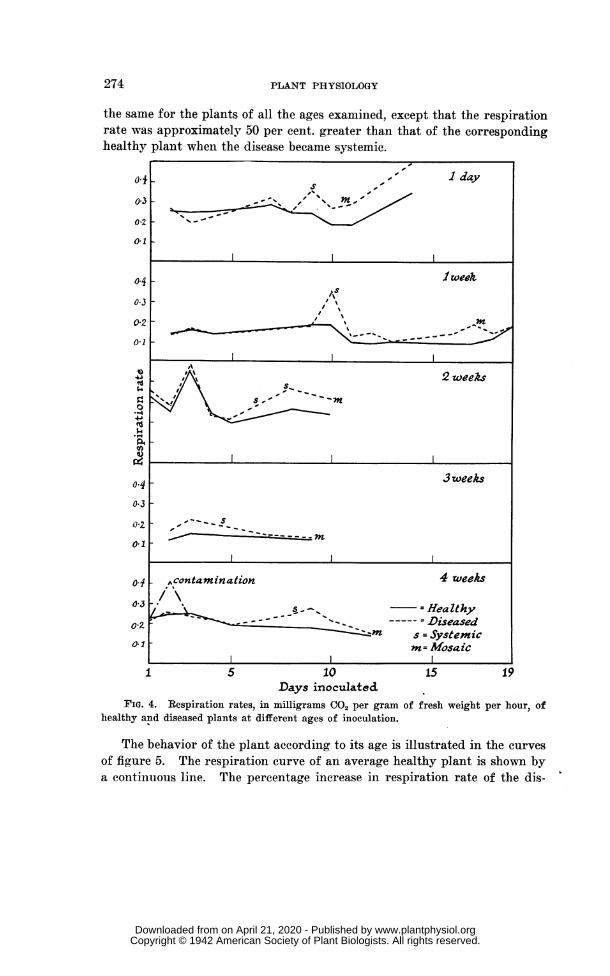
The respiration rates were all calculated in terms of milligrams of car-boll dioxide evolved per gram of fresh weight per hour. The results canbest be understood by reference to the curves in figure 4, which are typicalexamnples takeu froimi each age group. It will be seen that the respirationrates of the healthy and diseased plants began at approximately equal levelsancd remained equal, within experimental error, up to the point at whichthe respiration rate of the diseased plants rose suddenly above the rate ofthe healthy plants. In the course of a few days the higher rate of the dis-eased plants gradually decreased and approached the rate of the healthyplants.
The special significance of these results lies in the observation that thesudden rise in the respiration rate of the inoculated plants coincided withthe time at which the virus becaiiie systemic, i.e., at the time of the clearingof the veins. Usually when the mosaic mottling appeared on the plant, therespiration rates of the diseased and the healthy plants were either almostor actually equal again. In most cases the experiments were not continuedpast that point. When the disease became systemic the diseased plantsshowed a negligible increase of weight whereas the output of carbon dioxideremained the same or was much greater than formerly so that the resultingrespiration rate was much higher.
If the conditions were such that the clearing did not show, the rise inrespiration rate occurred at such a period before the mottling that it wasreasonable to assume that the systemic spread of the virus had coincidedwith the rise, especially as other indications such as crinkling of the leaveswere usually present. Omi one occasion, when circumstances prevented theobservation of the clearing of the veins, the highest rise of respiration ratewas missed, but the initial rise and subsequent fall were measured. Onanother occasion, one unit of the healthy plants showed a sudden rise inrespiration: it was found that one of the plants was contaminated with thevirus. In the same batch of plants one of the inoculated plants gave asudden rise on the second day of inoculation. Again one of these plantswas seen to be contaiiiinated and, upon removing it, the respiration rate ofthe unit fell back to normal, rising on the 8th day when the disease becamesy-stemnic in the remaining plants.
The respiration rate of the healthy plants varied from about 0.30 mg.CO,/gm./hour for the young plants to about 0.075 mg./gm./hour for theolder plants with a tendency to decrease regularly with increasing age andweight. With the inoculated plants the course of the respiration rate was
www.plantphysiol.orgon April 21, 2020 - Published by Downloaded from Copyright © 1942 American Society of Plant Biologists. All rights reserved.
274 PLANT PHYSIOLOGY
the same for the plants of all the ages examined, except that the respirationrate was approximately 50 per cent. greater than that of the correspondinghealthy plant when the disease became systemic.
01
o-3
0.2
0-1
4
0.3
02
0
4J"S$49
0.4
0:1
03
on4
aZ
04
0a3
ol0.1
7 I day, --,;/t4'-
t ~~~~~~~~~~lwee*f~~~ y
S= ~~~~~~~~~~2 weeks
I ~ ~~~~~~~~~~~I
l ~~~~~~~~~~3weeks
S-.
II
,contamination 4 weeks
HHealthyDi-seased
7n s Systemicm= Mosaic
15 10 15 19Days inoculatedc
FIG. 4. Respiration rates, in milligrams CO, per gram of fresh weight per hour, ofhealthy and diseased plants at different ages of inoculation.
The behavior of the plant according to its age is illustrated in the curvesof figure 5. The respiration curve of an average healthy plant is shown bya continuous line. The percentage increase in respiration rate of the dis-
www.plantphysiol.orgon April 21, 2020 - Published by Downloaded from Copyright © 1942 American Society of Plant Biologists. All rights reserved.

GLASSTONE: RESPIRATION IN HEALTHY AND MOSAIC-INFECTED TOBACCO 275
eased plants inoculated at the various ages is drawn in a series of dottedcurves. It will be seen that the percentaoe rise, when the disease becomessystemic, is approximiiately the same irrespective of the ages of the healthyplants at the time of inoculation. Wheni the mosaic develops, the per-cenitage rise apparenitly decreases with the inereasingy age of the inoculatedplants.
These results indicate that at the time when the virus becomes systemicthere is a (reat increase in the metabolic activity within the plant as indi-cated by the greater rate of respiration of the inoculated as compared withthe healthy plant. A numiiber of investigators have studied the changesand distribution of the virus subsequent to inoculationi (5, 7, 14, 16, 19).Their results all inidicate that with tobacco-mosaic virus the clearing(-of-the-
200Z .
,,) s7n
'
1005S..
Heatthy---------Diseased
s = Systemicm = Mosaic
1 7 14 21 28Days in solution
FIG. 5. The percentage increase in respiration rate of the diseased plants inoculatedat various ages.
veins period corresponds to the rapid movement and inierease of the virus,which would account for a high expenditure of energy whatever actlualprocess is taking place. Further experiments are now being carried out onthe respiration problems in order to obtain a fuller interpretation of theseresults.
SummaryThe paper describes the apparatus and method for comparingo the respi-
ration of entire healthy and mosaic-diseased tobacco plants from the timeof inoculation until the appearanee of the mottling disease. The sameplants were used throughout the period of each experimenit.
It was found that the respiration ratio of the diseased plants and healthyplants remained at the same level until the disease became systemie. Whenthe clearing of the veins was apparent, the respiration rate of the diseasedplants rose rapidly, followed by a decrease until in the older plants it be-came approximately equal to that of the healthy plants by the time that themosaic mottling had developed. The percentage increase in respiration rate
I
www.plantphysiol.orgon April 21, 2020 - Published by Downloaded from Copyright © 1942 American Society of Plant Biologists. All rights reserved.

PLANT PHYSIOLOGY
was approximately 50 per cent. higher than the rate of the correspondinighealthy plants.
DEPARTMENT OF ANIMAL AND PLANT PATHOLOGYTHE ROCKEFELLER INSTITIJTE FOR MEDICAL RESEARCH
PRINCETON, NEW JERSEY
LITERATURE CITED1. BENNETT, C. W. Relation of food translocation to movement of virus
of tobacco mosaic. Jour. Agr. Res. 60: 361-390. 1940.2. BRONFENBRENNER, J. Further studies on so-called bacteriophage.
Proc. Soc. Exp. Biol. Med. 22: 81-82. 1924.3. Does bacteriophage respire? Science n.s. 63: 51-52.
1926.4. , and REICHERT, P. Respirationi of so-called filterable
viruses. Proc. Soc. Exp. Biol. Med. 24: 176-177. 1926-1927.5. CALDWELL, J. The physiology of virus diseases in plants. V. The
movement of the virus agent in tobacco and tomato. Ann. Appl.Biol. 21: 191-205. 1934.
6. . The physiology of virus diseases in plants. VI. Someeffects of mosaic on the metabolism of the tomato. Ann. Appl.Biol. 21: 206-224. 1934.
7. CRAFTS, A. S. Movement of viruses, auxins, and cheiuical indicatorsin plants. Bot. Rev. 5: 471-504. 1939.
8. DUFRIENOY, J. Modifications pathologiques du metabolisme cellulairechez les tabacs. Ann. I:piphyties 18: 259-280, 281-316. 1932.
9. DUNLAP, A. A. The total nitrogen and carbohydrates, and the relativerates of respiration, in virus-infected plants. Amer. Jour. Bot.17: 348-357. 1930.
10. GLASSTONE, V. F. C. Passage of air through plants and its relationi tomeasurement of respiration and assimilation. Amer. Jour. Bot.29: 1942. (In press.)
11. HOAGLAND, D. R., and ARNON, D. I. The water-culture method forgrowing plants without soil. Univ. California Agr. Exp. Sta. Circ.347. 1939.
12. HOLMES, F. 0. Inoculating methods in tobacco mosaic studies. Bot.Gaz. 87: 56-63. 1929.
13. . Local and systemic increase of tobacco mosaic virus.Amer. Jour. Bot. 17: 789-805. 1930.
14. . Movement of mosaic virus from primary lesions inNicotiana tabacum L. Contrib. Boyce Thompson Inst. 4: 297-322.1932.
15. KEMPNER, W. Chemical nature of the oxygen-transferring fermenit ofrespiration in plants. Plant Physiol. 11: 605-613. 1936.
276
www.plantphysiol.orgon April 21, 2020 - Published by Downloaded from Copyright © 1942 American Society of Plant Biologists. All rights reserved.
GLASSTONE: RESPIRATION IN HEALThIY AND MOSAIC-INFECTED TOBACCO 277
16. KUNKEL, L. 0. Movemiient of tobacco-miiosaic virus in tomato plants.Phytopath. 29: 684-700. 1939.
17. LEMIMON, P. Comparative studies on metabolism of healthy andmosaic-infected tobacco leaves. Respiration studies. (Abstract)Amer. Jour. Bot. 22: 912. 1935.
18. MACK, W. B. The relation of temperature and the partial pressure ofoxygen to respiration and growth in germinating wheat. PlantPhysiol. 5: 1-68. 1930.
19. MATSUMOTO, T. Serological studies on the distribution and concentra-tion of tobacco mosaic virus in host plants. I, II. Trans. Nat.Hist. Soc. Formosa 31: 201-215, 275-285. 1941.
20. THUNG, T. H. Physiologisch onderzoek met betrekking tot het virusder bladrolziekte van der aardappelplant, Solanum tuberosum L.Tijdschr. Planteuiz. 34: 1-74. 1928.
21. WHITEHEAD, T. The physiology of potato leaf-roll. 1. On the respi-ration of healthy and leaf-roll infected potatoes. Ann. Appl. Biol.21: 48-77. 1934.
www.plantphysiol.orgon April 21, 2020 - Published by Downloaded from Copyright © 1942 American Society of Plant Biologists. All rights reserved.