Embed Size (px)
Citation preview
11
Motor Neuron Disease
Hamdy N. El Tallawy Assiut University
Egypt
1. Introduction
Neurologists in the 19th century recognized that muscle weakness could be due to primary disorders of muscle or secondary to loss of neuromuscular integrity, as happens when peripheral nerves are cut or when motor neurons degenerate. Furthermore, it was observed that there are forms of motor neuron degeneration which selectively affect upper motor neurons or lower motor neurons. A combination of upper and lower motor neuron dysfunction was named amyotrophic lateral sclerosis (ALS) by Charcot and Joffroy (Ringel, et al 1993). Jean-Martin Charcot first characterized the disease in 1874, naming the illness Amyotrophic lateral sclerosis (ALS) (Swash, 2001). In USA, ALS or Lou Gehrig's disease are terms used to describe all forms of the disease, whatever the combination of upper and lower motor neuron involvement (Ringel, et al 1993). ALS is now a term which classifies the most common form of the illness and is often used synonymously with MND (Swash, 2001). In the UK the umbrella term motor neuron disease (MND) is more common. MND is a disease of middle to late life with a mean age of onset of 58 years, (Ringel, et al 1993).
Actually, motor neuron diseases (MND) are a group of degenerative disorders that selectively affect motor neurons in the brain and spinal cord. Two groups of motor neurons are involved, lower motor neurons located in ventral horns of the spinal cord and brainstem motor nuclei, and upper motor neurons located in the cerebral cortex together with pyramidal tracts in spinal cord. The term MND is a broad spectrum term including amyotrophic lateral sclerosis.
2. Definitions and terminology of motor neuron disease (MND)
MND is a group of incurable progressive neurodegenerative disorders in which degeneration involves upper and lower motor neurons in different body regions, resulting in progressive weakness of bulbar, limbs and respiratory musculature, in different combination.
MND is an adult onset neurodegenerative disease which leads inexorably via weakness of limb, bulbar and respiratory muscles to death from respiratory failure three to five years later. (Allum and Shaw 2010).
3. Epidemiology of motor neuron disease
The prevalence of MND is 4-6 per 100,000 in most parts of the world, except the Western Pacific foci, (Leigh, 1991). However, its annual incidence is between 1.5 and 2/100,000 and
Neuromuscular Disorders
202
males are more commonly affected than females (1.4:1). The incidence increases with age with a mean age of onset of 63 years, (Ringel, et al 1993). It ranks as the third most common neurological degenerative disorder after Alzheimer's and Parkinson's disease (Talbot 2002).
In Guam, the incidence of MND has fallen from 87/100,000 in 1962 to 5/100,000 in 1985, (Rodgers-Johanson, et al. 1986).
Within the Caucasian population of Europe and North America, where most of the studies have been conducted, the lowest reported incidence of MND was 0.6 per 100.000 person –years in Italy,(De Domenice, et al. 1988) and the highest reported was 2.4 per 100.000 person-years in Finland(Murros and Fogelholm.1983). However, a lower incidence rate of 0.3/100,000 person-years was reported among Asian population, in China, (Fong, et al. 1996).
In the only well-conducted study of MND incidence among black African population, the incidence of MND was noted to be 0.9 per 100,000 person-years in Libya, (Radhakrishnan, et al. 1986).
The incidence of MND is said to be increasing, but this is probably the result of improved diagnosis, better awareness of the disease and an aging population, (Leigh and Ray-Chaudhuri.1994). The incidence increases after the age of 40 years, peaks in the late 60s and early 70s, and declines rapidly after that, (Logroscino, et al. 2008).
4. Aetiology, pathology and pathogenesis of motor neuron disease
MND is one of the complex and misunderstood diseases that health care professionals may encounter for various different reasons: (1) there are multiple different forms of the disease (Strong and Rosenfeld.2003), (2) the pathogenesis is not fully delineated (Wijesekera and Leigh.2009), (3) diagnosis can occur only by exclusion, (4) there exists only FDA approved medication, riluzole, for the treatment of ALS2 (Washington.2007), and despite this, (5) there is no cure for this disease.
Environmental exposures during the Gulf war have been proposed as the explanation for an increased incidence of ALS among Gulf War veterans (Haley 2003, Horner et al, 2003).
4.1 Neuropathological findings in MND
Gross changes were frontotemporal atrophy, which was usually mild to moderate, neither circumscribed nor of a 'Knife-blade' type, and atrophy of the anterior roots in the cervicothoracic spinal cord, which was seen in cases with definite lower motor neuron involvement. Cortical atrophy was marked in the limbic system including the temporal pole, parahippocampus and amygdala but usually spared the hippocampus in typical ALS – Dementia (ALS-D) cases. (Yoshida 2004).
Histological changes included neuronal loss and gliosis with sponginess in layers II and III of frontotemporal cortices with predominant involvement of the limbic system including the anterior cingulated gyrus, anterior temporal and insular lobes, parahippocampus, subiculum and amygdale in typical cases. (Yoshida 2004).
The full pathogenesis of ALS is not well understood as it has not been fully elucidated by medical research. However, several key factors can be noted including: (1) Genetics, (2)
Motor Neuron Disease
203
Excitotoxicity, (3) Oxidative stress, (4) Mitochondrial dysfunction, (5) Impaired axonal transport, (6) Neurofilament aggregation, (7) Protein aggregation, (8) Inflammatory dysfunction and contribution of non-neuronal cells, (9) Deficits of neurotrophic factors and dysfunction of signaling pathways, and (10) Apoptosis, (Wijesekera and Leigh.2009 and Shaw. 2005).
4.2 Genetics
Up to 90% of all ALS cases, occurs without family history, (sporadic ALS) and about 10% of cases are familial ALS (FALS). SALS is clinically indistinguishable from FALS, but the average age of onset in FALS is somewhat earlier, (Celveland and Rothstein (2001). Enteroviral infections and mutations of superoxide dismutase 1gene (SOD1) have been implicated in the pathogenesis of MND (oluwale et al 2001). About 25% of ALS cases, (Celveland and Rothstein (2001), and 2% of the sporadic cases, are linked to mutations in the gene encoding copper/zinc superoxide dismutase (SOD1). It is Known that there may be as many as six gene loci that code for the ALS phenotype, but only three have been identified. Several other mutations have also been documented to possibly take part in the pathogenesis of ALS, (Wijesekera and Leigh, 2009). Since the link between SOD1 and FALS was first established, >90 FALS-linked SOD1 mutations have been discovered, (Celveland and Rothstein (2001). Most of these mutations are point missense mutations, (Anderson, et al. 2003). Most of the genetics are transmitted via the autosomal dominant route, though some are autosomal recessive and others may be sex-linked, (Wijesekera and Leigh.2009).
4.3 Excitotoxicity
Excitotoxicity is a term used to signify the damage that occurs to neuronal cells that are characterized by overstimulated glutamate receptors, as glutamate is the major excitatory neurotransmitter in the human central nervous system (Riluzol monograph. 2011). As SOD1 codes for the major reuptake protein of glutamate, a mutation limits the concentration levels of that reuptake protein, allowing an excessive amount of glutamate to be present in the neuronal synapse. It is also postulated that glutamatergic toxicity plays a direct role in the destruction of neuronal cells in patients with ALS.( Shaw. 2005).
4.4 Oxidative stress
Oxidative stress is of particular interest to researchers due to the fact that the SOD1 gene mutation that is known to cause ALS, normally codes for an anti-oxidant protein. (Riluzol momgraph. 2011)
4.5 Mitochondrial dysfunction
There are many new data which supports the theory that mitochondrial dysfunction plays an important role in the pathogenesis of ALS. Multiple cases of dysfunctional mitochondria have been noted in post-mortem analyses of ALS patients. (Wijesekera and Leigh.2009)
Dysfunctional mitochondria have also been linked to the SOD1 gene mutation in mice models. (Shaw. 2005)
Neuromuscular Disorders
204
4.6 Impaired axonal transport
The theory that axonal transport is a key to ALS pathogenesis stems from SOD1 transgenic mice models. Mice from these models often show slowed anterograde and retrograde axonal transport. Although no human ALS patient has presented with this problem, yet it is known to occur in several other neuromuscular disorders of the human body. (Wijesekera and Leigh.2009)
4.7 Neurofilament aggregation
Abnormal neuronal assembly, including accumulation of neurofilaments, are often seen in ALS patients. Neurofilaments can combine with a toxic form of peripherin, an intermediate filament protein, and become toxic to neurons even at modest concentration levels. This combination has been found in the spinal cord of ALS patients and not in controls. This evidence points to neurofilament aggregation as being a part of ALS pathogenesis.
(Wijesekera and Leigh.2009)
4.8 Protein aggregation
Long debates as to whether protein aggregation take a part in disease pathogenesis have been occurred. It is possible that these inclusions may simply be innocent bystanders, or even beneficial to the cells. (Wijesekera and Leigh.2009)
4.9 Inflammatory dysfunction and contribution of non-neuronal cells
It has been recently discovered that SOD1 mutations alone are insufficient to cause ALS in transgenic mice, making the case that non-neuronal cells, as microglial and dendritic cells, may play a part in ALS pathogenesis (Shaw, 2005). ALS patients commonly experience activation of the non-neuronal microglial and dendritic cells. This activation has been shown to produce inflammatory cytokines such as interleukins and tumor necrosis factor (TNF). However, recent trials have yet to see success in utilizing immunomodulatory drug therapies to curve the progression of ALS. (Wijesekera and Leigh.2009).
4.10 Deficits of neurotrophic factors and dysfunction of signaling pathways
Lowered levels of neurotrophic factors have been noticed in post-mortem analysis of several ALS patients. In addition, three mutations in the Vascular Endothelial Growth Factors (VEGF) gene were thought to be associated with an increased risk for developing ALS. However, recently these finding have come under scrutiny due to an inability to replicate research results. (Wijesekera and Leigh.2009)
4.11 Apoptosis
Current research also skews towards examining if ALS motor neuron destruction occurs via a programmed cell death, or apoptosis. Several studies have shown that cell death due to ALS often occurs because of this programmed apoptosis, yet these findings are still being reviewed and discussed heavily. ( Shaw. 2005).
Motor Neuron Disease
205
5. Clinical presentation of motor neurone disease
Dilemmas of symptoms and signs are described for diagnosis of MND and its subtypes. Motor symptoms of both upper and lower motor neuron dysfunction can occur in any muscle group, including limbs, and, or bulbar regions. Combinations of the previous motor symptoms is not characteristic for MND and can be observed in many diseases, known as "MND Mimic Disorders" such as, Kennedy's diseases, multifocal motor neuropathy, brainstem lesions (syrinx, stroke,………. etc)
Meticulous evaluation by thorough history and examination by two or more specialists of neurology are required for diagnosis of MND. Special investigations are required for diagnosis of MND and differentiation from MND mimics.
Preclinical stage Diagnosis stage Progression stage Terminal stage
Long duration (months or even years
Disease spread through motor system
Usually Asymptomatic
Selective involvement of motor neurons in the anterior horns of the spinal cord early and late in the disease (Swash et al. 1986)
corticospinal pathways in the cord are similarly, asymmetrically affected (Ingram and Swash 1987)
Onuf''s sacral nucleus (innervate anal and urinary sphincter muscles) are always spared (Schroder, and Reske Nelson 1984)
Ocular motor nuclei are also relatively resistant (Mulder 1982)
weakness of one leg followed by other leg and patient will require a wheelchair to assist him
Swallowing Problems
Respiratory muscle weakness and dyspnea
All these symptoms are observed in different combinations according to different subtypes of MND
Respiratory insufficiency symptoms orthopnea, excessive daytime sleepiness, poor concentration, memory and appetite and signs of tachypnea, tachycardia, syncope and confusion, (Radunovic et al 2007.)
Nursed and care for 24 hours
Medication, Diet, Hygiene and special attention for respiration.
Table 1. Stages of Motor Neuron Disease (MND)
6. Bad prognostic factors for MND
1. Late age of onset. 2. Severe and early presentations of bulbar symptoms. 3. Respiratory complications.
The classical concept that MND only affects the motor system is obsolete. MND is considered to be a multisystem neurodegenerative disease. There is increasing clinical evidence for autonomic dysfunction (Baltadzhieva, et al 2006 and Takeda, et al, 1994), sensory abnormalities (Takeda et al, 1994) (Pugdahl et al, 2007) and ophthalmoplegia
Neuromuscular Disorders
206
(Takeda, et al, 1994) in MND. On the other hand, there are good pathological accounts of the involvement of sympathetic and parasympathetic neurons, (Takeda, et al, 1994), Onuf's nucleus (which innervates the pelvic floor sphincteric muscles), (Takeda, et al, 1994) peripheral sensory nerves, (Isaacs, et al. 2007) and oculomotor nuclei. (Takeda, et al, 1994). However, in practical terms, the presence of prominent ophthalmoplegia, sensory signs or sphincter dysfunction should raise doubts regarding the diagnosis of MND, unless there are a clear alternative explanation. Death usually results from ventilatory muscle weakness causing respiratory failure.
7. Motor neuron disease presenting to non neurological specialists
Respiratory: Progressive respiratory Failure
Ear, nose, and throat: Dysphagia.
Orthopaedics/ neurosurgery: Foot drop or other symptoms suggestive of compressive radiculopathy
Elderly care: Difficulty walking. (Talbot 2002).
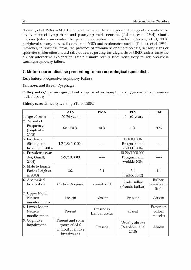
ALS PMA PLS PBP 1. Age of onset 50-70 years 40 – 60 years 2. Percent of
Frequency (Leigh et al 2003)
60 – 70 % 10 % 1 % 20%
3. Incidence (Strong and Rosenfeld, 2003)
1,2-1,8/100,000 ----- 1/1000,000-
Brugman and wokkle 2004
-----
4. Prevalence (van der, Graaff, 2004)
5-9/100,000 ----- 10-20/1000,000-Brugman and wokkle 2004
-----
5. Male to female Ratio ( Leigh et al 2003)
3:2 3:4
3:1 (Talbot 2002)
1:1
6. Anatomical localization Cortical & spinal spinal cord
Limb, Bulbar (Pseudo bulbar)
Bulbar, Speech and
limb 7. Upper Motor
Neuron manifestations
Present Absent Present Absent
8. Lower Motor Neuron manifestation
Present Present in
Limb muscles absent
Present in bulbar
muscles 9. Cognitive
impairment Present and some
group of ALS without cognitive
impairment
Present Usually absent (Raaphorst et al
2010) Absent
Motor Neuron Disease
207
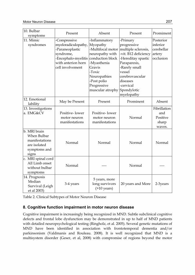
10. Bulbar symptoms
Present Absent Present Promiment
11. Mimic syndromes
-Compressive myeloradiculopathy, -Paraneoplastic syndrome, -Encephalo-myelitis with anterion horn cell involvement
-Inflammatory Myopathy -Multifocal motor neuropathy with conduction block-Myasthenia Gravis -Toxic Neuropathies -Post polio Progressive muscular atrophy
-Primary progressive multiple sclerosis, -vit. B12 deficiency -Hereditay spastic Paraparesis, -Rarely small vessel cerebrovascular diseases -cervical Spondylotic myelopathy
Posterior inferior cerebellar artery occlusion
12. Emotional lability
May be Present Present Prominent Absent
13. Investigotions a. EMG&CV Positive- lower
motor neuron manifestations
Positive- lower motor neuron manifestations
Normal
Fibrillation and
Positive sharp
waves. b. MRI brain
When Bulbar manifestations are isolated symptoms and signs
Normal Normal Normal Normal
c. MRI spinal cord All Limb onset without bulbar symptoms
Normal ---- Normal ----
14. Prognosis Median Survival (Leigh et al 2003)
3-4 years 5 years, more
long survivors (>10 years)
20 years and More 2-3years
Table 2. Clinical Subtypes of Motor Neuron Disease
8. Cognitive function impairment in motor neuron disease
Cognitive impairment is increasingly being recognized in MND. Subtle subclinical cognitive defects and frontal lobe dysfunction may be demonstrated in up to half of MND patients with detailed neuropsychological testing (Ringholz, et al. 2005). Several genetic mutations of MND have been identified in association with frontotemporal dementia and/or parkinsonism (Valdmanis and Rouleau. 2008). It is well recognized that MND is a multisystem disorder (Geser, et al, 2008) with compromise of regions beyond the motor
Neuromuscular Disorders
208
system, including cortical areas which are consistently involved in FTD. It comes as no surprise, therefore, that a proportion of patients presenting with MND manifest cognitive and/or behavioural changes which may be severe enough in some instances to reach criteria for frank FTD.(Irwin, et al, 2007).
8.1 Amyotrophic lateral sclerosis and frontotemporal dementia (ALS-FTD)
There is icreasing clinical, imaging and neurophysiological evidence that ALS represents a multisystem neurodegerative disease. Neurodegeneration is not restricted to motor neurons, but also includes parts of the brain other than the motor cortex, especially the preforntal and/or anterior temporal lobe, that contribute to the clinical syndrome. In some cases an evident dementia that resembles frontotemporal degeneration (FTD) was observed. It is now suggested that ALS and FTD are closely related conditions with overlapping clinical, pathological, radiological, and genetic characteristics. The presence of a frontal dementia in ALS has also crucial practical consequences for management of the patients, whose disorder requires critical life decisions for enteral nutrition and respiratory complications. (Zago, et al. 2010).
8.2 The new classification of cognitive and behavioral disorder in ALS
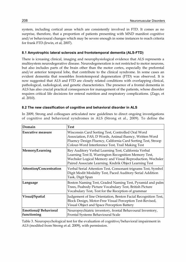
In 2009, Strong and colleagues articulated new guidelines to direct ongoing investigations of cognitive and behavioral syndromes in ALS (Strong et al., 2009). To define the
Domain Test
Executive measure Wisconsin Card Sorting Test, Controlled Oral Word Association, FAS, D Words, Animal fluency, Written Word Fluency Design Fluency, California Card Sorting Test, Stroop Colour-Word Interference Test, Trail Making Test
Memory/Learning Rey Auditory Verbal Learning Test, California Verbal Learning Test II, Warrington Recognition Memory Test, Wechsler Logical Memory and Visual Reproduction, Wechsler Paired Associate Learning. Kndrik Object Learning Test
Attention/Concentration Verbal Serial Attention Test, Consonant trigrams Test, Symbol Digit Modit Modality Test, Paced Auditory Serial Addition Task, Digit Span
Language Boston Naming Test, Graded Naming Test, Pyramid and palm Trees, Peabody Picture Vocabulary Test, British Picture Vocabulary Test, Test for the Reception of grammar
Visual/Spatial Judgement of line Orientation, Benton Facial Recognition Test, Block Design, Motor-Free Visual Perception Test-Revised, Visual Object and Space Perception Battery
Emotional/ Behavioral functioning
Neuropsychiatric inventory, frontal Behavoural Inventory, Frontal Systems Behavioural Scale
Table 3. Neuropsychological test for the evaluation of cognitive/behavioral impairment in ALS (modifed from Strong et al. 2009), with permission.
Motor Neuron Disease
209
neuropsychological status of ALS patients a framework was based on four different axes. Axis I is based on the EL Escorial criteria proposed in 1998, that includes possible, probable, and definite ALS clinical subtypes. This multidimensional approach incorporates several criteria (Brooks et al, 2000). The novelty of the classification lies primarily in Axis II with the proposal of five categories which classify ALS patients along a continuum: (1) ALS patients cognitively and behaviorally intact: (2) ALS patients with mild cognitive impairments; (3) ALS patients with mild behavioral impairment; (4) ALS with a full-fledged fronto-temporal dementia; (5) ALS with other non FTD-forms of dementia.
Axis III indicates the presence, in addition to frontotemporal impairments, of additional non-motoneuronal disease manifestations such as extrapyramidal signs, cerebellar degenerations, autonomic dysfunctions, sensory impairments, and ocular motility abnormalities. The absence of the above indicates a "pure form," while their presence defines "complicated forms" with additional pathological motor aspects. Axis IV, instead, provides the search for factors which could modify the course of the disease. Several disease modifiers have been reported in literature associated with longer survival, age at symptom onset (< 45 years), gender (male/sex), and site of the disease onset (bulbar or limb).
9. Language
Language deficits are occasionally found in the early stages of the disease. (Abrahams, et al. 2004).
The spectrum of language impairment in MND is wider than simply a problem in speech production due to dysarthria, but it is yet to be fully characterized. Reduced verbal output (adynamism) evolving into mutism has been reported, as well as echolalia. Perseverations, stereotypical expressions, (Bak and Hodges. 2004), true non-fluent aphasia with phonological and/or syntactic deficits and comprehension impairment have been reported in isolated cases. (Tsuchiya, et al. 2000)
MND has also been associated with apraxia of speech, in which there is breakdown in articulatroy planning, producing slowed, effortful and dysprosodic speech with problems repeating multisyllabic words. Apraxia of speech is often accompanied by orobuccal apraxia but not necessarily with aphasia.(Duffy, et al ,2007).
10. Memory
It has been difficult to categorize the pattern of memory impairment, but current evidence suggests that memory problems are related to abnormalities in retrieval of the information secondary to frontal dysfunction. (Neary, et al. 2000). Memory problems involve primarily immediate recall, (Phukan. et al. 2007) but impairment of visual memory also has been implicated,(Kew, et al.1993)
Frontal, temporal and thalamic hypoperfusion on SPECT has been shown to correlate with the severity of memory impairment. (Montovan, et al. 2003).
Most strikingly, learning and memory were found to be significantly improved in patients in the later stages of the disease, (Lakerveld et al, 2008).
Neuromuscular Disorders
210
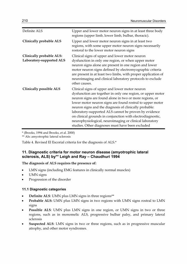
Definite ALS Upper and lower motor neuron signs in at least three body regions (upper limb, lower limb, bulbar, thoracic).
Clinically probable ALS Upper and lower motor neuron signs in at least two regions, with some upper motor neuron signs necessarily rostoral to the lower motor neuron signs
Clinically probable ALS: Laboratory-supported ALS
Clinical signs of upper and lower motor neuron dysfunction in only one region, or when upper motor neuron signs alone are present in one region and lower motor neuron signs defined by electromyographic criteria are present in at least two limbs, with proper application of neuroimaging and clinical laboratory protocols to exclude other causes.
Clinically possible ALS Clinical signs of upper and lower motor neuron dysfunction are together in only one region, or upper motor neuron signs are found alone in two or more regions, or lower motor neuron signs are found rostral to upper motor neuron signs and the diagnosis of clinically probable: laboratory-supported ALS cannot be proven by evidence on clinical grounds in conjunction with electrodiagnostic, neurophysiological, neuroimaging or clinical laboratory studies. Other diagnoses must have been excluded
* (Brooks, 1994 and Brooks, et al. 2000) ** Als: amyotrophic lateral sclerosis
Table 4. Revised El Escorial criteria for the diagnosis of ALS.*
11. Diagnostic criteria for motor neuron disease (amyotrophic lateral sclerosis, ALS) by** Leigh and Ray – Chaudhuri 1994
The diagnosis of ALS requires the presence of:
LMN signs (including EMG features in clinically normal muscles) UMN signs Progression of the disorder
11.1 Diagnostic categories
Definite ALS: UMN plus LMN signs in three regions** Probable ALS: UMN plus LMN signs in two regions with UMN signs rostral to LMN
signs Possible ALS: UMN plus LMN signs in one region, or UMN signs in two or three
regions, such as in monomelic ALS, progressive bulbar palsy, and primary lateral sclerosis
Suspected ALS: LMN signs in two or three regions, such as in progressive muscular atrophy, and other motor syndromes.
Motor Neuron Disease
211
11.2 The diagnosis of ALS requires the absence of
Sensory signs Sphincter disturbances Visual disturbances Autonomic dysfunction Parkinson's disease Alzheimer-type dementia ALS "mimic" syndromes
11.3 The diagnosis of ALS is supported by
Fasciculation in one or more regions Neurogenic change in EMG studies Normal motor and sensory nerve conduction (distal motor latencies may be increased) Absence of conduction block
Regions are defined as follows: brainstem, brachial, thorax and trunk, crural. UMN= Upper motor neuron; LMN= lower motor neuron.
12. Management of motor neuron disease (MND)
12.1 Investigations
There are no specific investigations for MND. Till now there are no specific biochemical or pathological markers of MND. The aim of Elctrophysiological, Imaging and laboratory investigations is to exclude MND mimics and/ or to support clinical signs presented by the patients.
Allum and Shaw (2010) clarified that investigations are important adjuncts to the clinical diagnosis of MND. Properly used they can provide supportive evidence of the clinical findings and help delineate the extent of disease. Investigations are also important to identify benign or treatable MND mimics.
12.2 Treatment plan of motor neuron disease
Although MND is still incurable disease up till now, in the last two decades MND management has evolved rapidly. Symptomatic treatment of MND still had the upper hand of management plan, especially for respiratory and bulbar complications. A team of work including neurologist, highly qualified nurses, ICU specialist in respiratory complications, psychologist, dietition, physiotherapy and speech therapist must be involved for management plan and follow up of MND patients, table (5).
Thus treatment strategy of MND was aimed towards;
1. Delay Progression of the disease and prevent further loss of motor neurons especially in the early stage of the disease
2. Symptomatomatic treatment to alleviate symptoms of the disease aiming to maintain quality of life
Neuromuscular Disorders
212
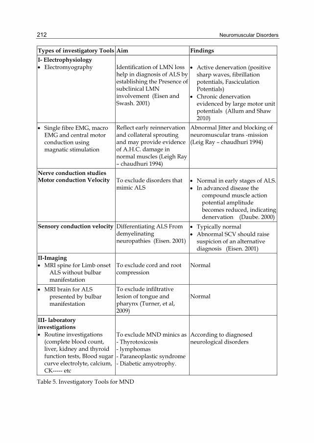
Types of investigatory Tools Aim Findings
I- Electrophysiology Electromyography Identification of LMN loss
help in diagnosis of ALS by establishing the Presence of subclinical LMN involvement (Eisen and Swash. 2001)
Active denervation (positive sharp waves, fibrillation potentials, Fasciculation Potentials)
Chronic denervation evidenced by large motor unit potentials (Allum and Shaw 2010)
Single fibre EMG, macro EMG and central motor conduction using magnatic stimulation
Reflect early reinnervation and collateral sprouting and may provide evidence of A.H.C. damage in normal muscles (Leigh Ray – chaudhuri 1994)
Abnormal Jitter and blocking of neuromuscular trans -mission (Leig Ray – chaudhuri 1994)
Nerve conduction studies Motor conduction Velocity To exclude disorders that
mimic ALS Normal in early stages of ALS. In advanced disease the
compound muscle action potential amplitude becomes reduced, indicating denervation (Daube. 2000)
Sensory conduction velocity Differentiating ALS From demyelinating neuropathies (Eisen. 2001)
Typically normal Abnormal SCV should raise
suspicion of an alternative diagnosis (Eisen. 2001)
II-Imaging MRI spine for Limb onset
ALS without bulbar manifestation
To exclude cord and root compression
Normal
MRI brain for ALS presented by bulbar manifestation
To exclude infiltrative lesion of tongue and pharynx (Turner, et al, 2009)
Normal
III- laboratory investigations Routine investigations
(complete blood count, liver, kidney and thyroid function tests, Blood sugar curve electrolyte, calcium, CK----- etc
To exclude MND minics as- Thyrotoxicosis - lymphomas - Paraneoplastic syndrome - Diabetic amyotrophy.
According to diagnosed neurological disorders
Table 5. Investigatory Tools for MND
Motor Neuron Disease
213
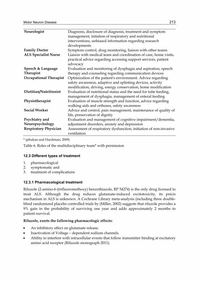
Neurologist Diagnosis, disclosure of diagnosis, treatment and symptom management, initation of respiratory and nutritional interventions, unbiased information regarding research developments
Family Doctor Symptom control, drug monitoring, liaison with other teams ALS Specialist Nurse Liaison with medical team and coordination of care, home visits,
practical advice regarding accessing support services, patient advocacy
Speech & Language Therapist
Evaluation and monitoring of dysphagia and aspiration, speech therapy and counseling regarding communication devices
Occupational Therapist Optimization of the patient's environment. Advice regarding safety awareness, adaptive and splinting devices, activity modification, driving, energy conservation, home modification
Dietitian/Nutritionist Evaluation of nutritional status and the need for tube feeding, management of dysphagia, management of enteral feeding
Physiotherapist Evaluation of muscle strength and function, advice regarding walking aids and orthoses, safety awareness
Social Worker Advice and control, pain management, maintenance of quality of life, preservation of dignity
Psychiatry and Neuropsychology
Evaluation and management of cognitive impairment/dementia, adjustment disorders, anxiety and depression
Respiratory Physician Assessment of respiratory dysfunction, initiation of non-invasive ventilation
* (phukan and Hardiman, 2009)
Table 6. Roles of the multidisciplinary team* with permission
12.3 Different types of treatment
1. pharmacological 2. symptomatic and 3. treatment of complications
12.3.1 Pharmacological treatment
Riluzole (2-amino-6-(trifluoromethoxy) benzothiazole, RP 54274) is the only drug licensed to treat ALS. Although the drug reduces glutamate-induced excitotoxicity, its précis mechanism in ALS is unknown. A Cochrane Library meta-analysis (including three double-blind randomized placebo controlled trials by (Miller, 2002) suggests that riluzole provides a 9% gain in the probability of surviving one year and adds approximately 2 months to patient survival.
Riluzole, exerts the following pharmacologic effects:
An inhibitory effect on glutamate release. Inactivation of Voltage – dependent sodium channels. Ability to interfere with intracellular events that follow transmitter binding at excitatory
amino acid receptor (Riluzole monograph 2011).
Neuromuscular Disorders
214
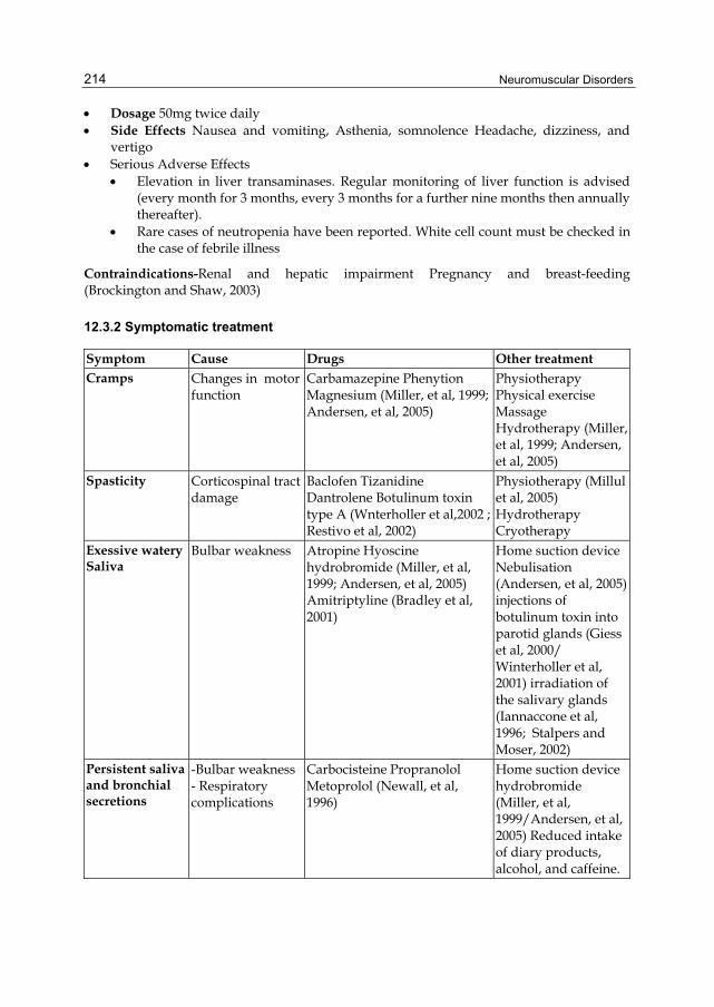
Dosage 50mg twice daily Side Effects Nausea and vomiting, Asthenia, somnolence Headache, dizziness, and
vertigo Serious Adverse Effects
Elevation in liver transaminases. Regular monitoring of liver function is advised (every month for 3 months, every 3 months for a further nine months then annually thereafter).
Rare cases of neutropenia have been reported. White cell count must be checked in the case of febrile illness
Contraindications-Renal and hepatic impairment Pregnancy and breast-feeding (Brockington and Shaw, 2003)
12.3.2 Symptomatic treatment
Symptom Cause Drugs Other treatment Cramps Changes in motor
function Carbamazepine Phenytion Magnesium (Miller, et al, 1999; Andersen, et al, 2005)
Physiotherapy Physical exercise Massage Hydrotherapy (Miller, et al, 1999; Andersen, et al, 2005)
Spasticity Corticospinal tract damage
Baclofen Tizanidine Dantrolene Botulinum toxin type A (Wnterholler et al,2002 ; Restivo et al, 2002)
Physiotherapy (Millul et al, 2005) Hydrotherapy Cryotherapy
Exessive waterySaliva
Bulbar weakness Atropine Hyoscine hydrobromide (Miller, et al, 1999; Andersen, et al, 2005) Amitriptyline (Bradley et al, 2001)
Home suction device Nebulisation (Andersen, et al, 2005) injections of botulinum toxin into parotid glands (Giess et al, 2000/ Winterholler et al, 2001) irradiation of the salivary glands (Iannaccone et al, 1996; Stalpers and Moser, 2002)
Persistent saliva and bronchial secretions
-Bulbar weakness - Respiratory complications
Carbocisteine Propranolol Metoprolol (Newall, et al, 1996)
Home suction device hydrobromide (Miller, et al, 1999/Andersen, et al, 2005) Reduced intake of diary products, alcohol, and caffeine.
Motor Neuron Disease
215
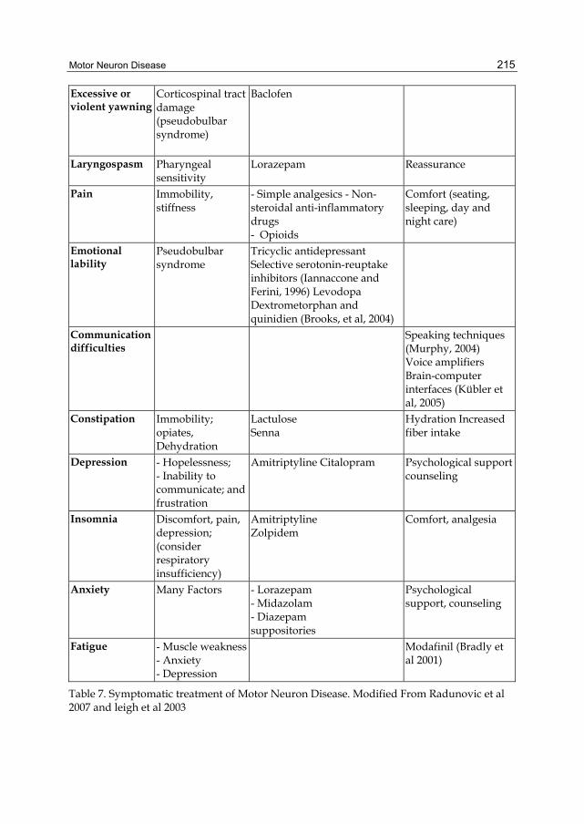
Excessive or violent yawning
Corticospinal tract damage (pseudobulbar syndrome)
Baclofen
Laryngospasm Pharyngeal sensitivity
Lorazepam Reassurance
Pain Immobility, stiffness
- Simple analgesics - Non-steroidal anti-inflammatory drugs - Opioids
Comfort (seating, sleeping, day and night care)
Emotional lability
Pseudobulbar syndrome
Tricyclic antidepressant Selective serotonin-reuptake inhibitors (Iannaccone and Ferini, 1996) Levodopa Dextrometorphan and quinidien (Brooks, et al, 2004)
Communication difficulties
Speaking techniques (Murphy, 2004) Voice amplifiers Brain-computer interfaces (Kübler et al, 2005)
Constipation Immobility; opiates, Dehydration
Lactulose Senna
Hydration Increased fiber intake
Depression - Hopelessness; - Inability to communicate; and frustration
Amitriptyline Citalopram Psychological support counseling
Insomnia Discomfort, pain, depression; (consider respiratory insufficiency)
Amitriptyline Zolpidem
Comfort, analgesia
Anxiety Many Factors - Lorazepam - Midazolam - Diazepam suppositories
Psychological support, counseling
Fatigue - Muscle weakness- Anxiety - Depression
Modafinil (Bradly et al 2001)
Table 7. Symptomatic treatment of Motor Neuron Disease. Modified From Radunovic et al 2007 and leigh et al 2003
Neuromuscular Disorders
216
12.3.3 Treatment of complication
a. Respiratory complications
Respiratory impairment is common in MND and may develop because of respiratory muscle weakness, impaired bulbar function causing aspiration or obstructive sleep apnea, or defects in central control. Dyspnea may be due to infection, pulmonary embolus, or airway obstruction from mucous plug or inhaled pharyngeal contents. (Howard and Wiles,1989).
Symptoms of respiratory insufficiency may be subtle and develop insidiously. Patients may report dyspnea, orthopnea, sleep fragmentation due to hypoventilation, morning headaches, daytime somnolence and fatigue, poor concentration/memory and nocturia. Others may be asymptomatic. Respiratory muscle weakness is an in dependent predictor of quality of life.(Bourke, et al, 2001) and respiratory failure is the most common cause of death in ALS patients. Assessment of respiratory insufficiency includes history, physical examination, rarly morning arterial blood gas, and overnight pulse oximetry.
Nocturnal hypoventilation may present as daytime hypersomnolence, lethargy, morning headaches, poor concentration, depression, anxiety, and irritability, while obstructive sleep apnea is characterized by snoring and restless sleep with abnormal movements. (Howard and orrell 2002).
Types of mechanical ventilation
There are several types of ventilatory aids. These are broadly classified in terms of invasive versus noninvasive.
As its name suggests, invasive techniques require an endotracheal tube or more commonly a tracheostomy. For patients in advanced respiratory failure (ie, no respiratory muscle function), invasive ventilators can assume complete control of ventilation. (Simonds. 2003).
Non- invasive ventilatory aids can be divided into two groups, negative or positive pressure ventilators. Negative pressure is exerted to the chest or abdominal wall mechanically to assist inspiration. Positive pressure devices can be set to deliver variable inspiratory and expiratory pressures, triggered by spontaneous effort (Simonds. 2003).
b. Management of Dysphagia
Management of Dysphagia includes modification of food and fluid consistency, postural advice (e.g. chin tuck: flexing the neck forward on swallowing to protect the airway), and parenteral feeding.
A percutaneous endoscopic gastrostomy (PEG) placement is indicated for those who have symptomatic dysphagia or significant weight loss. (Miller, et al. 2002). Patients and their families should be suitably counseled regarding the benefits and risks of the procedure.
13. Conclusion
Motor neuron disease is of the most common neurodegenerative disorders of unknown etiology, and had no specific treatment. ALS is the commonest type, and in most literatures is used as a synonym for motor neuron disease. Diagnosis is still clinical, mainly, and the investigatory tools have a definite role for diagnosis of other motor neuron mimics. Once motor neuron disease is diagnosed, the prognosis is usually bad, especially when bulbar, and respiratory complications are evident.
Motor Neuron Disease
217
14. References
Abrahams S., Goldstein L.H., Simmons A., et al. (2004). World retrieval in amyotrophic lateral sclerosis: a functional magnetic resonance imaging study. Brain;127:1507-17
Allum C.W,& Shaw P. (2010). Motor neurone disease: a practical update on diagnosis and management. Clinical Medicine, Vol 10. No 3: 252-8
Andersen P.M., Borasio G.D., Dengler R., et al. (2005). EFNS task force on management of amyotrophic lateral sclerosis: guidelines for diagnosing and clinical care of patients and relatives. Eur J Neurol;12:921-38.
Andersen P.M., Sims Xin W.W., et al. (2003). Sixteen novel mutations in the Cu/Zn superoxide dismutase gene in amyotrophic lateral sclerosis: a decade of discoveries, defects and disputes. Amyotroph Lateral Scler Other Motor Neuron Disorders;4:62-73.
Bak T.H.,& Hodges J.R.& (2004). The effects of motor neuron disease on language: further evidence. Brain Lang;89:354-61.
Baltadzhieva R., Gurevich T.,& Korezyn A.D. (2006). Autonomic impairment in amyotrophic lateral sclerosis. Curr Opin Neurol;18:487-93.
Bourke S.C., Shaw P.J.,& Gibson G.J. (2001). Respiratory function vs sleep-disorderd breathing as predictors of Qol in ALS. Neurology 57:2040-2044.
Bradley W.G., Anderson F., Bromberg M., et al. (2001). Current management of ALS: comparison of the ALS CARE Database and the AAN Practice Parameter. Neurology;57:500-04.
Brockington A.,& Shaw P. (2003). Developments in the treatment of Motor Neurone Disease. Acnr. Vol 3 N 5 November/December.
Brooks B.R. (1994). EL Escorial World Federation of Neurology criteria for the diagnosis of amyotrophic lateral sclerosis. Subcommittee on Motor Neuron Disease/Amyotrophic Lateral Sclerosis of the World Federation of Neurology Research Group on Neuromuscular Disease and El Escorial " Clinical limits of amyotrophic lateral sclerosis" workshop contributors. J Neurol Sci 124:96-107.
Brooks B.R., Miller R.G., Swash M. & Munsat T.L. (2000). World Federation of Neurology Research Group on Motor Neuron Disease: El Escorial revisited: revised criteria for the diagnosis of amyotrophic lateral sclerosis. Amyotrophic Lateral Scler. Other Motor Neuron Disord, 1:293-239.
Brooks B.R., Thisted R.A., Appel S.H., et al. (2004). Treatment of pseudobulbar affect in ALS with dextromethorphan/quinidine, Neurology;63:1363-70.
Brugman F., & Wokkle J.H.J. (2004). Primary Lateral Sclerosis. Orphanet Encyclopedia. April, http://www.orpha.net/data/patho/GB/uk-PLS.bdf.
Carter G.T., Weiss M.D., Lou J.S., et al. (2005). Modafinil to treat fatigue in amyotrophic lateral sclerosis: an open label pilot study. Am J Hosp Palliat Care;22:55-59.
Cleveland D.W.& Rothstein. J.D. (2001). Nat. Rev Neurosci 11,806-819. Daube J.R. (2000). Electrodiagnostic studies in amyotrophic lateral sclerosis and other motor
neuron disorders. Muscle Nerue; 23:1488-502. De Domenice P., Malara C.E., Marabello L., et al.( 1988). Amyotrophic lateral sclerosis; an
epidemiological study in the Province of Messina, Italy, 1976-1985. Neuroepidemiology;7:152-8.
Duffy J.R., Peach R.K., & Strand E.A. (2007). Progressive apraxia of speech as a sign of motor neuron disease. Am J Neuroradial;16:198-208.
Neuromuscular Disorders
218
Eisen A. (2001). Clinical electrophysiology of the upper and lower motor neuron in amyotrophic lateral sclerosis. Sem Neurol;21:141-54.
Eisen A., & Swash M. (2001). Clinical neurophysiology of ALS. Clin Neurophysiol;112:2190-201.
Fong K.Y., Yu Y.L., Chan Y.W., et al. (1996). Motor neuron disease in Hong Kong Chinese: epidemiology and clinical picture. Neuroepidemiology;15:239-45.
Geser F., Brandmeir N.J., Kwong L.K., et al. (2008). Evidence of multisystem disorder in whole – brain map of pathological TDP-43 in amyotrophic lateral sclerosis. Arch Neurol;65:636-41.
Giess R., Naumann M., Werner E., et al. (2000). Injections of botulinum toxin A into the salivary glands improve sialorrhoea in amyotrophic lateral sclerosis. J Neurol Neurosurg Psychiatry;69:121-23.
Haley R.W. (2003). Excess incidence of ALS in young Gulf War veterans. Neurology;61:750-6. Horner R.D., Kamins K.G., Feussner J.R. et al. (2003). Occurrence of amyotrophic lateral
sclerosis among Gulf War veterans. Neurology;61:742-9. Howard R.S., Wiles C.M., & Loh L. (1989). Respiratory complications and their management
in motor neurone disease. Brain;112:1155-70. Howard R.S.,& Orrell R.W. (2002). Management of motor neuron disease, Postgrad Med J;
78:736-741. Iannaccone S.,& Ferini-Strambi L. (1996). Pharmacological treatment of emotional lability.
Clin Neuropharmacol;19:532-35. Ingram D.A.,& Swash M. (1987). Central motor conduction is abnormal in motor neuron
disease. J Nerol Neurosurg Psychiatry;50:159-66 Irwin D., Lippa C.F., Swearer J.M. (2007). Cognition and amyotrophic lateral sclerosis (ALS).
Am J Alzheimer Dis Other Demen;22:300-12. Isaacs J.D., Dean A.F., Shaw C.E., et al. (2007). Amyotrophic lateral sclerosis with sensory
neuropathy: part of a multisystem disorders? J Neurol Neurosurg Psychiatry; 78:750-3.
Kew J.J.M., Goldstein L.H., Leigh P.N., et al. (1993). The relationship between abnormalities of cognitive function and cerebral activation in amyotrophic lateral sclerosis: a neuropsychological and positron emission tomography study. Brain;116:1399-423.
Kübler A., Nijboer F., Mellinger J., et al. (2005). Patients with ALS can use sensorimotor rhythms to operate a brain-computer interface. Neurology;64:1775-77.
Lakerveld J., Kotchoubey B.,& Kubler A. (2008). Cognitive function in patients with late stage amyotrophic lateral sclerosis. J Neurol Neurosurg Psychiatry;79:25-29. doi:10.1136/jnnp.2007.116178.
Leigh P.N. (1991). Amyotrophic lateral sclerosis and other motor neuron disorders. Curr Opin Neurol Neurosurg;4:586-96.
Leigh P.N., Abrahams S., Al-Chalabi A., Ampong M.-A., Goldstein L.H., Johnson J., et al. (2003). King's MND Care and Research Team. The management of motor neurone disease. J Neurol Neurosurg Psychiatry;74: iv 32-47.
Leigh P.N., & Ray-Chaudhuri K. (1994). Motor neuron disease. J Neurol Neurosurg Psychiatry;57:886-96.
Logroscino G., Traynor B.J., Hardiman O., et al. (2008). Descriptive epidemiology of amyotrophic lateral sclerosis: new evidence and unsolved issues. J Neurol Neurosurg Psychiatry;79:6-11.
Motor Neuron Disease
219
Miller R.G., Mitchell J.D., Lyon M.,& Moore D.H. (2002). Riluzole for amyotrophic lateral sclerosis (ALS)/ motor neuron disease (MND). Cochrane Database Syst Rev; CD001447.
Miller R.G., Rosenberg J.A., Gelinas D.F., et al. (1999). Practice parameter: the care of the patient with amyotrophic lateral sclerosis (an evidence-based review): report of the Quality Standards Subcommittee of the American American Academy of Neurology. Neurology;52:1311-23.
Millul A., Beghi E., Logroscino G., Micheli A., Vielli E.,& Zardi A. (2005). Survival of patients with amyotrophic lateral sclerosis in a population-based registry. Neuroepidemiology;25:114-19.
Montovan M.C., Baggio L., Dalla Barba G., et al. (2003). Memory deficits and retrieval processes in ALS. Eur J Neurol;10:221-7.
Mulder D.W. (1982). Clinical limits of amyotrophic lateral sclerosis. In Rowtand LP (ed). Human Motor Neuron Diseases. New York: Raven Press:15-22
Murphy J. (2004). Communication strategies of people with ALS and their partners. Amyotroph Lateral Scler Other Motor Disord;5:121-26.
Murros K. &, Fogelholm R. (1983). Amyotrophic lateral sclerosis in Middle-Finland: an epidemiological study. Acta Neurol Scand;67:41-7.
Neary D., Snowden J.S., & Mann D.M.A. (2000); Cognitive change in motor neurone disease/ amyotrophic lateral sclerosis (MND/ALS). J Neurol Sci;180:15-20.
Newall A.R., & Orser R. (1996). Hunt M. The control of oral secretions in bulbar ALS/MND. J Neurol Sci;139:43-44.
Oluwole O.S.A., Conradi S., Kristensson K.,& Karlsson H. (2004). Human Endogenous Retrovirus W and Motor Neurone Disease. Annals of badan Post graduate Medicine. Vol. 2No 2dec.
Phukan J., Pender N.P, & Hardiman O. (2007). Cognitive impairment in amyotrophic lateral Sclerosis. Lancet Neurol;6:994-1003.
Phukan J., & Hardiman O (2009). The management of amyotrophic lateral. J Neurol. DOI 10.1007/s00415-0090142-9.
Pugdahl K., Fuglsang-Frederiksen A., de Carvalho M., et al. (2007). Generalised sensory system abnormalities in amyotrophic lateral sclerosis: a European multicentre study. J Neurol Neurosurg Psychiatry;78:746-9.
Raaphorst J., de Visser M., Linssen W.H., de Haan R.,J., & Schmand B. (2010). The cognitive profile of amyotrophic lateral sclerosis: A meta-analysis. Amyotroph. Lateral Scler., 11:27-37.
Radhakrishnan K., Ashok P.P., Sridharan R., Mousa M.E. (1986). Descriptive epidemiology of motor neuron disease in Benghazi, Libya. Neuroepidemiology;5:47-54.
Radunovic A., Mitsumoto H.,& Leigh P.N. (2007). Clinical Care of Patients with Amyotrophic Lateral Sclerosis. Lancet Neurology.; 10:931-25.
Restivo D.A., Lanza S., Marchese-Ragona R.,& Plameri A. (2002). Improvement of masseter spasticity by botulinum toxin facilitates PEG placement in amyotrophic lateral sclerosis. Gasreoenterology;123:1749-50.
Riluzole monograph. Facts and comparisons online. [cited 2011 Feb 19] Available at: http://online.factsandcomparisons.com. Ringel S.P., Murphy J.R., Alderson MK, et al.( 1993). The natural history of amyotrophic
lateral sclerosis. Neurology;43:1316-22. Ringholz G.M. Appel S.H, Bradshaw M., et al. (2005). Prevalence and patterns of cognitive
impairment in sporadic ALS. Neurology;65:586-90.
Neuromuscular Disorders
220
Rodgers-Johanson P., Garruto R.M., Yanagihara R., Chen K.M., Gajdusek D.C.,& Gibbs C.J. (1986). Amyotrophic lateral sclerosis and parkinsonism-dementia on Guam: a 30 year evaluation and neuropathologic trends. Neurology;36:7-13.
Schroder H.D.,& Reske-Nielsen E. (1984). Preservation of the nucleus – pelvic floor motor system in amyorophic lateral sclerosis. Clin Neuropathol;3:210-6
Shaw PJ. (2005). Molecular and cellular pathways of neurodegeneration in motor neuron disease. J Neurol Neurosurg Psychiatry; 76:1046-1057.
Simonds A.K. (2003). Home ventitation. Eur Respir J; 22:38s-46s. Stalpers L.J., & Moser E.C. (2002). Results of radiotherapy for drooling in amyotrophic
lateral sclerosis. Neurology;58:1308. Strong M., & Rosenfeld J. (2003). Amyotrophic lateral sclerosis: a review of current concepts.
ALS and other motor neuron disorders;4:136-143 Strong M.J., Grace G.M., Freedman M., Lomen-Hoerth C, Woolley S, Goldstein L.H. Murphy
J. ,Shoesmith C., Rosenfeld J., Leigh P.N., Bruijn L., Ince P.,& Figlewiez D. (2009). Consensus criteria for the diagnosis of front-otemporal cognitive and behavioral syndromes in amyotrophic lateral sclerosis. Amyotroph. Lateral Scler. 10:131-146,.
Swash M. (2001). Amyotrophic lateral sclerosis: current understanding. J Neurosci Nurs;33:245-53.
Swash M., Leader M., Browen A.,& Swettenhan K. (1986). Focal loss of anterior horn cells in the cervical cord in motor neuron disease. Brain;109:939-52
Takeda S., Yamada M., Kawasaki K., et al. (1994). Motor neuron disease with multi-system involvement presenting as tetraparesis, ophthalmoplegia and sensori-autonomic dysfunction. Acta Neuropathol;88:193-200.
Talbot K. (2002). Motor neurone disease, Postgrad Med J.;78:513-519. Tsuchiya K., Ozawa E., Fukushima J., et al. (2000). Rapidly progressive aphasia and motor
neuron disease: a clinical, radiological, and pathological study of an autopsy case with circumscribed lobar atrophy. Acta Neuropathol;99:81-7.
Turner M.R., Kiernan M.C, Leigh P.N., & Talbot K. (2009). Biomarkers in amyotrophic lateral sclerosis. Lancet Neurol;8:94-109.
Valdmanis P.N., & Rouleau G.A. (2008). Genetics of familial amyotrophic lateral sclerosis. Neurology;70:144-52.
Van der Graaff M. (2004). Amyotrophic lateral sclerosis. Orphanet Encyclopedia. September. http://www.prpha.net/data/patho/GB/uk-ALS.pdf.
Washington, D.C. (c2007). The ALS Association; [updated 2008 Oct; cited 2011.Feb 17]. Available from http://www.alsa.org/.
Wijesekera L.C. & Leigh P.N. (2009). Amyotrophic lateral sclerosis. Orphanet Journal of Rare Diseases [cited 2011 Feb 17] ; 4(3) [about 22p.]. Avalilable from:
http://www.ojrd.com/content/4/1/3. Winterholler M.G., Erbguth F.J., Wolf S.,& Kat S. (2001). Botulinum toxin for the treatment of
sialorrhoea in ALS: serious side effects of a transductal approach. J Neurol Neurosurg Psychiatry;70:417-18.
Winterholler M.G., Heckmann J.G. Hecht M. & Erbguth F.J. (2002). Recurrent trismus and stridor in an ALS patient: successful treatment with botulinum toxin. Neurology;58:502-03.
Yoshida M. (2004). Amyotrophic lateral sclerosis with dementia: The clinicopothological spectrum, Neuropathology;24:87-102.
Zago S., Poletti B., Morelli C., Doretti A., & Silani V. (2010). Archives italiennes de Biologie, 149;39:56,.

© 2012 The Author(s). Licensee IntechOpen. This is an open access articledistributed under the terms of the Creative Commons Attribution 3.0License, which permits unrestricted use, distribution, and reproduction inany medium, provided the original work is properly cited.