Embed Size (px)
Citation preview
Mouse Hepatic Oval Cells Require Met-Dependent PI3Kto Impair TGF-b-Induced Oxidative Stress and ApoptosisAdoracion Martınez-Palacian1, Gaelle del Castillo1¤a, Amileth Suarez-Causado1¤b, Marıa Garcıa-Alvaro1,
Diego de la Morena-Frutos1, Margarita Fernandez1, Cesareo Roncero1, Isabel Fabregat2,
Blanca Herrera1", Aranzazu Sanchez1*"
1 Dep. Bioquımica y Biologıa Molecular II, Facultad de Farmacia, Universidad Complutense, Instituto de Investigacion Sanitaria del Hospital Clınico San Carlos (IdISSC),
Madrid, Spain, 2 Laboratori dOncologia Molecular and Departament de Ciencies Fisiologiques II, Universitat de Barcelona, Institut dInvestigacio Biomedica de Bellvitge,
LHospitalet de Llobregat, Barcelona, Spain
Abstract
We have previously shown that oval cells harboring a genetically inactivated Met tyrosine kinase (Met2/2 oval cells) aremore sensitive to TGF-b-induced apoptosis than cells expressing a functional Met (Metflx/flx), demonstrating that the HGF/Met axis plays a pivotal role in oval cell survival. Here, we have examined the mechanism behind this effect and have foundthat TGF-b induced a mitochondria-dependent apoptotic cell death in Metflx/flx and Met2/2 oval cells, associated with amarked increase in levels of the BH3-only proteins Bim and Bmf. Bmf plays a key role during TGF-b-mediated apoptosis sinceknocking down of BMF significantly diminished the apoptotic response in Met2/2 oval cells. TGF-b also induced oxidativestress accompanied by NADPH oxidase 4 (Nox4) mRNA up-regulation and decreased protein levels of antioxidant enzymes.Antioxidants inhibit both TGF-b-induced caspase 3 activity and Bmf up-regulation, revealing an oxidative stress-dependentBmf regulation by TGF-b. Notably, oxidative stress-related events were strongly amplified in Met2/2 oval cells, emphasizingthe critical role of Met in promoting survival. Pharmacological inhibition of PI3K did impair HGF-driven protection from TGF-b-induced apoptosis and increased sensitivity of Metflx/flx oval cells to TGF-ß by enhancing oxidative stress, reachingapoptotic indices similar to those obtained in Met2/2 oval cells. Interestingly, both PI3K inhibition and/or knockdown itselfresulted in caspase-3 activation and loss of viability in Metflx/flx oval cells, whereas no effect was observed in Met2/2 ovalcells. Altogether, results presented here provide solid evidences that both paracrine and autocrine HGF/Met signalingrequires PI3K to promote mouse hepatic oval cell survival against TGF-b-induced oxidative stress and apoptosis.
Citation: Martınez-Palacian A, del Castillo G, Suarez-Causado A, Garcıa-Alvaro M, Morena-Frutos Ddl, et al. (2013) Mouse Hepatic Oval Cells Require Met-Dependent PI3K to Impair TGF-b-Induced Oxidative Stress and Apoptosis. PLoS ONE 8(1): e53108. doi:10.1371/journal.pone.0053108
Editor: Tetsuo Takehara, Osaka University Graduate School of Medicine, Japan
Received July 25, 2012; Accepted November 23, 2012; Published January 2, 2013
Copyright: � 2013 Martınez-Palacian et al. This is an open-access article distributed under the terms of the Creative Commons Attribution License, whichpermits unrestricted use, distribution, and reproduction in any medium, provided the original author and source are credited.
Funding: AMP was recipient of a research-training contract (grant SAF2006-12025) from the Ministry of Education and Science. GC was recipient of a research-training contract from the Ministry of Education and Science. ASC was recipient of an Alban scholarship program and then a research assistant contract (grantSAF2009-12477). MGA is recipient of a research assistant contract (grant S2010/BMD-2402). This work has been supported by grants SAF2006-12025 from Ministryof Education and Science (Spain), SAF2009-12477 from Ministry of Science and Innovation (Spain), and 920359 (CAM-UCM, BSCH-UCM). The funders had no role instudy design, data collection and analysis, decision to publish, or preparation of the manuscript.
Competing Interests: The authors have declared that no competing interests exist.
* E-mail: [email protected]
" These authors are joint senior authors on this work.
¤a Current address: Instituto de Investigaciones Biomedicas ‘‘Alberto Sols’’, Madrid, Spain¤b Current address: Universidad de Cartagena, Cartagena de Indias, Colombia
Introduction
Oval cells constitute a bi-potential progenitor cell population
from adult liver. When hepatocyte proliferation and/or function is
impaired by chronic liver disease or hepatotoxin administration,
oval cells emerge from the periportal area, in particular the canals
of Hering, the terminal smallest branch of the biliary tree, and
expand into the damaged parenchyma, a process known as ‘‘oval
cell response’’ or ‘‘ductular reaction’’. Oval cells can give rise to
both hepatocytes and biliary epithelial cells and are characterized
by the co-expression of both hepatocytic and cholangiocytic
markers as well as hematopoietic and neuroepithelial markers,
which reflects their immature phenotype [1,2,3]. These cells are
nowadays a matter of intense investigation. On the one hand, due
to their facultative role in liver regeneration, oval cells have been
postulated as a therapeutic tool in acute or chronic liver diseases.
On the other hand a link has been established between oval cells
and hepatocarcinogenesis. Indeed, a substantial body of evidence
in the literature supports the hypothesis that oval cells could be the
origin of at least a subset of hepatocellular carcinoma (HCC) [4].
Understanding the intricate growth factor network and signaling
events that regulate oval cell biology will help us to clarify these
contrasting roles of oval cells in liver regeneration and tumor
development.
Transforming growth factor beta (TGF-b), a member of the
TGF-b superfamily ligands, has highly pleiotropic effects that
depend on the dose, duration of signal and the type and state of
the target cell. It has a crucial role during development, tissue
remodelling and homeostasis by controlling many cellular
processes, such as differentiation, proliferation, apoptosis and
motility in many types of cells [5]. TGF-b initiates the intracellular
PLOS ONE | www.plosone.org 1 January 2013 | Volume 8 | Issue 1 | e53108

signaling through binding to transmembrane serine-threonine
kinase receptors and subsequent activation of SMAD proteins,
which regulate gene expression [6]. Besides the canonical SMAD-
mediated pathway, TGF-b triggers a variety of intracellular
signaling pathways, commonly referred to as ‘‘non-SMAD’’ or
‘‘non-canonical’’ pathways that include mitogen-activated protein
kinases (MAPKs), Rho-like guanosine triphosphatases (GTPases)
and phosphatidilinositol-3-kinase PI3K/AKT pathways [7]. In
hepatocytes, the best characterized effects of TGF-b are undoubt-
edly the induction of growth arrest and apoptosis, which target
cells at different stages of differentiation and pathophysiological
conditions, i.e. adult, fetal and regenerating hepatocytes
[8,9,10,11,12]. Surprisingly, the effects of TGF-b on oval cell
biology are not yet well defined. In vivo and in vitro data indicate
that TGF-b negatively controls oval cell activation but the
mechanisms underlying its effects have not been fully explored.
Thus, transgenic mice expressing active TGF-b in the liver show
an impaired oval cell response after hepatic chronic injury induced
by a 3,5-diethoxycarbonyl-1,4-dihydro-collidine (DDC)-contain-
ing diet [13]. Furthermore, coinciding with the oval cell
proliferation an increased expression of TGF-b1 in hepatic stellate
cells is observed, followed by a peak in apoptosis of oval cells [14].
In agreement with these in vivo observations, TGF-b decreases rat
oval cell growth in vitro although to a lesser extent than in
hepatocytes [15]. We have also shown that TGF-b decreases cell
viability and induces caspase-3 activation in oval cells in vitro [16].
Interestingly, CD133+/CD45- oval cells isolated from methionine
adenosyltransferase 1a (Mat1a2/2) deficient mice, a cell popula-
tion that resemble cancer stem cells, show cell growth inhibition in
response to TGF-b but appear to be resistant to its apoptotic
effects [17].
Met is a proto-oncogene that encodes for the Hepatocyte
Growth Factor (HGF) receptor. HGF binds to Met to mediate its
multiple cellular activities including mitogenesis, motogenesis,
morphogenesis and survival in a variety of cell types [18,19]. Upon
HGF binding, Met is activated and autophosphorylated in specific
tyrosine residues, leading to the recruitment of signal transducers
through the C-terminal docking site. Among the signaling
pathways activated by Met in response to HGF are PI3K/AKT,
phospholipase-C gamma (PLC-c), sarcoma protein kinase (Src),
signal transducer and activator of transcription 3 (STAT3),
nuclear factor-kappa B (NF-kB) and MAPKs including Ras/
extracellular signal-regulated kinases (ERKs), c-Jun N-terminal
kinases (JNK) and p38 [19]. The HGF/Met signaling pathway is
critical during tissue formation and homeostasis, playing a major
role in liver physiology and pathology, both during development
and adulthood. This notion is supported by the fact that deletion
of either HGF or Met causes embryonic lethality due to multiple
abnormalities including massive hepatoblasts apoptosis [20,21].
Additionally, liver specific Met or HGF conditional knock-out
mice show an impaired regenerative response associated with an
increased cell death, delayed and decreased proliferation and
healing [22,23]. The increased apoptotic cell death is a common
trait in all these genetic ablation models, highlighting the
remarkable action of HGF/Met in the suppression of apoptosis.
Antiapoptotic action of HGF/Met in liver does not exclusively
target hepatocytes. To the contrary, HGF has proved to be a
survival factor for oval cells against different apoptotic insults, such
as Tumor Necrosis Factor (TNF-a), serum withdrawal and TGF-b[24,25]. Accordingly, we have demonstrated that oval cells
harboring a genetically inactivated Met tyrosine kinase (Met2/2
oval cells) are more sensitive to TGF-b-induced apoptosis than
their normal counterparts (Metflx/flx oval cells) [24].
Seeking to understand how HGF/Met exerts its survival effect
against TGF-b-induced apoptosis, we conducted a broad analysis
on the apoptotic signaling cascade induced by TGF-b in both
Metflx/flx and Met2/2 oval cell lines. Here we report that TGF-binduces a mitochondrial apoptotic cell death in oval cells, with up-
regulation of BIM and BMF proteins, two BH3-only members of
the Bcl-2 family. Furthermore, we reveal a signaling pathway in
which TGF-b induces oxidative stress associated with up-
regulation of Nox4 and down-regulation of the intracellular
antioxidant defenses, which leads to Bmf up-regulation and
subsequent cell apoptosis. Although both Metflx/flx and Met2/2
oval cells do respond to TGF-b, alteration of both mitochondrial
function and oxidative homeostasis are amplified in Met2/2 oval
cells, providing one mechanism for the increased sensitivity to
TGF-b-triggered apoptosis in Met-deficient oval cells. Finally, our
results provide strong evidence that PI3K may be a key player in
mediating anti-apoptotic signals via Met in oval cells by acting as
an antioxidant signal.
Materials and Methods
Reagents and AntibodiesMouse recombinant HGF was purchased from R&D Systems
(Minneapolis, MN). Human recombinant TGF-b, ERK inhibitor
PD90059, p38 inhibitor SB203580 and PI3K inhibitor LY294002
were from Calbiochem (La Jolla, CA). SP600125 JNK inhibitor
was from Alexis Biochemical (Madrid, Spain), Dulbecco’s
modified Eagle’s medium (DMEM), fetal bovine serum (FBS)
and trypsin-EDTA were from Gibco-Invitrogen (Barcelona,
Spain). Ascorbate, pyrrolidine carbodithioic acid (PDTC), peni-
cillin, streptomycin, HEPES, bovine serum albumin (fraction V,
fatty-acid free), propidium iodide, DNA oligos and buffer reagents
were from Sigma-Aldrich (Tres Cantos, Madrid, Spain). 29,79-
dichlorofluorescein-diacetate (DCFH-DA) was from Molecular
Probes (Eugene, OR). RNeasy Kit was from Qiagen (Valencia,
CA). SuperScript III RNase H Reverse Transcriptase was from
Invitrogen. Oligo-dT was from Roche Diagnostics (Sant Cugat del
Valles, Barcelona, Spain). Horseradish peroxidase-conjugated
secondary antibody and ECL reagent were from GE Healthcare
Europe (Barcelona, Spain). Caspase-3 substrate was obtained from
PharMingen (San Diego, CA).
The rabbit polyclonal antibodies against phospho-Smad2 (Ser
465/467) (CS3101), phospho-p38 (Thr180/Tyr1829) (CS9211)
and GADPH (CS2118) were purchased from Cell Signaling
(Beverly, MA). Rabbit polyclonal against p38 (SC-535) and mouse
monoclonal against phospho-JNK (SC-6254) antibodies were from
Santa Cruz Biotechnology, Inc., (Paso Robles, CA). Mouse
monoclonal anti-Cytochrome C (556433) and rabbit polyclonal
anti-Bim (559685) and anti-Bcl-x (610211) antibodies were from
BD Biosciences. Anti-b-actin (clone AC-15) and anti-Catalase
(C0979) mouse monoclonal antibodies were from Sigma-Aldrich.
Polyclonal antibodies anti-Manganese Superoxide Dismutase 2
(SOD2) (06–984) and anti-PI3K p85 (06–195) were from
Millipore, anti-gamma-Glutamylcysteine Synthetase (c-GCS) from
Abcam (40929) and mouse monoclonal antibody anti-Bmf from
Alexis Biochemicals (ALX-804-342).
Cell Lines and Culture ConditionsMetflx/flx and Met2/2 oval cell lines were generated as
described previously [24]. Cells were routinely maintained in
DMEM supplemented with 10% FBS in a humidified incubator at
37uC and a 5% CO2 atmosphere. Medium was replaced every
three days, and cells were harvested at 80% to 90% confluence
using trypsin-EDTA and replated at 1:10 dilution for mainte-
Met Protects Oval Cells against TGF-b via PI3K
PLOS ONE | www.plosone.org 2 January 2013 | Volume 8 | Issue 1 | e53108

nance. After an overnight attachment period, medium was
replaced by serum-free DMEM. Cells were maintained in
serum-free medium for 4–12 hours prior to treatment with growth
factors. Where indicated, cells were pretreated with HGF for at
least 6 hours followed by TGF-b treatment. PD98059, SB203580,
LY294002, SP600125, ascorbate and PDTC were added 30
minutes before addition of growth factors.
Analysis of Apoptosis by Phosphatidylserine ExposureCells were collected by centrifugation at 1300 rpm for 5 min
and washed once with PBS. 500,000 cells were resuspended with
195 ml of binding buffer (10 mM HEPES, pH 7.4, 2.5 mM CaCl2,
140 mM NaCl) supplemented with 5 ml annexin V-FITC (BD
Pharmingen) and incubated for 10 min at room temperature.
Samples were centrifuged and resuspended with 300 ml of binding
buffer containing 1 mg/ml propidium iodide. Fluorescence inten-
sity was analyzed using a FACSCalibur flow cytometer. 10,000
cells were recorded in each analysis.
Measurement of Intracellular ROSFor the analysis of intracellular ROS by flow cytometry, the
oxidation-sensitive probe DCFH-DA was used, as previously
described [12]. Cells were detached by trypsinization, collected by
centrifugation at 1300 rpm for 5 min and washed once with PBS.
Samples were then incubated with 5 mM DCFH-DA for 30
minutes at 37uC. Cellular fluorescence intensity was measured in a
FACScan flow cytometer. Propidium iodide (0.005%) was used to
detect dead cells. For each analysis 10,000 events were recorded.
Glutathione DeterminationCells were washed twice with PBS, scraped off and pelleted at
4uC. Cellular glutathione was extracted in a buffer containing
0.2% Triton X-100, 2.5% sulfosalicylic acid. After centrifugation
at 12000 rpm for 15 min at 4uC, the supernatant was used for the
determination of total glutathione, using the method of Griffith
modified as described previously [26]. Using reduced glutathione
as standard, glutathione content is expressed as nmol/mg protein.
Analysis of Mitochondrial Transmembrane PotentialCells were collected by centrifugation at 1300 rpm for 5 min,
washed once and incubated with 2 mM 5,59,6,69-tetrachloro-
1,19,3,39-tetraethylbenzimidazolylcarbocyanine iodide (JC-1) (Mo-
lecular Probes) for 1 hour at 37uC. Then, samples were
centrifuged and resuspended with 500 ml DMEM. Fluorescence
intensity (FL1 and FL2) was measured in a FACScan flow
cytometer. 10,000 cells were recorded in each analysis. JC-1 dye
exhibits potential-dependent accumulation in mitochondria. Cells
with healthy mitochondria show both red (FL2) (aggregate,
mitochondrial) and green (monomeric, cytoplasmic) fluorescence.
Mitochondria depolarization is indicated by a decrease in the red/
green (FL2/FL1) fluorescence intensity ratio, which is due to loss
of red J-aggregate fluorescence and cytoplasmic diffusion of green
monomer fluorescence.
Transcriptional Reporter AssaysTranscriptional reporter assays were performed using Cignal
SMAD Reporter (luc) kit (CCS-017L) from Qiagen. Cells were
plated in 96 well dishes. The next day, DNA was transfected into
the cells with Fugene transfection reagent, according to manufac-
turer recommendations. After 15 hours, medium was replaced
with serum-free medium for additional 15 hours. Cells were then
incubated with or without TGF-b and firefly and renilla
(normalizing transfection control) luciferase activities were mea-
sured using ‘‘Dual luciferase Reporter Assay System’’ (Promega) in
a luminometer Fluostar Omega (BMG labtech).
Quantitative Reverse Transcriptase-Polymerase ChainReaction (qRT-PCR) Analysis
Total cellular RNA was isolated using the RNeasy Kit (Qiagen,
Valencia, CA). RNA yield and purity were analyzed using a
spectrophotometer (UV-visible recording spectrophotometer Spe-
cord 205, AnalytikJena). 1 mg total RNA was reverse-transcribed
into complementary DNA using SuperScript III RNase H Reverse
Transcriptase and oligo-dT as a primer. Quantitative PCR was
performed using SYBRGREEN (Roche) and Amplified products
were analysed in ABI Prism 7900 HT Fast Real-Time (Applied
Biosystems). The relative amount of target mRNA was determined
after normalization against reference gene (Gusb) in each sample.
Primers used in the study are presented in supporting information,
table S1.
Measurement of Caspase-3-Like Enzymatic ActivityA fluorometric assay in the presence of Ac-DEVD-AMC as
fluorogenic Caspase-3 substrate was used following a previously
described procedure [26]. Cleavage of the substrate was moni-
tored in a Microplate Fluorescence Reader FL600 (Bio-Tek)
(excitation, 380 nm; emission, 440 nm). A unit of caspase activity
is the amount of enzyme that will lead to a one unit increase in the
fluorescence intensity. Protein concentration was estimated and
results are expressed as units of activity per microgram of protein.
ImmunoblottingFor standard western blotting total cell extracts were prepared
in modified RIPA buffer (30 mM Tris pH 7.5; 150 mM sodium
chloride; 1% NP40; 0.5% sodium deoxycholate; 0.1% SDS;
5 mM EDTA) supplemented with 1 mM phenylmethylsulfonyl
fluoride, 10 mg/ml aprotinin and leupeptin and 1 mM sodium
orthovanadate. 30 to 80 mg of protein were separated in 10–12%
acrylamide sodium dodecyl sulfate-polyacrylamide electrophoresis
gels and blotted to Immobilon-P membranes (Millipore, Bedford,
MA). Membranes were probed with the primary antibodies
diluted 1:500 to 1:1000 in Tris-buffered saline containing 0.1%
Tween 20 and 0.5% non-fat dried milk or 0.5% bovine serum
albumin according to manufacturer’s instructions. Detection was
done using the enhanced chemiluminescence method and
autoradiography.
Measurement of Apoptotic IndexMeasurement of apoptotic index was performed as previously
described [24]. After staining with propidium iodide, cells
undergoing apoptosis were scored under inverted fluorescence
microscope (Eclipse TE300, Nikon) at high magnification (x60)
following standard morphological criteria. Apoptotic indices were
calculated after counting a minimum of 1000 cells per treatment in
a blinded manner.
siRNA Knockdown AssayssiRNA knockdown assays were performed as previously
described [27]. For transient siRNA transfection, cells were seeded
at 50–60% confluence. On the following day, cells were
transfected with a SMARTpool siRNA directed to the mouse
p85a regulatory subunit of PI3K (50 nM) or to mouse Bmf
(20 nM) or the control non-targeting siRNA (50 nM and 20 nM,
respectively) (Dharmacon, Lafayette, CO) using TransIT-siQuest
reagent (Mirus, Madison, WI) according to manufacturers
instructions. Transfected cells were grown for 24 hours in
Met Protects Oval Cells against TGF-b via PI3K
PLOS ONE | www.plosone.org 3 January 2013 | Volume 8 | Issue 1 | e53108

complete medium, then tripsinized, diluted to the appropriate cell
density, and replated in dishes for subsequent assays.
Adenovirus-mediated Met KnockdownIn vitro inactivation of Met was achieved following previous
protocols [24]. Twenty-four hours after plating, Metflx/flx cells
were infected with adenovirus expressing Cre recombinase (Ad-
CMVCre, Vector Biolabs, Philadelphia, PA) to disrupt endoge-
nous met allele, or an empty adenovirus (Ad-CMVNull) used as
control. Virus was diluted in infection medium (growth medium
supplemented with 2.5 mg/ml polybrene) at a multiplicity of
infection of 10. Cells were incubated in infection medium for one
hour in the incubator with occasional shaking and then fresh
medium was added to complete volume. After 24 hours of
infection, cells were serum starved for 4 hours and treated with
TGF-b for additional 24 hours to analyze cell apoptosis. In
parallel, infected cells were used to isolate genomic DNA following
standard procedures to identify the deleted allele by PCR using
specific oligonucleotides [23].
Statistical AnalysisStatistical analysis was performed by Studentst-test method. The
differences were assumed significant at P,0.05.
Results
Absence of a Functional Met Receptor does not AffectTGF-b Induced Activation of Canonical and Non-canonical Signaling Pathways in Oval Cells
Previous results from our group have shown that mouse oval cell
lines harboring a genetically inactivated Met tyrosine kinase
(Met2/2) respond differently to apoptotic insults than Metflx/flx
oval cells and show an increased sensitivity to TGF-b and serum
starvation-induced apoptosis [24]. Indeed, caspase-3 activity
increased in oval cells upon TGF-b addition, being this increase
significantly higher in Met2/2 than in Metflx/flx oval cells
(figure 1A). Caspase-3 activation occurred concomitantly with
other apoptotic hallmarks. Thus, TGF-b promoted phosphatilser-
ine exposure on the outer leaflet of the plasma membrane, which
started after 15 hours of treatment and was maintained for several
hours (figure 1B). It is noteworthy that the annexin V positive/
propidium iodide negative subpopulation, corresponding to the
apoptotic cell subpopulation, was larger in Met2/2 oval cells
compared to their normal counterparts, serving as additional proof
that absence of a functional Met sensitizes oval cells to apoptosis.
As an alternative approach Met was transiently knocked down in
oval cells by infection with adenovirus expressing Cre recombinase
under a CMV promoter (Ad-CMVCre). Subsequently, cells were
treated with TGF-b and apoptosis was measured. Cells infected
with the Ad-CMVCre virus elicited a stronger apoptotic response
to TGF-b compared to control cells infected with an empty
adenovirus vector (Ad-CMVNull) (figure 1C) demonstrating the
specificity of the changes observed in the cell lines. Aiming to
understand the functional relevance of Met in the TGF-b-
triggered apoptosis in oval cells, we next study whether Met
mutant cells have an altered signaling response to TGF-b. In rat
oval cells, TGF-b induces phosphorylation and nuclear transloca-
tion of SMAD2 [15] but nothing is known about activation of
other signaling pathways by this factor in oval cells. On this basis,
we first checked TGF-b ability to induce SMAD2 phosphorylation
in mouse oval cells by western blot. SMAD2 phosphorylation was
maximal after 30 minutes of treatment with TGF-b and was
maintained up to 6 hours. No differences in kinetics or intensity of
SMAD2 phosphorylation were detected between Metflx/flx and
Met2/2 oval cells (figure 2A). Consistently, nuclear p-SMAD2
protein levels were similarly increased upon TGF-b treatment in
Metflx/flx and Met2/2 oval cells (data not shown). To further
demonstrate SMAD-dependent signaling in both cell lines,
transcriptional reporter assays were performed using a commercial
construct containing Smad Binding Elements (SBE) linked to
luciferase, which can be considered as a read out for transcrip-
tional responses to TGF-b. Cells were transfected with SBE-
containing construct and incubated in the presence or absence of
TGF-b for 8 hours. A three to four-fold induction in luciferase
reporter activity was observed upon TGF-b treatment in both
Metflx/flx and Met2/2 oval cells (figure 2B). Altogether, these
results indicate that activation of the SMAD pathway in response
to TGF-b in mouse oval cells is not affected by the absence of a
functional Met. TGF-b also activates other non-SMAD signaling
pathways [7]. Among them, p38 and JNK MAPKs pathways have
been reported to play a relevant role in controlling the apoptotic
response to TGF-b in a variety of cell types [28]. Therefore, we
next checked whether TGF-b triggers these signaling cascades by
western blot analysis using antibodies against the phosphorylated
(active) form of p38 and JNK. Results in figure 2C show that
treatment with TGF-b activates JNK and p38 in both Metflx/flx
and Met2/2 cells and no major differences were observed between
cell lines. To evaluate whether these kinases are involved in TGF-
b-mediated apoptotic response in mouse oval cells, Metflx/flx and
Met2/2 cells were preincubated with the synthetic p38 inhibitor
SB203580 or JNK inhibitor SP600125 and subsequently treated
with TGF-b. TGF-b was able to increase caspase-3 activity
regardless of presence or absence of the inhibitors (supporting
information, figure S1), demonstrating that neither p38 nor JNK
activation is required for the TGF-b apoptotic effect in oval cells.
Whether these signal effectors are involved in other biological
responses to TGF-b in oval cells remains to be investigated.
Mitochondrial Apoptotic Program Triggered by TGF-b isEnhanced in Oval Cells Lacking a Functional Met
Our next goal was to perform a detailed analysis of the
apoptotic cascade induced by TGF-b in oval cells. TGF-b has
been described to induce mitochondrial apoptosis in different cells
types including hepatocytes and HCC cells [26,28,29]. To assess if
mitochondria was implicated in the apoptosis induced by TGF-bin mouse oval cells, we analyzed changes in the mitochondrial
inner transmembrane potential (DYm) by flow cytometry after
incubation of the cells with the fluorescent probe JC1. TGF-bprovoked a decrease in DYm both in Metflx/flx and Met2/2 cells
(figure 3A) that was accompanied by cytochrome c release from
the mitochondria to the cytosol (data not shown). Importantly, our
data revealed that this effect was magnified in Met2/2 cells since
the dissipation of DYm was significantly lower in Met2/2 oval cells
than in Metflx/flx cells at the earlier time point analyzed (15 hours
of treatment), which is consistent with an increased caspase-3
activity in the Met2/2 oval cells (figure 1A). Since Bcl-2 family
members act as sentinels of mitochondrial outer membrane
permeabilization (MOMP) in the mitochondrial apoptotic path-
way [30], we next tested the possibility that TGF-b could
modulate the expression of some members of this family. In fact,
it has been previously described that TGF-b up-regulates the pro-
apoptotic Bcl-2 family members Bim and Bmf [31,32,33] and
down-regulates the anti-apoptotic member Bcl-xL [26] at the
transcriptional level. In oval cells both Bim and Bmf mRNA levels
were up-regulated by TGF-b (figure 3B and 3C). Bim mRNA
levels increased rapidly after treatment reaching maximum levels
after 5 hours and dropped to basal levels or even below after 12
and 24 hours, respectively, in both cell lines. Bmf mRNA
Met Protects Oval Cells against TGF-b via PI3K
PLOS ONE | www.plosone.org 4 January 2013 | Volume 8 | Issue 1 | e53108

expression follows similar kinetics in Metflx/flx cells, but interest-
ingly, up-regulation of Bmf mRNA was significantly stronger in
Met2/2 cells and remained elevated longer than in Metflx/flx cells.
Data were confirmed at the protein level, which showed an early
increase in BIM upon TGF-b treatment (6 hours) both in Metflx/flx
and Met2/2 cells that display similar maximum levels. BMF was
also up-regulated in both cell types, but showed much higher levels
in Met2/2 oval cells. It is worth noting that basal levels of BIM
and BMF were higher in Met2/2 cells than in Metflx/flx cells,
which is likely related to the increased apoptosis observed in oval
cells lacking a functional Met under serum deprivation [34]. No
significant modulation of BCL-XL (figure 3D) was observed upon
TGF-b treatment. Altogether, these results evidence a role for the
mitochondria in the apoptotic pathway induced by TGF-b in oval
cells. Remarkably, absence of a functional Met in oval cells results
in an enhancement of the mitochondrial apoptotic events, such as
dissipation of DYm and Bmf up-regulation induced by TGF-b. In
fact, differences in Bmf regulation suggested an important role for
this protein in TGF-b-mediated apoptosis in oval cells, and
therefore in amplification of the apoptotic response observed in
Met2/2 oval cells. To evaluate this hypothesis, we performed
transient knockdown experiments using specific siRNA targeting
Bmf in Met2/2 oval cells. A Bmf silencing efficiency of 70%
(figure 3E) significantly reduced TGF-b-induced apoptosis in oval
cells, as demonstrated by a clear decrease in the percentage of
apoptotic cells (annexin-V positive/IP negative cells) measured by
flow cytometry (figure 3F) and a significant reduction in caspase-3
activity (figure 3G). These results served as unequivocal proof that
Bmf plays an essential role in the TGF-b-triggered apoptotic
signaling cascade in oval cells.
TGF-b Induces Oxidative Stress in Oval Cells that isAmplified in the Absence of a Functional Met
ROS production is an early event of the apoptotic process
induced by TGF-b in a number of epithelial cells, including
normal and transformed hepatocytes [12,26,31,35,36]. We
hypothesized that TGF-b might act through a similar mechanism
in oval cells. Indeed, results shown in figure 4A indicate that TGF-
b increases intracellular ROS content as measured by flow
cytometry after incubation with the fluorescent probe DCFH-DA.
ROS increase was observed in both cell lines but was significantly
larger in Met2/2 cells after 24 hours of TGF-b treatment.
Intracellular ROS accumulation correlated with a depletion of
glutathione levels that occurred in both cell lines upon TGF-btreatment (figure 4B). It is noteworthy that while in Metflx/flx cells
glutathione content completely recovered to the normal levels after
48 hours, in Met2/2 cells glutathione levels still remained low. To
further characterize the oxidative stress process induced by TGF-bin these cells, we analyzed the expression of antioxidant enzymes
such as mitochondrial MnSOD (SOD2), catalase and c-GCS.
SOD activity is the major intracellular antioxidant defense against
superoxide anion, being SOD2 one of the three mammalian SOD
isoforms. Catalase catalyzed the decomposition of hydrogen
peroxide to water and oxygen while c-GCS is the rate limiting
enzyme of the glutathione biosynthesis pathway [37]. SOD2,
Figure 1. Increased sensitivity to TGF-b-induced apoptosis inoval cells lacking a functional Met receptor. A. Mouse Metflx/flx
and Met2/2 oval cell lines were serum-starved and incubated in theabsence (C) or presence of 1 ng/ml TGF-b (Tb) for different periods oftime. Cells were lysed and caspase-3 activity was measured. Data aremean6SEM of eight independent experiments. B. Cells were treated asin A, detached by tripsinization and incubated with annexin V and PI.Subsequently, fluorescence intensity was measured in a FACScan flowcytometer and the percentage of annexin V positive/PI negative cellswas calculated. Data are expressed as fold induction over untreatedcells and are mean6SEM of four independent experiments. C. Cells
were infected with Ad-CMVCre or Ad-CMVNull for 24 hours, thenserum-starved for 4 hours and treated with 1 ng/ml TGF-b for 24 hours.Apoptotic nuclei were visualized and counted after PI staining under afluorescence microscope. A minimum of 1000 nuclei was counted percondition. D. PCR genotyping of genomic DNA to confirm met deletionin infected cells. Black bars, Metflx/flx cells. White bars, Met2/2 cells.*P,0.05; **P,0.01; ***P,0.001 (treated versus untreated); #P,0.05(treated Met2/2 versus treated Metflx/flx).doi:10.1371/journal.pone.0053108.g001
Met Protects Oval Cells against TGF-b via PI3K
PLOS ONE | www.plosone.org 5 January 2013 | Volume 8 | Issue 1 | e53108

catalase and c-GCS protein levels were strongly down-regulated
upon TGF-b challenge (figure 4C). SOD2 down-regulation by
TGF-b was also observed at the mRNA levels (data not shown),
indicating a TGF-b-mediated transcriptional regulation of Sod2
gene as previously described [38]. Importantly, coincident with
accentuated ROS increase and glutathione depletion, Met2/2
oval cells display a stronger reduction in SOD2 and c-GCS
protein levels after TGF-b treatment as compared to their normal
counterparts, suggesting that Met helps counteract loss of
antioxidant defenses induced by TGF-b. Besides antioxidant
enzymes down-regulation, previous data of our group had
reported the implication of the NADPH oxidase Nox4 in TGF-
b-triggered ROS production and apoptosis in hepatocytes
[31,36,39]. We analyzed Nox4 expression in oval cells and found
an early and sustained up-regulation of Nox4 mRNA by TGF-b(figure 4D) that precedes ROS accumulation (figure 4A) suggesting
a role for Nox4 as a source of ROS in oval cells. Met2/2 oval cells
display higher up-regulation of Nox4 mRNA than Metflx/flx cells,
although differences did not reach statistical significance. Next, we
raised the question of whether ROS production induced by TGF-
b was involved in the apoptotic response of oval cells. To answer
this question, cells were incubated with ascorbate and PDTC, two
radical scavenger agents that have previously proved to be very
efficient in counteracting ROS effects in fetal hepatocytes [12,26].
Indeed, pretreatment of oval cells with these antioxidant agents
completely abrogated ROS accumulation induced by TGF-b(supporting information, figure S2), and more importantly,
significantly inhibited TGF-b-induced caspase-3 activity
(figure 4E) demonstrating their capacity to counteract the
apoptotic response. Additionally, we analyzed the effect of
antioxidants on Bmf and Bim mRNA expression levels (figure 4F
and data not shown, respectively) and found that antioxidants
were able to inhibit TGF-b-mediated induction of Bmf, but not
Bim. Collectively, these results show that TGF-b induces apoptosis
in oval cells through an oxidative stress process involving Bmf
induction. Furthermore, we provide strong evidence of a Met-
mediated antioxidant protective effect in oval cells.
PI3K is Required for HGF Anti-apoptotic Activity in OvalCells
We and others have previously shown that HGF readily
activates ERKs MAPK, p38 MAPK, PI3K/AKT, STAT3 and
NF-kB in oval cells. Furthermore, some of these pathways have
been involved in HGF-mediated proliferative effect on these cells
[25,27,40,41,42]. However, their potential implication in the
protective effect of Met revealed in oval cells is not yet known.
Certainly, PI3K signaling pathway has long been involved in
HGF-mediated cell survival in many cellular contexts including
liver cells [25,43,44,45,46,47] therefore being a good candidate for
analysis. Exogenously added HGF partially suppressed TGF-b-
induced apoptosis in Metflx/flx oval cells, effect that was lost when
cells were pre-incubated with the PI3K inhibitor LY294002
(figure 5A). These results indicated that PI3K activity is required
for HGF anti-apoptotic effect. Furthermore, combined treatment
of Metflx/flx oval cells with LY294002 and TGF-b caused an
increase in the percentage of apoptotic cells that reached similar
levels to those seen in TGF-b-treated Met2/2 oval cells, hence,
demonstrating that LY294002 sensitizes Metflx/flx oval cells to
TGF-b-induced apoptosis mimicking Met2/2 oval cells phenotype
(figure 5B). Strikingly, the increase in apoptosis observed in Metflx/
flx cells treated with LY294002 plus TGF-b was coincident with
deeper glutathione depletion (figure 5C) and enhanced TGF-b-
mediated up-regulation of Nox4 and Bmf mRNA (figures 5D and
5E). These data provide strong evidence that PI3K impairs TGF-b
Figure 2. Comparison of the TGF-b-induced canonical and non-canonical signaling in oval cell expressing a functionally activeor inactive Met receptor. A. Mouse Metflx/flx and Met2/2 oval celllines were serum-starved and treated with 1 ng/ml TGF-b for differentperiods of time as indicated. Untreated cells were included as control.Whole cell lysates were collected and used for immunoblotting withanti-phosphoSmad2 antibody. b-actin was analyzed as loading control.A representative experiment of two is shown. B. Mouse Metflx/flx andMet2/2 oval cell lines were transiently transfected with SBE containingconstruct linked to a luciferase reporter (Cignal Smad reporter). Cellswere serum-starved and incubated in the absence (C) or presence of1 ng/ml TGF-b (Tb) for 8 hours. Normalized luciferase activity is shownas fold induction relative to untreated cells. Data are mean6SEM ofthree independent experiments run in triplicates. C. Cells were treatedas in A. Whole cell lysates were collected and used for immunoblottingwith indicated antibodies. A representative experiment of two is shown.Black bars, Metflx/flx cells. White bars, Met2/2 cells. ***P,0.001(treated versus untreated).doi:10.1371/journal.pone.0053108.g002
Met Protects Oval Cells against TGF-b via PI3K
PLOS ONE | www.plosone.org 6 January 2013 | Volume 8 | Issue 1 | e53108

Figure 3. TGF-b induces mitochondrial depolarization and changes in expression of Bcl-2 family members in oval cells. Mouse Metflx/
flx and Met2/2 oval cell lines were serum-starved and incubated in the absence (C) or presence of 1 ng/ml TGF-b (Tb) for different periods of time. A.After 1 hour incubation with JC1 (2 mM), fluorescence intensity (FL1 and FL2) was measured in a FACScan flow cytometer. Results are expressed asFL2/FL1 fluorescence intensity ratio, as indicative of changes in the mitochondrial membrane potential. Data are mean6SEM of three independentexperiments run in duplicate. *P,0.05; **P,0.01; ***P,0.001 (treated versus untreated), #P,0.05 (treated Met2/2 versus treated Metflx/flx). B and C.
Met Protects Oval Cells against TGF-b via PI3K
PLOS ONE | www.plosone.org 7 January 2013 | Volume 8 | Issue 1 | e53108
induced apoptosis by counteracting the oxidative stress induced by
this factor.
Interestingly, we observed that treatment with LY294002
resulted in an increase in the basal apoptotic index in Metflx/flx
oval cell (data not shown). As these experiments were performed in
the absence of serum or any exogenously added growth factor,
data suggested the existence of a PI3K-mediated autocrine
antiapoptotic signaling in oval cells. These data pushed us to
explore this effect in more detail. Addition of LY294002 to oval
cells had a different effect depending on the presence or the
absence of a functional Met receptor. Thus, while PI3K activity
inhibition caused a decrease in cell viability and increase in
caspase-3 activity in Metflx/flx oval cells, no significant effect was
observed in Met2/2 oval cells (figures 6A and 6B). The specificity
of these effects was subsequently proved by cell transfection with
siRNA targeting p85a, the PI3K regulatory subunit. We found
that a p85a silencing efficiency of 70% (figure 6C) lead to
significant decrease in cell viability and increase in caspase-3
activity (figure 6D and 6E). Together, all these data strongly
suggest that PI3K signaling downstream both autocrine and
exogenous HGF is responsible for Met-mediated anti-apoptotic
activity in oval cells.
Discussion
In the present study, we have mechanistically addressed the
Met-mediated protective effect against TGF-b-triggered apoptosis
in mouse oval cells. We have found that autocrine Met-dependent
signaling helps counteract TGF-b-induced oxidative stress and
mitochondrial dysfunction. Additionally, our results have revealed
a pivotal role for PI3K activity in Met-mediated antioxidant and
antiapoptotic activities in oval cells.
It has been described that HGF can interfere with SMAD-
mediated signaling at different levels. Thus, HGF can negatively
regulate SMAD nuclear translocation [48,49] and SMAD-
dependent transcriptional activity by increasing the expression of
the SMAD co-repressor SnoN [50]. Therefore, it was plausible
that autocrine HGF signaling in Metflx/flx oval cells may impair or
modulate TGF-b signaling leading to an impairment of TGF-b-
mediated apoptosis. Our results demonstrate that TGF-b readily
induces SMAD2 phosphorylation and nuclear translocation in
both Metflx/flx and Met2/2 oval cells. Although Nguyen et al have
shown that intensity of SMAD signaling in response to TGF-b in
oval cells is weaker than in hepatocytes [15], we prove that the
level of induction of the canonical pathway in oval cells is sufficient
to control gene transcription. In fact, TGF-b is capable of
regulating the expression of prototypical target genes such as
plasminogen activator inhibitor-1 (Pai-1), inhibitor of differentia-
tion-1 and -2 (Id1, Id2) or follistatin (data not shown). Importantly,
we could not observe major differences neither in the intensity nor
the dynamics of the SMAD signaling between Metflx/flx and
Met2/2 oval cells, thus discarding a Met-mediated alteration in
SMAD signaling in oval cells. Besides SMADs, our results show
that JNK and p38 are activated in oval cells in response to TGF-b,
providing the first experimental evidence for activation of these
signaling pathways by TGF-b in oval cells. However, none of them
seems to be required for TGF-b-elicited apoptotic response. These
data oppose to previous work in murine hepatocytes (AML12) and
rat and human hepatoma cells showing a critical role for these two
MAPK in TGF-b-induced apoptosis [51]. Nonetheless, mecha-
nisms mediating TGF-b apoptosis have proven to be cell and
context dependent. In line with this, data obtained in mouse oval
cells are consistent with previous results from our laboratory using
fetal rat hepatocytes, which indicated a ROS-dependent activation
of p38 by TGF-b, which is dispensable for apoptosis [52].
Furthermore, epidermal growth factor (EGF) treatment, which
blocks TGF-b-induced apoptosis in fetal hepatocytes, does not
affect JNK activation by TGF-b, serving as evidence that JNK is
not essential in the apoptotic response [53]. Nevertheless, our data
cannot rule out a role for p38 and JNK in regulation of other
biological effects of TGF-b in oval cells, such as epithelial to
mesenchymal transition (EMT) (unpublished results), hypothesis
that is being currently tested.
Mechanisms involved in TGF-b-induced apoptosis include the
two major apoptotic pathways, death receptor-mediated and
mitochondrial apoptosis [28]. Here, we present novel evidence
showing that TGF-b-induced apoptosis in oval cells occurs
through a mitochondrial-dependent pathway. In our model,
TGF-b up-regulates the expression of two pro-apoptotic Bcl-2
family members, Bim and Bmf, as part of the mitochondrial
apoptotic program. This is consistent with previous results
reported in other cell types [31,32,33]. Furthermore, by using
gene silencing and other experimental approaches and models, a
fundamental role for Bim and Bmf has been demonstrated during
TGF-b-induced apoptosis [54,55,56]. However, our data show a
differential regulation of Bmf in Metflx/flx and Met2/2 oval cells,
not observed for Bim, which lead us to hypothesize on a
differential role for Bmf and Bim during the apoptotic response.
In this regard, by using transient knockdown approaches we have
demonstrated a critical role for Bmf up-regulation in the oval cell
apoptotic response triggered by TGF-b in Met2/2 oval cells.
Interestingly, our results also show that Bmf, but not Bim up-
regulation, is dependent on TGF-b-mediated oxidative stress. This
contrasts with previous data reporting that both radical scavengers
and Nox4 silencing do impair TGF-b-mediated Bim and Bmf up-
regulation, indicating that ROS are involved in the regulation of
both genes [31,33]. Further experiments are needed to clarify the
mechanism behind Bim induction by TGF-b in oval cells and its
contribution, if any, to the apoptotic response. Pending further
investigation, our data provide strong evidence in favor of a
Bim and Bmf mRNA levels were analyzed by qRT-PCR and normalized to the housekeeping gene Gusb. Data represent fold change relative tountreated samples and are mean6S.E.M of at least four independent experiments. *P,0.05; **P,0.01; ***P,0.001 (treated versus untreated),#P,0.05 (treated Met2/2 versus treated Metflx/flx). D. Whole cell lysates were collected and used for immunoblotting with the indicated antibodies.b-actin was analyzed as loading control. A representative experiment is shown. E, F and G. Mouse Met2/2 oval cells were transfected with eithernon-targeting negative control siRNA (si NT) or BMF targeting siRNA (si BMF). E. Twenty-four hours after transfection, cells were serum starved andtreated or not with TGF-b (1 ng/ml) for 5 hours. RNA was isolated and Bmf mRNA levels were analyzed by qRT-PCR and normalized to thehousekeeping gene Gusb. Data represent fold change relative to si NT untreated cells and are mean6S.D of a representative experiment out of two.F. Twenty-four hours after transfection, cells were serum starved and treated or not with TGF-b (1 ng/ml) for 24 hours. Fluorescence intensity wasmeasured in a FACScan flow cytometer and the percentage of annexin V positive/PI negative cells was calculated. Data are expressed as foldinduction over untreated cells and are mean6SD of a representative experiment out of two. G. Twenty-four hours after transfection, cells were serumstarved and treated or not with TGF-b (1 ng/ml) for 15 hours. Cells were lysed and caspase-3 activity was measured. Data are mean6SEM of threeindependent experiments. *P,0.05; **P,0.01; ***P,0.001 (treated versus untreated), #P,0.05 (as indicated). Black bars, Metflx/flx cells. Whitebars, Met2/2 cells.doi:10.1371/journal.pone.0053108.g003
Met Protects Oval Cells against TGF-b via PI3K
PLOS ONE | www.plosone.org 8 January 2013 | Volume 8 | Issue 1 | e53108
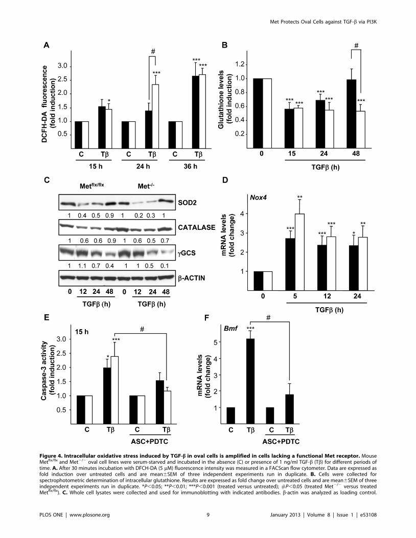
Figure 4. Intracellular oxidative stress induced by TGF-b in oval cells is amplified in cells lacking a functional Met receptor. MouseMetflx/flx and Met2/2 oval cell lines were serum-starved and incubated in the absence (C) or presence of 1 ng/ml TGF-b (Tb) for different periods oftime. A. After 30 minutes incubation with DFCH-DA (5 mM) fluorescence intensity was measured in a FACScan flow cytometer. Data are expressed asfold induction over untreated cells and are mean6SEM of three independent experiments run in duplicate. B. Cells were collected forspectrophotometric determination of intracellular glutathione. Results are expressed as fold change over untreated cells and are mean6SEM of threeindependent experiments run in duplicate. *P,0.05; **P,0.01; ***P,0.001 (treated versus untreated); #P,0.05 (treated Met2/2 versus treatedMetflx/flx). C. Whole cell lysates were collected and used for immunoblotting with indicated antibodies. b-actin was analyzed as loading control.
Met Protects Oval Cells against TGF-b via PI3K
PLOS ONE | www.plosone.org 9 January 2013 | Volume 8 | Issue 1 | e53108

relevant role for Bmf in TGF-b induced apoptosis in mouse oval
cells.
Our results also strongly suggest that autocrine signaling
through Met negatively regulates the increase in Bmf mRNA and
protein levels by TGF-b. Little is known about Bmf regulation by
growth factors. Up to date, an EGF-dependent Bmf down-
regulation has been described in human mammary epithelial
cells [57,58]. No data are currently available regarding potential
modulation of Bmf by HGF, although anti-apoptotic signaling
through HGF/Met has been largely associated to regulation of
members of Bcl-2 family, particularly to induction of anti-
apoptotic BCL-2 proteins, including BCL-2 or myeloid cell
leukemia-1 (MCL-1) [45,59]. Awaiting further investigation, we
provide novel evidence that negative regulation of pro-apoptotic
BCL-2 family proteins, such as BMF, might be an alternative
mechanism contributing to Met-dependent anti-apoptotic signal-
ing.
Studies conducted in hepatocytes and HCC cells have
demonstrated that oxidative stress is involved in TGF-b-mediated
apoptosis in hepatic cells [12,26,31,60,61]. Additionally, we have
proposed a model in which TGF-b-induced ROS have two main
sources, mitochondrial and extramitochondrial via Nox4
[31,36,38,39]. Oval cells seem to follow a similar model.
Evidence supporting this notion are: i) increased intracellular
ROS content and decreased glutathione levels in response to
TGF-b; ii) TGF-b-mediated down-regulation of SOD2, catalase
and c-GCS antioxidant enzymes; iii) up-regulation of Nox4
mRNA preceding the increase in ROS upon TGF-b treatment;
iv) impaired activation of caspase-3 and Bmf up-regulation in the
presence of radical scavengers. More importantly, our data
evidence an antioxidant role for HGF/Met axis in oval cells
since absence of Met results in an exacerbated oxidative stress
process. Thus, Met2/2 cells show higher increase in ROS,
sustained glutathione depletion and stronger down-regulation of
SOD2 and c-GCS protein and up-regulation of Nox4 mRNA. All
these data undoubtedly show that Met mutant oval cells display a
profound redox imbalance in response to TGF-b. This is
consistent with previous works reporting that HGF acts as an
antioxidant factor able to protect against oxidative stress-induced
cell death by increasing the expression and/or activity of c-GCS
and antioxidant enzymes, namely SOD1 and catalase [62,63,64].
In addition, similarly to Met2/2 oval cells, Met mutant
hepatocytes display a dysregulation in oxidative stress-responsive
genes and genes involved in glutathione metabolism [65].
Interestingly, HGF not only acts promoting cellular antioxidant
defenses but it can also prevent ROS production. Indeed, HGF
is able to attenuate the increase in NADPH oxidase activity
observed in hippocampus cells after ischemia [66]. Moreover, in
mesangial cells exposed to high glucose-mediated oxidative stress
HGF exerts a dual antioxidant action, both attenuating the
induction of expression of p22(phox), a component of the
NADPH oxidase system, and impeding a reduction in c-GCS
expression [67]. Based on our findings, our hypothesis is that
something similar might happen in oval cells. As to what are the
signaling pathways mediating the pro-survival antioxidant effect
of HGF in oval cells, a growing body of evidence suggests that
PI3K/AKT pathway is a good candidate. Thus, PI3K/AKT
signaling plays a critical role for HGF-mediated protection
against CD95- or bile acids-induced apoptosis in primary human
and rat hepatocytes, respectively [45,46]. It also mediates the
HGF pro-survival effect on DNA damaging agents-induced
apoptosis and oxidative damage caused by ethanol in hepatic
tumor cell lines [43,64]. In oval cells, PI3K signaling pathway
has been shown to be required for HGF proliferative effect
[41,42], but nothing is known about its role on the pro-survival
effect of HGF. Our results clearly show that treatment with
LY294002 completely suppresses the anti-apoptotic effect
induced by exogenous HGF demonstrating that PI3K is required
for the survival signaling triggered by HGF. Additionally, we
show that either LY294002 or siRNA-mediated PI3K silencing
by themselves decrease cell viability and increase apoptosis in the
absence of serum or any exogenously added growth factor,
indicating that PI3K is also mediating autocrine survival signals.
We and others have shown that oval cells have an autocrine
production of a number of growth factors, including PDGF, EGF
and HGF [24,27,68], all of them being putative PI3K activators.
We have also demonstrated that autocrine signaling through Met
and EGFR promotes oval cell survival [24,27]. The fact that
PI3K inhibition or silencing only increased apoptosis in Metflx/flx
but not in Met2/2 oval cells strongly evidences a Met-dependent
PI3K autocrine survival signaling. Besides, PI3K inhibition
results in an amplified TGF-b-triggered apoptotic response in
Metflx/flx cells resembling that observed in Met2/2 cells, with
analogous apoptotic indices and importantly, with a magnified
oxidative stress and Nox4 and Bmf up-regulation. It has been
shown in other cell systems that constitutive activation of AKT
by different means, including expression of an active oncogenic
mutant catalytic subunit of PI3K (p110a) or active myristoylated
AKT1 or AKT3 reduces and/or prevents Bmf induction [57,69].
Our data strongly suggest that amplification of TGF-b-mediated
Bmf up-regulation in the presence of LY294002 is a consequence
of the enhanced oxidative stress. Additionally, in agreement with
previous data in our laboratory reporting that PI3K inhibition
increases TGF-b-mediated up-regulation of Nox4 in fetal rat
hepatocytes [39], the increased Nox4 expression in the presence
of LY294002 suggest a negative regulatory role for PI3K on
Nox4 expression. Although additional work is required to fully
characterize the molecular mechanism involved in the regulation
of these genes in oval cells, altogether these data allow us to
propose a role for PI3K as a critical intracellular negative
regulator of Bmf and Nox4 during TGF-b-mediated apoptosis in
oval cells.
In summary, both inactivating Met and suppressing PI3K
signaling do result in an impairment of oval cell survival. Data
presented here constitute the first evidence that the HGF/Met/
PI3K signaling axis plays a central role in protecting oval cells
Optical density values relative to loading control were calculated. A representative experiment of two is shown. D. RNA was isolated and Nox4 mRNAlevels were analyzed by qRT-PCR and normalized to the housekeeping gene Gusb. Data represent fold change relative to untreated samples and aremean6S.E.M of at least four independent experiments. *P,0.05; **P,0.01; ***P,0.001 (treated versus untreated). E. Cells were pretreated or notwith radical scavengers (1 mM ascorbate +50 mM PDTC) for 1 hour followed by treatment with TGF-b for 15 hours. Cells were lysed and caspase-3activity was measured. Data are expressed as fold induction relative to untreated cells and are mean6SEM of three independent experiments run induplicate. F. Mouse Metflx/flx oval cells were pretreated or not with radical scavengers (1 mM ascorbate +50 mM PDTC) for 1 hour prior to TGF-btreatment (1 ng/ml). After 5 hours, RNA was isolated and Bmf mRNA levels were analyzed by qRT-PCR and normalized to the housekeeping geneGusb. Data represent fold change relative to untreated samples and are mean6S.D from one representative experiment out of two. *P,0.05;**P,0.01; ***P,0.001 (treated versus untreated); #P,0.05 (TGF-b treated versus ASC+PDTC+TGF-b treated). Black bars, Metflx/flx cells. White bars,Met2/2 cells.doi:10.1371/journal.pone.0053108.g004
Met Protects Oval Cells against TGF-b via PI3K
PLOS ONE | www.plosone.org 10 January 2013 | Volume 8 | Issue 1 | e53108
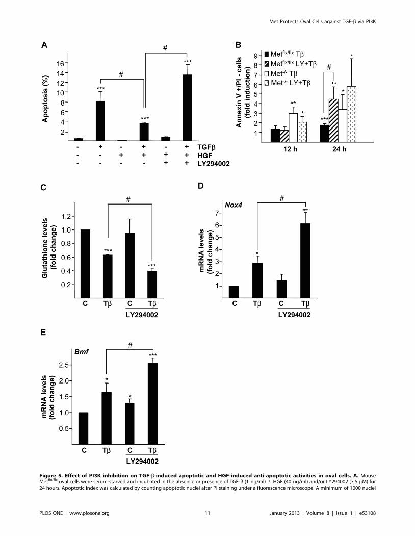
Figure 5. Effect of PI3K inhibition on TGF-b-induced apoptotic and HGF-induced anti-apoptotic activities in oval cells. A. MouseMetflx/flx oval cells were serum-starved and incubated in the absence or presence of TGF-b (1 ng/ml) 6 HGF (40 ng/ml) and/or LY294002 (7.5 mM) for24 hours. Apoptotic index was calculated by counting apoptotic nuclei after PI staining under a fluorescence microscope. A minimum of 1000 nuclei
Met Protects Oval Cells against TGF-b via PI3K
PLOS ONE | www.plosone.org 11 January 2013 | Volume 8 | Issue 1 | e53108

against TGF-b-induced oxidative stress and apoptosis. These
findings have important implications in vivo. Oval cells expand
during liver damage and are exposed to apoptotic insults such us
TGF-b, which is known to be elevated in several human chronic
liver pathologies or other liver injuries involving an oxidative stress
process [70]. Hence, antioxidant and anti-apoptotic signaling
through Met/PI3K could have a fundamental role in promoting
oval cell survival. This mechanism may be relevant for oval cell-
mediated liver regeneration but also for its role in HCC
development.
was counted per condition. Data are mean6S.D. of a representative experiment performed with triplicate dishes. B. Mouse Metflx/flx and Met2/2 ovalcells were serum-starved and incubated in the absence or presence of TGF-b (1 ng/ml) 6 LY294002 (7.5 mM) for different periods of time.Fluorescence intensity was measured in a FACScan flow cytometer and the percentage of annexin V positive/PI negative cells was calculated. Dataare expressed as fold induction over untreated cells and are mean6SEM of four independent experiments. C, D and E. Mouse Metflx/flx oval cellswere serum-starved and incubated in the absence or presence of TGF-b (1 ng/ml) 6 LY294002 (7.5 mM). C. After 8 hours of treatment, cells werecollected for spectrophotometric determination of intracellular glutathione. Results are expressed as fold change over untreated cells and aremean6SEM of two independent experiments run in duplicate. D and E. After 12 hours of treatment RNA was isolated and Nox4 and Bmf mRNAlevels were analyzed by qRT-PCR and normalized to the housekeeping gene Gusb. Data represent fold change relative to untreated samples and aremean6S.E.M of three independent experiments. Black bars, Metflx/flx cells. White bars, Met2/2 cells. *P,0.05; **P,0.01; ***P,0.001 (treated versusuntreated); #P,0.05 (T+LY treated versus T treated).doi:10.1371/journal.pone.0053108.g005
Figure 6. Effect of PI3K inhibition or silencing on basal oval cell viability and apoptosis. A. Mouse Metflx/flx and Met2/2 oval cells wereserum-starved and incubated in the absence or presence of LY294002 (7.5 mM) for 24 hours. Cell viability was assayed by crystal violet staining. Dataare expressed as fold change over untreated cells and are mean6S.D of one representative experiment out of two run in triplicate. B. Cells weretreated as in B for 15 hours and caspase-3 activity was measured. Data are expressed as fold change over untreated cells and are mean6S.E.M of fiveindependent experiments. C, D and E. Mouse Metflx/flx oval cell lines were transfected with non-targeting negative control siRNA (siRNA NT) or p85targeting siRNA (siRNA p85) for 24 hours and serum-starved for additional 24 hours. C. Whole cell lysates were collected and used forimmunoblotting with anti-PI3K p85 antibody. b-actin was analyzed as loading control. A representative experiment of five is shown (upper panel).Optical density values relative to loading controls were calculated and expressed as fold change relative to siRNA NT, given an arbitrary value of 1(bottom panel). Data are mean6SEM of five independent experiments. D. Caspase-3 activity was analyzed. Data are expressed as fold change oversiRNA NT and are mean6SEM of two independent experiments run in duplicate. E. Cell viability by crystal violet staining was assayed. Data areexpressed as fold change over siRNA NT and are mean6SEM of four independent experiments run in duplicate. Black bars, Metflx/flx cells. Whitebars, Met2/2 cells. *P,0.05; **P,0.01; ***P,0.001 (treated versus untreated); #P,0.05 (treated Met2/2 versus treated Metflx/flx).doi:10.1371/journal.pone.0053108.g006
Met Protects Oval Cells against TGF-b via PI3K
PLOS ONE | www.plosone.org 12 January 2013 | Volume 8 | Issue 1 | e53108

Supporting Information
Figure S1 Effect of p38 and JNK MAPKs inhibition onTGF-b-induced apoptosis in oval cells. Mouse Metflx/flx and
Met2/2 oval cell lines were serum-starved and incubated for 15
hours in the absence or in the presence of 1 ng/ml TGF-b, and in
the absence or the presence of (A) p38 inhibitor, SB203580
(10 mM) or (B) JNK inhibitor SP600125 (30 mM) added 30
minutes before TGF-b. Cells were lysed and caspase-3 activity
assayed. Data are mean6SEM of at least three independent
experiments.
(PDF)
Figure S2 Effect of antioxidant agents on intracellularROS content in oval cells. Mouse Metflx/flx and Met2/2 oval
cell lines were serum-starved, pretreated or not with radical
scavengers (1 mM ascorbate +50 mM PDTC) for 1 hour prior to
TGF-b (1 ng/ml) treatment for 24 hours. After 30 minutes
incubation with DFCH-DA (5 mM) fluorescence intensity was
measured in a FACScan flow cytometer. Data are expressed as
fold induction over untreated cells and are mean6SEM of two
independent experiments run in duplicate. Black bars, Metflx/flx
cells. White bars, Met2/2 cells. ns = not significant; *P,0.05;
**P,0.01 (treated versus untreated); #P,0.05 (T treated versus
T+Antioxidants treated).
(PDF)
Table S1 Primers used for qRT-PCR.
(PDF)
Acknowledgments
We express appreciation to Dr. S.S. Thorgeirsson and Dr. V.M. Factor for
generously providing the c-met conditional knockout mouse-derived
parental oval cells, from which the Metflx/flx and Met2/2 oval cell clones
were generated. We also acknowledge Dr. A. Porras for helpful discussions.
Author Contributions
Conceived and designed the experiments: MF CR IF BH AS. Performed
the experiments: AMP GC ASC MGA DMF. Analyzed the data: AMP
GC ASC MGA DMF MF CR IF BH AS. Wrote the paper: BH AS.
References
1. Duncan AW, Dorrell C, Grompe M (2009) Stem cells and liver regeneration.
Gastroenterology 137: 466–481.
2. Tanaka M, Itoh T, Tanimizu N, Miyajima A (2011) Liver stem/progenitor cells:
their characteristics and regulatory mechanisms. J Biochem 149: 231–239.
3. Fausto N, Campbell JS (2003) The role of hepatocytes and oval cells in liver
regeneration and repopulation. Mech Dev 120: 117–130.
4. Mishra L, Banker T, Murray J, Byers S, Thenappan A, et al. (2009) Liver stem
cells and hepatocellular carcinoma. Hepatology 49: 318–329.
5. Heldin CH, Landstrom M, Moustakas A (2009) Mechanism of TGF-beta
signaling to growth arrest, apoptosis, and epithelial-mesenchymal transition.
Curr Opin Cell Biol 21: 166–176.
6. Schmierer B, Hill CS (2007) TGFbeta-SMAD signal transduction: molecular
specificity and functional flexibility. Nat Rev Mol Cell Biol 8: 970–982.
7. Zhang YE (2009) Non-Smad pathways in TGF-beta signaling. Cell Res 19: 128–
139.
8. Nakamura T, Tomita Y, Hirai R, Yamaoka K, Kaji K, et al. (1985) Inhibitory
effect of transforming growth factor-beta on DNA synthesis of adult rat
hepatocytes in primary culture. Biochem Biophys Res Commun 133: 1042–
1050.
9. Oberhammer F, Bursch W, Parzefall W, Breit P, Erber E, et al. (1991) Effect of
transforming growth factor beta on cell death of cultured rat hepatocytes.
Cancer Res 51: 2478–2485.
10. Russell WE, Coffey RJ, Jr., Ouellette AJ, Moses HL (1988) Type beta
transforming growth factor reversibly inhibits the early proliferative response to
partial hepatectomy in the rat. Proc Natl Acad Sci U S A 85: 5126–5130.
11. Sanchez A, Alvarez AM, Benito M, Fabregat I (1995) Transforming growth
factor beta modulates growth and differentiation of fetal hepatocytes in primary
culture. J Cell Physiol 165: 398–405.
12. Sanchez A, Alvarez AM, Benito M, Fabregat I (1996) Apoptosis induced by
transforming growth factor-beta in fetal hepatocyte primary cultures: involve-
ment of reactive oxygen intermediates. J Biol Chem 271: 7416–7422.
13. Preisegger KH, Factor VM, Fuchsbichler A, Stumptner C, Denk H, et al. (1999)
Atypical ductular proliferation and its inhibition by transforming growth factor
beta1 in the 3,5-diethoxycarbonyl-1,4-dihydrocollidine mouse model for chronic
alcoholic liver disease. Lab Invest 79: 103–109.
14. Park DY, Suh KS (1999) Transforming growth factor-beta1 protein,
proliferation and apoptosis of oval cells in acetylaminofluorene-induced rat
liver regeneration. J Korean Med Sci 14: 531–538.
15. Nguyen LN, Furuya MH, Wolfraim LA, Nguyen AP, Holdren MS, et al. (2007)
Transforming growth factor-beta differentially regulates oval cell and hepatocyte
proliferation. Hepatology 45: 31–41.
16. del Castillo G, Alvarez-Barrientos A, Carmona-Cuenca I, Fernandez M,
Sanchez A, et al. (2008) Isolation and characterization of a putative liver
progenitor population after treatment of fetal rat hepatocytes with TGF-beta.
J Cell Physiol 215: 846–855.
17. Ding W, Mouzaki M, You H, Laird JC, Mato J, et al. (2009) CD133+ liver
cancer stem cells from methionine adenosyl transferase 1A-deficient mice
demonstrate resistance to transforming growth factor (TGF)-beta-induced
apoptosis. Hepatology 49: 1277–1286.
18. Nakamura T, Mizuno S (2010) The discovery of hepatocyte growth factor
(HGF) and its significance for cell biology, life sciences and clinical medicine.
Proc Jpn Acad Ser B Phys Biol Sci 86: 588–610.
19. Trusolino L, Bertotti A, Comoglio PM (2010) MET signalling: principles andfunctions in development, organ regeneration and cancer. Nat Rev Mol Cell
Biol 11: 834–848.
20. Schmidt C, Bladt F, Goedecke S, Brinkmann V, Zschiesche W, et al. (1995)Scatter factor/hepatocyte growth factor is essential for liver development.
Nature 373: 699–702.
21. Uehara Y, Minowa O, Mori C, Shiota K, Kuno J, et al. (1995) Placental defect
and embryonic lethality in mice lacking hepatocyte growth factor/scatter factor.Nature 373: 702–705.
22. Borowiak M, Garratt AN, Wustefeld T, Strehle M, Trautwein C, et al. (2004)
Met provides essential signals for liver regeneration. Proc Natl Acad Sci U S A101: 10608–10613.
23. Huh CG, Factor VM, Sanchez A, Uchida K, Conner EA, et al. (2004)
Hepatocyte growth factor/c-met signaling pathway is required for efficient liver
regeneration and repair. Proc Natl Acad Sci U S A 101: 4477–4482.
24. Del Castillo G, Murillo MM, Alvarez-Barrientos A, Bertran E, Fernandez M, etal. (2006) Autocrine production of TGF-beta confers resistance to apoptosis after
an epithelial-mesenchymal transition process in hepatocytes: Role of EGFreceptor ligands. Exp Cell Res 312: 2860–2871.
25. Yao P, Zhan Y, Xu W, Li C, Yue P, et al. (2004) Hepatocyte growth factor-
induced proliferation of hepatic stem-like cells depends on activation of NF-
kappaB. J Hepatol 40: 391–398.
26. Herrera B, Alvarez AM, Sanchez A, Fernandez M, Roncero C, et al. (2001)Reactive oxygen species (ROS) mediates the mitochondrial-dependent apoptosis
induced by transforming growth factor (beta) in fetal hepatocytes. Faseb J 15:741–751.
27. Martinez-Palacian A, del Castillo G, Herrera B, Fernandez M, Roncero C, et al.
(2012) EGFR is dispensable for c-Met-mediated proliferation and survivalactivities in mouse adult liver oval cells. Cell Signal 24: 505–513.
28. Sanchez-Capelo A (2005) Dual role for TGF-beta1 in apoptosis. Cytokine
Growth Factor Rev 16: 15–34.
29. Freathy C, Brown DG, Roberts RA, Cain K (2000) Transforming growth factor-
beta(1) induces apoptosis in rat FaO hepatoma cells via cytochrome c release andoligomerization of Apaf-1 to form a approximately 700-kd apoptosome caspase-
processing complex. Hepatology 32: 750–760.
30. Chipuk JE, Moldoveanu T, Llambi F, Parsons MJ, Green DR (2011) The BCL-2 family reunion. Mol Cell 37: 299–310.
31. Caja L, Sancho P, Bertran E, Iglesias-Serret D, Gil J, et al. (2009) Overactivation
of the MEK/ERK pathway in liver tumor cells confers resistance to TGF-
{beta}-induced cell death through impairing up-regulation of the NADPHoxidase NOX4. Cancer Res 69: 7595–7602.
32. Kiyono K, Suzuki HI, Matsuyama H, Morishita Y, Komuro A, et al. (2009)
Autophagy is activated by TGF-beta and potentiates TGF-beta-mediatedgrowth inhibition in human hepatocellular carcinoma cells. Cancer Res 69:
8844–8852.
33. Ramjaun AR, Tomlinson S, Eddaoudi A, Downward J (2007) Upregulation oftwo BH3-only proteins, Bmf and Bim, during TGF beta-induced apoptosis.
Oncogene 26: 970–981.
34. del Castillo G, Factor VM, Fernandez M, Alvarez-Barrientos A, Fabregat I, et
al. (2008) Deletion of the Met tyrosine kinase in liver progenitor oval cellsincreases sensitivity to apoptosis in vitro. Am J Pathol 172: 1238–1247.
35. Black D, Lyman S, Qian T, Lemasters JJ, Rippe RA, et al. (2007) Transforming
growth factor beta mediates hepatocyte apoptosis through Smad3 generation ofreactive oxygen species. Biochimie 89: 1464–1473.
Met Protects Oval Cells against TGF-b via PI3K
PLOS ONE | www.plosone.org 13 January 2013 | Volume 8 | Issue 1 | e53108

36. Sancho P, Bertran E, Caja L, Carmona-Cuenca I, Murillo MM, et al. (2009)
The inhibition of the epidermal growth factor (EGF) pathway enhances TGF-beta-induced apoptosis in rat hepatoma cells through inducing oxidative stress
coincident with a change in the expression pattern of the NADPH oxidases
(NOX) isoforms. Biochim Biophys Acta 1793: 253–263.37. Mates JM (2000) Effects of antioxidant enzymes in the molecular control of
reactive oxygen species toxicology. Toxicology 153: 83–104.38. Herrera B, Murillo MM, Alvarez-Barrientos A, Beltran J, Fernandez M, et al.
(2004) Source of early reactive oxygen species in the apoptosis induced by
transforming growth factor-beta in fetal rat hepatocytes. Free Radic Biol Med36: 16–26.
39. Carmona-Cuenca I, Roncero C, Sancho P, Caja L, Fausto N, et al. (2008)Upregulation of the NADPH oxidase NOX4 by TGF-beta in hepatocytes is
required for its pro-apoptotic activity. J Hepatol 49: 965–976.40. Hu Z, Evarts RP, Fujio K, Marsden ER, Thorgeirsson SS (1993) Expression of
hepatocyte growth factor and c-met genes during hepatic differentiation and
liver development in the rat. Am J Pathol 142: 1823–1830.41. Kato H, Shimomura T, Murai R, Gonda K, Ishii K, et al. (2007) Regulation of
hepatic oval cell proliferation by adenoviral mediated hepatocyte growth factorgene transfer and signal transduction inhibitors. Hepatogastroenterology 54:
821–825.
42. Okano J, Shiota G, Matsumoto K, Yasui S, Kurimasa A, et al. (2003)Hepatocyte growth factor exerts a proliferative effect on oval cells through the
PI3K/AKT signaling pathway. Biochem Biophys Res Commun 309: 298–304.43. Grotegut S, Kappler R, Tarimoradi S, Lehembre F, Christofori G, et al. (2010)
Hepatocyte growth factor protects hepatoblastoma cells from chemotherapy-induced apoptosis by AKT activation. Int J Oncol 36: 1261–1267.
44. Hu ZX, Geng JM, Liang DM, Luo M, Li ML (2010) Hepatocyte growth factor
protects human embryonic stem cell derived-neural progenitors from hydrogenperoxide-induced apoptosis. Eur J Pharmacol 645: 23–31.
45. Schulze-Bergkamen H, Brenner D, Krueger A, Suess D, Fas SC, et al. (2004)Hepatocyte growth factor induces Mcl-1 in primary human hepatocytes and
inhibits CD95-mediated apoptosis via Akt. Hepatology 39: 645–654.
46. Webster CR, Anwer MS (2001) Phosphoinositide 3-kinase, but not mitogen-activated protein kinase, pathway is involved in hepatocyte growth factor-
mediated protection against bile acid-induced apoptosis in cultured rathepatocytes. Hepatology 33: 608–615.
47. Yu X, Song M, Chen J, Zhu G, Zhao G, et al. (2010) Hepatocyte growth factorprotects endothelial progenitor cell from damage of low-density lipoprotein
cholesterol via the PI3K/Akt signaling pathway. Mol Biol Rep 37: 2423–2429.
48. Li Y, Wen X, Spataro BC, Hu K, Dai C, et al. (2006) hepatocyte growth factor isa downstream effector that mediates the antifibrotic action of peroxisome
proliferator-activated receptor-gamma agonists. J Am Soc Nephrol 17: 54–65.49. Yang J, Dai C, Liu Y (2003) Hepatocyte growth factor suppresses renal
interstitial myofibroblast activation and intercepts Smad signal transduction.
Am J Pathol 163: 621–632.50. Yang J, Dai C, Liu Y (2005) A novel mechanism by which hepatocyte growth
factor blocks tubular epithelial to mesenchymal transition. J Am Soc Nephrol 16:68–78.
51. Park HJ, Kim BC, Kim SJ, Choi KS (2002) Role of MAP kinases and theircross-talk in TGF-beta1-induced apoptosis in FaO rat hepatoma cell line.
Hepatology 35: 1360–1371.
52. Herrera B, Fernandez M, Roncero C, Ventura JJ, Porras A, et al. (2001)Activation of p38MAPK by TGF-beta in fetal rat hepatocytes requires radical
oxygen production, but is dispensable for cell death. FEBS Lett 499: 225–229.53. Carmona-Cuenca I, Herrera B, Ventura JJ, Roncero C, Fernandez M, et al.
(2006) EGF blocks NADPH oxidase activation by TGF-beta in fetal rat
hepatocytes, impairing oxidative stress, and cell death. J Cell Physiol 207: 322–330.
54. Ohgushi M, Kuroki S, Fukamachi H, O’Reilly LA, Kuida K, et al. (2005)
Transforming growth factor beta-dependent sequential activation of Smad, Bim,
and caspase-9 mediates physiological apoptosis in gastric epithelial cells. Mol
Cell Biol 25: 10017–10028.
55. Ramesh S, Qi XJ, Wildey GM, Robinson J, Molkentin J, et al. (2008) TGF beta-
mediated BIM expression and apoptosis are regulated through SMAD3-
dependent expression of the MAPK phosphatase MKP2. EMBO Rep 9: 990–
997.
56. Wildey GM, Patil S, Howe PH (2003) Smad3 potentiates transforming growth
factor beta (TGFbeta )-induced apoptosis and expression of the BH3-only
protein Bim in WEHI 231 B lymphocytes. J Biol Chem 278: 18069–18077.
57. Schmelzle T, Mailleux AA, Overholtzer M, Carroll JS, Solimini NL, et al. (2007)
Functional role and oncogene-regulated expression of the BH3-only factor Bmf
in mammary epithelial anoikis and morphogenesis. Proc Natl Acad Sci U S A
104: 3787–3792.
58. Whelan KA, Caldwell SA, Shahriari KS, Jackson SR, Franchetti LD, et al.
(2010) Hypoxia suppression of Bim and Bmf blocks anoikis and luminal clearing
during mammary morphogenesis. Mol Biol Cell 21: 3829–3837.
59. Yamamoto K, Morishita R, Hayashi S, Matsushita H, Nakagami H, et al. (2001)
Contribution of Bcl-2, but not Bcl-xL and Bax, to antiapoptotic actions of
hepatocyte growth factor in hypoxia-conditioned human endothelial cells.
Hypertension 37: 1341–1348.
60. Franklin CC, Rosenfeld-Franklin ME, White C, Kavanagh TJ, Fausto N (2003)
TGFbeta1-induced suppression of glutathione antioxidant defenses in hepato-
cytes: caspase-dependent post-translational and caspase-independent transcrip-
tional regulatory mechanisms. Faseb J 17: 1535–1537.
61. Kayanoki Y, Fujii J, Suzuki K, Kawata S, Matsuzawa Y, et al. (1994)
Suppression of antioxidative enzyme expression by transforming growth factor-
beta 1 in rat hepatocytes. J Biol Chem 269: 15488–15492.
62. Kannan R, Jin M, Gamulescu MA, Hinton DR (2004) Ceramide-induced
apoptosis: role of catalase and hepatocyte growth factor. Free Radic Biol Med
37: 166–175.
63. Tsuboi S (1999) Elevation of glutathione level in rat hepatocytes by hepatocyte
growth factor via induction of gamma-glutamylcysteine synthetase. J Biochem
126: 815–820.
64. Valdes-Arzate A, Luna A, Bucio L, Licona C, Clemens DL, et al. (2009)
Hepatocyte growth factor protects hepatocytes against oxidative injury induced
by ethanol metabolism. Free Radic Biol Med 47: 424–430.
65. Gomez-Quiroz LE, Factor VM, Kaposi-Novak P, Coulouarn C, Conner EA, et
al. (2008) Hepatocyte-specific c-Met deletion disrupts redox homeostasis and
sensitizes to Fas-mediated apoptosis. J Biol Chem 283: 14581–14589.
66. Niimura M, Takagi N, Takagi K, Mizutani R, Tanonaka K, et al. (2006) The
protective effect of hepatocyte growth factor against cell death in the
hippocampus after transient forebrain ischemia is related to the improvement
of apurinic/apyrimidinic endonuclease/redox factor-1 level and inhibition of
NADPH oxidase activity. Neurosci Lett 407: 136–140.
67. Li H, Jiang T, Lin Y, Zhao Z, Zhang N (2006) HGF protects rat mesangial cells
from high-glucose-mediated oxidative stress. Am J Nephrol 26: 519–530.
68. Lau CK, Yang ZF, Ho DW, Ng MN, Yeoh GC, et al. (2009) An Akt/hypoxia-
inducible factor-1alpha/platelet-derived growth factor-BB autocrine loop
mediates hypoxia-induced chemoresistance in liver cancer cells and tumorigenic
hepatic progenitor cells. Clin Cancer Res 15: 3462–3471.
69. Shao Y, Aplin AE (2010) Akt3-mediated resistance to apoptosis in B-RAF-
targeted melanoma cells. Cancer Res 70: 6670–6681.
70. Giannelli G, Mazzocca A, Fransvea E, Lahn M, Antonaci S (2011) Inhibiting
TGF-beta signaling in hepatocellular carcinoma. Biochim Biophys Acta 1815:
214–223.
Met Protects Oval Cells against TGF-b via PI3K
PLOS ONE | www.plosone.org 14 January 2013 | Volume 8 | Issue 1 | e53108