Embed Size (px)
Citation preview


§MQ; MODELS FOR THE FORMATION OF 5.85 RIBOSOMAL RNA DIMER
Ü bvChris Hays Dove
‘(ABSTRACT)
Ribosomal 5.85 RNA is a component of the large (605)
ribosomal subunit in eucaryotes. Studies of 5.85 rRNA in
solution have shown that under certain conditions, including
standard isolation procedures, the molecule complexes with
itself to form dimers and higher multimers. Two models have
been proposed in the literature to explain the intermolecu-
lar interactions responsible for 5.85 rRNA dimer formation.
The terminal interaction model of Sitz et al. (Biochem. 17,
5811-5815, 1978) proposes that the dimer forms through
base-pairing of the 5' and 3' terminal sequences of two 5.85
rRNA molecules. Pavlakis et al. (Nucl. Acids Res. 7,
2213-2237, 1978) showed that 5.85 rRNA lacking the 3' termi-
nal region was capable of forming dimer. They proposed an
alternative model for 5.85 rRNA dimer formation in which an
entirely different part of the molecule interacts to form a
double-stranded palindrome.
In this study, enzymatic probing techniques and stabil-
ity measurements (both experimental and theoretical) were
used to determine which model most accurately describes the

intermolecular interactions of the 5.88 rRNA dimer. The
methods used for determining stability were not able to dis-
criminate between the models for 5.88 rRNA dimer formation.
Results from the structural probing studies, however, sup-
port the terminal interaction model and indicate that the
palindrome interaction does not occur in 5.88 rRNA dimer
formed from the intact molecule.
ACKNOWLEDGEMENTS
There are several people to whom I wish to extend sinc-
ere thanks. Dr. Thomas O. Sitz, my major advisor, provided
me with an abundance of information, and I thank him for the
time he spent in teaching me, academically, as well as his
excellent training in laboratory technique. I am also
grateful for his patience, encouragement, and good humor.
My committee members, Drs. Mark L. Failla, John L. Johnson,
and John E. Wiktorowicz, I thank for their time, interest,
and. willingness to discuss the researdh project and for
their helpful suggestions.
I would like to thank those graduate students, faculty
and staff members who extended themselves to make this a
more enjoyable and memorable experience. I would also like
to thank Dr. Thomas Keenan for providing financial assis-
tance, which made this work possible, and, for her
invaluable asssistance in the preparation of this manu-
script.· A
‘ With special gratitude I acknowledge the encouragement
and support of my family and friends, particularly my hus-
band, , whose unfailing confidence i11 my abilities helped
me through the most difficult of times.
iv
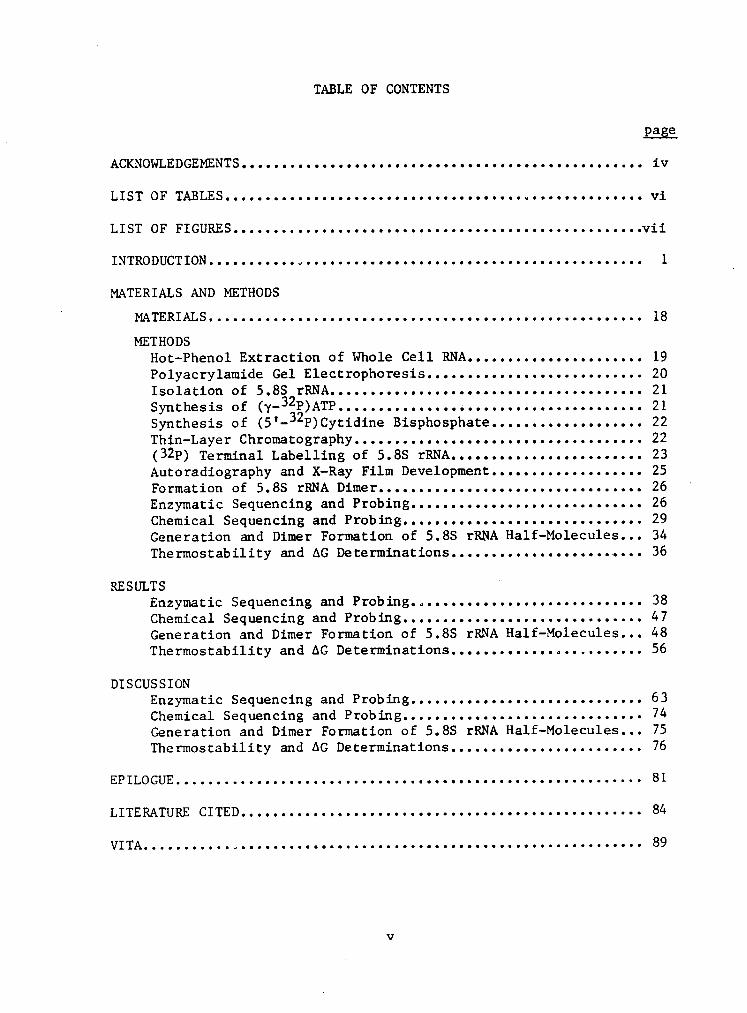
TABLE OF CONTENTS
page
LIST OF FIGURES...................................................vii
INTRODUCTION...........„ .......................................... 1
MATERIALS AND METHODS
MATERIALS...................................................... 18
METHOD8Hot—Phenol Extraction of Whole Cell RNA...................... 19Polyacrylamide Gel Electrophoresis........................... 20
Synthesis of (5'-32P)Cytidine Bisphosphate................... 22Thin—Layer Chromatography.................................... 22(3ZP) Terminal Labelling of 5.8S rRNA........................ 23Autoradiography and X—Ray Film Development................... 25Formation of 5.88 rRNA Dimer................................. 26 '
Enzymatic Sequencing and Probing............................. 26Chemical Sequencing and Probing.............................. 29Generation and Dimer Formation of 5.88 rRNA Half—Molecules... 34Thermostability and AG Determinations......................... 36
RESULTS 'Enzymatic Sequencing and Probing............................. 38Chemical Sequencing and Probing.............................. 47 ·
Generation and Dimer Formation of 5.88 rRNA Half-Molecules... 48Thermostability and AG Determinations........................ 56
DISCUSSIONEnzymatic Sequencing and Probing............................. 63Chemical Sequencing and Probing.............................. 74Generation and Dimer Formation of 5.88 rRNA Half—Molecules... 75Thermostability and AG Determinations........................ 76
LITERATURE CITED.................................................. 84
VITA...........„.................................................. 89
v
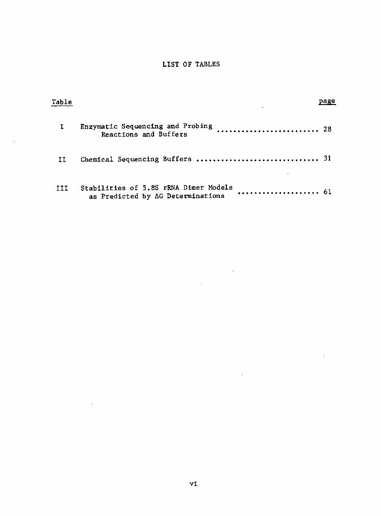
LIST OF TABLES
Table gage
I Enzymatic Sequencing and Probing 28I I I I I I I I I I I I I I I I I I I I I I I I I
II Chemical Sequencing Buffers ........•..................... 31
III Stabilities of 5 . 8S 1*RNA Dimer Models 61as Predicted by AG Decerminations ° ° ° ° ° ’°'°°° ° ° ' ° ° ' ' ° °
vi
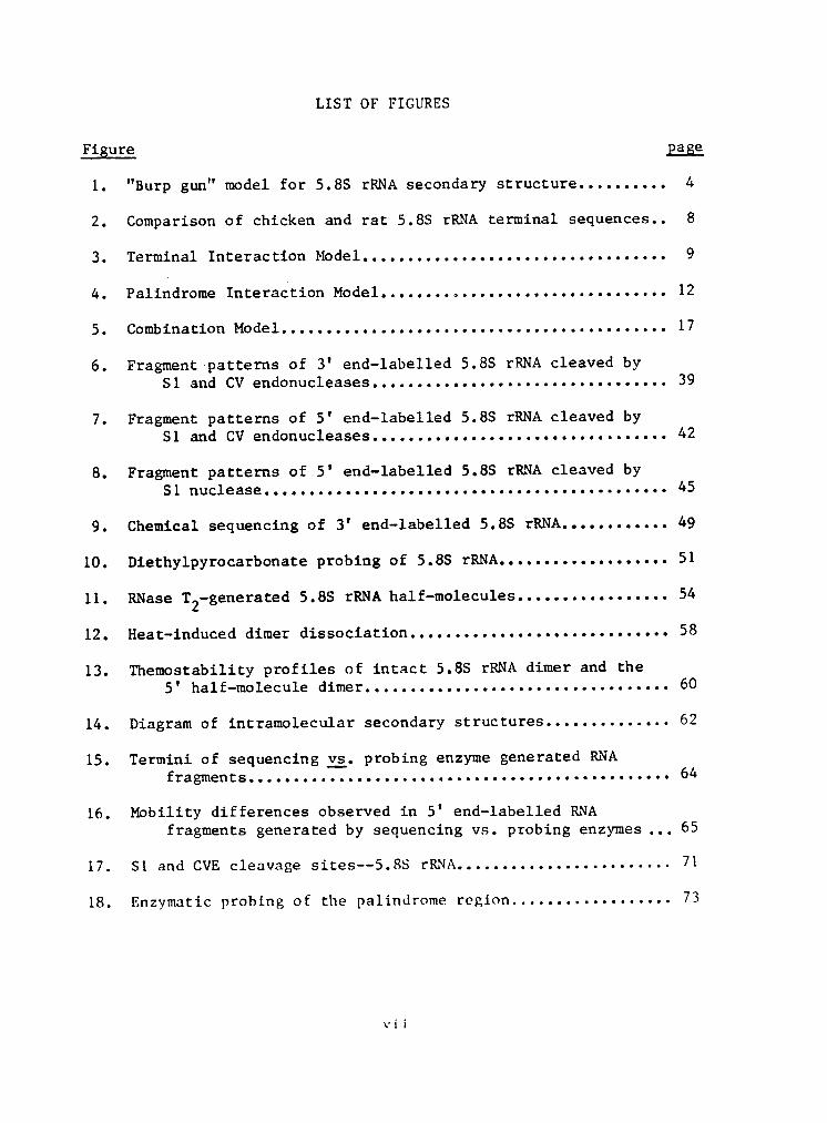
LIST OF FIGURESFigure pägg1. "Burp gun" model for 5.85 rRNA secondary structure.......... 4
2. Comparison of chicken and rat 5.8S rRNA terminal sequences.. 8
3. Terminal Interaction Model.................................. 9
4. Palindrome Interaction Model................................ 12
5. Combination Model........... .............. .................. 17
6. Fragment patterns of 3' end-labelled 5.8S rRNA cleaved byS1 and CV endonucleases................................. 39
7. Fragment patterns of S' end-labelled 5.8S rRNA cleaved byS1 and CV endonucleases................................. 42
8. Fragment patterns of 5' end-labelled 5.85 rRNA cleaved byS1 nuclease............................................. 45
9. Chemical sequencing of 3' end-labelled 5.88 rRNA............ 49
10. Diethylpyrocarbonate probing of 5.88 rRNA................... 51
11. RNase T2—generated 5.8S rRNA half-molecules................. 54
12. Heat-induced dimer dissociation............................. 58
13. Themostability profiles of intact 5.85 rRNA dimer and the5' half-molecule dimer.................................. 60
14. Diagram of intramolecular secondary structures.............. 62
15. Termini of sequencing gg. probing enzyme generated RNAfragments............................................... 64
16. Mobility differences observed in 5' end-labelled RNAfragments generated by sequencing vs. probing enzymes... 65
17. S1 and CVE cleavage sites--5.85 rRNA........................ 71
18. Enzymatic probing of the palindrome region.................. 73
vii
INTRODUCTION
Ribosomes have been studied extensively since the
l950's. Rapid progress has been made with respect to the
ribosome's role in protein synthesis, with theories describ-
ing the translational process proposed as early as 1960
(Bielka, 1982 [review]). At the same time, study of the
structure of the ribosome was also initiated; the ribosome
was found to consist of a large and a small subunit, each
comprised of various proteins and ribonucleic acids. While
much work has been done to characterize the individual com-
ponents, little is known about the role each plays in pro-
tein synthesis, or the intermolecular interactions of the
various components in the intact ribosome.
‘Ribosomal 5.85 RNA is a component of the large (605)
ribosomal subunit in eucaryotes. This low molecular weight
RNA species was first characterized by Pene and coworkers
(1968) and is hydrogen-bonded to the high molecular weight
RNA component (285 in mammals) in that same subunit. The
5.85 rRNA varies slightly in chain length and composition
from species to species, but is approximately 160 nucleo-
tides in length. Nomenclature for the molecule varied in
the early literature (satellite RNA, 75 rRNA, 5.55 rRNA) but
the term 5.85 rRNA has become universally accepted (see re-
1
2
views by Perry, 1976; Bielka, 1982; Nazar, 1982; Pace,
1983). Structural probing studies of the ribosome have in-
dicated that the 5.85 rRNA is located at the interface of
the two subunits and has therefore been implicated in subu-
nit association. Other roles have also been proposed, such
as tRNA binding during translation, but the actual function
of 5.85 rRNA is not known (see reviews).
Synthesis of 5.85 rRNA occurs in the nucleolus, where,
it is transcribed as part of a large precursor molecule (455
RNA). The 455 nuclear RNA also contains the 185 and 285
rRNA A sequences, with the following order:
_
(5'—-+-18-+-—-+-5.85—+---+——285--—-3'). The rRNA sequences
are separated by transcribed spacers. The 455 pre-rRNA and
newly synthesized ribosomal proteins from the cytoplasm are
packaged in the nucleolus into complexes referred to as
pre-ribosomal or ribonucleoprotein (RNP) particles. Pro-
cessing of the 455 pre-rRNA occurs primarily in the nucleo-
lus as part—of
the RNP particle. This particle is then
transported to the cytoplasm, where further modification
events may occur, producing a mature ribosomal subunit (Na-
zar, 1975b; Bielka, 1982).
The nucleotide sequence of 5.85 rRNA was first deter-
mined for the yeast Saccharomyces cerevisiae (Rubin, 1973).
Since that time, the molecule has been sequenced from more

3
than 20 different species (Erdmann, 1983; Ursi, 1983.) Com-
parison of the 5.8S sequences from different species has
revealed a high degree of homology. For example, the se- _
quences of 5.8S rRNA from yeast and man are 75% homologous,
indicating retention of the sequence during evolution (Nazar
et al., 1975). The sequences of rat and human 5.8S rRNAs
(Nazar· et al., 1975c, 1976) are identical except for· the
presence of an extra nucleotide at the 3' end of human 5.8S
rRNA. Although there is not a unique rRNA molecule in pro-
caryotes equivalent to eucaryotic 5.8S rRNA, the 5' end of
the procaryotic 23S rRNA contains a sequence homologous to
that of 5.8S rRNA, suggesting that the 5.8S rRNA evolved
from the 23S rRNA (Nazar, 1980). Such a proposal is sup-
ported by another finding in which the nucleotide residues
157 - 290 of E. coH 23S rRNA are homologous to the 5' end of
yeast and Xenopusl¤evß·28S rRNA (Cox & Kelly, 1981).
A number of models for the secondary structure of 5.8S
rRNA have been proposed, based on studies utilizing such
techniques as Raman spectroscopy, chemical, enzymatic and
fluorescence probing, and base-pairing as predicted by free
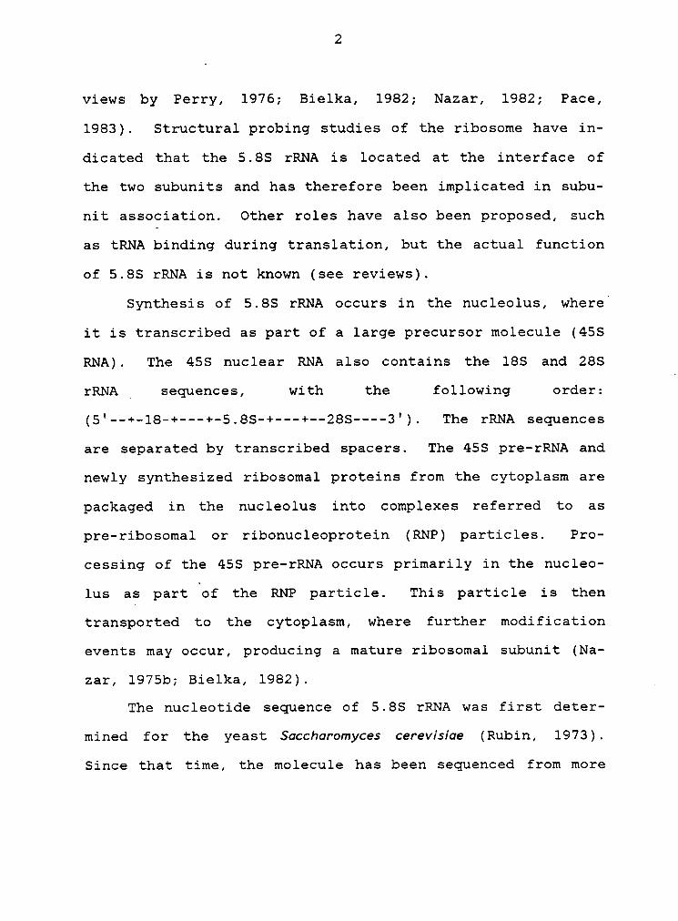
energy calculations (see reviews). The "burp gun" uwdel
(Fig. 1) of Nazar et al. (1975) has been the most consistent
with data derived from various physicochemical studies (Na-
zar, 1982). New models continue to be presented, usually
‘·,
~
c^cAU AC
Gu =• 6 G vc ¤ AA AA Ü
P(¢)GAC CUUAGCGGU•G GC GUGCGUCGAUG CGCAG UAUCGCUGcGAGUCUGuCg°GC2-6 UUC AC CG U
uu-AAGAUCG“° ‘ G•C “°
A A A-U eoG•C G C G-UG•C CUU U·AG•C mn U·A
wc-Gm A•UC-G C-GC-G A•U
U U C·G1oc J '°A A
C G AGA UUCG•
Figure 1: "ßurg gun" model for 5.85 rRNA secondag structure
(Nazar et al., 1975). Sequence shown is the cor-
rected sequence for rat 5.85 rRNA (Smith ec al.,
1984). »

5
retaining most of the structural domains proposed in the
"burp gun" model, but with slight variations (Ursi et al.,
1982, 1983.) In the "burp gun" model, the 5' and 3' termi-
nal regions of 5.8S rRNA interact. through base pairing.
Studies of the 5.8S-28S rRNA complex, however, indicate
these terminal regions of 5.8S rRNA to be the very sites of
interaction with the 28S rRNA (Nazar et al., 1980, 1981;
Pace et al., 1977; Walker et al., 1982). For this reason,
there has been some controversy regarding the base-pairing
of the terminal regions of 5.8S rRNA. Pace (1983; Pace et
al., 1977; Peters et al., 1982), one of the most outspoken
opponents of this proposed terminal interaction, states that
it is unlikely that there would be base-pairing between the
ends of the molecule because this would obstruct the binding·
of 5.8S to 28S rRNA. The "burp gun" model has been proposed
only as a model of the secondary structure of Boküed 5.8S
rRNA; the secondary and tertiary structure of 5.8S rRNA in
the 6OS ribosomal subunit is most certainly influenced not
only by its binding to 28S rRNA, but also by its interac-
tions with surrounding ribosomal proteins. This has been
verified by structural studies, which indicate that there
are significant differences between the structure of isolat-
ed 5.8S rRNA and the molecule at various stages of ribosome
reconstitution (Pace et al., 1977; Nazar et al., 1981, Lo &
Nazar, 1982; Walker et al., 1982).
6
In studying the temperature dependence of 5.85 rRNA
reassociation with 285 rRNA, Pace et al. (1977) observed anIalteration in the electrophoretic mobility' of 5.85 rRNA.
When 5.85 rRNA was heated to temperatures of 65OC and above,
a species that migrated more slowly than unheated 5.85 rRNA
was observed. They proposed that this structure had approx-
imately the molecular weight expected for a dimer of 5.85
rRNA, or that it could be a denatured form of the molecule
with a much greater radius of gyration.
In the following year, dimerization of 5.85 rRNA was
verified in a report by Sitz et al. (1978). Using a two-di-
mensional electrophoresis system, 75% of an RNA componentl
which migrated as 85 in the first dimension (non-denaturing
conditions) was found to migrate as 5.85 in the second di-
mension (denaturing conditions). Further analysis indicated
that this was due to the dissociation of a 5.85 rRNA dimer,
which co—migrated with an unrelated 85 molecule in the first
dimension. Further studies were performed to characterize
the dimer, and a model for dimer formation was proposed.
The Terminal Interaction Mogel
Formation of the dimer requires heating of the 5.85
rRNA molecule, indicating that some melting or alteration of

7
secondary structure is necessary. Sitz et al. (1978) deter-
mined the optimal incubation temperatures and concentrations
required for the formation of 5.8S rRNA dimers for chicken
and rat. Dimers of 5.8S rRNA from these species formed at
relatively low concentrations (0.1 - 1.0 mg/ml) as compared
to concentrations required for the formation of other RNA
dimers (15 — 40 mg/ml for tRNA). The optimum temperatures
of formation were determined to be 62OC and 67OC for chicken
and rat 5.8S rRNA dimers, respectively. The thermal stabil-
ities of the 5.8S rRNA dimers were also studied, expressed
as the temperature required to produce 50% dissociation of
the dimer. A five degree difference between the two species
was observed, chicken 5.8S rRNA dimers dissociating at 55OC
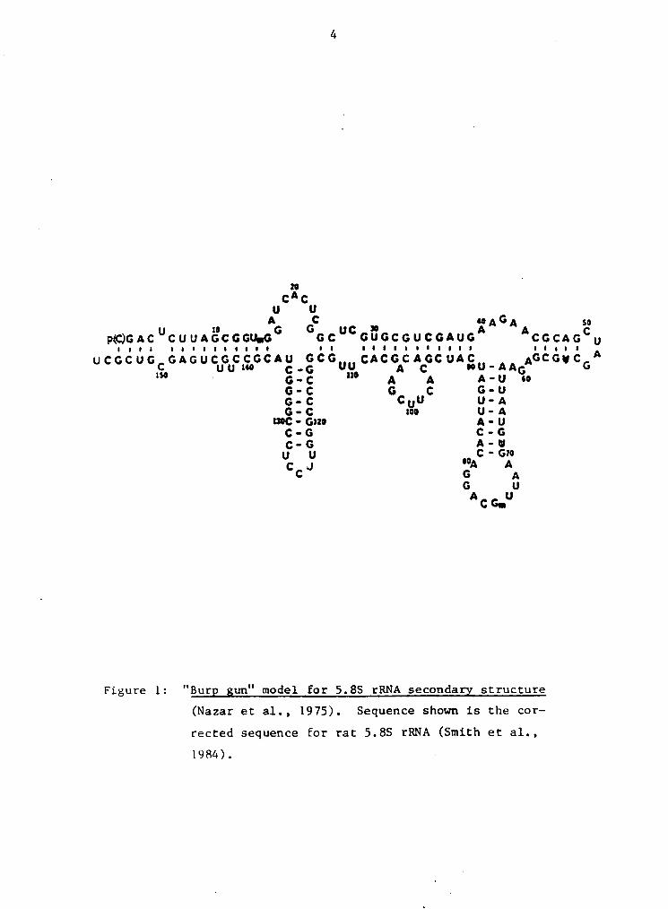
and rat 5.8S rRNA dimers at 60°C. Comparisons of the pri-
mary structures for rat and chicken 5.8S rRNA reveal that
the sequences differ in only three positions, all of which
occur in the terminal regions of the molecule (Fig. 2).
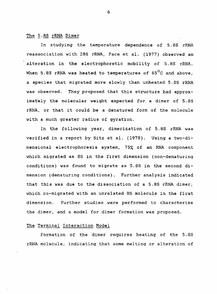
Based on the sequence data and the differences in ther-
mal stabilities and temperatures of formation, a model for
5.8S rRNA dimer formation was proposed in which the 5' and
3' ends of one 5.8S rRNA molecule interact with the 3' and
5' ends of another 5.8S rRNA molecule (Fig. 3). The most
important feature of this model is that the intermolecular
base-pairing between the terminal regions of two 5.8S rRNA

8
rat
S' Up(C)GAC CUUAG C GGU(m)G—-——\IIIIIIIIIIIIII I
UCGCUGCGAGUCUGUCCG C""3l
gv I chickenp(C) A A C U CUUAGCGGU(m)G—--—\ 'I I I I I I I I I I )
C GA GU CCGUC C G C---“G
C U IU C G
3·UI
Figure 2: Comgarison of chicken and rat 5.8S rRNA terminal
seguences. Arrows denote point mutations. Rest
of molecule, indicated by slashed line, has se- _
quence identical to that of rat 5.8S rRNA, shown
in Fig. 1. (Sitz et al., 1981)

9
s' ‘3, lIl|I|IIIIIII ¤¢u$n•u·|uu•
‘5
heat (65°—70°C)50
3l re—anneal,
3,5]
3' S' 5: Ig 's"*·
='llgll
2 E "*···-2 EIl.;-
¤ I ·_
5, \•
ä 3 ä •‘•""'”·=
E E 3 2 ß
-? 5
S' - 3.•___,,„»! 3,
ä
Figure 3: Terminal Interaction Model (Sitz et al., 1978)Schematic illustration of a proposed model for
5.8S rRNA dimer and multimer formation.
10
molecules in the dimer is exactly the same as the terminal
interaction proposed for the 5.85 rRNA monomer. Differences
observed in the temperatures of formation and thermal sta-
bility of 5.85 rRNA dimers in this study correlate with the
sequences of each species. This model was supported by a
later study·using human and turtle 5.85 rRNA (Sitz et al.,
1981). The formation of higher mulitimers (trimers and tet-
ramers), was also observed (Sitz et al., 1978), the occur-
rence of which can be explained by the terminal interaction
model (Fig. 3).
The Palindrome MegeT
Studies of the ribosomal RNA from Lhpsophüa reveal that
the 5.85 rRNA from this species has a unique feature when
compared to the 5.85 rRNA from other eucaryotes studied thus
far. Dmßophüa 5.85 rRNA is actually a split molecule, com-
posed of two molecules which associate through base- pairing
to produce a_structure that is comparable to the 5.85 rRNA
of other eucaryotes. This split in the molecule is the re-
sult of a secondary cleavage near the 3' end, producing a 30
nucleotide fragment, the 25 rRNA (Jordan, 1976).
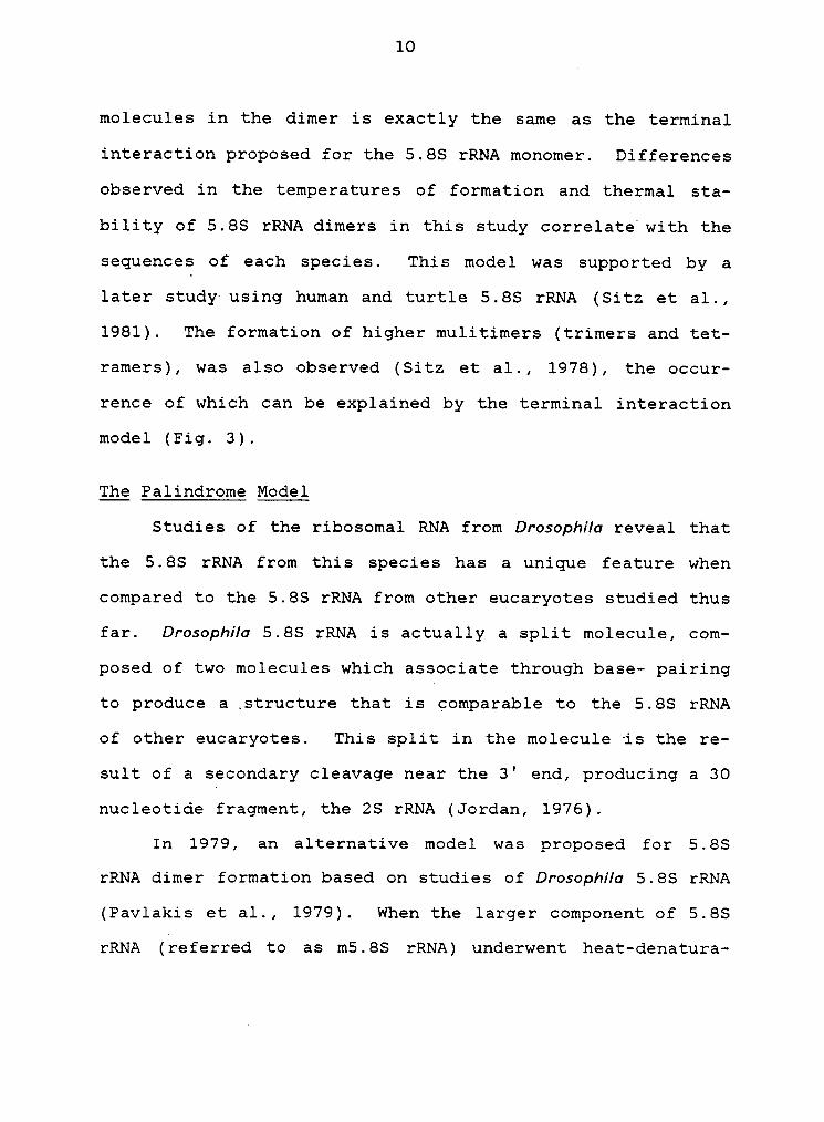
In 1979, an alternative model was proposed for 5.85
rRNA dimer formation based on studies of Lhpsophüa 5.85 rRNA
(Pavlakis et al., 1979). When the larger component of 5.85
rRNA (referred to as m5.85 rRNA) underwent heat—denatura-

„ 11
tion, a species with a slower electrophoretic mobility than
native m5.8S rRNA was observed. Computer analysis of the
sequence of m5.8S rRNA revealed a possible interaction bet-
ween two m5.8S rRNA molecules at nucleotides 17 — 39, form-
ing a long palindrome (Pavlakis et al., 1979). Since this
region differs from HeLa cell 5.85 rRNA by only two nucleo-
tides, they proposed that a similar interaction was there-
fore possible among mammalian 5.85 rRNA (Fig. 4). As addi-
tional support for their model, they state that the
calculated Gibb's free energy of formation indicates that
the palindrome structure is more stable than intramolecular
interactions for that region. Therefore, they propose that
reannealing of the 5.85 rRNA at high temperatures would fa-
vor the dimer.
° More recently, Peters et al. (1982) have published work —
supporting formation of the 5.85 rRNA dimer occurring
through interactions of the 5' portion of the molecule, alt-
hough the specific intermolecular interactions are not ad-
dressed. Mouse 5.85 rRNA was subjected to a limited diges-
tion by T2 ribonuclease. Under appropriate conditions, the
molecule is cleaved only at the loop of the A-U rich arm,
generating two "half-molecules." Of the two halves, only
the 5' half-molecule of 5.85 rRNA was observed to form di-
mers. From this they conclude that the 3' half of 5.85 rRNA
is not involved in dimer formation in the intact molecule.

12
3"" G A ....-3IG A 20C so 4oA G _UCA UCGGCUCGUGCGUCGA UGAIlllllllilllllllllélAAGUAGCUGCGUGCUCCGCUCA U_
G 40 ao 20 A G -„Ä —-— A G
___)
3'A
SI
·\\xxxxx
g g
IIIIIIIIIIIIIIIIIIIs Z
·
Q : .\\‘
S!
3l
Figure 4: Palindrome Interaction Model (Pavlakis et al_., 1979)
Intermolecular interaction shown in top diagram from
Pavlakis et al., 1979.

13
Thesis Objective ggg Experimental Approach
Two fundamental questions are being addressed in this’
thesis: What are the intermolecular interactions of the
5.8S rRNA dimer? And secondly, which of the proposed models
most accurately represents that interaction?
The models for 5.8S rRNA dimer formation differ signi-
ficantly with respect to the molecule's secondary structure. 4
Therefore, methods developed to probe higher-order molecular
structure should distinguish between the proposed models.
Both enzymatic and chemical structural probing techniques
n have been developed for the study of nucleic acids and are
used in this study. S1 nuclease, isolated from Aspeqßüus
oryum, cleaves the phosphodiester bond of single-stranded
nucleic acids and can therefore be used to locate those re-
gions of the molecule not involved in higher-order structur-
al interactions (Ando, 1966; Khan and Maden, 1976). Cobra
venom endonuclease from NqM*n¤M oxwna, on the other hand,
is specific for double-stranded nucleic acids, and provides
an alternative enzymatic probe for secondary- and tertiary-
structure analysis, giving information about those parts of
the molecule involved in helix formation (Toots et al.,
1981).
Chemical probes are base-specific rather than struc-
ture-specific; however, they react only with those bases not

14
involved in higher-order structure. The direct chemical se-
quencing method for RNA, developed by Peattie (1979) makes
use of chemicals that modify specific nucleotides, generat-
ing a site that is susceptible to cleavage by a second chem-
ical. For sequencing purposes, the initial modification
reaction is performed under denaturing conditions to allow
random modification over the length of the molecule. These
same reactions, carried out under milder, non-denaturing
conditions, provide a probe for higher-order structure
(Peattie, 1980, 1983). V
A second approach taken in this study to distinguish
between the models for 5.85 rRNA dimer formation is the com-
parison of the stabilities of the intact 5.85 rRNA dimer and
the dimer formed by the 5' half-molecule. The Tm value foreach complex, measured experimentally and defined here as
the temperature at which 50 per cent of the dimer dissoci-
ates, can be used to determine if the interactions of the
two complexes are different. Alternatively, free energy va-
lues for the regions of interaction proposed in the 5.85
rRNA dimer models can be calculated and compared to estab-
lish relative theoretical stabilities. The stability of a
double—stranded helix has been found to be influenced by the
stacking of adjacent bases as well as the hydrogen bonding
of the base-pairs. Furthermore, the sequence of the bases

15
affects the stability of the stacking (Borer et al., 1974).
Free energy (AG) values have been assigned to "doublets"
(adjacent base-pairs), and these values are dependent on the
order of the four bases in the doublet. The free energy va-
lue for a double-stranded region is derived from the sum of
the favorable (—AG) values for each doublet and the unfavo-
rable (+AG) values for unpaired bases, which destabilize the
helix. The original AG values were obtained from thermody-
namic studies using double-stranded synthetic oligonucleo-
tides (Gralla & Crothers, 1973; Borer et al., 1974). Cur-
rent values used by different investigators vary somewhat,
and are derived from thermodynamic, statistical and empiri-
cal models (see Ninio, 1979). A great deal of controversy
exists regarding the destabilizing effects of unpaired bases
(Ninio, 1979; Pipas and McMahon, 1975; Salser, 1977). Of
particular concern to this study are disagreements regarding
1) the stacking of double—stranded helices across bulges,
internal loops and multi-branched loops; and 2) the extent
to which bulges, dangling ends, and the various loop struc-
tures destabilize double—he1ical regions.
The dimer formed by the 5' half of the 5.8S rRNA molec-
ule (Peters et al., 1982) has not been physically character-
ized, and the model to define this dimer interaction is
based only on theoretical parameters (Pavlakis et al.,1979).

16
The fact that this 5' half-molecule dimerization does occur
raises the following question. In the formation of the 5.8S
rRNA dimer, does the presence of the 3' half of the 5.8S mo-
lecule (i.e. the intact molecule) preclude the interaction
responsible for dimerization of the 5' half—molecule? If
this is not true, then perhaps the actual intermolecular in-
teraction of the 5.8S rRNA dimer is some combination. of
these two models. The palindrome interaction involves nu-
cleotides 18 - 40, which leaves the terminal regions open.
Perhaps the terminal interaction can occur simultaneously
with the palindrome interaction, as shown in Figure 5.
Results from the structural probing of the monomer and
dimer forms of 5.8S rRNA and Tm and AG determinations forthe intact 5.8S rRNA and 5' half-molecule dimers are pre-
~ sented and discussed in the following sections.

17
3”5* I
1: Qg _
E·
Zi
E’0
f
Z IIIIIIIIIIIIIIIIIII ,4
ll,//I
0 ·: CÜ• ·;1 EEZ i€Ö •-’
5
·
S'3l
Figure 5: "Combination Model" for 5.8S rRNA dimer formation.
Formation of the 5.8S rRNA dimer by base—pairing
at both the terminal and palindrome interaction
sites.
MATERIALS AND METHODS
MATERIALS
Radioactive phosphate was obtained from ICN Pharmaceu-
ticals, Inc. as (32P)-H3PO4. ATP, NAD and cyti-
dine-3'·monophosphate were purchased from Sigma Chemical
Company. Alkaline phosphatase was obtained from Boehringer
Mannheim. P—L„ Biochemicals, Inc. was the source of all
other enzymes. Dimethyl sulfate, diethylpyrocarbonate, an-
hydrous hydrazine and aniline were purchased from Aldrich
Chemical Company. Polyethyleneimine—cellulose was obtained
from Brinkmann Instruments, Inc. All other chemicals were
reagent grade. » _
Animals were supplied by the Virginia Tech Lab Animal
Resources Center. Human placenta was provided by Dr. Miguel
Langebeck, Montgomery County Hospital, Virginia.
18

19
METHODS
Hot-Phenol Extraction ef yhele gell RNA.
Whole cell RNA was extracted from rat liver using the
hot-phenol extraction procedure of Steele et al., (1965).
The tissue was homogenized with a stainless-steel blender in
20 Volumes of SDS buffer (0.14 M NaCl, 0.05 M Na acetate,
0.3% sodium dodecyl sulfate [SDS], pH 5.1), followed by an
equal Volume of phenol—cresol reagent (1625 ml phenol, 325
ml cresol, 2 g 8-OH quinoline/2 liters). The mixture was
heated to 55°C, stirring continuously, and then brought to
room temperature by cooling on ice. The mixture was sepa-
rated into aqueous and organic phases by centrifugation at
1300 x g for 30 min at4OC
in a Beckman low—speed centrifuge
(Model J6—B); centrifugation was stopped without the use ofV
a brake to avoid disturbing the interface. The upper aque-
ous phase containing the whole cell RNA was collected with a
. sterile syringe fitted with a sterile stainless-steel cannu-
la, and the RNA was precipitated with 2.5 - 3.0 Volumes of
95% ethanol-2% potassium acetate at -2000 overnight. The
RNA was pelleted by centrifugation at 1700 x g for 15 min
and the pellet was washed and concentrated by a series of
aqueous resuspensions and ethanol-potassium acetate precipi-
tations. The final precipitate was washed with 95% ethanol,
dried in a Savant Speed-Vac Concentrator attached to a Lab

20
Con Co. lyophilizer, and then re-dissolved in a small volume
of autoclaved water. RNA concentration was determined by UV
absorbance at 260 nm using a Gilford 250 spectrophotometer.
nm, 1 cm = ZOO]
Polyacrylamide gel Electrophoresis
Polyacrylamide (PA) gel electrophoresis was used for
purification of 5.8S rRNA, separation of monomer and dimer
forms, and for the separation of fragments generated by var-
ious chemical and enzymatic treatments. All gels used a
Tris-Borate buffer system. Gels used for the isolation of
5.8S rRNA were 10% PA (bis-acrylamide:acrylamide, 1:39), 7 M
urea in Tris-Borate buffer (89 mM Boric acid, 2.5 mM disodi-
um ethylenediamine tetracetic acid [Na2EDTA], 89 mM
Tris(hydroxymethyl)aminomethane [Tris], pH 8.3) and were 20
x 40 cm, with a thickness of 0.4 cm. Sequencing gels were
12% PA (bis-acrylamide:acrylamide, 1:19), 8.5 M urea, in
0.5x Tris-Borate buffer and were 40 x 30 cm with a thickness
of either 0.4 or 0.8 mm. All other gels were 10% PA (bis-a-
crylamidezacrylamide, 1:39) j'7 M urea, in Tris-Borate buf-
fer, with a gel thickness of 0.8 or 1.6 mm. Before samples
were applied to the sequencing gels, the gels were electro-
phoresed until bromophenol blue had migrated the length of
the gel; other gels were pre-electrophoresed for about 30
min.

21
Isolation gf §.§§ ERNA
Whole cell RNA (less than 40 mg/ 2 ml) was mixed with
an equal volume of a formamide/ dye mixture (0.1% bromophe-
nol blue, 0.1% xylene cyanol), heated to 60OC for 5 min and
quickly chilled in an ethanol/ice bath. This mixture was
then applied to a 10% PA, 7 M urea gel. Electrophoresis was
carried out at 250 — 500 V until the xylene cyanol dye mark-
er reached the bottom of the gel (approximately 20 hours).0
The gel was stained with 0.1% methylene blue in 5% acetic
acid. After destaining the gel with water, the band corres-
ponding to 5.8S RNA was cut out and homogenized in 10 — 15
ml of 0.15 mM NaCl, 1 mM EDTA, 1 mM morpholinopropane- sul-
fonic acid (MOPS)„ pH 7.2. This acrylamide/RNA suspension
was frozen and thawed three times to facilitate RNA extrac-
tion from the gel. The acrylamide was then pelleted by cen-
trifugation at 1700 x g for 15 min, and the supernatant con-
taining the 5.8S RNA was precipitated, washed and
concentrated as described above (Ro-Choi et al., 1973).
Synthesis gf (X—32P)ATP
The X-phosphate of ATP was exchanged with radioactive
phosphate in a coupled reverse-reaction system utilizing
phosphoglycerate kinase (E.C. 2.7.2.3) and D-glyceraldehyde
phosphate dehydrogenase (E.C. 1.2.1.12) (Maxmn & Gilbert,
1980). Briefly, 10 mCi of 32P were incubated with 0.66 uMl

22
ATP, 8.3 uM NAD, in 50 mM Tris, pH 8.0, 5 mM MgCl2, 10 mM
beta-mercaptoethanol, 1 mM 3—phosphoglyceric acid. The
reaction was started with the addition of phosphoglycerate
kinase and D-glyceraldehyde phosphate dehydrogenase (0.2
units each), and allowed to proceed for 45 min at room temp-
erature. Incorporation of the label into ATP was determined
by thin layer chromatography (see TLC).
1Svnthesis pf (5'—32P) cytidine bisphosphate (pgp)
(X-32P) ATP was reacted with 8 nmoles of Cp and 20
units of polynucleotide kinase (E.C. 2.7.1.78) in 10 ul of
kinase reaction buffer (5 mM dithiothreitol [DTT], 10 mM
MgCl2, 50 mM Tris, pH 9.0) at 37OC for 1 h (Maxam & Gilbert,
1980). The reaction mixture was then heated at 90oC for 5-7
A min to denature the enzymes. Synthesis of radiolabelled pCp
was determined by TLC (see below.)I
Thin-Layer Chromatography (Tgg)
Samples were spotted on polyethyleneimine (PEI) - cel-
lulose plates, dried and chromatographad in 0.4 M sodium
phosphate, pH 3.5 or 0.2 M sodium phosphate for better reso-
lution of pCp. X—ray film was exposed to the dried PEI—cel-
lulose plate to localize radioactive spots. The spots were.
then cut from the plate and the radioactivity of each com-
pound was determined by Cherenkov counting.

23
(32P)-Terminal Labelling gf 5;§§ RNA -
The 5.85 rRNA was radiolabelled at either the 3' or 5'
end (reaction conditions described below). Because rat 5.85
rRNA exhibits heterogeneity at the 5'_terminus (some molec-
ules have an additional C, Nazar et al., 1974), human 5.85
rRNA was used for 5' end-labelling. After labelling, the
RNA was further purified by PA gel electrophoresis. The
bands were visualized by autoradiography, excised from the
gel and homogenized in 1 — 2 ml of 0.15 M NaCl, l mM EDTA, 1
mM MOPS, pH 7.2. The RNA was then extracted from the gel as
described above.
QL End-labelling. Radiolabelling of the 3' end of the
RNA was done by the ligation of (5'-32P)pCp to the
3'-hydroxyl group : 10-15 ug of 5.85 rRNA were taken up in
2 ul of 10x RNA ligase reaction buffer (0.15 M MgCl2, 33 mM
DTT, 0.5 M N-2-hydroxyethylpiperazine [HEPES], pH 7.5), 2 ul
of bovine serum albumin (0.1 mg/ml), 2 ul of 0.26 mM ATP, 2
ul of dimethyl sulfoxide (DMSO), and 0.1-0.4 mCi
(5'-32P)pCp, mixed well and chilled on ice. The reaction,
in a final volume of 20 ul, was initiated with the addition
of 10 units of T—4 RNA ligase (E.C. 6.5.1.3) and the reac-
tion was incubated for 24 hrs at 4OC (England & Uhlenbeck,
1978). During this time the reaction tube was stored in a
lead container in the refrigerator. At 24 hours, 20 ul of

. 24
water and 40 ul of formamide/dye mix were added, the solu-
Q tion heated at 55OC for 3-5 min, quick-chilled, and loaded
on a 10% PA, 7 M urea gel.
QL End-labelling. The 5'-hydroxyl of the RNA was radi-
olabelled by the addition of 32-P from the X position of
ATP. Prior to the end-labelling reaction, the 5' phosphate
was removed by treatment of the RNA with alkaline phospha-
· tase (E.C. 3.1.3.1): 10 ug of 5.8S rRNA were brought up to
a final concentration of 0.2 ug/ml with 25 mM Tris—HCl, pH
8.3. Alkaline phosphatase (0.3 units/ 1 ul) was added and
the solution was incubated at 37OC for 1 h. The reaction
was stopped and the RNA extracted by the hot-phenol proce-
dure described earlier. Fifty ul of SDS buffer and 100 ul
phenol/cresol reagent were added and the sample was heated
for 15 min at 55OC, mixing every few minutes. The mixture
was then centrifuged (12,000 x g) in an Eppendorf centrifuge
for 5 min and the aqueous upper phase collected. The phenol
phase was then washed with a small Volume of SDS-buffer,
centrifuged, and the wash was added to the collected sample.
The RNA was then precipitated in 2.5 Volumes of 95% etha-
nol—2% potassium acetate at -20OC, pelleted by centrifuga-
tion, washed with 95% ethanol and dried as described earli-
er. The de-phosphorylated 5.8S rRNA was dissolved in 10 ul
of 1 mM spermidine, 0.1 mM EDTA, 10 mM Tris—HCl, pH 7.4,
25
heated at 55OC for 3 min, and quickly chilled in an ethanol/
ice bath. To this solution 2 ul of 10x kinase reaction buf-
fer (0.1 M MgCl2, 50 mM DTT, 0.5 M Tris-HC1, pH 9.0), and
0.1 - 0.4 mCi of (X-32P)ATP were added to a final volume of _
20 ul. Polynucleotide kinase (10 units/ 1 ul) was added and
the reaction was incubated for 30 min at 37oC. The reaction
was stopped with the addition of 30 ul of water and 50 ul of
formamide/dye mix, heated at 55OC for 3-5 min, chilled _
quickly and loaded on a 10% PA, 7M urea gel (Silberklang et
al., 1977).
Autoradioqraphy ggg X;ggy Film Development
Radiolabelled material, resolved by polyacrylamide gel
electrophoresis or thin layer chromatography, was visualized
by autoradiography using Kodak X-Omat XAR-5 X—ray film.
' Films were exposed to gels that did not contain urea at room
temperature, sometimes with the use of G. E. Blue Max 2 in-
tensifying screens. Films exposed to denaturing gels of 0.4
and 0.8 mm thickness were stored at -80OC during the expo-
sure, with intensifying screens. Exposure of films to high-
ly radioactive gels (such as the preparative gels used after
terminal labelling) and thin layer plates used for
(K-32P)ATP and (5'-32P)pCp analysis were carried out at room
temperature, without the use of intensifying screens. X—ray
films were developed in a standard developing tank using Ko-
dak GBX developer and fixer solutions.
26
Formation gf 5.8S rRNA Qimgr
The 5.8S rRNA dimer was formed under conditions estab-
lished by Sitz et al. (1978), with some modifications. 5.8S
rRNA (1.0 - 1.5 mg/ml) was incubated at 67.5OC for 5 min in
_0.15 M NaCl, 1 mM EDTA, 1 mM MOPS, pH 7.2 and allowed to
cool slowly to room temperature. The 5.8S rRNA monomer and
dimer were then resolved on a non—denaturing 10% PA gel, vi-
sualized by exposure to X-ray film, cut out of the gel and
extracted in 0.15 M NaCl buffer as described earlier. Dur-
ing the extraction, the sample was kept cold to prevent dis-
sociation of the dimer.
Enzvmatic Sequencing ang Probing
Four different enzymes were employed for the sequencing
of RNA using a procedure established by Donis-Keller et al.
(1977). Each enzyme is specific for one or two nucleotides,
and when used in parallel provide a digestion pattern from
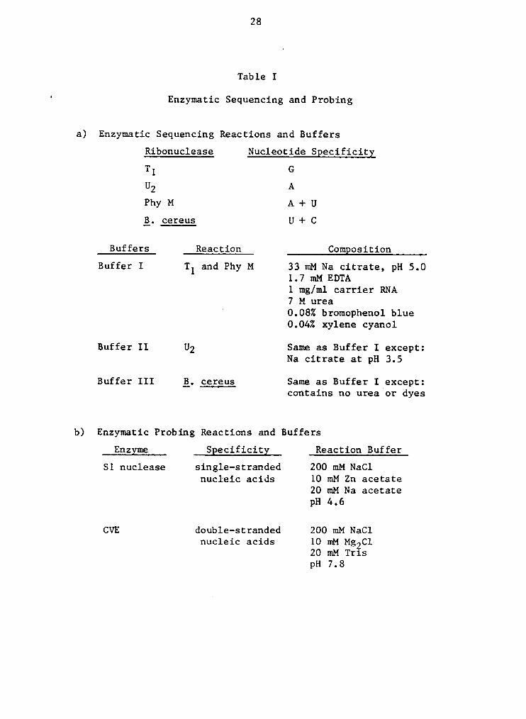
which the RNA sequence can be derived. Table I(a) lists the
enzymes and buffers used for the individual reactions. Ac-
tivities of different enzyme preparations vary and so it was
necessary to determine the dilution required to give compa-
rable extents of digestion between the four enzymes. All

27 .
sequencing enzymes used in this study were from the same
lot. One ul of stock T1, U2, Phy M and B. cereus RNases
were diluted separately in 30, 15, 6,and 10 ul of autoclaved
water, respectively. Six samples of (32P) end-labelled RNA
were dried in separate microfuge tubes. One sample was used
as a control to show the presence of nicks in the untreated
RNA. A second sample underwent an alkaline hydrolysis with
sodium carbonate at 90OC for 4 min. The four other samples
were dissolved in 3 ul of the appropriate reaction buffer
and chilled on ice. Reactions were initiated by the addi-
tion of 1 ul of diluted enzyme, and the solution was incu-
bated at 55OC for 12 min. Reactions were then quenched on
ice, 3 ul of 1% formamide/dye mix were added to the B. cer-
eus reaction tube, and the samples were loaded on a sequenc-
ing gel. Reactions were performed just prior to loading on
the gel, or were stored at -80OC and thawed just before
loading. ‘
S1 nuclease from AspergHum· oryzae (E.C. 3.1.4.21)
cleaves single—stranded nucleic acids (Ando, 1966); cobra
venom endonuclease (CVE) from Naja naja oxiana cleaves double-
stranded helical regions (Toots et al., 1981). Partial dig-
ests of the monomer and dimer forms of 5.8S rRNA under non-
denaturing conditions were electrophoresed on sequencing
gels alongside samples from the sequencing reactions, which
28
Table I
' Enzymatic Sequencing and Ptobing
a) Enzymatic Sequencing Reactions and Buffets
Ribonuclease Nucleotide Specificity
Tl G
Phy M A + U
Q. ceteus U + C
Buffets Reaction Composition
Buffet I T1 and Phy M 33 mM Na cittate, pH 5.01.7 mM EDTA1 mg/ml cattiet RNA7 M utea
° 0.08% btomophenol blue0.04% xylene cyanol
Buffet II U2 Same as Buffet I except:Na cittate at pH 3.5
Buffet III Q, ceteus Same as Buffet I except:contains no utea ot dyes
b) Enzymatic Ptobing Reactions and Buffets
Enzyme Specificity Reaction Buffet
S1 nuclease single—sttanded 200 mM NaClnucleic acids 10 mM Zn acetate
20 mM Na acetatepH 4.6
CVE double—sttanded 200 mM NaClnucleic acids 10 mM Mg2Cl
20 mM TtispH 7.8

29
were used as markers. End-labelled monomer and dimer sam-
ples were dissolved in 3 ul of the appropriate buffer. Ta-
ble I(b) lists the reaction buffer used with each enzyme.
The reactions were initiated by the addition of 1 ul of di-
luted S1 or CV enzyme solutions, and stopped by chilling on
ice and adding an equal volume of formamide/dye mix. Both
enzymes were tested at various concentrations and under
different reaction conditions for use in probing higher-ord-
er structure. Reaction conditions are given in the figure
legends for experiments shown in figures 6 - 8.
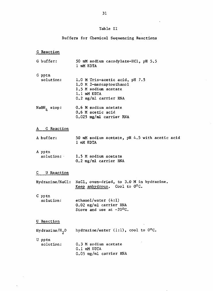
Chemical Seqgencing ang Probing
The chemical sequencing method (Peattie, 1979, 1983)
uses the same principle as the enzymatic method. Four
different base-specific chemical reactions are used to gen-
erate the sequence pattern. Terminally labelled RNA is used
and the fragment products are resolved by polyacrylamide gel
electrophoresis. The chemical process which results in RNA
cleavage is a two step reaction, however. In the first step,
the base is covalently modified in a base—specfic reaction;
this modification results in a site that is susceptible to
cleavage by a second chemical (aniline). Table II lists the
buffers used for the individual reactions. All reactions
were carried out in 1.5 ml Eppendorf microfuge tubes that
30
had been previously treated with a 5% solution of dimethyl-
dichlorosilane in hexane. Centrifugation was done at 12,000
x g in an Eppendorf microcentrifuge. Aniline was re-dis-
tilled by simple distillation under vacuum.
The G-specific reaction was performed under a variety
of conditions to establish optimum reaction conditions. The
following variables were tested: the pH of the reaction
buffer (pH 5.5-7.5), the presence and concentration of car-
rier RNA during the dimethyl sulfate (DMS) reaction, heat-
denaturation of the RNA before the DMS reaction, concentra-
tion of DMS, and length of reaction time. The reaction was
chilled on ice and 75 ul of cold G precipitation (pptn) so-
lution added, followed by 900 ul of cold absolute ethanol.
The solution was then mixed well and chilled at -70OC (dry
ice/ethanol bath) for 5 min. The precipitated RNA was pel-
leted by centrifugation for 5 min, resuspended in 200 ul of
· cold 0.3 M sodium acetate, and re—precipitated with 600 ul1
of absolute ethanol. The sample was centrifuged for 5 min,
the pellet rinsed with 500 ul of absolute ethanol (-2000),
chilled at -7000 for 2 min, recentrifuged and the pellet
dried under vacuum. The RNA was then dissolved in 10 ul of
1.0 M Tris-HC1, pH 8.2 10 ul of freshly made 0.2 M sodium
borohydride were added, and the sample was quickly mixed and
centrifuged before incubating at OOC for 30 min in the dark.
31
Table II
Buffers for Chemical Sequencing Reactions
G Reaction
G buffer: 50 mM sodium cac0dylate—HCl, pH 5.51 mM EDTA
G pptnsolution: 1.0 M Tris—acetic acid, pH 7.5 ~
1.0 M 2-mercaptoethanol1.5 M sodium acetate1.1 mM EDTA0.2 mg/ml carrier RNA
NaBH4 stop: 0.6 M sodium acetate0.6 M acetic acid0.025 mg/ml carrier RNA
A G Reaction
A buffer: 50 mM sodium acetate, pH 4.5 with acetic acid1 mM EDTA
A pptnsolution:· 1.5 M sodium acetate
0.2 mg/ml carrier RNA
C U Reaction
Hydrazine/NaCl: NaCl, oven-dried, to 3.0 M in hydrazine.Keep anhgdrous. Cool to 0°C.
C pptnsolution: ethanol/water (4:1)
0.02 mg/ml carrier RNAStore and use at —20°C.
U Reaction
Hydrazine/H20 hydrazine/water (1:1), cool to O°C.
U pptn _solution: 0.3 M sodium acetate
0.1 mM EDTA0.05 mg/ml carrier RNA
32
At 30 min, 200 ul of the sodium borohydride stop reagent was
added, followed by 600 ul of cold absolute ethanol. The RNA
was then precipitated, washed and dried as described above.
For the A>G reaction, 3' end-labelled RNA was dissolved
in 200 ul of A buffer, mixed and chilled to o°c. One ul of
diethyl pyrocarbonate (DEPC) was added and the reaction in-
cubated at 90OC for 10 min. The sample was then chilled on
ice and 50 ul of cold A pptn solution added, followed by 750
ul of cold absolute ethanol. Precipitations and washes were
followed as described for the G reaction.
For cleavage at U residues, lyophilized 3' end-labelled_
RNA was reacted with 10 ul of a freshly made hydrazine/water1
solution (1:1, OOC). The reaction was incubated on ice for
10 min, followed by the addition of 200 ul U pptn solution
and 600 ul of cold absolute ethanol. Precipitation and sub-
sequent washes were followed as described for the G reac-
tion.
For the C>U reaction, lyophilized 3' end-labelled RNA
was reacted with 10 ul of 3.0 M NaCl in anhydrous hydrazine
(freshly made). The reaction was incubated on ice for 15
min, followed by the addition of 500 ul of C pptn solution
(-20°C). Precipitations and. washes were followed as de-
scribed for the G reaction.

33
In an alternate C>U reaction, the 3' end-labelled RNA
was dissolved in 300 ul of G buffer and reacted with DMS as
described for the G reaction. Instead of the sodium borohy-
dride reaction, however, the RNA was then reacted with hy-
drazine/water (1:1) as described for the C reaction.
Samples which were modified as described above were
then reacted with 20 ul of aniline solution (freshly made)
for 20 min at 60OC in the dark. Reactions were stopped by
freezing the samples in a -70OC dry ice/ethanol bath; the
samples were then dried under Vacuum, washed 3 times with 20
ul of autoclaved water, drying under Vacuum after each wash.
The samples were resuspended in 2 - 4 ul autoclaved water,
followed by an equal volume of 1% formamide/dye mix. The
solution was heated at 60OC for 2.5 min, chilled quickly and
loaded on a sequencing gel.
DEPC was used to probe for A residues not involved in
higher-order structure (Lo, 1981; Nazar, 1983; Peattie,
1980, 1983). The RNA was dissolved in 200 ul of G buffer
(at pH 7.5). Various amounts of DEPC were added (1-20 ul)
and the reaction conditions were varied with respect to both
temperature and time to find suitable conditions for prob-
ing. Reactions conditions for the experiment shown in Fig-
ure 10 are given in the legend.
34
Generation ggg Qimgr Formation gg 5.88 rRNA Half-molecules
The 5.8S rRNA was digested under limiting conditions
with RNase T2 as described by Peters et al. (1982) with some
modifications. An initial experiment was performed to eval-
uate the reaction conditions. Two samples of 3' end-la-
belled 5.8S rRNA were dissolved in 10 mM NaCl, 3 mM EDTA, 15
mM MOPS, pH 7.3, at a concentration less than 0.08 mg/ml.
One of the samples was heated at 70OC for 5 min and then
slowly cooled to room temperature over a 4 h period. Both
samples were then brought up to 0.2 M NaCl and 0.5 mg RNA/ml
with tRNA and chilled on ice. RNase T2 (13 units/ml) was
added and the solution was incubated at 1lOC for 20 min.
The reaction was quenched on ice and an equal volume of for-
mamide/dye mix added. The sample was resolved on a 10% PA,
7 M urea gel and analyzed by autoradiography.
Digestions of non-radioactive 5.8S rRNA were done as
followsz 50 ug of 5.8S rRNA (<0.08 mg/ml) were incubated in
0.2 M NaCl,3-mM
EDTA, 15 mM MOPS, pH 7.3, for 5 min at 75OC
and then slowly cooled to room temperature over a 3-4 h per-
iod. The sample was then chilled on ice and RNase T2 (2
units/ml) added; the reaction was incubated for 20 min at
ll°C, and then quenched on ice. The reaction was stopped
and the RNA extracted by the addition of one volume of SDS-
buffer and two volumes of phenol-cresol reagent. The RNA
35
fragments were then precipitated, treated with alkaline
phosphatase and labelled on their 3' ends with (5'-32P) pCp
as described in the above sections.
The fragments visualized by the autoradiograph were ex-
cised and extracted from the gel in 0.15 M NaCl buffer as
described earlier. These were then characterized by enzy-
matic sequencing and terminal analysis. A small aliquot of
each terminally-labelled sequence was subjected to a com-
plete hydrolysis and the nucleotides were resolved by high-
voltage paper electrophoresis. Approximately 1000 cpm of
each fragment were precipitated with 25 ug of unlabelled
carrier tRNA and dried under vacuum. The pellets were re-
dissolved in 10 ul of 0.3 M Na0H and incubated at 37°C for
16-24 h, mixing intermittently. The samples were then spot-
ted on a 70 cm sheet of Whatman 3 mm filter paper sprayed ·
immediately prior to sample application with 5% acetic acid
adjusted to a pH of 3.5 with ammonium hydroxide. The sam-
ples were electrophoresed at 1000-2000 volts for 3-5 h in a
Savant HV tank. After electrophoresis, the paper was dried
and the resolved nucleotides localized by ultraviolet illu-
mination. The nucleotides from each fragment were cut out
and the filter paper was immersed in a scintillation fluor
and counted for radioactivity. The homogeneity of the frag-
ment termini was then calculated. The fragments were also
36
characterized and identified using the enzymatic sequencing
method described earlier.A
Both 5.85 rRNA 3' and 5' fragments were tested for the
ability to form dimers under the conditions described earli-
er for intact 5.85 rRNA dimer formation. Concentrations of
the fragments could not be determined directly but were es-
timated. based on radioactivity. Monomer and dimer forms
were resolved on a 10% native polyacrylamide gel, and were
recovered from the gel as previously described.
Thermostability ggg gg Determinations
The thermostabilities of intact 5.85 rRNA and 5' half-
molecule dimers were determined as follows (Sitz et al.,
1978). Dimer samples (in 0.15 M NaCl, 1 mM EDTA, 1 mM MOPS,
pH 7.2) were diluted ten-fold with autoclaved water; small
aliquots (400 — 1500 cpm/ 10 ul) were sealed in glass capil-
laries and chilled on ice. The samples were incubated in a
water bath for 5 min at various temperatures (35°- 70°C), ·
immediately chilled in an ice/water bath, and kept cold un-
til each sample had been heated at the appropriate tempera-
ture. The RNAs were then resolved on a native 10% PA gel.
The monomer and dimer bands were visualized by autoradiogra-
phy, excised from the gel, and the radioactivities deter-
mined by Cherenkov counting. In some cases, the autoradio-

37
graphs were scanned using a densitometer (E—C Apparatus
Corp.).
Free energy (AG) determinations were made using· the
methods of Ninio (1979), Salser (1977); and Pipas and McMa—
hon (1975). Assumptions regarding the stability of various
secondary structures pertinent to the two dimer models are
defined in Table III, p. 61.
RESULTS
The average yield of whole cell RNA from rat liver was
7.2 mg/g of tissue. From this appreximately 0.8 ug of 5.8S‘
rRNA were recovered per mg ef whole cell RNA. Synthesis ef
radioactive ATP and pCp resulted in 77 and 69 per cent in-
corperation ef the 32P radiolabel, respectively. Terminal
labelling ef the 3' end was eight times more efficient than
5' end-labelling.
Enzymatic Seguencing ggg Prebing
Cleavage patterns resulting from partial digestions of _
5.8S rRNA with Sl and CV endenucleases are shown in Figures
6a and 6b (3' end-labelled 5.8S rRNA) and Figures 7a, 7b,
and 8 (5' end-labelled 5.8S rRNA). Digestion ef the monomer
and dimer forms ef 5.8S rRNA produced nearly identicali
cleavage patterns fer the regions studied. S1 and CV ende-
nuclease prebing of 3' end-labelled rat liver 5.8S rRNA me-
nomer and dimer were repeated five times, with similar re-
- sults. Digestiens of the 5' labelled human 5.8S rRNA were
repeated four times. Electropheresis ef monomer and dimer
· forms of 5.8S rRNA en 10% native PA gels showed that 80 - 90
38
39
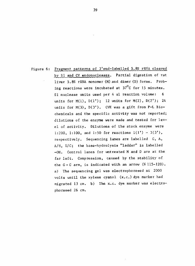
Figure 6: Fragment patterns of 3'end-labelled 5.88 rRNA cleaved
by Sl and CV endonucleases. Partial digestion of rat”
liver 5.88 rRNA monomer OO and dimer UD forms. Prob-
ing reactions were incubated at 37oC for 15 minutes.
S1 nuclease units used per 4 ul reaction volume: 6
units for M(1), D(l'); 12 units_for M(2), D(2'); 24
units for M(3), D(3'). CVE was a gift from P-L Bio-
chemicals and the specific activity was not reported;
dilutions of the enzyme were made and tested for lev-
el of activity. Dilutions of the stock enzyme were
1:200, 1:100, and 1:50 for reactions 1(1') - 3(3'),
respectively. Sequencing lanes are labelled G, A,
A/U, U/C; the base-hydrolysis "ladder" is labelled
-0H. Control lanes for untreated M and D are at the
far left. Compression, caused by the stability of
the G·-C arm, is indicated with an arrow (N 115-120).
a) The sequencing gel was electrophoresed at 2000
volts until the xylene cyanol (x.c.) dye marker had
migrated 13 cm. b) The x.c. dye marker was electro-
phoresed 26 cm.

42
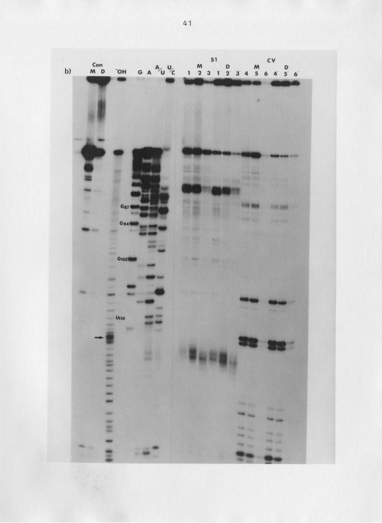
Figure 7: Fragment. gatterns of 5'end—labelled 5.88 rRNA cleaved
bx Sl and CV endonucleases. Partial digestion of hu-
man osteosarcoma 5.88 rRNA monomer (M) and dimer (D)
forms. Reaction conditions and figure nomenclature
as in Fig.6.


45
Figure 8: Fragment patterns of 5'end-labelled 5.8S rRNA cleaved
bz S1 nuclease. Partial digestion of human placenta
5.8S rRNA monomer (M) and dimer (D) forms. Probing
reactions were incubated at 37°C for 15 minutes. S1
nuclease units used per 4 ul reaction volume: 1.5,
3, 6, 12, 24 units for reactions 1(1') - 5(5'), re-
spectively. Figure nomenclature as in Fig.6. The
differences seen between monomer and dimer, partic-
ularly in lanes M(3) and D(3'), are not representa—
tive of results from other experiments.


47
per cent of the dimer was maintained in that form, and mo-
nomer remained at 90 - 100 per cent. The autoradiographs
focus on regions of the molecule proposed to be involved in
dimer formation, namely, nucleotides 1-15 and 137-156 (the
5' and 3' terminal regions) and nucleotides 18-42 (the re-
gion for the palindrome interaction). Additionally, I chose
to look at that section of the molecule opposite the palin-
drome interaction site, nucleotides 90-114.
Major CVE cleavage sites were nucleotides 6-8, 12-14,
23, 28, 33-37, 46-47, 107, 115-122, 128-141, 146 and'
151-154; major S1 nuclease cleavage sites were 1-3, 8-10,
38-41, 123-127 and 154. Although the sequencing gels were
not done to look at the nucleotide region 50-90 (and there-
fore these bands are not well resolved), the probing enzymes
appear to cleave in the following general regions: CVE,
54-58, 62-70 and 80-90; S1, 50-53, 60-63 and 70-80. In as-
signing the probing enzyme cleavage sites, it is important
to note that S1 and CV endonucleases leave a 5' phosphate on
the product, whereas the sequencing enzymes leave a 3'
phosphate (for details see Discussion).
Chemical Seguencing ggg Probing
The optimal conditions for the G-specific reaction were
determined to be as follows: one minute pre-incubation of

48
the sample at 90oC immediately before addition of DMS, 5 ug
of carrier RNA present during the DMS reaction a DMS concen-
tration of 0.5 ul/reaction volume and a one minute reaction
time (at 90°C). The pH did not influence the reaction. The
most important component was the presence of carrier RNA
(see Fig. 9a).
Difficulties with the C > Q reaction were due to an er-
ror in the concentration of carrier RNA in the precipitation
buffer in the published protocol (Peattie 1983). Figure 9b
shows an autoradiograph of a chemical sequencing gel in
which the sequence can be read clearly.
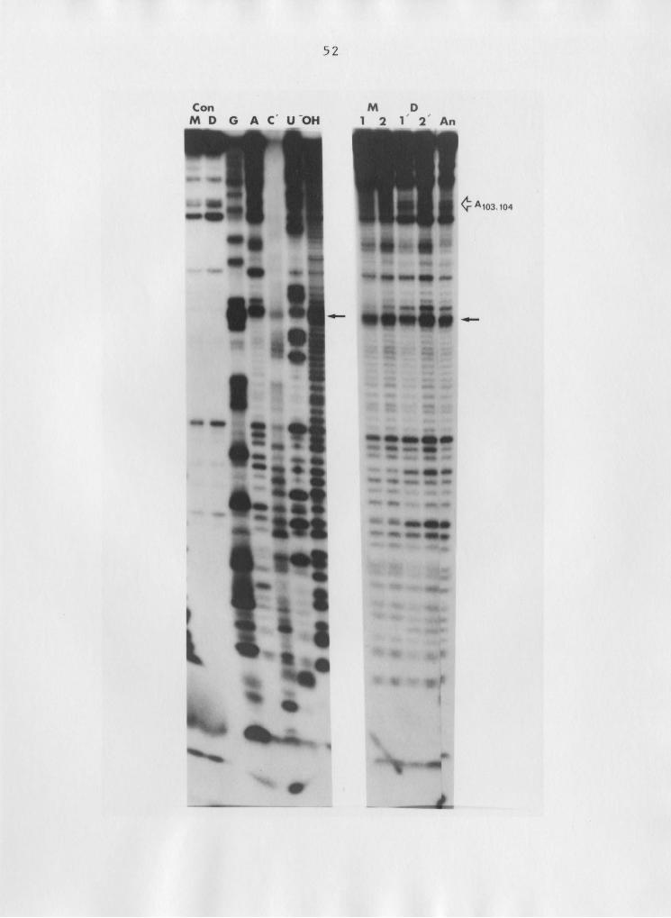
DEPC probing of adenine residues is shown in Figure 10.
An increase in reactivity is seen at positions AlO3’lO4 but
not at other adenines between that site and the 3' termini
(see figure legend for reaction conditions). However, non-
specific "background" cleavages by aniline make interpreta-
tion of the probing reactions difficult (see aniline control
lane).
Generation ang Qimgg Formation gf 5.8S rRNA Half-molecules
The digestion of 5.8S rRNA by RNase T2 was tested with
and without a pre-heat treatment (see Methods) prior to the
RNase T2 reaction. When 3' end-labelled 5.8S rRNA was not
pre—heated, 46 per cent of the radioactivity was recovered

49
Figure 9: Chemical seguencing of 3' end—labe1led 5.85 rRNA.
a) Effect of carrier. RNA on DMS (G—specific) re-
action. Lanes 1 and 2, without carrier; lanes 3
and 4, 5 ug carrier RNA present during reaction.
Concentration of DMS per 300 ul reaction volume:
0.5 ul DMS, lanes 1and3;1 ul DMS, lanes 2and4.
b) Sequence of rat 5.85 rRNA from nucleotides
94 - 156. C' denotes alternate C reaction (see
Methods). Control lanesz 1, aniline control;
2, RNA control. The x.c. dye marker was elec-
trophoresed 21 and 12 cm. ' 4


51
Figure 10: Diethzlpxrocarbonate grobing of 5.85 rRNA. Sequence
of 3' end—labelled rat 5.85 rRNA shown on left (G, A,
C', U). DEPC probing of adenine residues of monomer
(M) and dimer (D) 5.8S rRNA shown on right. An: an-
iline control. Reaction conditions: 5 ul DEPC/ ZOO
ul reaction; 1 h (1, I') and 2 h (Z, Z') at 25OC.
53
as intact 5.85 rRNA. When the RNA was pre-heated, the un-
digested 5.85 rRNA was reduced to 19 per cent, with an in-
crease in both the 3' half-molecule fragments and in bands
seen near the bottom of the gel.
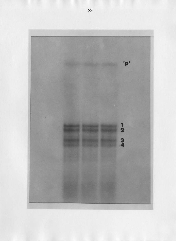
Non—radioactive 5.85 rRNA (from rat liver) was digested q
with RNase T2 and the fragment products were 3' end-labelled
with (5'-32P) pCp as described in the Methods section. Re-
solution of the labelled fragments by 10% PA, 7 M urea gel
electrophoresis resulted in four major bands, as shown in
Figure 11.
Characterization of the two more slowly migrating bands
(1 and 2) by enzymatic sequencing and terminal analysis rev-
ealed that they were both 5.85 rRNA 3' half-molecules, hav-
ing heterogeneous termini equal to that of the "parent" mo-
lecule (see Fig. 11): approximately 75 per cent uracil and
21 per cent cytosine. The 5' half-molecules had greater mo-
bilities; fragment 3 had a 90 per cent uracil 3' terminus,
representing either U73 or U74. The sequence for band 4 was
difficult to read because of the high degree of terminal
heterogeneity; terminal analysis showed that 64 per cent of
the fragments had a 3' uracil; 24 per cent, adenine; 8 per
cent, cytosine; and 4 per cent guanine. The sequence pat-
tern for band 4 resembled that of fragment 3 much more than
the patterns for bands 1 and 2, however, and the fragments
were considered to be 5' half-molecules of 5.85 rRNA.
54
Figure 11: Ribonuclease T2-generated 5.8S rRNA half-molecules.Partial digestion of 5.8S rRNA, followed by 3' end-
labelling and resolution of the fragments on a 10%
PA, 7b1urea gel. "Parent" band has the same mobil-
ity as intact 5.8S rRNA, but was slightly digested
· at the 3' end, producing terminal heterogeneity
(see text).
56
In the first RNase T2 digestion using cold 5.88 rRNA,
the 5.88 rRNA concentration was maintained at 0.5 mg/ml (as
in the initial testing reaction) and the RNA was not pre-
heated. The 3' and 5' half-molecules had the same relative
mobilities as described above. The termini of both 3' and
5' half-molecules exhibited a higher degree of heterogenei-
ty, however. .
_ The 3' and 5' half-molecules were tested for dimer for-
mation under the same conditions used for intact 5.88 rRNA
dimer formation. The 3' halves did not form dimer, while 5'
half-molecule "monomers" converted to 45 - 50 per cent di-
mer.
In a separate experiment using human 5' labelled 5.88
rRNA, a 5' fragment of approximately 50-55 nucleotides was
recovered with the same radioactivity as a 5' fragment about
„ 70 nucleotides in length. Incubation of these two fragments
under the same conditions resulted in dimer formation of the
longer fragment, but not of the shorter one.
Thermostability ggg Ag Determinations
The thermal stabilities of the intact 5.88 rRNA and 5'
half-molecule dimers were determined three times for each
complex in a total of five experiments. In the fifth exper-
iment, the two complexes were mixed and sealed in the samel

57
capillaries, since the 5.8S rRNA monomer and the 5' half-mo-
lecule dimer have different mobilities (Fig. 12). The re-
sults from these studies are shown in Figure 13. The Tm va-
lues for the two complexes can not be considered signifi-
cantly different, due to the high degree of variability in
the results across the three repetitions.
The AG values for the palindrome interaction region and
one terminal interaction region using the method of Salser
(1977) are -21.0 kcal and -24.5 kcal, respectively. Using
values from Ninio's empirical (variant) model, the AG values
are -21.3 kcal for the palindrome region and -16 kcal for
the terminal region. Table III lists other AG values, de-
pendent on various assumptions used in the stability models.

58
Figure 12: Heat—induced dimer dissociation. Resolution of
monomer and dimer forms of intact 5.8S rRNA and
5'ha1f—molecules after heat treatments (see text).
D, Dlg, and M represent controls for the intact
5.8S rRNA dimer, 5'half-molecule dimer, and the
5.8S rR.NA monomer, respectively. Incubation
temperatures of the samples were 550, 57.50, 600,
62.50, 650, and 7o°c for 16666 1-6, respectively.


60
100 '
90X
80
70
gf] .2 60 _ O 0
”
· E' x T = 61 + 2 C form
•-•
N both (·) and (x)*· 0é 5 ><~l-*
EX lä - 40
Z
30
¥20 ii
10 ‘‘
50 55 60 65 70
1*1=;M1=ERATuRE (°c)
Figure 13: Thermostability profiles of intact 5.88 rRNA dimer '
Tand the 5' half—molecule dimer. The amount of per
cent dimer (control) varied from the different pre-
parations; to allow comparison from the different
thermostability experiments, dimer is expressed in
terms of relative per cent. (·) intact 5.88, (x)
5' half-molecule.
61
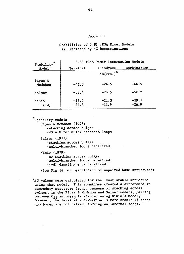
Table III
Stabilities of 5.8S rRNA Dimer Modelsas Predicted by AG Determinations
. a 5.8S rRNA Dimer Interaction ModelsStability
Model Terminal Palindrome Combination
AG(kcal)bPipas &
McMahon -42.0 -24.5 -66.5
Salser -38.4 -24.5 -58.2
Ninio -26.0 -21.3 -39.7
" (+d) -22.8 -11.9 -26.9
aStability Models4
Pipas 6. McMahon (1975)-stacking across bulges-AG = 0 for multi-branched loops
Salser (1977) ·
-stacking across bulges·multi-branched loops penalized ~
Ninio (1979)-no stacking across bulges-multi-branched loops penalized-(+d) dangling ends penalized
(See Fig 14 for description of unpaired-base structures)
' bAGvalues were calculated for the most stable structure
using that model. This sometimes created a difference insecondary structure (e.g., because of stacking acrossbulges, in the Pipas & McMahon and Salser models, pairingbetween C11 and Gl&3 is stable; using Ninio's model,however, the terminal interaction is more stable if thesetwo bases are not paired, forming an internal loop).
. 62
I °
"
IIII
I ¤gfE
__
-—
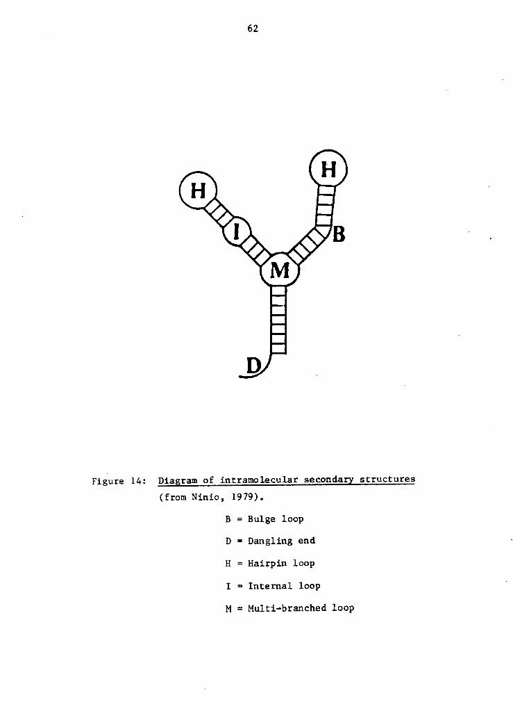
Figure 14: Diagram of intramolecular secondagx structures
(from Ninio, 1979).
B = Bulge loop
D = Dangling end ·
H = Hairpin loop
I = Internal loop '
M = Multi-branched loop

DISCUSSION
Enzymatic Seguencing ggg Probing
The enzymes used for the sequencing reactions cleave
RNA differently than do Sl and CV endonucleases. This is
important to consider when interpreting the autoradiographs.
The sequencing enzymes (T1, U2, Phy M, and B. cereus are
specific for ribonucleic acids and cleave the phosphodiester
bond via a cyclic intermediate, generating a product with a
3' phosphate group. S1 nuclease and CVE, on the other hand,
· cleave both RNA and DNA, therefore utilizing a reaction me-
chanism not dependent on the 2' hydroxyl group. This re-
sults in a fragment containing a 5' phosphate (Pavlakis et
al., 1979) (see Fig. 15). This difference in cleavage caus-
es fragments of equal nucleotide length to have different
electrophoretic mobilities, due to the difference in nega-
tive charge. When the RNA is labelled at the 3' end, the
radiolabelled fragments cleaved by the sequencing enzymes
have a greater mobility than fragments generated by Sl or
CVE cleavage at the same site; the reverse is true with RNA
labelled at the 5' end.
Figure 16 depicts the dramatic difference in mobilities
of various fragments, and demonstrates the care that must be
taken in using the sequencing enzyme products as markers for
63

64
SF ic
QHZYTHES
F''’°°°"''’°'°°'°‘°’’°°°°’'’''’°°°'’’°'’°'‘°’'’°‘‘‘‘°‘°‘°°‘‘°°°‘‘s3* • 3
F sF________________________________
S1, CV probing enzymes
5'<*> =~ 3*
Figure 15: Termini of seguencing vs. probing enzyme-generated
RNA fragments. Schematic representation depicting
the location of the phosphate group after cleavage
of the phosphodiester bond of RNA by the sequencing
enzymes vs. S1 and CV endonucleases.
65
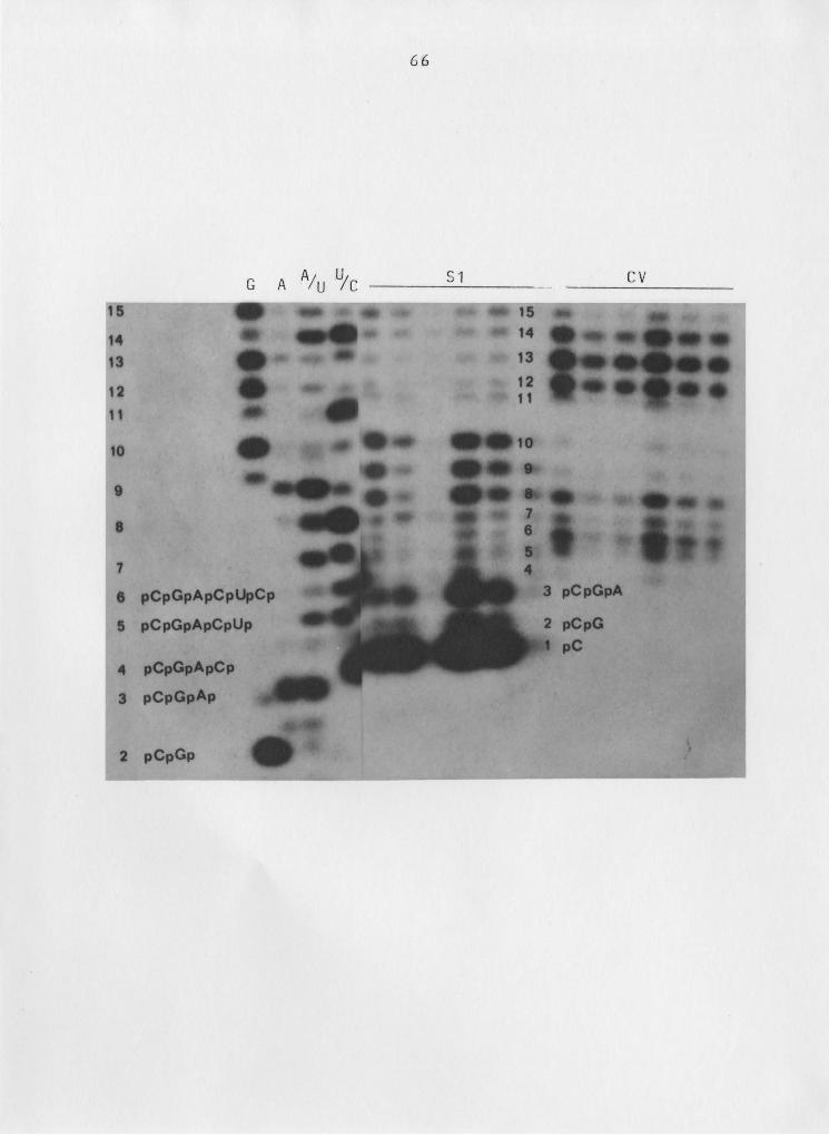
Figure 16: Mobility differences observed in 5' end—labelled RNA
fragments generated by seguencing vs. Rrobing enzyges.
Autoradiograph shown in Fig. 7, focusing on the 5'
terminal region. Fragment assignments are superimposed
on the autoradiograph. Figure nomenclature as in
Fig. 7.
67
assigning S1 and CVE cleavage sites, particularly near the
labelled termini. Although significant differences in mo-
bility are seen with the very short fragments, it appears
that the presence (or absence) of an additional terminal
phosphate has less effect on the electrophoretic mobility of
the fragment as its length increases. This is probably a
function of the decrease in the charge to mass ratio. Dif-
ferences in the relative mobilities seem to disappear alto-
gether when the chain length reaches about ten nucleotides
(using this gel system). Therefore, probing and sequencing
enzyme products of equal nucleotide length greater than 10-
nucleotides were considered to have equal electrophoretic
mobilities. This is in contrast to other studies (Pavlakis
et al., 1979; Toots et al., 1981) in which assignments of
probing enzyme cleavage sites were based on the differences
in mobilities being maintained throughout the sequence.
Other factors important to consider in evaluating the
probing studies include the following.
1) Digestion patterns of the molecule at higher enzyme
concentrations (or increased reaction times) may not reflect
the native conformation of the molecule, because initial _
cleavages may produce alterations in the higher—order struc-
ture. Therefore it is important to distinguish between
"first cuts" and those that occur later.

68
2) In comparing the digestion patterns of the monomer
and dimer, differences in the relative intensity of the
bands at given positions may be influenced by how much of
the sample actually migrated into the gel. The radioactivi-
ties of the samples were equal prior to the enzymatic reac-
tions. Radioactivities of the sample tubes (and micro-pi-
pette tips) were determined after loading the samples on the
gel to account for any discrepancies in relative radioactiv-
ities. Nonetheless, some samples had significant counts re-
maining in the wells. For this reason, the tops of the gels
are included in the autoradiographs.1
3) "Nicks" in the untreated RNA controls were not al-
ways the same for monomer and dimer samples, making probing
at those sites appear different. It is not known whetheru
these are the result of nuclease contamination or non—enzy-
matic degradation, but the former seems un1ikely' as the
breaks are not limited to one specific base, nor to pyrimi-
dines or purines. Also, incubation of the molecule at 37°C
did not increase the occurrence of cleavage at these sites.
The significant differences in single- and double-
stranded regions of the 5.85 rRNA dimer in the two published
models for dimer formation (Nazar et al., 1978; Pavlakis et
al., 1979) led to the choice of 51 and CV endonucleases as
probes for investigating the 5.85 rRNA dimer interaction

69 .
site. Partial digestion of the dimer with Sl and CV endonu-
cleases (specific for single- and double-stranded nucleo-
tides, respectively) should easily distinguish between these
· two models. In the terminal interaction model (Sitz et al.,
1978), base-pairing in the regions of contact in the dimer
are the same as those that occur in the monomer. It was
predicted, therefore, that if this model was correct, diges-
tions of the dimer and monomer forms of 5.8S RNA with Sl and
CV endonucleases would produce the same fragment patterns.
Alternatively, if the 5.8S rRNA dimer forms as described by
Pavlakis et al. (1979), the fragment patterns for 5.8S rRNA
dimer would be different than that of the monomer. Compari-
sons of the two patterns, however, are only legitimate if it
can be shown that the dimer form of 5.8S rRNA is maintained
during the probing reaction, and likewise, that the monomer
does not convert to dimer. Controls for the probing reac-
tions indicate that both the monomer and dimer forms of 5.8S
rRNA are maintained under the reaction conditions.
Partial digests of the monomer and dimer forms of 5.8S ·
rRNA with S1 and CV endonucleases produced identical frag-
ment patterns for the nucleotide regions studied (viz., N
1-45, 90-156). Some differences were observed between mo-
nomer and dimer digestion patterns for nucleotides 55-90.
The differences were not consistent, however, across the
70
probing studies; the_ cause of these differences is not
known. Probing studies by others (Lo & Nazar, 1982) indi-
cate changes in the secondary structure of the A-U rich arm
(nucleotides 62-90) of 5.85 rRNA upon its association with
285 rRNA and ribosomal proteins. It has also been suggested
that 5.85 rRNA has two stable configurations (Walker & Pace,
1983). Further studies are necessary to clarify the struc-
tural changes occurring in that part of the molecule.
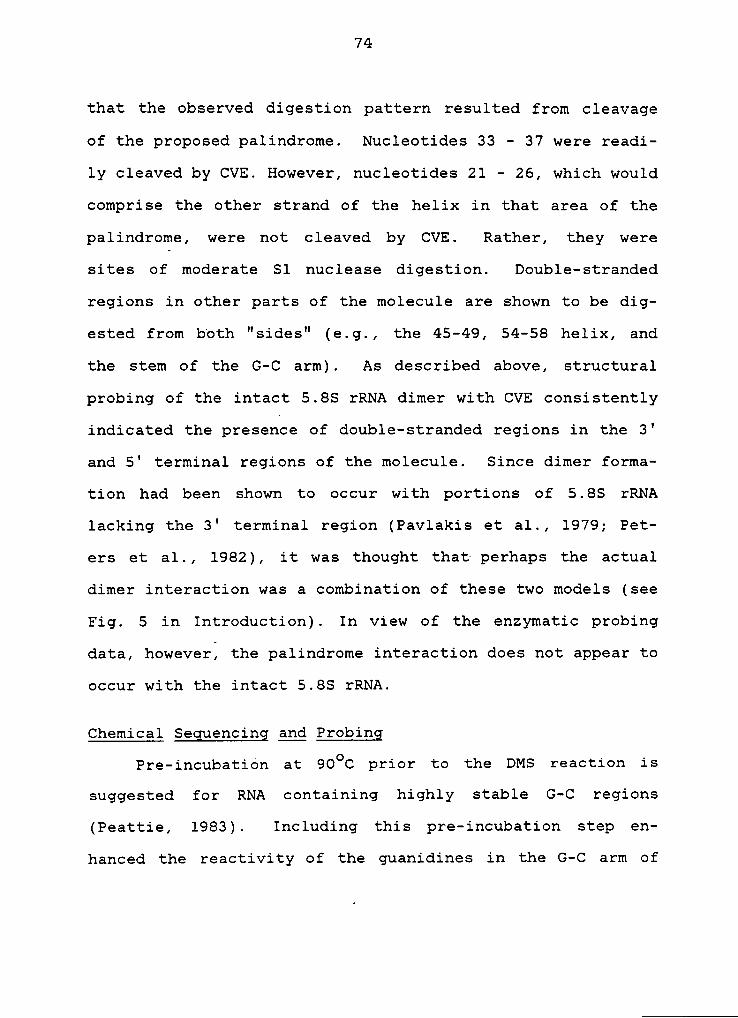
Analysis of the fragment patterns shown in figures 6 —
8 support the terminal interaction model for 5.85 rRNA dimer
formation. The cleavage sites for 51 and CVE, determined
from the autoradiographs, are depicted in Figure 17 and are
in general agreement with the "burp gun" model for 5.85 rRNA
secondary structure (Nazar et al., 1975). Cleavage observed‘
at the following sites are exceptions: CVE cleavage at nu-
cleotides 16, 23 and 28; 51 cleavage at 1-3, 8-10, 31, 37,
and 38. Cleavage at proposed single-stranded regions by CVE
may be due ta teritary interactions of 5.85 rRNA, of which
little is known. For cleavage by 51 nuclease, the very ini-
tial cuts occur at the 50-53 loop, 70-80 loop, the 123-127
loop and the very terminal 3' end. One would expect the
16-24 loop to also be attacked early. Perhaps this loop is
involved in some type of tertiary interaction that makes at
least a portion of it inaccessible to 51 nuclease (and nu-

71
8)Ö
Ö xu ur { { ”A C +{‘°AGA sou ¤• 6 G uc ¤ A A CP©GAC CUUAGCGGI-|•G GC GUGCGUCGAUG CGCAG U••••nn••••••••
Géé•Aéé•Ä••••• é••••A
GC G GAG GCCGCAUC U1%
Ucuuuu 2,6 uuc
Ac cGcUA£U·AAGA CGUCG
6-c“°
A A A·u so ‘
f 6-c JG c 6-uG·C }¤¤•\ U-A
*¤¤C-Cm A.UC·G C-G
- o6*% é‘E.•¤· ¤^ ^..
G U ßxA U
. CG;b CVE '°)
lc^cU -
U*6 GQ {{ A A so+1+ Nb G 1 111++ ·· G
P(C)GACUCUUAgCGGU•\Ö GCUCGSGCGUCGAUGA AC1GttGCUn••••••••••••• éé•••AUCGCUG GAGUCGCCGCAU UQAUUIM 6,6 C _ „U-AAG^ GVC6
1 A 6-c A A A·u •¤G·C G C G·uG-C CUU U-AG·C •¤• U·A
¤¤C•G¤z¤ A•UC·G C·GC·G A·U
U U C-C10c u
•°AA
C G AGA uuCG•
Figure 17: S1 and CVE cleavage sites--5.8S rRNA. "Burp gun" model
used to represent terminal interaction model as base-
pairing scheme is the same. Arrows indicate sites of
cleavage by a) S1 nuclease and b) cobra venom endonu—
clease. (*) indicates earliest cleavages; open arrows
indicate cligestion observed only at high enzyme con-
centrations. Size of arrow correlates with degree of
_ digestion at that site.
72
cleotide 23 a site for CV cleavage). The lack of cleavage
at this loop by S1 nuclease has also been observed by Khan
and Maden (1976). They suggest that the molecule may fold
back on itself, with interaction between loop structures as
is seen with tRNA. Cleavage by S1 nuclease was seen at nu-
cleotides 16-21 in Figure 7, but the high frequency of nick-
ing at C19-A20 makes interpretation of the probing reactions
in this area difficult. Cleavage by S1 nuclease at other
sites on the xnolecule (e.g., 1-3 and 8-10) may actually
represent secondary cleavages caused by changes in the mo-
lecule's conformation induced by first cuts. An alternative
possibilty is that opening and closing of the helix occurs,
providing temporary single-stranded sites for S1 nuclease.
Tritium-exchange studies of double-strandad nucleic acids
suggest that helical regions "breathe," existing in an equi-
librium between open and closed states (Englander, 1972).
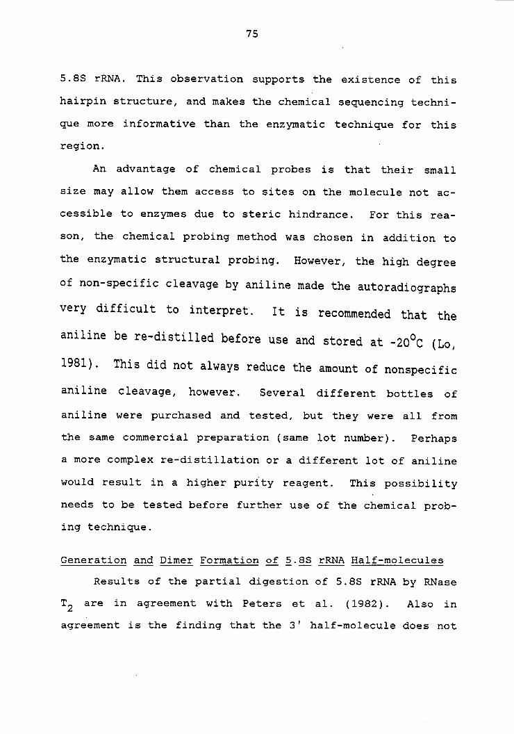
The alternative model for dimer formation (Pavlakis et
al., 1979) proposes that nucleotides 18-40 form the sites
for dimer interaction, creating a palindrome. Figure 18
shows the base-pairing scheme for the palindrome region,
with S1 nuclease and CVE cleavage sites indicated. The dig-
estion patterns for this region do not appear to support the
palindrome interaction model. Little is available in the
literature regarding CV endonuclease, but it seems unlikely

73
a)Sl
-„___G 20 40 A___D GA lHCH 30l IHIG 3'UCAUCGGCUCGUGCGUCGAUGAlll lllll llll lllll Iél
I3.___AcAl A so 20 cG___5
b)CVE
6*---6 30 A..;3·GA 2%* ll +lU**ll“°A¤UCAUCGGCUCGUGCGUCGAUGAlll IIIII llll lllll lélAAGUAGCUGCGUGCUCGGCUCA UA• G 40***tT 39*
*
ZQ G •3--·A 9---5
by a) S1 nuclease and b) cobra venom endo—
nuclease. Arrows as in Fig. 17.
74
that the observed digestion pattern resulted from cleavage
of the proposed palindrome. Nucleotides 33 - 37 were readi-
ly cleaved by CVE. However, nucleotides 21 - 26, which would
comprise the other strand of the helix in that area of the
palindrome, were not cleaved by CVE. Rather, they were
sites of moderate S1 nuclease digestion. Double—stranded
regions in other parts of the molecule are shown to be dig-
ested from both "sides" (e.g., the 45-49, 54-58 helix, and
the stem of the G-C arm). As described above, structural
probing of the intact 5.8S rRNA dimer with CVE consistently
indicated the presence of double-stranded regions in the 3'
and 5' terminal regions of the molecule. Since dimer forma-
tion had been shown to occur with portions of 5.8S rRNA
lacking the 3' terminal region (Pavlakis et al., 1979; Pet-
ers et al., 1982), it was thought that perhaps the actual
dimer interaction was a combination of these two models (see
Fig. 5 in Introduction). In view of the enzymatic probing
data, however, the palindrome interaction does not appear to
occur with the intact 5.8S rRNA.
Chemical Seguencing and Probing
Pre-incubation at 90oC jprior to the DMS reaction is
suggested for RNA containing highly stable G-C regions
(Peattie, 1983). Including this pre-incubation step en-
hanced the reactivity of the guanidines in the G-C arm of
75
5.85 rRNA. This observation supports the existence of this
hairpin structure, and makes the chemical sequencing techni-
que more informative than the enzymatic technique for this
region. °
An advantage of chemical probes is that their small
size may allow them access to sites on the molecule not ac-
cessible to enzymes due to steric hindrance. For this rea-
son, the chemical probing method was chosen in addition to
the enzymatic structural probing. However, the high degree
of non-specific cleavage by aniline made the autoradiographs
very difficult to interpret. It is recommended that the
aniline be re-distilled before use and stored at -2OOC (Lo,
1981). This did not always reduce the amount of nonspecific
aniline cleavage, however. Several different bottles of
aniline were purchased and tested, but they were all from
the same commercial preparation (same lot number). Perhaps
a more complex re-distillation or a different lot of aniline
would result in a higher purity reagent. This possibility
needs to be tested before further use of the chemical prob-
ing technique.
Generation ang Qimeg Formation gf 5.85 gggg Half-molecules
Results of the partial digestion of 5.85 rRNA by RNase
T2 are in agreement with Peters et al. (1982). Also in
agreement is the finding that the 3' half-molecule does not

. 76
form dimer. However, the formation of higher aggregates of
the 5' half—mo1ecule, reported by Peters et al. (1982), was
not observed in this study.
In forming the 5' half-molecule dimer an interesting
result was observed, viz., a fragment containing the nucleo-
tide sequence for the palindrome interaction did not form
dimer. This was the only experiment in which this particu-
lar length fragment was characterized and used to form di-
mer. Therefore, speculation must be limited. However, this
result raises the interesting possibility that the 5' half-
molecule dimer forms via an interaction other than the pal-
indrome interaction.
Thermostability ggg gg Determinations .
The Tm determined for the intact rat 5.8S rRNA dimer-
was about 5OC higher than that determined by Sitz et al.
(1978). This difference in thermal stability may be due to
the difference in buffer systems used (0.015 M NaCl, 0.1 mM
EDTA, 0.1 mM MOPS, pH 7.2 gg. 0.015 M NaCl, 0.1 mM EDTA,
0.1 mM Tris, pH 7.5); or the method of analysis, which was
also different in the two studies (quantitation by radioac—
tivity gg. staining of unlabelled 5.8S rRNA). Another pos-
sibility is that because the Volumes in which the dimer sam-
ples were dissolved were different, influences on the salt

77
concentration due to residual salt from the RNA sample might
be different. This in turn would affect the dimer stabili-W
ty, as salt concentration is known to play a dramatic role
in structural stability (see text by Cantor and Schimmel,
1980).
4 A significant difference in the Tm values for the in-
tact 5.8S rRNA and 5' half—molecule dimers was not observed.
However, the high degree of variablilty in the dissociation
of the dimers makes comparisons of the Tm values difficult.
This variability may be due to residual salt in the diffe-
rent RNA preparations. Mixing the two complexes and treat-
ing them as one sample, as in the fifth experiment, is a
better technique for comparing the Tm values because the two
complexes are in the same environment. This_method reduces
the degree of experimental error and will be used for future
Tm determinations. Done in this way, the Tm for each com-
plex could be compared for each individual experiment, rath-
er than averaging the Tm for each complex across several ex-
periments and comparing the averaged values.
If the Tm value for the intact 5.88 rRNA dimer was sig-
nificantly different from that of the 5' half-molecule di-
mer, one could argue that the interactions are indeed diffe-
rent. Equivalent Tm values, however, do not necessarily
mean the interactions are the same; it indicates that the
78
stabilites of the intact 5.8S rRNA and5’
half-molecule di-
mers are comparable, but it gives no information as to the
specifics of the intermolecular interactions.
If the stabilities of the regions of interaction in the
two dimer models are comparable, it would be expected that
the Tm values would also be comparable. Models for predict-
ing RNA secondary structure use free energy (AG) values for
predicting the most stable secondary structure (see Intro-
duction). Controversy regarding the destabilizing effects
of unpaired-bases leads to differences in AG values calcu-
lated for various structures, depending on. the stability
parameters of the model used.
Using the assumption of Pipas & NmMahon (1974) that
multi-branched loops have a AG value of zero, the terminal
interaction is more stable than the palindrome model (see
Table III). Salser considers multi-branched loops to be so-
mewhat destabilizing, but not very much, and the AG values
for the two dimer models are comparable to those calculated
using the model of Pipas & McMahon. Ninio's model (1979),
on the other hand, penalizes multi-branched loops quite sev-
erely, causing the AG values for the two dimer models to be
similar if dangling ends are not taken into account. If
dangling ends are also considered, the terminal interaction
is nearly twice as stable as the palindrome interaction.

79
However, all stability models predicted the combination mo-
del to be more stable than either the palindrome or terminal
interaction model, as might be expected.
In comparing the thermal stability data and the AG va-
lues derived from the various models, the AG values for the
two dimer models support equivalent Tm values for the two
complexes only if the multi-branched loop is considered a
very destabilizing structure (Ninio, 1979) and either dan-
gling ends are not as destablizing gs predicted by Ninio, or
the nucleotide region 1 - 17 does not constitute a danglingl
end in the palindrome model (perhaps it is involved in some
type of secondary or tertiary interactions). Obviously,
more work is necessary in defining the parameters for accu-
rately predicting secondary structure. It should also be
mentioned that in all of the stability models, tertiary in-
teractions are not taken into account, which may alter the
predicted stabilities of structures such as the dimer com-
plexes discussed in this study.
The interaction responsible for dimer formation of the
5' half-molecule still remains uncharacterized. A 5.85 rRNA
fragment containing the palindrome interaction sequence, but
shorter than the 5' .half-molecule, failed. to forun dimer,
suggesting that either formation of the 5' half-molecule di-
mer occurs through a different interaction than that pro-

80
posed, or the shorter fragment is capable of folding back on
itself in a way that obstructs the palindrome interaction
site. Study of the sequence has not revealed an alternative
dimer interaction that is more stable than the palindrome
interaction. This was also concluded by Pavlakis et al.
(1979) using a computer program for intermolecular interac-
tions. Characterization of the RNase T2—generated 5' frag-
ments (both monomer and dimer) by enzymatic probing should”
help to clarify the actual interaction. Results from the
enzymatic probing of 5.85 rRNA dimer support the terminal
interaction model for dimer formation (Sitz et al., 1978)
and indicate that the palindrome interaction (Pavlakis et
al, 1979) does not occur in dimer formed from the intact mo-
lecule.
EPILOGUE
The formation of the 5.85 rRNA dimer is an "in vitro
artifact" and as such bears no physiological relevance.
However, the study of such a complex is not without merit.
The techniques used in this study are being used by several
investigators to study the higher-order structure of RNA mo-
lecules (Wurst et al., 1978; Lockard et al., 1981; Toots et
al., 1981) and intermolecular interactions of complexes such
as the 5.85-285 rRNA junction (Nazar & Sitz, 1980; Walker et
al., 1982). Although the binding site of 5.85 rRNA for 285
rRNA has been determined with some certainty (see Introduc-
tion), there is still limited information regarding how 285
rRNA interacts with 5.85 rRNA (Walker & Pace, 1983
· [review]). The 285 rRNA is a very large molecule, approxi—
mately 5000 nucleotides in mammals. Determining its higher-
order structure and the sites involved in 5.85 rRNA binding
will be a difficult task.
The 5.85 rRNA dimer provides a model system for study-
ing RNA-RNA interactions using a molecule that is much sim-‘
pler and better characterized than a molecule such as 285
rRNA. It is the hope that characterization of the structure
of the 5.85 rRNA dimer by various techniques will help in
assessing what techniques are most reliable for determining
81 .

82 .
the intermolecular interactions of more complex RNA associa-2
tions. In comparing different techniques used to arrive at
the same information, a more complete and accurate pictureA8'”''
·
can be assembled. It also allows for a more critical evalu-
ation of a technique--its usefulness, its weaknesses, and
the pitfalls involved in interpreting data derived from stu-
dies using that technique. _
The data presented in this thesis support the terminal
interaction model for 5.8S rRNA dimer formation. However, if
this study is to serve as a model system for studying
RNA-RNA interactions, further studies are necessary to sub-
stantiate that the intermolecular interactions of the 5.8S
rRNA dimer occur as defined by the terminal interaction mo-
del. Below are outlined several studies that would be help-
ful in verifying this model.
l) A heat—denaturation profile (melt curve) of the mo-
nomer and dimer forms of 5.8S rRNA xuay give information
about the degree of similarity between the two structures.
It would be predicted that if the 5.8S rRNA dimer forms via
the palindrome interaction, the melt curve of the 5.8S rRNA
monomer and dimer would be substantially different.
2) Characterization of the 5' half—molecule dimer by
enzymatic probing should verify whether the palindrome in-
teraction is responsible for dimer formation of this com-
83
plex. A comparison of the digestion patterns of the 5'
half-molecule dimer and the intact 5.8S rRNA dimer should
indicate whether these two complexes form through the same
interaction. This study would require that the radiola-
belled 5' half-molecule termini be homogeneous.
3) If sufficient funds were available, short fragments
complementary to the palindrome region (N 18-40) and the
terminal regions (N 1-15; N 138-156) could be synthesized.
These fragments could then be used (separately) in competi-
tion experiments to determine what region of the 5.8S rRNA
molecule is necessary for dimer formation. ·
4) Finally, X-ray crystallography studies of the 5.8S
rRNA dimer would be useful in determining the specific in-
termolecular interactions. These studies would be dependent
not only on sufficient funding, but also on the ability to
produce 5.8S rRNA dimer crystals, as well as a competent in-
terpretation of crystallgraphic data.

_ LITERATURE
CITEDBielkaH., Bommer U-A., Noll F., Stahl J., Welfle H., Wes-terman P. (1982) The Eukaryotic Ribosome (Bielka, H.,Ed.) Springer-Verlag, New York.
Borer P.N., Dengler B., Tinoco Jr. I., Uhlenbeck 0.C. (1974)Stability of ribonucleic acid <iouble-stranded helices.Q. Mol. Biol. gg, 843-853. .
Cantor C.R., Schimmel P.R. (1980) Biophysical Chemistry PartIII: Behavior gi Biological Macromolecules. W.H. Free-man and Company.
Cox R.A., Kelly J.M. (1981) Comments on the evolution of23-28 S rRNA: Mature 23 S rRNA of prokaryotes appearshomologous with the precursor of 25-28 S rRNA of euka-ryotes. FEBS Lett. 130, 1-6.
V
England T.E., Uhlenbeck 0.C. (1978) 3' Terminal labelling ofRNA with T4 RNA ligase. Nature 275, 560-561.
VEng1ander J.J. (1972) H—exchange of polynucleotides in tRNA.Q. Mol. Biol. gg, 153, 169.
VErdmann V.A. (1976) Structure and function of 5S and 5.8S
RNA. In: Nucleic Acid Research Q Molecular Biology(Cohn W.E., ed.) lg, 45-90.
. Erdmann V.A.„ Huysmans E., Vandenberghe A., Dewatcher R.(1983) Collection of published 5S and 5.8S ribosomal RNAsequences. In: Nucleic Acid Research Q Molecular Biolo-gy (Cohn W.E., ed.), ll, r105-r133.
Gralla J., Crothers D.M. (1973) Free energy of imperfect nu-cleic acid helices. Q. Mol. Biol. lg, 301-319.
Jordan B.R., Jourdan R., Jacq B. (1976) Late steps in thematuration of Lhvsophüa 26S ribosomal RNA: Generation of5.8S and 25 RNAs by cleavages occurring in the cytoplasm.Q. Mol. Biol. 101, 85-105.
Khan M.S.N., Maden B.E.H. (1976) Conformation of mammalian5.8S ribosomal RNA: S1 nuclease as a probe. FEBS Lett.lg, 105-110.
84

85
Liu W., Lo A.C., Nazar R.N. (1983) Structure of theribosome-associated 5.8S ribosomal RNA. Q. Mol. Biol.171, 217-224.
Lo A.C. (1983) Studies on the eukaryotic 5.8S ribosomal RNA.Ph.D. Thesis.
Lo A.C., Nazar R.N. (1982) Topography of 5.8S rRNA in ratliver ribosomes--identification. of ZDEPC-reactive sites.Q. Biol. Chem. 257, 3516-3524.
Lockard R.E., Kumar A. (1981) Mapping tRNA structure in so-lution using double-strand-specific ribonuclease V1 fromcobra venom. Nucl. Acids Res. 5, 5125.
Maxam A., Gilbert W. (1980) Methods 15 Enzvmologv (ColowickS.P. and Kaplan N.O., eds.) 65-499. Academic Press, NewYork.
Nazar R.N. (1980) A 5.8S rRNA-like sequence in prokaryotic23S rRNA. FEBS Lett. 119, 212-214.
Nazar R.N. (1982) The eukaryotic 5.8 and 5S ribosomal RNAsand related DNAs. The Cell Nucleus 11, 1-28.
Nazar R.N., Sitz T.O., Busch H. (1974) Heterogeneity in the° methylation and 5' termini of Novikoff ascites hepatoma
of 5.8S ribosomal RNA. FEBS Lett. 44, 206-212.
Nazar R.N., Sitz T.O., Busch H. (1975a) Homologies in euka-ryotic 5.8S ribosomal RNA. Biochem. Biophys. Res. Com.55, 736-743.
Nazar R.N., Owens T.W., Sitz T.O., Busch H. (1975b) Matura-tion pathway for Novikoff ascites hepatoma 5.8S ribosomalRNA. Evidence for its presence in 32S rRNA. Q. Biol.Chem., 250 2475-2481.
Nazar R.N., Sitz T.O., Busch H. (1975c) Structural analysesof mammalian ribosomal ribonucleic acid and its precur-sors. The nucleotide sequence of ribosomal 5.8S ribonu-cleic acid. Q. Biol. Chem. 250, 8591-8597.
Nazar R.N., Sitz T.O., Busch H. (1976) Sequence homologiesin mammalian 5.8S ribosomal RNA. Biochemistry 15,505-508.
Nazar R.N., Sitz T.O. (1980) Role of the5‘
terminal se-quence in the RNA binding site of yeast 5.8S rRNA. FEBSLett. 115, 71-76.

86
Ninio J. (1979) Prediction of pairing schemes in RNA molec-ules-loop contributions and energy of wobble and non-wob-ble pairs. Biochimie gl, 1133-1150.
Pace N.R., Walker T.A., Schroeder E. (1977) Structure of the5.8S RNA component of the 5.8S-28S ribosomal RNA junctioncomplex. Biochemistry lg, 5321-5328.
Pavlakis G.N., Jordan B.R., Wurst R.M., Vournakis J.N.(1979) Sequence and secondary structure of Drosophi/0 mela-nogasuw 5.8S and 2S rRNAs and of the processing site bet-ween them. Nucl. Acids Res. g, 2213-2237.
Peattie D.A. (1979) Direct chemical method for sequencingRNA. Proc. Natl. Acad. Sci. USA gg, 1760-1764.
Peattie D.A. (1983) Direct chemical method for sequencingend-labeled ribonucleic acids. In: Methods gg DNA andRNA Seggencing (Weissman S.M., Ed.), 261-364. PraegerPublishers, New York. _
Peattie D.A., Gilbert W. (1980) Chemical probes for higher-order structure in RNA. Proc. Natl. Acad. Sci. USA gg,4679-4682.
Pene J.J., Knight E.Jr., Darnell J.E.Jr. (1968) Characteri-zation of a new low molecular weight RNA in HeLa cell ri-bosomes. g. Mol. Biol. gg, 609-623.
Perry R.P. (1976) Processing of RNA. Ann. Rev. Biochem. gg,610-617.
Peters M.A., Walker T.A., Pace N.R. (1982) Independent bind-ing sites in mouse 5.8S ribosomal ribonucleic acid for28S ribosomal ribonucleic acid. Biochemistry gl,
‘
2329-2335.
Pipas J.M., McMahon J.E. (1975) Method for predicting RNAsecondary structure. Proc. Nat. Acad. Sci. USA gg,2017-2021.
Ro-choi T.S., Choi Y.C., Savage H.E., Busch H. (1973) In:Methods lg Cancer Research (Busch H., ed.) Vol IX,71-153, Academic Press, New York.
Rubin G.M. (1973) The nucleotide sequence of Saccharomyces ·cerevhwae 5.8 S ribosomal ribonucleic acid. g. Biol.Chem. 248, 3860-3875.
87
Salser W. (1977) Globin mRNA sequences: Analysis of basepairing and evolutionary implications. Cold Spring Har-bor gygp. guant. Biol. gg, 985-1002.
Silberklang M., Prochiantz A., Haenni A-L., RajBhandary U.L.(1977) Studies on the sequence of the 3'-terminal regionof Turnip-Yellow- Mosaic-Virus RNA. Eur. Q. Biochem. gg,465-478.
Sitz T.0., Kuo S-C., Nazar R.N. (1978) Multimer forms of eu-karyotic 5.88 ribosomal RNA. Biochemistry gg, 5811-5815.
Sitz T.O., Banerjee N., Nazar R.N. (1981) Effect of pointmutations on 5.88 ribosomal ribonucleic acid secondary
„ structure and the' 5.88-288 ribosomal ribonucleic acidjunction. Biochemistrv gg, 4029-4033.
Smith 8.D., Banerjee N., Sitz T.0. (1984) Gene heterogenei-ty: A basis for alternative 5.88 rRNA processing. Bio-chemistry gg, 3648-3652. -
Steele W.J., Okamura N., Busch H. (1965) Effects of Thioace-tamide on the composition and biosynthesis of nucleolarand nuclear ribonucleic acid in rat liver. Q. Biol.Chem. 240, 1742-1749.
Tinoco I.J., Borer P.N., Dengler B., Levine M.D., Uhlenbeck0.C., Crothers D.M., Gralla J. (1973) Improved estima-tion of secondary structure in ribonucleic acids. Nature(New Biology) 246, 40-41.
Toots I., Metspalu A., Villems R., Saarma M. (1981) Locationof single-stranded and double-stranded regions in ratliver ribosomal 58 RNA and 5.88 RNA. Nucl. Acids Res. g,5331-5343.
Ursi D., Vandenberghe A., DeWachter R. (1982) The sequenceof the 5.88 ribosomal RNA of the crustacean /h%emk1s¤Hn¤.With a proposal for a general secondary structure modelfor 5.88 ribosomal RNA. Nucl. Acids Res. gg, 3517-3528.
Ursi D., Vandenberghe A., Dewachter R. (1983) Nucleotide se-quences of the 5.88 rRNAs of a mollusc and a porifer, andconsiderations regarding the secondary structure of 5.88rRNA and its interaction with 288 rRNA. Nucl. Acids Res.gg, 8111-8121.
Van N.T., Nazar R.N., Sitz T.0. (1977) Comparative studieson the secondary structure of eukaryotic 5.88 ribosomalRNA. Biochemistry gg, 3754-3759.

88
Walker T.A., Johnson K.D., Kimberly D., Olsen G.J., PetersM.A., Pace N.R. (1982) Enzymatic and chemical structuremapping of mouse 28S ribosomal ribonucleic acid contactsin 5.8S ribosomal ribonucleic acid. Biochemistry 21,2320-2329. h
Walker T.A., Pace N.R. (1983) 5.8S ribosomal RNA. Cell 33,320-322.
Wurst R.M., Vournakis J.N., Maxam A.M. (1978) Structure map-
ping of 5'32P-labeled RNA with Sl nuclease. Biochemistry
ll, 4493-4499.

_ VITA
Chris Ann Hays was born in Tokyo, Japan
1956. She was educated primarily in southeastern Michigan
and graduated from Harry S. Truman High School in 1974. She
attended Oakland University, Rochester, Michigan and re-
ceived a Bachelor of Arts degree in biology in 1980. During
her studies at Oakland University, Chris participated in un-
dergraduate research and also received a teaching assistant-
ship. Before beginning her graduate studies, she worked as
a research technician for the American Red Cross. In 1981,
she married Dennis C. Dove of Pontiac, Michigan. In 1982,
they moved to Blacksburg, Virginia, where Chris began her
studies toward a Master of Science degree in biochemistry at
Virginia Polytechnic Institute and State University.·
89