Embed Size (px)
Citation preview
MR Advance Techniques
Special Procedures
Class V
1
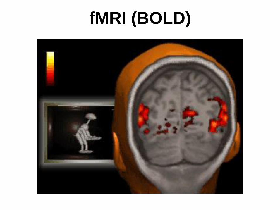
fMRI
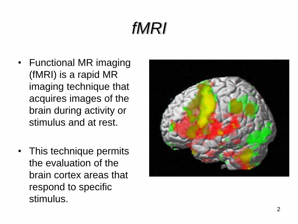
• Functional MR imaging
(fMRI) is a rapid MR
imaging technique that
acquires images of the
brain during activity or
stimulus and at rest.
• This technique permits
the evaluation of the
brain cortex areas that
respond to specific
stimulus.2
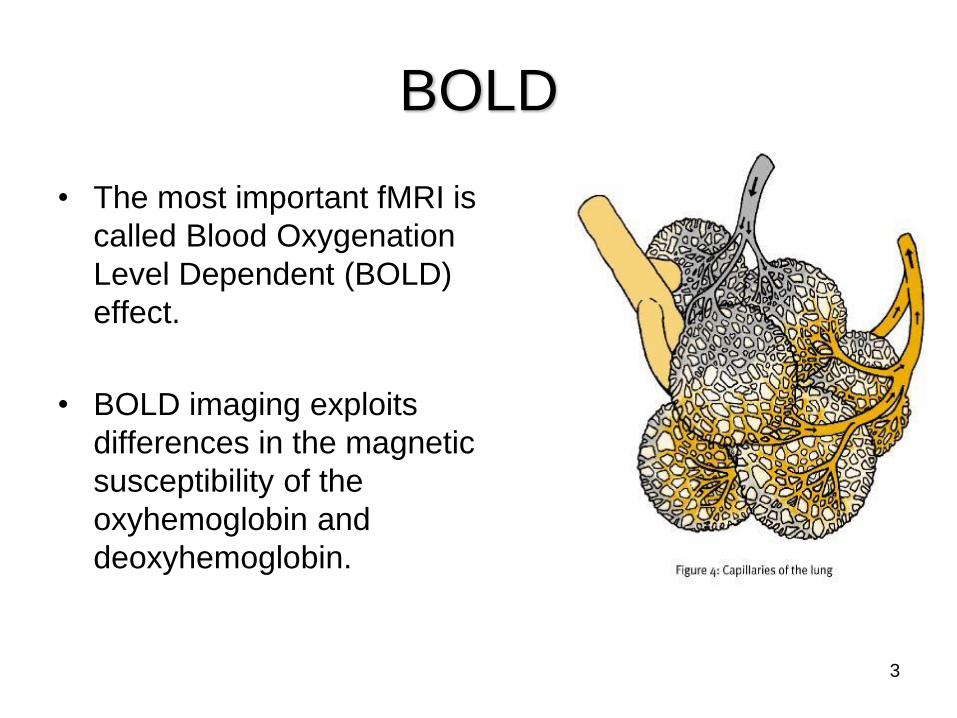
BOLD
• The most important fMRI is
called Blood Oxygenation
Level Dependent (BOLD)
effect.
• BOLD imaging exploits
differences in the magnetic
susceptibility of the
oxyhemoglobin and
deoxyhemoglobin.
3

BOLD
• Because deoxyhemoglobin is paramagnetic,
vessels containing a significant amount of this
molecule create local field inhomogeneities
causing dephasing and therefore signal loss.
Deoxyhemoglobin
4
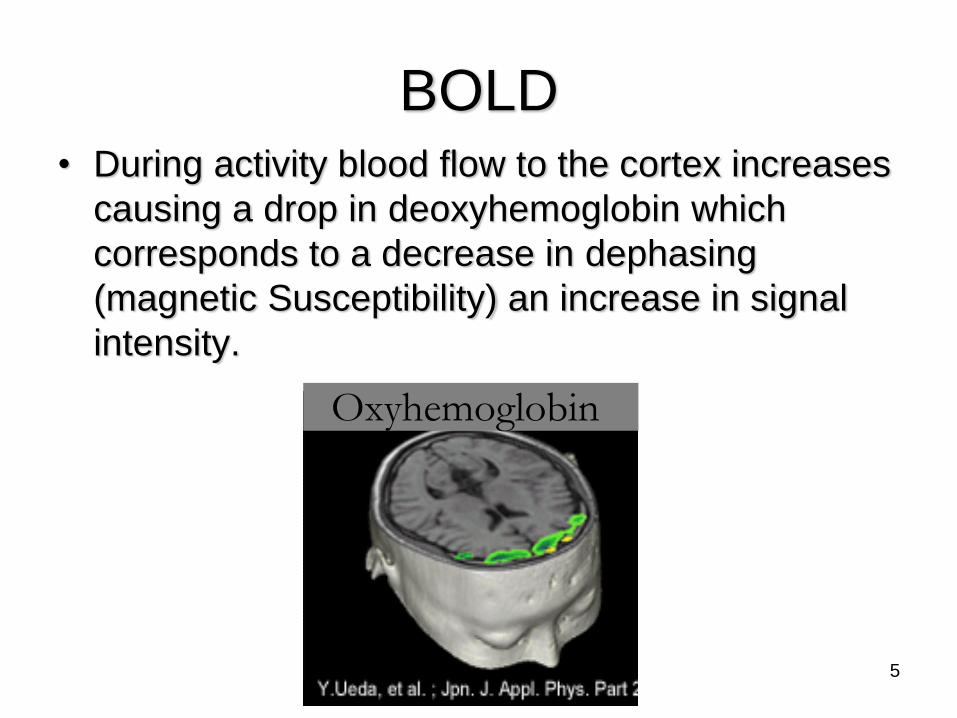
BOLD• During activity blood flow to the cortex increases
causing a drop in deoxyhemoglobin which
corresponds to a decrease in dephasing
(magnetic Susceptibility) an increase in signal
intensity.
Oxyhemoglobin
5
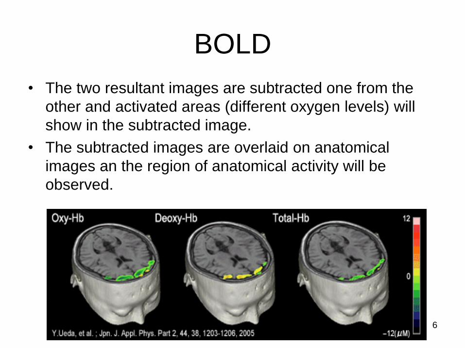
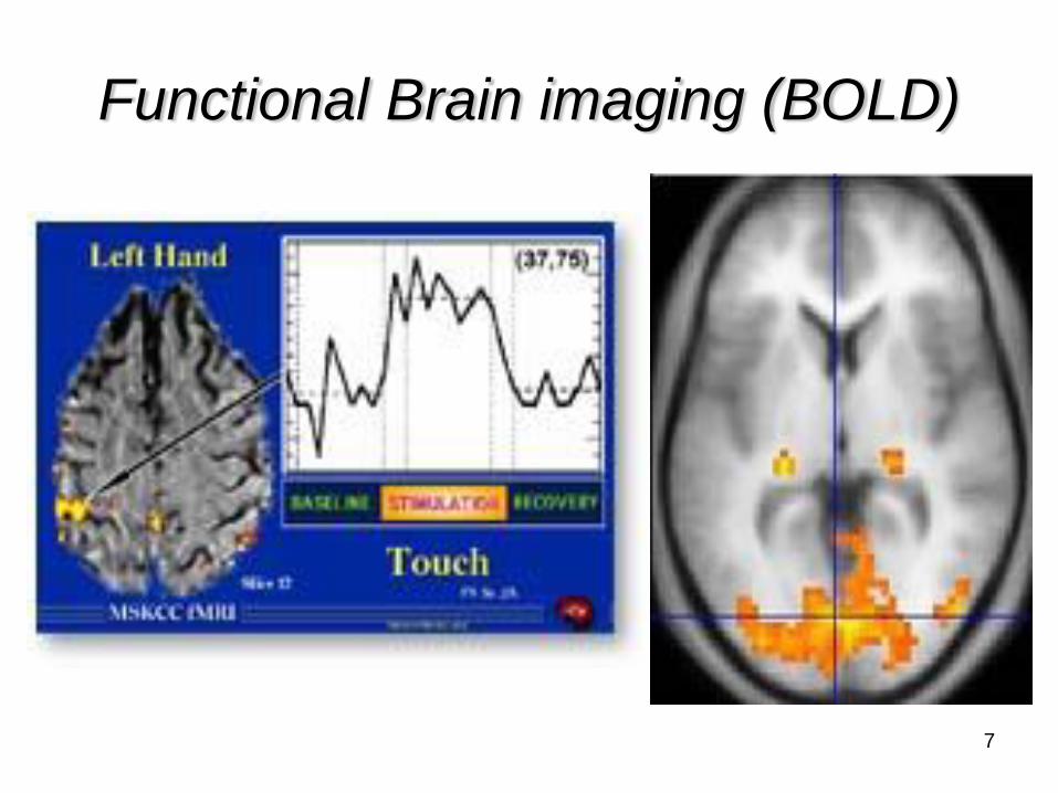
BOLD
• The two resultant images are subtracted one from the
other and activated areas (different oxygen levels) will
show in the subtracted image.
• The subtracted images are overlaid on anatomical
images an the region of anatomical activity will be
observed.
6
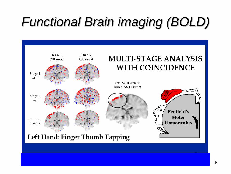
Functional Brain imaging (BOLD)
8
9
Clinical Applications of fMRI
• In the future these technique will develop our
understanding of brain function and will have
several clinical applications including the
evaluation of:
– Brain Surgery Planning
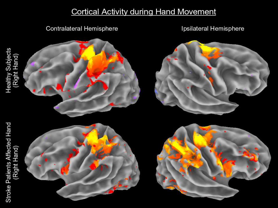
– Strokes
– Epilepsy
– Pain
– Behavioral problems
11
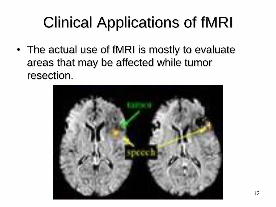
Clinical Applications of fMRI
• The actual use of fMRI is mostly to evaluate
areas that may be affected while tumor
resection.
12
13
14

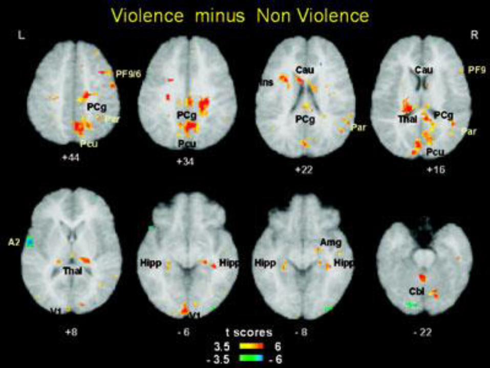
Behavioral Problems
• Some studies suggests that criminal
psychopaths show less activity than non-criminal
subjects in specific emotion-processing areas of
the brain.
15
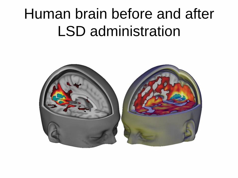
Human brain before and after
LSD administration

Therapeutic
• Interventional MRI
• Effective on the treatment of:
– Parkinson
– Depression
– OCD (obsessive compulsive disorder)
– Alzheimer’s
– Autism
• Surgery planning
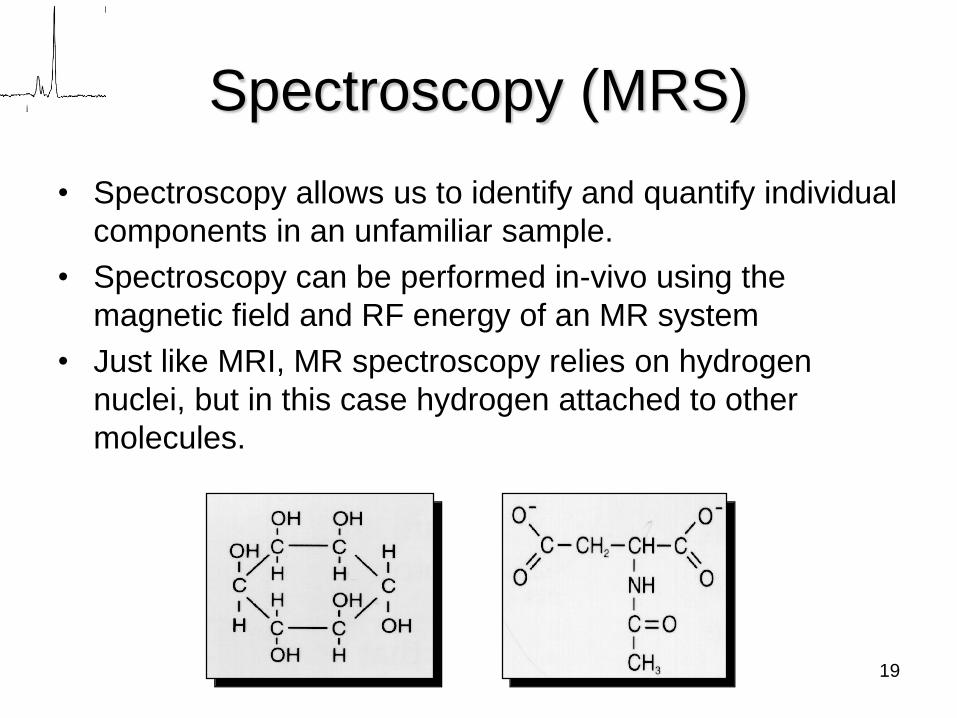
Spectroscopy (MRS)
• Spectroscopy allows us to identify and quantify individual
components in an unfamiliar sample.
• Spectroscopy can be performed in-vivo using the
magnetic field and RF energy of an MR system
• Just like MRI, MR spectroscopy relies on hydrogen
nuclei, but in this case hydrogen attached to other
molecules.
19
Spectroscopy
• The basic principle that enables MR
spectroscopy (MRS) is the electron cloud around
an atom that shields the nucleus from the
magnetic field to a greater or lesser degree.
• This naturally results in slightly resonant
frequencies, which in turn return a slightly
different signal.
20
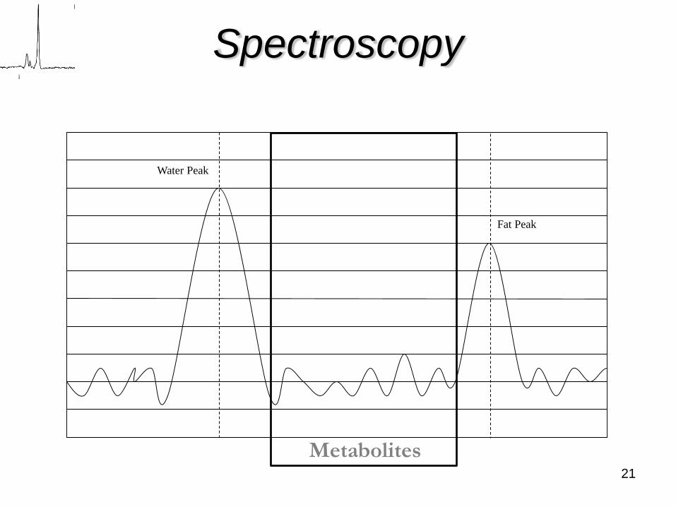
Spectroscopy
Water Peak
Fat Peak
Metabolites21
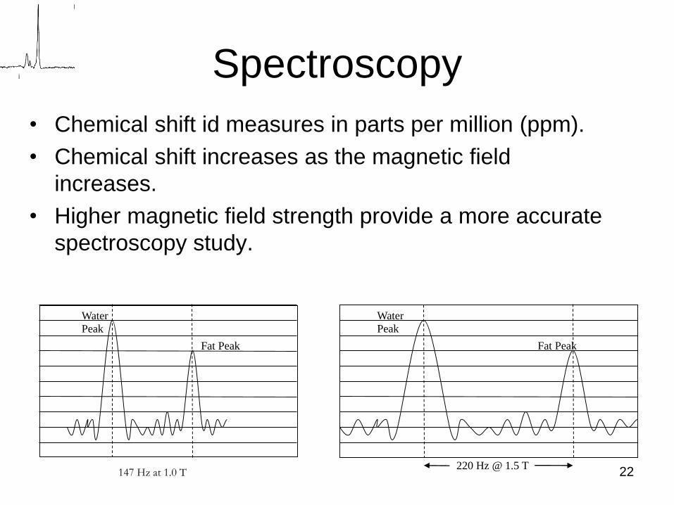
Spectroscopy
• Chemical shift id measures in parts per million (ppm).
• Chemical shift increases as the magnetic field
increases.
• Higher magnetic field strength provide a more accurate
spectroscopy study.
Water
Peak
Fat Peak
220 Hz @ 1.5 T
Water
Peak
Fat Peak
147 Hz at 1.0 T 22
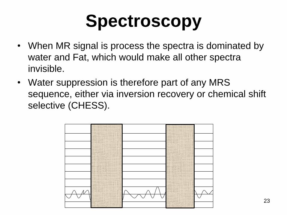
Spectroscopy
• When MR signal is process the spectra is dominated by
water and Fat, which would make all other spectra
invisible.
• Water suppression is therefore part of any MRS
sequence, either via inversion recovery or chemical shift
selective (CHESS).
Water
Peak
Fat
Peak
23
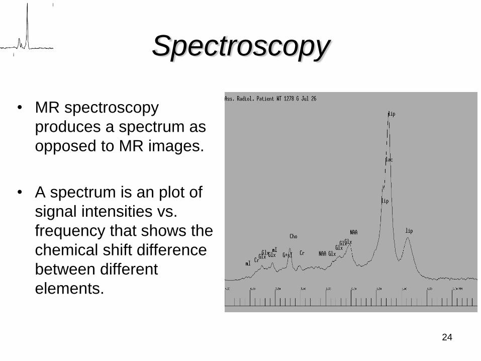
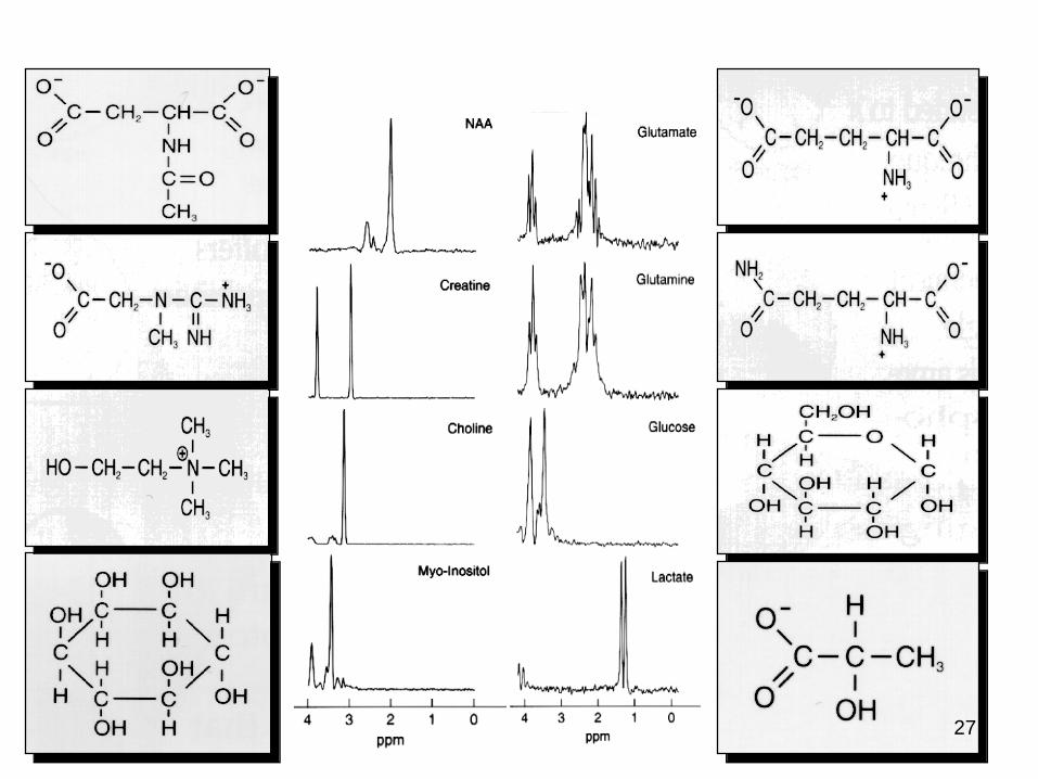
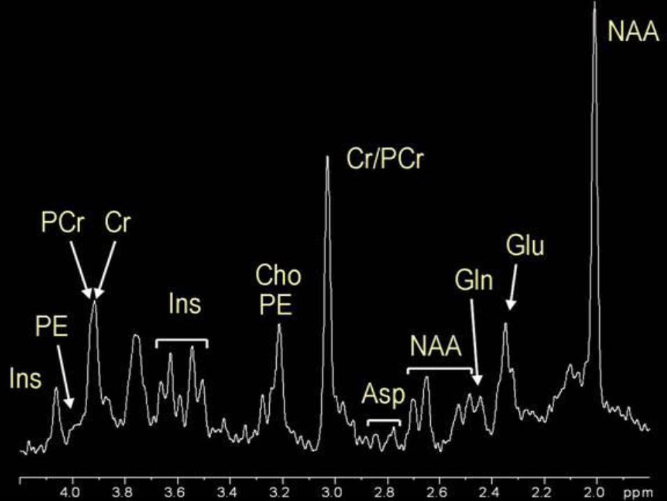
Spectroscopy
• MR spectroscopy
produces a spectrum as
opposed to MR images.
• A spectrum is an plot of
signal intensities vs.
frequency that shows the
chemical shift difference
between different
elements.
24
25
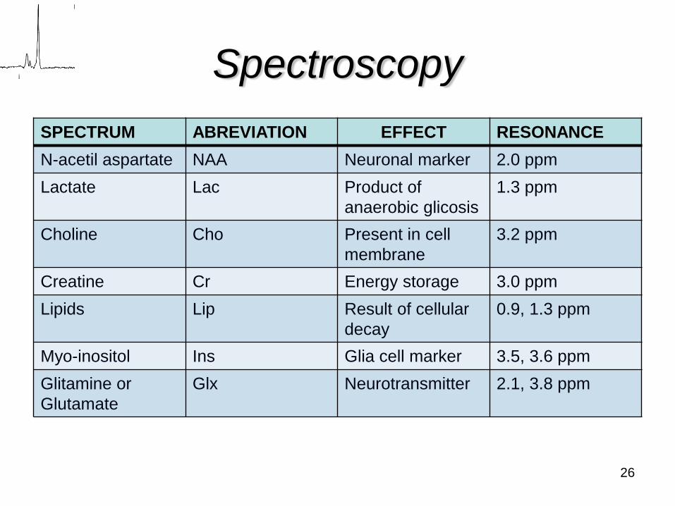
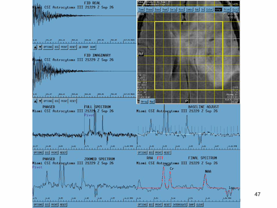
Spectroscopy
SPECTRUM ABREVIATION EFFECT RESONANCE
N-acetil aspartate NAA Neuronal marker 2.0 ppm
Lactate Lac Product of
anaerobic glicosis
1.3 ppm
Choline Cho Present in cell
membrane
3.2 ppm
Creatine Cr Energy storage 3.0 ppm
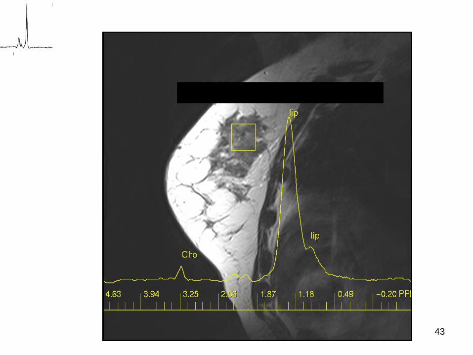
Lipids Lip Result of cellular
decay
0.9, 1.3 ppm
Myo-inositol Ins Glia cell marker 3.5, 3.6 ppm
Glitamine or
Glutamate
Glx Neurotransmitter 2.1, 3.8 ppm
26
27
28
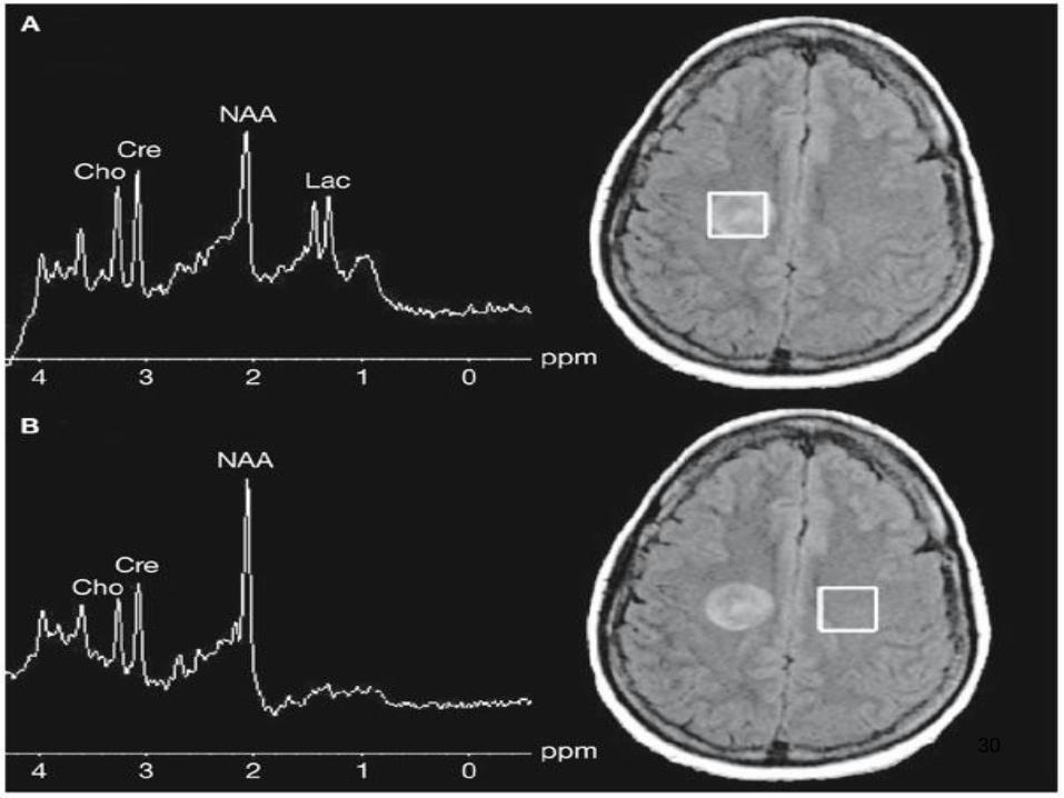
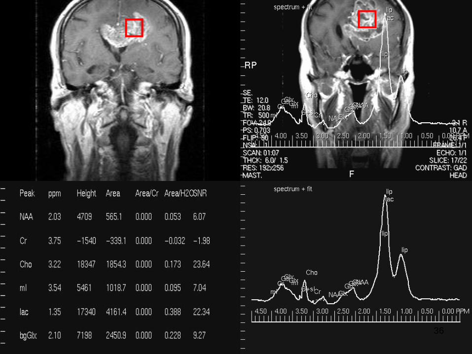
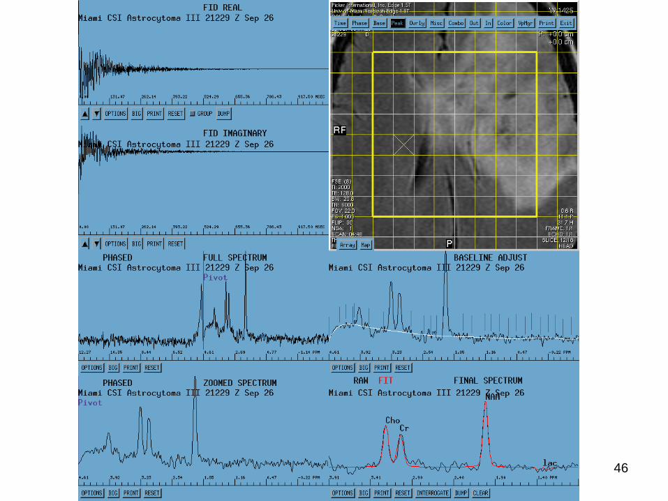
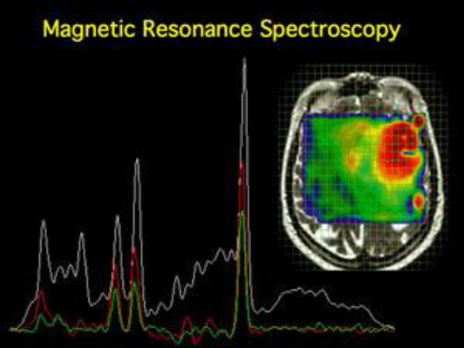
Spectroscopy
• Changes in the fallowing are indicators of tumors:
– NAA drop indicates tumor cell invasion
– Choline elevation indicates tumor growth
– Lactate changes indicates anaerobic status
– Lipid elevation indicates tumor necrosis
– Creatine mostly constant, it can be reduced in
high grade gliomas.
29
30
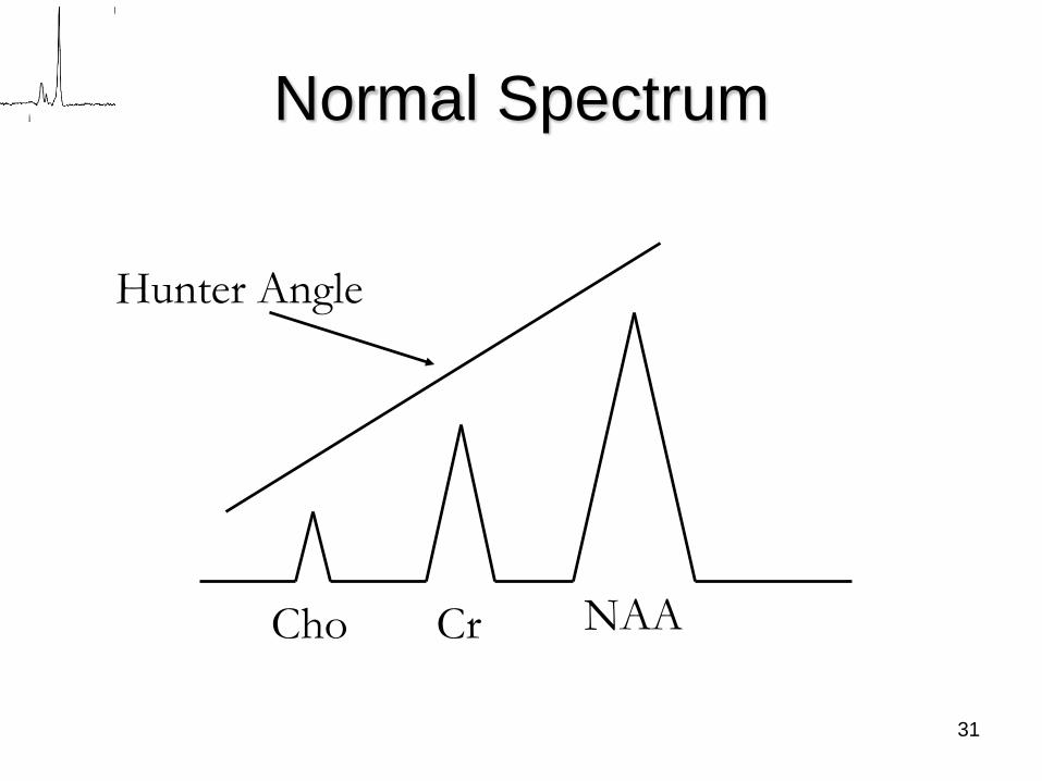
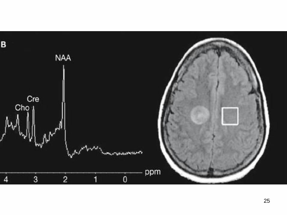
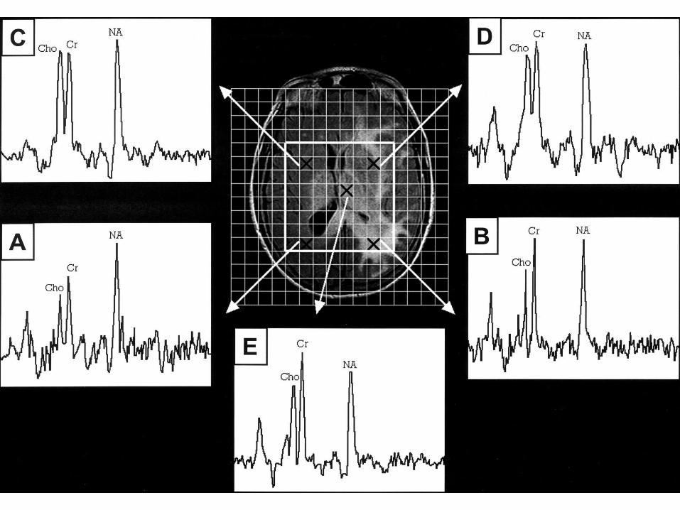
Normal Spectrum
Cho Cr NAA
Hunter Angle
31
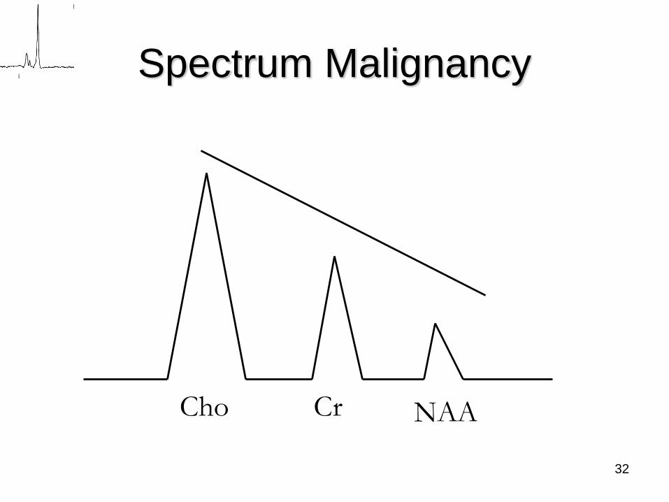
Spectrum Malignancy
Cho Cr NAA
32
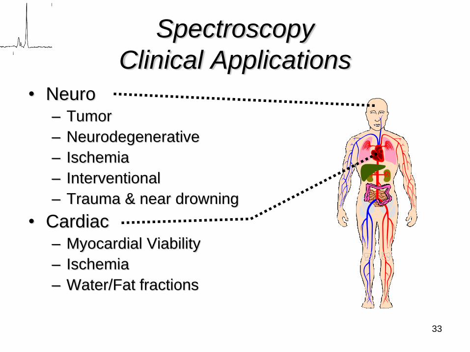
Spectroscopy
Clinical Applications
• Neuro– Tumor
– Neurodegenerative
– Ischemia
– Interventional
– Trauma & near drowning
• Cardiac– Myocardial Viability
– Ischemia
– Water/Fat fractions
33
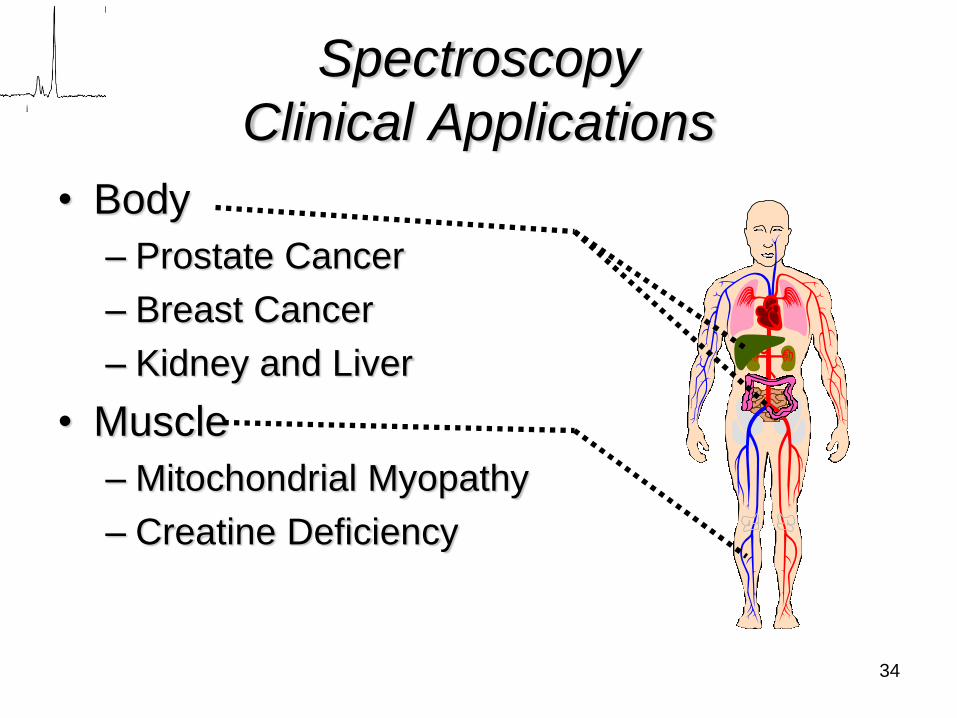
Spectroscopy
Clinical Applications
• Body
– Prostate Cancer
– Breast Cancer
– Kidney and Liver
• Muscle
– Mitochondrial Myopathy
– Creatine Deficiency
34
Spectroscopy
A spectrum is located in one or two ways:
• Single voxel:
– Stimulated Echo Acquisition Mode (STEAM)
(TE 20)
– Point Resolved Spectroscopy Spin Echo
(PRESS) (TE 135)
• Multivoxel (more efficient)35
36

37
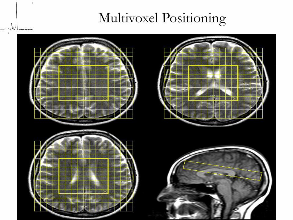
Multivoxel Positioning
38
39
MR Spectroscopy Uses
• MRS is used in the following ways:
– To diagnose in conjunction with MRI
– To plan for therapy
– Biopsy guidance
– To aid in prognosis
– Therapy monitoring
40
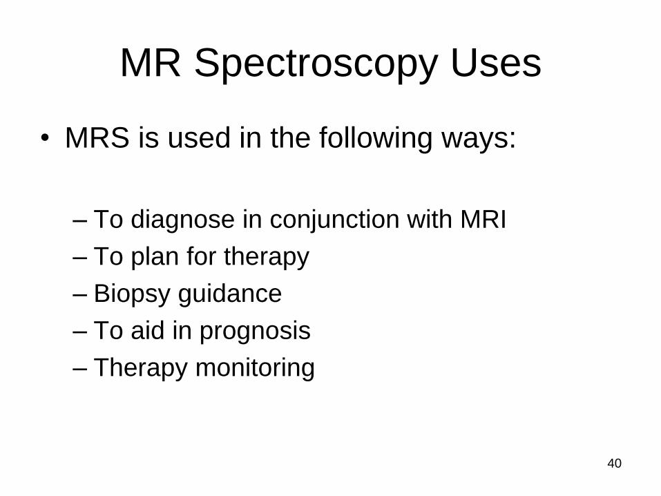
Neck Tumor
The choline peak is
obvious in this neck
lesion. Biopsy
proved it to be a
malignant papillary
cancer.
41
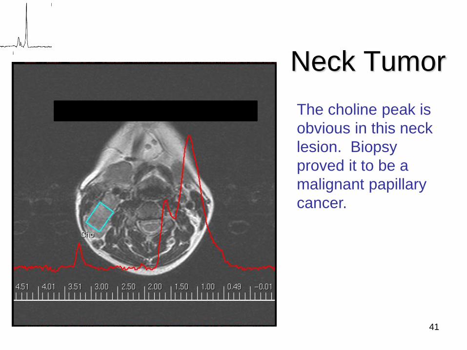
Bone Lesion (humeral head)
42
43
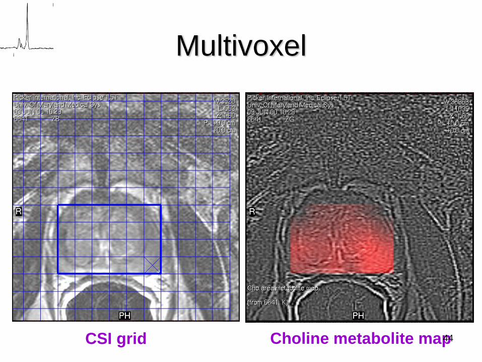
CSI grid Choline metabolite map
Multivoxel
44
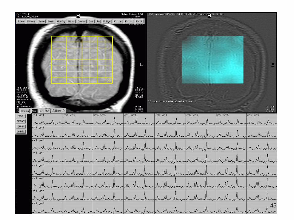
45
46
47
48
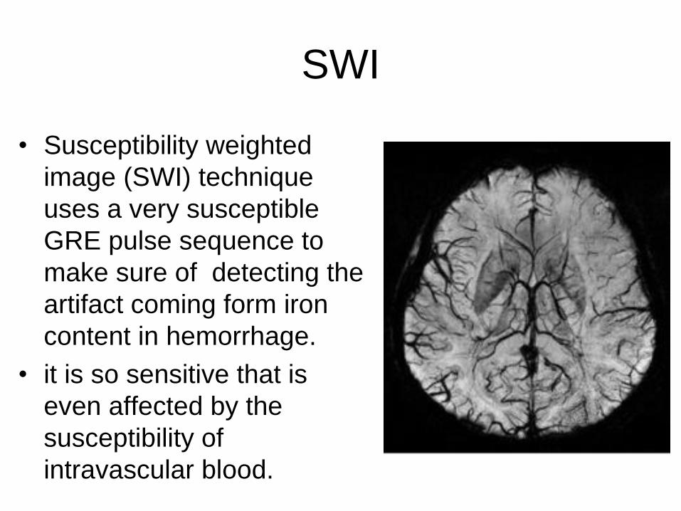
SWI
• Susceptibility weighted
image (SWI) technique
uses a very susceptible
GRE pulse sequence to
make sure of detecting the
artifact coming form iron
content in hemorrhage.
• it is so sensitive that is
even affected by the
susceptibility of
intravascular blood.
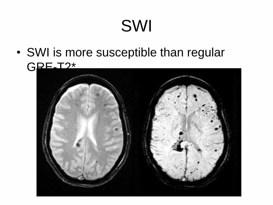
SWI
• SWI is more susceptible than regular
GRE-T2*.
SWI
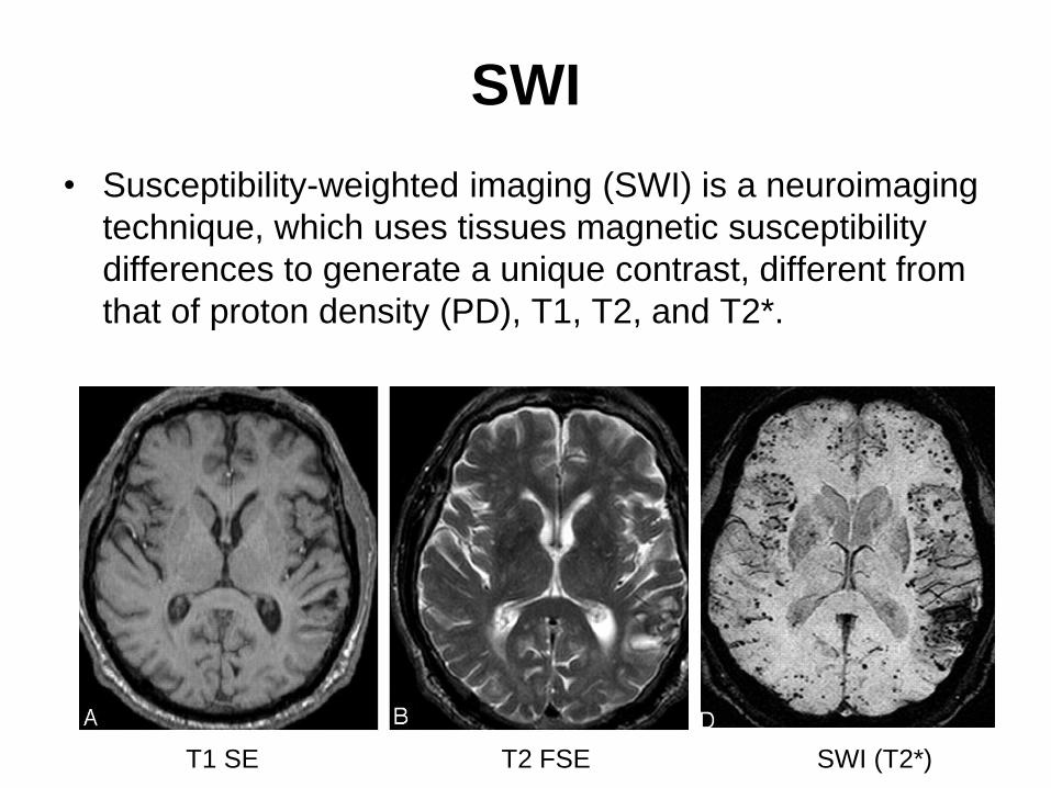
T1 SE T2 FSE SWI (T2*)
• Susceptibility-weighted imaging (SWI) is a neuroimaging
technique, which uses tissues magnetic susceptibility
differences to generate a unique contrast, different from
that of proton density (PD), T1, T2, and T2*.
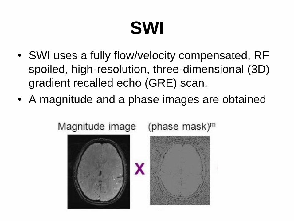
SWI
• SWI uses a fully flow/velocity compensated, RF
spoiled, high-resolution, three-dimensional (3D)
gradient recalled echo (GRE) scan.
• A magnitude and a phase images are obtained
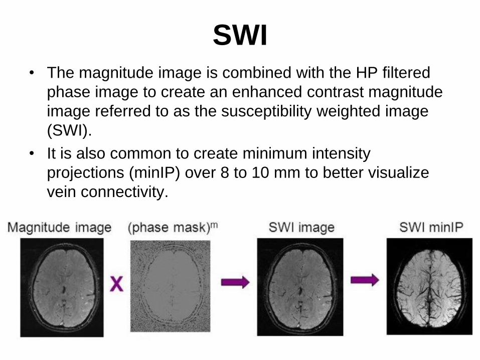
SWI• The magnitude image is combined with the HP filtered
phase image to create an enhanced contrast magnitude
image referred to as the susceptibility weighted image
(SWI).
• It is also common to create minimum intensity
projections (minIP) over 8 to 10 mm to better visualize
vein connectivity.
SWI
• SWI can be used better at higher field strengths.
– First of all, magnetic susceptibility increases
accordingly to the square of the magnetic field
strength.
– Moreover, the high signal-to-noise (SNR) ratio
available at higher magnetic fields allows
higher resolution scans.
– Finally, stronger magnetic fields allow shorter
echo times (TE) without a loss of contrast
which can reduce scan time and motion
related artifacts.
Clinical Applications• Improved detection of hemorrhage,
microbleeding (diffuse axonal injury) and
hemorrhagic transformation (stroke).
• Tumor characterization. Ability to detect tumor
vasculature and micro-hemorrhages.
• Detection of occult vascular disease
(cavernomas, angiomas, telangiectasias).
• Identification of iron and other mineral
deposition.
• Helpful in MR diagnosis of neurodegenerative
diseases (Alzheimer’s, multiple sclerosis, etc.)
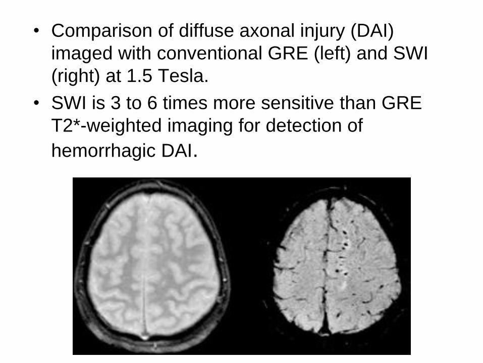
• Comparison of diffuse axonal injury (DAI)
imaged with conventional GRE (left) and SWI
(right) at 1.5 Tesla.
• SWI is 3 to 6 times more sensitive than GRE
T2*-weighted imaging for detection of
hemorrhagic DAI.
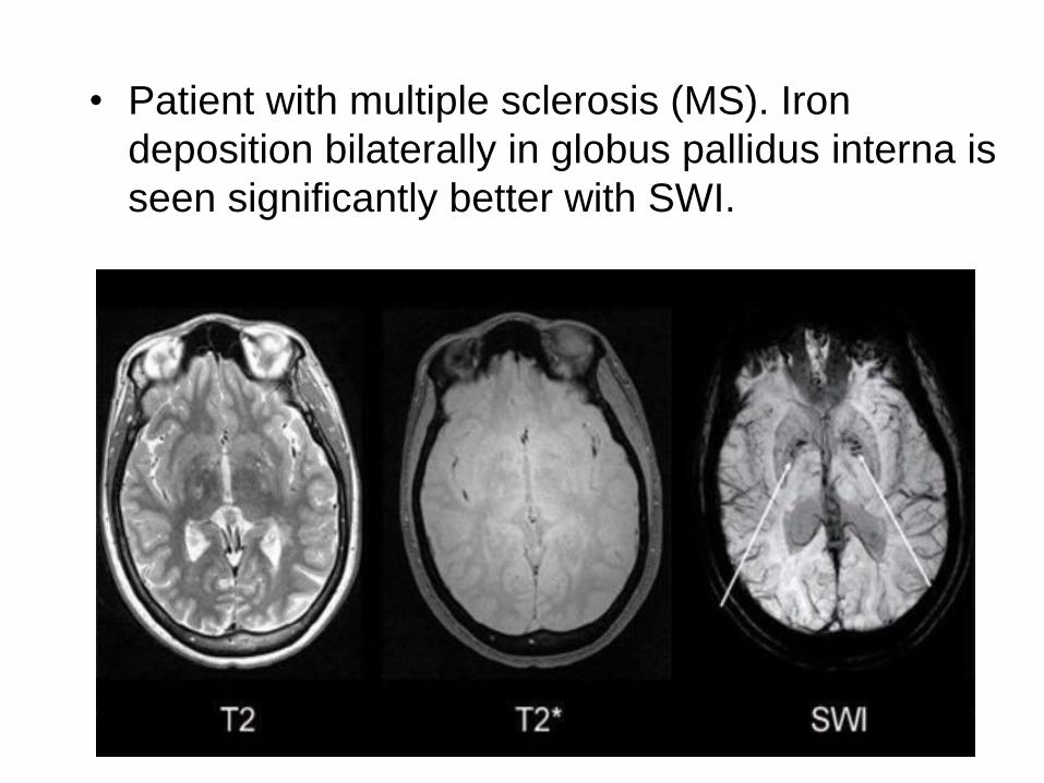
• Patient with multiple sclerosis (MS). Iron
deposition bilaterally in globus pallidus interna is
seen significantly better with SWI.
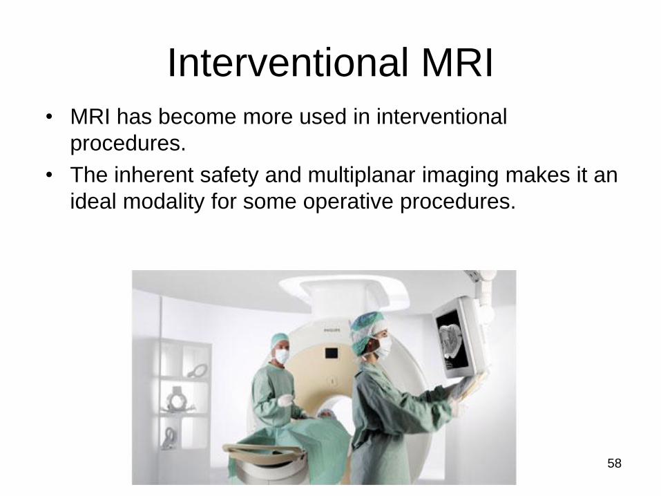
Interventional MRI
• MRI has become more used in interventional
procedures.
• The inherent safety and multiplanar imaging makes it an
ideal modality for some operative procedures.
58
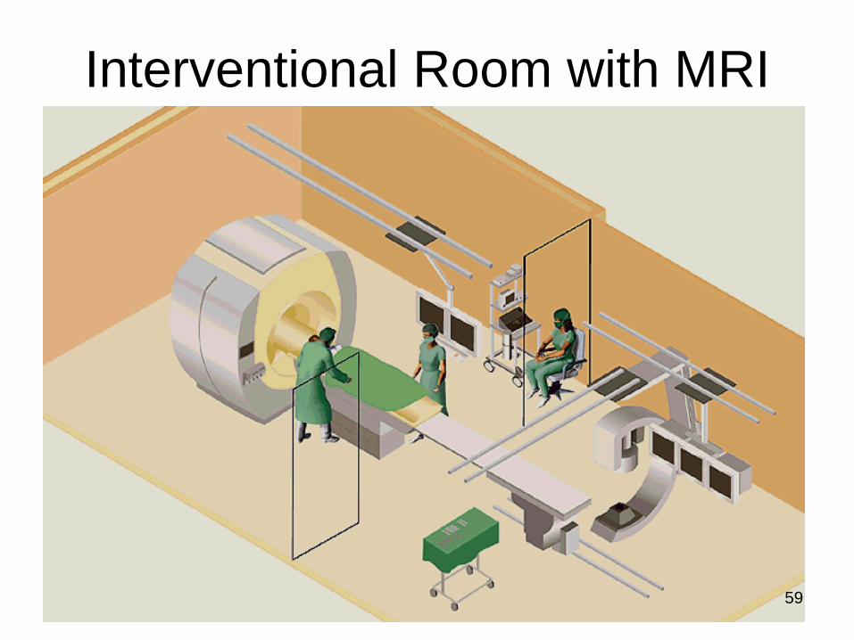
Interventional Room with MRI
59
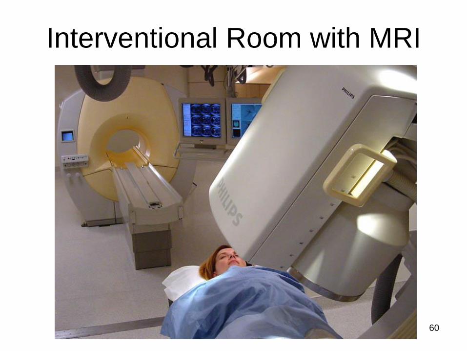
Interventional Room with MRI
60
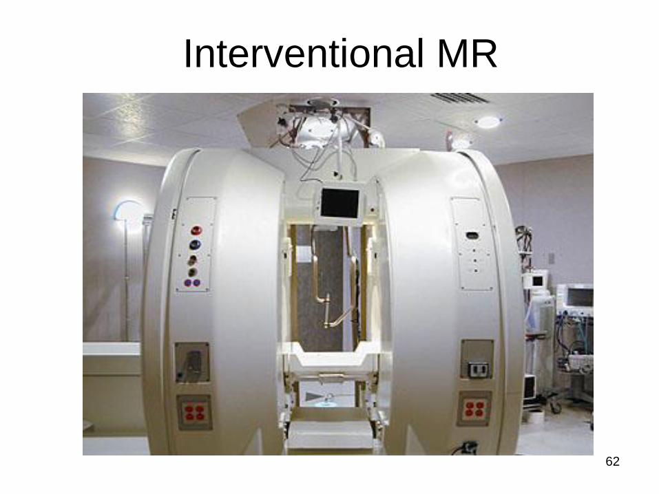
Interventional MRI
• The development of this technique has required several
modifications to existing hardware and software options.
• Due to the restricted nature of superconductive magnets
a more open design is required to permit easy access to
the patient.
• An interventional system uses a semi-conducting 0.5T
system shaped like two doughnuts which permits easy
access to the patient.
61
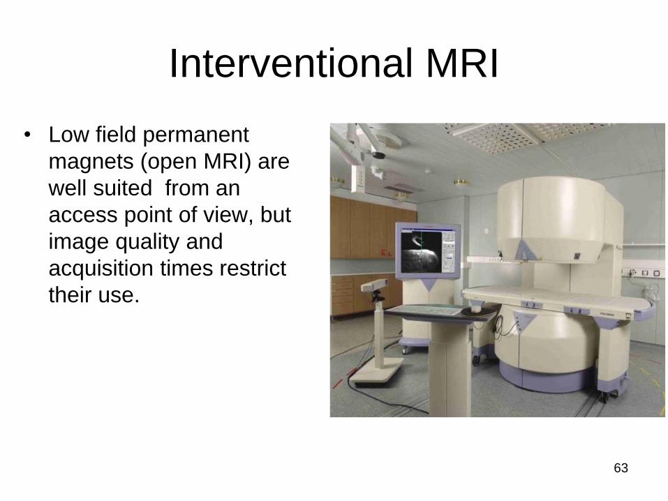
Interventional MRI
• Low field permanent
magnets (open MRI) are
well suited from an
access point of view, but
image quality and
acquisition times restrict
their use.
63

Interventional MRI
• This system permits:
– Intra-operative acquisition of MR images without moving the patient.
– Online images-guided stereotactic without pre-operative imaging.
– Real time tracking of instrument.
– Precise location of the area under examination.
– Monitor the procedure in 3D
64
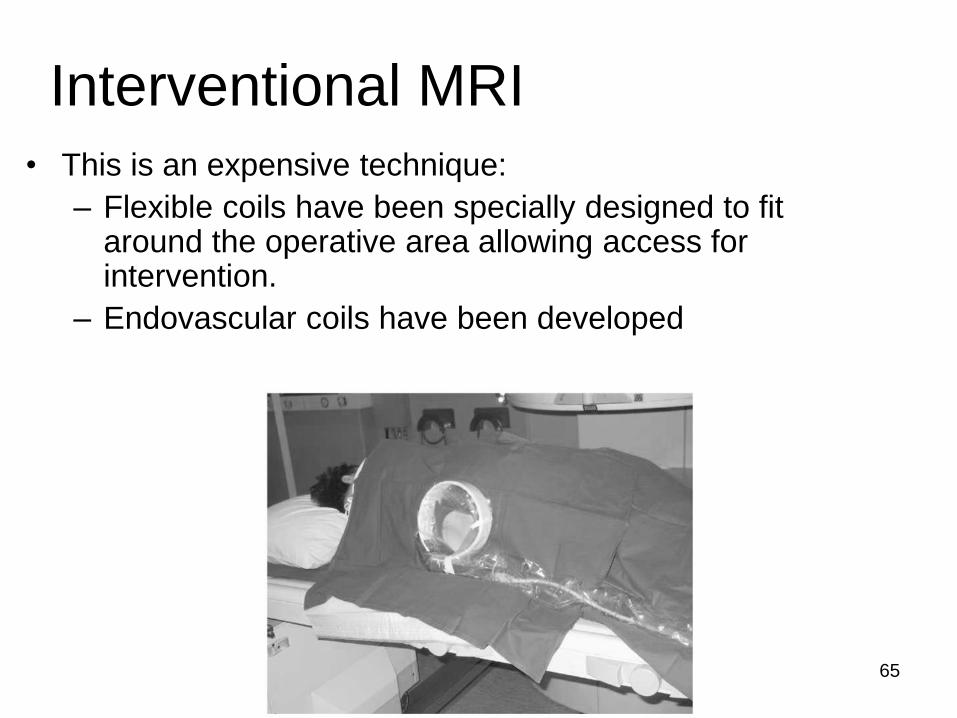
Interventional MRI
• This is an expensive technique:
– Flexible coils have been specially designed to fit around the operative area allowing access for intervention.
– Endovascular coils have been developed
65
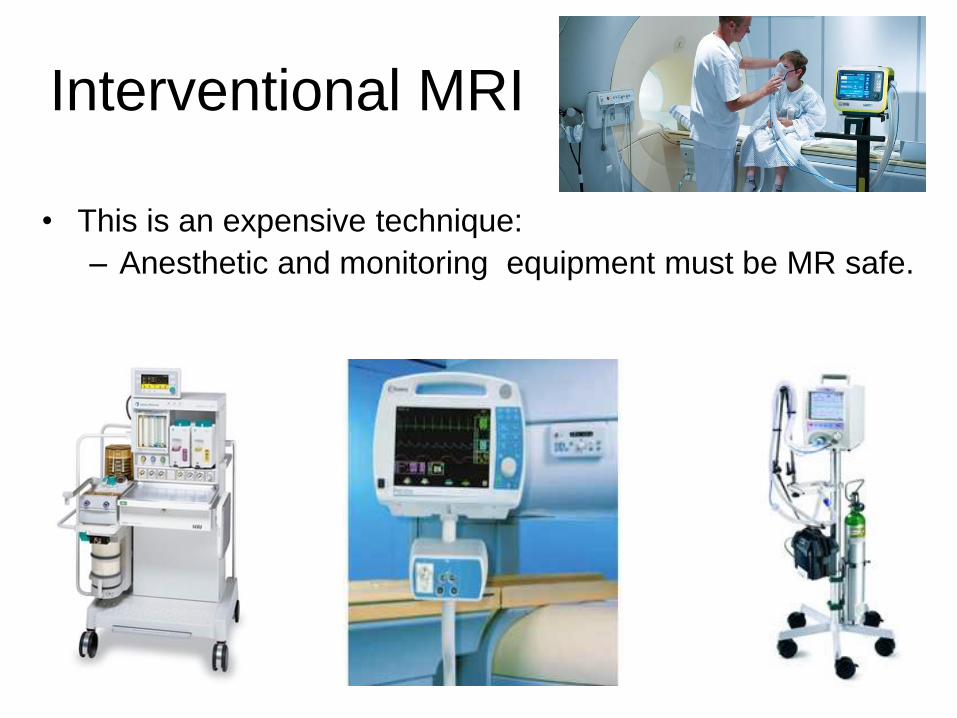
Interventional MRI
• This is an expensive technique:
– Anesthetic and monitoring equipment must be MR safe.
66
67
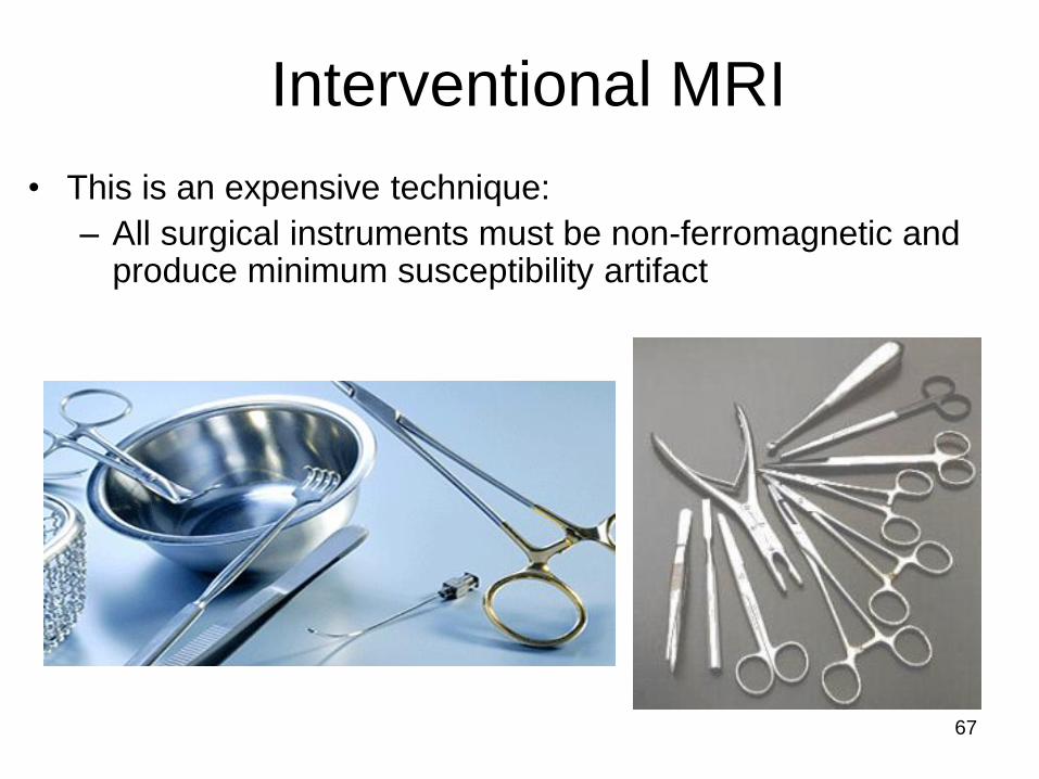
• This is an expensive technique:
– All surgical instruments must be non-ferromagnetic and produce minimum susceptibility artifact
Interventional MRI
Interventional MRI Uses
• Some of the most important uses of interventional MRI are:
– Liver imaging and tumor oblation
– Breast imaging and benign lump excision
– Orthopedic and kinematic studies
– Biopsies
– functional endoscopic sinus surgery
68
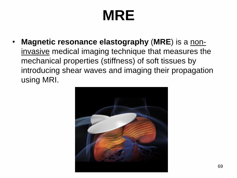
MRE
• Magnetic resonance elastography (MRE) is a non-
invasive medical imaging technique that measures the
mechanical properties (stiffness) of soft tissues by
introducing shear waves and imaging their propagation
using MRI.
69

MRE
• Pathological tissues are often stiffer than the
surrounding normal tissue.
• For instance, malignant breast tumors are much harder
than healthy fibro-glandular tissue.
70
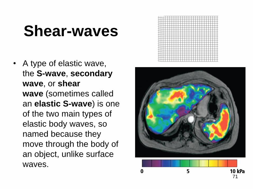
Shear-waves
• A type of elastic wave,
the S-wave, secondary
wave, or shear
wave (sometimes called
an elastic S-wave) is one
of the two main types of
elastic body waves, so
named because they
move through the body of
an object, unlike surface
waves.
71
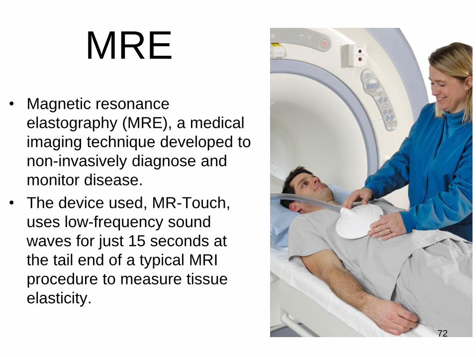
MRE
• Magnetic resonance
elastography (MRE), a medical
imaging technique developed to
non-invasively diagnose and
monitor disease.
• The device used, MR-Touch,
uses low-frequency sound
waves for just 15 seconds at
the tail end of a typical MRI
procedure to measure tissue
elasticity.
72
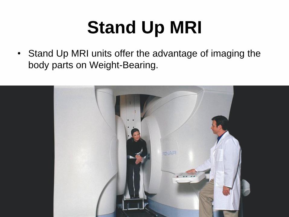
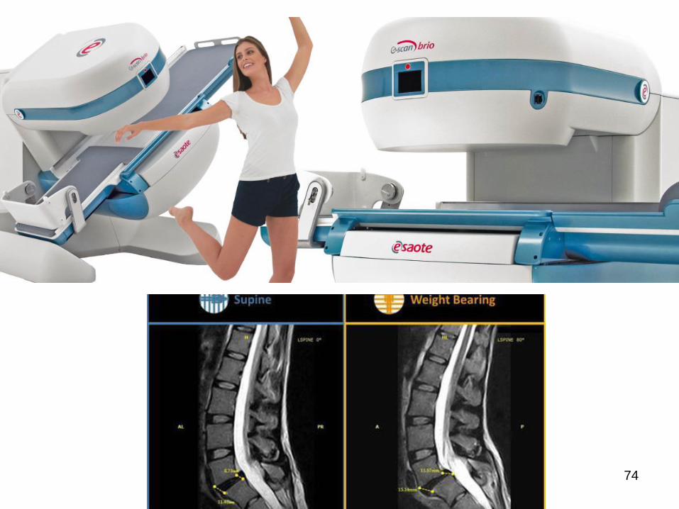
Stand Up MRI
• Stand Up MRI units offer the advantage of imaging the
body parts on Weight-Bearing.
73
74
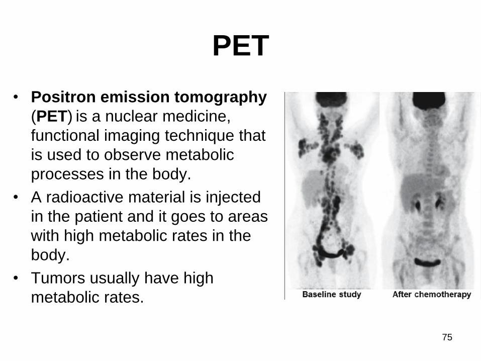
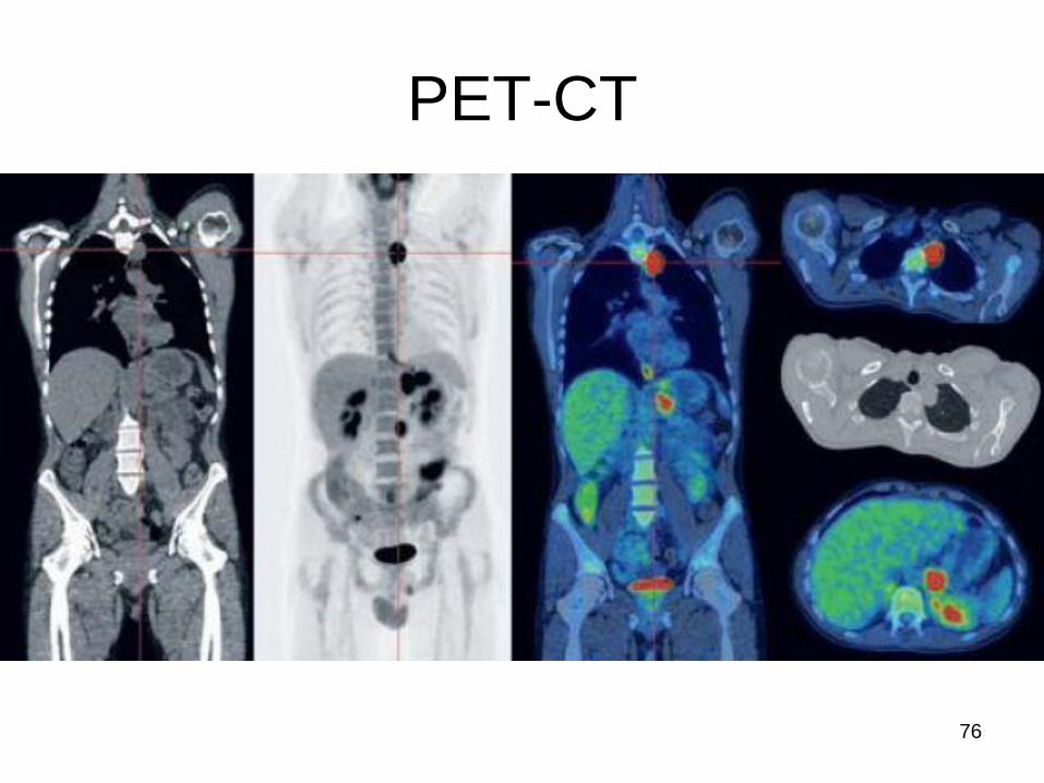
PET
• Positron emission tomography
(PET) is a nuclear medicine,
functional imaging technique that
is used to observe metabolic
processes in the body.
• A radioactive material is injected
in the patient and it goes to areas
with high metabolic rates in the
body.
• Tumors usually have high
metabolic rates.
75
77