Embed Size (px)
Citation preview

978
MR Imaging Characteristics of a Neurenteric Cyst Glen K. Geremia,1.2 Eric J. Russell,1 and Raymond A. Clasen3
MR studies are correlated with histologic findings in a case of intradural extramedullary neurenteric cyst. No associated vertebral anomalies were present, and the origin of the cyst could be suggested but not confirmed before histologic examination.
Case Report
A 34-year-old man presented with a 2-year history of progressive bilateral leg weakness , paresthesias, and gait incoordination. Of later onset was nocturnal enuresis. On admission to the medical center, a cystometrogram revealed spastic neurogenic bladder. Physical examination demonstrated symmetrical patchy areas of decreased pinprick sensation below the T4 level. Four beats of clonus were elicited at the right ankle, and sustained clonus was noted on the left. There was a left Babinski sign , and the gait was broad and awkward.
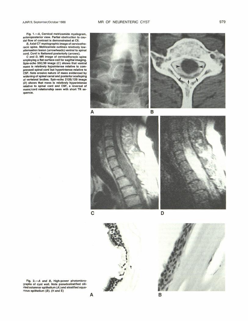
Metrizamide myelography performed via lateral cervical puncture revealed a ventral intradural extramedullary mass extending from the C6 to the T2 level (Fig. 1 A). The cervical spinal cord was displaced posteriorly and severely flattened . There was an incomplete block to the caudal flow of contrast at T2 . CT myelography of the cervicothoracic spine better defined the fusiform , hypodense, extramedullary soft-tissue mass (Fig. 1 B). MR imaging of the cervicothoracic spine was performed by using a 0.5-T superconductive imager' with a bore size of 100 cm. A flat-surface receiving coil was placed over the region of interest and spin-echo images were acquired at 530/30 (TR/TE) (Fig . 1 C) and at 2120/60,120 (Fig. 10). These revealed the lesion to be hypointense relative to spinal cord but slightly brighter than CSF on 530 T1-weighted images, and hyperintense relative to spinal cord and CSF on both 60 and 120 images at TR = 2120. The lesion was seen clearly to lie ventral to and separate from the cord.
Surgery was performed via anterior approach. The C6-C7 and C7 - T1 intervertebral disks along with the C7 vertebral body were removed. The dura was identified and incised longitudinally in the midline. Immediately, a large cystic mass deep to the dura and anterior to the spinal cord bulged forward. The cyst was punctured with a 25-gauge needle, but attempts to aspirate the gelatinous fluid were unsuccessful. The cyst wall was then widely opened and a mucoidtype material exuded forth, resulting in rapid collapse of the cyst wall. The mid-posterior wall of the cyst was found to be attached to the ventral pia of the spinal cord. The exposed cord was flattened . The cyst wall was removed in its entirety and sent for pathologic study. Biochemical analysis was not performed on the mucoid aspirate .
• Teslacon, Technicare Corp., Solon, OH .
Received August 8 , 1986; accepted after revision March 7, 1987.
Histologic sections of the cyst wall are shown in Figures 2A and 2B. The epithelial lining consisted predominately of pseudostriatified ciliated columnar epithelium and simple columnar epithelium with basal nuclei and slightly basophilic cytoplasm. In addition, a small portion of nonkeratinizing stratified squamous epithelium was found at the site of pial attachment. The pseudostriatified ciliated epithelium clearly resembled the lining of the respiratory tract and the simple columnar epithelium resembled the lining of the gastrointestinal tract. On the basis of these histologic features the final pathologic diagnosis was neurenteric cyst.
Discussion
Intraspinal enteric cyst may occur as a solitary lesion, although it should be considered as part of the spectrum of developmental disorders referred to as the split notochord syndrome [1]. The notochord is abnormally split by a perSistent connection between gut and dorsal skin in the embryo, and the cyst originates during the first three weeks of fetal life [2]. A variety of malformations may also result. Dorsal enteric fistula (enteriC patent communication between the intestinal cavity and dorsal skin) is the most severe form [3, 4]. The fistula may course through prevertebral tissue, vertebral bodies, the spinal canal, and finally the dorsal skin surface [5]. Any segment of the fistulous tract may perSist, leaving isolated diverticula, duplications, cysts, fibrous cords, and/or sinuses at any point along the segmental tract [5]. The tract may involve the viscera of the chest or abdomen, and incorporate abnormal tissue remnants of enteric derivation.
In a recent review of 31 neurenteric cysts [6], four of 31 contained pseudostratified columnar epithelium with or without cilia, and 10 of 31 contained simple ciliated epithelium. The remainder showed simple columnar epithelium, cuboidal epithelium, and goblet cells . An additional reported case showed a mixture of ciliated epithelium, goblet cells, and squamous epithelium [7] . The three different types of epithelium were confirmed by electron microscopy. The mixed epithelial pattern seen in our case indicates a foregut origin of the cyst.
The clinical symptoms associated with enterogenous cysts depend on the site of the lesion. In about one-half of the
, Department of Radiology, Rush-Presbyterian-St. Luke Medical Center, 1753 w. Congress Pkwy., Chicago, IL 60612 . Address reprint requests to G. Geremia. 2 Department of Radiology, University of Chicago Hospitals, 5841 S. Maryland Ave. , Chicago, IL 60637. 3 Department of Pathology, Rush-Presbyterian-St. Luke Medical Center, 1753 w. Congress Pkwy. , Chicago, IL 60612 .
AJNR 9:978-980, September/October 1988 0195-6108/88/0905-0978 © American Society of Neuroradiology
AJNR:9. September/October 1988
Fig. 1.-A, Cervical metrizamide myelogram, anteroposterior view. Partial obstruction to caudal flow of contrast is demonstrated at CS.
B, Axial CT myelographic image of cervicothoracic spine. Metrizamide outlines relatively lowattenuation lesion (arrowheads) ventral to spinal cord. Cord is flattened posteriorly (arrows) .
C and 0, MR image of cervicothoracic spine employing a flat surface coil for sagittal imaging. Spin-echo 350/30 image (C) shows that ventral mass is relatively hypo intense relative to compressed spinal cord but hyperintense relative to CSF. Note erosive nature of mass evidenced by widening of spinal canal and posterior scalloping of vertebral bodies. Spin-echo 2120/120 image (0) shows that mass is relatively hyperintense relative to spinal cord and CSF, a reversal of mass/cord relationship seen with short TR sequence.
Fig. 2.-A and B, High-power photomicrographs of cyst wall. Note pseudostratified ciliated columnar epithelium (A) and stratified squamous epithelium (B). (H and E)
A
c
A
MR OF NEURENTERIC CYST 979
B
o
, • • I
.. ' •
B

980 GEREMIA ET AL. AJNR:9 , September/October 1988
cases, the progression of symptoms is episodic, so that it may be difficult to distinguish this disorder from multiple sclerosis [8]. Males predominate over females (3:2) and the usual age distribution is 20-40 years old with infrequent presentation in infancy and in the newborn period. The cysts usually extend over three vertebral segments and are most common in the cervical region. Their occurrence in association with spina bifida is well known [8, 9] . Spina bifid a or vertebral fusion is reportedly seen in about half the cases. Secondary changes such as widening of the spinal canal are uncommon. Myelography will suggest an intradural space-occupying lesion but is not histologically diagnostic. In our case, metrizamide myelography and CT were also nonspecific and revealed an intradural extramedullary lesion flattening the cord. The MR pattern in our case of neurenteric cyst was more indicative of the cystic nature of the lesion than was myelography or CT myelography. Several recent reports describe the MR features of cystic lesions in the kidney and brain [10-13]. We are aware of only one report of MR findings in intraspinal neurenteric cyst [14]. In that case the presence of associated spinal anomalies led to a correct preoperative diagnosis. The cyst contained gastric mucosa and appeared to be intramedullary on MR studies and at surgery, although attachment to the tract through an anomalous vertebra suggested an extramedullary origin. In our case, no such anomaly or connection was present, and a secure diagnosis of cyst origin was not possible before histologic study.
The intensity pattern of cystic lesions on MR varies with the nature (particularly the protein content) of the intracavitary fluid . Arachnoid cysts contain CSF, as do most postoperative cysts, and therefore are similar to CSF intensity on all MR pulse sequences [15]. Hemorrhagic cysts and colloid cysts exhibit significantly higher intensity than CSF on both long and short TR spin-echo sequences [11]. Proteinaceous nonhemorrhagic cysts (tumor, inflammatory) have an intermediate range of intensity, which is similar to or slightly higher than CSF on short TR spin-echo sequences and higher than CSF on long TR sequences. The MR appearance of our case of enteric cyst suggests high protein content without hemorrhage (lower intensity than spinal cord, higher intensity than CSF on short TR sequences). Another case of spinal neurenteric cyst reported by Levin and Antin [1 6] revealed high protein content. The protein shortens both T1 and T2 relaxation times of cystic fluid. T1 shortening causes the lesion to be of higher intenSity than CSF on short TR images, and this T1 effect predominates even on relatively long TR (T2-weighted) sequences such as 2000/60 used in our case, due to the extremely long T1 of CSF (> 1200 msec). The T1 effect overcomes the T2 effect, which explains the relatively bright intensity of the lesion despite T2 shortening. While a 2000/ 60 sequence is reasonably T2-weighted for solids, it is T1-weighted for fluids [13] .
Despite T1 and T2 shortening by protein, both relaxation times are still relatively long relative to the solid spinal cord.
Therefore, the cyst is less intense than cord with a short TR sequence, and brighter than cord at 2000/60 and 2000/120 sequences.
The intensity characteristics would also be dependent on whether the cystic contents of the mass were pulsatile or nonpulsatile. This is exemplified by the CSF flow-void phenomenon seen in the brain and spinal canal , whereby the CSF pulsations decreased signal intensity. This is due to spindephasing and time-of-flight effects caused by the pulsatile CSF motion [17] . Since the contents of this cyst were gelatinous and not free-flowing, these factors should not have contributed to its signal intensity appearance.
Although our main diagnostic considerations were neurofibroma/schwannoma after both CT myelography and MR, we considered proteinaceous cyst only after reviewing the MR images.
REFERENCES
1. Burrows FGO, Sutcliffe J. The split notochord syndrome. Br J Radiol 1968;41 :844-847
2. Gimeno A, Lopez F, Figuera D, Rodrigo L. Neuroenteric cysts. Neuroradiology 1972;3:167-172
3. Bentley JFR, Smith JR. Developmental posterior enteric remnants and spinal malformations: the split notochord syndrome. Arch Dis Child 1960;35:76-86
4. Smith JR. Accessory enteric formations: a classification and nomenclature. Arch Dis Child 1960;35:87-89
5. Naidich TP, McLone DG, Harwood-Nash DC. Spinal dysraphism. In: Newton III, Potts DG, eds. Computed tomography of the spine and the spinal cord. San Anselmo, CA: Raven Press, 1983:299-353.
6. Lerma S, Roda JM, Villarejo F, Perez-Higueras A, Gutierrez-Moina M, Blazques MG. Intradural neurenteric cyst: review and discussion. Neurochirurgie 1985;28:228-231
7. Matsushima T, Fukui M, Egami H. Epithelial cells in a so-called intraspinal neurenteric cyst: a light and electron microscopic study. Surg Neuro 1985;24:656-660.
8. Angolo ALA, Albrecht L, Schonmayr R. Enterogenous intraspinal cyst. J
Neurosurg 1984;61 :834-840 9. Odake G, Yamaki T, Naruse S. Neurenteric cyst with meningomyelocele
Case report. J Neurosurg 1976;45:352-356 10. Choyke PL, Kressel HY, Pollack HM, Arger PM, Axel L, Mamourian AC
Focal renal masses: magnetic resonance imaging. Radiology 1984 152:471-477
11 . Hricak H, Crooks L, Sheldon P, Kaufman L. Nuclear magnetic resonanCE imaging of the kidney. Radiology 1983;146:425-432
12. Hricak H, Williams RD, Moon KL, et al. Nuclear magnetic resonanCE imaging of the kidney: renal masses. Radiology 1983;147:765-772
13. Kjos BO, Brant-Zawadzki M, Kucharczyk W, Kelly WM , Norton D, Newtor TH . Cystic intracranial lesions: magnetic resonance imaging. Radiology 1985;155:363-369
14. Kantrowitz LR , Pais MJ, Burnett K, Choi B, Pritz MB. Intraspinal neurenteric cyst containing gastric mucosa: CT and MRI findings . Pediatr Radio 1986;16:324-327
15. Heier LA, Zimmerman RD, Russell EJ, et al. MR imaging of arachnoic cysts. Presented at the 71 st annual meeting of the RSNA, Chicago November 1985
16. Levin P, Antin SP. Intraspinal neurenteric cyst in the cervical area. Neurol ogy 1964;14:727-730
17. Sherman J, Citrin C, Gangarosa R, Bowen B. The MR appearance of CSF pulsation in the spinal canal. AJNR 1986;7:879-884