Embed Size (px)
Citation preview
MRI interpretation of the foot
Waraporn S. Boonsaeng, M.D.
Department of Radiology, Thammasat University

Outlines
¤ Technique
¤ Forefoot
¤ First MTP joint
¤ Lesser MTP & metatarsalgia
¤ Plantar plate injury
¤ Intermetatarsal space (morton neuroma, intermetatarsalbursitis)
¤ Pressure lesions: soft tissue callus, adventitial bursitis
¤ Midfoot
¤ Lisfranc ligament complex
¤ Osseous structures: BME

Standard MRI sequences
¤ Often focusing on a specific portion of the foot:
ankle/hindfoot, midfoot, or forefoot
¤ Sagittal, short axis (coronal ankle) and long axis (axial
ankle) planes relative to metatarsals
¤ Sagittal and short axis images: plantar plates, sesamoid
bones and flexor and extensor tendons; intermetatarsal
structures (short axis)
¤ Long axis images: collateral ligaments, Lisfranc ligament
complex

Arnold G, et al. Magn Reson Imaging Clin N Am 19 (2011) 655–679

From approximately the mid subtalar joint to the proximal metatarsals
Coronal, parallel to the bottom of the foot; Axial, transverse to the longitudinal arch of the foot.
Sofka CM. Magn Reson Imaging Clin N Am 25 (2017) 1–10
Suggested protocol for imaging the midfoot

From approximately the naviculocuneiform articulation through the toes.
Coronal, parallel to the bottom of the foot; Axial, Transverse to the longitudinal arch of the foot.
Sofka CM. Magn Reson Imaging Clin N Am 25 (2017) 1–10
Suggested protocol for imaging the forefoot

Lesser MTP and Metatarsalgia
•Plantar plate
•Morton neuroma
•Intermetatarsal bursitis
•Soft tissue callus
•Adventitial bursitis

Plantar plate
¤ Primary stabilizer of the lesser MTP joints, especially in the
dorsal–plantar direction
¤ Articulates directly with the plantar surface of the lesser
metatarsal head
¤ A firm, flexible fibrocartilaginous structure that has a
mean length of 20 mm and average thickness of 2 mm at
the second MTP joint

¤ Uniform dark signal deep to the metatarsal head
¤ Flexor digitorum tendon
courses beneath and non-discernable
intervening cleavage plane.
¤ Normal capsular
recess, 47% in the
midsagittal plane*
Plantar plate anatomy
*Mohana-BorgesAVR,et al. Radiology 2003; 227:175–82.

Plantar plate anatomy
¤ Plantar plate: a thick c-
shaped low signal band
¤ Central groove for flexor
digitorum longus and
brevis tendons
¤ Proper collateral
ligaments blend with the
plantar plate at base of
proximal phalanx
insertions.

Medial and lateral collateral ligaments
q Best for evaluating the attachment of collateral
ligaments onto the
bilateral base of the proximal phalanges
q Collateral ligaments have a close relationship with
the interosseous, abductor
digiti minimi, and flexor digiti minimi brevis tendons

Plantar plate injury
¤ Typically a chronic acquired degenerative condition
¤ Common at 2nd MTP
¤ Predisposing factors:
¤ A long second metatarsal
¤ Relative shortening of the first ray: cavus foot, mildly increased metatarsus adductus, a supinated foot, or a forefoot varus deformity (plantar flexed “shortened” position of the first ray)
¤ Plantar plate rupture: MC at the distal, lateral insertion onto the proximal phalangeal base

Second metatarsal protrusion
¤ > 4 mm, trend toward correlation with plantar
plate tear*
*Umans R, et al. Radiological Society of North America 2014 Scientific Assembly and Annual Meeting. Chicago, IL, November 30-December 5, 2014.

The “crossover toe” end-stage disabling deformity
Image from Magn Reson Imaging Clin N Am 25 (2017) 127–144

Plantar plate tear
¤ Partial or complete discontinuity at the insertion
¤ Focal high T2W SI of the plantar plate
Clin Sports Med 25 (2006) 763–780
buttonhole appearance
Nery C, et al. Magn Reson Imaging Clin N Am 25 (2017) 127–144

¤ Thinning or non-visualization of the plantar plate
¤ Increased distance between the distal margin of plantar
plate and the base of the proximal phalanx
¤ Distortion of the interosseous tendon and collateral
ligament complex
Plantar plate tear

Plantar plate tear

Morton neuroma
¤ Fibrosis and neural degeneration surrounding the plantar digital nerve
¤ More commonly in women, possibly a result of wearing higher heeled shoes
¤ Most likely due to repetitive compression and irritation of the nerve
¤ 2nd and 3rd intermetatarsal spaces, along plantar aspect of transverse intermetatarsal ligament
www.mortonsneuroma.com

Morton neuroma
¤ Rounded, or dumbbell-shaped masses between the metatarsal
heads
¤ Isointense to muscle on T1W, hypointense relative to fat on T2W
and varying enhancement
T1W PDW

Morton neuroma
¤ Dumbbell-shaped masses between the metatarsal heads
¤ Isointense to muscle on T1W, hypointense relative to fat on T2W and varying enhancement
T1W
STIR
T1+Gd

¤ Homogeneous low T1/high T2 mass between metatarsal head, peripheral enhancement
¤ Extends dorsal to the level of intermetatarsal ligament, no plantar extension
¤ Small fluid collections with a transverse diameter ≤ 3 mm in the 1st three intermetatarsal bursae may be physiologic.
Intermetatarsal bursitis
T1W T1+Gd

Pressure lesions - soft tissue callus
¤ Benign fibroblastic response to chronic mechanical pressure
¤ Focal masslike infiltration in superficial plantar subcutaneous fat
¤ Typically in forefoot (beneath the 1st&5th metatarsal heads and distal phalanx of the hallux) and heel; deep to the cuboid in rocker bottom deformities
¤ MRI: low signal compared with surrounding fat on T1W and T2W, enhances
¤ In DM, can become ulcerated and infected, and a conduit for deep infection

STIRT1+Gd
Soft tissue callus
T1W
T1W T2W

Adventitial bursitis
¤ Develop sporadically owing to increased friction
¤ Chronic friction at the callus can lead to overlying adventitial bursitis.
¤ May be asymptomatic or present as a painful mass (when inflamed)
¤ In the fat plantar to 1st metatarsal & plantar and lateral to 5th metatarsal head; retro-Achilles bursa; and malleolarbursae (medial > lateral)
¤ Inflamed bursae à fill with fluid and/or thickened synovium

¤ Tend to have a pliable, discoid shape
¤ Iso- or slightly hyperintense to muscle on T1W, high SI on T2W, thin rim of enhancement
¤ Inflamed bursae: thickened, enhancing rim peripherally and more complex internal enhancement
¤ Fibrosis predominately: low T2 signal
Adventitial bursitisT1W
STIR
T1Gd

Midfoot
•Lisfranc ligament complex

Lisfranc joint complex
Tafur M, et al. Magn Reson Imaging Clin N Am 25 (2017) 95–125

Lisfranc ligament
¤ 3 parts
¤ Interosseous Lisfranc ligament (strongest)
¤ Dorsal component, weak
Oblique ligaments extend from medial cuneiform to 2nd metatarsal base.
¤ Plantar component: C1 to bases of M2 & M3
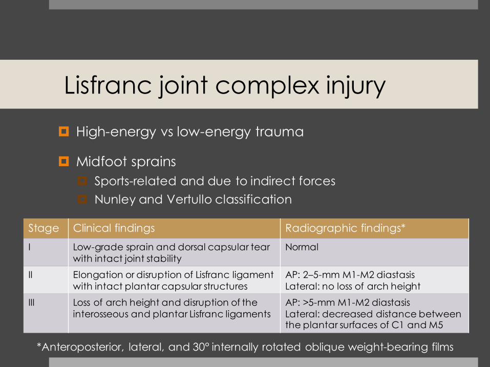
Lisfranc joint complex injury
¤ High-energy vs low-energy trauma
¤ Midfoot sprains
¤ Sports-related and due to indirect forces
¤ Nunley and Vertullo classification
Stage Clinical findings Radiographic findings*
I Low-grade sprain and dorsal capsular tear
with intact joint stability
Normal
II Elongation or disruption of Lisfranc ligament
with intact plantar capsular structures
AP: 2–5-mm M1-M2 diastasis
Lateral: no loss of arch height
III Loss of arch height and disruption of the
interosseous and plantar Lisfranc ligaments
AP: >5-mm M1-M2 diastasis
Lateral: decreased distance between the plantar surfaces of C1 and M5
*Anteroposterior, lateral, and 30° internally rotated oblique weight-bearing films

¤ Fluid surrounding the Lisfranc ligament
¤ Ligament irregularity or frank disruption
¤ Abnormal signal intensity within the ligament
Lisfranc ligament injury
Primary signs
Images from http://radiologycases.blogspot.com

Lisfranc ligament injury
Tafur M, et al. Magn Reson Imaging Clin N Am 25 (2017) 95–125

Lisfranc ligament injury
Fleck-sign: small avulsion fractures at base of the M2 or C1
C1
M2

Lisfranc ligament injury
¤ Fractures along the 2nd cuneometatarsal joint
¤ Contusions at the tarsometatarsal joints
¤ Soft tissue edema surrounding the 2nd metatarsal
¤ Edema in the 1st dorsal interosseous muscle
Secondary signs
Tafur M, et al. Magn Reson Imaging Clin N Am 25 (2017) 95–125

¤ Thickening of the interosseous
Lisfranc ligament, particularly in
setting of tarsometatarsalosteoarthrosis à typically
indicate old midfoot sprain.
Lisfranc ligament injury
Tafur M, et al. Magn Reson Imaging Clin N Am 25 (2017) 95–125

Bone marrow edema pattern
¤ Challenging in diabetic foot
¤ Early osteomyelitis vs stress response/bone marrow reaction
¤ Early neuropathic arthropathy vs infection
¤ Early osteomyelitis
¤ Identify a site of direct inoculation (skin/soft tissue defect
with a sinus tract or abscess extending to the bones)
¤ Focal or diffuse replacement of normal marrow fat on T1W à
most reliable
¤ Frank cortical destruction and/or periosteal reaction
¤ Geographic enhancement on T1 post contrast

DM with direct osteomyelitis from ulcer
T1W
T1W
T1Gd
STIR

Bone marrow response to surrounding soft tissue infection
T1W STIR

Bone marrow edema patterns
Early neuropathic arthropathy
¤ Diffuse soft tissue and bone marrow edema with
increased enhancement
¤ Common at Lisfranc, Chopartor MTP joints
¤ Multiple bone and joint involvements
¤ Joint centered, and tend to
occur symmetrically on either side of the joint
Early osteomyelitis
¤ Site of direct inoculation
¤ Occur distal to TMT joint, and
in malleoli and calcaneus
¤ Single bone involvement
¤ Tend to be limited to one side
of a joint (in absence of septic arthritis)

Neuropathic arthropathy-DM

Reference
¤ Sofka CM. Technical Considerations Best Practices for MR Imaging of the Foot and Ankle. Magn Reson Imaging Clin N Am 2017 Feb;25(1):1-10.
¤ Nery C, Baumfeld D, Umans H, Yamada AF. MR Imaging of the Plantar Plate: Normal Anatomy, Turf Toe, and Other Injuries. Magn Reson Imaging Clin N Am 2017 Feb;25(1):127-144.
¤ Hochman MG, Wu JS. MR Imaging of Common Soft Tissue Masses in the Foot and Ankle. Magn Reson Imaging Clin N Am 2017 Feb;25(1):159-181.
¤ Siddiqui NA, Galizia MS, Almusa E, et al. Evaluation of the tarsometatarsal joint using conventional radiography, CT, and MR imaging. Radiographics 2014; 34: 514–531.
¤ Tafur M, Rosenberg ZS, Bencardino JT. MR Imaging of the Midfoot Including Chopart and Lisfranc Joint Complexes. Magn Reson Imaging Clin N Am 2017 Feb;25(1):95-125.
¤ McCarthy E, Morrison WB, Zoga AC. MR Imaging of the Diabetic Foot. Magn ResonImaging Clin N Am. 2017 Feb;25(1):183-194.
¤ Arnold G, et al. Normal Magnetic Resonance Imaging Anatomy of the Ankle & Foot. Magn Reson Imaging Clin N Am 2011; 19:655–679.
¤ Umans HR. Imaging sports medicine Injuries of the foot and toes. Clin Sports Med 2006; 25:763-780.
¤ Linklater JM. Imaging of sports injuries in the foot. AJR 2012; 199:500–508.
¤ Ashman CJ, Klecker RJ, Yu JS. Forefoot pain involving the metatarsal region: differential diagnosis with MR imaging. RadioGraphics 2001;21:1425–1440.