Embed Size (px)
Citation preview
281ISSN 1758-427210.2217/IJR.12.15 © 2012 Future Medicine Ltd Int. J. Clin. Rheumatol. (2012) 7(3), 281–285
PersPective
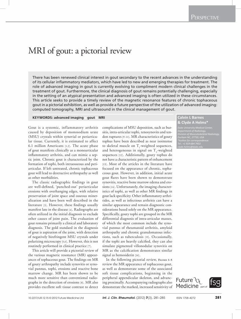
MRI of gout: a pictorial review
Gout is a systemic, inf lammatory arthritis caused by deposition of monosodium urate (MSU) crystals within synovial or periarticu-lar tissue. Currently, it is estimated to affect 6.1 million Americans [1,2]. The acute phase of gout manifests clinically as a monoarticular inflammatory arthritis, and can mimic a sep-tic joint. Chronic gout is characterized by the formation of tophi, both intraosseous and peri-articular. If left untreated, chronic tophaceous gout will lead to destructive arthropathy as well as other morbidities.
The classic radiographic findings in gout are well-defined, ‘punched-out’ periarticular erosions with overhanging edges, with relative preservation of joint space and osseous miner-alization and have been well described in the literature [3]. However, these findings usually manifest late in the disease [4]. Radiographs are often utilized in the initial diagnosis to exclude other causes of joint pain. The evaluation of gout remains primarily a clinical and laboratory diagnosis. The gold standard in the diagnosis of gout is aspiration of the joint, with detection of negatively birefringent MSU crystals under polarizing microscopy [5,6]. However, this is not routinely performed in clinical practice [7].
This article will provide a pictorial review of the various magnetic resonance (MR) appear-ances of tophaceous gout. The findings on MR of gouty arthropathy include synovitis or syno-vial pannus, tophi, erosions and reactive bone marrow change. MR has been shown to be much more sensitive than conventional radio-graphs in the detection of erosions [8]. MR also provides excellent soft tissue contrast to detect
complications of MSU deposition, such as bur-sitis, intra-articular tophi, tenosynovits and ten-don ruptures [9–11]. MR characteristics of gouty tophus have been described as near isointense to skeletal muscle on T
1-weighted sequences,
and heterogeneous in signal on T2-weighted
sequences [12]. Additionally, gouty tophus do not have a characteristic pattern of enhancement [13]. Most of the articles in the literature have focused on the appearance of chronic, topha-ceous gout. However, in addition, initial acute gout flares have been shown to demonstrate synovitis, reactive bone marrow edema and ero-sions [14]. Unfortunately, the imaging character-istics of tophi, as well as other MR findings in gout lack specificity. Other inflammatory arthri-tides, as well as infectious arthritis can have a similar appearance and remain diagnostic con-siderations based solely on the MR appearance. Specifically, gouty tophi are grouped in the MR differential diagnosis of intra-articular masses, of which the most common include the syno-vial pannus of rheumatoid arthritis, amyloid arthropathy and chronic granulomatous infec-tions, such as tuberculosis [15]. Occasionally, if the tophi are heavily calcified, they can also simulate pigmented villonodular synovitis on MR as the calcification demonstrates similar signal as hemosiderin [16].
In the following pictorial review, Figures 1–5 review the MR appearance of tophaceous gout, as well as demonstrate some of the associated soft tissue complications, beginning in the peripheral appendicular skeleton, and advanc-ing proximally. Accompanying radiographs also demonstrate the marked, increased sensitivity of
There has been renewed clinical interest in gout secondary to the recent advances in the understanding of its cellular inflammatory mediators, which have led to new and emerging therapies for treatment. The role of advanced imaging in gout is currently evolving to compliment modern clinical challenges in the treatment of gout. Furthermore, the clinical diagnosis of gout remains potentially challenging, especially in the setting of an atypical presentation and advanced imaging is often utilized in these circumstances. This article seeks to provide a timely review of the magnetic resonance features of chronic tophaceous gout in a pictorial exhibition, as well as provide a future perspective of the utilization of advanced imaging: computed tomography, MRI and ultrasound in the clinical management of gout.
KEYWORDS: advanced imaging n gout n MRI Calvin L Barnes & Clyde A Helms*Duke University Medical Center, Department of Radiology, Division of Musculoskeletal Radiology, Durham NC, 27710, USA *Author for correspondence: Tel.: +1 919 684 7453 [email protected]
part of
PersPective Barnes & HelmsPersPective Barnes & Helms
Int. J. Clin. Rheumatol. (2012) 7(3)282 future science group
MRI of gout: a pictorial review PersPective
MRI. Additionally, Figure 6 and Figure 7, which are cases of amyloid arthropathy and rheuma-toid arthritis in the shoulder respectively, were selected to demonstrate the limitations of MR in the initial diagnosis of gout due to relative lack of specificity in the MR findings.
Future perspective There has been recent interest in utilizing advanced imaging to quantify tophus burden as a marker of response to therapy. Schumacher et al. reported a multicenter study evaluating the intra- and inter-reader variability in calculating tophus volumes utilizing MR [17]. They found that there was excellent intrareader reproduc-ibility, with no statistical difference in volume measurements between two visits. They did find a statistical difference in the inter-reader
variability, although the actual difference in the volume measurement between readers was small. This study was limited by inherent artifacts in some of the MR protocols and there is a paucity of other studies to corroborate their results.
Dual-energy computed tomography (CT) is a relatively recent technology that allows acquisition of data simultaneously at two dif-ferent energy levels (kilovolts). Given the dif-ferent chemical composition of calcium and MSU deposits, their attenuation properties at the two different energy levels allow them to be distinguished from each other. Moreover, CT post-processing techniques, such as color map-ping, 3D surface rendering and volumetric ana-lysis provide accurate and readily interpretable images, and this technology may become very useful in both initial diagnosis in atypical clinical
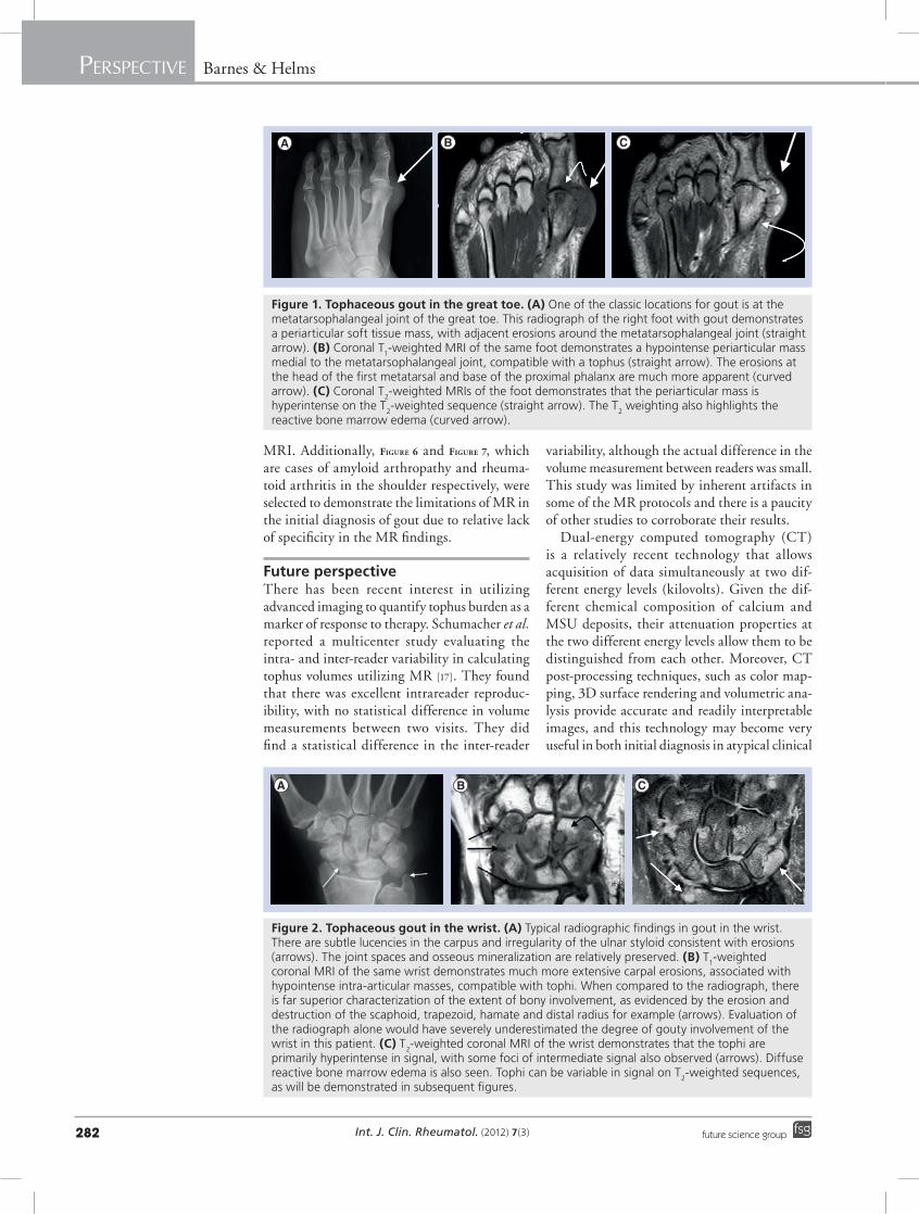
Figure 1. Tophaceous gout in the great toe. (A) One of the classic locations for gout is at the metatarsophalangeal joint of the great toe. This radiograph of the right foot with gout demonstrates a periarticular soft tissue mass, with adjacent erosions around the metatarsophalangeal joint (straight arrow). (B) Coronal T
1-weighted MRI of the same foot demonstrates a hypointense periarticular mass
medial to the metatarsophalangeal joint, compatible with a tophus (straight arrow). The erosions at the head of the first metatarsal and base of the proximal phalanx are much more apparent (curved arrow). (C) Coronal T
2-weighted MRIs of the foot demonstrates that the periarticular mass is
hyperintense on the T2-weighted sequence (straight arrow). The T
2 weighting also highlights the
reactive bone marrow edema (curved arrow).
Figure 2. Tophaceous gout in the wrist. (A) Typical radiographic findings in gout in the wrist. There are subtle lucencies in the carpus and irregularity of the ulnar styloid consistent with erosions (arrows). The joint spaces and osseous mineralization are relatively preserved. (B) T
1-weighted
coronal MRI of the same wrist demonstrates much more extensive carpal erosions, associated with hypointense intra-articular masses, compatible with tophi. When compared to the radiograph, there is far superior characterization of the extent of bony involvement, as evidenced by the erosion and destruction of the scaphoid, trapezoid, hamate and distal radius for example (arrows). Evaluation of the radiograph alone would have severely underestimated the degree of gouty involvement of the wrist in this patient. (C) T
2-weighted coronal MRI of the wrist demonstrates that the tophi are
primarily hyperintense in signal, with some foci of intermediate signal also observed (arrows). Diffuse reactive bone marrow edema is also seen. Tophi can be variable in signal on T
2-weighted sequences,
as will be demonstrated in subsequent figures.
PersPective Barnes & HelmsPersPective Barnes & Helms
www.futuremedicine.com 283future science group
MRI of gout: a pictorial review PersPective
presentations as well as assessing response to var-ious therapies [18–20]. The advantages over MR include cost and time, although one drawback is ionizing radiation. Additionally, dual-energy CT is highly specific for gout and may obviate the need for joint aspiration [18].
Another method of assessing tophi includes ultrasound [21]. Superficial structures, such as the olecrenon bursa and prepatellar bursa as well as peripheral joints within the hand and feet can be easily accessed and examined with a high-frequency transducer. Ultrasound has been shown to be capable of monitoring treatment response in tophaceous gout [22]. Limitations of ultrasound include an inherent operator depen-dence to reliably reproduce images and can be time intensive, pending patient body habitus. Ultrasound has also been demonstrated to be less sensitive than MR, especially in the assess-ment of subclinical disease or osseous erosions [8]. However, a recent review of the litera-ture regarding all current methods of tophus assessment did find that physical measure-ment techniques and ultrasound most closely meet the criteria of the Outcomes Measure in Rheumatology [23].
The initial diagnosis of gout will most likely remain a clinical- and laboratory-based diag-nosis in the perceivable future. Although MR is very sensitive to detection of disease, it lacks specificity and is not cost effective for routine evaluation. MR does and will continue to have a role in the evaluation of patients with atypi-cal presentations, or in patients with clinical suspicion of internal derangement. There have been recent advances in the understanding of the underlying inflammatory pathways in gout, with IL-1 being implicated as the crucial mediator of inflammation [24]. As a result, new drugs targeting IL-1 activation are currently being heavily investigated [25]. It is very likely that as the pharmacologic treatment of chronic
rheumatologic conditions, such as gout, become more sophisticated, the role of MR, as well as CT and ultrasound will expand to be utilized in the monitoring of disease response.
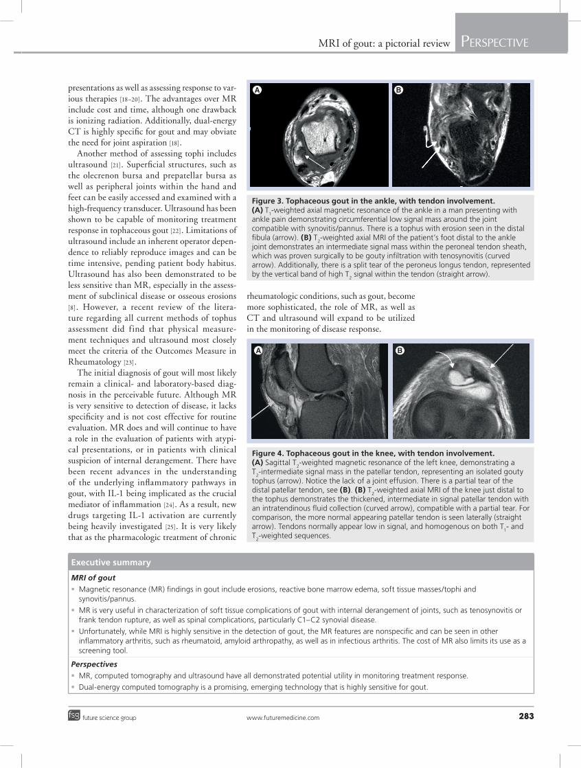
Figure 3. Tophaceous gout in the ankle, with tendon involvement. (A) T
1-weighted axial magnetic resonance of the ankle in a man presenting with
ankle pain demonstrating circumferential low signal mass around the joint compatible with synovitis/pannus. There is a tophus with erosion seen in the distal fibula (arrow). (B) T
2-weighted axial MRI of the patient’s foot distal to the ankle
joint demonstrates an intermediate signal mass within the peroneal tendon sheath, which was proven surgically to be gouty infiltration with tenosynovitis (curved arrow). Additionally, there is a split tear of the peroneus longus tendon, represented by the vertical band of high T
2 signal within the tendon (straight arrow).
Executive summary
MRI of gout
� Magnetic resonance (MR) findings in gout include erosions, reactive bone marrow edema, soft tissue masses/tophi and synovitis/pannus.
� MR is very useful in characterization of soft tissue complications of gout with internal derangement of joints, such as tenosynovitis or frank tendon rupture, as well as spinal complications, particularly C1–C2 synovial disease.
� Unfortunately, while MRI is highly sensitive in the detection of gout, the MR features are nonspecific and can be seen in other inflammatory arthritis, such as rheumatoid, amyloid arthropathy, as well as in infectious arthritis. The cost of MR also limits its use as a screening tool.
Perspectives
� MR, computed tomography and ultrasound have all demonstrated potential utility in monitoring treatment response.
� Dual-energy computed tomography is a promising, emerging technology that is highly sensitive for gout.
Figure 4. Tophaceous gout in the knee, with tendon involvement. (A) Sagittal T
2-weighted magnetic resonance of the left knee, demonstrating a
T2-intermediate signal mass in the patellar tendon, representing an isolated gouty
tophus (arrow). Notice the lack of a joint effusion. There is a partial tear of the distal patellar tendon, see (B). (B) T
2-weighted axial MRI of the knee just distal to
the tophus demonstrates the thickened, intermediate in signal patellar tendon with an intratendinous fluid collection (curved arrow), compatible with a partial tear. For comparison, the more normal appearing patellar tendon is seen laterally (straight arrow). Tendons normally appear low in signal, and homogenous on both T
1- and
T2-weighted sequences.
PersPective Barnes & HelmsPersPective Barnes & Helms
Int. J. Clin. Rheumatol. (2012) 7(3)284 future science group
MRI of gout: a pictorial review PersPective
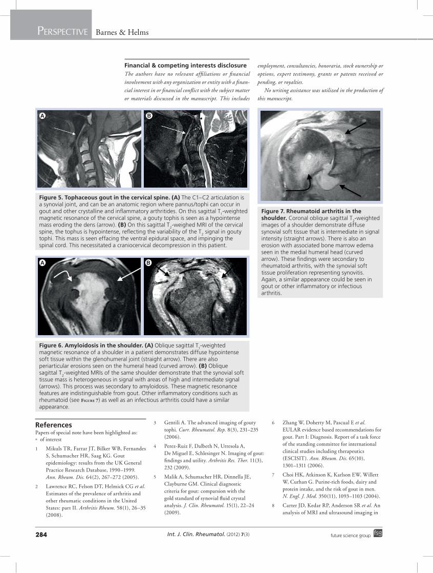
Figure 5. Tophaceous gout in the cervical spine. (A) The C1–C2 articulation is a synovial joint, and can be an anatomic region where pannus/tophi can occur in gout and other crystalline and inflammatory arthritides. On this sagittal T
1-weighted
magnetic resonance of the cervical spine, a gouty tophis is seen as a hypointense mass eroding the dens (arrow). (B) On this sagittal T
2-weighed MRI of the cervical
spine, the tophus is hypointense, reflecting the variability of the T2 signal in gouty
tophi. This mass is seen effacing the ventral epidural space, and impinging the spinal cord. This necessitated a craniocervical decompression in this patient.
Figure 6. Amyloidosis in the shoulder. (A) Oblique sagittal T1-weighted
magnetic resonance of a shoulder in a patient demonstrates diffuse hypointense soft tissue within the glenohumeral joint (straight arrow). There are also periarticular erosions seen on the humeral head (curved arrow). (B) Oblique sagittal T
2-weighted MRIs of the same shoulder demonstrate that the synovial soft
tissue mass is heterogeneous in signal with areas of high and intermediate signal (arrows). This process was secondary to amyloidosis. These magnetic resonance features are indistinguishable from gout. Other inflammatory conditions such as rheumatoid (see Figure 7) as well as an infectious arthritis could have a similar appearance.
Figure 7. Rheumatoid arthritis in the shoulder. Coronal oblique sagittal T
2-weighted
images of a shoulder demonstrate diffuse synovial soft tissue that is intermediate in signal intensity (straight arrows). There is also an erosion with associated bone marrow edema seen in the medial humeral head (curved arrow). These findings were secondary to rheumatoid arthritis, with the synovial soft tissue proliferation representing synovitis. Again, a similar appearance could be seen in gout or other inflammatory or infectious arthritis.
Financial & competing interests disclosureThe authors have no relevant affiliations or financial involvement with any organization or entity with a finan-cial interest in or financial conflict with the subject matter or materials discussed in the manuscript. This includes
employment, consultancies, honoraria, stock ownership or options, expert testimony, grants or patents received or pending, or royalties.
No writing assistance was utilized in the production of this manuscript.
ReferencesPapers of special note have been highlighted as:n of interest
1 Mikuls TR, Farrar JT, Bilker WB, Fernandes S, Schumacher HR, Saag KG. Gout epidemiology: results from the UK General Practice Research Database, 1990–1999. Ann. Rheum. Dis. 64(2), 267–272 (2005).
2 Lawrence RC, Felson DT, Helmick CG et al. Estimates of the prevalence of arthritis and other rheumatic conditions in the United States: part II. Arthritis Rheum. 58(1), 26–35 (2008).
3 Gentili A. The advanced imaging of gouty tophi. Curr. Rheumatol. Rep. 8(3), 231–235 (2006).
4 Perez-Ruiz F, Dalbeth N, Urresola A, De Miguel E, Schlesinger N. Imaging of gout: findings and utility. Arthritis Res. Ther. 11(3), 232 (2009).
5 Malik A, Schumacher HR, Dinnella JE, Clayburne GM. Clinical diagnostic criteria for gout: comparsion with the gold standard of synovial fluid crystal analysis. J. Clin. Rheumatol. 15(1), 22–24 (2009).
6 Zhang W, Doherty M, Pascual E et al. EULAR evidence based recommendations for gout. Part I: Diagnosis. Report of a task force of the standing committee for international clinical studies including therapeutics (ESCISIT). Ann. Rheum. Dis. 65(10), 1301–1311 (2006).
7 Choi HK, Atkinson K, Karlson EW, Willett W, Curhan G. Purine-rich foods, dairy and protein intake, and the risk of gout in men. N. Engl. J. Med. 350(11), 1093–1103 (2004).
8 Carter JD, Kedar RP, Anderson SR et al. An ana lysis of MRI and ultrasound imaging in
PersPective Barnes & HelmsPersPective Barnes & Helms
www.futuremedicine.com 285future science group
MRI of gout: a pictorial review PersPective
patients with gout who have normal plain radiographs. Rheumatology 48(11), 1442–1446 (2009).
9 Bond JR, Sim FH, Sundaram M. Radiologic case study. Gouty tophus involving the distal quadriceps tendon. Orthopedics 27(1), 90–92 (2004).
10 Lagoutaris ED, Adams HB, Didomenico LA, Rothenberg RJ. Longitudinal tears of both peroneal tendons associated with tophaceous gouty infiltration. A case report. J. Foot Ankle Surg. 44(3), 222–224 (2005).
11 Yu KH, Lien LC, Ho HH. Limited knee joint range of motion due to invisible gouty tophi. Rheumatology 43(2), 191–194 (2004).
12 Yu JS, Chung C, Recht M, Dailiana T, Jurdi R. MR imaging of tophaceous gout. Am. J. Roentgenol. 168(2), 523–527 (1997).
13 Gerster JC, Landry M, Dufresne L, Meuwly JY. Imaging of tophaceous gout: computed tomography provides specific images compared with magnetic resonance imaging and ultrasonography. Ann. Rheum. Dis. 61(1), 52–54 (2002).
14 Cimmino MA, Zampogna G, Parodi M et al. MRI synovitis and bone lesions are common in acute gouty arthritis of the wrist even during the first attack. Ann. Rheum. Dis. 70(12), 2238–2239 (2011).
n Recent article highlighting the magnetic resonance findings in acute gout, and demonstrating early arthritic and bone changes.
15 Sheldon PJ, Forrester DM, Learch TJ. Imaging of intraarticular masses. Radiographics 25(1), 105–119 (2005).
16 Ritchie DA. MR imaging of synovial tumours and tumour-like lesions. Br. J. Radiol. 72(854), 212–218 (1999).
17 Schumacher HR Jr, Becker MA, Edwards NL et al. Magnetic resonance imaging in the quantitative assessment of gouty tophi. Int. J. Clin. Pract. 60(4), 408–414 (2006). [Erratum in: Int. J. Clin. Pract. 60(5), 630 (2006)].
18 Choi HK, Al-Arfaj AM, Eftekhari A et al. Dual energy computed tomography in tophaceous gout. Ann. Rheum. Dis. 68(10), 1609–1612 (2009).
n Recent article highlighting dual-energy computed tomography (CT).
19 Desai MA, Peterson JJ, Garner HW, Kransdorf MJ. Clinical utility of dual-energy CT for evaluation of tophaceous gout. Radiographics 31(5), 1365–1375 (2011).
n Recent article highlighting dual-energy CT.
20 Nicolaou S, Yong-Hing CJ, Galea-Soler S, Hou DJ, Louis L, Munk P. Dual-energy CT as a potential new diagnostic tool in the management of gout in the acute setting. Am. J. Roentgenol. 194(4), 1072–1078 (2010).
n Recent article highlighting dual-energy CT.
21 Thiele RG. Role of ultrasound and other advanced imaging in the diagnosis and management of gout. Curr. Rheumatol. Rep. 13(2), 146–153 (2011).
n Relevant, current review of ultrasound imaging with regard to tophaceous gout.
22 Thiele RG, Schlesinger N. Ultrasonography shows disappearance of monosodium urate crystal deposition on hyaline cartilage after sustained normouricemia is achieved. Rheumatol. Int. 30(4), 495–503 (2010).
23 Dalbeth N, Schauer C, Macdonald P et al. Methods of tophus assessment in clinical trials of chronic gout. a systematic literature review and pictorial reference guide. Ann. Rheum. Dis. 70(4), 597–604 (2011).
n Consise and recent literature review with comparison of magnetic resonance, CT and ultrasound in tophus assessment.
24 Busso N, So A. Mechanisms of inflammation in gout. Arthritis Res. Ther. 12(2), 206 (2010).
25 Neogi T. Clinical practice. Gout. N. Engl. J. Med. 364(5), 443–452 (2011).