Embed Size (px)
Citation preview
Mte1 interacts with Mph1 and promotescrossover recombination and telomeremaintenanceSonia Silva,1,9 Veronika Altmannova,2 Sarah Luke-Glaser,3 Peter Henriksen,4 Irene Gallina,1
Xuejiao Yang,1 Chunaram Choudhary,4 Brian Luke,3 Lumir Krejci,2,5,6,7 and Michael Lisby1,8
1Department of Biology, University of Copenhagen, DK-2200 Copenhagen N, Denmark; 2Department of Biology, MasarykUniversity, CZ-62500 Brno, Czech Republic; 3Institute of Molecular Biology gGmbH (IMB), 55128 Mainz, Germany; 4The NovoNordisk Foundation Center for Protein Research, Faculty of Health Sciences, University of Copenhagen, DK-2200 Copenhagen N,Denmark; 5National Centre for Biomolecular Research, Masaryk University, CZ-62500 Brno, Czech Republic; 6InternationalClinical ResearchCenter, 7Center for Biomolecular andCellular Engineering, St. Anne’sUniversityHospital Brno,CZ-656 91 Brno,Czech Republic; 8Center for Chromosome Stability, Department of Cellular and Molecular Medicine, University of Copenhagen,DK-2200 Copenhagen N, Denmark
Mph1 is a member of the conserved FANCM family of DNA motor proteins that play key roles in genome main-tenance processes underlying Fanconi anemia, a cancer predisposition syndrome in humans. Here, we identifyMte1as a novel interactor of the Mph1 helicase in Saccharomyces cerevisiae. In vitro, Mte1 (Mph1-associated telomeremaintenance protein 1) binds directly to DNA with a preference for branched molecules such a D loops and forkstructures. In addition, Mte1 stimulates the helicase and fork regression activities of Mph1 while inhibiting theability of Mph1 to dissociate recombination intermediates. Deletion ofMTE1 reduces crossover recombination andsuppresses the sensitivity ofmph1Δmutant cells to replication stress. Mph1 and Mte1 interdependently colocalizeatDNAdamage-induced foci and dysfunctional telomeres, andMTE1 deletion results in elongated telomeres. Takentogether, our data indicate that Mte1 plays a role in regulation of crossover recombination, response to replicationstress, and telomere maintenance.
[Keywords: homologous recombination; telomere maintenance; genome integrity; DNA repair; Mph1; Mte1]
Supplemental material is available for this article.
Received December 8, 2015; revised version accepted February 17, 2016.
Several human genome instability diseases are caused bymutations in DNA helicases. For example, mutations inFANCM cause Fanconi anemia (FA), which is a conditionassociated with bone marrow failure, physical abnormali-ties, organ defects, and cancer predisposition. FA has beenascribed to defects in repair of DNA interstrand cross-links, protein–DNA adducts, and DNA double-strandbreaks (DSBs) (for review, seeDuxin andWalter 2015). An-other example is RTEL1, where mutations give rise toHoyeraal-Hreidarsson syndrome, which is associatedwith bone marrow failure, microcephaly, immunodefi-ciency, telomere fragility, and predisposition to cancer(for review, see Vannier et al. 2014). Mph1 appears to bethe functional homolog of FANCMandRTEL1 in the bud-ding yeast model system (Whitby 2010; Luke-Glaser et al.2012; Vannier et al. 2014; Xue et al. 2015b), where it serves
as amutation suppressor and a negative regulator of cross-over (CO) recombination (Prakash et al. 2005, 2009).The canonical homologous recombination (HR) path-
way for repairing DSBs in yeast is initiated by Rad52-me-diated loading of Rad51 on the 3′ ssDNA tails generatedby resection of a DSB that consequently facilitates dis-placement of replication protein A (RPA). The Rad51 nu-cleoprotein filament is subsequently responsible for thecrucial homology search and strand invasion into a ho-mologous donor duplex to form a characteristic displace-ment loop (D loop) (Sung 1994; Symington et al. 2014).After initial extension of the invading 3′ end, the extend-ed D loop can be processed by several alternative path-ways. In mitotic cells, breaks are primarily repaired bysynthesis-dependent strand annealing (SDSA), in whichthe extended D loop is displaced to yield non-CO
9Present address: Centro Andaluz de Biología Molecular y MedicinaRegenerativa CABIMER, Universidad de Sevilla, Seville 41004, Spain.Corresponding author: [email protected] published online ahead of print. Article and publication date areonline at http://www.genesdev.org/cgi/doi/10.1101/gad.276204.115.
© 2016 Silva et al. This article is distributed exclusively by Cold SpringHarbor Laboratory Press for the first six months after the full-issue publi-cation date (see http://genesdev.cshlp.org/site/misc/terms.xhtml). Aftersix months, it is available under a Creative Commons License (At-tribution-NonCommercial 4.0 International), as described at http://creativecommons.org/licenses/by-nc/4.0/.
GENES & DEVELOPMENT 30:1–18 Published by Cold Spring Harbor Laboratory Press; ISSN 0890-9369/16; www.genesdev.org 1
Cold Spring Harbor Laboratory Press on March 12, 2022 - Published by genesdev.cshlp.orgDownloaded from
(NCO) products (Symington et al. 2014). If the extendedstrand is instead stabilized, second end capture can en-sue, giving rise to a double Holliday junction (dHJ) thatcan be either resolved or dissolved to yield CO or NCOevents, respectively (see the Discussion for details; Sy-mington et al. 2014). CO events are a potential threatto genome integrity because they can result in genomicrearrangements and loss of heterozygosity (LOH). Oneimportant negative regulator of CO events is the 3′–5′
helicase Mph1. Mph1 has the ability to disrupt D loopsformed by Rad51 and catalyze migration of branchedDNA structures (Prakash et al. 2005, 2009; Whitby2010; Zheng et al. 2011; Mitchel et al. 2013). Independentof its role in D-loop unwinding, the Mph1 helicase pro-motes fork regression and branch migration during repli-cation-associated recombinational repair (Prakash et al.2005, 2009; Mankouri et al. 2009; Panico et al. 2010;Zheng et al. 2011; Xue et al. 2014). The role of Mph1 inreplication fork rescue is negatively regulated by theSmc5/6 complex by direct binding to the regulatorydomain of Mph1 at its C terminus. Furthermore, the in-hibition of Mph1 by the Smc5/6 complex is attenuatedby the Mhf1–Mhf2 histone fold complex (MHF) (Xueet al. 2015a). In mutants of the Smc5/6 complex, Mph1is responsible for accumulation of replication intermedi-ates that lead to increased sensitivity to replication stressand genomic instability (Chen et al. 2009, 2013; Qiu et al.2013; Xue et al. 2014).
Mph1 has recently been implicated in telomeremainte-nance, promoting telomere uncapping and acceleratingsenescence when overexpressed in telomerase-negativecells (Luke-Glaser and Luke 2012). In yeast, telomeresconsist of 250- to 300-base-pair (bp) arrays of TG1–3 repeatswith a 3′ ssDNA overhang of 12–14 nucleotides (G-tail)that is specifically recognized and bound byCdc13 togeth-er with Stn1 and Ten1 (CST complex) (Wellinger andZakian 2012). The yeast telomeres are also bound in thedouble-stranded region by the Ku complex and by Rap1and its binding partners, Rif1–Rif2. The association ofthese protein complexeswith telomericDNA forms a pro-tective capping structure that accounts for two crucialoutcomes: the full replication of telomeres and protectionof telomeres from recognition by the recombination andcheckpointmachineries and associated nuclease and heli-case activities (Bartsch et al. 2000; Raschle et al. 2004;Lisby and Geli 2009; Dewar and Lydall 2012). In the ab-sence of telomerase, telomeres shorten progressivelywith each round of replication. Within ∼60–80 cell divi-sions, telomeres become critically short, rendering chro-mosomes unstable, and cells lose viability in a processcalled replicative senescence (Lundblad and Szostak1989; Abdallah et al. 2009). Telomere shortening leadsto the accumulation of ssDNA at chromosome ends,which elicits the activation of the DNA damage check-point and recruitment of repair factors that are usually ex-cluded from telomeres, such as Rad52 (d’Adda di Fagagnaet al. 2003; Takai et al. 2003; Abdallah et al. 2009; Khada-roo et al. 2009; Lin et al. 2009). Despite the loss of telome-rase, some cells are able to use a RAD52-dependent HRmechanism to elongate their telomeres, thereby bypass-
ing senescence and surviving (Sugiyama et al. 1998;Le et al. 1999).
In this study, we identify and characterize theYGR042W-encoded protein Mte1 (Mph1-associated telo-mere maintenance protein 1) as a novel factor involvedin the DNA damage response. Mte1 has homology withthe human zinc finger protein ZGRF1 (C4ORF21; impli-cated in HR and DNA interstrand cross-link repair) (Smo-gorzewska et al. 2010; Adamson et al. 2012) and thefission yeast Dbl2 (Yu et al. 2013). The Mte1-GFP fusionprotein has been shown to form nuclear foci in responseto replication stress (Huh et al. 2003; Tkach et al. 2012;Yimit et al. 2015). Moreover, high-throughput screenshave implicated Mte1 in telomere length maintenance(Askree et al. 2004) and indicated that it can interactwith Cmr1 (Gilmore et al. 2012), a chromatin-bindingprotein associated with replication stress response (Choiet al. 2012; Tkach et al. 2012; Gallina et al. 2015).In this report, we show that Mte1 is a D-loop-bindingprotein that interacts with the Mph1 helicase and hasan antagonizing role toMph1 in recombination outcomesby enhancing CO formation. Furthermore, we show thatMte1 is involved in telomere length maintenance andassociates with dysfunctional telomeres together withMph1.
Results
The Mph1 helicase interacts with Mte1
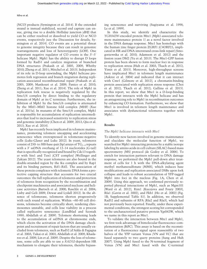
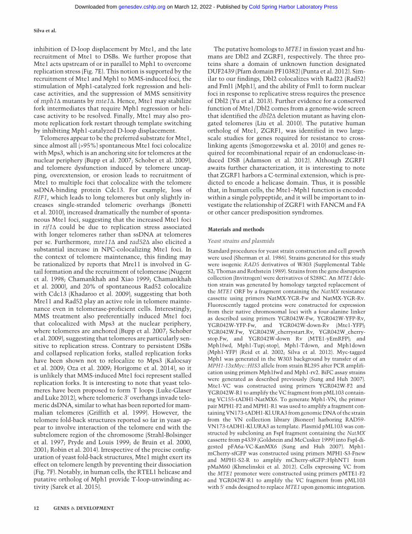
To identify new factors involved in genome maintenanceand elucidate the molecular function of Mph1, wesearched forMph1-interacting proteins by a stable isotopelabeling by amino acids in cell culture (SILAC)-basedmassspectrometry (MS) protocol (de Godoy et al. 2006). Toenrich for interaction partners relevant to DNA damageresponse, we performed the Mph1 pull-down after treat-ment of cells for 1 h with the DNA-alkylating agentmethyl methanesulfonate (MMS), which induces basemodifications and replication-associated DSBs upon forkcollapse and leads to robust accumulation of YFP-taggedMph1 into foci in the nucleus (Fig. 1A; Chen et al.2009). Using this approach, we confirmed previously re-ported physical interactions of Mph1, such as Mgm101(Ward et al. 2012), Rim1 (Kucejova and Foury 2003),Rfa1 (Gavin et al. 2002), and Fkh1 (Ho et al. 2002) (Fig.1B; Supplemental Table S1). In addition, we observedRad52 and subunits of RPA (Rfa2 and Rfa3), which hadnot previously been reported. Finally, under these experi-mental conditions, the strongest scoring hit correspondedto the uncharacterized putative protein Ygr042W, whichwe name in this report as Mte1.
To validate the interaction between Mte1 and Mph1,we first took advantage of bimolecular fluorescence com-plementation (BiFC). This assay is based on the reconsti-tution of a fluorescence signal upon reassembly of twohalves of the YFP variant Venus (VN and VC) that arefused separately to two query proteins (Sung and Huh2007). Using Mph1 fused to the N-terminal fragment ofVenus (VN) and Mte1 fused with the C-terminal
Silva et al.
2 GENES & DEVELOPMENT
Cold Spring Harbor Laboratory Press on March 12, 2022 - Published by genesdev.cshlp.orgDownloaded from
complementary fragment (VC), we found that Mph1 andMte1 directly interact in the nucleus (Fig. 1C,D). Second,in a strain coexpressing YFP-tagged Mte1 and myc-taggedMph1, the two proteins coimmunoprecipitated in bothuntreated and MMS-treated conditions (Fig. 1E), indicat-ing that the interaction is independent of DNA damage.Furthermore, the interaction is likely to be independentof DNA, as treatment of extracts with DNase prior toimmunoprecipitation did not reduce the amount ofMph1 retrieved by pull-down of Mte1 (cf. Fig. 1E and Sup-plemental Fig. 1A,B).
DNA damage-induced Mte1 and Mph1 fociare interdependent
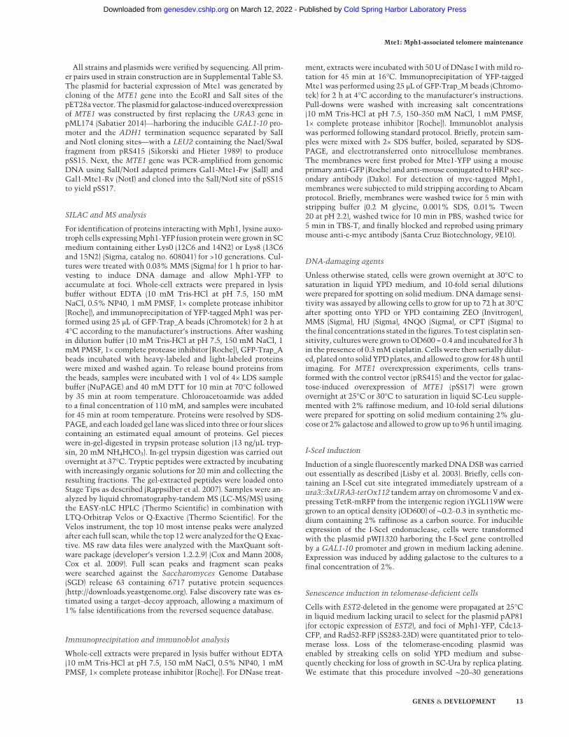
The formation of nuclear foci has been observed for manyfactors involved in DNA damage response, and HR and iscommonly used as marker for sites of DNA damage andongoing repair (Lisby and Rothstein 2015). To gain furtherunderstanding of the relationship between Mph1 andMte1, we monitored Mte1 and Mph1 focus formationand colocalization between the two proteins in untreatedcells and after exposure to various types of DNA damag-ing agents. For this purpose we generated YFP-taggedMte1 at the C terminus, which showed the expected nu-clear localization (Huh et al. 2003; Srikumar et al. 2013;Yimit et al. 2015). In untreated cells, only a subset of
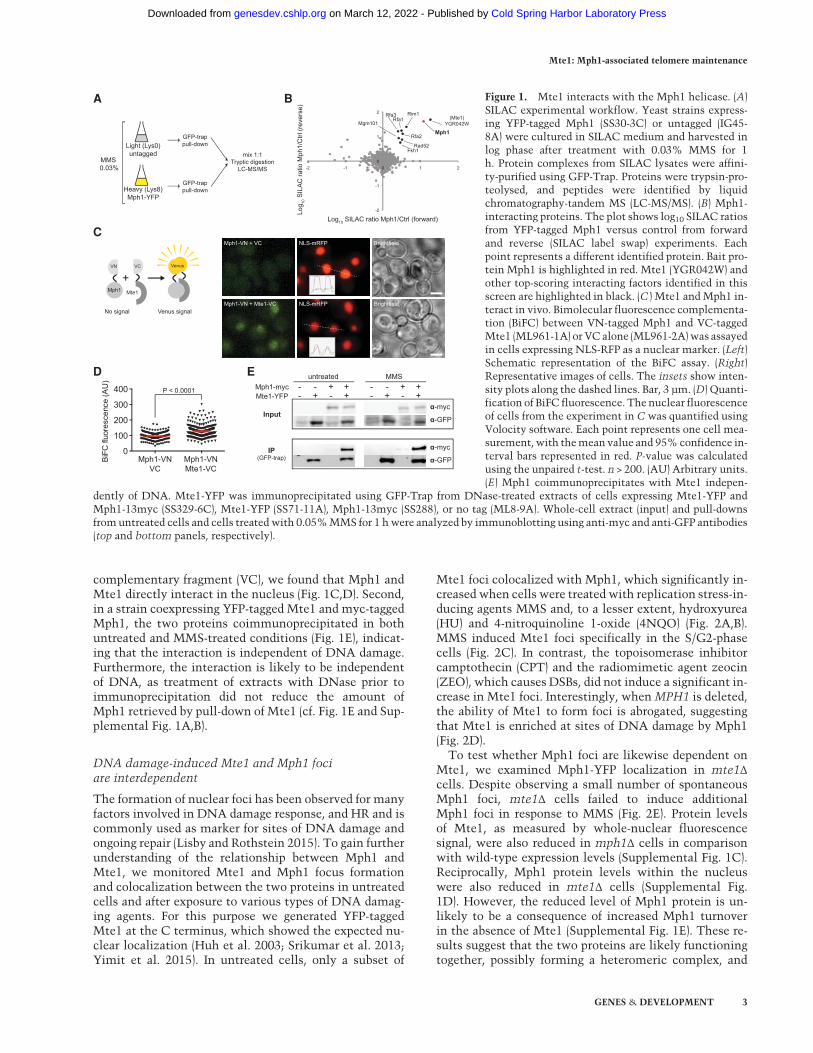
Mte1 foci colocalized with Mph1, which significantly in-creased when cells were treated with replication stress-in-ducing agents MMS and, to a lesser extent, hydroxyurea(HU) and 4-nitroquinoline 1-oxide (4NQO) (Fig. 2A,B).MMS induced Mte1 foci specifically in the S/G2-phasecells (Fig. 2C). In contrast, the topoisomerase inhibitorcamptothecin (CPT) and the radiomimetic agent zeocin(ZEO), which causes DSBs, did not induce a significant in-crease in Mte1 foci. Interestingly, whenMPH1 is deleted,the ability of Mte1 to form foci is abrogated, suggestingthat Mte1 is enriched at sites of DNA damage by Mph1(Fig. 2D).To test whether Mph1 foci are likewise dependent on
Mte1, we examined Mph1-YFP localization in mte1Δcells. Despite observing a small number of spontaneousMph1 foci, mte1Δ cells failed to induce additionalMph1 foci in response to MMS (Fig. 2E). Protein levelsof Mte1, as measured by whole-nuclear fluorescencesignal, were also reduced in mph1Δ cells in comparisonwith wild-type expression levels (Supplemental Fig. 1C).Reciprocally, Mph1 protein levels within the nucleuswere also reduced in mte1Δ cells (Supplemental Fig.1D). However, the reduced level of Mph1 protein is un-likely to be a consequence of increased Mph1 turnoverin the absence of Mte1 (Supplemental Fig. 1E). These re-sults suggest that the two proteins are likely functioningtogether, possibly forming a heteromeric complex, and
A
Light (Lys0)untagged
Heavy (Lys8)Mph1-YFP
MMS 0.03%
GFP-trappull-down
GFP-trappull-down
mix 1:1Tryptic digestion
LC-MS/MS
B
C
Log 10
SIL
AC
ratio
Mph
1/C
trl (r
ever
se)
Log10 SILAC ratio Mph1/Ctrl (forward)
Mph1
(Mte1)YGR042W
Rim1Rfa1
Rfa3
Rfa2
Rad52Fkh1
Mgm101
-2
-1
0
1
2
-2 -1 0 1 2
VN VC Venus
Mte1Mph1
No signal Venus signal
+
D E
Input
untreated++
+-
-+
--
Mph1-mycMte1-YFP
MMS++
+-
-+
--
α-myc++
α-GFP
IP (GFP-trap) α-GFP
α-myc0
100
200
300
400
BiF
C fl
uore
scen
ce (A
U)
Mph1-VN + Mte1-VC NLS-mRFP Brightfield
Mph1-VN + VC NLS-mRFP Brightfield
Mph1-VNVC
P < 0.0001
Mph1-VNMte1-VC
Figure 1. Mte1 interacts with the Mph1 helicase. (A)SILAC experimental workflow. Yeast strains express-ing YFP-tagged Mph1 (SS30-3C) or untagged (IG45-8A) were cultured in SILAC medium and harvested inlog phase after treatment with 0.03% MMS for 1h. Protein complexes from SILAC lysates were affini-ty-purified using GFP-Trap. Proteins were trypsin-pro-teolysed, and peptides were identified by liquidchromatography-tandem MS (LC-MS/MS). (B) Mph1-interacting proteins. The plot shows log10 SILAC ratiosfrom YFP-tagged Mph1 versus control from forwardand reverse (SILAC label swap) experiments. Eachpoint represents a different identified protein. Bait pro-tein Mph1 is highlighted in red. Mte1 (YGR042W) andother top-scoring interacting factors identified in thisscreen are highlighted in black. (C ) Mte1 andMph1 in-teract in vivo. Bimolecular fluorescence complementa-tion (BiFC) between VN-tagged Mph1 and VC-taggedMte1 (ML961-1A) or VC alone (ML961-2A)was assayedin cells expressing NLS-RFP as a nuclear marker. (Left)Schematic representation of the BiFC assay. (Right)Representative images of cells. The insets show inten-sity plots along the dashed lines. Bar, 3 µm. (D) Quanti-fication of BiFC fluorescence. The nuclear fluorescenceof cells from the experiment in C was quantified usingVolocity software. Each point represents one cell mea-surement,with themean value and 95%confidence in-terval bars represented in red. P-value was calculatedusing the unpaired t-test. n > 200. (AU) Arbitrary units.(E) Mph1 coimmunoprecipitates with Mte1 indepen-
dently of DNA. Mte1-YFP was immunoprecipitated using GFP-Trap from DNase-treated extracts of cells expressing Mte1-YFP andMph1-13myc (SS329-6C), Mte1-YFP (SS71-11A), Mph1-13myc (SS288), or no tag (ML8-9A). Whole-cell extract (input) and pull-downsfromuntreated cells and cells treated with 0.05%MMS for 1 hwere analyzed by immunoblotting using anti-myc and anti-GFP antibodies(top and bottom panels, respectively).
Mte1: Mph1-associated telomere maintenance
GENES & DEVELOPMENT 3
Cold Spring Harbor Laboratory Press on March 12, 2022 - Published by genesdev.cshlp.orgDownloaded from
thus deleting one of the components might affect the oth-er component’s localization and ability to respond toDNA damage.
To estimate the abundance of Mte1 and Mph1 proteinin vivo, we quantitated the total nuclear YFP fluorescenceof the tagged proteins in both haploid and diploid cellsusing Rad52-YFP as a reference (Fig. 2F; Lisby et al.2003). Based on this method, there were slightly morethan 300 molecules of Mte1 in a haploid nucleus androughly twice that amount in diploid cells. By compari-son, Mph1 is twice as abundant as Mte1 in haploid cells,while, in diploid cells, the two proteins are present inequal amounts. By quantitating the fraction of fluores-cence within foci, we found that both spontaneous andMMS-induced foci contain approximately four molecules
of either protein (Fig. 2G), indicating that Mte1 andMph1are present at stoichiometric amounts at sites of DNAdamage.
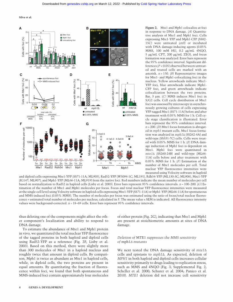
Deletion of MTE1 suppresses the MMS sensitivityof mph1Δ mutants
We next tested the DNA damage sensitivity of mte1Δcells and epistasis to mph1Δ. As expected, deletion ofMPH1 in both haploid and diploid cells increases cellularsensitivity primarily to drugs leading to replication stress,such as MMS and 4NQO (Fig. 3; Supplemental Fig. 2;Scheller et al. 2000; Schurer et al. 2004; Panico et al.2010). MTE1 deletion did not increase cell sensitivity
Mte1 fociMph1 foci
Cel
ls w
ith fo
ci (%
)
untreated0.05% MMS
mph1Δwt
Cel
ls w
ith M
ph1
foci
(%)
mte1Δwt
untreated0.05% MMS
0
20
40 *
colocalizing foci
0
25
50
75
MMS HU 4NQO CPTctrl
1h 2h
**
**
ZEO1h 2h 1h 2h 1h 2h 2h
��
�
�
�
�
�
�
��
�Mte1-YFP Mph1-CFP
merge DIC
0
20
40
60
Cel
ls w
ith M
te1
foci
(%)
*0
20
40
60
S/G2 G2/M G1
spontaneous fociMMS-induced foci
total cells (%) budded unbuddeduntreated 53% 47% MMS 56% 44%
Cel
ls w
ith M
te1
foci
(%)
Mte1
Num
ber o
f mol
ecul
es p
er c
ell
Rad52 Rdh54 Mrc1 Mph1
334 647 2300 4721 1567 673 404 843 581 645
3.5±0.5
4.1±0.3 4.5±0.63.8±0.4
A B
E
F
DC
G
1n 2n 1n 2n 1n 2n 1n 2n 1n 2n 1h- 1h-
Mph1Mte1MMS
0
5
10
15
20
Mol
ecul
es/fo
cus
100
1000
10000
Figure 2. Mte1 and Mph1 colocalize at fociin response to DNA damage. (A) Quantita-tive analysis of Mte1 and Mph1 foci. Cellsexpressing Mte1-YFP and Mph1-CFP (SS82-15C) were untreated (ctrl) or incubatedwith DNA damage-inducing agents (0.05%MMS, 100 mM HU, 0.2 μg/mL 4NQO,5 μg/mL CPT, 200 μg/mL ZEO), and focusformation was analyzed. Error bars representthe 95% confidence interval. Significant dif-ferences (P < 0.05) observed between untreat-ed and treated cells are marked with anasterisk. n > 150. (B) Representative imagesfor Mte1- and Mph1-colocalizing foci in thenucleus. Yellow arrowheads indicate Mte1-YFP foci, blue arrowheads indicate Mph1-CFP foci, and green arrowheads indicatecolocalization between the two proteins.Bar, 3 µm. (C ) MMS induces Mte1 foci inS/G2 cells. Cell cycle distribution of Mte1fociwas assessed bymicroscopy in asynchro-nously growing cultures of cells expressingYFP-taggedMte1 (SS71-11A) before and aftertreatment with 0.05%MMS for 1 h. Cell cy-cle stage classification is illustrated. Errorbars represent the 95% confidence interval.n > 200. (D) Mte1 focus formation is abrogat-ed in mph1 mutant cells. Mte1 focus forma-tion was analyzed in mph1Δ (SS262-4A) andwild-type (SS331-7C) cells. Cells were treat-ed with 0.05% MMS for 1 h. (E) DNA dam-age induction of Mph1 foci is dependent onMte1. Mph1 foci were quantitated inmte1Δ (SS260-24B) and wild-type (ML66-11A) cells before and after treatment with0.05% MMS for 1 h. (F ) Estimation of thenumber of Mte1 molecules per cell. Totalnuclear YFP fluorescence intensities weremeasured using Volocity software in haploid
and diploid cells expressing Mte1-YFP (SS71-11A; ML905), Rad52-YFP (W5094-1C; ML191), Rdh54-YFP (ML130-3C; ML906), Mrc1-YFP(IG147; ML907), and Mph1-YFP (ML66-11A; ML919) from the native loci. Red numbers indicate the mean number of molecules per cellbased on normalization to Rad52 in haploid cells (Lisby et al. 2003). Error bars represent 95% confidence intervals. n = 100–200. (G) Es-timation of the number of Mte1 and Mph1 molecules per focus. Focus and total nuclear YFP fluorescence intensities were measuredat the single-cell level using Volocity software in haploid cells expressingMte1-YFP (SS71-11A) orMph1-YFP (ML66-11A) for spontaneousand MMS-induced foci (0.05% MMS). The number of molecules per focus was estimated using the ratio of focus/total nuclear fluores-cence × estimated total number of molecules per nucleus, calculated in F. The mean value ± SEM is indicated. All fluorescence intensityvalues were background-corrected. n = 18–69 cells. Error bars represent 95% confidence intervals.
Silva et al.
4 GENES & DEVELOPMENT
Cold Spring Harbor Laboratory Press on March 12, 2022 - Published by genesdev.cshlp.orgDownloaded from
to DNA-damaging agents. Interestingly, we observed amodest but reproducible rescue of MMS sensitivity ofmph1Δ by mte1Δ in haploid cells (Fig. 3A). Conversely,overexpression of Mte1 from a galactose-inducible pro-moter enhanced the sensitivity of cells to genotoxic stress(Fig. 3B; Supplemental Fig. 2B). This suggests that Mte1produces or stabilizes a repair intermediate acted uponby Mph1.
Mte1 binds branchedDNA structures in vitro, stimulatesMph1 helicase and fork regression activities, and inhibitsMph1-catalyzed D-loop displacement
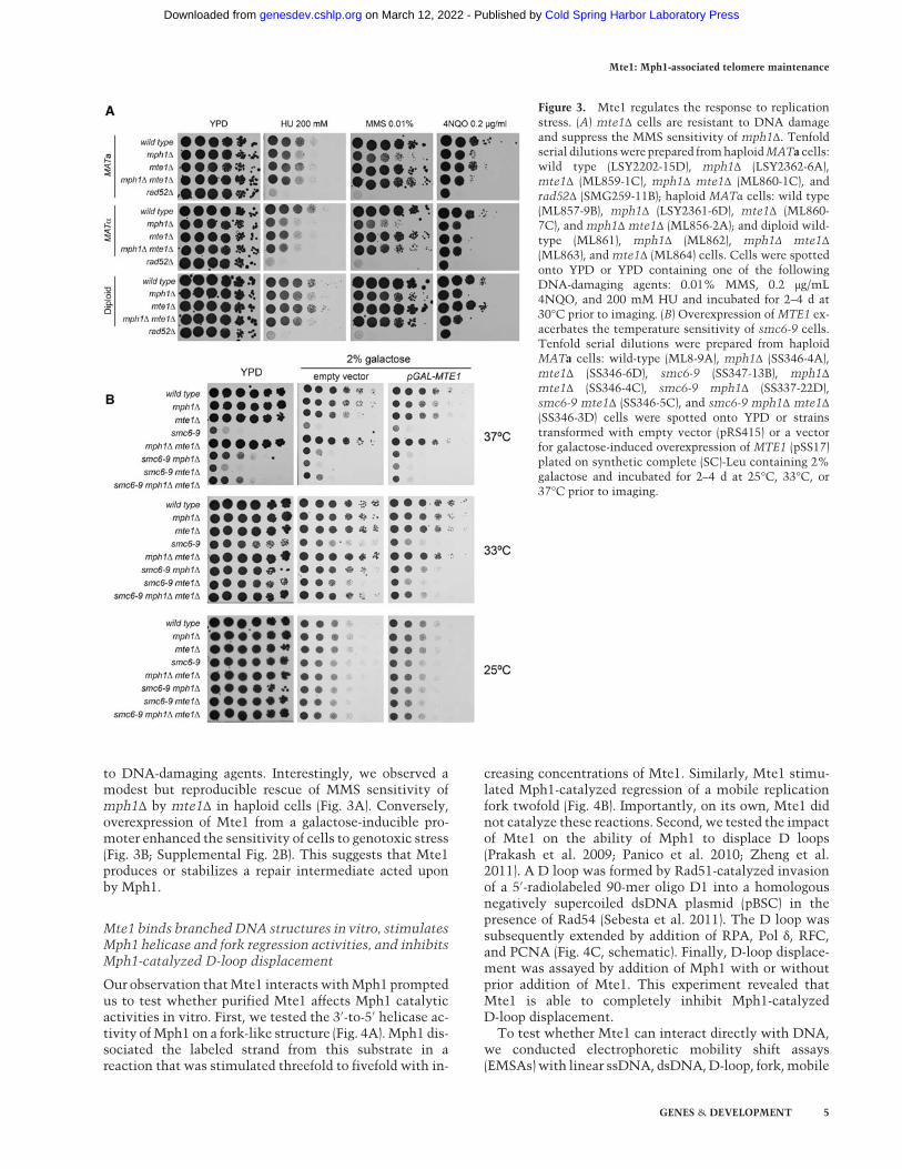
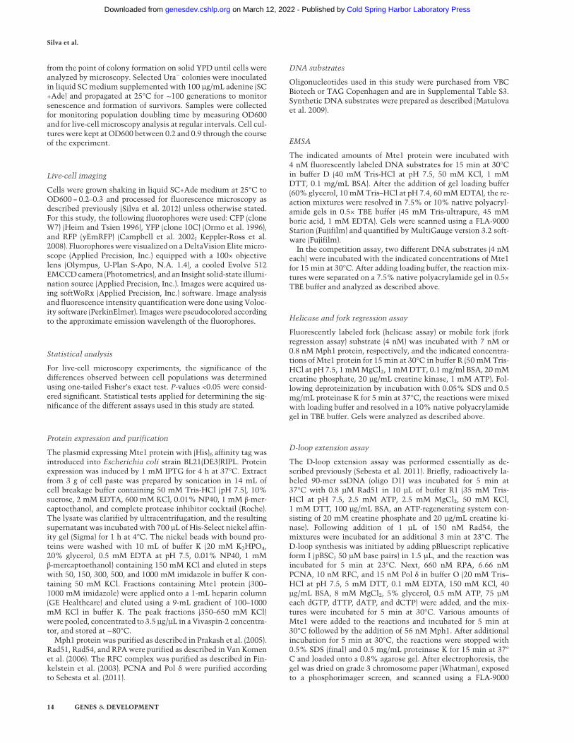
Our observation thatMte1 interacts withMph1 promptedus to test whether purified Mte1 affects Mph1 catalyticactivities in vitro. First, we tested the 3′-to-5′ helicase ac-tivity ofMph1 on a fork-like structure (Fig. 4A). Mph1 dis-sociated the labeled strand from this substrate in areaction that was stimulated threefold to fivefold with in-
creasing concentrations of Mte1. Similarly, Mte1 stimu-lated Mph1-catalyzed regression of a mobile replicationfork twofold (Fig. 4B). Importantly, on its own, Mte1 didnot catalyze these reactions. Second, we tested the impactof Mte1 on the ability of Mph1 to displace D loops(Prakash et al. 2009; Panico et al. 2010; Zheng et al.2011). A D loop was formed by Rad51-catalyzed invasionof a 5′-radiolabeled 90-mer oligo D1 into a homologousnegatively supercoiled dsDNA plasmid (pBSC) in thepresence of Rad54 (Sebesta et al. 2011). The D loop wassubsequently extended by addition of RPA, Pol δ, RFC,and PCNA (Fig. 4C, schematic). Finally, D-loop displace-ment was assayed by addition of Mph1 with or withoutprior addition of Mte1. This experiment revealed thatMte1 is able to completely inhibit Mph1-catalyzedD-loop displacement.To test whether Mte1 can interact directly with DNA,
we conducted electrophoretic mobility shift assays(EMSAs)with linear ssDNA, dsDNA,D-loop, fork,mobile
Figure 3. Mte1 regulates the response to replicationstress. (A) mte1Δ cells are resistant to DNA damageand suppress the MMS sensitivity of mph1Δ. Tenfoldserial dilutionswere prepared fromhaploidMATa cells:wild type (LSY2202-15D), mph1Δ (LSY2362-6A),mte1Δ (ML859-1C), mph1Δ mte1Δ (ML860-1C), andrad52Δ (SMG259-11B); haploid MATα cells: wild type(ML857-9B), mph1Δ (LSY2361-6D), mte1Δ (ML860-7C), and mph1Δ mte1Δ (ML856-2A); and diploid wild-type (ML861), mph1Δ (ML862), mph1Δ mte1Δ(ML863), and mte1Δ (ML864) cells. Cells were spottedonto YPD or YPD containing one of the followingDNA-damaging agents: 0.01% MMS, 0.2 μg/mL4NQO, and 200 mM HU and incubated for 2–4 d at30°C prior to imaging. (B) Overexpression of MTE1 ex-acerbates the temperature sensitivity of smc6-9 cells.Tenfold serial dilutions were prepared from haploidMATa cells: wild-type (ML8-9A), mph1Δ (SS346-4A),mte1Δ (SS346-6D), smc6-9 (SS347-13B), mph1Δmte1Δ (SS346-4C), smc6-9 mph1Δ (SS337-22D),smc6-9 mte1Δ (SS346-5C), and smc6-9 mph1Δ mte1Δ(SS346-3D) cells were spotted onto YPD or strainstransformed with empty vector (pRS415) or a vectorfor galactose-induced overexpression of MTE1 (pSS17)plated on synthetic complete (SC)-Leu containing 2%galactose and incubated for 2–4 d at 25°C, 33°C, or37°C prior to imaging.
Mte1: Mph1-associated telomere maintenance
GENES & DEVELOPMENT 5
Cold Spring Harbor Laboratory Press on March 12, 2022 - Published by genesdev.cshlp.orgDownloaded from
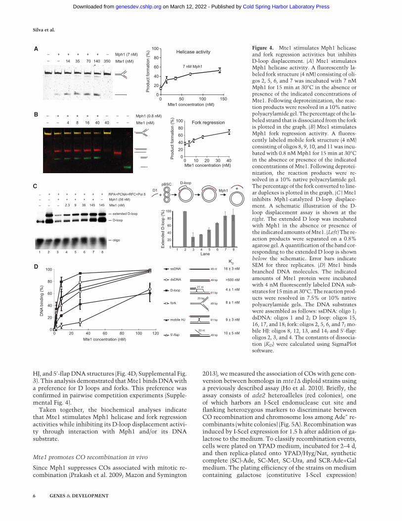
HJ, and 5′-flapDNA structures (Fig. 4D; Supplemental Fig.3). This analysis demonstrated thatMte1 bindsDNAwitha preference for D loops and forks. This preference wasconfirmed in pairwise competition experiments (Supple-mental Fig. 4).
Taken together, the biochemical analyses indicatethat Mte1 stimulates Mph1 helicase and fork regressionactivities while inhibiting its D-loop displacement activi-ty through interaction with Mph1 and/or its DNAsubstrate.
Mte1 promotes CO recombination in vivo
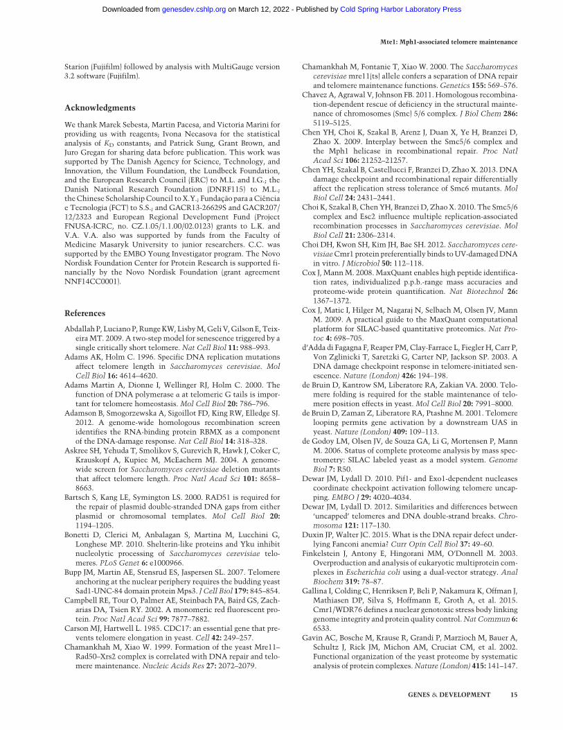
Since Mph1 suppresses COs associated with mitotic re-combination (Prakash et al. 2009; Mazon and Symington
2013), wemeasured the association of COswith gene con-version between homologs in mte1Δ diploid strains usinga previously described assay (Ho et al. 2010). Briefly, theassay consists of ade2 heteroalleles (red colonies), oneof which harbors an I-SceI endonuclease cut site andflanking heterozygous markers to discriminate betweenCO recombination and chromosome loss among Ade+ re-combinants (white colonies) (Fig. 5A). Recombinationwasinduced by I-SceI expression for 1.5 h after addition of ga-lactose to the medium. To classify recombination events,cells were plated on YPAD medium, incubated for 2–4 d,and then replica-plated onto YPAD/Hyg/Nat, syntheticcomplete (SC)-Ade, SC-Met, SC-Ura, and SCR-Ade+Galmedium. The plating efficiency of the strains on mediumcontaining galactose (constitutive I-SceI expression)
0
20
40
60
80
100
0 20 40 60 80 100 120
DN
A bi
ndin
g (%
)
Mte1 concentration (nM)
ssDNA 16 ± 3 nM
>500 nM
4 ± 1 nM
8 ± 1 nM
9 ± 3 nM
10 ± 5 nM
KD
49 nt
21 nt
20 bp
20 nt
dsDNA
D-loop
fork
mobile HJ
5'-flap
61 bp
61 bp
49 bp
49 bp
49 bp
0
20
40
60
80
100
0 50 100 150
7 nM Mph1
Helicase activity
Fork regression
Pro
duct
form
atio
n (%
)
Mte1 concentration (nM)
_ Mte1 (nM)14 3501407035_
_ + + + + + _ Mph1 (7 nM)
_ Mte1 (nM)16 4040_
_ + + +
4 8
+ + ___
__Mph1 (0.8 nM)
0
20
40
60
80
100
0 10 20 30 40 Pro
duct
form
atio
n (%
)Mte1 concentration (nM)
Mte1 (nM)2.3 9 36 145 145 _+ + + + _ Mph1 (56 nM)
D-loop
extended D-loop
RPA+PCNA+RFC+Pol δ+ + + + +__ _
_ _
oligo
++ +
0
20
40
60
80
100
1 2 3 4 5 6 7 8
Ext
ende
d D
-loop
(%)
Lane1 2 3 4 5 6 7 8
A
B
D
C+
+D1pBSC D-loop
Mph1
Figure 4. Mte1 stimulates Mph1 helicaseand fork regression activities but inhibitsD-loop displacement. (A) Mte1 stimulatesMph1 helicase activity. A fluorescently la-beled fork structure (4 nM) consisting of oli-gos 2, 5, 6, and 7 was incubated with 7 nMMph1 for 15 min at 30°C in the absence orpresence of the indicated concentrations ofMte1. Following deproteinization, the reac-tion products were resolved in a 10% nativepolyacrylamide gel. Thepercentage of the la-beled strand that is dissociated from the forkis plotted in the graph. (B) Mte1 stimulatesMph1 fork regression activity. A fluores-cently labeled mobile fork structure (4 nM)consisting of oligos 8, 9, 10, and 11was incu-bated with 0.8 nMMph1 for 15 min at 30°Cin the absence or presence of the indicatedconcentrations of Mte1. Following deprotei-nization, the reaction products were re-solved in a 10% native polyacrylamide gel.The percentage of the fork converted to line-ar duplexes is plotted in the graph. (C ) Mte1inhibits Mph1-catalyzed D-loop displace-ment. A schematic illustration of the D-loop displacement assay is shown at theright. The extended D loop was incubatedwith Mph1 in the absence or presence ofthe indicated amounts ofMte1. (Left) The re-action products were separated on a 0.8%agarose gel. A quantification of the band cor-responding to the extended D loop is shownbelow the schematic. Error bars indicateSEM for three replicates. (D) Mte1 bindsbranched DNA molecules. The indicatedamounts of Mte1 protein were incubatedwith 4 nM fluorescently labeled DNA sub-strates for 15min at 30°C.The reactionprod-ucts were resolved in 7.5% or 10% nativepolyacrylamide gels. The DNA substrateswere assembled as follows: ssDNA: oligo 1;dsDNA: oligos 1 and 2; D loop: oligos 15,16, 17, and 18; fork: oligos 2, 5, 6, and 7; mo-bile HJ: oligos 8, 12, 13, and 14; and 5′-flap:oligos 2, 3, and 4. The constants of dissocia-tion (KD) were calculated using SigmaPlotsoftware.
Silva et al.
6 GENES & DEVELOPMENT
Cold Spring Harbor Laboratory Press on March 12, 2022 - Published by genesdev.cshlp.orgDownloaded from

relative to growth on glucose-containing medium wasused to normalize the fractions of recombination events.Recombination events were classified as NCO, CO, orbreak-induced replication (BIR) based on LOH forthe Nat and Hph markers. Half-sectored colonies (red/white) were generated by a recombination event andcould be scored with less ambiguity than solid white orsolid red colonies (Ho et al. 2010). Analysis of the red/white-sectored colonies revealed that mte1Δ cells had asignificantly lower CO frequency than the wild type (4%and 9%; P = 0.02), and the mph1Δ mte1Δ double mutant
reverted the higher frequency of COs seen in the mph1Δsingle mutant to wild-type levels (8% and 18%; P = 0.002)(Fig. 5B).We also tested whether Mte1 affected spontaneous mi-
totic direct-repeat recombination in haploid cells. Therate of direct-repeat recombination was largely unaffectedby the deletion of mte1Δ (Fig. 5C,D). The mph1Δ mte1Δdouble mutant showed a significant decrease in Leu+Ura−
recombinants, which could reflect either increased single-strand annealing (SSA) or unequal sister chromatid ex-change (USCE).
WT
mte1∆
mph1∆
mph1∆
mte1
∆
Leu+ Ura-
*
Leu+ Ura+
Rec
ombi
natio
n ra
te (p
er 1
05 )∆B leu2 ∆E LEU2
LEU2 ∆B leu2 ∆E
∆B leu2 ∆E ∆B leu2 ∆E
URA3
URA3
URA3
LEU2or
or
MET22 ade2-I Hph
* ade2-n NatURA3
Chr. XV I-SceI cs
0 20 40 60 80
WT
WT
mph1∆
mph1∆
mte1∆
mte1∆
mph1∆ mte1∆
mph1∆mte1∆
whiteredred/white
Distribution of colony colors after I-SceI induction (%)
C
E
A
B
D
0 20 40 60 80 100
NCO CO BIR
Events among red/white colonies normalized to PE (%)
WT(90%)
mph1∆(100%)
mte1∆(77%)
mph1∆ mte1∆(93%)
* *
**
0
1
2
3
4
5
0 5
10 15 20 25 30 35 40
Rat
e of
Can
R (p
er 1
07 )
Figure 5. Mte1 promotes CO formation during interhomolog recombination. (A) Schematic representation of the assay used to infer COfrequency showing the positions of genetic markers on chromosome XV. The arrow indicates the I-SceI cut site within the ade2-I allele.The asterisk notes the location of the ade2-nmutation present on the homolog.Hph andNat cassettes are inserted at theHIS3 locus, 150kb downstream from the ade2 locus. The solid circle indicates the centromere.Not drawn to scale. (B)Mte1 promotesCO formation. (Left)Distribution of colony colors after I-SceI induction and plating on YPD. (Right) Distribution of NCO, CO, and BIR products for red/white-sectored colonies for wild-type (ML861), mph1Δ (ML862), mph1Δ mte1Δ (ML863), and mte1Δ (ML864) strains. Plating efficiency (PE) isindicated in parentheses. (C ) Schematic representation of the assay used for measuring spontaneous mitotic direct repeat recombinationbetween leu2-ΔEcoRI and leu2-ΔBstEII heteroalleles. (D) Mte1 and Mph1 synergistically suppress single-strand annealing (SSA)/unequalsister chromatid exchange (USCE). Recombination rates were calculated using theMSS-MLEmethod via the FALCORWeb tool for wild-type (ML829-13A),mph1Δ (ML829-12B),mte1Δ (ML829-9B), andmph1Δmte1Δ (ML829-11C) cells. For each strain, the fluctuation anal-ysis was based on nine trials. Error bars represent 95% confidence intervals. (∗) Fraction of Leu+ Ura− recombinants significantly differentfrom thewild type; P = 0.02, Student’s t-test. (E)mte1Δ cells exhibit thewild-typemutation rate. Forwardmutation at theCAN1 locuswasdetermined by fluctuation analysis of 10 trials for wild type (ML904-14C), mph1Δ (ML904-6D), mte1Δ (ML904-14A), and mph1Δ mte1Δ(ML904-7C) strains.
Mte1: Mph1-associated telomere maintenance
GENES & DEVELOPMENT 7
Cold Spring Harbor Laboratory Press on March 12, 2022 - Published by genesdev.cshlp.orgDownloaded from
MTE1 overexpression exacerbates the replication stresssensitivity of the smc6 mutant
Deleting MPH1 relieves the replication stress sensitivityof smc5/6 mutant cells by reducing the formation of re-combination intermediates (Chen et al. 2009, 2013;Choi et al. 2010; Chavez et al. 2011). To explore a possibleparticipation of Mte1 in the formation or stabilization ofsuch recombination intermediates together with Mph1,we tested the effect ofmte1Δ on the replication stress sen-sitivity of the smc6-9mutants. In cells grown at the semi-permissive temperature, accumulation of recombinationintermediates leads to genomic instability and increasedcell death. Contrary tomph1Δ, we observed no phenotyp-ic rescue of smc6-9 by mte1Δ (Fig. 3B). However, overex-pression of MTE1 further enhanced the temperaturesensitivity of smc6-9 cells similar to the increased DNAdamage sensitivity upon MTE1 overexpression (Supple-mental Fig. 2B).
MPH1 was originally identified on the basis of elevatedspontaneousmutation rates of haploid cells (Scheller et al.2000). We therefore tested whetherMTE1 is related to themutator phenotype ofmph1Δ cells bymonitoring forwardmutation at the CAN1 locus, which leads to canavanineresistance. This analysis showed that mph1Δ leads to aneightfold increase in the rate of CAN1 mutation indepen-dently of MTE1 (Fig. 5E). Taken together, we concludethat Mte1 promotes CO recombination in both the pres-ence and absence of Mph1, andmte1Δ does not relieve ei-ther replication stress of smc5/6 cells or the mutatorphenotype of mph1Δ.
MTE1 deletion increases telomere length
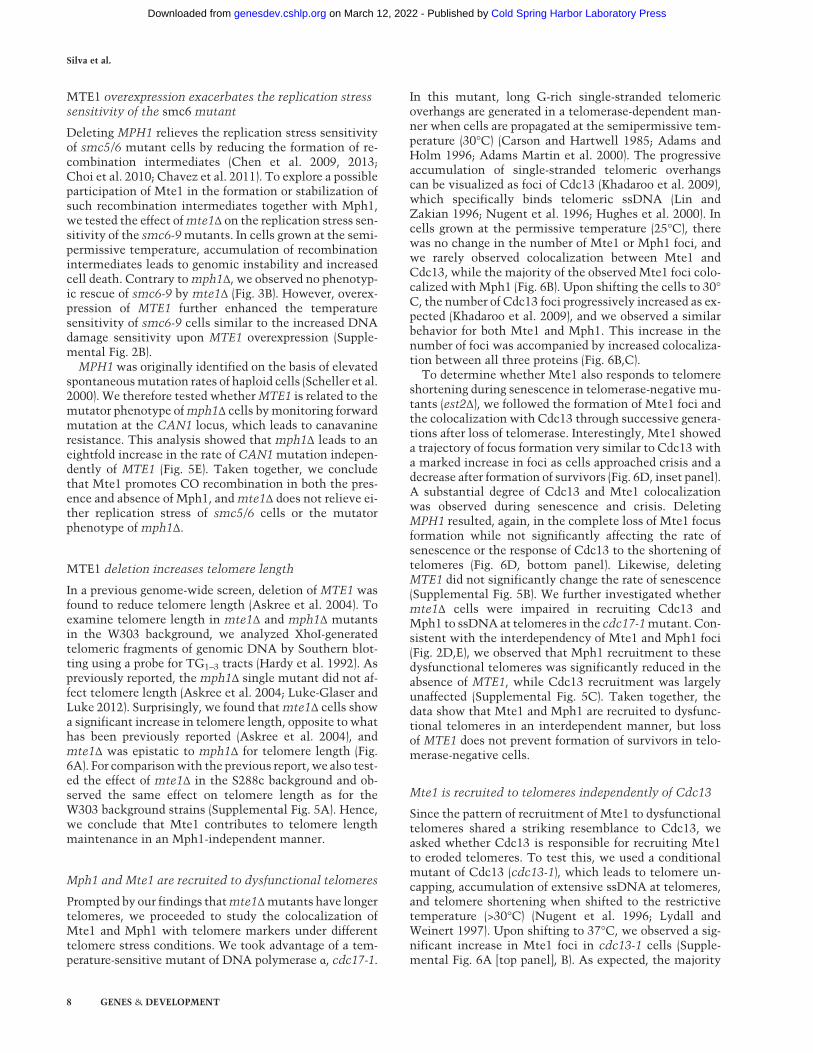
In a previous genome-wide screen, deletion of MTE1 wasfound to reduce telomere length (Askree et al. 2004). Toexamine telomere length in mte1Δ and mph1Δ mutantsin the W303 background, we analyzed XhoI-generatedtelomeric fragments of genomic DNA by Southern blot-ting using a probe for TG1–3 tracts (Hardy et al. 1992). Aspreviously reported, the mph1Δ single mutant did not af-fect telomere length (Askree et al. 2004; Luke-Glaser andLuke 2012). Surprisingly, we found thatmte1Δ cells showa significant increase in telomere length, opposite to whathas been previously reported (Askree et al. 2004), andmte1Δ was epistatic to mph1Δ for telomere length (Fig.6A). For comparisonwith the previous report, we also test-ed the effect of mte1Δ in the S288c background and ob-served the same effect on telomere length as for theW303 background strains (Supplemental Fig. 5A). Hence,we conclude that Mte1 contributes to telomere lengthmaintenance in an Mph1-independent manner.
Mph1 and Mte1 are recruited to dysfunctional telomeres
Prompted by our findings thatmte1Δmutants have longertelomeres, we proceeded to study the colocalization ofMte1 and Mph1 with telomere markers under differenttelomere stress conditions. We took advantage of a tem-perature-sensitive mutant of DNA polymerase α, cdc17-1.
In this mutant, long G-rich single-stranded telomericoverhangs are generated in a telomerase-dependent man-ner when cells are propagated at the semipermissive tem-perature (30°C) (Carson and Hartwell 1985; Adams andHolm 1996; Adams Martin et al. 2000). The progressiveaccumulation of single-stranded telomeric overhangscan be visualized as foci of Cdc13 (Khadaroo et al. 2009),which specifically binds telomeric ssDNA (Lin andZakian 1996; Nugent et al. 1996; Hughes et al. 2000). Incells grown at the permissive temperature (25°C), therewas no change in the number of Mte1 or Mph1 foci, andwe rarely observed colocalization between Mte1 andCdc13, while the majority of the observed Mte1 foci colo-calized with Mph1 (Fig. 6B). Upon shifting the cells to 30°C, the number of Cdc13 foci progressively increased as ex-pected (Khadaroo et al. 2009), and we observed a similarbehavior for both Mte1 and Mph1. This increase in thenumber of foci was accompanied by increased colocaliza-tion between all three proteins (Fig. 6B,C).
To determine whether Mte1 also responds to telomereshortening during senescence in telomerase-negative mu-tants (est2Δ), we followed the formation of Mte1 foci andthe colocalization with Cdc13 through successive genera-tions after loss of telomerase. Interestingly, Mte1 showeda trajectory of focus formation very similar to Cdc13 witha marked increase in foci as cells approached crisis and adecrease after formation of survivors (Fig. 6D, inset panel).A substantial degree of Cdc13 and Mte1 colocalizationwas observed during senescence and crisis. DeletingMPH1 resulted, again, in the complete loss of Mte1 focusformation while not significantly affecting the rate ofsenescence or the response of Cdc13 to the shortening oftelomeres (Fig. 6D, bottom panel). Likewise, deletingMTE1 did not significantly change the rate of senescence(Supplemental Fig. 5B). We further investigated whethermte1Δ cells were impaired in recruiting Cdc13 andMph1 to ssDNAat telomeres in the cdc17-1mutant. Con-sistent with the interdependency of Mte1 and Mph1 foci(Fig. 2D,E), we observed that Mph1 recruitment to thesedysfunctional telomeres was significantly reduced in theabsence of MTE1, while Cdc13 recruitment was largelyunaffected (Supplemental Fig. 5C). Taken together, thedata show that Mte1 and Mph1 are recruited to dysfunc-tional telomeres in an interdependent manner, but lossof MTE1 does not prevent formation of survivors in telo-merase-negative cells.
Mte1 is recruited to telomeres independently of Cdc13
Since the pattern of recruitment of Mte1 to dysfunctionaltelomeres shared a striking resemblance to Cdc13, weasked whether Cdc13 is responsible for recruiting Mte1to eroded telomeres. To test this, we used a conditionalmutant of Cdc13 (cdc13-1), which leads to telomere un-capping, accumulation of extensive ssDNA at telomeres,and telomere shortening when shifted to the restrictivetemperature (>30°C) (Nugent et al. 1996; Lydall andWeinert 1997). Upon shifting to 37°C, we observed a sig-nificant increase in Mte1 foci in cdc13-1 cells (Supple-mental Fig. 6A [top panel], B). As expected, the majority
Silva et al.
8 GENES & DEVELOPMENT
Cold Spring Harbor Laboratory Press on March 12, 2022 - Published by genesdev.cshlp.orgDownloaded from
mph1∆ 2.9 ± 2.2 mte1∆ 39.6 ± 1.7 *mph1∆ mte1∆ 48.5 ± 3.2 *
Telomere length difference from WT (bp ± SD)Genotype
1.0
Mkb
3.0
2.0
TG1-3
wild
type
mph
1Δ m
te1Δ
mph
1Δ
mte
1Δ
3μm
� �
�
� �
��
� �
�
� �
Mte1-RFP Mph1-YFP Brightfield
Cdc13-CFP merge
Mte1+Mph1+Cdc13 Colocalization:
Mte1+Cdc13 Mte1+Mph1 Mte1 alone Fr
actio
n of
M
te1
foci
0.2
0
0.4
0.6
0.8
1.0
*
*n.s.
n.s.
*
*n.s.
n.s.
1 Mte1 focus/cell≥2 Mte1 foci/cellMph1 fociCdc13 foci
0
20
40
60
80
100 25°C 30°C 1h30
Cel
ls w
ith fo
ci (%
)
* *
*
*
30°C 5h**
**
****
25°C 30°C 1h30
30°C 5h
BA
C
cdc17-1
Cdc13 foci Mte1 foci colocalizing foci
Cel
ls w
ith fo
ci (%
)
est2∆
80
I II III IV V VI0
40 Population doublings
Tim
e / d
oubl
ing
(h)
0
2
4
6
0 20 40 60 80 100
III
IV
V VI
III est2∆ mph1∆
I II III IV V
0
4
8
12
16
0 20 40 60 80
Tim
e / d
oubl
ing
(h)
Population doublings
III
IV
VIII
80
0
40
Cel
ls w
ith fo
ci (%
)
D
Figure 6. Mte1 affects telomere homeostasis. (A) mte1Δ mutants exhibit long telomeres. Genomic DNA was isolated from wild-type(ML829-13A), mph1Δ (ML829-12B), mte1Δ (ML829-9B), and mte1Δ mph1Δ (ML829-11C) cells; digested with XhoI; and subjected toSouthern blotting using a telomeric TG1–3 repeat probe. Terminal restriction fragment analysis was used to calculate the average telo-mere length difference between the wild type and the deletionmutants. (B) Mte1 andMph1 colocalize at dysfunctional telomeres. Quan-titative analysis of Mte1, Mph1, and Cdc13 foci in cdc17-1 cells. Cells with the temperature-sensitive cdc17-1 mutant allele andexpressing fluorescently tagged Mte1-RFP, Mph1-YFP, and Cdc13-CFP (SS303-6A) were pregrown at the permissive temperature(25°C) and subsequently transferred to the semipermissive temperature (30°C) and examined by microscopy at 0, 1.5, and 5 h after tem-perature shift. Error bars represent 95% confidence intervals. n > 200 cells. Asterisks mark values significantly different from measure-ments at the permissive temperature. P < 0.05. (n.s.) Not significantly different from the permissive temperature. (C ) Mte1 and Mph1colocalize with the telomeric single-stranded binding protein Cdc13. The panels show representative images for cells at the semipermis-sive temperature. Red arrowheads mark Mte1-yEmRFP foci, yellow arrowheads indicate Mph1-YFP foci, blue arrowheads indicateCdc13-CFP foci, and green arrowheads mark RFP–YFP–CFP-colocalizing foci. Bar, 3 µm. (D) Mte1 colocalizes with Cdc13 foci duringsenescence. Mte1-YFP and Cdc13-CFP foci were monitored during senescence and formation of survivors in telomerase-negativeMPH1 (SS261-12C) andmph1Δ (SS272-13D) (est2Δ) cells. Senescence was induced by loss of the pAP81 plasmid carrying EST2 and prop-agation in liquid medium at 25°C. Population doubling times were monitored by measuring OD600, and a representative growth curvefor each strain is presented at the top right corner of each panel. Red dots and numbers represent the points at which the microscopyimages were acquired for each culture.
Mte1: Mph1-associated telomere maintenance
GENES & DEVELOPMENT 9
Cold Spring Harbor Laboratory Press on March 12, 2022 - Published by genesdev.cshlp.orgDownloaded from
of these Mte1 foci colocalized with Rad52 (SupplementalFig. 6A; Abdallah et al. 2009; Khadaroo et al. 2009). To alsotest whether resection of uncapped telomeres is requiredfor recruitment of Mte1 to foci, we analyzed the effect ofexo1Δ and pif1Δ on Mte1 focus formation. The Exo15′-to-3′ exonuclease and Pif1 helicase have a prominentrole in resection of uncapped telomeres (Maringele andLydall 2002; Vega et al. 2007; Dewar and Lydall 2010).Recruitment of Mte1 and Rad52 to foci at 37°C was sig-nificantly reduced in exo1Δ and pif1Δ mutant cells, sug-gesting that resection and the formation of ssDNA attelomeres promote Mte1 recruitment rather than Cdc13itself.
Defective HR and telomere dysfunction induce Mte1focus formation
To further characterize the genetic requirements forMte1 focus formation, we tested factors involved inDNA repair and telomere maintenance: Rad52, the majorrecombination mediator in yeast (for review, see Syming-ton et al. 2014), and Mre11, a component of the Mre11–Rad50–Xrs2 complex involved in the initial recognitionand short-range resection of DSBs and in telomere lengthmaintenance (Goudsouzian et al. 2006; Sabourin et al.2007; Longhese et al. 2010). In both the rad52Δ andmre11Δ cells, we observed an increase in both spontane-ous and MMS-induced Mte1 foci (Supplemental Fig. 7A),indicating that Mte1 responds to a defective HR pathway.We also tested a knockout of the Ku complex (yKu70–yKu80), which binds and regulates telomeres and pro-motes nonhomologous end joining (NHEJ) as an alterna-tive DSB repair pathway (Milne et al. 1996; Gravel et al.1998). The yku70Δ cells showed Mte1 foci at levels com-parable with the wild type. The final factor tested wasRif1, a negative regulator of telomere length that bindsRap1 together with Rif2 at telomeres to promote gene si-lencing and end protection (Hardy et al. 1992; Marcandet al. 1997; Ghaemmaghami et al. 2003). In rif1Δ cells,which have longer telomeres than wild type (Teixeiraet al. 2004), we observed a marked increase in Mte1 foci,with a significant percentage of the cells exhibitingmulti-ple foci. Taken together, these results indicate that Mte1responds to defective HR and dysfunctional telomeres butis unaffected by NHEJ defects.
Mte1 colocalizes with persistent DSBs and Mps3at the nuclear periphery
Wenoticed thatMte1 foci often localize at the nuclear pe-riphery, which is reminiscent of uncapped telomeres andpersistent DSBs. These have been shown to relocalize tothe nuclear periphery to promote alternative repair path-ways (Nagai et al. 2008; Kalocsay et al. 2009; Khadarooet al. 2009; Oza et al. 2009). To examine the localizationof Mte1 to a persistent DNA lesion, we introduced an in-ducible I-SceI endonuclease cut site on chromosome V ad-jacent to an array of Tet repressor (TetR)-binding sites incells expressing mRFP-tagged TetR (Fig. 7A). We usedcells expressing the nuclear pore complex (NPC) compo-
nent CFP-Nup49 to infer the position of the break andMte1 foci relative to the nuclear periphery (Fig. 7B). Theposition of Mte1 foci relative to the nuclear peripheryand themarked DSB were analyzed before and at differenttime points after induction of I-SceI expression by galac-tose (Fig. 7C). Notably, the spontaneous Mte1 foci in theuninduced condition localized almost exclusively at thenuclear periphery (95%). Interestingly, a significant de-gree of relocalization ofMte1 to themarked DSB occurredonly after 3 h of continuous I-SceI induction, and less thanhalf of thoseMte1-recognizedDSBs localized at the nucle-ar periphery (Fig. 7C, top panel). The colocalization ofMte1 with the I-SceI cut site was largely independent ofRad52 (Fig. 7C, bottom panel). In mre11Δ mutant cellsthat are defective in short-range resection of DSBs (Mim-itou and Symington 2008), the number of Mte1 foci wasdramatically increased even prior to cleavage of the I-SceI site, but localization of Mte1 to the I-SceI cut sitewas reduced.
To examine the localization ofMMS-inducedMte1 focirelative to telomere-anchoring sites at the nuclear periph-ery, we fluorescently tagged Mps3, an inner nuclearmembrane protein of the SUN family (Sad1/UNC-84 ho-mology) that tethers telomeres to the nuclear peripheryand suppresses their recombination (Bupp et al. 2007;Schober et al. 2009). The majority (>80%) of both sponta-neous and MMS-induced Mte1 foci colocalized with pe-ripheral Mps3 (Supplemental Fig. 7B–D), suggestingeither thatMMS preferentially induces Mte1 recruitmentto Mps3-anchored telomeres or that Mte1 recognizes atype of DNA damage that relocalizes to Mps3.
Discussion
In this study, we identify Mte1 (YGR042W) as a novelinteractor of the Mph1 helicase and show that the twoproteins interdependently colocalize at DNA damage-in-duced foci. The interaction appears to be constitutiveand independent of DNA. In vitro, Mte1 stimulates thehelicase and fork regression activities of Mph1 while in-hibiting the ability of Mph1 to dissociate D loops. Mte1also binds directly to DNAwith a preference for branchedmolecules, such as D loops and fork structures. We notethat similar conclusions were reached in an independentstudy by Xue et al. (2016). Deletion of MTE1 reduces COrecombination and suppresses the MMS sensitivity ofmph1Δ mutant cells, suggesting that Mte1 acts upstreamof Mph1 to promote a DNA repair pathway that requiresMph1. Deletion of MTE1 alone does not cause pro-nounced sensitivity toDNAdamage but results in elonga-tion of telomeres by ∼40 bp. Consistently, Mph1 andMte1 colocalize at dysfunctional telomeres.
Despite the physical interaction and interdependencyof Mte1 and Mph1 foci, the epistasis analysis indicatesthat Mte1 and Mph1 are not strictly dependent on eachother. For example, MTE1 is epistatic to MPH1 for telo-mere length regulation, while MPH1 is epistatic toMTE1 for mutation avoidance. This complex relationshipmay be rationalized by the fact that both Mph1 and Mte1
Silva et al.
10 GENES & DEVELOPMENT
Cold Spring Harbor Laboratory Press on March 12, 2022 - Published by genesdev.cshlp.orgDownloaded from

can interact directly with DNA and thereby potentiallyact in DNA repair independently of each other. Hence,the physical interaction of the two proteinsmay primarilyserve to stabilize their coordinated recruitment to sites ofDNA damage. We also note that the epistasis analysis in-dicated that focus formation of Mph1 and Mte1 is not
strictly required for function. Based on our data, we pro-pose a model in which Mte1 stabilizes D loops eitherdirectly or through inhibition of Mph1-catalyzed dissoci-ation, thereby promoting CO recombination (Fig. 7D).This model is consistent with the reduced CO recom-bination observed in mte1Δ mutants, the biochemical
Cel
ls w
ith M
te1-
YFP
foc
i (%
)
Chr V
I-SceI cs112xtetO
A
B
C
D
F
E
0.0
0.25
0.50
0.75
1.0
Mte1 at nuclear peripheryMte1-TetO at nuclear peripheryMte1-TetO within nucleoplasm
25
50
75
100wt rad52∆ mre11∆
0
Dis
tribu
ition
of M
te1
foci
wt rad52∆ mre11∆
Time after I-SceI induction (min)
Time after I-SceI induction (min)0 18012090 0 18012090 0 18012090
�� �
��
Mte1-YFP
Mte1-YFPTetR-mRFP
TetR-mRFP
Mte1-YFPTetR-mRFPCFP-Nup49
Brightfield
0 18012090 0 18012090 0 18012090
�
regression
reversal
Stalled fork
DSB
T-loop
cleavage
Mph1
Smc5-Smc6 Mte1
Mte1 strand exchange
restart
5’ 5’
5’5’
MHF
templateswitch
Mph1
Mte1
Mph1
extension
5’
5’
displacement
displacement2nd end capture
5’
3’
3’
3’
5’
3’
strand exchange
resolution gap filling
non-crossovercrossover
5’
3’
3’
3’
5’
3’
5’
3’
5’
3’
5’
3’
3’
3’
3’
�
displacement
extension
Mte1
3’
3’
Figure 7. Mte1 localizes to persistentDSBs at the nuclear periphery. (A) Schematic illustration of a construct to visualize an I-SceI-induc-ible DSB. Red boxes represent the relative positions of tandem lacO sites on the left arm of chromosome V. (Blue circle) I-SceI cut site (I-SceIcs); (solid circle) centromere. (B) Mte1 foci localize to the nuclear periphery. The panels show YFP, CFP, and RFP and combinationsof themerged images as well as a bright-field image of representative cells (SS142-23B) after I-SceI induction. Arrowheads indicate two pe-ripheral Mte1 foci (yellow), a colocalizing TetR focus (red), and the NPCs (blue). (C ) Localization of Mte1 to a DSB is delayed inmre11Δ.Wild-type (SS142-23B), rad52Δ (SS159-1B), andmre11Δ (SS160-6D) cells carrying the inducible DSB and expressing Mte1-YFPwere trans-formedwith the plasmids pWJ1320, expressing the I-SceI endonuclease under the control of a galactose-inducible promoter, and pNEB21,expressing the fluorescently taggedNPCsubunitCFP-Nup49.Cellswere grown in syntheticmediumsupplementedwith 2%raffinose andlacking adenine prior to I-SceI induction.To induce expression of the nuclease, galactosewas added to a final concentration of 2%. (Bottompanel) Mte1 focus formation and relative localization to the DSB and NPCwasmonitored by fluorescence microscopy prior to adding ga-lactose and at 90, 120, and 180 min after I-SceI induction. Error bars represent 95% confidence intervals. (D) Model for promotion of COrecombination byMte1. The homologousDNA donor strand is shown in red. (E) Model for regulation ofMph1 byMte1 during replicationstress. (F ) Model for regulation of telomere extension by Mte1. Red strands indicate subtelomeric DNA or telomeric TG1–3 repeats.
Mte1: Mph1-associated telomere maintenance
GENES & DEVELOPMENT 11
Cold Spring Harbor Laboratory Press on March 12, 2022 - Published by genesdev.cshlp.orgDownloaded from
inhibition of D-loop displacement by Mte1, and the laterecruitment of Mte1 to DSBs. We further propose thatMte1 acts upstream of or in parallel to Mph1 to overcomereplication stress (Fig. 7E). This notion is supported by therecruitment of Mte1 and Mph1 to MMS-induced foci, thestimulation of Mph1-catalyzed fork regression and heli-case activities, and the suppression of MMS sensitivityof mph1Δ mutants by mte1Δ. Hence, Mte1 may stabilizefork intermediates that require Mph1 regression or heli-case activity to be resolved. Finally, Mte1 may also pro-mote replication fork restart through template switchingby inhibiting Mph1-catalyzed D-loop displacement.
Telomeres appear to be the preferred substrate forMte1,since almost all (>95%) spontaneous Mte1 foci colocalizewithMps3, which is an anchoring site for telomeres at thenuclear periphery (Bupp et al. 2007; Schober et al. 2009),and telomere dysfunction induced by telomere uncap-ping, overextension, or erosion leads to recruitment ofMte1 to multiple foci that colocalize with the telomeressDNA-binding protein Cdc13. For example, loss ofRIF1, which leads to long telomeres but only slightly in-creases single-stranded telomeric overhangs (Bonettiet al. 2010), increased dramatically the number of sponta-neous Mte1 foci, suggesting that the increased Mte1 fociin rif1Δ could be due to replication stress associatedwith longer telomeres rather than ssDNA at telomeresper se. Furthermore, mre11Δ and rad52Δ also elicited asubstantial increase in NPC-colocalizing Mte1 foci. Inthe context of telomere maintenance, this finding maybe rationalized by reports that Mre11 is involved in G-tail formation and the recruitment of telomerase (Nugentet al. 1998; Chamankhah and Xiao 1999; Chamankhahet al. 2000), and 20% of spontaneous Rad52 colocalizewith Cdc13 (Khadaroo et al. 2009), suggesting that bothMre11 and Rad52 play an active role in telomere mainte-nance even in telomerase-proficient cells. Interestingly,MMS treatment also preferentially induced Mte1 focithat colocalized with Mps3 at the nuclear periphery,where telomeres are anchored (Bupp et al. 2007; Schoberet al. 2009), suggesting that telomeres are particularly sen-sitive to replication stress. Contrary to persistent DSBsand collapsed replication forks, stalled replication forkshave been shown not to relocalize to Mps3 (Kalocsayet al. 2009; Oza et al. 2009; Horigome et al. 2014), so itis unlikely that MMS-induced Mte1 foci represent stalledreplication forks. It is interesting to note that yeast telo-meres have been proposed to form T loops (Luke-Glaserand Luke 2012), where telomeric 3′ overhangs invade telo-meric dsDNA, similar towhat has been reported formam-malian telomeres (Griffith et al. 1999). However, thetelomere fold-back structures reported so far in yeast ap-pear to involve interaction of the telomere end with thesubtelomere region of the chromosome (Strahl-Bolsingeret al. 1997; Pryde and Louis 1999; de Bruin et al. 2000,2001; Robin et al. 2014). Irrespective of the precise config-uration of yeast fold-back structures, Mte1might exert itseffect on telomere length by preventing their dissociation(Fig. 7F). Notably, in human cells, the RTEL1 helicase andputative ortholog of Mph1 provide T-loop-unwinding ac-tivity (Sarek et al. 2015).
The putative homologs toMTE1 in fission yeast and hu-mans are Dbl2 and ZGRF1, respectively. The three pro-teins share a domain of unknown function designatedDUF2439 (Pfam domain PF10382) (Punta et al. 2012). Sim-ilar to our findings, Dbl2 colocalizes with Rad22 (Rad52)and Fml1 (Mph1), and the ability of Fml1 to form nuclearfoci in response to replicative stress requires the presenceof Dbl2 (Yu et al. 2013). Further evidence for a conservedfunction of Mte1/Dbl2 comes from a genome-wide screenthat identified the dbl2Δ deletion mutant as having elon-gated telomeres (Liu et al. 2010). The putative humanortholog of Mte1, ZGRF1, was identified in two large-scale studies for genes required for resistance to cross-linking agents (Smogorzewska et al. 2010) and genes re-quired for recombinational repair of an endonuclease-in-duced DSB (Adamson et al. 2012). Although ZGRF1awaits further characterization, it is interesting to notethat ZGRF1 harbors a C-terminal extension, which is pre-dicted to encode a helicase domain. Thus, it is possiblethat, in human cells, the Mte1–Mph1 function is encodedwithin a single polypeptide, and it will be important to in-vestigate the relationship of ZGRF1with FANCM and FAor other cancer predisposition syndromes.
Materials and methods
Yeast strains and plasmids
Standard procedures for yeast strain construction and cell growthwere used (Sherman et al. 1986). Strains generated for this studywere isogenic RAD5 derivatives of W303 (Supplemental TableS2; Thomas andRothstein 1989). Strains from the gene disruptioncollection (Invitrogen) were derivatives of S288C. AnMTE1 dele-tion strain was generated by homology targeted replacement ofthe MTE1 ORF by a fragment containing the NatMX resistancecassette using primers NatMX-YGR-Fw and NatMX-YGR-Rv.Fluorescently tagged proteins were constructed for expressionfrom their native chromosomal loci with a four-alanine linkeras described using primers YGR042W-Fw, YGR042W-YFP-Rv,YGR042W-YFP-Fw, and YGR042W-down-Rv (Mte1-YFP);YGR042W.Fw, YGR042W_cherrystart.Rv, YGR042W_cherry-stop.Fw, and YGR042W-down Rv (MTE1-yEmRFP); andMph1fwd, Mph1-Tup(-stop), Mph1-Tdown, and Mph1down(Mph1-YFP) (Reid et al. 2002; Silva et al. 2012). Myc-taggedMph1 was generated in the W303 background by transfer of anMPH1-13xMyc::HIS3 allele from strain BL295 after PCR amplifi-cation using primers Mph1fwd and Mph1-rv2. BiFC assay strainswere generated as described previously (Sung and Huh 2007).Mte1-VC was constructed using primers YGR042W-F2 andYGR042W-R1 to amplify theVC fragment frompML103 contain-ing VC155-tADH1-NatMX6. To generate Mph1-VN, the primerpairMPH1-F2 andMPH1-R1was used to amplify a fragment con-tainingVN173-tADH1-Kl.URA3 fromgenomicDNAof the strainfrom the VN collection library (Bioneer) harboring RAD59-VN173-tADH1-Kl.URA3 as template. Plasmid pML103 was con-structed by subcloning an FspI fragment containing the NatMXcassette from p4339 (Goldstein andMcCusker 1999) into FspI-di-gested pFA6a-VC-KanMX6 (Sung and Huh 2007). Mph1-mCherry-sfGFP was constructed using primers MPH1-S3-Fnewand MPH1-S2-R to amplify mCherry-sfGFP::HphNT1 frompMaM60 (Khmelinskii et al. 2012). Cells expressing VC fromthe MTE1 promoter were constructed using primers pMTE1-F2and YGR042W-R1 to amplify the VC fragment from pML103with 5′ ends designed to replaceMTE1 upon genomic integration.
Silva et al.
12 GENES & DEVELOPMENT
Cold Spring Harbor Laboratory Press on March 12, 2022 - Published by genesdev.cshlp.orgDownloaded from
All strains and plasmids were verified by sequencing. All prim-er pairs used in strain construction are in Supplemental Table S3.The plasmid for bacterial expression of Mte1 was generated bycloning of the MTE1 gene into the EcoRI and SalI sites of thepET28a vector. The plasmid for galactose-induced overexpressionof MTE1 was constructed by first replacing the URA3 gene inpML174 (Sabatier 2014)—harboring the inducible GAL1-10 pro-moter and the ADH1 termination sequence separated by SalIand NotI cloning sites—with a LEU2 containing the NaeI/SwaIfragment from pRS415 (Sikorski and Hieter 1989) to producepSS15. Next, the MTE1 gene was PCR-amplified from genomicDNA using SalI/NotI adapted primers Gal1-Mte1-Fw (SalI) andGal1-Mte1-Rv (NotI) and cloned into the SalI/NotI site of pSS15to yield pSS17.
SILAC and MS analysis
For identification of proteins interacting withMph1, lysine auxo-troph cells expressingMph1-YFP fusion proteinwere grown in SCmedium containing either Lys0 (12C6 and 14N2) or Lys8 (13C6and 15N2) (Sigma, catalog no. 608041) for >10 generations. Cul-tures were treated with 0.03% MMS (Sigma) for 1 h prior to har-vesting to induce DNA damage and allow Mph1-YFP toaccumulate at foci. Whole-cell extracts were prepared in lysisbuffer without EDTA (10 mM Tris-HCl at pH 7.5, 150 mMNaCl, 0.5% NP40, 1 mM PMSF, 1× complete protease inhibitor[Roche]), and immunoprecipitation of YFP-tagged Mph1 was per-formed using 25 μL of GFP-Trap_A beads (Chromotek) for 2 h at4°C according to the manufacturer’s instructions. After washingin dilution buffer (10 mM Tris-HCl at pH 7.5, 150 mM NaCl, 1mMPMSF, 1× complete protease inhibitor [Roche]), GFP-Trap_Abeads incubated with heavy-labeled and light-labeled proteinswere mixed and washed again. To release bound proteins fromthe beads, samples were incubated with 1 vol of 4× LDS samplebuffer (NuPAGE) and 40 mM DTT for 10 min at 70°C followedby 35 min at room temperature. Chloroacetoamide was addedto a final concentration of 110 mM, and samples were incubatedfor 45 min at room temperature. Proteins were resolved by SDS-PAGE, and each loaded gel lanewas sliced into three or four slicescontaining an estimated equal amount of proteins. Gel pieceswere in-gel-digested in trypsin protease solution (13 ng/µL tryp-sin, 20 mM NH4HCO3). In-gel trypsin digestion was carried outovernight at 37°C. Tryptic peptides were extracted by incubatingwith increasingly organic solutions for 20 min and collecting theresulting fractions. The gel-extracted peptides were loaded ontoStage Tips as described (Rappsilber et al. 2007). Samples were an-alyzed by liquid chromatography-tandem MS (LC-MS/MS) usingthe EASY-nLC HPLC (Thermo Scientific) in combination withLTQ-Orbitrap Velos or Q-Exactive (Thermo Scientific). For theVelos instrument, the top 10 most intense peaks were analyzedafter each full scan,while the top 12were analyzed for theQExac-tive. MS raw data files were analyzed with the MaxQuant soft-ware package (developer’s version 1.2.2.9) (Cox and Mann 2008;Cox et al. 2009). Full scan peaks and fragment scan peakswere searched against the Saccharomyces Genome Database(SGD) release 63 containing 6717 putative protein sequences(http://downloads.yeastgenome.org). False discovery rate was es-timated using a target–decoy approach, allowing a maximum of1% false identifications from the reversed sequence database.
Immunoprecipitation and immunoblot analysis
Whole-cell extracts were prepared in lysis buffer without EDTA(10 mM Tris-HCl at pH 7.5, 150 mM NaCl, 0.5% NP40, 1 mMPMSF, 1× complete protease inhibitor [Roche]). For DNase treat-
ment, extracts were incubatedwith 50U of DNase I withmild ro-tation for 45 min at 16°C. Immunoprecipitation of YFP-taggedMte1 was performed using 25 μL of GFP-Trap_M beads (Chromo-tek) for 2 h at 4°C according to the manufacturer’s instructions.Pull-downs were washed with increasing salt concentrations(10 mM Tris-HCl at pH 7.5, 150–350 mM NaCl, 1 mM PMSF,1× complete protease inhibitor [Roche]). Immunoblot analysiswas performed following standard protocol. Briefly, protein sam-ples were mixed with 2× SDS buffer, boiled, separated by SDS-PAGE, and electrotransferred onto nitrocellulose membranes.The membranes were first probed for Mte1-YFP using a mouseprimary anti-GFP (Roche) and anti-mouse conjugated toHRP sec-ondary antibody (Dako). For detection of myc-tagged Mph1,membranes were subjected to mild stripping according to Abcamprotocol. Briefly, membranes were washed twice for 5 min withstripping buffer (0.2 M glycine, 0.001% SDS, 0.01% Tween20 at pH 2.2), washed twice for 10 min in PBS, washed twice for5 min in TBS-T, and finally blocked and reprobed using primarymouse anti-c-myc antibody (Santa Cruz Biotechnology, 9E10).
DNA-damaging agents
Unless otherwise stated, cells were grown overnight at 30°C tosaturation in liquid YPD medium, and 10-fold serial dilutionswere prepared for spotting on solid medium. DNA damage sensi-tivity was assayed by allowing cells to grow for up to 72 h at 30°Cafter spotting onto YPD or YPD containing ZEO (Invitrogen),MMS (Sigma), HU (Sigma), 4NQO (Sigma), or CPT (Sigma) tothe final concentrations stated in the figures. To test cisplatin sen-sitivity, cultureswere grown toOD600 = 0.4 and incubated for 3 hin the presence of 0.3mMcisplatin. Cells were then serially dilut-ed, plated onto solidYPDplates, and allowed to grow for 48 huntilimaging. For MTE1 overexpression experiments, cells trans-formed with the control vector (pRS415) and the vector for galac-tose-induced overexpression of MTE1 (pSS17) were grownovernight at 25°C or 30°C to saturation in liquid SC-Leu supple-mented with 2% raffinose medium, and 10-fold serial dilutionswere prepared for spotting on solid medium containing 2% glu-cose or 2%galactose and allowed to growup to 96huntil imaging.
I-SceI induction
Induction of a single fluorescentlymarked DNADSBwas carriedout essentially as described (Lisby et al. 2003). Briefly, cells con-taining an I-SceI cut site integrated immediately upstream of aura3::3xURA3-tetOx112 tandem array on chromosomeV and ex-pressing TetR-mRFP from the intergenic region iYGL119Wweregrown to an optical density (OD600) of ∼0.2–0.3 in synthetic me-dium containing 2% raffinose as a carbon source. For inducibleexpression of the I-SceI endonuclease, cells were transformedwith the plasmid pWJ1320 harboring the I-SceI gene controlledby a GAL1-10 promoter and grown in medium lacking adenine.Expression was induced by adding galactose to the cultures to afinal concentration of 2%.
Senescence induction in telomerase-deficient cells
Cells with EST2-deleted in the genome were propagated at 25°Cin liquid medium lacking uracil to select for the plasmid pAP81(for ectopic expression of EST2), and foci of Mph1-YFP, Cdc13-CFP, and Rad52-RFP (SS283-23D) were quantitated prior to telo-merase loss. Loss of the telomerase-encoding plasmid wasenabled by streaking cells on solid YPD medium and subse-quently checking for loss of growth in SC-Ura by replica plating.We estimate that this procedure involved ∼20–30 generations
Mte1: Mph1-associated telomere maintenance
GENES & DEVELOPMENT 13
Cold Spring Harbor Laboratory Press on March 12, 2022 - Published by genesdev.cshlp.orgDownloaded from
from the point of colony formation on solid YPD until cells wereanalyzed by microscopy. Selected Ura− colonies were inoculatedin liquid SCmedium supplemented with 100 µg/mL adenine (SC+Ade) and propagated at 25°C for ∼100 generations to monitorsenescence and formation of survivors. Samples were collectedfor monitoring population doubling time by measuring OD600and for live-cell microscopy analysis at regular intervals. Cell cul-tures were kept at OD600 between 0.2 and 0.9 through the courseof the experiment.
Live-cell imaging
Cells were grown shaking in liquid SC+Ade medium at 25°C toOD600 = 0.2–0.3 and processed for fluorescence microscopy asdescribed previously (Silva et al. 2012) unless otherwise stated.For this study, the following fluorophores were used: CFP (cloneW7) (Heim and Tsien 1996), YFP (clone 10C) (Ormo et al. 1996),and RFP (yEmRFP) (Campbell et al. 2002; Keppler-Ross et al.2008). Fluorophoreswere visualized on aDeltaVision Elitemicro-scope (Applied Precision, Inc.) equipped with a 100× objectivelens (Olympus, U-Plan S-Apo, N.A. 1.4), a cooled Evolve 512EMCCDcamera (Photometrics), and an Insight solid-state illumi-nation source (Applied Precision, Inc.). Images were acquired us-ing softWoRx (Applied Precision, Inc.) software. Image analysisand fluorescence intensity quantification were done using Voloc-ity software (PerkinElmer). Images were pseudocolored accordingto the approximate emission wavelength of the fluorophores.
Statistical analysis
For live-cell microscopy experiments, the significance of thedifferences observed between cell populations was determinedusing one-tailed Fisher’s exact test. P-values <0.05 were consid-ered significant. Statistical tests applied for determining the sig-nificance of the different assays used in this study are stated.
Protein expression and purification
The plasmid expressing Mte1 protein with (His)6 affinity tag wasintroduced into Escherichia coli strain BL21(DE3)RIPL. Proteinexpression was induced by 1 mM IPTG for 4 h at 37°C. Extractfrom 3 g of cell paste was prepared by sonication in 14 mL ofcell breakage buffer containing 50 mM Tris-HCl (pH 7.5), 10%sucrose, 2 mM EDTA, 600 mM KCl, 0.01% NP40, 1 mM β-mer-captoethanol, and complete protease inhibitor cocktail (Roche).The lysate was clarified by ultracentrifugation, and the resultingsupernatant was incubatedwith 700 µL of His-Select nickel affin-ity gel (Sigma) for 1 h at 4°C. The nickel beads with bound pro-teins were washed with 10 mL of buffer K (20 mM K2HPO4,20% glycerol, 0.5 mM EDTA at pH 7.5, 0.01% NP40, 1 mMβ-mercaptoethanol) containing 150 mM KCl and eluted in stepswith 50, 150, 300, 500, and 1000 mM imidazole in buffer K con-taining 50 mM KCl. Fractions containing Mte1 protein (300–1000 mM imidazole) were applied onto a 1-mL heparin column(GE Healthcare) and eluted using a 9-mL gradient of 100–1000mM KCl in buffer K. The peak fractions (350–650 mM KCl)were pooled, concentrated to 3.5 µg/µL in a Vivaspin-2 concentra-tor, and stored at −80°C.Mph1 protein was purified as described in Prakash et al. (2005).
Rad51, Rad54, and RPAwere purified as described in Van Komenet al. (2006). The RFC complex was purified as described in Fin-kelstein et al. (2003). PCNA and Pol δ were purified accordingto Sebesta et al. (2011).
DNA substrates
Oligonucleotides used in this study were purchased from VBCBiotech or TAG Copenhagen and are in Supplemental Table S3.Synthetic DNA substrates were prepared as described (Matulovaet al. 2009).
EMSA
The indicated amounts of Mte1 protein were incubated with4 nM fluorescently labeled DNA substrates for 15 min at 30°Cin buffer D (40 mM Tris-HCl at pH 7.5, 50 mM KCl, 1 mMDTT, 0.1 mg/mL BSA). After the addition of gel loading buffer(60% glycerol, 10 mMTris–HCl at pH 7.4, 60 mM EDTA), the re-action mixtures were resolved in 7.5% or 10% native polyacryl-amide gels in 0.5× TBE buffer (45 mM Tris-ultrapure, 45 mMboric acid, 1 mM EDTA). Gels were scanned using a FLA-9000Starion (Fujifilm) and quantified by MultiGauge version 3.2 soft-ware (Fujifilm).In the competition assay, two different DNA substrates (4 nM
each) were incubated with the indicated concentrations of Mte1for 15 min at 30°C. After adding loading buffer, the reaction mix-tures were separated on a 7.5% native polyacrylamide gel in 0.5×TBE buffer and analyzed as described above.
Helicase and fork regression assay
Fluorescently labeled fork (helicase assay) or mobile fork (forkregression assay) substrate (4 nM) was incubated with 7 nM or0.8 nM Mph1 protein, respectively, and the indicated concentra-tions of Mte1 protein for 15 min at 30°C in buffer R (50 mMTris-HCl at pH 7.5, 1 mMMgCl2, 1 mMDTT, 0.1 mg/ml BSA, 20mMcreatine phosphate, 20 µg/mL creatine kinase, 1 mM ATP). Fol-lowing deproteinization by incubation with 0.05% SDS and 0.5mg/mL proteinase K for 5 min at 37°C, the reactions were mixedwith loading buffer and resolved in a 10% native polyacrylamidegel in TBE buffer. Gels were analyzed as described above.
D-loop extension assay
The D-loop extension assay was performed essentially as de-scribed previously (Sebesta et al. 2011). Briefly, radioactively la-beled 90-mer ssDNA (oligo D1) was incubated for 5 min at37°C with 0.8 µM Rad51 in 10 µL of buffer R1 (35 mM Tris-HCl at pH 7.5, 2.5 mM ATP, 2.5 mM MgCl2, 50 mM KCl,1 mM DTT, 100 µg/mL BSA, an ATP-regenerating system con-sisting of 20 mM creatine phosphate and 20 µg/mL creatine ki-nase). Following addition of 1 µL of 150 nM Rad54, themixtures were incubated for an additional 3 min at 23°C. TheD-loop synthesis was initiated by adding pBluescript replicativeform I (pBSC; 50 µM base pairs) in 1.5 µL, and the reaction wasincubated for 5 min at 23°C. Next, 660 nM RPA, 6.66 nMPCNA, 10 nM RFC, and 15 nM Pol δ in buffer O (20 mM Tris–HCl at pH 7.5, 5 mM DTT, 0.1 mM EDTA, 150 mM KCl, 40µg/mL BSA, 8 mM MgCl2, 5% glycerol, 0.5 mM ATP, 75 µMeach dGTP, dTTP, dATP, and dCTP) were added, and the mix-tures were incubated for 5 min at 30°C. Various amounts ofMte1 were added to the reactions and incubated for 5 min at30°C followed by the addition of 56 nM Mph1. After additionalincubation for 5 min at 30°C, the reactions were stopped with0.5% SDS (final) and 0.5 mg/mL proteinase K for 15 min at 37°C and loaded onto a 0.8% agarose gel. After electrophoresis, thegel was dried on grade 3 chromosome paper (Whatman), exposedto a phosphorimager screen, and scanned using a FLA-9000
Silva et al.
14 GENES & DEVELOPMENT
Cold Spring Harbor Laboratory Press on March 12, 2022 - Published by genesdev.cshlp.orgDownloaded from
Starion (Fujifilm) followed by analysis with MultiGauge version3.2 software (Fujifilm).
Acknowledgments
We thankMarek Sebesta, Martin Pacesa, and Victoria Marini forproviding us with reagents; Ivona Necasova for the statisticalanalysis of KD constants; and Patrick Sung, Grant Brown, andJuro Gregan for sharing data before publication. This work wassupported by The Danish Agency for Science, Technology, andInnovation, the Villum Foundation, the Lundbeck Foundation,and the European Research Council (ERC) to M.L. and I.G.; theDanish National Research Foundation (DNRF115) to M.L.;theChinese ScholarshipCouncil to X.Y.; Fundação para aCiênciae Tecnologia (FCT) to S.S.; and GACR13-26629S and GACR207/12/2323 and European Regional Development Fund (ProjectFNUSA-ICRC, no. CZ.1.05/1.1.00/02.0123) grants to L.K. andV.A. V.A. also was supported by funds from the Faculty ofMedicine Masaryk University to junior researchers. C.C. wassupported by the EMBO Young Investigator program. The NovoNordisk Foundation Center for Protein Research is supported fi-nancially by the Novo Nordisk Foundation (grant agreementNNF14CC0001).
References
AbdallahP, LucianoP,RungeKW,LisbyM,GeliV,GilsonE,Teix-eiraMT. 2009. A two-stepmodel for senescence triggered by asingle critically short telomere. Nat Cell Biol 11: 988–993.
Adams AK, Holm C. 1996. Specific DNA replication mutationsaffect telomere length in Saccharomyces cerevisiae. MolCell Biol 16: 4614–4620.
Adams Martin A, Dionne I, Wellinger RJ, Holm C. 2000. Thefunction of DNA polymerase α at telomeric G tails is impor-tant for telomere homeostasis. Mol Cell Biol 20: 786–796.
Adamson B, Smogorzewska A, Sigoillot FD, King RW, Elledge SJ.2012. A genome-wide homologous recombination screenidentifies the RNA-binding protein RBMX as a componentof the DNA-damage response. Nat Cell Biol 14: 318–328.
Askree SH, Yehuda T, Smolikov S, Gurevich R, Hawk J, Coker C,Krauskopf A, Kupiec M, McEachern MJ. 2004. A genome-wide screen for Saccharomyces cerevisiae deletion mutantsthat affect telomere length. Proc Natl Acad Sci 101: 8658–8663.
Bartsch S, Kang LE, Symington LS. 2000. RAD51 is required forthe repair of plasmid double-stranded DNA gaps from eitherplasmid or chromosomal templates. Mol Cell Biol 20:1194–1205.
Bonetti D, Clerici M, Anbalagan S, Martina M, Lucchini G,Longhese MP. 2010. Shelterin-like proteins and Yku inhibitnucleolytic processing of Saccharomyces cerevisiae telo-meres. PLoS Genet 6: e1000966.
Bupp JM, Martin AE, Stensrud ES, Jaspersen SL. 2007. Telomereanchoring at the nuclear periphery requires the budding yeastSad1-UNC-84 domain proteinMps3. J Cell Biol 179: 845–854.
Campbell RE, Tour O, Palmer AE, Steinbach PA, Baird GS, Zach-arias DA, Tsien RY. 2002. A monomeric red fluorescent pro-tein. Proc Natl Acad Sci 99: 7877–7882.
Carson MJ, Hartwell L. 1985. CDC17: an essential gene that pre-vents telomere elongation in yeast. Cell 42: 249–257.
Chamankhah M, Xiao W. 1999. Formation of the yeast Mre11–Rad50–Xrs2 complex is correlated with DNA repair and telo-mere maintenance. Nucleic Acids Res 27: 2072–2079.
Chamankhah M, Fontanie T, Xiao W. 2000. The Saccharomycescerevisiaemre11(ts) allele confers a separation of DNA repairand telomere maintenance functions.Genetics 155: 569–576.
Chavez A, Agrawal V, Johnson FB. 2011. Homologous recombina-tion-dependent rescue of deficiency in the structural mainte-nance of chromosomes (Smc) 5/6 complex. J Biol Chem 286:5119–5125.
Chen YH, Choi K, Szakal B, Arenz J, Duan X, Ye H, Branzei D,Zhao X. 2009. Interplay between the Smc5/6 complex andthe Mph1 helicase in recombinational repair. Proc NatlAcad Sci 106: 21252–21257.
Chen YH, Szakal B, Castellucci F, Branzei D, Zhao X. 2013. DNAdamage checkpoint and recombinational repair differentiallyaffect the replication stress tolerance of Smc6 mutants. MolBiol Cell 24: 2431–2441.
Choi K, Szakal B, ChenYH, Branzei D, ZhaoX. 2010. The Smc5/6complex and Esc2 influence multiple replication-associatedrecombination processes in Saccharomyces cerevisiae. MolBiol Cell 21: 2306–2314.
Choi DH, Kwon SH, Kim JH, Bae SH. 2012. Saccharomyces cere-visiaeCmr1 protein preferentially binds toUV-damagedDNAin vitro. J Microbiol 50: 112–118.
Cox J, MannM. 2008. MaxQuant enables high peptide identifica-tion rates, individualized p.p.b.-range mass accuracies andproteome-wide protein quantification. Nat Biotechnol 26:1367–1372.
Cox J, Matic I, Hilger M, Nagaraj N, Selbach M, Olsen JV, MannM. 2009. A practical guide to the MaxQuant computationalplatform for SILAC-based quantitative proteomics. Nat Pro-toc 4: 698–705.
d’Adda di Fagagna F, Reaper PM,Clay-Farrace L, Fiegler H, Carr P,Von Zglinicki T, Saretzki G, Carter NP, Jackson SP. 2003. ADNA damage checkpoint response in telomere-initiated sen-escence. Nature (London) 426: 194–198.
de Bruin D, Kantrow SM, Liberatore RA, Zakian VA. 2000. Telo-mere folding is required for the stable maintenance of telo-mere position effects in yeast. Mol Cell Biol 20: 7991–8000.
de Bruin D, Zaman Z, Liberatore RA, PtashneM. 2001. Telomerelooping permits gene activation by a downstream UAS inyeast. Nature (London) 409: 109–113.
de Godoy LM, Olsen JV, de Souza GA, Li G, Mortensen P, MannM. 2006. Status of complete proteome analysis by mass spec-trometry: SILAC labeled yeast as a model system. GenomeBiol 7: R50.
Dewar JM, Lydall D. 2010. Pif1- and Exo1-dependent nucleasescoordinate checkpoint activation following telomere uncap-ping. EMBO J 29: 4020–4034.
Dewar JM, Lydall D. 2012. Similarities and differences between‘uncapped’ telomeres and DNA double-strand breaks. Chro-mosoma 121: 117–130.
Duxin JP, Walter JC. 2015. What is the DNA repair defect under-lying Fanconi anemia? Curr Opin Cell Biol 37: 49–60.
Finkelstein J, Antony E, Hingorani MM, O’Donnell M. 2003.Overproduction and analysis of eukaryotic multiprotein com-plexes in Escherichia coli using a dual-vector strategy. AnalBiochem 319: 78–87.
Gallina I, Colding C, Henriksen P, Beli P, Nakamura K, Offman J,Mathiasen DP, Silva S, Hoffmann E, Groth A, et al. 2015.Cmr1/WDR76 defines a nuclear genotoxic stress body linkinggenome integrity and protein quality control.Nat Commun 6:6533.
Gavin AC, Bosche M, Krause R, Grandi P, Marzioch M, Bauer A,Schultz J, Rick JM, Michon AM, Cruciat CM, et al. 2002.Functional organization of the yeast proteome by systematicanalysis of protein complexes.Nature (London) 415: 141–147.
Mte1: Mph1-associated telomere maintenance
GENES & DEVELOPMENT 15
Cold Spring Harbor Laboratory Press on March 12, 2022 - Published by genesdev.cshlp.orgDownloaded from
Ghaemmaghami S, Huh WK, Bower K, Howson RW, Belle A,Dephoure N, O’Shea EK, Weissman JS. 2003. Global analysisof protein expression in yeast.Nature (London) 425: 737–741.
Gilmore JM, SardiuME, Venkatesh S, Stutzman B, Peak A, SeidelCW, Workman JL, Florens L, Washburn MP. 2012. Character-ization of a highly conserved histone related protein,Ydl156w, and its functional associations using quantitativeproteomic analyses. Mol Cell Proteomics 11: M111.011544.
Goldstein AL, McCusker JH. 1999. Three new dominant drug re-sistance cassettes for gene disruption in Saccharomyces cere-visiae. Yeast 15: 1541–1553.
Goudsouzian LK, Tuzon CT, Zakian VA. 2006. S. cerevisiaeTel1p and Mre11p are required for normal levels of Est1pand Est2p telomere association. Mol Cell 24: 603–610.
Gravel S, Larrivee M, Labrecque P, Wellinger RJ. 1998. Yeast Kuas a regulator of chromosomal DNA end structure. Science280: 741–744.
Griffith JD,Comeau L, Rosenfield S, Stansel RM, BianchiA,MossH, de Lange T. 1999. Mammalian telomeres end in a large du-plex loop. Cell 97: 503–514.
Hardy CF, Sussel L, Shore D. 1992. A RAP1-interacting proteininvolved in transcriptional silencing and telomere length reg-ulation. Genes Dev 6: 801–814.
Heim R, Tsien RY. 1996. Engineering green fluorescent proteinfor improved brightness, longer wavelengths and fluorescenceresonance energy transfer. Curr Biol 6: 178–182.
Ho Y, Gruhler A, Heilbut A, Bader GD, Moore L, Adams SL,Millar A, Taylor P, Bennett K, Boutilier K, et al. 2002. System-atic identification of protein complexes in Saccharomycescerevisiae by mass spectrometry. Nature (London) 415:180–183.
HoCK,MazonG, LamAF, Symington LS. 2010.Mus81 and Yen1promote reciprocal exchange duringmitotic recombination tomaintain genome integrity in budding yeast. Mol Cell 40:988–1000.
Horigome C, Oma Y, Konishi T, Schmid R, Marcomini I, HauerMH, Dion V, Harata M, Gasser SM. 2014. SWR1 and INO80chromatin remodelers contribute to DNA double-strandbreak perinuclear anchorage site choice. Mol Cell 55:626–639.
Hughes TR,Weilbaecher RG,WalterscheidM, Lundblad V. 2000.Identification of the single-strand telomeric DNA bindingdomain of the Saccharomyces cerevisiae Cdc13 protein.Proc Natl Acad Sci 97: 6457–6462.
HuhWK, Falvo JV,Gerke LC,Carroll AS,HowsonRW,WeissmanJS, O’Shea EK. 2003. Global analysis of protein localization inbudding yeast. Nature (London) 425: 686–691.
KalocsayM,HillerNJ, Jentsch S. 2009. Chromosome-wide Rad51spreading and SUMO-H2A.Z-dependent chromosome fixa-tion in response to a persistent DNA double-strand break.Mol Cell 33: 335–343.
Keppler-Ross S, Noffz C, DeanN. 2008. A new purple fluorescentcolor marker for genetic studies in Saccharomyces cerevisiaeand Candida albicans. Genetics 179: 705–710.
Khadaroo B, Teixeira MT, Luciano P, Eckert-Boulet N, GermannSM, Simon MN, Gallina I, Abdallah P, Gilson E, Geli V, et al.2009. The DNA damage response at eroded telomeres andtethering to the nuclear pore complex. Nat Cell Biol 11:980–987.
Khmelinskii A, Keller PJ, BartosikA,MeurerM, Barry JD,MardinBR, Kaufmann A, Trautmann S, Wachsmuth M, Pereira G,et al. 2012. Tandem fluorescent protein timers for in vivoanalysis of protein dynamics. Nat Biotechnol 30: 708–714.
Kucejova B, Foury F. 2003. Search for protein partners of mito-chondrial single-stranded DNA-binding protein Rim1p using
a yeast two-hybrid system. Folia Microbiol (Praha) 48:183–188.
Le S, Moore JK, Haber JE, Greider CW. 1999. RAD50 and RAD51define two pathways that collaborate tomaintain telomeres inthe absence of telomerase. Genetics 152: 143–152.
Lin JJ, Zakian VA. 1996. The Saccharomyces CDC13 protein is asingle-strand TG1–3 telomeric DNA-binding protein in vitrothat affects telomere behavior in vivo. Proc Natl Acad Sci93: 13760–13765.
Lin YH, Chang CC, Wong CW, Teng SC. 2009. Recruitment ofRad51 and Rad52 to short telomeres triggers aMec1-mediatedhypersensitivity to double-stranded DNA breaks in senescentbudding yeast. PLoS One 4: e8224.
Lisby M, Geli V. 2009. DNA damage response to eroded telo-meres. Cell Cycle 8: 3617–3618.
Lisby M, Rothstein R. 2015. Cell biology of mitotic recombina-tion. Cold Spring Harb Perspect Biol 7: a016535.
Lisby M, Mortensen UH, Rothstein R. 2003. Colocalization ofmultiple DNA double-strand breaks at a single Rad52 repaircentre. Nat Cell Biol 5: 572–577.
Liu NN, Han TX, Du LL, Zhou JQ. 2010. A genome-wide screenfor Schizosaccharomyces pombe deletion mutants that affecttelomere length. Cell Res 20: 963–965.
Longhese MP, Bonetti D, Manfrini N, Clerici M. 2010. Mecha-nisms and regulation of DNA end resection. EMBO J 29:2864–2874.
Luke-Glaser S, Luke B. 2012. The Mph1 helicase can promotetelomere uncapping and premature senescence in buddingyeast. PLoS One 7: e42028.
Luke-Glaser S, Poschke H, Luke B. 2012. Getting in (and out of)the loop: regulating higher order telomere structures. FrontOncol 2: 180.
Lundblad V, Szostak JW. 1989. A mutant with a defect in telo-mere elongation leads to senescence in yeast. Cell 57:633–643.
Lydall D, Weinert T. 1997. Use of cdc13–1-induced DNA damageto study effects of checkpoint genes on DNA damage process-ing. Methods Enzymol 283: 410–424.
Mankouri HW, Ngo HP, Hickson ID. 2009. Esc2 and Sgs1 act infunctionally distinct branches of the homologous recombina-tion repair pathway in Saccharomyces cerevisiae. Mol BiolCell 20: 1683–1694.
Marcand S, Gilson E, Shore D. 1997. A protein-counting mecha-nism for telomere length regulation in yeast. Science 275:986–990.
Maringele L, Lydall D. 2002. EXO1-dependent single-strandedDNA at telomeres activates subsets of DNA damage and spin-dle checkpoint pathways in budding yeast yku70Δ mutants.Genes Dev 16: 1919–1933.
Matulova P, Marini V, Burgess RC, Sisakova A, Kwon Y, Roth-stein R, Sung P, Krejci L. 2009. Cooperativity of Mus81.Mms4with Rad54 in the resolution of recombination and rep-lication intermediates. J Biol Chem 284: 7733–7745.
Mazon G, Symington LS. 2013. Mph1 andMus81–Mms4 preventaberrant processing of mitotic recombination intermediates.Mol Cell 52: 63–74.
Milne GT, Jin S, Shannon KB, Weaver DT. 1996. Mutations intwo Ku homologs define a DNA end-joining repair pathwayin Saccharomyces cerevisiae. Mol Cell Biol 16: 4189–4198.
Mimitou EP, Symington LS. 2008. Sae2, Exo1 and Sgs1 collabo-rate in DNA double-strand break processing.Nature (London)455: 770–774.
Mitchel K, Lehner K, Jinks-Robertson S. 2013. HeteroduplexDNA position defines the roles of the Sgs1, Srs2, and Mph1
Silva et al.
16 GENES & DEVELOPMENT
Cold Spring Harbor Laboratory Press on March 12, 2022 - Published by genesdev.cshlp.orgDownloaded from
helicases in promoting distinct recombination outcomes.PLoS Genet 9: e1003340.
Nagai S, Dubrana K, Tsai-Pflugfelder M, Davidson MB, RobertsTM, Brown GW, Varela E, Hediger F, Gasser SM, Krogan NJ.2008. Functional targeting of DNA damage to a nuclearpore-associated SUMO-dependent ubiquitin ligase. Science322: 597–602.
Nugent CI, Hughes TR, LueNF, Lundblad V. 1996. Cdc13p: a sin-gle-strand telomeric DNA-binding protein with a dual role inyeast telomere maintenance. Science 274: 249–252.
Nugent CI, Bosco G, Ross LO, Evans SK, Salinger AP, Moore JK,Haber JE, Lundblad V. 1998. Telomere maintenance is depen-dent on activities required for end repair of double-strandbreaks. Curr Biol 8: 657–660.
OrmoM,Cubitt AB, Kallio K, Gross LA, Tsien RY, Remington SJ.1996. Crystal structure of the Aequorea victoria green fluores-cent protein. Science 273: 1392–1395.
Oza P, Jaspersen SL,Miele A, Dekker J, Peterson CL. 2009.Mech-anisms that regulate localization of a DNA double-strandbreak to the nuclear periphery. Genes Dev 23: 912–927.
Panico ER, Ede C, Schildmann M, Schurer KA, Kramer W. 2010.Genetic evidence for a role of Saccharomyces cerevisiaeMph1 in recombinationalDNA repair under replicative stress.Yeast 27: 11–27.
Prakash R, Krejci L, Van Komen S, Anke Schurer K, Kramer W,Sung P. 2005. Saccharomyces cerevisiaeMPH1 gene, requiredfor homologous recombination-mediated mutation avoid-ance, encodes a 3′ to 5′ DNA helicase. J Biol Chem 280:7854–7860.
Prakash R, Satory D, Dray E, Papusha A, Scheller J, Kramer W,Krejci L, Klein H, Haber JE, Sung P, et al. 2009. Yeast Mph1helicase dissociates Rad51-made D-loops: implications forcrossover control in mitotic recombination. Genes Dev 23:67–79.
Pryde FE, Louis EJ. 1999. Limitations of silencing at native yeasttelomeres. EMBO J 18: 2538–2550.
PuntaM, Coggill PC, Eberhardt RY, Mistry J, Tate J, Boursnell C,PangN, ForslundK, CericG, Clements J, et al. 2012. The Pfamprotein families database.Nucleic Acids Res 40:D290–D301.
Qiu Y, Antony E, Doganay S, Koh HR, Lohman TM, Myong S.2013. Srs2 prevents Rad51 filament formation by repetitivemotion on DNA. Nat Commun 4: 2281.
Rappsilber J, MannM, Ishihama Y. 2007. Protocol for micro-puri-fication, enrichment, pre-fractionation and storage of peptidesfor proteomics using StageTips. Nat Protoc 2: 1896–1906.
Raschle M, Van Komen S, Chi P, Ellenberger T, Sung P. 2004.Multiple interactions with the Rad51 recombinase governthe homologous recombination function of Rad54. J BiolChem 279: 51973–51980.
Reid R, Lisby M, Rothstein R. 2002. Cloning-free genome alter-ations in Saccharomyce cerevisiae using adaptamer-mediatedPCR. Methods Enzymol 350: 258–277.
Robin JD, Ludlow AT, Batten K, Magdinier F, Stadler G, WagnerKR, Shay JW, Wright WE. 2014. Telomere position effect: reg-ulation of gene expression with progressive telomere shorten-ing over long distances. Genes Dev 28: 2464–2476.
Sabatier P. 2014. Intrabody engineering for monitoring of DNAdamaged induced post-translational modifications. Masterthesis, University of Copenhagen, Denmark.
Sabourin M, Tuzon CT, Zakian VA. 2007. Telomerase and Tel1ppreferentially associate with short telomeres in S. cerevisiae.Mol Cell 27: 550–561.
Sarek G, Vannier JB, Panier S, Petrini JH, Boulton SJ. 2015. TRF2recruits RTEL1 to telomeres in S phase to promote t-loop un-winding. Mol Cell 57: 622–635.
Scheller J, Schurer A, Rudolph C, Hettwer S, Kramer W. 2000.MPH1, a yeast gene encoding a DEAH protein, plays a rolein protection of the genome from spontaneous and chemicallyinduced damage. Genetics 155: 1069–1081.
Schober H, Ferreira H, Kalck V, Gehlen LR, Gasser SM. 2009.Yeast telomerase and the SUN domain protein Mps3 anchortelomeres and repress subtelomeric recombination. GenesDev 23: 928–938.
Schurer KA, Rudolph C, Ulrich HD, Kramer W. 2004. YeastMPH1 gene functions in an error-free DNA damage bypasspathway that requires genes from Homologous recombina-tion, but not from postreplicative repair. Genetics 166:1673–1686.
Sebesta M, Burkovics P, Haracska L, Krejci L. 2011. Recon-stitution of DNA repair synthesis in vitro and the role of poly-merase and helicase activities. DNA Repair (Amst) 10:567–576.
Sherman F, Fink GR, Hicks JB. 1986.Methods in Yeast Genetics.Cold Spring Harbor Laboratory, Cold Spring Harbor, NY.
Sikorski RS, Hieter P. 1989. A system of shuttle vectors and yeasthost strains designed for efficient manipulation of DNA inSaccharomyces cerevisiae. Genetics 122: 19–27.
Silva S, Gallina I, Eckert-Boulet N, Lisby M. 2012. Live cell mi-croscopy of DNA damage response in Saccharomyces cerevi-siae. Methods Mol Biol 920: 433–443.
Smogorzewska A, Desetty R, Saito TT, Schlabach M, Lach FP,Sowa ME, Clark AB, Kunkel TA, Harper JW, Colaiacovo MP,et al. 2010. A genetic screen identifies FAN1, a Fanconi ane-mia-associated nuclease necessary for DNA interstrand cross-link repair. Mol Cell 39: 36–47.
Srikumar T, Lewicki MC, Costanzo M, Tkach JM, van Bakel H,Tsui K, Johnson ES, Brown GW, Andrews BJ, Boone C, et al.2013. Global analysis of SUMO chain function reveals multi-ple roles in chromatin regulation. J Cell Biol 201: 145–163.
Strahl-Bolsinger S, Hecht A, Luo K, Grunstein M. 1997. SIR2 andSIR4 interactions differ in core and extended telomeric hetero-chromatin in yeast. Genes Dev 11: 83–93.
SugiyamaT,New JH, Kowalczykowski SC. 1998.DNAannealingby RAD52 protein is stimulated by specific interaction withthe complex of replication protein A and single-strandedDNA. Proc Natl Acad Sci 95: 6049–6054.
Sung P. 1994. Catalysis of ATP-dependent homologous DNApairing and strand exchange by yeast RAD51 protein. Science265: 1241–1243.
SungMK, HuhWK. 2007. Bimolecular fluorescence complemen-tation analysis system for in vivo detection of protein–proteininteraction in Saccharomyces cerevisiae. Yeast 24: 767–775.
Symington LS, Rothstein R, LisbyM. 2014. Mechanisms and reg-ulation of mitotic recombination in Saccharomyces cerevi-siae. Genetics 198: 795–835.
TakaiH, SmogorzewskaA, de LangeT. 2003.DNAdamage foci atdysfunctional telomeres. Curr Biol 13: 1549–1556.
Teixeira MT, Arneric M, Sperisen P, Lingner J. 2004. Telomerelength homeostasis is achieved via a switch between telome-rase- extendible and -nonextendible states.Cell 117: 323–335.
Thomas BJ, Rothstein R. 1989. Elevated recombination rates intranscriptionally active DNA. Cell 56: 619–630.
Tkach JM, Yimit A, Lee AY, Riffle M, Costanzo M, Jaschob D,Hendry JA, Ou J, Moffat J, Boone C, et al. 2012. DissectingDNA damage response pathways by analysing protein locali-zation and abundance changes during DNA replication stress.Nat Cell Biol 14: 966–976.
Van Komen S, Macris M, Sehorn MG, Sung P. 2006. Purificationand assays of Saccharomyces cerevisiae homologous recombi-nation proteins. Methods Enzymol 408: 445–463.
Mte1: Mph1-associated telomere maintenance
GENES & DEVELOPMENT 17
Cold Spring Harbor Laboratory Press on March 12, 2022 - Published by genesdev.cshlp.orgDownloaded from
Vannier JB, Sarek G, Boulton SJ. 2014. RTEL1: functions of a dis-ease-associated helicase. Trends Cell Biol 24: 416–425.
Vega LR, Phillips JA, Thornton BR, Benanti JA, Onigbanjo MT,Toczyski DP, Zakian VA. 2007. Sensitivity of yeast strainswith long G-tails to levels of telomere-bound telomerase.PLoS Genet 3: e105.
Ward TA, Dudasova Z, Sarkar S, BhideMR, Vlasakova D, Chova-necM,McHugh PJ. 2012. Components of a Fanconi-like path-way control Pso2-independent DNA interstrand crosslinkrepair in yeast. PLoS Genet 8: e1002884.
Wellinger RJ, Zakian VA. 2012. Everything you ever wanted toknow about Saccharomyces cerevisiae telomeres: beginningto end. Genetics 191: 1073–1105.
Whitby MC. 2010. The FANCM family of DNA helicases/trans-locases. DNA Repair (Amst) 9: 224–236.
Xue X, Choi K, Bonner J, Chiba T, Kwon Y, Xu Y, Sanchez H,Wyman C, Niu H, Zhao X, et al. 2014. Restriction of replica-tion fork regression activities by a conserved SMC complex.Mol Cell 56: 436–445.
Xue X, Choi K, Bonner JN, Szakal B, ChenYH, PapushaA, SaroD,Niu H, Ira G, Branzei D, et al. 2015a. Selective modulation of
the functions of a conserved DNA motor by a histone foldcomplex. Genes Dev 29: 1000–1005.
Xue X, Sung P, Zhao X. 2015b. Functions and regulation of themultitasking FANCM family of DNA motor proteins. GenesDev 29: 1777–1788.
Xue X, Papusha A, Choi K, Bonner JN, Kumar S, Niu H, Kaur H,Zheng X-F, Donnianni RA, Lu L, et al. 2016. Differential reg-ulation of the anti-crossover and replication fork regressionactivities of Mph1 by Mte1. Genes Dev (this issue). doi:10.1101/gad.276139.115.
Yimit A, Riffle M, Brown GW. 2015. Genetic regulation of Dna2localization during the DNA damage response.G3 (Bethesda)5: 1937–1944.
Yu Y, Ren JY, Zhang JM, Suo F, Fang XF, Wu F, Du LL. 2013. Aproteome-wide visual screen identifies fission yeast proteinslocalizing to DNA double-strand breaks. DNA Repair(Amst) 12: 433–443.
Zheng XF, Prakash R, Saro D, Longerich S, Niu H, Sung P. 2011.Processing ofDNAstructures viaDNAunwinding and branchmigration by the S. cerevisiae Mph1 protein. DNA Repair(Amst) 10: 1034–1043.
Silva et al.
18 GENES & DEVELOPMENT
Cold Spring Harbor Laboratory Press on March 12, 2022 - Published by genesdev.cshlp.orgDownloaded from
10.1101/gad.276204.115Access the most recent version at doi: published online March 10, 2016Genes Dev.
Sonia Silva, Veronika Altmannova, Sarah Luke-Glaser, et al. telomere maintenanceMte1 interacts with Mph1 and promotes crossover recombination and
Material
Supplemental
http://genesdev.cshlp.org/content/suppl/2016/03/08/gad.276204.115.DC1
Related Content
Genes Dev. March , 2016 30: 687-699
Xiaoyu Xue, Alma Papusha, Koyi Choi, et al.activities of Mph1 by Mte1Differential regulation of the anti-crossover and replication fork regression
Published online March 10, 2016 in advance of the full issue.
License
Commons Creative
.http://creativecommons.org/licenses/by-nc/4.0/at Creative Commons License (Attribution-NonCommercial 4.0 International), as described
). After six months, it is available under ahttp://genesdev.cshlp.org/site/misc/terms.xhtmlsix months after the full-issue publication date (see This article is distributed exclusively by Cold Spring Harbor Laboratory Press for the first
ServiceEmail Alerting
click here.right corner of the article or
Receive free email alerts when new articles cite this article - sign up in the box at the top
Published by © 2016 Silva et al.; Published by Cold Spring Harbor Laboratory Press
Cold Spring Harbor Laboratory Press on March 12, 2022 - Published by genesdev.cshlp.orgDownloaded from