Embed Size (px)
Citation preview

MUC1 Expression is upregulated in CSC-Enriched Populations of Luminal Breast Cancer Cell Lines
Yulia Evgenyevna Kravchenko, Shemyakin-Ovchinnikov Institute of Bioorganic Chemistry of the Russian Academy of Sciences
117997, Russian Federation, Moscow, GSP-7, Ulitsa Miklukho-Maklaya, 16/10 Engelhardt Institute of Molecular Biology, Russian Academy of Sciences
119991 Moscow, Russia
Raysa Abyasovna Alibaeva, Elena Ivanovna Frolova, Stepan Petrovich Chumakov Shemyakin-Ovchinnikov Institute of Bioorganic Chemistry of the Russian Academy of Sciences
117997, Russian Federation, Moscow, GSP-7, Ulitsa Miklukho-Maklaya, 16/10
Abstract Investigating CSC properties is crucial for understanding tumor development mechanisms. Different strategies for CSC isolation succeeded in primary along with established tumor cell cultures, starting with surface markers up to very sensitive reporter constructs, ultimately allowing deep study of CSC phenotypes of various cancer subtypes. Epithelial markers were thought to be associated with less aggressive properties and therefore their expression on CSC subset is less likely. Assessing MUC1 expression on CSC of luminal subtype of BC resulted in detection of elevated levels of MUC1. Furthermore, shRNA knockdown led to significant reduction of CSC fraction. This altogether confirms that MUC1 expression is crucial for CSC maintenance in luminal BC cultures and that this epithelial marker is preserved in CSC subset.
Keywords: cancer stem cells, CSC, MUC1, epithelial-mesenchymal transition, breast cancer, luminal
INTRODUCTION Since initial discovery of breast cancer cells with
stem-like properties, various strategies were proposed for their detection and isolation. Initially, these cells were described as a subpopulation with CD44+/CD24-/low phenotype which is able to initiate tumors in xenograft models with 10-1000-fold greater tumorigenicity when compared to normal tumor cells of the same origin. It has been also shown that breast cancer cells with CD44+/CD24+ phenotype also may have stem-like properties and act as progenitors for CD44+/CD24-/low subpopulation [1]. Expression of aldehyde dehydrogenase ALDH1 – a detoxifying enzyme responsible for oxidation of intracellular aldehydes which plays important role in stem cell differentiation through metabolism of retinal to retinoic acid – has been proposed as an alternative marker for breast CSC identification and isolation [2]. It has been reported that ALDH-based isolation of breast CSCs with ALDEFLUOR yields cells with higher tumorigenicity compared to CD44+/CD24-/low population [3], although some data suggest the contrary [4].
Recently, several reporter systems were constructed aimed specifically for CSC isolation. These reporters contain fluorescent proteins expressed under control of Oct4, Sox2 or Nanog-responsive promoters [5, 6]. These reporters allow easier and more reproducible isolation of CSC from primary and secondary cancer cell cultures.
Breast cancer cell lines of luminal origin are characterized by more epithelial-like properties when compared to basal breast cancer cell lines. They exhibit less CSC frequency compared and express more epithelial markers, including mucins. Mucin expression is tightly linked with cancer invasive potential and tumor ability to harness immune response [7]. Data obtained on breast
cancer cell lines of basal origin indicate that mucins are actively involved in breast CSC maintenance. On the other hand, breast cancer SCs were shown to have more mesenchymal properties than terminally differentiated tumor cells due to being farther in the process of EM transition. This allows to hypothesize that these cells may express less mucins.
In this study assess expression of tumor-associated mucin MUC1 on CSC-enriched and CSC-depleted fractions of breast cancer cells of luminal subtype and to test how induced variations in MUC1 expression may affect CSC frequency in breast cancer cell culture.
MATERIALS AND METHODS Plasmid construction Fluorescent protein tagRFP (Evrogen, Russia) was
amplified by PCR with primers tagRFP dir (AGAGAGTCTAGAACCACCATGGTGTCTAAGGGCGAAG) and tagRFP PEST rev (CGGGAAGCCATGGCTAAGATTAAGTTTGTGCCCCAGTTTG), purified from reaction mix and again amplified with synthetic PEST signal (IDT, USA) with primers tagRFP dir and PEST rev(AGAGAGGATCCTTACACATTGATCCTAGCAGAAGC). Resulting tagRFP-PEST fusion DNA was again purified, digested with restriction XbaI and BamHI and cloned into lentivirus reporter construct downstream of mCMV promoter. Sox2 and Oct4 responsive elements were synthesized (IDT, USA) and cloned upstream of mCMV.
Cell culture Аdherently growing human embryonic kidney
(HEK) cell line HEK 293T and luminal breast cancer cell line T47D were cultured in 50/50 mixture of Dulbecco’s modified Eagle’s medium and Ham’s nutrient mixture F12
Yulia Evgenyevna Kravchenko et al /J. Pharm. Sci. & Res. Vol. 9(12), 2017, 2539-2543
2539

(DMEM/F12, high glucose (4.5 g/L) with 2 mM L-alanyl-L-glutamine) supplemented with 10% (v/v) heat inactivated fetal calf serum (FCS) (Gibco, USA) and 1% (v/v) of a 10.000 U/mL penicillin and 10 mg/mL streptomycin stock solution (Paneco, Russia) at 37°C, 5% CO2.
Lentiviral packaging and virus collection Twenty-four hours before transfection, HEK 293T
cells growing at ~70% confluency were trypsinized, and cell concentration was adjusted to 1.0×10⁶ cells/mL with complete culture medium. Cells were seeded on 10-cm cell culture dishes and cultured for 24 hours. On the day of transfection, cells were at 75%–85% confluency. Lentivirus reporter construct and two packaging plasmids, psPAX2 and pMD2-G were co-transfected into HEK 293T cells using TurboFect reagent (Thermo Fisher Scientific, USA) in accordance with manufacturer's protocol. 8 hours post-transfection, cell culture medium was replaced with fresh DMEM/F12 medium supplemented with PeproGrow-1 Serum-Free Cell Culture Supplement Kit (Peprotech, USA) and 4mM caffeine (Sigma-Aldrich, USA). After 24 h post-transfection, tagRFP expression was visually examined by fluorescent microscope (Zoe, Bio-Rad, USA). 48 h post-transfection, culture medium was collected and filtered through a 0.45-μM syringe filters to remove debris. After that, PEG-8000 was added to a final concentration of 12% and supernatant was incubated on ice for 24 h. To obtain high virus titer, supernatant was centrifuged at 6000×g at 4 °C for 30 min and white precipitate was resuspended in 1/10 of initial volume of phosphate-buffered saline (PBS).
Virus transduction and puromycin selection T47D cells were seeded at 5×10⁵ cells per well in
12-well plates in DMEM/F12 medium containing 10% FBS. After 24 hours of incubation, the cells were transduced with lentivirus stock. T47D cells were then incubated 24-48 h after transduction prior to selection with puromycin. For selection, 1 ug/ml of puromycin was added to culture media and cells were cultivated for at least two weeks with media renewal in every 2 to 3 days. After selection process was completed, tagRFP-positive fraction was measured by flow cytometry (BD FACSVantage SE, USA).
Aldefluor assay BODIPY-aminoacetalaldehyde diethyl acetal
(BAAA-DA, Cayman Chemicals, USA) was incubated in 2M HCl (1:1) solution for 2 hours to transform reagent to active BODIPY-aminoacetalaldehyde (BAAA). Staining buffer consisted of PBS with 50 µM verapamil. 0.15 mМ N,N-dietilaminobenzaldehyde (DEAB) was added to staining buffer for negative control. Next, BODIPY-aminoacetaldehyde stock solution (125 µM) in dilutions from 1/10 to 1/250 was added to the solution, cell culture medium was removed and cells were incubated in staining buffer. After 30 minutes of incubation, cells were centrifuged and resuspended in PBS, and ALDH activity was assessed by flow cytometry (BD FACSVantage SE, USA).
Cell sorting Reporter-bearing T47D cells were washed with
PBS, trypsinized and resuspended in complete growth media. After resuspension, cells were centrifuged at 200G for 5 minutes, supernatants removed and cells were resuspended in PBS, filtered through 40 um mesh and subject to cell sorting. For tagRFP fluorescence detection, a combination of channels were used, FL2 with excitation by 473-nm blue laser and detection at 585/42 band, and FL6 with excitation by 532-nm green laser and same detection band as FL2. Same channel set was used for cytomentric assessment of CSC population. Aldefluor-stained cells were measured with standard FL1 settings (472 excitation, 530/30 detection band).
SDS-PAGE electrophoresis and Western blotting
Sorted populations of T47D cells were centrifuged at 200G for 15 minutes, supernatants removed and cells were lysed with NET lysis buffer, protein concentration was measured by Bradford protein assay, then samples were mixed with sample buffer and run on a 10% SDS–PAGE gel under reducing conditions. Separated proteins were transferred to PVDF membrane (Hybond, GE Healthcare, USA). Membrane was blocked with PBS-T containing 3% BSA and incubated with primary monoclonal mouse antibodies specific to MUC1 (sc-7313, Santa Cruz Biotechnology, USA) for 10-16 hours at +4C followed by three steps of washing with PBS containing 0.1 % Tween 20 and incubated with a peroxidase-labelled rabbit anti-mouse antibodies. Peroxidase activity was visualized femtoLUCENT luminol solution (GBioscience, USA) and visualized by GeneGnome (Syngene, USA). Relative band intensities were calculated with GeneTools 4.02 (Syngene, USA).
siRNA and transfection anti-MUC1 (sc-35985) and anti-E6 (sc-156008)
siRNAs were transfected into T47D cells bearing reporter construct with Lipofectamine 2000 transfection reagent (Thermo Fisher Scientific, USA) according to manufacturer’s protocol. All assessments and measurements were performed 48 hours post-transfection.
Muc1 transcript detection by QPCR Total RNA was extracted from transfected cells
with ExtractRNA reagent (Evrogen, Russia), according to manufacturer’s protocol. To generate cDNA, reverse transcription was performed with reverse transcriptase SuperScript III (Thermo Fisher Scientific, USA) with oligo(dТ)12-18 primer on 2 ug of total RNA according to manufacturer’s protocol. cDNA aliquots were subject to realtime-PCR with TaqMan-style primers and probe sets.
For Muc1 detection, the following sequences of primers were used:
Muc1 direct AGTGCTTACAGTTGTTACGGGT
Muc1 reverse AGTAGTCGGTGCTGGGATCT
Muc1 probe TGCAAGCTCTACCCCAGGTGGAGA
Yulia Evgenyevna Kravchenko et al /J. Pharm. Sci. & Res. Vol. 9(12), 2017, 2539-2543
2540

To compare MUC1 expression between samples, an RPII housekeeper control transcript was detected with the following primers:
RPII direct GCACCACGTCCAATGACAT
RPII reverse GTGCGGCTGCTTCCATAA
RPII probe TACCACGTCATCTCCTTTGATGGCTCCTA
The temperature of primers annealing was calculated in the OligoAnalyser application that is available at the Internet page of the IDT DNA (www.idtdna.com/scitools).
RESULTS To assess MUC1 expression in luminal-subtype breast cancer cells, we established a reporter cell line. For this purpose, we used lentiviral reporter cassette expressing destabilized fluorescent protein mCherry under control of minimal cytomegalovirus promoter with Oct4 and Sox2 responsive elements put upstream. Reporter construct also had pac selectable marker gene for puromycin selection of
transduced cells. Breast cancer cell line of luminal subtype, T47D, was transduced with lentiviral reporter and, after 2-week selection, percentage of tagRFP-positive cells was assessed by flow cytometry (Figure 1). Flow cytometry of T47D cell culture after reporter introduction and selection revealed that this cell line contains very little cells with CSC phenotype. This was further confirmed with Aldefluor staining. Both detection strategies indicated that CSC population is below 1% (0.89% +- 0.1% for Aldefluor and 0.71% +- 0.1% for Sox2/Oct4 reporter). As been reported previously, T47D cells express substantial amounts of MUC1 [8]. Reporter-bearing culture was subject to cell sorting and split onto CSC-enriched fraction (~1.5% of cells that exhibit most fluorescence in red channel) and CSC-depleted (50% of least fluorescent cells with normal FSC/SSC profile). MUC1 detection by Western-blotting revealed that CSC-enriched fraction does not differ substantially from CSC-depleted, in fact, MUC1 expression in CSCs was slightly higher than in CSC-depleted population (Figure 2).
Figure 1. A, Aldefluor-stained T47D cells. B, Aldefluor-stained DEAB-treated control for T47D CSC population. C,
Reporter-bearing T47D cells. D, CSC content measured by Aldefluor staining (gate is Figure d in A) and by reporter fluorescence detection (gate is Figure d in C).
Yulia Evgenyevna Kravchenko et al /J. Pharm. Sci. & Res. Vol. 9(12), 2017, 2539-2543
2541
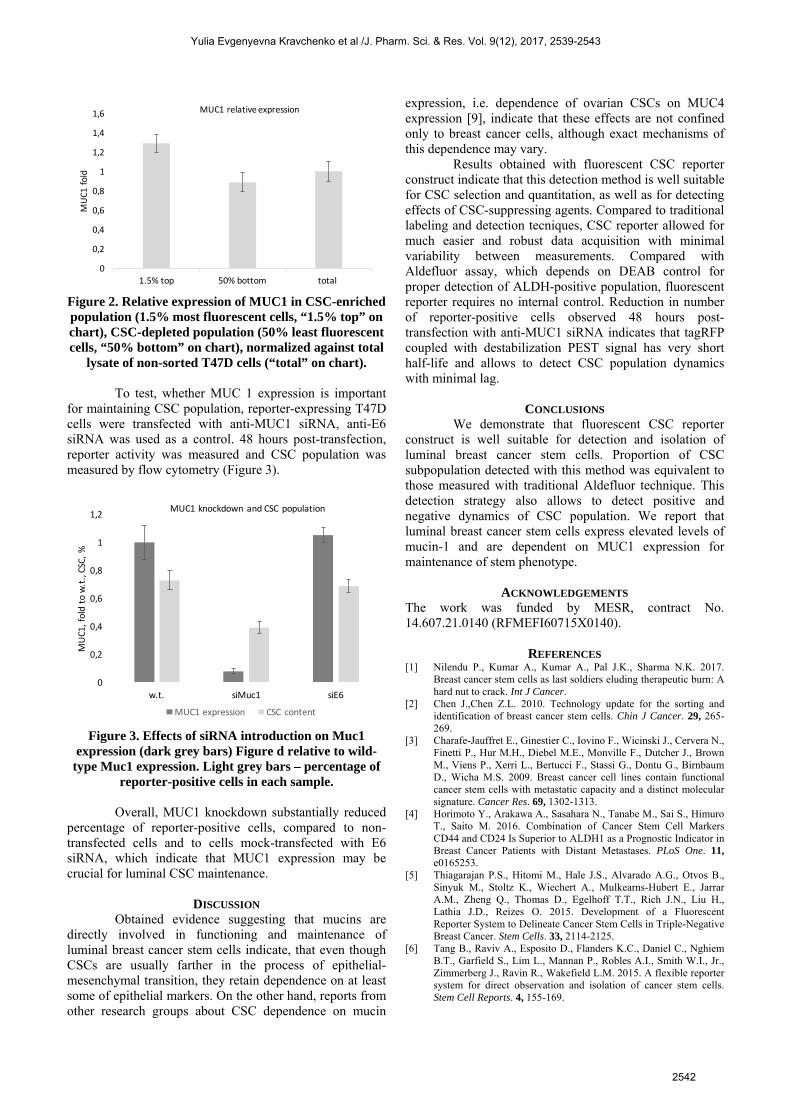
Figure 2. Relative expression of MUC1 in CSC-enriched population (1.5% most fluorescent cells, “1.5% top” on chart), CSC-depleted population (50% least fluorescent cells, “50% bottom” on chart), normalized against total
lysate of non-sorted T47D cells (“total” on chart). To test, whether MUC 1 expression is important for maintaining CSC population, reporter-expressing T47D cells were transfected with anti-MUC1 siRNA, anti-E6 siRNA was used as a control. 48 hours post-transfection, reporter activity was measured and CSC population was measured by flow cytometry (Figure 3).
Figure 3. Effects of siRNA introduction on Muc1
expression (dark grey bars) Figure d relative to wild-type Muc1 expression. Light grey bars – percentage of
reporter-positive cells in each sample. Overall, MUC1 knockdown substantially reduced percentage of reporter-positive cells, compared to non-transfected cells and to cells mock-transfected with E6 siRNA, which indicate that MUC1 expression may be crucial for luminal CSC maintenance.
DISCUSSION Obtained evidence suggesting that mucins are directly involved in functioning and maintenance of luminal breast cancer stem cells indicate, that even though CSCs are usually farther in the process of epithelial-mesenchymal transition, they retain dependence on at least some of epithelial markers. On the other hand, reports from other research groups about CSC dependence on mucin
expression, i.e. dependence of ovarian CSCs on MUC4 expression [9], indicate that these effects are not confined only to breast cancer cells, although exact mechanisms of this dependence may vary. Results obtained with fluorescent CSC reporter construct indicate that this detection method is well suitable for CSC selection and quantitation, as well as for detecting effects of CSC-suppressing agents. Compared to traditional labeling and detection tecniques, CSC reporter allowed for much easier and robust data acquisition with minimal variability between measurements. Compared with Aldefluor assay, which depends on DEAB control for proper detection of ALDH-positive population, fluorescent reporter requires no internal control. Reduction in number of reporter-positive cells observed 48 hours post-transfection with anti-MUC1 siRNA indicates that tagRFP coupled with destabilization PEST signal has very short half-life and allows to detect CSC population dynamics with minimal lag.
CONCLUSIONS We demonstrate that fluorescent CSC reporter construct is well suitable for detection and isolation of luminal breast cancer stem cells. Proportion of CSC subpopulation detected with this method was equivalent to those measured with traditional Aldefluor technique. This detection strategy also allows to detect positive and negative dynamics of CSC population. We report that luminal breast cancer stem cells express elevated levels of mucin-1 and are dependent on MUC1 expression for maintenance of stem phenotype.
ACKNOWLEDGEMENTS The work was funded by MESR, contract No. 14.607.21.0140 (RFMEFI60715X0140).
REFERENCES [1] Nilendu P., Kumar A., Kumar A., Pal J.K., Sharma N.K. 2017.
Breast cancer stem cells as last soldiers eluding therapeutic burn: A hard nut to crack. Int J Cancer.
[2] Chen J.,Chen Z.L. 2010. Technology update for the sorting and identification of breast cancer stem cells. Chin J Cancer. 29, 265-269.
[3] Charafe-Jauffret E., Ginestier C., Iovino F., Wicinski J., Cervera N., Finetti P., Hur M.H., Diebel M.E., Monville F., Dutcher J., Brown M., Viens P., Xerri L., Bertucci F., Stassi G., Dontu G., Birnbaum D., Wicha M.S. 2009. Breast cancer cell lines contain functional cancer stem cells with metastatic capacity and a distinct molecular signature. Cancer Res. 69, 1302-1313.
[4] Horimoto Y., Arakawa A., Sasahara N., Tanabe M., Sai S., Himuro T., Saito M. 2016. Combination of Cancer Stem Cell Markers CD44 and CD24 Is Superior to ALDH1 as a Prognostic Indicator in Breast Cancer Patients with Distant Metastases. PLoS One. 11, e0165253.
[5] Thiagarajan P.S., Hitomi M., Hale J.S., Alvarado A.G., Otvos B., Sinyuk M., Stoltz K., Wiechert A., Mulkearns-Hubert E., Jarrar A.M., Zheng Q., Thomas D., Egelhoff T.T., Rich J.N., Liu H., Lathia J.D., Reizes O. 2015. Development of a Fluorescent Reporter System to Delineate Cancer Stem Cells in Triple-Negative Breast Cancer. Stem Cells. 33, 2114-2125.
[6] Tang B., Raviv A., Esposito D., Flanders K.C., Daniel C., Nghiem B.T., Garfield S., Lim L., Mannan P., Robles A.I., Smith W.I., Jr., Zimmerberg J., Ravin R., Wakefield L.M. 2015. A flexible reporter system for direct observation and isolation of cancer stem cells. Stem Cell Reports. 4, 155-169.
0
0,2
0,4
0,6
0,8
1
1,2
1,4
1,6
1.5% top 50% bottom total
MUC1 fo
ld
MUC1 relative expression
0
0,2
0,4
0,6
0,8
1
1,2
w.t. siMuc1 siE6
MUC1, fold
to w.t., CSC, %
MUC1 knockdown and CSC population
MUC1 expression CSC content
Yulia Evgenyevna Kravchenko et al /J. Pharm. Sci. & Res. Vol. 9(12), 2017, 2539-2543
2542
[7] Lee K.M., Nam K., Oh S., Lim J., Kim R.K., Shim D., Choi J.H.,Lee S.J., Yu J.H., Lee J.W., Ahn S.H., Shin I. 2015. ECM1regulates tumor metastasis and CSC-like property throughstabilization of beta-catenin. Oncogene. 34, 6055-6065.
[8] Lawrence R.T., Perez E.M., Hernandez D., Miller C.P., Haas K.M.,Irie H.Y., Lee S.I., Blau C.A., Villen J. 2015. The proteomiclandscape of triple-negative breast cancer. Cell Rep. 11, 630-644.
[9] Ponnusamy M.P., Seshacharyulu P., Vaz A., Dey P., Batra S.K.2011. MUC4 stabilizes HER2 expression and maintains the cancerstem cell population in ovarian cancer cells. J Ovarian Res. 4, 7.
Yulia Evgenyevna Kravchenko et al /J. Pharm. Sci. & Res. Vol. 9(12), 2017, 2539-2543
2543