Embed Size (px)
Citation preview

European Journal of Pharmaceutical Sciences 24 (2005) 1–13
Multicomponent complex formation between vinpocetine,cyclodextrins, tartaric acid and water-soluble polymers
monitored by NMR and solubility studies
Laura Ribeiroa, Rui A. Carvalhob, Domingos C. Ferreirac, Francisco J.B. Veigaa,∗a Laboratory of Pharmaceutical Technology, Faculty of Pharmacy, University of Coimbra, 3000-295 Coimbra, Portugal
b Department of Biochemistry, Faculty of Sciences and Technology, University of Coimbra, Coimbra, Portugalc Laboratory of Pharmaceutical Technology, Faculty of Pharmacy, University of Porto, Porto, Portugal
Received 25 February 2004; received in revised form 25 May 2004; accepted 14 September 2004Available online 14 November 2004
Abstract
( monitoredb nd int ciencyoec and H-5)c ity of both� ut in ordert shment ofs of VP isi l sidec©
K
1
sgrsCw
osi-–CHatedares of
woco-vitydt va-lting
nds,
0d
This work deals with multicomponent complex formation of vinpocetine (VP) with�-cyclodextrin (�CD), sulfobutyl ether�-cyclodextrinSBE�CD) and tartaric acid (TA), in the presence or absence of water-soluble polymers, in aqueous solution. Complexation wasy phase-solubility and proton nuclear magnetic resonance (1H NMR) studies. TA demonstrated a synergistic effect on VP solubility, a
he complexation efficiency of�CD and SBE�CD. Additionally, water-soluble polymers increased even more the complexation effif the CDs that was reflected by a 2.1–2.5 increase onKC values for VP–CD–TA–polymer multicomponent complexes. SBE�CD was moreffective in VP solubilization, asKC values of VP–SBE�CD–TA multicomponent complexes were notably higher than in corresponding�CDomplexes. The large chemical shift displacements from protons located in the interior of the hydrophobic CD cavities (i.e., H-3oupled with significant chemical shift displacements of VP aromatic protons suggested that this moiety was included in the cavCD and SBE�CD. Two-dimensional rotating frame nuclear Overhauser effect spectroscopy (ROESY) experiments were carried o
o obtain information about the multicomponent complex geometry in solution. Inspection of ROESY spectra allowed the establipatial proximities between all aromatic protons of VP and the internal protons of the CDs, confirming that the aromatic moietyncluded in CD cavities being deeply inserted in SBE�CD multicomponent complexes, since additional interactions with the sulfobutyhains were evidenced.2004 Elsevier B.V. All rights reserved.
eywords:Vinpocetine; Cyclodextrins; Water-soluble polymers; Phase-solubility studies;1H NMR; Multicomponent complexes
. Introduction
Cyclodextrins (CDs) are naturally available water-oluble cyclic oligosaccharides composed of�-1,4-linkedd-lucopyranose units. The most commonly used forms of theseing-shaped molecules are�-, �-, and�-CDs formed by six,even and eight glucose units, respectively (Szejtli, 1988).Ds are toroidal molecules with a truncated cone structurehere the secondary hydroxyl groups are located on the wider
∗ Corresponding author. Tel.: +351 2398 37850; fax: +351 2398 37731.E-mail address:[email protected] (F.J.B. Veiga).
side of the ring, while the primary hydroxyl groups are ptioned on the opposite, narrower side of the torus. Thegroups carrying the H-1, H-2 and H-4 protons are locon the exterior of the molecule and the hydroxyl groupsoriented to the cone exterior, making the external faceCDs hydrophilic. The interior of the torus is lined by trings of –CH groups (H-3 and H-5) and by a ring of glysidic “ether oxygens” (O-4), with H-6 located near the ca(Uekama and Otagiri, 1987). This low polarity central voiis able to encapsulate either partially or entirely a greariety of guest molecules of suitable size and shape resuin a stable association without formation of covalent bo
928-0987/$ – see front matter © 2004 Elsevier B.V. All rights reserved.oi:10.1016/j.ejps.2004.09.003

2 L. Ribeiro et al. / European Journal of Pharmaceutical Sciences 24 (2005) 1–13
Fig. 1. Structure of (a) SBE�CD and (b)�CD.
being the resultant entity known as host–guest complex orinclusion complex (Saenger, 1980). When compared to hostand guest separately, these inclusion complexes often displayvery different properties such as altered solubility, reducedvolatitility, reduced or enhanced stability, modified chemicalreactivity and altered bioavailability.
Among the above mentioned CDs,�CD, with an innercavity diameter of 6.0–6.5A and a depth of 7.9A, is themost widely used. Its internal cavity has an excellent abil-ity to incorporate hydrophobic aromatic guests in aqueoussolution, provided that the sizes of the host internal cavityand the entering portion of a guest molecule are suitablefor complexation (Saenger, 1980). However, its anomalouslow aqueous solubility is a serious handicap in its widerutilization (Szejtli, 1988). To overcome those difficulties,chemical modifications of the CD torus by substitution atthe hydroxyl groups, at positions 2, 3 and 6 of the�-d-glucose have been made to enhance and expand the functionalities of CDs, leading to derivatives that provide bettersolubility. One of the most prominent groups of modifiedCDs, as far as pharmaceutical applications are concerned, arethe sulfobutyl-substituted CDs (Fig. 1), among which is the(sulfobutylether)7M-�-cyclodextrin (SBE�CD). SBE�CD isa polyanionic CD derivative, with an average degree of sub-stitution of seven and a much greater solubility in water thantg icb CD(
therl fre-q ter-i e of
CD can be dramatically improved by addition of a suit-able third component such as�-hydroxy-acids, and water-soluble polymers followed by heating the complexation me-dia (Loftsson et al., 1999; Chiesi et al., 1999).
CD inclusion complexes formed by molecular encapsu-lation of guest compounds in the cavities of macrocyclichosts can yield non-covalent multicomponent associationswith suitable counter ions of guest molecules. Such multi-component associations are of current interest in the field ofsupramolecular systems and are of scientific interest and tech-nological relevance for their physical, chemical and biologi-cal properties (Selva et al., 1998). In the field of pharmaceu-tical preparations, multicomponent associations of drug–CDinclusion complexes can dramatically enhance the solubilityin water of sparingly soluble drugs (Chiesi et al., 1999).
Previously, we have reported the preparation and physico-chemical characterization of vinpocetine (VP) multicom-ponent complexes with�CD, SBE�CD, tartaric acid (TA)and the water-soluble polymers hydroxypropylmethylcellu-lose (HPMC) and polyvinylpyrrolidone K30 (PVP), in solid-state, by scanning electron microscopy, differential scanningcalorimetry, X-ray diffractometry and Fourier-transform in-frared spectroscopy (Ribeiro et al., 2003a,b). However, thesetechniques can hardly suggest if guest molecules form a com-plex or not and cannot provide a clear answer about the typeo uc-tP yb ances dis-t l in-t culess f theg vity(i ariedc andt gesi for-m CDc cav-i estm hy-d H-6)w .N hy-d tiona bi-d tat-i SY)(
uc-t aticm av-i , asa VP,
he parent CD (�CD). The inclusion ability of SBE�CD isenerally greater than that of�CD due to the hydrophobutyl side arms that extend the hydrophobic cavity of theZia et al., 2001).
Frequently, the complexation efficiency of CDs is raow and consequently a significant amount of CDs isuently needed to solubilize small amounts of a wa
nsoluble drug. However, drug solubility in the presenc
-
f complex formed (inclusion or adsorption) or the strural conformation of the molecules involved (Djedaini anderly, 1991; Veiga et al., 2001). This information can onle provided by high resolution nuclear magnetic resonpectroscopy (NMR) since this technique allows a clearinction between inclusion and other possible externaeraction processes by observing guest and host moleimultaneously and is capable to differentiate the part ouest molecule involved in the interaction with the CD caFernandes et al., 2003). Monitoring changes in the1H chem-cal shifts as the composition of theses complexes is van elucidate the stoichiometry of inclusion complexeshe dynamics of their formation. Thus, chemical shift chann the1H spectra have been used to monitor the complex
ation process, since if a guest is incorporated into theavity, the hydrogen atoms located in the interior of thety (H-3 and H-5) will be considerably shielded by the gu
olecule, causing a significant upfield shift, whereas therogen atoms on the outer surface (H-1, H-2, H-4 andill either be unaffected or experience a marginal shift1HMR can thus provide information about structure androgen bonding in CDs; whereas, more detailed informabout their conformations is available from a variety ofimensional NMR experiments, including NOESY and ro
ng frame nuclear Overhauser effect spectroscopy (ROESzejtli, 1988; Schneider et al., 1998).
In this work, taking into account that the molecular strure of VP is characterized by the presence of an aromoiety that is potentially able to interact with the CD c
ty (Fig. 2), we have employed phase-solubility studiesprevious screening of the complex formation between

L. Ribeiro et al. / European Journal of Pharmaceutical Sciences 24 (2005) 1–13 3
Fig. 2. Structure of VP.
�CD, SBE�CD, TA, PVP and HPMC in order to evaluate thesolubilizing power of CDs, in association with an hydroxy-acid and water-soluble polymers, towards VP, and to deter-mine the apparent stability constants and stoichiometry ofthe complexes. A series of1H NMR experiments were thenundertaken to prove the real inclusion of VP in VP–CD mul-ticomponent complexes, to confirm the stoichiometry of theputative inclusion complexes and a reliable structure of those.
2. Materials and methods
2.1. Materials
Hydroxypropylmethylcellulose 4000 cps (HPMC),polyvinylpyrrolidone K30 (PVP), and tartaric acid (TA)were purchased from Sigma Chemical Co. (St. Louis, USA).Vinpocetine (VP) was purchased from Covex (Madrid,Spain).�-Cyclodextrin (�CD; Kleptose®; MW 1135) and(sulfobutylether)7M-�-cyclodextrin (SBE�CD; CaptisolTM;TDS 6.8; MW 2160) were kindly donated by Roquette(Lestrem, France) and Cydex (Kansas City, USA). The latterwas dried at 40◦C for 12 h before use. Deuterium oxide(D2O; 99.90%) was purchased from SDS (Peypin, France).All other chemicals were of analytical reagent grade, andd
2
o-l em ,1 askst ingi ith-o Ca ers,g untilr withp autoc for7 ectedo VPa enti ing
polymer concentrations and no significant degradation of VPin acidic medium was found after autoclavation at 120◦Cfor 20 min. All suspensions were filtered through a 0.45-�m membrane filter (Millipore) and VP concentrations an-alyzed spectrophotometrically (UV-1603, Shimadzu, Japan)at 316 nm. Each experiment was repeated at least three timesand the results reported are the mean values. The apparent sta-bility constants (KC), assuming that a 1:1 (VP:CD) complexwas initially formed, were calculated from the straight lineof the phase-solubility diagrams according to the equation ofHiguchi and Connors.
2.3. 1H NMR studies
One-dimensional1H NMR spectra were recorded at 25◦Con a Varian 500 MHz spectrometer using a 5 mm NMR probeand a simple pulse-acquire sequence with solvent presatura-tion. Acquisition parameters consisted of 16K points cover-ing a sweep width of 5300 Hz, a pulse width of 19�s and atotal repetition time of 13 s. Digital zero filling to 64K anda 0.5 Hz exponential were applied before Fourier transfor-mation. The resonance at 4.700 ppm due to residual solvent(HOD) was used as the internal reference. Samples were pre-pared by dissolving an appropriate amount of the solid com-plexes in D2O to achieve a VP concentration of 6.8 mM, di-r lidm liza-tR mMw s thecc e in-c g tot
ptedt ns of1 /v)o ain6 olu-t tocks e ofm ingt m-i sa
2
ls ames f VPi 2K( oreF by2 pliedi
eionized water was used throughout the study.
.2. Phase-solubility studies
Solubility studies were carried out in TA 16.6 mM sutions at room temperature (22± 1◦C) according to th
ethod of Higuchi and Connors (Higuchi and Connors965). Excess amounts of VP were weighted into glass fl
o which were added 10 ml of 16.6 mM solutions containncreasing amounts of CDs (0.001–0.025 M) with or wut a fixed polymer concentration of 0.10% (w/v) for HPMnd 0.25% (w/v) for PVP. For systems without polymlass containers were sealed and mechanically stirredeaching equilibrium (about 72 h). In the case of systemsolymers, glass containers were sealed and heated in anlave at 120◦C for 20 min and then allowed to equilibrate2 h. The polymer and TA concentrations used were seln the basis of preliminary studies carried out betweennd polymers or VP and TA, since no further improvem
n the solubility values of VP was achieved by increas
-
ectly on 5-mm RMN tubes (0.6 ml total volume). The soulticomponent complexes were prepared by the lyophi
ion method as previously described (Ribeiro et al., 2003a).eference samples containing pure VP and CDs at 6.8ere also prepared in the same acidic environment aomplexes (TA 16.6 mM).1H NMR chemical shifts (�δ)aused upon complexation were measured to confirm thlusion of VP in acidic medium and calculated accordinhe formula:�δ = δ (complex)− � (free).
The continuous variation method (Job’s plot) was adoo assess the stoichiometry of the complexes. TA solutio6.6 mM were prepared, with or without 0.1% HPMC (wr 0.25% (w/v) in D2O, and subsequently used to obt.8 mM stock solutions of VP and both CDs. A series of s
ions were prepared by mixing variable volumes of both solutions in varying proportions so that a complete rangole ratios was sampled (0 > [VP]/[VP] + [CD] > 1), keep
otal concentration constant ([VP] + [CD] = 6.8 mM). Checal shift differences (�δ) × [VP] (or [�CD]) were plotted a
function of mole ratio (r) (Mitra et al., 1998).
.4. COSY experiments
Standard absorptive two-dimensional1H–1H chemicahift correlation spectra (COSY) were acquired in the spectrometer to allow the chemical shift assignment on TA solution. Each spectrum consisted of a matrix ofF2) by 0.5K (F1) covering a sweep width of 5000 Hz. Befourier transformation, the matrix was zero filled to 4KK and standard sinebell apodization functions were ap
n both dimensions.
4 L. Ribeiro et al. / European Journal of Pharmaceutical Sciences 24 (2005) 1–13
2.5. ROESY experiments
The average extent of penetration and the directionof inclusion in the host cavity were determined by two-dimensional phase sensitive nuclear Overhauser effect spec-troscopy by the detection of intermolecular nuclear Over-hauser effects (NOEs) between VP and CDs. ROESY spec-tra were acquired in the phase sensitive mode using the samespectrometer. Each spectrum consisted of a matrix of 2K (F2)by 1K (F1) covering a sweep width of 5000 Hz. Spectra wereobtained with the samples prepared from lyophilized multi-component complexes used for1H NMR studies, using spin-lock mixing periods of 500 ms. Before Fourier transforma-tion, the matrix was zero filled to 4K by 4K and sinebellapodization functions were applied in both dimensions toenhance spectral resolution. The ROESY spectra were nor-malized and plotted with similar intensity contour levels forall systems studied.
3. Results and discussion
3.1. Phase-solubility studies
Since VP is a poorly water-soluble base-type drug, mul-t ten-s inm ith-o outt pro-fi ora ares
oth� -
solubility diagrams in the CD concentration range studied(Higuchi and Connors, 1965) and indicating the formationof 1:1 stoichiometry VP–CD complexes in 16.6 mM TA so-lution, in the presence or absence of HPMC and PVP. Theincrement of VP solubility seems to be related to the inclu-sion ability of the CD molecules in TA solution. As the slopesof these solubility diagrams were all less than 1, it was pos-sible to calculate the apparent stability constants (KC) of themulticomponent complexes. The estimatedKC and VP sol-ubility values achieved in the multicomponent systems arecollected inTable 1. As it can be observed, the addition ofthe water-soluble polymers to the complexation media re-sulted in an increase on the diagram slopes that was reflectedon enhanced VP solubility. Thus, VP solubility in the pres-ence of CDs, TA and polymers resulted in a synergistic ef-fect and in an increase on CD complexation efficiency. Thebinding potential and the solubilization effect of SBE�CDwere higher than that of�CD. This effect may be due tothe butyl micellar arms of SBE�CD that extend the depth ofthe hydrophobic cavity of the CD (Zia et al., 2001). There-fore,KC values of VP–SBE�CD–TA multicomponent com-plexes were greater than those obtained with VP–�CD–TAmulticomponent complexes, since the complexation with theSBE�CD may involve the CD cavity and the hydrophobicsulfobutyl ether substituents.
us( Pm ers,w d int iliza-tT pres-e o-c tiono
F nction�C ntst
icomponent complexation with TA was attempted to inify solubilization by CDs. Phase-solubility studies of VPulticomponent systems with CDs and TA, with and wut polymers, were performed to obtain information ab
he drug solubilization mechanism. The phase-solubilityles of VP in 16.6 mM TA–CD solutions, in the presencebsence of HPMC 0.10% (w/v) and PVP 0.25% (w/v)hown inFig. 3.
VP solubility increased linearly as a function of bCD and SBE�CD concentrations, giving AL-type phase
ig. 3. Phase-solubility profiles of VP, in 16.6 mM TA solution, as a fuhe mean of three determinations.
Similar solubility results were previously reported byRibeiro et al., 2003a,b) concerning the solubility of Verely in the presence of CDs and water-soluble polymhere a VP synergistic solubility effect was also observe
he presence both PVP and HPMC, with a superior solubion and complexation effect being attributed to SBE�CD.he major difference between these two studies is thence of TA. TA is an�-hydroxy-acid commonly used as complexing agent with CDs to promote complex formaf basic type drugs (Redenti et al., 2000). The mechanism
ofD and SBE�CD, with or without PVP and HPMC. Each point represe

L. Ribeiro et al. / European Journal of Pharmaceutical Sciences 24 (2005) 1–13 5
Table 1Values of apparent stability constant (KC ± standard deviation) and drug solubility in VP complexes (n= 3)
S1a S2
b S2/S0c D2/D1
d KC (M−1)
VP–�CDe 0.51× 10⁻² 1.17× 10⁻² 2.3 – 75± 3VP–�CD–PVPe 0.89× 10⁻² 2.24× 10⁻² 4.4 2.1 142± 4VP–�CD–HPMCe 0.72× 10⁻² 3.23× 10⁻² 6.3 3.5 242± 7VP–�CD–TA 5.80 7.59 1490 183.1 15 ± 3VP–�CD–TA–PVP 6.06 9.44 1850 37± 4VP–�CD–TA–HPMC 5.96 8.41 1650 343.4 23 ± 4VP–SBE�CDe 0.51× 10⁻² 0.14 27.5 – 340± 8VP–SBE�CD–PVPe 0.89× 10⁻² 0.21 41.2 1.5 490± 12VP–SBE�CD–HPMCe 0.72× 10⁻² 0.16 31.4 1.4 390± 8VP–SBE�CD–TA 5.80 10.62 2080 109.0 73 ± 5VP–SBE�CD–TA–PVP 6.06 12.46 2440 144.3 155± 12VP–SBE�CD–TA–HPMC 5.96 11.83 2320 136.0 129± 12
a VP solubility (mg/ml) in water, PVP 0.25% (w/v), HPMC 0.10% (w/v), TA 16.6 mM solution or in their combinations.b VP solubility (mg/ml) in CD solutions (15 mM�CD or 60 mM SBE�CD in the absence of TA 16.6 mM or 25 mM for either�CD or SBE�CD in the
presence of TA 16.6 mM) with and without PVP 0.25% (w/v) or HPMC 0.10% (w/v).c Ratio between VP solubility achieved in the complexes and VP intrinsic solubility in water (S0).d D2/D1 is the ratio between the slopes of the phase-solubility diagrams achieved in VP–CD–TA multicomponent systems and in the corresponding binary
ones.e Ribeiro et al. (2003a).
by which TA seems to enhance the solubilization and com-plexation of these drugs has been related to its ability to in-teract with CDs by forming hydrogen bonds with their nu-merous hydroxyl groups (Fenyvesi et al., 1999). It is evidentfrom all examined systems that multicomponent complexesformed between VP, CDs and TA were clearly more effec-tive in enhancing VP solubility than the ternary complexesVP–CD–polymers. In fact, VP solubilization efficiency wasimproved nearly 4–41 times in VP–CD–polymers complexesand 1650–2440 times in VP–CD–TA–polymer complexes, incomparison with VP intrinsic solubility (S0 ≈ 5�g/ml). How-ever, in all cases, a decrease in drug–CD interaction was alsoexperimented, as indicated by the decrease inKC values (seeTable 1). Such an effect was explained on the basis of thehigher initial drug solubility due to an increased ionizationof VP in the presence of TA with consequent less affinityto the apolar cavity (Mura et al., 2001). Although CD com-plexes of un-ionized drugs are usually of stronger stabilitythan those of their anionic counterparts, the achieved totalsolubility (free ionized drug + un-ionized drug) usually in-creases (Redenti et al., 2000). Therefore, when TA was addedto the complexation media, a greater overall VP solubility wasachieved by using a combined approach of CD complexationand drug ionization. Indeed, CDs and TA had a synergisticeffect on VP solubility, since a greater extent on the solubi-l andT evenm PVPo f thep ownt CDs( ec heirr dh
increased 2.1–2.5 times on for VP–CD–TA–polymer multi-component complexes, in comparison with VP–CD–TA com-plexes. Thus, even in acidic medium, the water-soluble poly-mers had an important role in the improvement of complexa-tion efficiency of CDs towards VP, allowing better solubilityresults to be achieved with a reduction in the amount of CDrequired to dissolve VP.
3.2. 1H NMR studies
As the increased solubility of a drug in the presence ofCDs observed in phase-solubility diagrams cannot be con-sidered as a definitive proof for the formation of inclusioncomplexes, we performed NMR studies. NMR techniqueshave been widely used to investigate supramolecular assem-blies in solution, their stoichiometries and structure of theresulting complexes, especially the orientation of the guestmolecule in the CD cavity (Djedaini and Perly, 1991). It iswell-known that the chemical shift (δ) of a given nucleusdepends on its shielding constant and in turn is sensitive tomedium effects. Therefore, changes in� (ppm) values of thehost and guest nuclei can provide a measure of the degreeof complex formation since significant changes in the mi-croenvironment are known to occur between the free andbound states (Wilson and Verral, 1998). As the chemicale tion,t (o ts).N truei enso ity,a ncesd uest.T n pro-c lded
ization effect, than that expected by the addition of CDA separately, was displayed when used together. Butore surprising was the observed effect of HPMC andn VP–CD–TA systems. The simultaneous presence oolymers and TA on the complexation media, both kn
o enhance separately the complexation efficiency ofLoftsson et al., 1999; Redenti et al., 2000), had a positivonsequence on VP solubility and CD complexation. Telative solubilizing efficiencies (D2/D1) were 109–343-foligher than that of binary complexes VP–CD, andKC values
nvironment of some protons changes upon complexahere is a consequent variation in the chemical shifts�δ)f 1H NMR resonances (shielding or deshielding effecMR spectroscopy provides the most direct evidence for
nclusion complex formation since H-3 and H-5 hydrogf the host, that point toward the interior of the CD cavre remarkably shielded, being the shift of their resonaue to magnetic anisotropic effects exerted by the ghe guest resonances are also affected by the inclusioess, being the chemical shift of the anisotropically shie

6 L. Ribeiro et al. / European Journal of Pharmaceutical Sciences 24 (2005) 1–13
Table 21H Chemical shifts corresponding to�CD in free and complexed state
�CD protons VP–�CD–TA VP–�CD–TA–PVP VP–�CD–TA–HPMC
δ(free) δ(complex) �δa δ(complex) �δa δ(complex) �δa
H-1 4.999 4.995 −0.004 4.995 −0.004 4.990 −0.009H-2 3.578 3.586 +0.008 3.588 +0.010 3.582 +0.004H-3 3.892 3.844 −0.048 3.839 −0.053 3.835 −0.057H-4 3.513 3.505 −0.008 3.505 −0.008 3.500 −0.013H-5 3.782 3.774 −0.008 3.773 −0.009 3.768 −0.014H-6 3.806 3.806 0.000 3.807 +0.001 3.809 +0.003
a �δ = δ(complex)− δ(free).
atoms modified in the NMR spectra (Ganza-Gonzalez et al.,1994).
3.2.1. VP–�CD–TA multicomponent complexesDue to fast exchange, separate sets of signals belonging
to the free and complexed forms were not detected in the1H NMR spectra. The insertion of VP into�CD cavity wasclearly demonstrated by changes in the1H chemical shiftvalues of VP and�CD protons in all VP–�CD–TA multi-component complexes.Table 2reports the chemical shift of�CD protons in the native and complexed forms andFig. 4the1H spectra of VP–�CD–TA multicomponent complexes.In the presence of VP, both H-3 and H-5 inner protons of
�CD undergo a consistent upfield shift, which demonstrateda clear involvement of these hydrogen atoms in host–guest in-teractions. As this upfield displacement has been essentiallyattributed to the anisotropic effect caused by the inclusionof groups rich in�-electrons of the guest molecules into thehydrophobic cavity of�CD, this observable fact was takenas an evidence of complex formation (Djedaini and Perly,1991), giving strong indications of the insertion of the aro-matic ring of VP into�CD cavity. Furthermore, because ofhigher shielding effect on H-3 proton (−0.048 to−0.0057)with respect to H-5 (−0.008 to−0.014) it could be hypothe-sized that VP penetrates into the�CD cavity from the moreaccessible wider side (secondary hydroxyl rim). Since all the
F(
ig. 4. Expansions from the1H NMR spectra of VP (I) and�CD (II), and from tV) complexes.
he VP–�CD–TA (III), VP–�CD–TA–PVP (IV) and VP–�CD–TA–HPMC
L. Ribeiro et al. / European Journal of Pharmaceutical Sciences 24 (2005) 1–13 7
exterior protons of�CD were more or less influenced, wecannot exclude the existence of interactions with externalsurface of the macrocycle that could be ascribed to hydrogenbond formation with the hydroxyl groups at the edge of theCD cavity with the guest molecule and the co-complexingagents, namely TA and polymers.
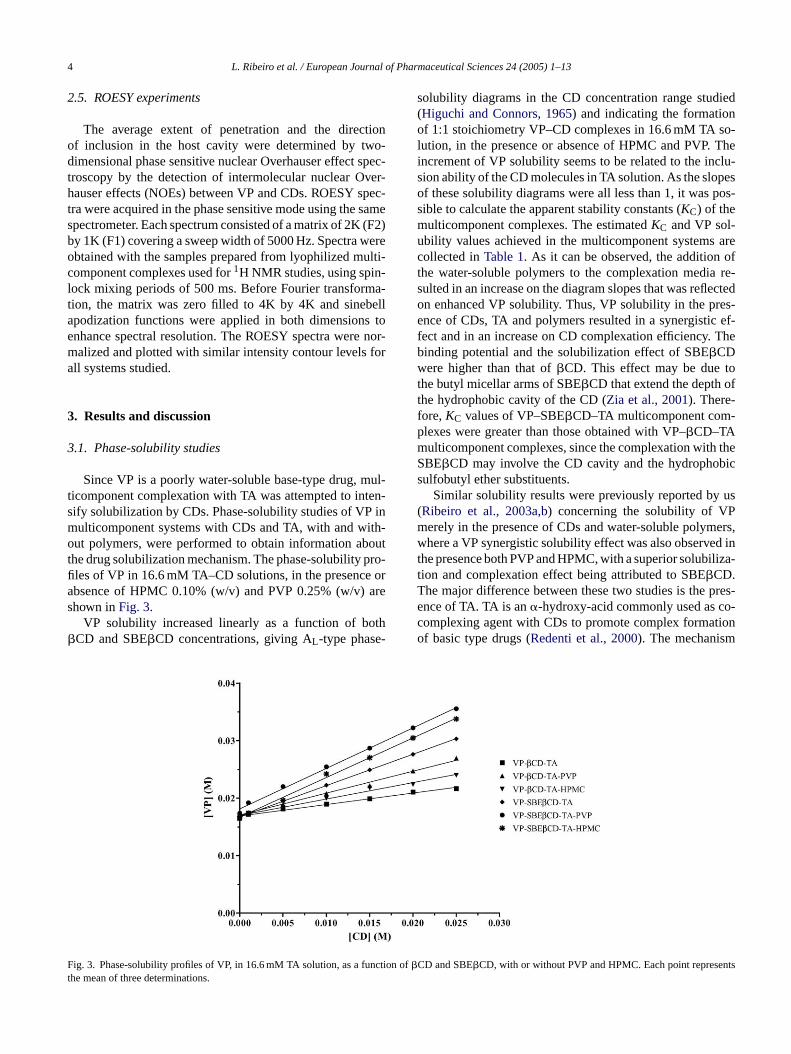
Protons of VP were assigned by analysis of one-dimensional1H NMR spectra and two-dimensional chemicalshift correlation spectra (COSY). In this process, the assign-ment of all individual aromatic protons of VP became diffi-cult due to the superposition of the signals of H-A1 and H-A2.Table 3summarizes all proton chemical shifts of VP in thefree and complexed forms.
Changes in the chemical shifts of the guest (VP) werealso monitored. The aromatic protons of the VP (δ: 7.1–7.6),H-A1/A2 and H-A4 (−0.004 to−0.024), were shifted up-field and the H-A3 proton was shifted downfield, with theexception on VP–�CD–TA–PVP, where all the aromatic pro-tons were shifted upfield. The extent of the displacementsdepended on the system studied, with a general tendencyfor higher�δ in the multicomponent complexes with PVP.The upfield shifts indicate that these protons are close to ahost atom which is rich in�-electrons, in this case asso-ciated with oxygen atoms, and also reflects conformationalchanges produced by inclusion. On the other hand, the down-fi arityo rac-ta ta eta ty ofV sig-n shiftia ulti-c na bly.I m-p tionsw n ofa itht entw -n 99;S ralt pro-t ntc itedt ons( r hy-d andw re-gc tiono
d tov e Ta
ble
31H
Che
mic
alsh
ifts
corr
espo
ndin
gto
VP
inV
P–C
D–T
Am
ultic
ompo
nent
com
plex
es
VP
prot
ons
VP
–�C
D–T
AV
P–�
CD
–TA
–HP
MC
VP
–�C
D–T
A–P
VP
VP
–SB
E�C
D–T
AV
P–S
BE�
CD
–TA
–HP
MC
VP
–SB
E�C
D–T
A–P
VP
δ (fr
ee)
δ (co
mpl
ex)
�δa
δ (co
mpl
ex)
�δa
δ (co
mpl
ex)
�δa
δ (co
mpl
ex)
�δa
δ (co
mpl
ex)
�δa
δ (co
mpl
ex)
�δa
A4
7.58
77.
566
−0.0
217.
569
−0.0
187.
563
−0.0
247.
595
+0.
008
7.57
2−0
.015
7.56
2−0
.025
A3
7.22
07.
225
+0.
005
7.22
4+
0.00
47.
218
−0.0
027.
194
−0.0
267.
182
−0.0
387.
173
−0.0
47A
1/A
27.
277
7.27
0−0
.007
7.27
3−0
.004
7.26
7−0
.010
7.25
8−0
.019
7.25
8−0
.019
7.24
1−0
.036
M0.
986
0.98
0−0
.006
0.98
0−0
.006
0.97
5−0
.011
1.01
9+
0.03
31.
004
+0.
018
0.99
8+
0.01
2N
/N′
1.81
81.
820
+0.
002
1.81
7−0
.001
1.81
2−0
.006
1.87
3+
0.05
51.
852
+0.
034
1.84
6+
0.02
8O
6.29
26.
295
+0.
003
6.29
3+
0.00
16.
288
−0.0
046.
350
+0.
058
6.32
7+
0.03
56.
321
+0.
029
P1.
321
1.31
3−0
.008
1.31
7−0
.004
1.31
2−0
.009
1.35
8+
0.03
71.
350
+0.
029
1.34
4+
0.02
3Q
/Q′
4.40
14.
404
+0.
003
4.40
4+
0.00
34.
398
−0.0
034.
436
+0.
035
4.42
5+
0.02
44.
419
+0.
018
S′1.
058
1.01
9−0
.039
1.02
2−0
.036
1.01
9−0
.039
0.96
1−0
.097
0.95
2−0
.106
0.94
7−0
.111
R4.
836
4.81
2−0
.024
4.80
3−0
.033
4.80
0−0
.036
4.91
0−0
.074
4.86
4+
0.02
84.
856
+0.
020
S/T
1.64
91.
643
−0.0
061.
640
−0.0
091.
635
−0.0
14–
––
––
–U
3.09
53.
084
−0.0
113.
083
−0.0
123.
079
−0.0
163.
131
+0.
036
3.09
2−0
.003
3.08
6−0
.009
V3.
673
3.65
8−0
.015
3.65
6−0
.017
3.65
1−0
.022
––
––
––
X3.
013
2.92
9−0
.084
2.93
7−0
.076
2.93
4−0
.079
3.00
0−0
.013
2.96
9−0
.044
2.95
9−0
.054
X′
3.19
23.
183
−0.0
093.
183
−0.0
093.
177
−0.0
153.
247
+0.
055
3.21
7+
0.02
53.
211
+0.
019
TA4.
599
4.53
8−0
.061
4.46
9−0
.130
4.46
2−0
.137
4.53
7−0
.062
4.47
0−0
.129
4.46
9−0
.130
a�
δ=
δ (co
mpl
ex)−
δ (fr
ee).
eld shift observed is probably due changes in local polr to deshielding effects caused by van der Waals inte
ions between the drug and carbohydrate chains (Djedaini etl., 1990; Ganza-Gonzalez et al., 1994; Uccello-Barretl., 1993). These findings suggest that the aromatic moieP is located inside the CD cavity. Moreover, the protonal corresponding to TA showed an appreciable upfield
ndicative of its involvement in interactions with the�CDnd VP molecules and, therefore, a possible role in momponent complex formation. From1H NMR data, we cassume that TA is strictly involved in the molecular assem
n particular, TA seems to be strictly implicated in the colexation process, by establishing electrostatic interacith protonated atoms of VP that results in the formation ion-pair, and by the formation of hydrogen bonds w
he hydroxy groups of�CD. These results are in agreemith previously reported1H NMR studies for multicompoent complexes (Faucci et al., 2000; Fenyvesi et al., 19elva et al., 1998). Additionally we did also observe a gene
endency for higher chemical shift displacements of theons of both VP and�CD molecules in the multicomponeomplexes with PVP and HPMC. This effect may be credo the ability of polymers in establishing different interactihydrophobic bonds, van der Waals dispersion forces orogen bonds) with the outer surface of the CD moleculesith drug–CD complexes forming drug–CD–polymer aggates (Hladon and Cwiternia, 1994; Valero et al., 2003) andonsequently reflected the important role in the stabilizaf VP–CD–TA multicomponent complexes.
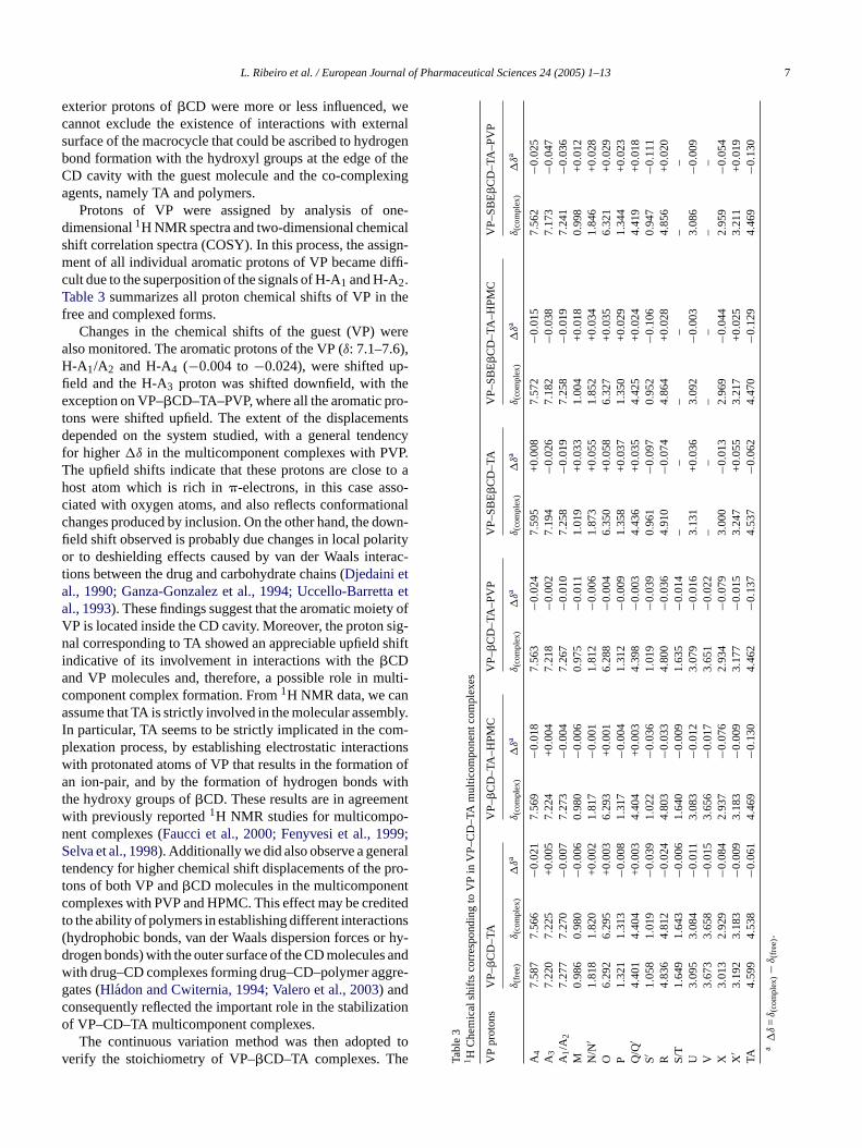
The continuous variation method was then adopteerify the stoichiometry of VP–�CD–TA complexes. Th
8 L. Ribeiro et al. / European Journal of Pharmaceutical Sciences 24 (2005) 1–13
Fig. 5. Continuous variation plots for VP and�CD protons in VP–�CD–TA (A), VP–�CD–TA–PVP (B) and VP–�CD–TA–HPMC (C) multicomponentcomplexes [protons of VP: (�) N/N′, (�) O and (�) U; protons of�CD: (�) H-3 and (�) H-5].
1H chemical shift (δ) was measured at different ratios ofdrug/CD, while keeping the total [VP] + [CD] constant. Thecalculated quantities�δ × [VP] (or �δ × [CD]) were propor-tional to the concentration of the inclusion complex and couldthus be plotted against “r”. The continuous variation plots(Fig. 5) for some of the most markedly affected protons of VPand�CD confirmed the 1:1 (VP–�CD) stoichiometry, sincethe maximum was atr = 0.5. These results are in completeaccordance with the above reported phase-solubility studies.
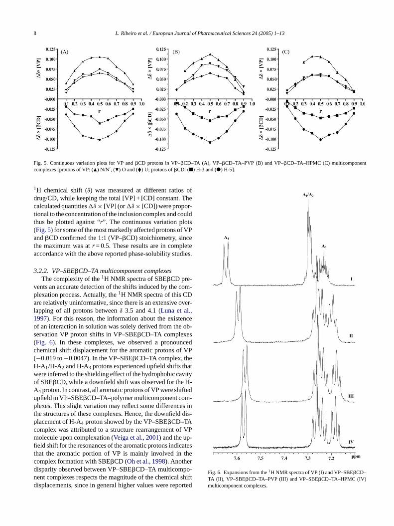
3.2.2. VP–SBE�CD–TA multicomponent complexesThe complexity of the1H NMR spectra of SBE�CD pre-
vents an accurate detection of the shifts induced by the com-plexation process. Actually, the1H NMR spectra of this CDare relatively uninformative, since there is an extensive over-lapping of all protons betweenδ 3.5 and 4.1 (Luna et al.,1997). For this reason, the information about the existenceof an interaction in solution was solely derived from the ob-servation VP proton shifts in VP–SBE�CD–TA complexes(Fig. 6). In these complexes, we observed a pronouncedchemical shift displacement for the aromatic protons of VP(−0.019 to−0.0047). In the VP–SBE�CD–TA complex, theH-A1/H-A2 and H-A3 protons experienced upfield shifts thatwere inferred to the shielding effect of the hydrophobic cavityof SBE�CD, while a downfield shift was observed for the H-A ftedu -p s int d dis-pc f VPm -fi atest hecdn l shiftd orted
Fig. 6. Expansions from the1H NMR spectra of VP (I) and VP–SBE�CD–TA (II), VP–SBE�CD–TA–PVP (III) and VP–SBE�CD–TA–HPMC (IV)multicomponent complexes.
4 proton. In contrast, all aromatic protons of VP were shipfield in VP–SBE�CD–TA–polymer multicomponent comlexes. This slight variation may reflect some difference
he structures of these complexes. Hence, the downfiellacement of H-A4 proton showed by the VP–SBE�CD–TAomplex was attributed to a structure rearrangement oolecule upon complexation (Veiga et al., 2001) and the up
eld shift for the resonances of the aromatic protons indichat the aromatic portion of VP is mainly involved in tomplex formation with SBE�CD (Oh et al., 1998). Anotherisparity observed between VP–SBE�CD–TA multicompo-ent complexes respects the magnitude of the chemicaisplacements, since in general higher values were rep

L. Ribeiro et al. / European Journal of Pharmaceutical Sciences 24 (2005) 1–13 9
for the VP–SBE�CD–TA–polymer complexes. Taking intoaccount that the extent of these displacements is dependenton the relative strength of the interactions between these aro-matic protons and the hydrogen atoms of the�CD cavity, wecan assume that a deeper insertion of the aromatic moietyof VP was produced by the presence of the polymers. Theseobservations are in total agreement with the reported resultsof the phase-solubility studies, since higherKC values wereobtained for VP–SBE�CD–TA–polymer complexes than forthe VP–SBE�CD–TA complex.
In what concerns guest resonances, although there is a gen-eral tendency for chemical shift displacements, upfield for thearomatic protons and downfield for the other ones, there isno simple explanation in terms of inclusion or non-inclusion.The chemical shift displacement of VP protons must be re-garded as a result of a combination of interactions with themacrocycle and time-averaged conformational changes of theincluded molecules (Redondo et al., 1999). Nevertheless, thedownfield shift of the resonances of the alkyl protons wasascribed to the interaction with the hydrophilic external partof the CD molecule (Uccello-Barretta et al., 1993) and toconformational changes produced by the inclusion (Redentiet al., 1999).
The same feature, as in VP–�CD–TA multicomponentcomplexes, was observed with the protons of TA, that is,a nceo ntsw om-p ers,wa dis-p� oi-e pho-b y-d erac-t n oft ore-
over, the anionic SBE�CD produces a downfield shift dis-placement of almost all non-aromatic protons, oppositelyto the �CD that induced preponderantly upfield displace-ment in these hydrogen atoms. This fact prompts for adifferent mode of interaction between VP and both CDs(Owens et al., 1997) and may also reflect a distinct chemicalmicroenvironment.
Considering all results mentioned above, we can pre-sume the existence of important structural differences inVP–�CD–TA and VP–SBE�CD–TA multicomponent com-plexes since SBE�CD has an average of seven negativecharges and therefore the most favourable position of posi-tively amino charged groups of VP must be not far away fromthe negatively moieties of the CD. The drug molecule has toarrange itself within the hydrophobic cavity to allow for theelectrostatic interactions with the charged substituents, and,at the same time, the protonated nitrogen atoms of the drugwill interact with TA molecules resulting in the creation of anion-pair, without the formation of chemical bonds (Massonet al., 1998). In summary, SBE�CD most likely encloses thearomatic ring of VP due to hydrophobic interaction and theresulting complex is synergistically stabilized by additionalelectrostatic interaction between sulfobutyl moieties of thehost and the protonated nitrogen atoms of the guest and byhydrogen bond forming with the 2- and 3-hydroxy groups oft
ap-pc m-p lots( -tp ithint stoi-c tureoc e for-m ,
F , VP–S tc
n upfield shift related to the co-complexing performaf the�-hydroxy-acid. Larger chemical shift displacemeere also observed for the VP protons in the multiconent complexes with both HPMC and PVP polymhich might reveal a tight association between VP, SBE�CDnd TA in their presence. The magnitudes of the shiftlacements of VP protons were greater in SBE�CD than inCD complexes. This may indicate that the aromatic mty of VP is included deeper into the expanded hydroic cavity of SBE�CD, since the sulbobutylation of the hroxyl groups may change the nature of host–guest int
ion and could therefore expand the hydrophobic regiohe CD cavity, enhancing the binding of the guest. M
ig. 7. Continuous variation plots for VP protons in VP–SBE�CD–TA (A)omplexes [protons of VP: () N/N′, (�) O, (�) Q/Q′ and (�) R].
he CD and the resulting ion-pair.The continuous variation method was subsequently
lied to all protons of the VP molecule in VP–SBE�CD–TAomplexes for confirming the stoichiometry of the colexes, yielding identical results. In all cases, Job’s pFig. 7) showed a maximum atr = 0.5, indicating the formaion of a complex where the complexing agent (SBE�CD) isresent in first-order degree with respect to the drug, w
he range of the investigated concentrations. The 1:1hiometry obtained is also strongly supported by the naf the SBE�CD molecule, given that each SBE�CD moleculearries an average of seven negative charges that makation of higher-order complexes difficult (Loftsson et al.
BE�CD–TA–PVP (B) and VP–SBE�CD–TA–HPMC (C) multicomponen
10 L. Ribeiro et al. / European Journal of Pharmaceutical Sciences 24 (2005) 1–13
2002). These results are once again in agreement with theabove related phase-solubility studies.
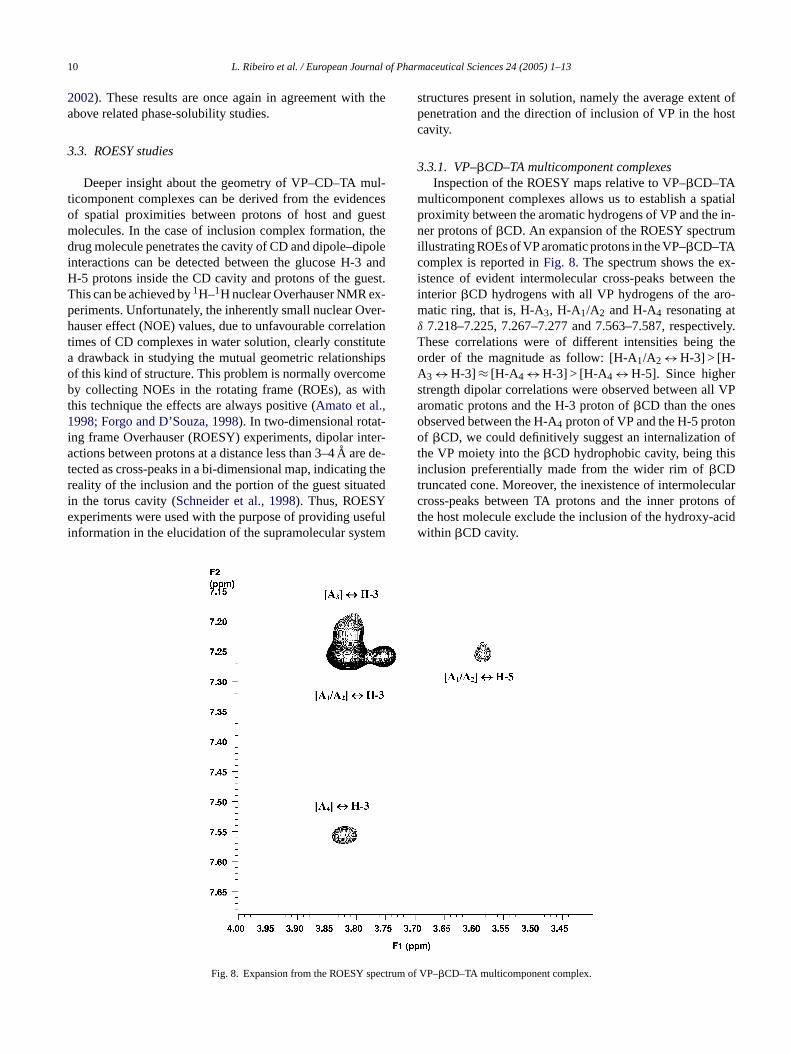
3.3. ROESY studies
Deeper insight about the geometry of VP–CD–TA mul-ticomponent complexes can be derived from the evidencesof spatial proximities between protons of host and guestmolecules. In the case of inclusion complex formation, thedrug molecule penetrates the cavity of CD and dipole–dipoleinteractions can be detected between the glucose H-3 andH-5 protons inside the CD cavity and protons of the guest.This can be achieved by1H–1H nuclear Overhauser NMR ex-periments. Unfortunately, the inherently small nuclear Over-hauser effect (NOE) values, due to unfavourable correlationtimes of CD complexes in water solution, clearly constitutea drawback in studying the mutual geometric relationshipsof this kind of structure. This problem is normally overcomeby collecting NOEs in the rotating frame (ROEs), as withthis technique the effects are always positive (Amato et al.,1998; Forgo and D’Souza, 1998). In two-dimensional rotat-ing frame Overhauser (ROESY) experiments, dipolar inter-actions between protons at a distance less than 3–4A are de-tected as cross-peaks in a bi-dimensional map, indicating thereality of the inclusion and the portion of the guest situatedie sefuli tem
structures present in solution, namely the average extent ofpenetration and the direction of inclusion of VP in the hostcavity.
3.3.1. VP–�CD–TA multicomponent complexesInspection of the ROESY maps relative to VP–�CD–TA
multicomponent complexes allows us to establish a spatialproximity between the aromatic hydrogens of VP and the in-ner protons of�CD. An expansion of the ROESY spectrumillustrating ROEs of VP aromatic protons in the VP–�CD–TAcomplex is reported inFig. 8. The spectrum shows the ex-istence of evident intermolecular cross-peaks between theinterior �CD hydrogens with all VP hydrogens of the aro-matic ring, that is, H-A3, H-A1/A2 and H-A4 resonating atδ 7.218–7.225, 7.267–7.277 and 7.563–7.587, respectively.These correlations were of different intensities being theorder of the magnitude as follow: [H-A1/A2 ↔ H-3] > [H-A3 ↔ H-3] ≈ [H-A4 ↔ H-3] > [H-A4 ↔ H-5]. Since higherstrength dipolar correlations were observed between all VParomatic protons and the H-3 proton of�CD than the onesobserved between the H-A4 proton of VP and the H-5 protonof �CD, we could definitively suggest an internalization ofthe VP moiety into the�CD hydrophobic cavity, being thisinclusion preferentially made from the wider rim of�CDtruncated cone. Moreover, the inexistence of intermolecularc ns oft acidw
n the torus cavity (Schneider et al., 1998). Thus, ROESYxperiments were used with the purpose of providing u
nformation in the elucidation of the supramolecular sys
Fig. 8. Expansion from the ROESY spectrum
ross-peaks between TA protons and the inner protohe host molecule exclude the inclusion of the hydroxy-ithin �CD cavity.
of VP–�CD–TA multicomponent complex.

L. Ribeiro et al. / European Journal of Pharmaceutical Sciences 24 (2005) 1–13 11
The VP–�CD–TA–polymer ROESY spectra demon-strated comparable results, with equivalent dipolar interac-tions evidenced between the inner protons of�CD and thearomatic protons of VP, which indicates that a similar inclu-sion process of the aromatic VP moiety must occur in thesesystems. However, the intensities of the cross-peaks weregreater in the presence of the polymers (PVP and HPMC),corroborating the previous results reported here, i.e., a some-how superior stabilization of the resultant supramolecular as-sembly by interaction with the outer surface of the inclusioncomplex.
From NMR data, there are clear evidences that the moreor less planar VP is tilted inside the�CD cavity and this ar-rangement allows the guest molecule to occupy most of theavailable cavity space, while keeping the polar susbstituentsclose to the hydroxy groups of the�CD rim. The polar sub-stituents, namely the basic nitrogen atoms, may then protrudeto the exterior environment and are stabilized by tartarate ionsand by the hydroxy groups of the CD rim.
3.3.2. VP–SBE�CD–TA multicomponent complexesIn the two-dimensional ROESY spectra of
VP–SBE�CD–TA multicomponent complexes, cross-peaks connecting two proton resonances were unambigu-ously observed between the internal protons of the�CDda ofV
intense ROE interactions were observed between H-A3,H-A1/A2 and H-A4 aromatic protons of VP, and both internalprotons of SBE�CD (in the 3.4–3.9 ppm region) for all threeVP–SBE�CD–TA complexes under study, indicating alsoin these systems the involvement of VP aromatic moiety inthe complexation. We also detected feeble intermolecularcross-peaks between the aromatic protons in VP, and theb–CH2 and c–CH2 hydrogens of the sulfobutyl side chainsof SBE�CD in theδ 2.85–2.75 and 1.60–1.75 regions, butthose were just observable in multicomponent containingpolymers. Using these NMR results, it is reasonable toascribe these effects to the formation of an inclusioncomplex between SBE�CD and VP, through the inclusionof the aromatic moiety of VP. As previously reported forVP–�CD–TA multicomponent complexes, the presence ofpolymers enhanced even more the magnitude of all observeddipolar interactions making it possible to visualize theinteraction between VP and the alkyl side chains of the CDderivative.
The greater ROE intensities observed inVP–SBE�CD–TA multicomponent complexes relativeto the corresponding complexes with�CD again suggestedthat VP was embedded in a more hydrophobic environ-ment in the former inclusion complexes; hence, SBE�CDexhibited the strongest inclusion property. This is duetn r thee
erivative and the aromatic protons of VP.Fig. 9 showscontour plot of a section of the ROESY spectrum
P–SBE�CD–TA–HPMC complex (δ: 1.60–4.0). Very
Fig. 9. Expansion from the ROESY spectrum of V
o attractive electrostatic interactions, since SBE�CD isegatively charged while VP is positively charged undexperimental conditions. As previously reported byZia et al.
P–SBE�CD–TA–PVP multicomponent complex.

12 L. Ribeiro et al. / European Journal of Pharmaceutical Sciences 24 (2005) 1–13
(2001), chemical modifications of�CD with sulfobutylethersubstituents may provide supplementary binding sites formolecules capable of forming ionic interactions with thecharged sulfonate moieties. The charged sulfonate groupsof each CD are likely to repel one another by extending outand away from each other providing a hydrophobic regionnear the cavity composed of only alkyl ether protons ofthe sulfobutyl groups. Thus, the alkyl chains of sulfobutylmoieties may provide additional hydrophobic regions forstabilization of the complex, potentially counterbalancingthe negative effects of steric interference. Sulfobutyl alkylchains may also provide an extension of the CD cavitywith which the guest molecule may interact. Consequently,the expansion of the hydrophobic CD cavity enhances thepossibility of VP binding by means of a hydrophobic effectand, in addition, complexation with VP seems to occur notonly via the CD cavity, but also via the alkyl chains near thecavity.
The acidic character conferred by TA allows the positivelycharged form of VP to be predominant in the complexationmedia. The experimental results show that the aromatic moi-ety of VP is deeply included into the hydrophobic cavity ofthe SBE�CD. Furthermore, we can assume that TA is kept inthe proximity of the external rim of SBE�CD by a concertedmechanism that involves the binding of the hydrophobic parto at isa tici-p itht ,2 ce-m e, its f VPi ter-a s oft
C ins to av froma merc con-t ngV ., ac d ap ,2
4
solu-b Ds,T lex-a P tot ncel ntc sol-
ubility was drastically improved. Given that the complexationefficiency is defined byKC ×S0 (Loftsson et al., 1999), thedecrease inKC values observed for the multicomponent com-plexes was significantly overcome by the increase inS0 in thepresence of the hydroxy-acid. Additionally, the polymers hada positive effect on theKC values of VP–CD–TA–polymermulticomponent complexes emphasizing even more VP sol-ubility and the complexation efficiency.
The stoichiometry of the multicomponent complexes pro-vided by the phase-solubility diagrams was further confirmedby the continuous variation method which indicated a 1:1 sto-ichiometry and the formation of complexes where the com-plexing agents (CDs) were present in first-order degree withrespect to VP.
Finally, high resolution1H NMR techniques made it pos-sible to identify atomic interactions between the guest andhost and to establish geometrical relationships. The hall-marks of the inclusion within CD cavities (chemical shiftdisplacements of the H-3 and H-5 protons of CDs and pro-tons of VP molecule, and intermolecular ROESY correla-tions between aromatic guest protons and the inner protonsof CDs) were clearly demonstrated. Moreover, the changeson the1H signals measured in the presence of�CD wereof lower magnitude than those induced by SBE�CD, andthe greater magnitude of ROEs in VP–SBE�CD–TA mul-t n ofV in am xesw thei CDc be-i sa hep ion-p VPs icals hisi ma-t CDs d ni-t tro-s utylm il-io
d-i ROEd up-p ro-c f thep abi-l xesb –TAmt aalsi
f the drug and simultaneous formation of an ion-pair thccommodated near the CD cavity. In addition, TA parates actively in the complex formation by interacting w
he external hydrogen bond system of CDs (Redenti et al.000) as proved by the significant chemical shift displaents of TA protons in VP complexes. At the same tim
eems that the naphthyridine protonated nitrogen atom os synergistically stabilized by additional electrostatic inction with the charged head group of sulfobutyl moietie
he host.Despite the uncertainty on the role of PVP and HPM
tabilizing the resultant multicomponent complex, dueery broad and undefined NMR spectra, we can presumell reported NMR data that the derived VP–CD–TA–polyomplexes must be additionally stabilized by outsideributions in a similar way as with micelles, formiP–CD–TA–polymer aggregates or a co-complex, i.eomplex between several VP–CD–TA molecules anolymer chain [(VP–CD–TA)n–polymer] (Ribeiro et al.003b).
. Conclusions
The present results suggest that the increases in VPility resulted from a synergistic effect in presence of CA and polymers, as well as from an increase on CD comption efficiency. Despite the decrease in the affinity of V
he hydrophobic CD cavity in the presence of TA and heower KC values obtained in VP–CD–TA multicomponeomplexes relatively to VP–CD complexes, the achieved
icomponent complexes indicated a stronger interactioP with the latter CD, suggesting that VP is embeddedore hydrophobic environment in the inclusion compleith SBE�CD. These observations are consistent with
nsertion of the aromatic moiety of VP molecule into theavity from the wider rim of the truncated cone of the CD,ng deeply inserted in SBE�CD multicomponent complexend less included in�CD complexes. We believe that tyridine protonated nitrogen of VP molecule forms anair with TA, as stated by the important improvement inolubility in these systems and by the significant chemhift displacements of TA protons in VP complexes. Ton-pair seems to participate actively in the complex forion by interacting with the external hydroxy groups ofecondary rim. In addition, the naphthyridine protonaterogen atom of VP is synergistically stabilized by an electatic interaction with the charged head group of sulfoboieties of the SBE�CD, resulting in the enhanced stab
ty of VP–SBE�CD–TA complexes relatively with the�CDnes.
The higherKC values obtained from the solubility stues along with the greater chemical shift differences andipolar correlations in VP–CD–TA–polymer complexes sort the involvement of polymers in the complexation pess. Accordingly, we presume that the conformation oolymer chains play an important role in the exterior st
ization of the above mentioned multicomponent compley forming co-complexes between several VP–CDolecules and a polymer chain [(VP–CD–TA)n–polymer]
hat involve relatively weak forces such as van der Wnteractions and hydrogen bonds.

L. Ribeiro et al. / European Journal of Pharmaceutical Sciences 24 (2005) 1–13 13
References
Amato, M.E., Likowitz, K.B., Lombardo, G.M., Pappalardo, G.C., 1998.Hight-field NMR spectroscopic techniques combined with moleculardynamics simulations for the study of the inclusion complexes of�- and �-cyclodextrin with the cognition activator 3-phenoxypiridinesulphate (Cl-844). Magn. Reson. Chem. 36, 693–705.
Chiesi, P., Ventura, P., Pasini, M., Szejtli, J., Vikmon, M., Redenti, E.1999. Highly soluble multicomponent inclusion complexes containinga base type drug, an acid and a cyclodextrin. USP 5,855,916.
Djedaini, F., Lin, S.-Z., Perly, B., Wouessidjewe, D., 1990. High-fieldnuclear magnetic resonance techniques for the investigation of a�-cyclodextrin:indomethacin inclusion complex. J. Pharm. Sci. 79,643–646.
Djedaini, F., Perly, B., 1991. Nuclear magnetic resonance of the stoi-chiometries in�-cyclodextrin:steroid inclusion complexes. J Pharm.Sci. 80, 1157–1161.
Faucci, M.T., Melani, F., Mura, P., 2000.1H NMR and molecular mod-elling techniques for the investigation of the inclusion complex ofeconazole with�-cyclodextrin in the presence of malic acid. J. Pharm.Biomed. Anal. 23, 25–31.
Fenyvesi, E., Vikmon, M., Szeman, J., Redenti, E., Delcanale, M.,Ventura, P., Szejtli, J., 1999. Interaction of hydroxy-acids with�-cyclodextrin. J. Incl. Phenom. Macroc. Chem. 33, 339–344.
Fernandes, C.M., Carvalho, R.A., Pereira da Costa, S., Veiga,F.J., 2003. Multimodal encapsulation of nicardipine hydrochorideby �-cyclodextrin, hydroxypopyl-�-cyclodextrin and triacetyl-�-cyclodextrin in solution. Structural studies by1H NMR and ROESYexperiments. Eur. J. Pharm. Sci. 18, 285–296.
Forgo, P., D’Souza, V.T., 1998. The application of selective ROE experi-riva-
G pinar,ance.
H nal.
H s be-0.
L .arm.
L cei. 9,
L .A.,copyalto-
M .,on-
M fer inolar
M m-ns. J.
Oh, I., Lee, M.-Y., Lee, Y.-B., Shin, S.-C., Park, I., 1998. Spectroscopiccharacterization of ibuprofen/2-hydroxypropyl-�-cyclodextrin. Int. J.Pharm. 175, 215–223.
Owens, P.K., Fell, A.F., Coleman, M.W., Kinns, M., Berridge, J.C., 1997.Use of1H NMR spectroscopy to determine the enantioselective mech-anism of neutral and anionic cyclodextrins in capillary electrophoresis.J. Pharm. Biomed. Anal. 15, 1603–1619.
Redenti, E., Szente, L., Szejtli, J., 2000. Drug/cyclodextrin/hydroxy acidmulticomponent systems. Properties and pharmaceutical applications.J. Pharm. Sci. 89, 1–8.
Redenti, E., Ventura, P., Fronza, G., Selva, A., Rivara, S., Plazzi, V.P.,Mor, M., 1999. Experimental and theorical analysis of the interactionof (+/−)-cis-ketoconazole with�-cyclodextrin in the presence of (+)-l-tartaric acid. J. Pharm. Sci. 88, 599–607.
Redondo, J., Blasquez, M.A., Torrens, A., 1999. Chiral discriminationof the analgesic cizolirtine by using cyclodextrins: a1H NMR onthe solution stuctures of their host–guest complexes. Chirality 11,694–700.
Ribeiro, L., Ferreira, D., Veiga, F., 2003a. Physicochemical investigationof the effects of water-soluble polymers on vinpocetine complexationwith �-cyclodextrin and its sulfobutyl ether derivative in solution andsolid state. Eur. J. Pharm. Sci. 20, 253–266.
Ribeiro, L., Loftsson, T., Ferreira, D., Veiga, F., 2003b. Investigation andphysicochemical characterization of vinpocetine-sulfobutyl ether�-cyclodextrin binary and ternary complexes. Chem. Pharm. Bull. 51,914–922.
Saenger, W., 1980. Cyclodextrin inclusion compounds in research andindustry. Angew. Chem. Int. Ed. Eng. 19, 344–362.
Schneider, H.-J., Hacket, F., Rudiger, V., 1998. NMR studies of cy-clodextrins and cyclodextrin complexes. Chem. Rev. 98, 1755–
S falenttry. J.
S trin45–
U 993.-(+)-s.
U ms.
V Pt.
V 2001.
s.
W fom-
Z rgerison
ments to study solution structures of cyclomaltooligosacharide detives and complexes. Carbohydr. Res. 306, 473–478.
anza-Gonzalez, A., Vila-Jato, J.L., Anguiano-Igea, S., Otero-EsF.J., Blanco-Mendez, J., 1994. A proton nuclear magnetic resonstudy of the inclusion complex of naproxen with�-cyclodextrin. IntJ. Pharm. 106, 179–185.
iguchi, T., Connors, K., 1965. Phase-solubility techniques. Adv. AChem. Instr. 4, 117–210.
ladon, T., Cwiternia, B., 1994. Physical and chemical interactiontween cellulose ethers and�-cyclodextrins. Pharmazie 49, 497–50
oftsson, T., Magnusdottir, A., Masson, M., Sigurjonsdottir, J., 2002Self-association and cyclodextrin solubilization of drugs. J. PhSci. 91, 2307–2316.
oftsson, T., Masson, M., Sirgurjonsdottir, J.F., 1999. Methods to enhanthe complexation efficiency of cyclodextrins. S.T.P Pharma Sc237–242.
una, E.A., Velde, D.G.V., Tait, R.J., Thompson, O.D., Rajewski, RStella, V.J., 1997. Isolation and characterization by NMR spectrosof three monosubstitued 4-sulfobutyl ether derivatives of cyclomheptaose (�-cyclodextrin). Carbohydr. Res. 299, 111–118.
asson, M., Loftsson, T., Jonsdottir, S., Friðriksdottir, H., Petersen, D.S1998. Stabilization of ionic drugs through complexation with nionic and ionic-cyclodextrins. Int. J. Pharm. 164, 45–55.
itra, S., Das, R., Mukherjee, S., 1998. Intramolecular proton transinclusion complexes of cyclodextrins; Role of water and highly pnon-aqueous media. J. Phys. Chem. B 102, 3730–3735.
ura, P., Faucci, M.T., Manderioli, A., Bramanti, G., 2001. Multicoponent systems of econazole with hydroxyacids and cyclodextriIncl. Phenom. Macroc. Chem. 39, 131–138.
1785.elva, A., Redenti, E., Ventura, P., Zanol, M.C.B., 1998. Study o�-
cyclodextrin–ketoconazole–tartaric acid multicomponent non-covassociation by positive and negative ionspray mass spectromeMass Spectrom. 33, 729–734.
zejtli, J., 1988. Cyclodextrins. In: Szejtli, J. (Ed.), CyclodexTechnology. Kluwer Academic Publishers, Dordrecht, pp.82.
ccello-Barretta, G., Chiavacci, C., Bertucci, C., Salvadori, P., 1Stereochemistry and dynamics of the inclusion complex of (S)fenoprofen with cyclomaltoheptaose (�-cyclodextrin). Carbohydr. Re243, 1–10.
ekama, K., Otagiri, M., 1987. Cyclodextrins in drug carrier systeCRC Crit. Rev. Therap. Drug Carr. Syst. 3, 1–40.
alero, M., Perez-Revuelta, B.I., Rodrıguez, L.J., 2003. Effect of PVK-25 on the formation of the naproxen:�–cyclodextrin complex. InJ. Pharm. 253, 97–110.
eiga, F.J.B., Fernandes, C.M., Carvalho, R.A., Geraldes, C.F.G.C.,Molecular modelling studies and1H NMR: ultimate tools for theinvestigation of tolbutamide:hydroxypropyl-�-cyclodextrin complexeChem. Pharm. Bull. 49, 1251–1256.
ilson, L.D., Verral, R.E., 1998. F and1H NMR investigation ocyclodextrin/fluorcarbon alkyl carboxylate surfactant inclusion cplexes. Langmuir 14, 4710–4717.
ia, V., Rajewski, R.A., Stella, V.J., 2001. Effect of cyclodextrin chaon complexation of neutral and charged substrates: compaof sulfobutylether-�-cyclodextrin to hydroxypropyl-�-cyclodextrin.Pharm. Res. 18, 668–673.