Embed Size (px)
Citation preview
MULTIMEDIA
M.Stabilimento / Plant
Via Bicocca, 14/c
40026 Imola - Bo (Italy)
tel. +39 0542 653441
fax +39 0542 653601
Sede legale ed amministrativa Headquarters
Cefla s.c.
Via Selice Provinciale, 23/a
40026 Imola - Bo (Italy)
tel. +39 0542 653111
fax +39 0542 653344
www.anthos.com
MULTIMEDIA
03
/20
17
A
MU
LGB
17
1S
00
Our
po
licy
of
cons
tant
tec
hno
log
ical
up
gra
din
g m
eans
tha
t te
chni
cal s
pec
ifica
tio
ns m
ay b
e su
bje
ct t
o c
hang
e w
itho
ut p
rio
r no
tice
. Acc
ord
ing
to
the
reg
ulat
ions
in f
orc
e, s
om
e p
rod
ucts
and
/o
r fe
atur
es m
ay h
ave
diff
eren
t av
aila
bili
ty a
nd c
hara
cter
isti
cs in
are
as o
utsi
de
of
the
Eur
op
ean
Uni
on.
Ple
ase
cont
act
your
loca
l dis
trib
uto
r.
FULLY INTEGRATED TECHNOLOGY
MULTIMEDIA
M.
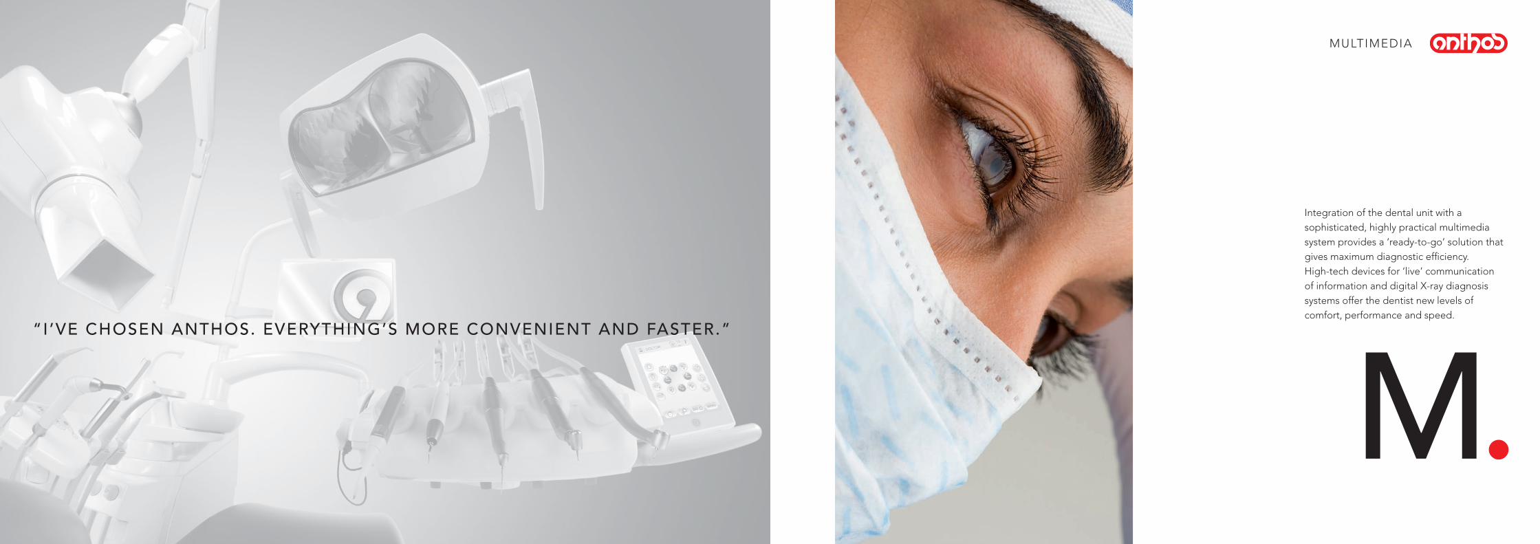
Integration of the dental unit with a sophisticated, highly practical multimedia system provides a ‘ready-to-go’ solution that gives maximum diagnostic efficiency.High-tech devices for ‘live’ communication of information and digital X-ray diagnosis systems offer the dentist new levels of comfort, performance and speed.
“I’VE CHOSEN ANTHOS. EVERYTHING’S MORE CONVENIENT AND FASTER.”
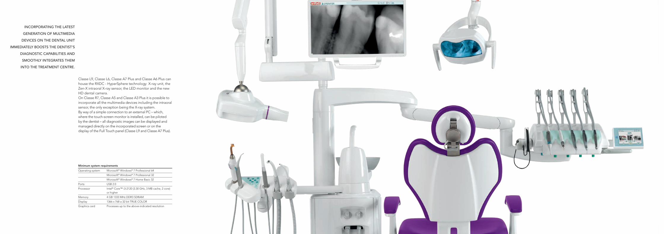
Minimum system requirements
Operating system Microsoft® Windows® 7 Professional 64
Microsoft® Windows® 7 Professional 32
Microsoft® Windows® 7 Home Basic 32
Ports USB 2.0
Processor Intel® Core™ i3-2120 (3.30 GHz, 3 MB cache, 2 core) or higher
Memory 4 GB 1333 MHz DDR3 SDRAM
Display 1366 x 768 a 32 bit TRUE COLOR
Graphics card Processes up to the above-indicated resolution
Classe L9, Classe L6, Classe A7 Plus and Classe A6 Plus can house the RXDC - HyperSphere technology X-ray unit, the Zen-X intraoral X-ray sensor, the LED monitor and the new HD dental camera. On Classe R7, Classe A5 and Classe A3 Plus it is possible to incorporate all the multimedia devices including the intraoral sensor, the only exception being the X-ray system.By way of a simple connection to an external PC – which, where the touch-screen monitor is installed, can be piloted by the dentist – all diagnostic images can be displayed and managed directly on the incorporated screen or on the display of the Full Touch panel (Classe L9 and Classe A7 Plus).
INCORPORATING THE LATEST
GENERATION OF MULTIMEDIA
DEVICES ON THE DENTAL UNIT
IMMEDIATELY BOOSTS THE DENTIST’S
DIAGNOSTIC CAPABILITIES AND
SMOOTHLY INTEGRATES THEM
INTO THE TREATMENT CENTRE.
127°
25°
65°
65°
127°
25°
65°
65°
Monitor technical specifications
Flat screen 22” – 16/9 IPS LCD
Number of pixels (H x V) 1920 x 1080
Brightness 250 cd/m2
Contrast ratio 1000/1
Screen resolution Full HD
Colours supported 16.7 millions of colours
Mount VESA 75
Video input VGA, DisplayPort, HDMI
Medical device certificate CE 93-42
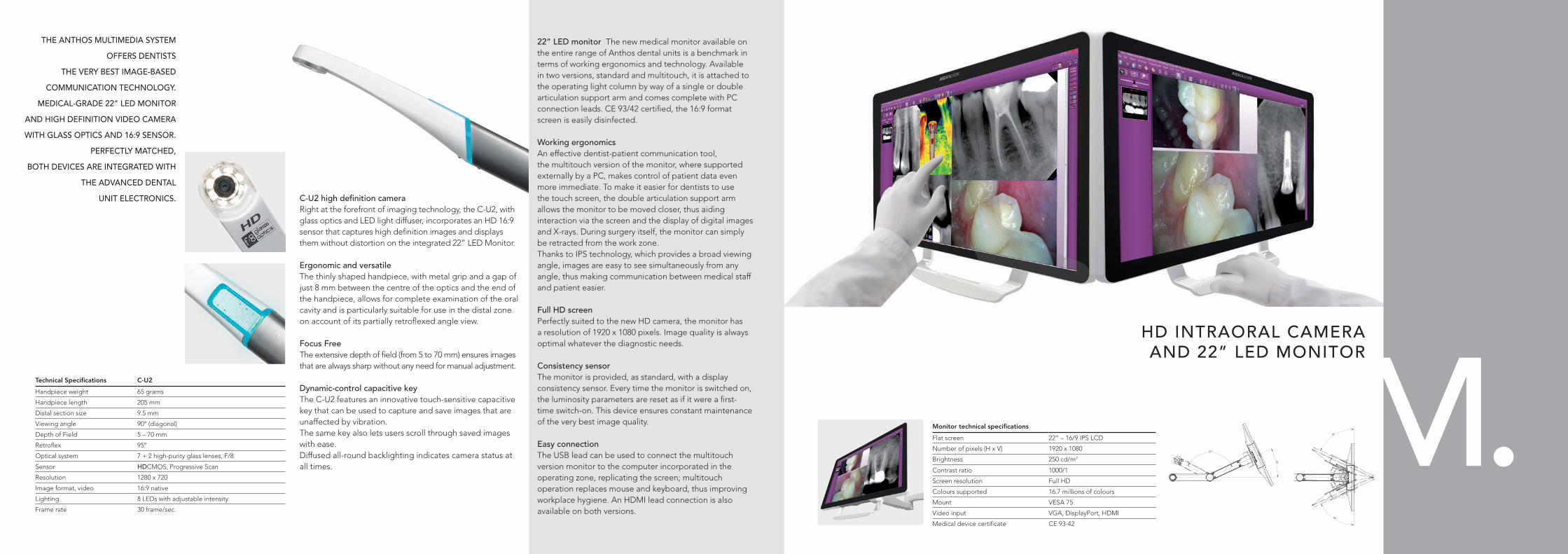
HD INTRAORAL CAMERA AND 22” LED MONITOR
22” LED monitor The new medical monitor available on the entire range of Anthos dental units is a benchmark in terms of working ergonomics and technology. Available in two versions, standard and multitouch, it is attached to the operating light column by way of a single or double articulation support arm and comes complete with PC connection leads. CE 93/42 certified, the 16:9 format screen is easily disinfected.
Working ergonomicsAn effective dentist-patient communication tool, the multitouch version of the monitor, where supported externally by a PC, makes control of patient data even more immediate. To make it easier for dentists to use the touch screen, the double articulation support arm allows the monitor to be moved closer, thus aiding interaction via the screen and the display of digital images and X-rays. During surgery itself, the monitor can simply be retracted from the work zone.Thanks to IPS technology, which provides a broad viewing angle, images are easy to see simultaneously from any angle, thus making communication between medical staff and patient easier.
Full HD screenPerfectly suited to the new HD camera, the monitor has a resolution of 1920 x 1080 pixels. Image quality is always optimal whatever the diagnostic needs.
Consistency sensorThe monitor is provided, as standard, with a display consistency sensor. Every time the monitor is switched on, the luminosity parameters are reset as if it were a first-time switch-on. This device ensures constant maintenance of the very best image quality.
Easy connectionThe USB lead can be used to connect the multitouch version monitor to the computer incorporated in the operating zone, replicating the screen; multitouch operation replaces mouse and keyboard, thus improving workplace hygiene. An HDMI lead connection is also available on both versions.
Technical Specifications C-U2
Handpiece weight 65 grams
Handpiece length 205 mm
Distal section size 9.5 mm
Viewing angle 90° (diagonal)
Depth of Field 5 – 70 mm
Retroflex 95°
Optical system 7 + 2 high-purity glass lenses, F/8
Sensor HDCMOS, Progressive Scan
Resolution 1280 x 720
Image format, video 16:9 native
Lighting 8 LEDs with adjustable intensity
Frame rate 30 frame/sec.
THE ANTHOS MULTIMEDIA SYSTEM
OFFERS DENTISTS
THE VERY BEST IMAGE-BASED
COMMUNICATION TECHNOLOGY.
MEDICAL-GRADE 22” LED MONITOR
AND HIGH DEFINITION VIDEO CAMERA
WITH GLASS OPTICS AND 16:9 SENSOR.
PERFECTLY MATCHED,
BOTH DEVICES ARE INTEGRATED WITH
THE ADVANCED DENTAL
UNIT ELECTRONICS. C-U2 high definition camera Right at the forefront of imaging technology, the C-U2, with glass optics and LED light diffuser, incorporates an HD 16:9 sensor that captures high definition images and displays them without distortion on the integrated 22” LED Monitor.
Ergonomic and versatileThe thinly shaped handpiece, with metal grip and a gap of just 8 mm between the centre of the optics and the end of the handpiece, allows for complete examination of the oral cavity and is particularly suitable for use in the distal zone on account of its partially retroflexed angle view.
Focus Free The extensive depth of field (from 5 to 70 mm) ensures images that are always sharp without any need for manual adjustment.
Dynamic-control capacitive keyThe C-U2 features an innovative touch-sensitive capacitive key that can be used to capture and save images that are unaffected by vibration. The same key also lets users scroll through saved images with ease. Diffused all-round backlighting indicates camera status at all times.
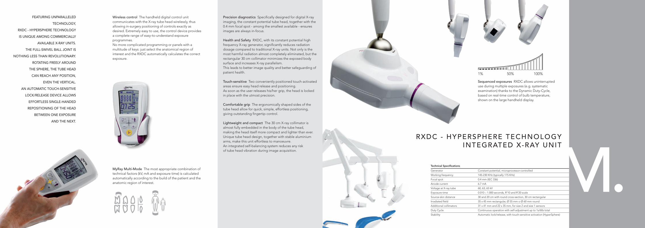
1% 100%50%
Technical Specifications
Generator Constant potential, microprocessor-controlled
Working frequency 145-230 KHz (typically 175 KHz)
Focal spot 0.4 mm (IEC 336)
Anode current 6.7 mA
Voltage at X-ray tube 60, 63, 65 kV
Exposure time 0.010 – 1.000 seconds, R’10 and R’20 scale
Source-skin distance 30 and 20 cm with round cross-section, 30 cm rectangular
Irradiated field 35 x 45 mm rectangular, Ø 55 mm o Ø 60 mm round
Additional collimators 31 x 41 mm and 22 x 35 mm, for size 2 and size 1 sensors
Duty Cycle Continuous operation with self-adjustment up to 1s/60s total
Stability Automatic lock/release, with touch-sensitive activation (HyperSphere)
Precision diagnostics Specifically designed for digital X-ray imaging, the constant potential tube head, together with the 0.4 mm focal spot - among the smallest available - ensures images are always in-focus.
Health and Safety RXDC, with its constant potential high frequency X-ray generator, significantly reduces radiation dosage compared to traditional X-ray units. Not only is the most harmful radiation almost completely eliminated, but the rectangular 30 cm collimator minimizes the exposed body surface and increases X-ray parallelism.This leads to better image quality and better safeguarding of patient health.
Touch-sensitive Two conveniently positioned touch-activated areas ensure easy head release and positioning.As soon as the user releases his/her grip, the head is locked in place with the utmost precision.
Comfortable grip The ergonomically shaped sides of the tube head allow for quick, simple, effortless positioning, giving outstanding fingertip control.
Lightweight and compact The 30 cm X-ray collimator is almost fully embedded in the body of the tube head,making the head itself more compact and lighter than ever.Unique tube head design, together with stable aluminium arms, make this unit effortless to manoeuvre.An integrated self-balancing system reduces any riskof tube head vibration during image acquisition.
Sequenced exposures RXDC allows uninterrupteduse during multiple exposures (e.g. systematic examination) thanks to the Dynamic Duty-Cycle,based on real-time control of bulb temperature,shown on the large handheld display.
FEATURING UNPARALLELED
TECHNOLOGY,
RXDC - HYPERSPHERE TECHNOLOGY
IS UNIQUE AMONG COMMERCIALLY
AVAILABLE X-RAY UNITS.
THE FULL-SWIVEL BALL JOINT IS
NOTHING LESS THAN REVOLUTIONARY.
ROTATING FREELY AROUND
THE SPHERE, THE TUBE HEAD
CAN REACH ANY POSITION,
EVEN THE VERTICAL.
AN AUTOMATIC TOUCH-SENSITIVE
LOCK/RELEASE DEVICE ALLOWS
EFFORTLESS SINGLE-HANDED
REPOSITIONING OF THE HEAD
BETWEEN ONE EXPOSURE
AND THE NEXT.
Wireless control The handheld digital control unit communicates with the X-ray tube head wirelessly, thus allowing in-surgery positioning of controls exactly as desired. Extremely easy to use, the control device provides a complete range of easy-to-understand exposure programmes.No more complicated programming or panels with a multitude of keys: just select the anatomical region of interest and the RXDC automatically calculates the correct exposure.
MyRay Multi-Mode The most appropriate combination of technical factors (kV, mA and exposure time) is calculated automatically according to the build of the patient and the anatomic region of interest.
RXDC - HYPERSPHERE TECHNOLOGYINTEGRATED X-RAY UNIT
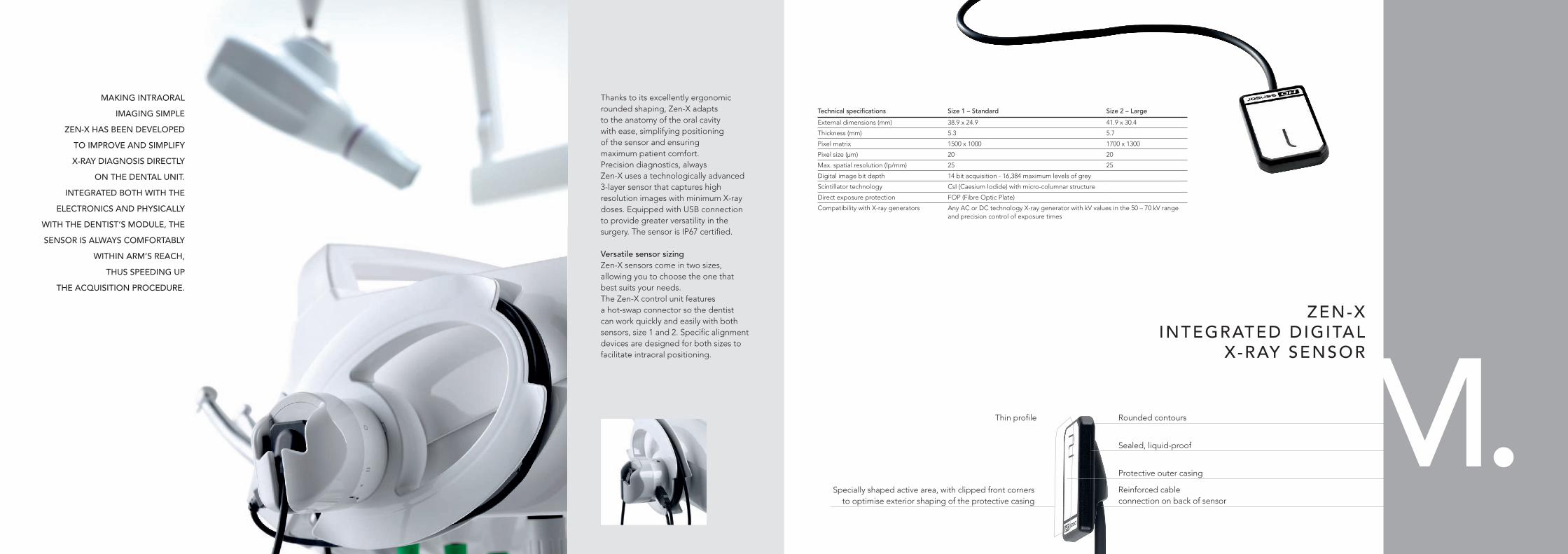
Thin profile Rounded contours
Sealed, liquid-proof
Reinforced cableconnection on back of sensor
Protective outer casing
Specially shaped active area, with clipped front corners to optimise exterior shaping of the protective casing
ZEN-XINTEGRATED DIGITAL
X-RAY SENSOR
Technical specifications Size 1 – Standard Size 2 – Large
External dimensions (mm) 38.9 x 24.9 41.9 x 30.4
Thickness (mm) 5.3 5.7
Pixel matrix 1500 x 1000 1700 x 1300
Pixel size (μm) 20 20
Max. spatial resolution (lp/mm) 25 25
Digital image bit depth 14 bit acquisition - 16,384 maximum levels of grey
Scintillator technology CsI (Caesium Iodide) with micro-columnar structure
Direct exposure protection FOP (Fibre Optic Plate)
Compatibility with X-ray generators Any AC or DC technology X-ray generator with kV values in the 50 – 70 kV range and precision control of exposure times
Thanks to its excellently ergonomic rounded shaping, Zen-X adaptsto the anatomy of the oral cavitywith ease, simplifying positioningof the sensor and ensuringmaximum patient comfort.Precision diagnostics, always Zen-X uses a technologically advanced 3-layer sensor that captures high resolution images with minimum X-ray doses. Equipped with USB connection to provide greater versatility in the surgery. The sensor is IP67 certified. Versatile sensor sizing Zen-X sensors come in two sizes, allowing you to choose the one that best suits your needs.The Zen-X control unit featuresa hot-swap connector so the dentist can work quickly and easily with both sensors, size 1 and 2. Specific alignment devices are designed for both sizes to facilitate intraoral positioning.
MAKING INTRAORAL
IMAGING SIMPLE
ZEN-X HAS BEEN DEVELOPED
TO IMPROVE AND SIMPLIFY
X-RAY DIAGNOSIS DIRECTLY
ON THE DENTAL UNIT.
INTEGRATED BOTH WITH THE
ELECTRONICS AND PHYSICALLY
WITH THE DENTIST’S MODULE, THE
SENSOR IS ALWAYS COMFORTABLY
WITHIN ARM’S REACH,
THUS SPEEDING UP
THE ACQUISITION PROCEDURE.