Embed Size (px)
Citation preview
0008-3194/2012/94–101/$2.00/©JCCA 2012
94 JCanChiroprAssoc2012;56(2)
MultipleMyelomapresentingassacroiliacjointpain:acasereportDanielle Southerst, BScH, DC*John Dufton, DC, MSc, MD†
Paula Stern, BSc, DC, FCCS(C)‡
* Canadian Memorial Chiropractic College, Division of Graduate Studies, 6100 Leslie Street, Toronto, ON, M2H 3J1, (416) 482-2344 x 287; [email protected].
† Queen’s University, Department of Diagnostic Radiology, Kingston, ON Canada.‡ Division of Graduate Studies, Canadian Memorial Chiropractic College. This case report was written with full verbal consent from the patient
to disclose medical information.© JCCA 2012
Le myélome multiple (MM) est le cancer primitif des os le plus fréquent chez les adultes. La manifestation clinique du MM est variée et dépend des lieux et du degré d’envahissement. Plus important encore pour les chiropraticiens, les signes cliniques prépondérants du MM sont liés au sarcome osseux et peuvent ressembler à une douleur d’origine musculosquelettique. Voici le cas d’un patient de 56 ans en chiropratique éprouvant depuis six mois une douleur aux articulations sacro-iliaques, qui avait préalablement reçu le diagnostic d’hématome et traité sans résultat par de multiples fournisseurs de soins. Après un examen physique, une imagerie médicale et des essais en laboratoire, le diagnostic de MM a été confirmé. Le rapport du cas décrit la pathophysiologie apparentée, la manifestation clinique, l’imagerie médicale et la prise en charge du MM, tout en illustrant les enjeux majeurs relatifs à la prise en charge du patient, car ils concernent la chiropratique ainsi que la reconnaissance de la pathologie dans le cadre d’une douleur musculosquelettique.(JCCA 2012; 56(2):94–101)
m o t s c l é s : myélome, cancer, diagnostic
Multiple Myeloma (MM) is the most common primary cancer of bone in adults. The clinical presentation of MM is varied and depends on the sites and extent of involvement. Most importantly for chiropractors, the leading clinical symptoms of MM are related to bone neoplasm and may mimic pain of musculoskeletal origin. The following is the case of a 56 year old male chiropractic patient presenting with a 6 month history of sacroiliac joint pain previously diagnosed and managed unsuccessfully as a hematoma by multiple providers. Physical examination, imaging, and laboratory investigations confirmed a diagnosis of MM. The case report describes relevant pathophysiology, clinical presentation, imaging, and management for MM, while illustrating key issues in patient management as they relate to chiropractic practice and the recognition of pathology in the context of musculoskeletal pain.(JCCA 2012; 56(2):94–101)
k e y w o r d s : myeloma, cancer, diagnosis
IntroductionMultiple Myeloma (MM) is a primary malignancy of bone marrow characterized by clonal proliferation of plasma cells and production of monoclonal immunoglobulin. It is the most common primary bone cancer in adults1,2 con-
tributing to 1.3% of new cancer cases in Canada and 1.9% of cancer deaths.3 In 2008, an estimated 6000 Canadians were living with the disease, including 2100 newly diag-nosed.3 Myeloma is slightly more prevalent in males4,5,6 and blacks.4,5,7 The median age at diagnosis is 66, with
JCanChiroprAssoc2012;56(2) 95
D Southerst, J Dufton, P Stern
the majority diagnosed over the age of 60;8,9 however in a review of 1027 patients diagnosed with MM, 30% were under the age of 60 and the age of diagnosis ranged from 20–92.8 The most common symptoms reported are those related to bone neoplasm including unexplained backache that is often severe and precipitated by movement.8,11 These symptoms may motivate a patient to seek conserva-tive care for what is assumed to be a complaint of muscu-loskeletal origin. This case emphasizes key components of patient management as they relate to chiropractic prac-tice and the recognition of pathology in the context of a patient presenting with pain of presumed musculoskeletal origin.
CasePresentation
HistoryA 56 year old male presented to a tertiary care centre with a six month history of pain in the left gluteal/sacro-iliac joint region. The complaint progressively worsened following its onset after heavy lifting. His pain varied in intensity and was often exacerbated following activity. The most recent and intense exacerbation occurred in the week prior to his hospital presentation and was insidious in onset. Symptoms included radiation of pain down the left leg of two days duration described as stabbing in na-ture with an intensity of 7/10. The patient did not report any associated numbness or paresthesias and was not ex-periencing any weakness. Aggravating factors included sitting and lying supine. In addition, he reported occa-sional waking during the night due to pain. The patient reported a mass in the left gluteal region which had slow-ly increased in size over the preceding month. This mass was diagnosed and managed as a hematoma 6 months prior to his hospital presentation. His family physician prescribed Tylenol-3 and physiotherapy. Two visits with a physiotherapist consisting of a passive assisted stretch-ing technique and traction aggravated his complaint. He then consulted a chiropractor who treated him with spinal manipulative therapy for more than 12 visits over 6 weeks. The patient reported minimal short-term relief over this period of time with no change in the soft tis-sue mass. Following his lack of response to conservative chiropractic management, the patient was referred by the chiropractor to the local hospital emergency department for further medical assessment.
The patient reported his health status as otherwise healthy. He denied a history of fever, night sweats, or re-cent changes in his weight and did not report any bowel or bladder dysfunction. A systems review was unremarkable.
Physical ExaminationUpon hospital presentation, examination revealed a palp-able warm soft tissue mass in the left buttock region, ap-proximately 10 cm in diameter. Hip and lumbar spine ranges of motion were pain-free and within normal limits. Neurological examination including sensory, motor, and reflex testing of the upper and lower limb was unremark-able. Straight leg raise was negative bilaterally. Sacroiliac (SI) compression and FABER (Flexion Abduction Exter-nal Rotation) were both positive on the left, reproducing pain over the patient’s left SI joint. No tenderness was noted on lumbar spine palpation. An abdominal examina-tion failed to reveal any tenderness or palpable masses and there was no evidence of lymphadenopathy peripherally. Vital signs were within normal limits.
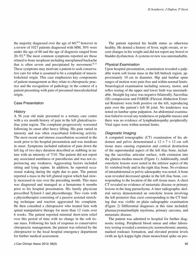
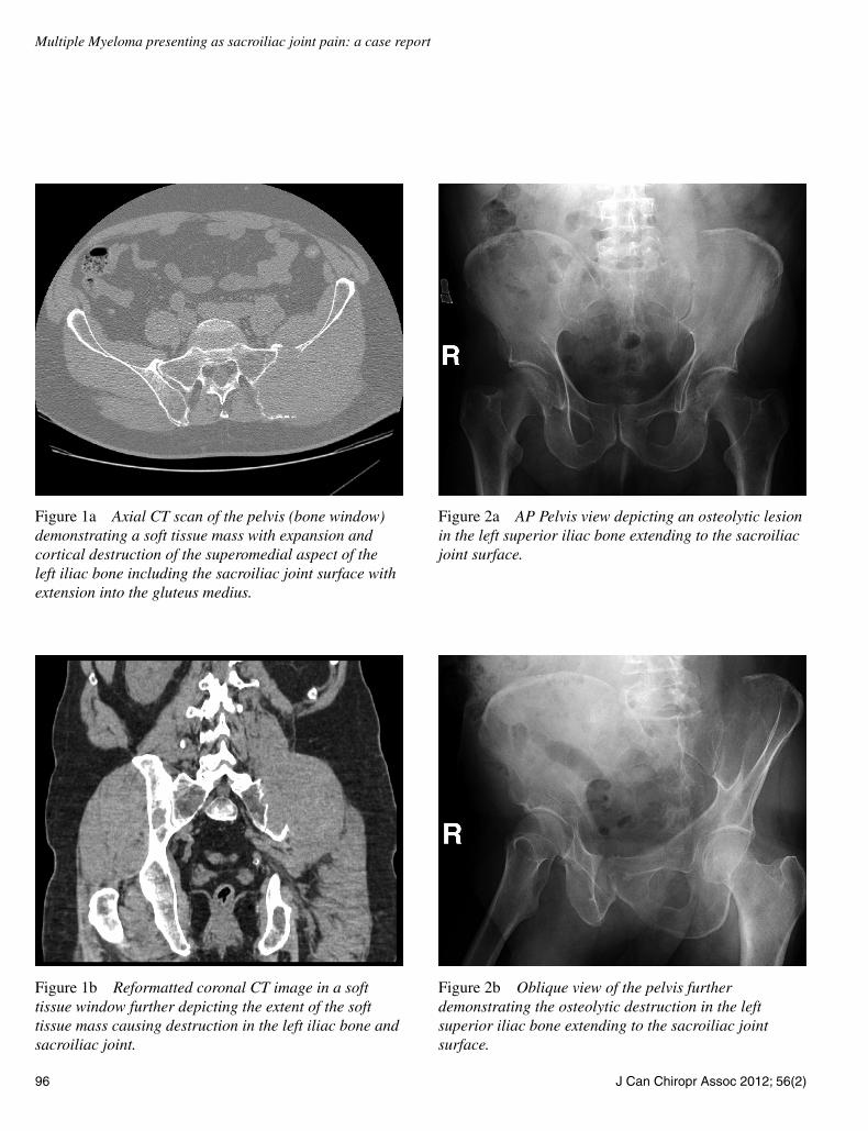
Diagnostic ImagingA computed tomographic (CT) examination of his ab-domen and pelvis demonstrated a 12 × 7 × 12 cm soft tissue mass causing expansion and cortical destruction of the superomedial aspect of the left iliac bone, includ-ing the sacroiliac articular surface, with extension into the gluteus medius muscle (Figure 1). Additionally, small osteolytic lesions were noted in the inferior aspect of the S1 vertebral body and in the right iliac bone. No evidence of intraabdominal or pelvic adenopathy was noted. A bone scan revealed decreased uptake in the left iliac bone, cor-responding to the location of the soft tissue mass. A chest CT revealed no evidence of metastatic disease or primary lesions in the lung parenchyma. A later radiographic skel-etal survey demonstrated an osteolytic lesion involving the left posterior iliac crest corresponding to the CT find-ing that was visible on plain radiographic examination (Figure 2) Differential diagnoses at this time included; plasmacytoma/multiple myeloma, primary sarcoma, and metastatic disease.
The patient was admitted to hospital for further diag-nostic testing. Peripheral blood smear and other labora-tory testing revealed a normocytic normochromic anemia, marked rouleaux formation, and elevated protein levels (with an IgA-kappa light chain monoclonal spike on pro-
96 JCanChiroprAssoc2012;56(2)
Multiple Myeloma presenting as sacroiliac joint pain: a case report
Figure 2b Oblique view of the pelvis further demonstrating the osteolytic destruction in the left superior iliac bone extending to the sacroiliac joint surface.
Figure 2a AP Pelvis view depicting an osteolytic lesion in the left superior iliac bone extending to the sacroiliac joint surface.
Figure 1a Axial CT scan of the pelvis (bone window) demonstrating a soft tissue mass with expansion and cortical destruction of the superomedial aspect of the left iliac bone including the sacroiliac joint surface with extension into the gluteus medius.
Figure 1b Reformatted coronal CT image in a soft tissue window further depicting the extent of the soft tissue mass causing destruction in the left iliac bone and sacroiliac joint.
JCanChiroprAssoc2012;56(2) 97
D Southerst, J Dufton, P Stern
tein electrophoresis). A bone marrow aspirate demon-strated plasma cells making up over 50% of the total cell population. These findings were consistent with multiple myeloma given the osseous lesions noted in the left iliac bone and S1 vertebral body.
ManagementOnce admitted to the hospital, the patient was referred for consultations with hematology and oncology. He received transfusions to correct his anemia and was administered his initial 4 day cycle of chemotherapy. His pain was controlled effectively through the use of regular strength Tylenol. A referral was made to radiation oncology for consideration of radiation therapy. The patient received 6 cycles of chemotherapy, subsequent radiation therapy and autologous stem cell transplant over the duration of one year. Despite some initial improvement in symptoms and activities of daily living, upon last follow up new lesions were discovered in his liver. His prognosis for recovery at the time of last follow-up was poor.
Discussion
PathophysiologyThe pathophysiology of MM begins with cytogenetic changes that occur in the plasma cell lineage within the bone marrow.10 Monoclonal expansion of myeloma plasma cells within the bone marrow interferes with the production of normal blood cells. Myeloma cells pro-duce abnormal immunoglobulin (M protein), light chain proteins (κ and λ), and other factors, such as cytokines. Excessive M protein causes hyperviscosity of the blood, whereas excessive light chains cause end organ damage (for example renal failure). Lesions of bone are largely caused by the release of cytokines7 that promote bone resorption via upregulation of osteoclast activity, dif-ferentiation, and maturation.7,9 Unrestrained osteoclast activation leads to the release of mediators that stimulate further clonal proliferation of myeloma cells and subse-quent tumour growth.10 The result is a vicious cycle of bone destruction and tumour growth, leading to further bone destruction.
Clinical Manifestations/PresentationSymptoms of MM are the result of bone marrow infiltra-tion, the development of bone neoplasms, and the effects
of the disease process on the renal system.5,7 Prolifera-tion of abnormal plasma cells within bone marrow results in reduced production of normal blood cells causing an-emia, thrombocytopenia, and leucopenia.7 Fatigue, weak-ness, and malaise are common symptoms experienced by approximately 1/3 of patients,8 most commonly due to anemia.7,8 Thrombocytopenia causes excessive bleed-ing and/or bruising, whereas leukopenia leads to frequent recurrent infections.7,10 The leading symptoms of mul-tiple myeloma are those related to bone neoplasm.11 Un-explained backache or bone pain in the long bones, ribs, skull, or pelvis are common presenting complaints7,10 and may be present in up to 58% of patients.8 Pain is often severe and precipitated by movement.8 Pathological fractures as a result of diffuse osteopenia or expansile tumours may be the presenting complaint in 26–34% of patients.5,7 Vertebral compression fractures are common and can result in spinal cord or nerve root compression.2 Peripheral neuropathies and paresthesias may also occur, the most common being carpal tunnel syndrome,7,10 Hyper calcemia becomes prominent as bone resorption continues, and can result in numerous symptoms such as: anorexia, nausea, vomiting, polydipsia, constipation, abdominal pain, bone pain, impaired concentration and memory, lethargy, muscle weakness, and itching.7,10,24
High levels of M protein in the blood can lead to symp-toms of hyperviscosity including headache and bruis-ing.10 Although there are many symptoms associated with multiple myeloma, studies have shown that up to 34% of patients report an absence of symptoms prior to their diagnosis.5
For patients experiencing pain, correct diagnosis relies on a thorough history. MM’s variable presentation makes it difficult to provide a list of symptoms that are highly sensitive or specific to confirm or rule out the diagnosis. Typically, pain of pathological origin is suspected in the presence of red flags including age over 50, a previous history of cancer, no relief with rest, and constitutional symptoms such as unexpected weight loss, fever, and fa-tigue.17,18 Individually, these indicators have high speci-ficity but low sensitivity.17,18 The specificity of a clinical test speaks to its ability to identify true negative test re-sults, whereas sensitivity is related to a test’s ability to identify true positive test results. With high specificity, the chance of receiving a false positive result is low; how-ever with a low sensitivity, there is a higher chance of
98 JCanChiroprAssoc2012;56(2)
Multiple Myeloma presenting as sacroiliac joint pain: a case report
receiving a false negative result. When trying to rule out a condition where there is a high cost for a missed diag-nosis such as malignancy, a clinical test must have high sensitivity. Due to their low sensitivity, the absence of any one red flag cannot accurately rule out the presence of sig-nificant pathology. When combined, red flags may have higher sensitivity, emphasizing the importance of asking about all possible red flags.17 Berenson et al. suggest that multiple myeloma should be considered as a diagnosis in a patient over the age of 50 with back pain persisting for more than one month and one or more of the following symptoms; pain worse in the supine position, pain worse at night or pain that awakens the patient from sleep, band like distribution of pain around the body, pain not re-sponding to conservative care or rest, constitutional symp-toms, or progressive neurological deficit.10 Upon hospital presentation, the patient discussed in this case reported aggravation with lying supine and occasional waking due to night pain, symptoms fulfilling these criteria that may or may not have been present initially, emphasizing the importance of continued evaluation to ensure the recogni-tion of latent symptoms of pathology.23
This patient also experienced a 6 month history of worsening pain and a growing mass. The mass was initial-ly diagnosed as a hematoma and was unresponsive to con-servative management. The presence of a growing mass is a red flag, particularly where a diagnosis of hematoma is given. The natural history of a hematoma indicates that the mass should have diminished in size, rather than increase over 4–6 weeks. Failure to respond to conservative care is another significant red flag. The Glenerin Guidelines (1996) suggest that lack of improvement after 12 visits may imply the diagnosis is incorrect, the treatment is in-correct, or there is a co-existing condition. After 6 weeks of no improvement, a referral is warranted.19 Guidelines, however, do not take the place of clinical reasoning. Clin-ical decision making is guided by case complexity, the best available evidence, provider expertise and experi-ence, and patient preference and beliefs.20,21,22 A decision must be made prior to implementation of care regarding expectations for improvement based on available evidence regarding natural history and individual patient factors or case complexity.20 A lack of expected improvement war-rants a change in treatment approach or referral.20,21,22 In-formation regarding a patient’s progress is obtained from continued frequent evaluation; even when a diagnosis has
been provided by another health professional. It is the con-sulting health professional’s responsibility to re-evaluate and formulate a diagnosis that is consistent with history and physical examination findings as well as the patient’s response to management given previous diagnoses. For the patient in this case who was treated for pain of mus-culoskeletal origin and a hematoma, the natural history of the complaint should have been considered. Combined with his emerging symptoms consistent with pathology, his lack of response to conservative management should have resulted in an earlier decision for imaging or referral.
Physical ExaminationWithin the primary care setting, the suspicion of MM may be based solely on information gathered in the history. When the most prominent symptom is pain, a physical examination is performed to confirm or rule out pain of musculoskeletal origin; however it is important to note that the pain associated with neoplasm of bone can be re-produced much like pain of musculoskeletal origin. Pain precipitated by range of motion is a common symptom in patients with bone neoplasm, including multiple my-eloma.8,12 A musculoskeletal examination of the patient in this case reproduced the chief complaint, emphasizing that positive musculoskeletal provocation tests do not rule out pain of pathological origin. In addition, chronicity of a complaint also does not rule out pain of a pathological origin. The authors of a retrospective case review involv-ing primary sarcomas of the pelvis (excluding multiple myeloma) suggested one of the reasons for delayed diag-nosis was that primary care practitioners were mislead by chronicity which is less consistent with rapidly growing malignancies. In the pelvis, tumours can occupy larger areas without detection due to the relatively large sur-rounding space.12 This patient developed chronic pain due to the lack of attention to his growing tumour which was originally diagnosed and managed as a hematoma, further placing him outside of the typical presentation of malignancy.
Imaging FindingsImaging plays a role in the diagnosis and prognostic clas-sification of multiple myeloma. Radiography is of key in-terest to chiropractors as many have access to this form of imaging. Clinical indications for radiographic imaging include the presence of red flags and lack of response to
JCanChiroprAssoc2012;56(2) 99
D Southerst, J Dufton, P Stern
treatment. Radiographic findings of multiple myeloma include punched out osteolytic lesions without reactive sclerosis, osteoporosis, and pathological compression fractures.2,8 The most frequently involved bones are the skull, pelvis, ribs, sternum, and long bones.2 Approxi-mately 79% of patients have positive radiographic find-ings at the time of their diagnosis.8 Up to 25% of those with an absence of radiographic findings subsequently de-veloped positive findings in follow-up examination, em-phasizing the lack of sensitivity of radiographic imaging in early phases of the disease.8,11 Radiography also lacks sensitivity in identifying myeloma-related osteoporosis, as 50% trabecular loss is required for its visualization.2 Due to this lack of sensitivity, additional imaging such as Computed Tomography (CT) or Magnetic Resonance Imaging (MRI) may be required when suspicions of mul-tiple myeloma are high.8,11 CT is able to provide detailed information regarding the extent of cortical involvement of the tumour, whereas MRI is able to demonstrate mar-row infiltration as well as diffuse patterns of infiltration that may not be adequately visualized using radiographic imaging alone.1,11 In addition, MRI is able to demon-strate the extent of soft tissue and neurovascular involve-ment.11 On MRI, myeloma tumours have a low T1 and high T2 weighted signal intensity with enhancement after the administration of intravenous contrast.2 As there is no increase in osteoblastic activity, bone scans are of less im-portance in MM and may result in false negative findings, leading to misdiagnosis.13 In the diagnosis of the patient in this case, CT, plain radiographic imaging, and bone scan provided findings that were consistent with findings of MM; however earlier referral for radiographic imaging or diagnostic ultrasound for his growing mass may have lead to more prompt diagnosis and management.
Laboratory FindingsLaboratory tests used to screen for MM include a com-plete blood count (CBC), peripheral blood smear, erythro-cyte sedimentation rate (ESR), chemistry panel (including electrolytes, calcium, uric acid), and serum and urine pro-tein electrophoresis. For confirmation of a diagnosis of MM, a bone marrow biopsy and immunofixation should also be performed. A CBC will demonstrate normocytic normochromic anemia in most patients8 and may also re-veal leukocytopenia and thrombocytopenia. Rouleaux, a characteristic finding of MM, will be seen with a periph-
eral blood smear in roughly half of all patients.8 ESR is often elevated.8,13 A chemistry panel will reveal hyper-uricemia and hypercalcemia13 and serum creatinine will be elevated in the case of renal insufficiency (due to renal failure, myeloma kidney, hypercalcemia).8 Serum albu-min may also be decreased.13 Protein electrophoresis will demonstrate a characteristic M-spike in the serum and/or urine of most patients, indicating monoclonal gam-mopathy.8,13 Bone marrow biopsy confirms a diagnosis of multiple myeloma through the demonstration of ma-lignant plasma cell infiltrates.13 In the current case, lab-oratory findings were used to rule out the differential diagnoses and confirm the diagnosis of multiple myeloma following the discovery of a tumour in the posterior ilium.
DiagnosisThe diagnosis of MM is dependent on findings from a number of investigations including clinical examina-tion, imaging, and laboratory testing. As chiropractors, the clinical picture and imaging are used to indicate the need for referral for appropriate follow-up diagnostic testing. In order to properly diagnose MM, World Health Organization criteria include plasmacytosis, an M spike on serum, plasma, or urine protein electrophoresis, and plasmacytoma proven on biopsy.10,13 Additional findings considered in the diagnosis of MM are osteolytic lesions and decreased polyclonal immunoglobulins.13 Differ-ential diagnoses vary depending on the clinical picture. For chiropractors, relevant differential diagnoses will be based on initial imaging findings of an osteolytic bone tumour or the patient’s clinical presentation. Differential diagnosis of an osteolytic bone tumour in a patient over the age of 50 includes metastasis, multiple myeloma, and lymphoma. Staging of MM is achieved using radiograph-ic skeletal surveys and further CT or MRI and is helpful in the development of a plan of management and in deter-mining a prognosis.
ManagementStandard medical management of symptomatic MM in-volves chemotherapy with or without an autologous stem cell transplant for patients under the age of 70.13 Intra-venous administration of bisphosphonates are used in conjunction with chemotherapy and have been shown to decrease the progression of osteolytic lesions and the development of vertebral and non-vertebral fractures, to
100 JCanChiroprAssoc2012;56(2)
Multiple Myeloma presenting as sacroiliac joint pain: a case report
treat hypercalcemia and bone pain, and improve quality of life.9,10,14,15,16 Bisphosphonate treatment is recommended for patients with radiological evidence of osteolytic le-sions or osteoporosis and is continued monthly for a period of 2 years.15 Supportive therapy for patients with MM may include maintenance of fluid and electrolytes with regular hydration, erythropoietin and transfusions to replace red blood cells and platelets, pain control using analgesics (NSAIDs are contraindicated due to potential for renal complications), promotion of mobility to prevent osteoporotic fractures, spinal decompression surgery for spinal complications, and radiation therapy for focal skel-etal lesions.4,7,13,15
PrognosisThe mean overall duration of survival after being diag-nosed with multiple myeloma is 33 months with con-siderable individual variation.8,13 Negative prognostic indicators include older age, a previously diagnosed plasma cell disorder, and key laboratory findings.2,8,10 Laboratory findings indicating a worse prognosis include elevated β-2 microglobulin, serum albumin, and C-react-ive protein.2,10 Bone marrow cytogenics can also be used to help determine prognosis upon diagnosis.5,10
ConclusionIn addition to a discussion on relevant clinical presenta-tion, diagnosis, treatment, and prognosis of multiple my-eloma, this case of a 56 year old male chiropractic patient with multiple myeloma illustrates key issues in patient management as they relate to chiropractic practice and the recognition of pathology in the context of musculoskel-etal pain. An estimated 0.7% of patients with back pain in the primary care setting have neoplastic disease.17 As di-agnosticians, the chiropractor’s role is to rule out these se-rious causes for a patient’s pain. Due to the low sensitivity of red flags, their absence cannot be used exclusively to rule out significant pathology.17,18 Further complicating the elimination of a pathological diagnosis is the fact that provocation of pain during the musculoskeletal examina-tion does not rule out the presence of serious pathology, nor does the chronicity of the complaint. Decisions must be made prior to implementation of care regarding ex-pectations for improvement based on available evidence regarding natural history, provider experience, and pa-tient-related factors.20,21 With continued frequent evalu-
ation, the recognition of a lack of expected improvement warrants a change in treatment approach or referral.20,21 This case illustrates that without proper benchmarking of care, pathology can be missed, even in the event of the inclusion of multiple health professionals. For chiroprac-tors, understanding the limitations of initial history and examination findings, as well as the importance of ongo-ing re-evaluation in the detection of a lack of expected improvement is crucial to appropriate patient manage-ment and the recognition of underlying pathology in the context of seemingly musculoskeletal pain presentations.
References 1 Dinter DJ, Neff WK, Klaus J, Bohm C, Hastka J, Weiss
C, Schoenburg SO, Metzgeroth G. Comparison of whole-body MR imaging and conventional X-ray examination in patients with multiple myeloma and implications for therapy. Ann Hematol. 2009; 88:457–464.
2 Mulligan ME, Badros AZ. PET/CT and MR imaging in myeloma. Skeletal Radiol. 2007; 36: 5–16.
3 Myeloma Canada. 2010. Retrieved August 3, 2010 from Myeloma Canada Website: http://www.myelomacanada.ca/en/default.htm
4 Beers MH, Porter RS, Jones TV, Kaplan JL, Berkwits M. The Merck Manual of Diagnosis and Therapy. 18th ed. New Jersey: Merck Research Laboratories, 2006:1129–1131.
5 Dynamed. (2011, Feb 22). Multiple Myeloma. Ipswitch, MA: EBSCO Publishing. Retrieved March 22, 2011 from, http://dynaweb.ebscohost.com/Detail?id=AN+116888&sid=83da546b-4168-4b73-9f13-84c75f5b4bd6@sessionmgr113.
6 Dores GM, Landgren O, McGlynn KA, Curtis RE, Linet MS, Devesa SS. Plasmacytoma of bone, extramedullary plasmacytoma, and multiple myeloma: incidence and survival in the United States, 1992–2004. Br J Hematol. 2008; 144:86–94.
7 Nau KC, Lewis WD. Multiple Myeloma: Diagnosis and Treatment. Am Fam Physician. 2008; 78(7):853–859.
8 Kyle RA, Gertz MA, Witzig TE, Lust JA, Lacy MQ, Dispenzieri A, Fonseca R, Rajkumar V, Offord JR, Larson MS, Plevak ME, Therneau TM, Greipp PR. Review of 1027 patients with newly diagnosed multiple myeloma. Mayo Clin Proc. 2003; 78:21–33.
9 Ashcroft AJ, Davies FE, Morgan GJ. Aetiology of bone disease and the role of bisphosphonates in multiple myeloma. The Lancet-Oncology. 2003; 4:284–292.
10 Berenson JR, Rajdev L, Broder M. Bone complications in multiple myeloma. Cancer Biol Ther. 2006; 5(9):1082–1085.
11 Mirzaei S, Filipits M, Keck A, Bermayer W, Knoll P, Koehn H, Ludwig H, Pecherstorfer M. Comparison of
JCanChiroprAssoc2012;56(2) 101
D Southerst, J Dufton, P Stern
Technietium-99m-MIBI imaging with MRI for detection of spine involvement in patients with multiple myeloma. BMC Nucl Med. 2003; 3(2).
12 Wurtz LD, Peabody TD, Simon MA. Delay in the diagnosis and treatment of primary bone sarcoma of the pelvis. J Bone Joint Surg Am. 1999; 81:317–325.
13 Finnish Medical Society Duodecim. Multiple myeloma (MM). In: EBM Guidelines. Evidence-Based Medicine [Internet]. Helsinki, Finland: Wiley Interscience. John Wiley and Sons; 2007 May 30.
14 He Y, Wheatley K, Glasmacher A, Ross H, Djulbegovic B. Early versus deferred treatment for early stage multiple myeloma. Cochrane Database of Syst Rev. 2003; Issue 1: Art. No.: CD004023. DOI: 10.1002/14651858.CD004023.
15 Lacey MQ, Dispenzieri A, Gertz MA, Greipp PR, Gollback KL, Hayman SR, Kumar S, Lust JA, Rajkumar v, Russell SJ, Witzig TE, Zeldenrust SR, Dingli D, Bergsagel L, Fonseca R, Reeder CB, Stewart K, Roy V, Dalton RJ, Carr AB, Kademani D, Keller E, Viozzi CF, Kyle R. Mayo Clinic Consensus Statement for the Use of Bisphosphonates in Multiple Myeloma. Mayo Clin Proc. 2006; 81(8):1047–1053.
16 Shipman CM, Oyajobi BO, Mundy GR. Advances in the management of myeloma bone disease. Expert Opin Pharmacother. 2005; 6(16):2781–2791.
17 Deyo RA. Diagnostic evaluation of LBP. Arch Intern Med. 2002; 162:1444–47.
18 Jarvik JG, Deyo RA. Diagnostic evaluation of low back pain with emphasis on imaging. Ann Int Med. 2002; 137(7):586–597.
19 Canadian Chiropractic Association. 1994. Clinical Guidelines for Chiropractic Practice in Canada (Glenerin Guidelines). Retrieved on March 22, 2011 from: http://www.chiropracticcanada.ca/enus/AboutUs/ClinicalPracticeGuidelines/GlenerinGuidelinesApril1993.aspx on March 1, 2010.
20 Triano JJ. What constitutes evidence for best practice? J Manip Physiol Ther. 2008; 31(9):637–43.
21 Von Korff M, Saunders K. The course of back pain in primary care. Spine. 1996; 21(24):2833–7.
22 Van Tulder MW, Tuut M, Pennick V, Bombardier C, Assendelft WJJ. Quality of primary care guidelines for acute low back pain. Spine. 2004; 29(17):E357–62.
23 Walsh NE, et al. Standards of care for acute and chronic musculoskeletal pain: The Bone and Joint Decade (2000–2010). Arch Phys Med Rehabil. 2008; 89: 1830–45.
24 Fauci As, Harrison TR. Harrison’s Manual of Medicine. New York: McGraw Hill Professional, 2009: 959.