Embed Size (px)
Citation preview
Multiple risk pathways for schizophrenia convergein serine racemase knockout mice, a mouse modelof NMDA receptor hypofunctionDarrick T. Balua,b, Yan Lia, Matthew D. Puhla,b, Michael A. Benneywortha,b, Alo C. Basua,b, Shunsuke Takagic,Vadim Y. Bolshakova, and Joseph T. Coylea,b,1
aDepartment of Psychiatry, Harvard Medical School, Boston, MA 02115; bLaboratory for Psychiatric and Molecular Neuroscience, Department of Psychiatry,McLean Hospital, Belmont, MA 02478; and cDepartment of Psychiatry and Behavioral Sciences, Graduate School of Medical and Dental Sciences, TokyoMedical and Dental University, Bunkyo-ku, Tokyo 113-8519, Japan
Edited* by Solomon H. Snyder, The Johns Hopkins University School of Medicine, Baltimore, MD, and approved May 7, 2013 (received for reviewMarch 5, 2013)
Schizophrenia is characterized by reduced hippocampal volume,decreased dendritic spine density, altered neuroplasticity signalingpathways, and cognitive deficits associated with impaired hippo-campal function. We sought to determine whether this diversepathology could be linked to NMDA receptor (NMDAR) hypofunc-tion, and thus used the serine racemase-null mutant mouse (SR−/−),which has less than 10% of normal brain D-serine, an NMDAR coa-gonist.We found that D-serinewas necessary for themaintenance oflong-term potentiation in the adult hippocampal dentate gyrus andfor full NMDAR activity on granule cells. SR−/− mice had reduceddendritic spines and hippocampal volume. These morphologicalchangeswere paralleled bydiminished BDNF/Akt/mammalian targetof rapamycin (mTOR) signaling and impaired performance on a trace-conditioning memory task. Chronic D-serine treatment normalizedthe electrophysiological, neurochemical, and cognitive deficits inSR−/− mice. These results demonstrate that NMDAR hypofunctioncan reproduce the numerous hippocampal deficits associated withschizophrenia, which can be reversed by chronic peripheral D-serinetreatment.
miR-132 | MeCP2 | glycogen synthase 3 kinase | CREB
Schizophrenia is a severe psychiatric disorder that affects 1% ofthe population worldwide (1). There are widespread mor-
phological, neurochemical, and functional changes in the brain inschizophrenia that have been linked to its symptomatic features(2). For example, the hippocampus of patients with schizophreniaexhibits reduced dendritic spine density (3), atrophy (4), and im-paired activation while performing cognitive tasks (5). The neu-roplasticity deficits observed in schizophrenia could be caused bya constellation of factors.Impaired neurotrophic signaling could be one mechanism un-
derlying these abnormalities. BDNF regulates a complex array ofprocesses, including neurite outgrowth and spine density, by sig-naling through tropomyosin receptor kinase B (TrkB), its high-affinity receptor (6). In postmortem studies, BDNF mRNA andprotein (7–9) levels, as well as TrkB mRNA (7, 10, 11) and pro-tein (12), are reduced in subjects with schizophrenia. V-akt mu-rine thymoma viral oncogene (Akt) is a kinase downstream ofTrkB. Not only is the Akt1 isoform a putative schizophrenia riskgene (13), its expression (14, 15) and the amount of phosphory-lated Akt (p-Akt) (16) in the dentate gyrus (DG) are reducedin schizophrenia.Aberrant microRNA (miR) processing might also be contribut-
ing to the pathophysiology of schizophrenia (17). These noncodingRNAs regulate neural plasticity by controlling the translation oftarget mRNA transcripts. Expression of the neuron-enrichedmiR-132 is reduced in schizophrenia (18); it regulates basal and activity-induced neurite outgrowth (19), and is up-regulated in vivo inresponse to external stimuli (20, 21). Importantly, both BDNF (22)
and miR-132 (17) expression are increased by NMDAR receptor(NMDAR) activation.Pharmacologic and biochemical evidence has converged to
support NMDAR hypofunction as a key etiological componentof schizophrenia (23–26). Furthermore, meta-analysis of geneticassociation studies (27) and recent large-scale, copy number var-iants analyses (28, 29) have implicated the NMDAR and proteinsassociated with the postsynaptic density in the etiology of schizo-phrenia. NMDAR activation requires postsynaptic depolarization,binding of glutamate, and binding of either glycine or D-serine atthe glycine modulatory site (GMS). D-serine is enriched in corti-colimbic regions of the brain, where its localization closely paral-lels that of NMDARs (30). Genetic and biochemical findingssuggest that serine racemase (SR), the enzyme that convertsL-serine to D-serine, and D-serine itself are reduced in schizophrenia(31–35).Therefore, our laboratory generated a mutant mouse in which
exon 1 of the SR gene was constitutively deleted (36). SR de-letion produces a 90% reduction in cortical D-serine, resulting inNMDAR hypofunction (36). In the present study, we charac-terize how NMDAR hypofunction in SR knockout (SR−/−) miceaffects numerous aspects of adult hippocampal neuroplasticityand behavior, and demonstrate that these deficits can be reversedwith chronic D-serine treatment.
Significance
We sought to determine whether the diverse hippocampal neu-ropathology observed in schizophrenia could be recapitulatedin an animal model of NMDA receptor (NMDAR) hypofunction.Serine racemase-deficient (SR−/−) mice, which lack one of theNMDAR coagonists D-serine, display impaired hippocampal plas-ticity, as well as the morphological, neurochemical, and cognitiveabnormalities consistent with what is observed in schizophrenia.Importantly, treatment in adulthood with D-serine reversed theelectrophysiological, neurochemical, and cognitive deficits. Theseresults demonstrate that NMDAR hypofunction can reproducethe hippocampal deficits associated with schizophrenia andpoint to potential interventions for the currently untreatablenegative and cognitive symptoms of this disorder.
Author contributions: D.T.B., Y.L., M.D.P., M.A.B., A.C.B., V.Y.B., and J.T.C. designed re-search; D.T.B., Y.L., M.D.P., M.A.B., and S.T. performed research; D.T.B., Y.L., and M.D.P.analyzed data; and D.T.B., Y.L., M.D.P., V.Y.B., and J.T.C. wrote the paper.
Conflict of interest statement: J.T.C. is a consultant for Biovail, Puretech, Abbott, andBristol-Myers Squibb, and owns stock in Abbott. A patent owned by Massachusetts Gen-eral Hospital for the use of D-serine as a treatment for serious mental illness could yieldroyalties for J.T.C..
*This Direct Submission article had a prearranged editor.1To whom correspondence should be addressed. E-mail: [email protected].
This article contains supporting information online at www.pnas.org/lookup/suppl/doi:10.1073/pnas.1304308110/-/DCSupplemental.
E2400–E2409 | PNAS | Published online May 31, 2013 www.pnas.org/cgi/doi/10.1073/pnas.1304308110
Dow
nloa
ded
by g
uest
on
Dec
embe
r 21
, 202
1
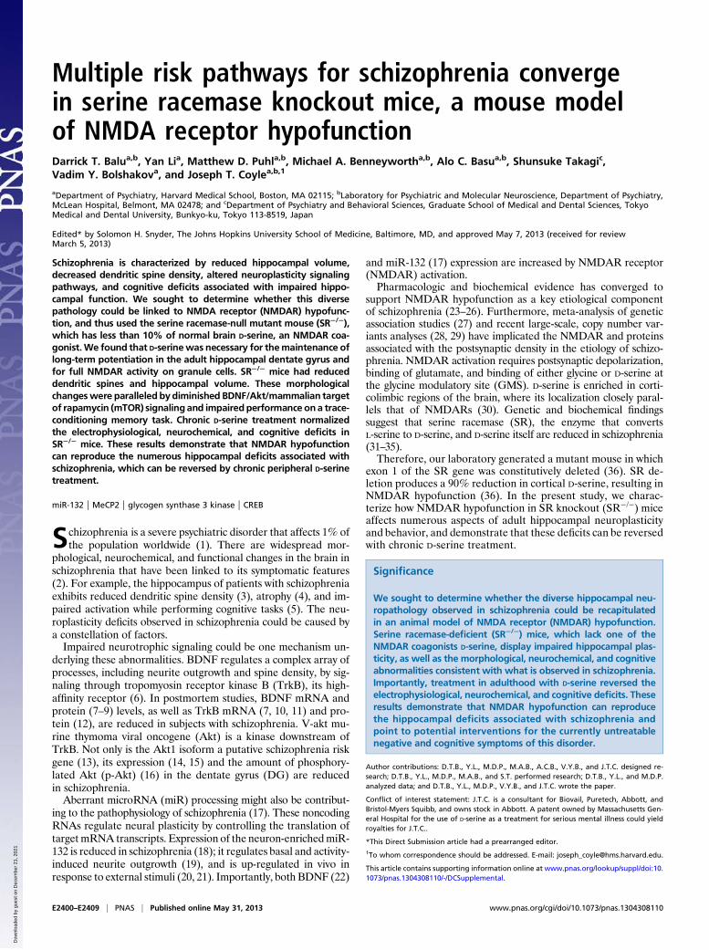
ResultsNMDAR-Mediated Miniature Excitatory Postsynaptic Current AmplitudeIs Decreased in DG Granule Cells of SR−/− Mice. To evaluate the con-tribution of endogenous D-serine to the activation of NMDARsin the DG, we assessed the parameters of miniature excitatorypostsynaptic currents (mEPSCs) recorded from granule cells incontrol and SR−/− mice. We targeted cells located in the outerthird of the granule cell layer to avoid recording from cells atdifferent stages of maturity as a result of adult hippocampalneurogenesis (37). Recorded neurons showed no differences intheir input resistance between the groups (four WT mice: 258.9 ±35.8 MΩ, n = 11 cells; four SR−/− mice: 232.6 ± 21.9 MΩ, n = 11cells, P= 0.27), indicating that deletion of SR had no effect on themembrane properties of granule cells. The NMDAR-mediatedcurrents (NMDARmEPSCs) were recorded at a holding potentialof –70 mV in the external medium without added Mg2+, andmeasured at 10 ms after the peak of averaged mEPSCs, which wasdetermined by activation of AMPA receptors (AMPARs). Wefound that the amplitude of NMDAR mEPSCs was significantlysmaller in slices from SR−/− mice (Fig. 1 B and C) (P < 0.05), butthe decay time was faster in SR−/− animals (Fig. 1 E and F) (P <0.05). Exogenously applied D-serine (10 μM) potentiated theamplitude and prolonged the decay time of NMDAR mEPSCs inboth WT (P < 0.05 for both amplitude and decay time) and SR−/−
mice (P = 0.006 for amplitude, P < 0.05 for decay time), con-firming the lack of NMDAR GMS saturation under conditions ofbasal synaptic transmission (38). In the presence of exogenouslyapplied D-serine, the difference in the amplitude and decay time ofNMDAR mEPSCs observed under control conditions was abol-ished, indicating that the functional properties of the NMDARsin DG granule cells were unaffected in SR−/− mice (39, 40). WTand SR−/− mice did not differ in the amplitude of AMPAR
mEPSCs, measured at the peak mEPSC amplitude in the presenceof the NMDAR antagonist D-AP5 (50 μM) (Fig. 1 D and G).
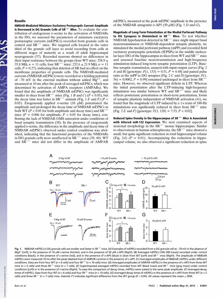
Magnitude of Long-Term Potentiation at the Medial Perforant Pathwayto DG Synapses Is Diminished in SR−/− Mice. To test whetherNMDAR hypofunction detected in SR−/−mice might impact basalneurotransmission or NMDAR-dependent synaptic plasticity, westimulated the medial perforant pathway (mPP) and recorded fieldexcitatory postsynaptic potentials (fEPSPs) in the middle molecu-lar layer DGof the hippocampus in slices fromWTand SR−/−miceand assessed baseline neurotransmission and high-frequencystimulation-induced long-term synaptic potentiation (LTP). Base-line synaptic transmission, assayed with input-output curves (Fig. 2A and B) [genotype: F(1, 133) = 0.51, P = 0.48] and paired pulseratio at the mPP to DG synapses (Fig. 2 C and D) [genotype: F(1,56) = 0.0002, P = 0.99] remained unchanged in slices from SR−/−
mice. However, we observed significant deficits in LTP. Whereasthe initial potentiation after the LTP-inducing high-frequencystimulation was similar between WT and SR−/− mice and likelyreflects posttetanic potentiation or short-term potentiation, formsof synaptic plasticity independent of NMDAR activation (41), wefound that the magnitude of LTP induced by a 1-s train of 100-Hzstimulations was significantly reduced in slices from SR−/− mice(Fig. 2 E and F) [genotype: F(1, 120) = 7.53, P < 0.02].
Reduced Spine Density in the Hippocampus of SR−/− Mice Is Associatedwith Altered miR-132 Expression. We next examined aspects ofneuronal morphology in the SR−/− mouse hippocampus. Similarto observations in human schizophrenia, the SR−/− mice showed asmall, but quite significant reduction in total hippocampal volume(Fig. 3A) (P = 0.01). Accompanying the reduction in hippo-campal volume, we also observed a significant reduction in spine
Fig. 1. NMDARmEPSCs in DG granule cells are smaller and faster in SR−/−mice. (A) Examples ofmEPSCs recorded from aDG granule cell at−70mV in the absence ofMg2+ (Left), in the presence of 10 μM D-serine (Center), and in the presence of 50 μM D-AP5 (Right). (B) Averaged mEPSCs (100–300 traces) recorded under controlconditions (black), in the presence of D-serine (red), and in the presence of D-AP5 (blue) in slices from WT (Left) and SR−/− mice (Right). The amplitude of NMDARmEPSCs were measured 10ms after the peak (dashed line) of AMPAR currents in the presence of D-AP5. (C) Averaged amplitudes of NMDARmEPSCs under differentconditions. Data are from fourWT (n = 6 cells) and four SR−/− (n = 10 cells) mice. (D) Averaged amplitudes of AMPARmEPSCs in the presence of D-AP5 from threeWTmice (n = 5 cells) and three SR−/− mice (n = 7 cells). (E) Superimposed averaged mEPSCs recorded from WT (black trace) and SR−/− mice (gray trace) under controlconditions (Left) or in the presence of D-serine (Right). To ease the comparison of decay times, mEPSCs were scaled to the same peak amplitude. (F) Averaged decaytimes of mEPSCs. Data from fourWT (n = 6 cells) and four SR−/−mice (n = 10 cells). (G) Averaged decay times of mEPSCs in the presence of D-AP5 from threeWT (n = 5cells) and three SR−/− (n = 7 cells) mice. Asterisk (*) indicates significant difference from the WT group (P < 0.05). All values represent the mean ± SEM.
Balu et al. PNAS | Published online May 31, 2013 | E2401
NEU
ROSC
IENCE
PNASPL
US
Dow
nloa
ded
by g
uest
on
Dec
embe
r 21
, 202
1
density on dendritic arbors of DG granule neurons from SR−/−
mice compared with WT mice (Fig. 3 B and C) (P = 0.03),similar to what we have observed in the cortex (42, 43). Con-sistent with these morphological abnormalities, the expression ofmiR-132, a known positive modulator of spine plasticity, wasreduced. As shown in Fig. 3D, the expression of the primary (P <0.05), precursor (P < 0.05), and mature (P < 0.05) transcripts ofmiR-132 were significantly reduced in SR−/− mice. We also ex-amined mature miR-212 expression because it is processed fromthe same primary transcript as miR-132 (44). However, there wasno difference in the expression of the mature miR-212 transcriptin SR−/− mice (WT: 100 ± 0.1%; SR−/−: 103 ± 0.09% P = 0.86).Others have also found changes in miR-132 expression withoutchanges in miR-212 following manipulations in vivo (45).
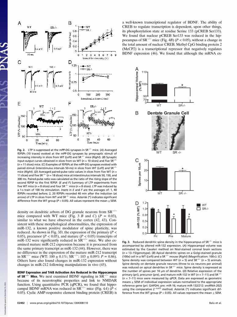
BDNF Expression and TrkB Activation Are Reduced in the Hippocampusof SR−/− Mice. We next examined BDNF signaling in SR−/− micebecause of its neurotrophic properties and link to NMDARfunction. Using quantitative PCR (qPCR), we found that hippo-campal BDNF mRNA was reduced in SR−/− mice (Fig. 4A) (P <0.05). Cyclic AMP-responsive element binding protein (CREB) is
a well-known transcriptional regulator of BDNF. The ability ofCREB to regulate transcription is dependent, upon other things,its phosphorylation state at residue Serine 133 (pCREB Ser133).We found that nuclear pCREB Ser133 was reduced in the hip-pocampus of SR−/− mice (Fig. 4B) (P < 0.05), without a change inthe total amount of nuclear CREB. Methyl CpG binding protein 2(MeCP2) is a transcriptional repressor that negatively regulatesBDNF expression (46). We found that although the mRNA ex-
Fig. 2. LTP is suppressed at the mPP-DG synapses in SR−/− mice. (A) AveragedfEPSPs (10 traces) evoked at the mPP-DG synapses by presynaptic stimuli ofincreasing intensity in slices from WT (Left) and SR−/− mice (Right). (B) Synapticinput-output curves obtained in slices from six WT (n = 10 slices) and five SR−/−
(n = 11 slices) mice. (C) Examples of fEPSPs at the mPP-DG synapses evoked withpaired stimuli (interstimulus intervals 50-ms) in slices from WT (Left) and SR−/−
mice (Right). (D) Averaged paired-pulse ratio values in slices from five WT (n =11 slices) and five SR−/− (n = 18 slices) mice at interstimulus intervals 50, 150, and300 ms. Paired-pulse ratio was calculated as the ratio of the rising slope of thesecond fEPSP to the first fEPSP. (E and F) Summary of LTP experiments fromfive WT mice (n = 8 slices) and four SR−/− mice (n = 8 slices). LTP was induced bya 1-s train of 100 Hz stimulation. Insets in E and F are the averages of: 1, 40fEPSPs recorded before; 2, 20 fEPSPs recorded 40 min after the induction (atarrow) of LTP in slices from WT and SR−/− mice. Asterisk (*) indicates significantdifference from the WT group (P < 0.05). All values represent the mean ± SEM.
A
B
C
D
Fig. 3. Reduced dendritic spine density in the hippocampus of SR−/− mice isaccompanied by altered miR-132 expression. (A) Hippocampal volume wasestimated by the Cavaleri method on Nissl-stained coronal brain sections(n = 13–15/genotype). (B) Apical dendritic spines on a Golgi-stained granule(100x) cell in a WT (Left) and a SR−/− mouse (Right) (Magnification: 100×). (C)Spine density was compared between WT (n = 5) and SR−/− (n = 5) animals.Spine density on dentate granule neurons (three to six neurons per animal)was reduced on apical dendrites in SR−/− mice. Spine density is expressed asthe number of spines per 10 μm of dendrite. (D) Relative expression of theprimary (pri), precursor (pre), and mature miR-132 in WT (n = 7–11) and SR−/−
(n = 7–11) mice were measured by qPCR. Data are expressed as geometricmeans ± SEM of individual expression values normalized to the appropriatereference gene (pri: GAPDH; pre: miR-16; mature miR-132/212: snoRNA-202)using the comparative 2−ΔΔCt method. Asterisk (*) indicates significant dif-ference from the WT group (P < 0.05). All values represent the mean ± SEM.
E2402 | www.pnas.org/cgi/doi/10.1073/pnas.1304308110 Balu et al.
Dow
nloa
ded
by g
uest
on
Dec
embe
r 21
, 202
1
pression level of MeCP2 was unaltered in the hippocampus ofSR−/− mice (WT: 100 ± 0.9%; SR−/−: 103 ± 0.1%; P = 0.85), theamount of protein was significantly increased in the nucleus (Fig.4C) (P < 0.05). Interestingly, MeCP2 mRNA is a known target formiR-132 degradation (47).We next determined whether reduced BDNF mRNA was as-
sociated with reduced protein. We indeed found that SR−/− micehad significantly reduced levels of free, mature BDNF protein inthe hippocampus (Fig. 4D) (P = 0.01). Although the total pro-tein amount of TrkB was unchanged (Fig. 4E), the amount ofphosphorylated (active) TrkB was reduced (Fig. 4F) (P < 0.05) inthe hippocampus of SR−/− mice.
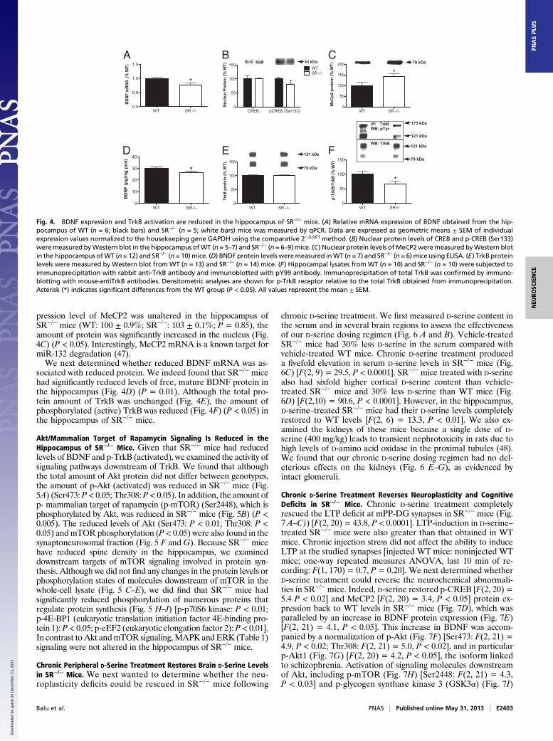
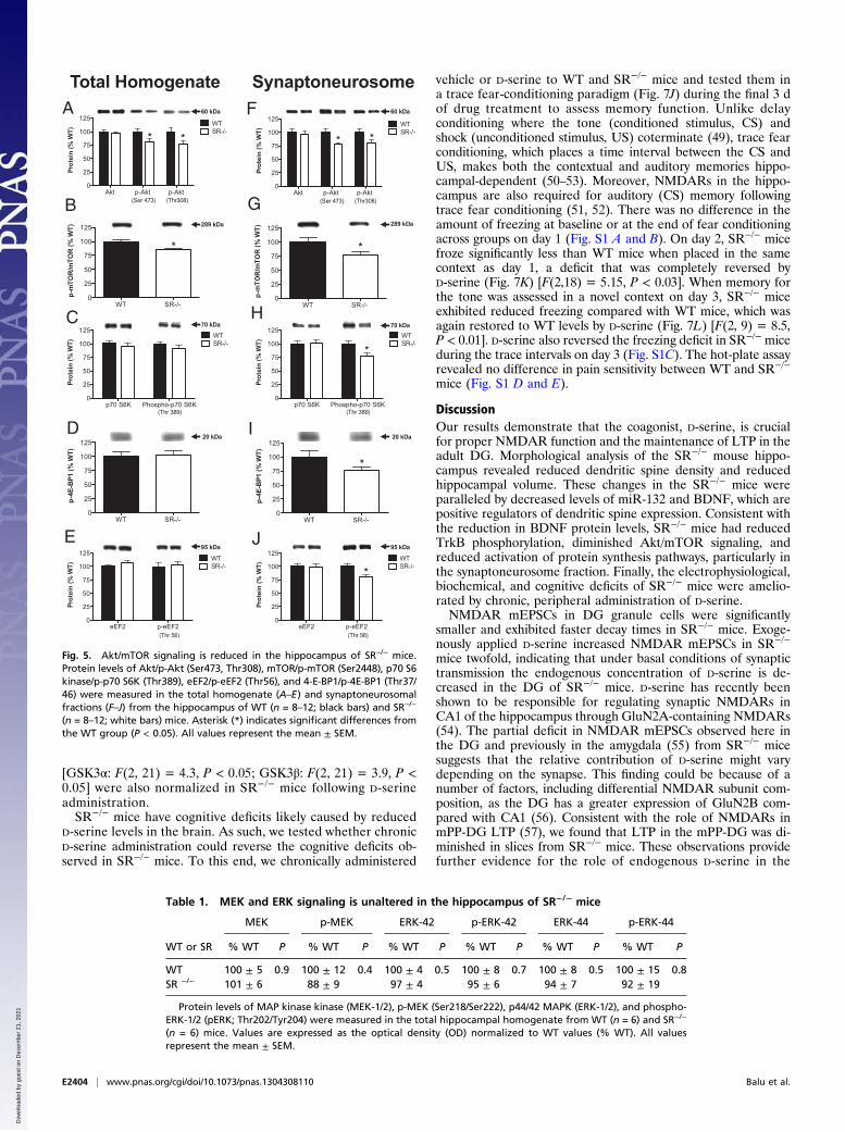
Akt/Mammalian Target of Rapamycin Signaling Is Reduced in theHippocampus of SR−/− Mice. Given that SR−/− mice had reducedlevels of BDNF and p-TrkB (activated), we examined the activity ofsignaling pathways downstream of TrkB. We found that althoughthe total amount of Akt protein did not differ between genotypes,the amount of p-Akt (activated) was reduced in SR−/− mice (Fig.5A) (Ser473: P< 0.05; Thr308: P< 0.05). In addition, the amount ofp- mammalian target of rapamycin (p-mTOR) (Ser2448), which isphosphorylated by Akt, was reduced in SR−/− mice (Fig. 5B) (P <0.005). The reduced levels of Akt (Ser473: P < 0.01; Thr308: P <0.05) andmTOR phosphorylation (P< 0.05) were also found in thesynaptoneurosomal fraction (Fig. 5 F and G). Because SR−/− micehave reduced spine density in the hippocampus, we examineddownstream targets of mTOR signaling involved in protein syn-thesis. Although we did not find any changes in the protein levels orphosphorylation states of molecules downstream of mTOR in thewhole-cell lysate (Fig. 5 C–E), we did find that SR−/− mice hadsignificantly reduced phosphorylation of numerous proteins thatregulate protein synthesis (Fig. 5 H–J) [p-p70S6 kinase: P < 0.01;p-4E-BP1 (eukaryotic translation initiation factor 4E-binding pro-tein 1): P< 0.05; p-eEF2 (eukaryotic elongation factor 2):P< 0.01].In contrast to Akt andmTOR signaling,MAPK and ERK (Table 1)signaling were not altered in the hippocampus of SR−/− mice.
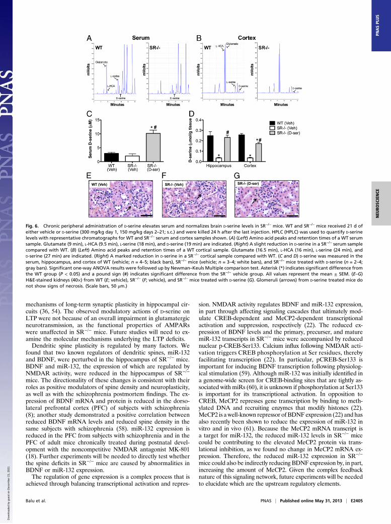
Chronic Peripheral D-Serine Treatment Restores Brain D-Serine Levelsin SR−/− Mice. We next wanted to determine whether the neu-roplasticity deficits could be rescued in SR−/− mice following
chronic D-serine treatment. We first measured D-serine content inthe serum and in several brain regions to assess the effectivenessof our D-serine dosing regimen (Fig. 6 A and B). Vehicle-treatedSR−/− mice had 30% less D-serine in the serum compared withvehicle-treated WT mice. Chronic D-serine treatment produceda fivefold elevation in serum D-serine levels in SR−/− mice (Fig.6C) [F(2, 9) = 29.5, P < 0.0001]. SR−/− mice treated with D-serinealso had sixfold higher cortical D-serine content than vehicle-treated SR−/− mice and 30% less D-serine than WT mice (Fig.6D) [F(2,10) = 90.6, P < 0.0001]. However, in the hippocampus,D-serine–treated SR−/− mice had their D-serine levels completelyrestored to WT levels [F(2, 6) = 13.3, P < 0.01]. We also ex-amined the kidneys of these mice because a single dose of D-serine (400 mg/kg) leads to transient nephrotoxicity in rats due tohigh levels of D-amino acid oxidase in the proximal tubules (48).We found that our chronic D-serine dosing regimen had no del-eterious effects on the kidneys (Fig. 6 E–G), as evidenced byintact glomeruli.
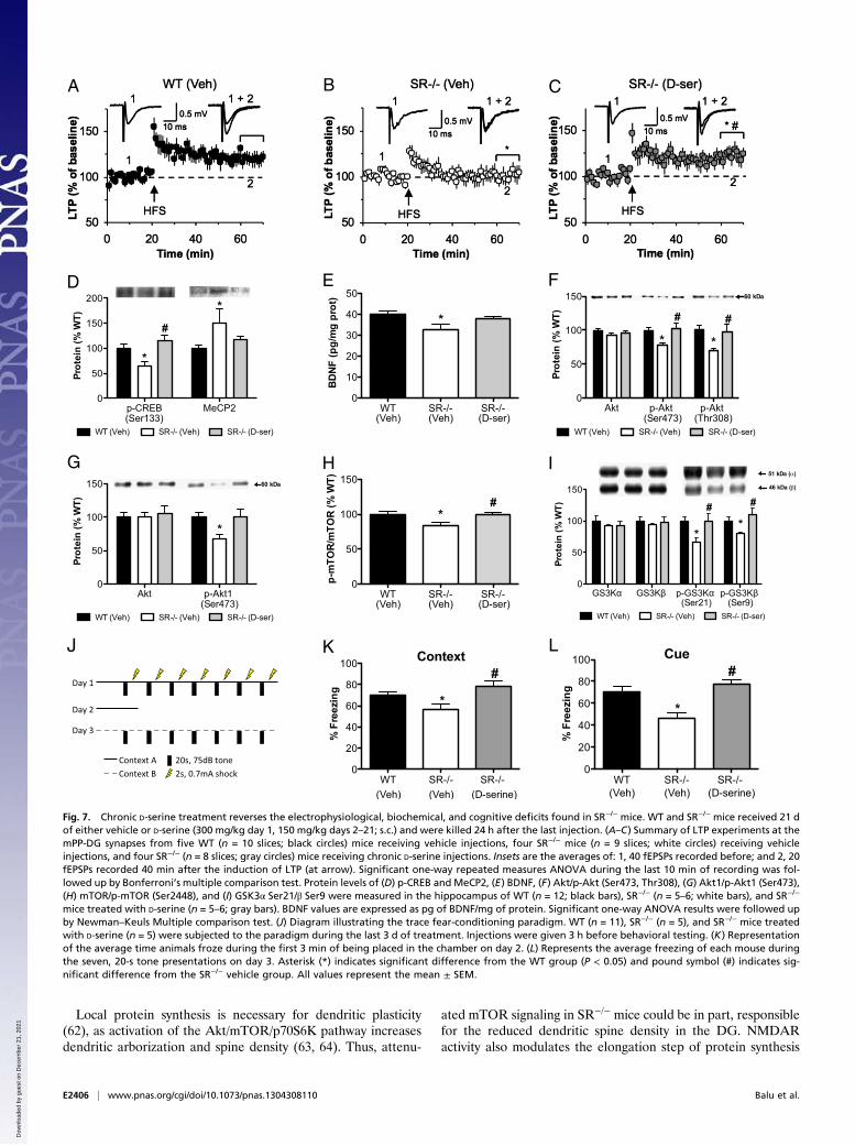
Chronic D-Serine Treatment Reverses Neuroplasticity and CognitiveDeficits in SR−/− Mice. Chronic D-serine treatment completelyrescued the LTP deficit at mPP-DG synapses in SR−/− mice (Fig.7 A–C)) [F(2, 20) = 43.8, P < 0.0001]. LTP-induction in D-serine–treated SR−/− mice were also greater than that obtained in WTmice. Chronic injection stress did not affect the ability to induceLTP at the studied synapses [injected WT mice: noninjected WTmice; one-way repeated measures ANOVA, last 10 min of re-cording: F(1, 170) = 0.7, P= 0.20]. We next determined whetherD-serine treatment could reverse the neurochemical abnormali-ties in SR−/− mice. Indeed, D-serine restored p-CREB [F(2, 20) =5.4 P < 0.02] and MeCP2 [F(2, 20) = 3.4, P < 0.05] protein ex-pression back to WT levels in SR−/− mice (Fig. 7D), which wasparalleled by an increase in BDNF protein expression (Fig. 7E)[F(2, 21) = 4.1, P < 0.05]. This increase in BDNF was accom-panied by a normalization of p-Akt (Fig. 7F) [Ser473: F(2, 21) =4.9, P < 0.02; Thr308: F(2, 21) = 5.0, P < 0.02], and in particularp-Akt1 (Fig. 7G) [F(2, 20) = 4.2, P < 0.05], the isoform linkedto schizophrenia. Activation of signaling molecules downstreamof Akt, including p-mTOR (Fig. 7H) [Ser2448: F(2, 21) = 4.3,P < 0.03] and p-glycogen synthase kinase 3 (GSK3α) (Fig. 7I)
MeC
p2 p
ro
tein
(%
WT
)
WT SR -/- 0
50
100
150
200
*
79 kDa
Nu
cle
ar P
ro
tein
(%
WT
)
CREB pCREB (Ser133)0
50
100
150WTSR -/-
*
43 kDa
WT SR -/-0.0
0.5
1.0
1.5
BD
NF
mR
NA
(%
WT
)
*
A CB
WT SR-/-0
10
20
30
40
BD
NF
(p
g/m
g p
ro
t)
*
Trk
B p
ro
te
in (
% W
T)
WT SR -/- 0
50
100
150
121 kDa
79 kDa
WT SR-/- 0
50
100
150
p-T
rk
B/T
rk
B (
% W
T)
*
121 kDa
121 kDa
175 kDa
WB: pTyr
WB: TrkB
IP: TrkB
79 kDa
D FE
Fig. 4. BDNF expression and TrkB activation are reduced in the hippocampus of SR−/− mice. (A) Relative mRNA expression of BDNF obtained from the hip-pocampus of WT (n = 6; black bars) and SR−/− (n = 5; white bars) mice was measured by qPCR. Data are expressed as geometric means ± SEM of individualexpression values normalized to the housekeeping gene GAPDH using the comparative 2−ΔΔCt method. (B) Nuclear protein levels of CREB and p-CREB (Ser133)weremeasured byWestern blot in the hippocampus ofWT (n = 5–7) and SR−/− (n = 6–9) mice. (C) Nuclear protein levels ofMeCP2weremeasured byWestern blotin the hippocampus ofWT (n = 12) and SR−/− (n = 10)mice. (D) BNDF protein levels weremeasured inWT (n = 7) and SR−/− (n = 6)mice using ELISA. (E) TrkB proteinlevels were measured by Western blot from WT (n = 13) and SR−/− (n = 14) mice. (F) Hippocampal lysates from WT (n = 10) and SR−/− (n = 10) were subjected toimmunoprecipitation with rabbit anti-TrkB antibody and immunoblotted with pY99 antibody. Immunoprecipitation of total TrkB was confirmed by immuno-blotting with mouse-antiTrkB antibodies. Densitometric analyses are shown for p-TrkB receptor relative to the total TrkB obtained from immunoprecipitation.Asterisk (*) indicates significant differences from the WT group (P < 0.05). All values represent the mean ± SEM.
Balu et al. PNAS | Published online May 31, 2013 | E2403
NEU
ROSC
IENCE
PNASPL
US
Dow
nloa
ded
by g
uest
on
Dec
embe
r 21
, 202
1
[GSK3α: F(2, 21) = 4.3, P < 0.05; GSK3β: F(2, 21) = 3.9, P <0.05] were also normalized in SR−/− mice following D-serineadministration.SR−/− mice have cognitive deficits likely caused by reduced
D-serine levels in the brain. As such, we tested whether chronicD-serine administration could reverse the cognitive deficits ob-served in SR−/− mice. To this end, we chronically administered
vehicle or D-serine to WT and SR−/− mice and tested them ina trace fear-conditioning paradigm (Fig. 7J) during the final 3 dof drug treatment to assess memory function. Unlike delayconditioning where the tone (conditioned stimulus, CS) andshock (unconditioned stimulus, US) coterminate (49), trace fearconditioning, which places a time interval between the CS andUS, makes both the contextual and auditory memories hippo-campal-dependent (50–53). Moreover, NMDARs in the hippo-campus are also required for auditory (CS) memory followingtrace fear conditioning (51, 52). There was no difference in theamount of freezing at baseline or at the end of fear conditioningacross groups on day 1 (Fig. S1 A and B). On day 2, SR−/− micefroze significantly less than WT mice when placed in the samecontext as day 1, a deficit that was completely reversed byD-serine (Fig. 7K) [F(2,18) = 5.15, P < 0.03]. When memory forthe tone was assessed in a novel context on day 3, SR−/− miceexhibited reduced freezing compared with WT mice, which wasagain restored to WT levels by D-serine (Fig. 7L) [F(2, 9) = 8.5,P < 0.01]. D-serine also reversed the freezing deficit in SR−/− miceduring the trace intervals on day 3 (Fig. S1C). The hot-plate assayrevealed no difference in pain sensitivity between WT and SR−/−
mice (Fig. S1 D and E).
DiscussionOur results demonstrate that the coagonist, D-serine, is crucialfor proper NMDAR function and the maintenance of LTP in theadult DG. Morphological analysis of the SR−/− mouse hippo-campus revealed reduced dendritic spine density and reducedhippocampal volume. These changes in the SR−/− mice wereparalleled by decreased levels of miR-132 and BDNF, which arepositive regulators of dendritic spine expression. Consistent withthe reduction in BDNF protein levels, SR−/− mice had reducedTrkB phosphorylation, diminished Akt/mTOR signaling, andreduced activation of protein synthesis pathways, particularly inthe synaptoneurosome fraction. Finally, the electrophysiological,biochemical, and cognitive deficits of SR−/− mice were amelio-rated by chronic, peripheral administration of D-serine.NMDAR mEPSCs in DG granule cells were significantly
smaller and exhibited faster decay times in SR−/− mice. Exoge-nously applied D-serine increased NMDAR mEPSCs in SR−/−
mice twofold, indicating that under basal conditions of synaptictransmission the endogenous concentration of D-serine is de-creased in the DG of SR−/− mice. D-serine has recently beenshown to be responsible for regulating synaptic NMDARs inCA1 of the hippocampus through GluN2A-containing NMDARs(54). The partial deficit in NMDAR mEPSCs observed here inthe DG and previously in the amygdala (55) from SR−/− micesuggests that the relative contribution of D-serine might varydepending on the synapse. This finding could be because of anumber of factors, including differential NMDAR subunit com-position, as the DG has a greater expression of GluN2B com-pared with CA1 (56). Consistent with the role of NMDARs inmPP-DG LTP (57), we found that LTP in the mPP-DG was di-minished in slices from SR−/− mice. These observations providefurther evidence for the role of endogenous D-serine in the
A F
B G
C H
D I
E J
Fig. 5. Akt/mTOR signaling is reduced in the hippocampus of SR−/− mice.Protein levels of Akt/p-Akt (Ser473, Thr308), mTOR/p-mTOR (Ser2448), p70 S6kinase/p-p70 S6K (Thr389), eEF2/p-eEF2 (Thr56), and 4-E-BP1/p-4E-BP1 (Thr37/46) were measured in the total homogenate (A–E) and synaptoneurosomalfractions (F–J) from the hippocampus of WT (n = 8–12; black bars) and SR−/−
(n = 8–12; white bars) mice. Asterisk (*) indicates significant differences fromthe WT group (P < 0.05). All values represent the mean ± SEM.
Table 1. MEK and ERK signaling is unaltered in the hippocampus of SR−/− mice
WT or SR
MEK p-MEK ERK-42 p-ERK-42 ERK-44 p-ERK-44
% WT P % WT P % WT P % WT P % WT P % WT P
WT 100 ± 5 0.9 100 ± 12 0.4 100 ± 4 0.5 100 ± 8 0.7 100 ± 8 0.5 100 ± 15 0.8SR −/− 101 ± 6 88 ± 9 97 ± 4 95 ± 6 94 ± 7 92 ± 19
Protein levels of MAP kinase kinase (MEK-1/2), p-MEK (Ser218/Ser222), p44/42 MAPK (ERK-1/2), and phospho-ERK-1/2 (pERK; Thr202/Tyr204) were measured in the total hippocampal homogenate from WT (n = 6) and SR−/−
(n = 6) mice. Values are expressed as the optical density (OD) normalized to WT values (% WT). All valuesrepresent the mean ± SEM.
E2404 | www.pnas.org/cgi/doi/10.1073/pnas.1304308110 Balu et al.
Dow
nloa
ded
by g
uest
on
Dec
embe
r 21
, 202
1
mechanisms of long-term synaptic plasticity in hippocampal cir-cuits (36, 54). The observed modulatory actions of D-serine onLTP were not because of an overall impairment in glutamatergicneurotransmission, as the functional properties of AMPARswere unaffected in SR−/− mice. Future studies will need to ex-amine the molecular mechanisms underlying the LTP deficits.Dendritic spine plasticity is regulated by many factors. We
found that two known regulators of dendritic spines, miR-132and BDNF, were perturbed in the hippocampus of SR−/− mice.BDNF and miR-132, the expression of which are regulated byNMDAR activity, were reduced in the hippocampus of SR−/−
mice. The directionality of these changes is consistent with theirroles as positive modulators of spine density and neuroplasticity,as well as with the schizophrenia postmortem findings. The ex-pression of BDNF mRNA and protein is reduced in the dorso-lateral prefrontal cortex (PFC) of subjects with schizophrenia(8); another study demonstrated a positive correlation betweenreduced BDNF mRNA levels and reduced spine density in thesame subjects with schizophrenia (58). miR-132 expression isreduced in the PFC from subjects with schizophrenia and in thePFC of adult mice chronically treated during postnatal devel-opment with the noncompetitive NMDAR antagonist MK-801(18). Further experiments will be needed to directly test whetherthe spine deficits in SR−/− mice are caused by abnormalities inBDNF or miR-132 expression.The regulation of gene expression is a complex process that is
achieved through balancing transcriptional activation and repres-
sion. NMDAR activity regulates BDNF and miR-132 expression,in part through affecting signaling cascades that ultimately mod-ulate CREB-dependent and MeCP2-dependent transcriptionalactivation and suppression, respectively (22). The reduced ex-pression of BDNF levels and the primary, precurser, and maturemiR-132 transcripts in SR−/− mice were accompanied by reducednuclear p-CREB-Ser133. Calcium influx following NMDAR acti-vation triggers CREB phosphorylation at Ser residues, therebyfacilitating transcription (22). In particular, pCREB-Ser133 isimportant for inducing BDNF transcription following physiolog-ical stimulation (59). Although miR-132 was initially identified ina genome-wide screen for CREB-binding sites that are tightly as-sociated with miRs (60), it is unknown if phosphorylation at Ser133is important for its transcriptional activation. In opposition toCREB, MeCP2 represses gene transcription by binding to meth-ylated DNA and recruiting enzymes that modify histones (22).MeCP2 is a well-known repressor of BDNF expression (22) and hasalso recently been shown to reduce the expression of miR-132 invitro and in vivo (61). Because the MeCP2 mRNA transcript isa target for miR-132, the reduced miR-132 levels in SR−/− micecould be contributing to the elevated MeCP2 protein via trans-lational inhibition, as we found no change in MeCP2 mRNA ex-pression. Therefore, the reduced miR-132 expression in SR−/−
mice could also be indirectly reducing BDNF expression by, in part,increasing the amount of MeCP2. Given the complex feedbacknature of this signaling network, future experiments will be neededto elucidate which are the upstream regulatory elements.
Fig. 6. Chronic peripheral administration of D-serine elevates serum and normalizes brain D-serine levels in SR−/− mice. WT and SR−/− mice received 21 d ofeither vehicle or D-serine (300 mg/kg day 1, 150 mg/kg days 2–21; s.c.) and were killed 24 h after the last injection. HPLC (HPLC) was used to quantify D-serinelevels with representative chromatographs for WT and SR−/− serum and cortex samples shown. (A) (Left) Amino acid peaks and retention times of a WT serumsample. Glutamate (9 min), L-HCA (9.5 min), L-serine (18 min), and D-serine (19 min) are indicated. (Right) A slight reduction in D-serine in a SR−/− serum samplecompared with WT. (B) (Left) Amino acid peaks and retention times of a WT cortical sample. Glutamate (16.5 min), L-HCA (16 min), L-serine (24 min), andD-serine (27 min) are indicated. (Right) A marked reduction in D-serine in a SR−/− cortical sample compared with WT. (C and D) D-serine was measured in theserum, hippocampus, and cortex of WT (vehicle; n = 4–5; black bars), SR−/− mice (vehicle; n = 3–4; white bars), and SR−/− mice treated with D-serine (n = 2–4;gray bars). Significant one-way ANOVA results were followed up by Newman–Keuls Multiple comparison test. Asterisk (*) indicates significant difference fromthe WT group (P < 0.05) and a pound sign (#) indicates significant difference from the SR−/− vehicle group. All values represent the mean ± SEM. (E–G)H&E-stained kidneys (40×) from WT (E; vehicle), SR−/− (F; vehicle), and SR−/− mice treated with D-serine (G). Glomeruli (arrows) from D-serine treated mice donot show signs of necrosis. (Scale bars, 50 μm.)
Balu et al. PNAS | Published online May 31, 2013 | E2405
NEU
ROSC
IENCE
PNASPL
US
Dow
nloa
ded
by g
uest
on
Dec
embe
r 21
, 202
1
Local protein synthesis is necessary for dendritic plasticity(62), as activation of the Akt/mTOR/p70S6K pathway increasesdendritic arborization and spine density (63, 64). Thus, attenu-
ated mTOR signaling in SR−/− mice could be in part, responsiblefor the reduced dendritic spine density in the DG. NMDARactivity also modulates the elongation step of protein synthesis
A B C
D E F
G H I
J K L
Fig. 7. Chronic D-serine treatment reverses the electrophysiological, biochemical, and cognitive deficits found in SR−/− mice. WT and SR−/− mice received 21 dof either vehicle or D-serine (300 mg/kg day 1, 150 mg/kg days 2–21; s.c.) and were killed 24 h after the last injection. (A–C) Summary of LTP experiments at themPP-DG synapses from five WT (n = 10 slices; black circles) mice receiving vehicle injections, four SR−/− mice (n = 9 slices; white circles) receiving vehicleinjections, and four SR−/− (n = 8 slices; gray circles) mice receiving chronic D-serine injections. Insets are the averages of: 1, 40 fEPSPs recorded before; and 2, 20fEPSPs recorded 40 min after the induction of LTP (at arrow). Significant one-way repeated measures ANOVA during the last 10 min of recording was fol-lowed up by Bonferroni’s multiple comparison test. Protein levels of (D) p-CREB and MeCP2, (E) BDNF, (F) Akt/p-Akt (Ser473, Thr308), (G) Akt1/p-Akt1 (Ser473),(H) mTOR/p-mTOR (Ser2448), and (I) GSK3α Ser21/β Ser9 were measured in the hippocampus of WT (n = 12; black bars), SR−/− (n = 5–6; white bars), and SR−/−
mice treated with D-serine (n = 5–6; gray bars). BDNF values are expressed as pg of BDNF/mg of protein. Significant one-way ANOVA results were followed upby Newman–Keuls Multiple comparison test. (J) Diagram illustrating the trace fear-conditioning paradigm. WT (n = 11), SR−/− (n = 5), and SR−/− mice treatedwith D-serine (n = 5) were subjected to the paradigm during the last 3 d of treatment. Injections were given 3 h before behavioral testing. (K) Representationof the average time animals froze during the first 3 min of being placed in the chamber on day 2. (L) Represents the average freezing of each mouse duringthe seven, 20-s tone presentations on day 3. Asterisk (*) indicates significant difference from the WT group (P < 0.05) and pound symbol (#) indicates sig-nificant difference from the SR−/− vehicle group. All values represent the mean ± SEM.
E2406 | www.pnas.org/cgi/doi/10.1073/pnas.1304308110 Balu et al.
Dow
nloa
ded
by g
uest
on
Dec
embe
r 21
, 202
1
through regulation of eEF2, a GTP-binding protein that controlsmRNA trafficking through the ribosome. Although phosphory-lation of eEF2 on Thr56 usually inhibits eEF2-ribosome bindingand arrests peptide chain elongation (65), it also increases thetranslation of some mRNAs localized to dendrites (66, 67).Reducing the amount of p-eEF2 decreases hippocampal spinedensity and stability, as well as dendritic BDNF protein expres-sion (68). Hence, impaired eEF2 signaling in the hippocampalsynaptoneurosome fraction of SR−/− mice could be anothercontributing factor to not only the reduced spine density, but alsothe reduced BDNF protein levels.The mTORC1 pathway described above can be activated by
NMDAR activity and by neurotrophins, such as BDNF. NMDARand TrkB activation lead to increases in both PI3K/Akt andMAPK/ERK signaling. Activation of these two pathways relievesthe constitutive inhibition placed on the mTORC1 complex andincreases protein synthesis. Akt signaling, but not MAPK/ERKsignaling, was reduced in the hippocampus of SR−/− mice. Thisfinding suggests that attenuated Akt activity is contributing to thereduced levels of p-mTOR and subsequent impaired mTORC1signaling. Interestingly, AKT1 has been associated with an in-creased risk for schizophrenia (13). Furthermore, reduced Aktsignaling likely contributes to the pathophysiology of the disorder,as the brains of patients with schizophrenia have reduced Akt andp-Akt levels (16, 69). Evidence from genetic animal models alsoimplicates Akt1 in processes that are altered in patients withschizophrenia, including hippocampal plasticity (i.e., LTP) andcognitive tasks (16, 70). We demonstrate here that SR−/− micehave reduced hippocampal p-Akt1, which fully accounts for thereduction in total p-Akt (Fig. 7) and highlights the importance ofthis particular isoform of Akt in the brain. Thus, impaired Akt1signaling in the hippocampus of SR−/− mice potentially contributesto the reductions in spine density and neuroplasticity via mTORC1.Although the current medications for schizophrenia, which
mainly block dopamine D2 receptors, are relatively effective inmanaging the psychosis, they are ineffective in treating the cog-nitive deficits and negative symptoms (71). These deficits are themost enduring and correlate with the degree of cortical atrophy.A recent meta-analysis found that GMS agonists, including D-serine,exert significant therapeutic effects onmultiple symptom domainsin schizophrenia in patients receiving antipsychotic medications,especially negative and cognitive symptoms (72). We found thatchronic, peripherally administered D-serine to SR−/− mice nor-malized the LTP deficit at the mPP-DG synapse, MeCP2, pCREB,and BDNF protein expression, as well as downstream Akt/mTOR/GS3K signaling. Our previous research has demonstrated thatSR−/− mice have impairments in spatial and contextual memory(36, 73). We demonstrate here that SR−/− mice also have impair-ments in fear-conditioned learning that can be rescued by D-serinetreatment. Specifically, SR−/−mice have impairments in contextualand cue-dependent memory following trace fear conditioning. Theintroduction of the trace interval during conditioning makes boththe contextual and auditory memories hippocampal-dependent, asdetermined by lesion (50, 53) and NMDAR antagonist infusion(51, 52) studies. Thus, normalizing NMDAR activity in SR−/−micewith D-serine treatment was able to rescue deficits in both con-textual and auditory trace fear conditioning. Future studies willneed to determine the duration of treatment needed for the re-storative effects of D-serine, as well as which signaling pathwaysdownstream of NMDAR activation and which brain regions arecritical for the cognitive enhancing effects.In conclusion, SR−/− mice display significant impairments in
hippocampal neuroplasticity, both at the electrophysiological andneurochemical level. Importantly, NMDAR hypofunction altersnumerous pathways, including BDNF/TrkB, Akt/mTOR/GS3K,and miR-132 that not are not only potent regulators of plasticityand spine dynamics, but have been found to be genetically asso-ciated with or perturbed in schizophrenia (13). Furthermore, a
recent study of de novo copy number variants implicated inschizophrenia demonstrated a highly significant enrichment ofgene products that are localized to the postsynaptic density andare associated with the NMDAR (28). Thus, morphological, neu-rochemical, functional and cognitive deficits of the hippocampusand cortex demonstrated in schizophrenia can be recapitulated inthe mouse by reduced availability of D-serine, one of the two coa-gonists at the NMDAR, resulting in NMDAR hypofunction (TableS1). Finally, these findings demonstrate that neuroplasticity andbehavioral deficits resulting from a constitutive genetic lesion canbe reversed with interventions that occur in adulthood and high-light the GMS on the NMDAR as a potential therapeutic targetfor schizophrenia.
Materials and MethodsFurther details on subcellular fractionation, Western blot analysis, HPLC,kidney staining, trace fear conditioning, and hot-plate test can be foundin the SI Materials and Methods.
Animals. SR−/− mice (36) were generated as previously described. SR+/− siresand dams were bred to produce WT and SR−/− offspring. Adult male mice(3–5 mo old) were used for all of the experiments, except where noted.Animals were housed in groups of four in polycarbonate cages and main-tained on a 12:12-h light/dark cycle in a temperature (22 °C) and humiditycontrolled vivarium. Animals were given access to food and water ad libi-tum. All animal procedures were approved by the McLean Hospital In-stitutional Animal Care and Use Committee.
Electrophysiology. Hippocampal slices (400 μm) were prepared from adult SR−/− orWTmice (littermates) with a vibratome. Slices were continuously perfusedin solution containing: 119 mM NaCl, 2.5 mM KCl, 2.5 mM CaCl2, 1.0 mMMgSO4, 1.25 mM NaH2PO4, 26.0 mM NaHCO3, 10 mM glucose, and 0.05 mMpicrotoxin, and equilibrated with 95% (vol/vol) O2 and 5% CO2 (pH 7.3–7.4) at22 °C. fEPSPs were recorded in the middle molecular layer of the DG of thehippocampus with a glass pipette filled with the extracellular solution. Syn-aptic responses were evoked at 0.033 Hz by stimulation of the mPP (74, 75)with a concentric stimulation electrode (76, 77). In LTP experiments, the ba-seline stimulation intensity was adjusted to produce fEPSPs with an amplitudethat was ∼30% of maximum amplitude fEPSP. LTP was induced by a 1-s trainof presynaptic stimulation at 100 Hz. The initial slope of the fEPSP’s risingphase was used to measure the changes in synaptic strength. Summary LTPgraphs were constructed by normalizing data in 60-s epochs to the meanvalue of the baseline fEPSPs slope. The magnitude of LTP was estimated ina 10-min time window 40 min after the induction. Whole-cell recordings (2- to7-mo-old male and female mice; groups were counterbalanced for age andsex) of spontaneous mEPSCs were obtained from granule cells of the DG atphysiological temperature (36 ± 1°) in the presence of 1 μM tetrodotoxin (TTX)to block action potential-induced neurotransmitter release. The NMDAR-me-diated currents were recorded under voltage-clamp conditions at –70 mVwithout Mg2+ in the external medium. The patch electrodes (3–4 MΩ re-sistance) in voltage-clamp experiments contained: 120 mM Cs-methanesulfo-nate, 5 mM NaCl, 1 mM MgCl2, 10 mM BAPTA, 10 mM Hepes, 2 mM Mg-ATP,and 0.1 mM Na-GTP (adjusted to pH 7.2 with CsOH). Spontaneous synapticcurrents were filtered at 1 kHz and digitized at 5 kHz. The mEPSCs wererecorded first in control external solution, then in the presence of 10 μM D-serine, and, finally, in the presence of the NMDA receptor antagonist D-AP5(50 μM). Series resistance was monitored throughout the experiment and wasin the range of 15–25 MΩ. The parameters of mEPSCs were analyzed with theMini Analysis Program (Synaptosoft).
Golgi Staining and Quantification of Dendritic Spine Density. Golgi stainingwas performed using the FD Rapid GolgiStain Kit (FD NeuroTechnologies) aspreviously described (42, 43, 78). Neurons were located between approxi-mately −1.46 mm to −2.20 mm posterior to bregma (79) and within themiddle third of the section. Spines were counted on two unobscured apicaldendritic branches (minimum second order) per neuron, with the averagespine density used as the value for that neuron. Spines were counted onthree to six neurons per animal. Only neurons in the outer two-thirds of thegranule cell layer of the DG were chosen for spine density analysis. Neuronsin the outer layers of the granule cell layer are not derived from the activelydividing cells of the subgranular zone associated with adult neurogenesis.The average dendritic length analyzed for spine density did not significantlydiffer between genotypes (WT = 28.9 ± 0.8 μm, SR−/− = 32.0 ± 1.9 μm).
Balu et al. PNAS | Published online May 31, 2013 | E2407
NEU
ROSC
IENCE
PNASPL
US
Dow
nloa
ded
by g
uest
on
Dec
embe
r 21
, 202
1
Dendrites were visualized at 100× (oil-immersion) on a Zeiss Axioskop40microscope and the number of spines was quantified using Neurolucida(MBF Bioscience). The experimenter was blind to genotype during tracing.
Nissl Staining and Hippocampal Volume Estimation. Brains from WT and SR−/−
were fixed, sectioned, and stained using cresyl echt violet solution (pH 3.5) aspreviously described (78). Hippocampal volume was estimated at 4× ona Zeiss Axioskop40 by the Cavalieri method (600-μm grid size; 7–9 sectionsper brain) using Stereo Investigator software (MBF Bioscience). The grid sizechosen estimated volumes with high precision (coefficient of error: 0.009;average total points per hippocampus: 177).
qPCR. RNA was isolated from the hippocampi of WT and SR−/− mice (n =6/genotype) as previously described (78). RNA was isolated from the tissuesamples using the miRvana miRNA isolation kit (Ambion). cDNA for eachRNA sample (2-μg input) was generated using the High Capacity cDNA Re-verse Transcription kit (Applied Biosystems). Mature miR-132 and sno-202cDNA (10-ng input) was generated using the TaqMan MicroRNA ReverseTranscription kit (Applied Biosystems). premiR-132 (10-μm primer) and miR-16 (10-μm primer) cDNA (500-ng input) were generated using the Super-Script III First-Strand Synthesis System (Invitrogen). An initial step of 80 °C for1 min was added to the SuperScript III protocol to denature the hairpinstructures in premiR-132 transcripts. The primers and protocol were basedon a previous study (80). qPCR for GAPDH, BDNF, and pri-miR-132 wereperformed using TaqMan gene expression assays (Applied Biosystems). Datawere collected using a 48-well MJ Minioption Personal thermal cycler (Bio-Rad). qPCR for mature miR-132 and snoRNA202 was performed using theTaqMan MicroRNA expression assays (Applied Biosystems). qPCR for premiR-132 and miR-16 was performed using the Platinum SYBR Green qPCRSuperMix UDG (Invitrogen), which was followed by a thermal denaturationprotocol to ensure amplification of a single product. For relative quantifi-cation of mRNA expression (BDNF, MeCP2, pri-miR132), geometric meanswere calculated using the comparative 2−ΔΔCt method, with the housekeepinggene GAPDH used as the endogenous reference. miR-16 and snoRNA202were used as the endogenous reference genes for premiR-132 and maturemiR-132, respectively. Each sample was assayed in triplicate. All primers arelisted in Table S2.
BDNF ELISA. BDNF levels from the hippocampi of WT and SR−/− mice weremeasured using a commercially available ELISA kit (Promega) as previouslydescribed (78)
Subcellular Fractionation. The fractionation procedure was modified from apreviously published protocol (81). Protein concentrations were measuredusing the Bradford protein assay (Bio-Rad).
Western Blot Analysis. Immunoblotting was performed as previously de-scribed (81). Primary antibodies are listed in Table S3. Chemiluminescentvalues of the protein of interest were divided by its corresponding β-actinchemiluminescent values. The ratio of each WT sample was divided by theaverage of all of the WT sample values in each gel and multiplied by 100.
The average of the normalized WT values from each gel was 100% ± SEM.The mutant values were normalized to WT values (percent of WT) collectedin parallel from the same gel. The normalized values were then averagedand used for statistical analysis.
Immunoprecipitation. A separate cohort of mice from the ELISA and Westernblot analyses was used for immunoprecipitation. Hippocampal tissue lysatewas immunoprecipitated with a TrkB antibody. TrkB phosphorylation wasmeasured by immunoblotting for phosphotyrosine, stripping the membrane,and then reprobing for TrkB.
Chronic D-Serine Treatment.Mice received once daily, subcutaneous injectionsof vehicle or D-serine for 20 d at a volume of 5 mL/kg. WT mice receivedvehicle (0.9% sodium chloride) and SR−/− mice received either vehicle orD-serine (Sigma-Aldrich). D-serine was given at an initial dose of 300 mg/kgon day 1, followed by 150 mg/kg for the remaining 20 d. Mice were killed onday 21 without receiving an injection. One hippocampal lobe was used forelectrophysiology and the other lobe was used for neurochemistry. Left andright hemispheres were counterbalanced for analysis.
HPLC Analysis of D-Serine. Brain tissue and trunk blood were obtained fromthe same chronically treated mice that were used in the LTP experiments.Blood was collected, allowed to clot for 10 min, and spun at 2,000 × g for15 min at room temperature. Brain tissue and serum content of D-serinewere analyzed as previously described (36, 82).
Trace Fear Conditioning. Mice were subjected to a trace fear conditioningprotocol adapted from ref. 50. Mice were trained in context A on day 1.Memory for context A was measured on day 2 and memory for the CS (tone)in context B was measured on day 3. All testing was performed using the TheNear Infrared Fear Conditioning System (Med Associates). Freezing behaviorwas quantified using VideoFreeze software.
Statistical Analyses. Dendritic spine density, Western blot, qPCR, and BDNFELISA results were compared using unpaired Student t test. Repeatedmeasures one-way ANOVA was used to analyze LTP results, and one-wayANOVA was used to analyze BDNF, Western blot, and fear conditioningresults following chronic D-serine treatment. Significant F values were sub-ject to post hoc analyses as indicated. Values of P < 0.05 were consideredstatistically significant.
ACKNOWLEDGMENTS. We thank Drs. Sabina Berretta, Ole Isacson, andUwe Rudolph for the generous use of their equipment and software;Dr. Christopher Cowan for advice; and Jiamin Feng for animal colonymaintenance and genotyping. The Rodent Histopathology Core Facility ofthe Dana-Farber/Harvard Cancer Center (P30 CA06516) performed thekidney staining. Phenopro performed the hot-plate test. This work wassupported in part by a postdoctoral National Research Service Award F32MH090697; an Andrew P. Merrill Research Fellowship; a Phyllis & Jerome LyleRappaport Mental Health Research Scholars Award (to D.T.B.); and NationalInstitutes of Health Grants R01MH05190 and P50MH0G0450 (to J.T.C.).
1. Perälä J, et al. (2007) Lifetime prevalence of psychotic and bipolar I disorders in
a general population. Arch Gen Psychiatry 64(1):19–28.2. Jarskog LF, Miyamoto S, Lieberman JA (2007) Schizophrenia: New pathological
insights and therapies. Annu Rev Med 58:49–61.3. Rosoklija G, et al. (2000) Structural abnormalities of subicular dendrites in subjects
with schizophrenia and mood disorders: Preliminary findings. Arch Gen Psychiatry
57(4):349–356.4. Adriano F, Caltagirone C, Spalletta G (2011) Hippocampal volume reduction in first-
episode and chronic schizophrenia: A review and meta-analysis. Neuroscientist 18(2):
180–200.5. Heckers S, et al. (1998) Impaired recruitment of the hippocampus during conscious
recollection in schizophrenia. Nat Neurosci 1(4):318–323.6. Binder DK, Scharfman HE (2004) Brain-derived neurotrophic factor. Growth Factors
22(3):123–131.7. Hashimoto T, et al. (2005) Relationship of brain-derived neurotrophic factor and its
receptor TrkB to altered inhibitory prefrontal circuitry in schizophrenia. J Neurosci
25(2):372–383.8. Weickert CS, et al. (2003) Reduced brain-derived neurotrophic factor in prefrontal
cortex of patients with schizophrenia. Mol Psychiatry 8(6):592–610.9. Wong J, et al. (2010) Promoter specific alterations of brain-derived neurotrophic
factor mRNA in schizophrenia. Neuroscience 169(3):1071–1084.10. Weickert CS, et al. (2005) Reductions in neurotrophin receptor mRNAs in the
prefrontal cortex of patients with schizophrenia. Mol Psychiatry 10(7):637–650.
11. Thompson Ray M, Weickert CS, Wyatt E, Webster MJ (2011) Decreased BDNF, trkB-TK+and GAD67 mRNA expression in the hippocampus of individuals with schizophrenia andmood disorders. J Psychiatry Neurosci 36(3):195–203.
12. Wong J, Rothmond DA, Webster MJ, Shannon Weickert C (2011) Increases in twotruncated TrkB isoforms in the prefrontal cortex of people with schizophrenia.Schizophr Bull 39(1):130–140.
13. Balu DT, Coyle JT (2011) Neuroplasticity signaling pathways linked to the pathophysiologyof schizophrenia. Neurosci Biobehav Rev 35(3):848–870.
14. Emamian ES, Hall D, Birnbaum MJ, Karayiorgou M, Gogos JA (2004) Convergentevidence for impaired AKT1-GSK3beta signaling in schizophrenia. Nat Genet 36(2):131–137.
15. Thiselton DL, et al. (2008) AKT1 is associated with schizophrenia across multiplesymptom dimensions in the Irish study of high density schizophrenia families. BiolPsychiatry 63(5):449–457.
16. Balu DT, et al. (2012) Akt1 deficiency in schizophrenia and impairment of hippocampalplasticity and function. Hippocampus 22(2):230–240.
17. Miller BH, Wahlestedt C (2010) MicroRNA dysregulation in psychiatric disease. BrainRes 1338:89–99.
18. Miller BH, et al. (2012) MicroRNA-132 dysregulation in schizophrenia has implicationsfor both neurodevelopment and adult brain function. Proc Natl Acad Sci USA 109(8):3125–3130.
19. Wayman GA, et al. (2008) An activity-regulated microRNA controls dendritic plasticityby down-regulating p250GAP. Proc Natl Acad Sci USA 105(26):9093–9098.
20. Nudelman AS, et al. (2009) Neuronal activity rapidly induces transcription of theCREB-regulated microRNA-132, in vivo. Hippocampus 20(4):492–498.
E2408 | www.pnas.org/cgi/doi/10.1073/pnas.1304308110 Balu et al.
Dow
nloa
ded
by g
uest
on
Dec
embe
r 21
, 202
1
21. Tognini P, Putignano E, Coatti A, Pizzorusso T (2011) Experience-dependent expressionof miR-132 regulates ocular dominance plasticity. Nat Neurosci 14(10):1237–1239.
22. Greer PL, Greenberg ME (2008) From synapse to nucleus: Calcium-dependent genetranscription in the control of synapse development and function. Neuron 59(6):846–860.
23. Coyle JT (2006) Glutamate and schizophrenia: Beyond the dopamine hypothesis. CellMol Neurobiol 26(4-6):365–384.
24. Krystal JH, et al. (1994) Subanesthetic effects of the noncompetitive NMDAantagonist, ketamine, in humans. Psychotomimetic, perceptual, cognitive, andneuroendocrine responses. Arch Gen Psychiatry 51(3):199–214.
25. Weickert CS, et al. (2012) Molecular evidence of N-methyl-D-aspartate receptorhypofunction in schizophrenia. Mol Psychiatry, 10.1038/mp.2012.137.
26. Steiner J, et al. (2013) Increased prevalence of diverse N-methyl-D-aspartateglutamate receptor antibodies in patients with an initial diagnosis of schizophrenia:Specific relevance of IgG NR1a antibodies for distinction from N-methyl-D-aspartateglutamate receptor encephalitis. JAMA Psychiatry 70(3):271–278.
27. Allen NC, et al. (2008) Systematic meta-analyses and field synopsis of geneticassociation studies in schizophrenia: The SzGene database. Nat Genet 40(7):827–834.
28. Kirov G, et al. (2012) De novo CNV analysis implicates specific abnormalities ofpostsynaptic signalling complexes in the pathogenesis of schizophrenia. Mol Psychiatry17(2):142–153.
29. Walsh T, et al. (2008) Rare structural variants disrupt multiple genes in neurodevelopmentalpathways in schizophrenia. Science 320(5875):539–543.
30. Schell MJ, Molliver ME, Snyder SH (1995) D-serine, an endogenous synapticmodulator: Localization to astrocytes and glutamate-stimulated release. Proc NatlAcad Sci USA 92(9):3948–3952.
31. Bendikov I, et al. (2007) A CSF and postmortem brain study of D-serine metabolicparameters in schizophrenia. Schizophr Res 90(1–3):41–51.
32. Goltsov AY, et al. (2006) Polymorphism in the 5′-promoter region of serine racemasegene in schizophrenia. Mol Psychiatry 11(4):325–326.
33. Labrie V, Wang W, Barger SW, Baker GB, Roder JC (2009) Genetic loss of D-amino acidoxidase activity reverses schizophrenia-like phenotypes in mice. Genes Brain Behav9(1):11–25.
34. Morita Y, et al. (2007) A genetic variant of the serine racemase gene is associated withschizophrenia. Biol Psychiatry 61(10):1200–1203.
35. Hashimoto K, et al. (2005) Reduced D-serine to total serine ratio in the cerebrospinalfluid of drug naive schizophrenic patients. Prog Neuropsychopharmacol BiolPsychiatry 29(5):767–769.
36. Basu AC, et al. (2009) Targeted disruption of serine racemase affects glutamatergicneurotransmission and behavior. Mol Psychiatry 14(7):719–727.
37. Balu DT, Lucki I (2009) Adult hippocampal neurogenesis: Regulation, functionalimplications, and contribution to disease pathology. Neurosci Biobehav Rev 33(3):232–252.
38. Li Y, Krupa B, Kang JS, Bolshakov VY, Liu G (2009) Glycine site of NMDA receptorserves as a spatiotemporal detector of synaptic activity patterns. J Neurophysiol102(1):578–589.
39. TownsendM, Yoshii A, Mishina M, Constantine-PatonM (2003) Developmental loss ofminiature N-methyl-D-aspartate receptor currents in NR2A knockout mice. Proc NatlAcad Sci USA 100(3):1340–1345.
40. Cull-Candy SG, Leszkiewicz DN (2004) Role of distinct NMDA receptor subtypes atcentral synapses. Sci STKE 2004(255):re16.
41. Zucker RS, Regehr WG (2002) Short-term synaptic plasticity. Annu Rev Physiol 64:355–405.
42. Balu DT, Coyle JT (2012) Neuronal D-serine regulates dendritic architecture in thesomatosensory cortex. Neurosci Lett 517(2):77–81.
43. DeVito LM, et al. (2011) Serine racemase deletion disrupts memory for order andalters cortical dendritic morphology. Genes Brain Behav 10(2):210–222.
44. Vo N, et al. (2005) A cAMP-response element binding protein-induced microRNAregulates neuronal morphogenesis. Proc Natl Acad Sci USA 102(45):16426–16431.
45. Mellios N, et al. (2011) miR-132, an experience-dependent microRNA, is essential forvisual cortex plasticity. Nat Neurosci 14(10):1240–1242.
46. Zhou Z, et al. (2006) Brain-specific phosphorylation of MeCP2 regulates activity-dependent Bdnf transcription, dendritic growth, and spine maturation. Neuron 52(2):255–269.
47. Klein ME, et al. (2007) Homeostatic regulation of MeCP2 expression by a CREB-induced microRNA. Nat Neurosci 10(12):1513–1514.
48. Orozco-Ibarra M, et al. (2007) Evaluation of oxidative stress in D-serine inducednephrotoxicity. Toxicology 229(1–2):123–135.
49. Phillips RG, LeDoux JE (1992) Differential contribution of amygdala and hippocampusto cued and contextual fear conditioning. Behav Neurosci 106(2):274–285.
50. Chowdhury N, Quinn JJ, Fanselow MS (2005) Dorsal hippocampus involvement intrace fear conditioning with long, but not short, trace intervals in mice. BehavNeurosci 119(5):1396–1402.
51. Misane I, et al. (2005) Time-dependent involvement of the dorsal hippocampus intrace fear conditioning in mice. Hippocampus 15(4):418–426.
52. Quinn JJ, Loya F, Ma QD, Fanselow MS (2005) Dorsal hippocampus NMDA receptorsdifferentially mediate trace and contextual fear conditioning. Hippocampus 15(5):665–674.
53. McEchron MD, Bouwmeester H, Tseng W, Weiss C, Disterhoft JF (1998) Hippocampectomydisrupts auditory trace fear conditioning and contextual fear conditioning in the rat.Hippocampus 8(6):638–646.
54. Papouin T, et al. (2012) Synaptic and extrasynaptic NMDA receptors are gated bydifferent endogenous coagonists. Cell 150(3):633–646.
55. Li Y, et al. (2013) Identity of enodgenous NMDAR glycine site agonist in amygdala isdetermined by synaptic activity level. Nat Commun 4:1760.
56. Coultrap SJ, Nixon KM, Alvestad RM, Valenzuela CF, Browning MD (2005) Differentialexpression of NMDA receptor subunits and splice variants among the CA1, CA3 anddentate gyrus of the adult rat. Brain Res Mol Brain Res 135(1-2):104–111.
57. Morris RG, Anderson E, Lynch GS, Baudry M (1986) Selective impairment of learningand blockade of long-term potentiation by an N-methyl-D-aspartate receptorantagonist, AP5. Nature 319(6056):774–776.
58. Hill JJ, et al. (2005) Analysis of pyramidal neuron morphology in an inducibleknockout of brain-derived neurotrophic factor. Biol Psychiatry 57(8):932–934.
59. Hong EJ, McCord AE, Greenberg ME (2008) A biological function for the neuronalactivity-dependent component of Bdnf transcription in the development of corticalinhibition. Neuron 60(4):610–624.
60. Impey S, et al. (2004) Defining the CREB regulon: A genome-wide analysis oftranscription factor regulatory regions. Cell 119(7):1041–1054.
61. Im HI, Hollander JA, Bali P, Kenny PJ (2010) MeCP2 controls BDNF expression andcocaine intake through homeostatic interactions with microRNA-212. Nat Neurosci13(9):1120–1127.
62. Tanaka J, et al. (2008) Protein synthesis and neurotrophin-dependent structuralplasticity of single dendritic spines. Science 319(5870):1683–1687.
63. Li N, et al. (2010) mTOR-dependent synapse formation underlies the rapidantidepressant effects of NMDA antagonists. Science 329(5994):959–964.
64. Jaworski J, Spangler S, Seeburg DP, Hoogenraad CC, Sheng M (2005) Control ofdendritic arborization by the phosphoinositide-3′-kinase-Akt-mammalian target ofrapamycin pathway. J Neurosci 25(49):11300–11312.
65. Ryazanov AG, Shestakova EA, Natapov PG (1988) Phosphorylation of elongationfactor 2 by EF-2 kinase affects rate of translation. Nature 334(6178):170–173.
66. Scheetz AJ, Nairn AC, Constantine-Paton M (2000) NMDA receptor-mediated controlof protein synthesis at developing synapses. Nat Neurosci 3(3):211–216.
67. Sutton MA, Taylor AM, Ito HT, Pham A, Schuman EM (2007) Postsynaptic decodingof neural activity: eEF2 as a biochemical sensor coupling miniature synaptictransmission to local protein synthesis. Neuron 55(4):648–661.
68. Verpelli C, et al. (2010) Synaptic activity controls dendritic spine morphology bymodulating eEF2-dependent BDNF synthesis. J Neurosci 30(17):5830–5842.
69. Emamian ES (2012) AKT/GSK3 signaling pathway and schizophrenia. Front MolNeurosci 5:33.
70. Lai WS, et al. (2006) Akt1 deficiency affects neuronal morphology and predisposes toabnormalities in prefrontal cortex functioning. Proc Natl Acad Sci USA 103(45):16906–16911.
71. Coyle JT, Basu A, Benneyworth M, Balu D, Konopaske G (2012) Glutamatergic synapticdysregulation in schizophrenia: Therapeutic implications. Handbook Exp Pharmacol213:267–295.
72. Tsai GE, Lin PY (2010) Strategies to enhance N-methyl-D-aspartate receptor-mediatedneurotransmission in schizophrenia, a critical review and meta-analysis. Curr PharmDes 16(5):522–537.
73. Benneyworth MA, Coyle JT (2012) Altered acquisition and extinction of amphetamine-paired context conditioning in genetic mouse models of altered NMDA receptorfunction. Neuropsychopharmacology 37(11):2496–2504.
74. Dahl D, Burgard EC, Sarvey JM (1990) NMDA receptor antagonists reduce medial, butnot lateral, perforant path-evoked EPSPs in dentate gyrus of rat hippocampal slice.Exp Brain Res 83(1):172–177.
75. Steward O (1976) Topographic organization of the projections from the entorhinalarea to the hippocampal formation of the rat. J Comp Neurol 167(3):285–314.
76. Shin RM, Tsvetkov E, Bolshakov VY (2006) Spatiotemporal asymmetry of associativesynaptic plasticity in fear conditioning pathways. Neuron 52(5):883–896.
77. Tsvetkov E, Carlezon WA, Benes FM, Kandel ER, Bolshakov VY (2002) Fear conditioningoccludes LTP-induced presynaptic enhancement of synaptic transmission in the corticalpathway to the lateral amygdala. Neuron 34(2):289–300.
78. Balu DT, Basu AC, Corradi JP, Cacace AM, Coyle JT (2012) The NMDA receptor co-agonists, D-serine and glycine, regulate neuronal dendritic architecture in thesomatosensory cortex. Neurobiol Dis 45(2):671–682.
79. Paxinos G, Franklin KBJ (2001) The Mouse Brain in Stereotactic Coordinates (AcademicPress, San Diego), 2nd Ed.
80. Wibrand K, et al. (2010) Differential regulation of mature and precursor microRNAexpression by NMDA and metabotropic glutamate receptor activation during LTP inthe adult dentate gyrus in vivo. Eur J Neurosci 31(4):636–645.
81. Balu DT, Coyle JT (2011) Glutamate receptor composition of the post-synaptic densityis altered in genetic mouse models of NMDA receptor hypo- and hyperfunction. BrainRes 1392:1–7.
82. Hashimoto A, Nishikawa T, Oka T, Takahashi K, Hayashi T (1992) Determination offree amino acid enantiomers in rat brain and serum by high-performance liquidchromatography after derivatization with N-tert.-butyloxycarbonyl-L-cysteine ando-phthaldialdehyde. J Chromatogr A 582(1–2):41–48.
Balu et al. PNAS | Published online May 31, 2013 | E2409
NEU
ROSC
IENCE
PNASPL
US
Dow
nloa
ded
by g
uest
on
Dec
embe
r 21
, 202
1