Embed Size (px)
Citation preview

RESEARCH ARTICLE◥
MULTIPLE SCLEROSIS
A noninflammatory mRNA vaccine for treatment ofexperimental autoimmune encephalomyelitisChristina Krienke1,2, Laura Kolb1*, Elif Diken1*, Michael Streuber1, Sarah Kirchhoff1, Thomas Bukur1,Özlem Akilli-Öztürk1, Lena M. Kranz3, Hendrik Berger3, Jutta Petschenka1,4, Mustafa Diken1,3,Sebastian Kreiter1,3, Nir Yogev5,6, Ari Waisman2,5, Katalin Karikó3, Özlem Türeci3,7, Ugur Sahin1,2,3†
The ability to control autoreactive T cells without inducing systemic immune suppression is the majorgoal for treatment of autoimmune diseases. The key challenge is the safe and efficient delivery ofpharmaceutically well-defined antigens in a noninflammatory context. Here, we show that systemicdelivery of nanoparticle-formulated 1 methylpseudouridine-modified messenger RNA (m1Y mRNA)coding for disease-related autoantigens results in antigen presentation on splenic CD11c+ antigen-presenting cells in the absence of costimulatory signals. In several mouse models of multiple sclerosis,the disease is suppressed by treatment with such m1Y mRNA. The treatment effect is associated with areduction of effector T cells and the development of regulatory T cell (Treg cell) populations. Notably,these Treg cells execute strong bystander immunosuppression and thus improve disease induced bycognate and noncognate autoantigens.
Antigen-specific tolerization for the treat-ment of autoimmune diseases may se-lectively blunt autoimmunity withoutcompromising normal immune function.In the past decades, various approaches
have been studied, including delivery of auto-immuneantigensusingDNA, synthetic peptides,recombinant proteins, coated nanoparticles, orimmunomodulatory cellular therapies [reviewedin (1)]. However, clinical translation remainedelusive, with largely negative or inconclusiveoutcomes in human studies, and only a fewapproaches are in early clinical testing. Oneimpediment is the polyclonal complexity ofautoimmune diseases driven by distinctive,diverse autoreactive immune cell repertoiresof patients. The interindividual variability re-quires either personalized treatment tailoredfor the autoantigenic immune profiles of thepatients or therapies that mediate bystandertolerance to suppress both cognate and non-cognate autoimmune lymphocytes withoutbroad immune suppression (2).
Thephysiological inductionandmaintenanceof peripheral tolerance is based on the pre-sentation of self-antigens by lymphoid antigen-presenting cells (APCs) with low-level surfaceexpression of costimulatorymolecules, such asCD86. We sought to develop a therapeutic ap-proach thatwould emulate naturalmechanismsof immune tolerance. We recently introduced aliposomal formulation for systemic delivery ofmRNA-encoded vaccine antigens (mRNA-LPX)into lymphoid tissue-resident CD11c+ APCs (3).mRNA vaccination induces strong type 1T helper (TH1) cell responses driven by highlevels of interferon-a (IFN-a), released fromAPCs upon Toll-like receptor (TLR) signaling(3). Replacement of uridine (U) by incorpo-ration of 1-methylpseudouridine (m1Y) duringin vitro transcription and subsequent removalof double-stranded mRNA contaminants isknown to abrogate TLR7-triggering activity andto reduce inflammatory properties of single-strandedmRNA (4–6). We hypothesized thatthe use of such nucleoside-modified, purifiedmRNA (m1Y mRNA) for in vivo delivery ofautoimmunedisease target antigens intoCD11c+
APCs in a noninflammatory context would en-able systemic tolerogenic antigen presentationin lymphoid tissues.
Noninflammatory delivery of antigen-encodingm1Y mRNA into the spleen expandsantigen-specific CD4+ regulatory T cells
To test this hypothesis, we engineerednanoparticle-formulatedmRNA-LPX (hereinreferred to as mRNA) consisting of nonim-munogenic (m1Y) or immunogenic (U)mRNAcomplexed with liposomes that lack inherentadjuvant activity. In a first experiment, mRNAcoding for the reporter gene firefly luciferase
(LUC) or saline as control was administeredintravenously into albino C57BL/6 mice, andthe translation and expression of the LUCprotein was assessed.In line with previous reports, administra-
tion of U mRNA led to strong activation ofCD11c+ APCs and lymphocytes, and secretionof high levels of IFN-a (Fig. 1, A to C, and fig.S1, A and B). By contrast, we did not observesecretion of IFN-a or other inflammatory cy-tokines or significant activation of CD11c+
APCs, CD8+ and CD4+ T cells, or natural killer(NK) and B cells in m1Y mRNA–treated mice(Fig. 1, A to C, and fig. S1, A and B). Notably,translation of LUC was profoundly higher andprolonged in m1Y mRNA–treated animals(Fig. 1, D and E). These findings suggest thatm1YmRNA is suitable for noninflammatorydelivery of proteins into splenic CD11c+ APCs.To study the effects of m1Y mRNA in an
autoimmune disease, we chose experimentalautoimmune encephalomyelitis (EAE), a clin-ically relevantmousemodel ofmultiple sclero-sis (MS), inwhichwe previously demonstratedtolerance induction by selectively expressingMOG35-55, the epitope of myelin oligodendro-cyte glycoprotein, in dendritic cells (DCs) (7).We assessed the effect of antigen-encodingm1YmRNA treatment on T cell expansion.Naïve Thy1.2+ C57BL/6 mice were immunizedwith MOG35-55 m1Y or U mRNA, and the ex-pansion of both endogenous T cells and adop-tively transferred MOG35-55–T cell receptortransgenic Thy1.1+ CD4+ T cells from 2D2mice(8) was assessed. Both MOG35-55–encodingmRNAs induced proliferation of adoptivelytransferred CD4+ 2D2 T cell, with MOG35-55
m1YmRNAbeing superior (Fig. 1F). Similarly,endogenous MOG35-55–specific CD4
+ T cells innaïve mice were expanded by both MOG35-55–encodingmRNAs (Fig. 1G). However, the func-tional properties of the T cells induced witheither of these mRNAs differed profoundly.MOG35-55 m1Y mRNA treatment was capableof expanding or inducing de novo Foxp3+
regulatory T cells (Treg cells) in both wild-typeC57BL/6 and 2D2-Foxp3-eGFP transgenicmice (Fig. 1H and fig. S2A), whereas overallfrequencies of CD4+ Foxp3+ T cells did notchange (Fig. 1H). CD4+ T cells from vaccinated2D2 animals inhibited the in vitro proliferationof antigen-specific naïve CD4 T cells in a dose-dependentmanner. By contrast, CD4+ T cells ofMOG35-55UmRNAor control-treatedmice showedlittle to no suppressive activity (fig. S2B).We studied the cytokine response profiles
upon in vitro antigen restimulation and thephenotypes of expanded T cells in repetitive-ly vaccinated C57BL/6 mice in more detail.Whereas MOG35-55 UmRNA–expanded T cellsexhibited a functional TH1 effector profile withsecretion of IFN-g, tumor necrosis factor–a(TNFa), interleukin-6 (IL-6), granulocyte-macrophage colony-stimulating factor, and
RESEARCH
Krienke et al., Science 371, 145–153 (2021) 8 January 2021 1 of 9
1TRON – Translational Oncology at the University MedicalCenter of the Johannes Gutenberg University gGmbH,Freiligrathstr. 12, Mainz 55131, Germany. 2Research Centerfor Immunotherapy (FZI), University Medical Center at theJohannes Gutenberg University, Langenbeckstr. 1, Mainz55131, Germany. 3Biopharmaceutical New Technologies(BioNTech) Corporation, An der Goldgrube 12, Mainz 55131,Germany. 4Cancer Immunology and Immune Modulation,Boehringer Ingelheim Pharma GmbH & Co. KG, BirkendorferStr. 65, 88397 Biberach an der Riss, Germany. 5Institute forMolecular Medicine, University Medical Center of theJohannes Gutenberg University, Mainz 55131, Germany.6Clinic and Polyclinic for Dermatology and Venereology,University Hospital Cologne, Kerpenerstr. 62, Cologne 50937,Germany. 7CI3 – Cluster for IndividualizedImmunointervention e.V., Hölderlinstraße 8, 55131 Mainz,Germany.*These authors contributed equally to this work.†Corresponding author. Email: [email protected]
on January 14, 2021
http://science.sciencemag.org/
Dow
nloaded from

Krienke et al., Science 371, 145–153 (2021) 8 January 2021 2 of 9
NK cells
Fig. 1. Antigen-encoding m1Y mRNA potently expands antigen-specificCD4+ Treg cells by noninflammatory delivery into the spleen. (A andB) Activation of splenic immune cells 24 hours after (n = 3) and (C) IFN-a serumlevels 6 hours after intravenous injection of LPX-formulated mRNAs and saline(control) in C57BL/6 mice (n = 6). (D and E) Bioluminescence imaging ofalbino C57BL/6 mice (n = 5) after intravenous injection of m1Y or U LUC mRNA.Representative mice are shown. (F) Frequency and proliferation profiles ofMOG35-55–specific CD4+ T cells isolated from Thy1.1+ 2D2 mice, cell traceviolet (CTV)–labeled, and transferred into naïve Thy1.2+ C57BL/6 recipient mice.Twenty-four hours after adoptive cell transfer, C57BL/6 mice were treatedwith mRNAs or saline (control). Mice were sacrificed on day 4, and spleenswere analyzed for proliferating CD4+ Thy1.1+ cells (n = 3). (G) Expansion of
endogenous MOG35-55–specific CD4+ T cells and (H) frequency of splenicFoxp3+ Treg cells (n = 6) in C57BL/6 mice after treatment (days 0, 3, 7, and10) with mRNA or saline (control) analyzed by MOG35-55–tetramer (tet)staining 3 days after last dosing (n = 4 to 6). (I) CD4+ T cells of mice from(G) tested for IFN-g secretion by enzyme-linked immune absorbent spot(ELISpot) upon restimulation with MOG35-55 peptide (n = 4 to 5). (J) Phenotypeof tet+ CD4+ T cells of mice from (G). Data were compared by using one-wayanalysis of variance (ANOVA) and post hoc Tukey’s test in (A) to (C), (F),and (I) or by unpaired two-tailed Student’s t test in (H) and (J). Mean fluorescenceintensity (MFI) of bulk CD4 T cells of control groups from (H) and (J) are depictedto show baseline expression levels. Error bars indicate mean ± SD. *P ≤ 0.05;**P ≤ 0.01; ***P ≤ 0.001; ****P ≤ 0.0001. LLOD, lower limit of detection.
RESEARCH | RESEARCH ARTICLEon January 14, 2021
http://science.sciencemag.org/
Dow
nloaded from

IL-2 (Fig. 1I and fig. S3), splenic CD4+ T cellsfromMOG35-55 m1Y mRNA–treated mice didnot secrete these proinflammatory cytokines,even when exposed to very high antigen con-centrations. The only measurable factors werelow levels of anti-inflammatory and TH2 type–associated cytokines such as IL-10, IL-5, andIL-13 (fig. S3). The T cell exhaustion markersTIGIT, TIM-3, PD-1, CTLA-4, and LAG-3 werestrongly up-regulatedonMOG35-55m1YmRNA–expanded tetramer+ T cells (Fig. 1J).
Exposure to m1Y mRNA does not impair thecapability to mount immune responses
CD11c+ APCs of mice first exposed to m1YmRNA and thereafter injected with U mRNAdid not show any impairment in their abilityto respond to this TLR-agonistic stimulus withup-regulation of costimulatory molecules andIFN-a secretion (Fig. 2, A and B). To inves-tigate whether the induction and expansionof MOG35-55–specific CD4+ Treg cells affectsde novo priming of antigen-specific immuneresponses, we exploited two broadly usedmodel systems. First, C57BL/6mice underwentprime-boost (days 6 and 13) vaccinationwithUmRNA encoding the ovalbumin (OVA) epitopeSIINFEKL and were concurrently exposed toMOG35-55–encoding m1Y mRNA (days 0, 3,7, and 10). SIINFEKL-specific CD8+ T cellswere expanded above 40% of total blood CD8+
T cells (Fig. 2C) and displayed properties ofeffector T (Teff) cells such as cognate IFN-g se-cretion and highly potent and antigen-specifickilling (Fig. 2, D and E), which suggested un-compromised T cell priming and expansion.In a second experiment,micewere immunizedintramuscularly with a self-amplifying RNA(saRNA) vaccine encoding influenza hemag-glutinin (HA) (day 6) concurrent to repeatedtreatment with MOG35-55 m1Y mRNA or con-trols. Again, the capability of mice to mount aprotective immune response and develop neu-tralizing antibodies was unimpaired (Fig. 2,F and G). Overall, both studies demonstratethat MOG35-55 m1Y mRNA–induced antigen-specific CD4+ Treg cells do not suppress func-tional immune responses against nonmyelinantigens.
Treatment with antigen-encoding m1Y mRNAameliorates EAE in mice
Next, we studied the tolerogenic potential ofMOG35-55 m1Y mRNA in C57BL/6 micewithMOG35-55–induced EAE. Treatment withMOG35-55 m1Y mRNA was capable of block-ing all clinical signs of EAE in mice (Fig. 3A),whereas control animals showed the typicalcourse of the disease with rapid monophasicprogression. Inmice started onMOG35-55 m1YmRNA treatment when a paralysis of the tailor beginning of the hindlimbs were noted(disease score of 1 to 2 of EAE), further diseaseprogression could be prevented, and motor
functions were restored (Fig. 3B and fig. S4A).This included occasional cases of reversion ofparalysis, which was most likely attributableto an anti-inflammatory effect rather than tis-sue repair.Various effectswere observed inmice treated
with antigen-encoding m1Y mRNA comparedwith control animals. In the brain and spinalcord, the total amount of infiltrating CD4+
T cells, MOG35-55–specific CD4+ T cells and
subsets of CD4+ T cells secreting IFN-g andIL-17A were considerably lower (Fig. 3, C, D,and F, and figs. S4B and S5). Demyelinationof the spinal cord was also considerably re-duced (Fig. 3E). In the spleen ofMOG35-55m1YmRNA–treated animals, we observed an in-crease of lymphocytes (fig. S5), includingMOG35-55–specific CD4
+T cellswith lowCD62L,CCR6, andCCR7 expression, and up-regulationof CD69 (fig. S6). CCR6, CCR7, and CD62L arecritical for access of T cells to the central ner-vous system (CNS) (9–12), and the transmem-brane C-type lectin CD69 is known to promotelymphocyte retention in the spleen (13).Next, we analyzed the autoantigen-specific
CD4+ T cells in those mice in which the man-ifestation of EAE was prevented by treatmentwith MOG35-55 m1Y mRNA on days 7 and 10.MOG35-55–specific splenic CD4
+ T cells fromtreated animals showed down-regulation ofthe activation marker CD44 and strong ex-pression of coinhibitory molecules (Fig. 3G). Atthe peak of disease (day 16 after disease induc-tion), CD5, ICOS, LAG-3, PD-1, CTLA-4, TIGIT,and TIM-3 were up-regulated in tetramer+
splenic CD4+ T cells. Furthermore, we detecteda highly activated Treg cell population (Fig. 3, Hand I) and lower numbers of TH1 and TH17MOG35-55–specific CD4
+T cells (Fig. 3H). Similarfindings inmicewith symptomatic EAE (diseasescore of 1 to 2 at start of treatment) furtherconfirmed the potent disease-suppressive activ-ity of antigen-encoding m1Y mRNA (fig. S4).We extended our study of the preventive
and therapeutic effect of m1Y mRNA to otherEAEmousemodels. The SJLmodel is based onautoreactivity against the PLP139-151 epitope andis characterized by recurring EAE symptomsresulting in a relapsing-remitting disease, sim-ilar to the clinical presentation of MS in pa-tients. Treatment of SJL mice with PLP139-151m1Y mRNA twice a week starting from day 7after EAE induction resulted in almost fulldisease control (fig. S7A). Even when the micewere treated after the first disease peak (start-ing on day 14 after disease induction), progres-sion of the disease was halted (fig. S7B).
Treatment with m1Y mRNA leads totherapeutically effective bystander tolerance
In another experimental setup, we addressed akey challenge in human MS, namely thatantigen spread leads to a complex antimyelinautoreactivity pattern and the specificity of
autoreactive T cell clones in individual pa-tients, and thus, the potential targets for di-rect antigen-specific tolerization is unknown.A clinically viable approach would be to usebystander tolerance by inducing Treg cells,which, once activated by their cognate antigen,would suppress T cells against other antigensin the inflamed tissue.We evaluated bystander activity in two ex-
perimental settings. F1 C57BL/6 x SJL micewith PLP139-151 peptide–induced EAE werevaccinated withm1YmRNA encoding eitherPLP139-151 (the disease-causing autoantigen),MOG35-55 (unrelated autoantigen, againstwhichm1Y mRNA is capable of inducing potenteffector Treg cells), or irrelevant m1YmRNA(Fig. 4A and fig. S7C). MOG35-55 m1YmRNAtreatment showed a dose-dependent thera-peutic effect on EAE, similar to the curativeeffect mediated by vaccination with PLP139-151m1Y mRNA, indicating a strong Treg cell–mediated bystander suppression, also giventhe fact that antigen spread has been describedfor this particular EAE model (14). Antigen-specific Treg cells were notably expanded andhighly activated, constituting >80% of de novoexpanded MOG35-55–specific CD4
+ splenicT cells (Fig. 4, B and C). Teff cell infiltrationinto the brain and spinal cord inm1YmRNA–treated mice was reduced (Fig. 4, D to F), andno signs of demyelination in the spinal cordwere detected (Fig. 4G).We also investigated a complex EAE model
driven by multiple pathogenic autoreactiveT cell clones against MOG35-55, PLP139-151,PLP178-191, MBP84-104, and MOBP15-36, whichcould be successfully treated with the mixtureof m1Y mRNAs coding for the correspondingfour disease-inducing autoantigenic epitopes.m1Y mRNA encoding MOG35-55 were thera-peutically almost as effective as those encod-ing the cocktail of all four disease targets. Thissuggests that even polyclonal autoimmunedisease driven by a broad autoreactive T cellrepertoire can be sufficiently controlled bym1Y mRNA targeting a strong bystandertolerance–mediating T cell epitope (fig. S7D).One potential risk associated with antigen-
specific tolerization is the induction of auto-antibodies against respective targets, which canexacerbate disease (15). Moreover, nucleoside-modified mRNA is known to be highly im-munogenic and to induce high antibody titerswhen formulated with immune stimulatorylipid nanoparticles (16). We therefore ana-lyzed anti–MOG35-55 immunoglobulin G (IgG)antibody responses in the sera of EAE miceuponm1YmRNA vaccination. First, wemea-sured anti–MOG35-55 levels in sera ofMOG35-55
peptide–induced EAE (C57BL/6 mice), whichwere vaccinated with MOG35-55 m1Y mRNAon days 7 and 10 after EAE induction. Anti–MOG35-55 IgG levels were not elevated in com-parison with those of control animals treated
Krienke et al., Science 371, 145–153 (2021) 8 January 2021 3 of 9
RESEARCH | RESEARCH ARTICLEon January 14, 2021
http://science.sciencemag.org/
Dow
nloaded from

Krienke et al., Science 371, 145–153 (2021) 8 January 2021 4 of 9
blood
blood
spleen
spleen
Fig. 2. Exposure to m1Y mRNA does not impair the capability to mountimmune responses. (A) Activation of splenic CD11c+ APCs 24 hours after(n = 3) and (B) IFN-a serum levels 6 hours after intravenous injection (day 3)of LPX-formulated U mRNA or saline (control) in C57BL/6 mice (n = 6), whichwere pretreated with m1Y mRNA or saline (control) at day 0. MHCII, majorhistocompatibility complex II. (C to E) De novo priming of SIINFEKL-specific CD8+
T cells in C57BL/6 mice with prior exposure to MOG35-55 or irrelevant m1YmRNA or saline (days 0, 3, 7, and 10) to SIINFEKL U mRNA prime-boostvaccination (days 6 and 13). Controls only received saline. (C) Frequency ofSIINFEKL-specific CD8+ T cells (OVA257-264 tet) in blood (n = 5 to 7) and spleen(n = 3 to 4). (D) IFN-g secretion was measured by ELISpot upon restimulationof total splenocytes of mice from (C) (n = 3 to 4) with SIINFEKL peptide and(E) in vivo antigen-specific killing of adoptively transferred CTV-labeled andpeptide-loaded splenocytes of naïve mice (n = 4 to 5). For in vivo cytotoxicity
assays, mice were adoptively transferred on day 18 with 0.5 mM (low) or5 mM (high) CTV-labeled naïve splenocytes pulsed with peptide (6 mg/ml). Ofthese target cells, 1.5 × 10⁶ cells were adoptively transferred into immunizedand control recipients at a ratio of 1:1 (irrelevant HA518-526 peptide–loadedCTVlow:SIINFEKL peptide–loaded CTVhigh). Recipient splenocytes were recoveredand analyzed by flow cytometry 18 hours after transfer, and antigen-specificlysis was determined as follows: specific lysis (%) = [1 − (percentage of cellspulsed with SIINFEKL/percentage of cells pulsed with HA) × 100]. (F) Total(n = 5 to 6) and (G) neutralizing (n = 9 to 11) anti-HA IgG in C57BL/6 mice withprior exposure to MOG35-55 or irrelevant m1Y mRNA or saline (days 0, 3, 7,and 10). Controls only received saline. Protective immune responses were measured28 days after mice were immunized with influenza HA-saRNA (1 mg intramuscularly)on day 6. Data were compared by using one-way ANOVA and post hoc Tukey’stest. Error bars, mean ± SD. DOD, change in optical density; VN, virus neutralizing.
RESEARCH | RESEARCH ARTICLEon January 14, 2021
http://science.sciencemag.org/
Dow
nloaded from

Krienke et al., Science 371, 145–153 (2021) 8 January 2021 5 of 9
Fig. 3. Treatment with antigen-encoding m1Y mRNA ameliorates EAE inmice. (A and B) Disease severity in MOG35-55–induced EAE (n = 6 to 8 C57BL/6mice per group) treated with m1Y mRNA or saline (control) (A) on days 7and 10 after disease induction or (B) when disease progressed to a score of 1 to2. (C) Frequency of CD4+ T cell and MOG35-55–specific CD4+ T cells in thespinal cord of mice treated on days 7 and 10 after disease induction (n = 3 to 4).Thy1.1+ 2D2 CD4+ T cells were transferred 1 day before EAE induction intoThy1.2+ recipient mice and analyzed in the target organs on day 16 afterdisease induction. (D) Representative CD4 staining in the spinal cord of EAEmice treated with m1Y mRNAs or saline (control) (n = 3) on days 7 and10 after disease induction and analyzed at day 16 after EAE induction.(E) Representative Luxol fast blue (LFB) staining revealing areas of demyelination
in the spinal cord of mice from (D) (n = 3). (F to I) Frequency of CD4+ IFN-g–and IL-17A–secreting cells in brain and spinal cord (n = 3 to 4) (F) andflow cytometry analysis of splenic tetramer+ CD4+ T cells (n = 4 to 6) [(G) to(I)] of EAE mice treated with m1Y mRNAs or saline (control) on days 7 and10 after disease induction and analyzed at day 16 after EAE induction.Area under the curve (AUC) was used to determine statistical significancethrough one-way ANOVA and Tukey’s multiple comparison test of the differentEAE disease development curves in (A) and (B). Data were compared byusing one-way ANOVA and post hoc Tukey’s test in (F) and (H). Error barsindicate mean ± SEM in (A) and (B) or mean ± SD in (C), (F), and (H). The scalebar in the upper row of (D) and (E) represents 200 mm and, in the lower rowof (D) and (E), represents 50 mm.
RESEARCH | RESEARCH ARTICLEon January 14, 2021
http://science.sciencemag.org/
Dow
nloaded from

Krienke et al., Science 371, 145–153 (2021) 8 January 2021 6 of 9
spleen spleen
brain brain
spinal cord spinal cord
Fig. 4. Treatment with m1Y mRNA leads to therapeutically effectivebystander tolerance. (A) Dynamics of EAE in PLP139-151–induced EAE mice(n = 13 to 15 F1 C57BL/6 x SJL mice) upon treatment with MOG35-55 m1Y,PLP139-151 m1Y, irrelevant m1Y mRNA, or saline (control) twice per week startingon days 7 and 10 after disease induction with 40 mg of m1Y mRNA. (B andC) Expansion of endogenous MOG35-55–specific CD4+ T cells (B) and frequencyof Tetramer+ Foxp3+ Treg cells and analysis of CD69 expression on respectivecell population (C) upon treatment with m1Y mRNA measured in the spleenon day 28 after EAE induction (n = 5). (D) Frequency of CD4+ IFN-g– and IL-17A–secreting cells upon PLP139-151–peptide restimulation and (E) total cell count of
lymphocytes in brain and spinal cord of EAE mice treated with different m1YmRNAs (n = 4 to 5) and analyzed on day 28 after EAE induction. (F) RepresentativeCD4 staining in the spinal cord of EAE mice from (A) (n = 2). (G) RepresentativeLFB staining revealing areas of demyelination in the spinal cord of mice from(A) (n = 2). AUC was used to determine statistical significance through one-wayANOVA and Tukey’s multiple comparison test of the different EAE diseasedevelopment curves (A). Data were compared by using one-way ANOVA andpost hoc Tukey’s test. Error bars indicate mean ± SEM in (A) or mean ± SD in (C)to (E). The scale bar in the upper row of (F) and (G) represents 200 mm and,in the lower row of (F) and (G), represents 50 mm.
RESEARCH | RESEARCH ARTICLEon January 14, 2021
http://science.sciencemag.org/
Dow
nloaded from

Krienke et al., Science 371, 145–153 (2021) 8 January 2021 7 of 9
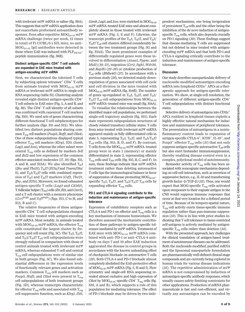
Fig. 5. Distinct splenicantigen-specific CD4+
T cell subsets are expandedin EAE mice treated withantigen-encoding m1YmRNA. (A to G) MOG35-55–specific CD4+ T cells isolatedfrom mice with MOG35-55–induced EAE treated withm1Y mRNA on days 7and 10 after disease inductionand analyzed by single-cellRNA sequencing on day 15or day 16, respectively.(A) A two-dimensionaluniform manifold approxima-tion and projection (UMAP)projection of MOG35-55–specific CD4+ T cells isolatedfrom different treatmentgroups (each dot representsone cell) identified byunsupervised clustering.(B) UMAP projection of singlecells, color-coded accordingto the identified cell subsets.(C) Frequency of differentcell subsets. (D) and (E)depict a UMAP projection ofclassical Treg cell markers(D) and effector Treg cellmarkers (E). (F) Genes up-regulated in an effector Tregsubpopulation upon MOG35-55m1Y mRNA treatment(adjusted P value < 0.05).av. exp., average expression.(G) Genes down-regulatedupon MOG35-55 m1Y mRNAtreatment in TH1, TH17, andTH1/TH17 cell subsets(adjusted P value < 0.05).(H and I) MOG35-55–inducedEAE in C57BL/6 mice(n = 8 per group) treatedwith m1Y mRNA on days 7and 10 after disease inductionin combination with (H),anti–PD-1, and (I) anti–CTLA-4 blocking antibodies orisotype controls administeredtwice per week. Statisticalsignificance of AUCdifferences of EAE diseasedevelopment curves wasassessed by using one-wayANOVA and Tukey’s multiplecomparison test in (H) and(I). Error bars representmean ± SEM in (H) and (I).
RESEARCH | RESEARCH ARTICLEon January 14, 2021
http://science.sciencemag.org/
Dow
nloaded from
with irrelevant m1Y mRNA or saline (fig. S8A).This suggests thatm1YmRNAapplication doesnot exacerbate preformed autoantibody re-sponses. Even after repetitive MOG35-55 m1YmRNA challenge (twice per week, 12 timesin total) of F1 C57BL/6 x SJL mice, no anti–MOG35-55 IgG antibodies were detected inthose where EAE was induced with PLP139-151–peptide immunization (fig. S8B).
Distinct antigen-specific CD4+ T cell subsetsare expanded in EAE mice treated withantigen-encoding m1Y mRNA
Next, we characterized the tolerized T cellsby subjecting splenic tetramer+ CD4+ T cellsfrom animals treated with MOG35-55 m1YmRNA or irrelevant m1Y mRNA to single-cellRNA sequencing (table S1). Clustering analysisrevealed eight distinct antigen-specific CD4+
T cell subsets in EAEmice (Fig. 5, A and B, andfig. S9). The CD4+ T cell identity of all subsetswas confirmed with canonical T cell markers(fig. S10). We used sets of genes characterizingdifferent functional T cell subphenotypes forfurther analysis (figs. S11 and S12). We iden-tified two distinct populations sharing com-mon Treg cell markers (Foxp3, Ikzf2, and Ctla4).One of these subpopulations displayed typicaleffector Treg cell markers (Klrg1, Il10, Gzmb,Lag3, and Icos), whereas the other subset werecentral Treg cells as defined by markers Selland Bcl2 and low expression of the commoneffector-associated molecules (17, 18) (figs. S11,A and B, and S13A). We also identified TH1(Ifng and Tbx21), TH17 (Il17a/f and Tmem176a/b), and TH1/TH17 cells with combined expres-sion of TH1 and TH17 markers (Csf2, Tbx21,Ifng, and Il17a).Moreover, we found exhaustedantigen-specific T cells (Lag3 and Cd160),T follicular helper (TFH) cells (Il4, Il21, andCxcr5),and a T cell cluster with amemory phenotype(Ccr7high and Tcf7high) (figs. S11, C to H, andS13, B and C).The relative frequencies of these antigen-
specific CD4+ T cell subpopulations changedin EAE mice treated with antigen-encodingm1Y mRNA. Most notably, in animals treatedwith MOG35-55 m1YmRNA, the effector Treg
cells constituted the largest cluster by fre-quency and cell count (Fig. 5C). The TH1, TH17,and TH1/TH17 Teff cell subpopulations werestrongly reduced in comparison with those ofcontrol animals treated with irrelevant m1YmRNA, whereas exhausted T cell and centralTreg cell subpopulations were of similar sizein both groups (Fig. 5C). We also found sub-stantial differences in the expression levelsof functionally relevant genes and activationmarkers. Common Treg cell markers such asFoxp3, Ikzf2, and Ctla4 were present in Treg
cell subpopulations of both treatment groups(Fig. 5D), whereas transcripts characteristicfor effector Treg cells and associated with Treg
cell suppressive function, such as Klrg1, Il10,
Gzmb,Lag3, and Icos,were enriched inMOG35-55
m1YmRNA–treated EAEmice and almost com-pletely absent in those treated with irrelevantm1Y mRNA (Fig. 5, E and F). Likewise, thetranscript profiles of the TH1, TH17, and TH1/TH17 Teff cell clusters differed considerably be-tween the two treatment groups (Fig. 5G andfig. S14A). The most prominent examples ofdifferentially regulated genes were those in-volved in differentiation (Anxa1, Ppp4c, andMid1) (19–21), migration (Ccr2, Itgb1,Wdr26,and Rap1b) (22–26) or cytokine production ofTeff cells (Bhlhe40) (27). In accordance with aprevious study (28), we detectedmainly down-regulation of genes associated with cell cycleand cell division in the mice treated withMOG35-55 m1YmRNA (fig. S14B). The numberof genes up-regulated in TH1, TH17, and TH1/TH17 Teff cell subpopulations from MOG35-55
m1YmRNA–treatedmicewas small (fig. S14A).To visualize the relationships between the
identifiedmajor cell populations, we performedsingle-cell trajectory analysis (fig. S15). Eachstate represents subpopulation structures ofclosely related transitory cellular states. T cellsfrommice treated with irrelevant m1YmRNAappeared mainly as fully differentiated cells instate 5, representing TH1, TH17, and TH1/TH17Teff cells (fig. S15, B, D, and F). By contrast,T cells from theMOG35-55m1YmRNA–treatedgroup were in state 1 (memory) or mainlydifferentiated in state 2, representing effectorTreg cells and TFH cells (fig. S15, B, C, and F). Insum, these findings indicate that m1Y mRNAtreatment rather than deleting autoreactiveT cells tips the immunological balance in favorof suppression of disease promotingMOG35-55–specific TH1, TH17, and TH1/TH17 Teff cells byexpanding effector Treg cells.
PD-1 and CTLA-4 signaling contribute to theinduction and maintenance of antigen-specifictolerance
Expression of coinhibitory receptors such asCTLA-4 and PD-1 on effector and Treg cells is akey mechanism of immune homeostasis. Wetherefore assessed the mechanistic contribu-tion of these pathways to antigen-specific tol-erancemediated bym1YmRNA. Treatment ofEAE mice with MOG35-55 m1Y mRNA com-bined with anti–PD-1 or anti–CTLA-4 anti-body on days 7 and 10 after EAE inductionaggravated the disease in control groups inaccordance with the known unleashing effectof checkpoint blockade on autoreactive T cells(29). Both CTLA-4 and PD-1 blockade almostcompletely abolished the EAE-protective effectof MOG35-55 m1Y mRNA (Fig. 5, H and I). Flowcytometry and single-cell RNA sequencing re-vealed almost exclusive and high expression ofCtla4 by MOG35-55–specific CD4
+ Treg cells (fig.S16, A and B), which supports a role of thispopulation for mediating tolerance. The effectof PD-1 blockade may be driven by two inde-
pendent mechanisms, one being invigorationof preexistent Teff cells and the other being theinhibition of the de novo induction of antigen-specific Treg cells, which also depends cruciallyon PD-1 signaling (30). These findings suggestthat disease-mediating T cells are suppressedbut not deleted in mice treated with antigen-encoding m1Y mRNA and that both PD-1 andCTLA-4 signaling critically contribute to theinduction andmaintenance of antigen-specifictolerance.
Discussion
Our study describes nanoparticulate delivery ofnucleoside-modified autoantigen-encodingmRNA into lymphoid CD11c+ APCs as a ther-apeutic approach for antigen-specific toler-ization. We show, at single-cell resolution, thegeneration of different antigen-specific CD4+
T cell subpopulations with distinct functionalstates.Selective delivery of autoantigens into CD11c+
APCs resident in lymphoid tissues exploits ahighly effective natural mechanism for induc-tion and maintenance of peripheral tolerance.The presentation of autoantigens in a nonin-flammatory context leads to expansion ofantigen-specific CTLA-4+, ICOS+, IL-10+, andFoxp3+ effector Treg cells (31) that not onlysuppress antigen-specific autoreactive Teff cellsbut also exert bystander immunosuppression,thereby enabling disease control even in acomplex, polyclonal model of autoimmunity.Bystander activity of Treg cells has been as-
sociatedwith noncognatemechanisms depend-ing on cell-cell interaction, such as secretion ofsuppressive factors, e.g., IL-10 and transforminggrowth factor–b (TGF-b) (32). Thus, one wouldexpect that MOG-specific Treg cells activatedupon reexposure to their cognate antigen in theCNS would suppress immune responses thatoccur at that very location for a defined periodof time. Because of its temporo-spatial nature,Treg cell activity exerts tissue-specific immuneregulation rather than pan-immune suppres-sion (33). This is in line with prior studies in-dicating that T cell tolerance to tissue-restrictedself-antigens is actively mediated by antigen-specific Treg cells rather than deletion (34).With the presented approach, key challenges
for clinical translation of antigen-based treat-ment of autoimmunediseases canbe addressed.Both the nucleoside-modified, purified mRNAand the liposomal nanoparticle formulationare pharmaceuticallywell-defined clinical-stagecompounds and are currently being explored inhuman trials for various disease indications(35). The repetitive administration of m1YmRNA is not compromised by induction ofautoantigen-specific antibody responses, whichusually causes safety-limiting constraints forother applications. Production of mRNA phar-maceuticals is fast and cost-efficient, and vir-tually any autoantigen can be encoded by
Krienke et al., Science 371, 145–153 (2021) 8 January 2021 8 of 9
RESEARCH | RESEARCH ARTICLEon January 14, 2021
http://science.sciencemag.org/
Dow
nloaded from
mRNA. Thus, tailoring the treatment for thedisease-causing antigens of individual patientsis conceivable, similar to that which has beensuccessfully executed in the setting of person-alized cancer vaccines (36, 37). Combination ofm1YmRNAs encoding eithermultiple person-alized autoantigens or autoantigens that con-fer bystander tolerance may enable control ofeven complex autoimmune diseases.
REFERENCES AND NOTES
1. P. Serra, P. Santamaria, Nat. Biotechnol. 37, 238–251 (2019).2. A. Miller, O. Lider, H. L. Weiner, J. Exp. Med. 174, 791–798
(1991).3. L. M. Kranz et al., Nature 534, 396–401 (2016).4. K. Karikó, M. Buckstein, H. Ni, D. Weissman, Immunity 23,
165–175 (2005).5. K. Karikó et al., Mol. Ther. 16, 1833–1840 (2008).6. K. Karikó, H. Muramatsu, J. Ludwig, D. Weissman, Nucleic
Acids Res. 39, e142 (2011).7. N. Yogev et al., Immunity 37, 264–275 (2012).8. E. Bettelli et al., J. Exp. Med. 197, 1073–1081 (2003).9. I. S. Grewal et al., Immunity 14, 291–302 (2001).10. Y. Arima et al., Cell 148, 447–457 (2012).11. A. Reboldi et al., Nat. Immunol. 10, 514–523 (2009).12. S. Noor, E. H. Wilson, J. Neuroinflammation 9, 77 (2012).13. L. R. Shiow et al., Nature 440, 540–544 (2006).14. R. Gold, C. Linington, H. Lassmann, Brain 129, 1953–1971 (2006).15. C. P. Genain et al., Science 274, 2054–2057 (1996).16. N. Pardi et al., Nat. Commun. 9, 3361 (2018).17. E. Cretney, A. Kallies, S. L. Nutt, Trends Immunol. 34, 74–80
(2013).18. R. J. Miragaia et al., Immunity 50, 493–504.e7 (2019).19. N. Paschalidis et al., J. Neuroinflammation 6, 33 (2009).
20. S. A. Apostolidis, T. Rauen, C. M. Hedrich, G. C. Tsokos,J. C. Crispín, J. Biol. Chem. 288, 26775–26784 (2013).
21. A. Collison et al., Nat. Med. 19, 232–237 (2013).22. E. E. Kara et al., Nat. Commun. 6, 8644 (2015).23. B. T. Fife, G. B. Huffnagle, W. A. Kuziel, W. J. Karpus, J. Exp.
Med. 192, 899–906 (2000).24. S. Glatigny, R. Duhen, M. Oukka, E. Bettelli, J. Immunol. 187,
6176–6179 (2011).25. C. Runne, S. Chen, Cell Adh. Migr. 7, 214–218 (2013).26. T. Kinashi, K. Katagiri, Immunol. Lett. 93, 1–5 (2004).27. C. C. Lin et al., Nat. Commun. 5, 3551 (2014).28. B. R. Burton et al., Nat. Commun. 5, 4741 (2014).29. L. M. Yshii, R. Hohlfeld, R. S. Liblau, Nat. Rev. Neurol. 13,
755–763 (2017).30. L. Wang et al., Proc. Natl. Acad. Sci. U.S.A. 105, 9331–9336
(2008).31. N. Ohkura, S. Sakaguchi, Nat. Immunol. 12, 283–284 (2011).32. A. M. Thornton, E. M. Shevach, J. Immunol. 164, 183–190
(2000).33. X. Clemente-Casares et al., Nature 530, 434–440 (2016).34. F. P. Legoux et al., Immunity 43, 896–908 (2015).35. N. Dammes, D. Peer, Trends Pharmacol. Sci. 41, 755–775
(2020).36. U. Sahin, Ö. Türeci, Science 359, 1355–1360 (2018).37. U. Sahin et al., Nature 547, 222–226 (2017).
ACKNOWLEDGMENTS
We thank V. Ames, K. Zwadlo, A. Plaschke, I. Beulshausen,E. Petscherskich, E. Daniel, R. Roth, B. Jesionek, M. Brkic, A. Selmi,M. Baiersdörfer, and S. Berl for technical assistance; S. Witzel,B. Tillmann, S. Wurzel, Z. Yildiz, and N. Blaumeuser for cloning ofconstructs; S. Fesser, K. Tillmann, J. Beckerle, E. Heintz, andC. Golletz for mRNA production; and P. Guna, A.-L. Popa, andH. Haas for providing liposomes. Additional support was providedby S. Attig and A. Hohberger for cell sorting. Moreover, we acknowledgeF. Vascotto for support and scientific discussion for the manuscript,
T. Regen for experimental advice, and L. Giese for supporting single-cellRNA sequencing. Furthermore, we thank the NIH Tetramer CoreFacility for providing the MOG35-55 MHC class II tetramer. Funding:This work has been supported by grants from the ImmunologyResearch Center (FZI) Mainz (FZI-TRP 2014-12 to U.S. and N.Y.) andthe Deutsche Forschungsgemeinschaft DFG (SFB/CRC-TR 128 toA.W.). Author contributions: U.S. was responsible for conceptionand experimental strategy of the study. Planning and analysisof the experiments were done by C.K., L.M.K., M.D., S.Kr., H.B., andJ.P., supported by N.Y. and A.W. C.K. and S.Ki. performedimmunological experiments. Processing and analysis of scRNA-seq data was done by L.K., E.D., and M.S., supported by T.B. Ö.A.-Ö.performed IHC experiments. C.K., Ö.T., and U.S. interpreted thedata and drafted the manuscript. L.K., E.D., M.S., M.D., K.K., andA.W. supported writing of the manuscript. All authors editedand approved the final manuscript. Competing interests: L.M.K.,H.B., K.K., Ö.T., and U.S. are employees at BioNTech SE (Mainz,Germany). M.D. and S.Kr. work as consultants for BioNTechSE (Mainz, Germany). C.K., J.P., L.M.K., M.D., S.Kr., K.K., and U.S.are inventors on patents and patent applications related to this study.Ö.T. and U.S. are stock owner and management board members ofBioNTech SE (Mainz, Germany). All other authors declare no competinginterests. Data and materials availability: Correspondence andrequest for materials should be addressed to U.S.
SUPPLEMENTARY MATERIALS
science.sciencemag.org/content/371/6525/145/suppl/DC1Materials and MethodsFigs. S1 to S16Tables S1 and S2References (38–52)
View/request a protocol for this paper from Bio-protocol.
13 June 2019; resubmitted 27 April 2020Accepted 17 November 202010.1126/science.aay3638
Krienke et al., Science 371, 145–153 (2021) 8 January 2021 9 of 9
RESEARCH | RESEARCH ARTICLEon January 14, 2021
http://science.sciencemag.org/
Dow
nloaded from

encephalomyelitisA noninflammatory mRNA vaccine for treatment of experimental autoimmune
Türeci and Ugur SahinKranz, Hendrik Berger, Jutta Petschenka, Mustafa Diken, Sebastian Kreiter, Nir Yogev, Ari Waisman, Katalin Karikó, Özlem Christina Krienke, Laura Kolb, Elif Diken, Michael Streuber, Sarah Kirchhoff, Thomas Bukur, Özlem Akilli-Öztürk, Lena M.
DOI: 10.1126/science.aay3638 (6525), 145-153.371Science
, this issue p. 145Scienceimmune suppression.vaccine delayed the onset and reduced the severity of established disease without showing overt symptoms of generalbystander suppression of autoreactive T cells against other myelin-specific autoantigens. In mouse models of MS, the antigen-specific effector regulatory T cell that suppresses autoreactivity against targeted autoantigens and promotesadjuvant activity and delivers MS autoantigens into lymphoid dendritic cells. This approach expands a distinct type of
designed a messenger RNA vaccine strategy that lackset al.effects such as increased risk of infections. Krienke tissue damage by autoreactive T lymphocytes. Current treatments can cause systemic immune suppression and side
Autoimmune diseases, such as multiple sclerosis (MS), result from a breach of immunological self-tolerance andPrecision therapy for immune tolerance
ARTICLE TOOLS http://science.sciencemag.org/content/371/6525/145
MATERIALSSUPPLEMENTARY http://science.sciencemag.org/content/suppl/2021/01/06/371.6525.145.DC1
CONTENTRELATED
http://stm.sciencemag.org/content/scitransmed/12/568/eaba0599.fullhttp://stm.sciencemag.org/content/scitransmed/11/498/eaav5519.fullhttp://stm.sciencemag.org/content/scitransmed/11/500/eaaw0044.fullhttp://stm.sciencemag.org/content/scitransmed/12/546/eaay6422.full
REFERENCES
http://science.sciencemag.org/content/371/6525/145#BIBLThis article cites 51 articles, 12 of which you can access for free
PERMISSIONS http://www.sciencemag.org/help/reprints-and-permissions
Terms of ServiceUse of this article is subject to the
is a registered trademark of AAAS.ScienceScience, 1200 New York Avenue NW, Washington, DC 20005. The title (print ISSN 0036-8075; online ISSN 1095-9203) is published by the American Association for the Advancement ofScience
Copyright © 2021, American Association for the Advancement of Science
on January 14, 2021
http://science.sciencemag.org/
Dow
nloaded from