Embed Size (px)
Citation preview
HAL Id: hal-03112393https://hal.archives-ouvertes.fr/hal-03112393
Submitted on 16 Jan 2021
HAL is a multi-disciplinary open accessarchive for the deposit and dissemination of sci-entific research documents, whether they are pub-lished or not. The documents may come fromteaching and research institutions in France orabroad, or from public or private research centers.
L’archive ouverte pluridisciplinaire HAL, estdestinée au dépôt et à la diffusion de documentsscientifiques de niveau recherche, publiés ou non,émanant des établissements d’enseignement et derecherche français ou étrangers, des laboratoirespublics ou privés.
Distributed under a Creative Commons Attribution - NonCommercial - NoDerivatives| 4.0International License
Multiplexed Analysis of Serum Breast and OvarianCancer Markers by Means of Suspension Bead–quantum
Dot MicroarraysKristina Brazhnik, Zinaida Sokolova, Maria Baryshnikova, Regina Bilan, Igor
Nabiev, Alyona Sukhanova
To cite this version:Kristina Brazhnik, Zinaida Sokolova, Maria Baryshnikova, Regina Bilan, Igor Nabiev, et al.. Multi-plexed Analysis of Serum Breast and Ovarian Cancer Markers by Means of Suspension Bead–quantumDot Microarrays. Physics Procedia, Elsevier, 2015, 73, pp.235 - 240. �10.1016/j.phpro.2015.09.163�.�hal-03112393�
Physics Procedia 73 ( 2015 ) 235 – 240
Available online at www.sciencedirect.com
1875-3892 © 2015 The Authors. Published by Elsevier B.V. This is an open access article under the CC BY-NC-ND license (http://creativecommons.org/licenses/by-nc-nd/4.0/).Peer-review under responsibility of the National Research Nuclear University MEPhI (Moscow Engineering Physics Institute)doi: 10.1016/j.phpro.2015.09.163
ScienceDirect
M
K
Abst
Multquaneach clearanalymark© 20Peer-
Keyw
1
Tet almicrclassfluororgaarrayextindiffecharal. (2FREan amark
* C
E
4th Inter
Multiplex
Kristina Br
aNational Reseab Blokhin Rus
cLaboratoire de
tract
tiplexed analysintitative detectio
marker by mer discriminationyte ones due toker profiling. 015 The Author-review under r
words: quantum d
1. Introductio
The developme. (2002); Elshroparticles to sical flow cytrophores, inor
anic dyes consy. QDs have nction coefficerent colors wracteristics, QD2012)). This s
ET-based suspalternative diakers of prostat
Corresponding autE-mail address: al
rnational Co
ed analyof s
razhnika, Zi
arch Nuclear Unissian Cancer Rese Recherche en N
is of cancer mon of three breaans of flow cyt
n between the o the simultane
rs. Published byresponsibility o
ots; cancer marke
on
ent of multiplehal et al. (2006obtain individtometry. Micrganic semicosiderably restrunique advan
cients and, henwith a single Ds can act as significantly i
pension arraysagnostic suspete pathology i
thor. Tel.: +33 62lyona.sukhanova@
onference P
ysis of sesuspensi
inaida Sok
iversity MEPhI (Msearch Center, RuNanosciences, LRN
arkers is cruciaast cancer marktometry. Antigesamples with rous detection o
y Elsevier B.V. f National Rese
ers; lab-on-a-bead
exed fluoresce6); Sukhanovadual spectral roparticles ca
onductor fluorrict the numbntages over cnce, a high brlight source aefficient donoimproves the s were demonension bead–Qin serum samp
25-683-663; fax: [email protected] (D
Photonics an
erum breion bead
kolovaa,b, MAlyon
Moscow Engineerussian Academy oN - EA4682, Univ
al for early tumkers in human sen-specific bearespect to the aof various mark
earch Nuclear U
d; multiplexed an
ent suspensiona et al. (2008)codes (Czarn
an be opticallrescent nanocrer of their poclassical orgarightness; narand a rock-staors for Försterdetection quastrated in our
QD based arraples from canc
33-326-918-127. Dr. Alyona Sukha
nd Informati
east and d–quantu
Maria Barysna Sukhanoring Physics Instiof Medical Scienciversité de Reims
mor diagnosis aserum. Quantumads reliably deteantigen levels. kers. Therefore
University MEP
nalysis; flow cyto
n arrays is cur)]. This technonik (1997)). Fly encoded wrystals or qua
ossible combinanic fluorophorrow, symmetrable photostar resonance enality and increr earlier studyay for identificer patients.
anova) or igor.na
ion Optics,
ovarian um dot m
shnikovaa,b
ovaa,c* itute), 31 Kashirsces, 24 KashirskoChampagne-Ard
and screening. m dots were usected CA 15-3,The novel mic the developed
PhI (Moscow En
ometry.
rrently of partology uses comluorophore-en
with either conantum dots (Qnations, limitiores (Resch-Grical fluoresce
ability (Nabievnergy transfereases the sens
y (Sukhanova fication of pro
PhIO 2015,
cancer mmicroarra
b, Regina B
skoe shosse, 1154oe shosse, 115201enne, 51 rue Cog
We have desiged as bead-bou, CEA, and CA
croarray is advad microarray is
ngineering Phy
ticular interestmbinations of ncoded beads nventional orgDs). Specific ng the numbe
Genger et al. ence peaks; thv et al. (2008r (FRET) to a sitivity of diaget al. (2007))
ostate-specific
m (Prof. Igor Nabi
, 28-30 Janu
markers ays
Bilana, Igor
409 Moscow, Rus1 Moscow, Russiagnacq Jay, 51100
gned lab-on-a-bund fluorescent A 125 in serumantageous overa promising to
ysics Institute).
t for clinical df fluorophores
can be rapidrganic dyes o
characteristicer of color set(2008)). The
he possibility 8)). Due to thsuitable accepgnostic assay). Recently, wc antigens serv
iev)
uary 2015
by mean
r Nabieva,c*
sian Federation an Federation 0 Reims, France
bead microarraytags for identifsamples, provi
r the routine sinool for serum tu
diagnostics (Ns incorporated ly analyzed ur novel advan
cs of conventits in a suspen
ese are, e.g., to excite QD
he unique speptor (Akinfievs. The benefit
we have develoving as molec
ns
*,
y for fying iding ngle-umor
Nolan into
using nced ional nsion high
Ds of ectral va et ts of oped cular
© 2015 The Authors. Published by Elsevier B.V. This is an open access article under the CC BY-NC-ND license (http://creativecommons.org/licenses/by-nc-nd/4.0/).Peer-review under responsibility of the National Research Nuclear University MEPhI (Moscow Engineering Physics Institute)
236 Kristina Brazhnik et al. / Physics Procedia 73 ( 2015 ) 235 – 240
Nomenclature
Abs antibodies ELISA enzyme-linked immunosorbent assay CA cancer antigen CEA carcinoembryonic antigen QDs quantum dots
In this study, we have designed a similar highly sensitive and specific diagnostic system based on QD-encoded microbeads to
detect specific markers of female reproductive system tumors. Preparation of optically encoded fluorescent microbeads of different sizes intended for immunodiagnostics is based on layer-by-layer electrostatic deposition of charged polymers onto the charged surface of polystyrene latex beads. Water-soluble CdSe/ZnS QDs emitting in the orange region (585 nm) were deposited between polymer layers to form an individual optical code (Brazhnik et al. (2014); Brazhnik et al. (2015)). This technology makes it possible to obtain an almost unlimited number of individual identification codes for biomolecule tagging by using multiple QD color combinations and different sizes of encoded microparticles.
We prepared sets of QD-encoded microbeads of different sizes and bound capture antibodies (Abs) against different cancer biomarkers (CA 15-3, carcinoembryonic antigen (CEA), and CA 125) to their surfaces. These cancer-specific biomarkers are generally found in serum of women with reproductive system disorders, in particular, breast cancer. The capture monoclonal Abs were chemically linked to the bead polymer shell. These antigen-specific microbeads have been calibrated with the calibrator standards and used to test serum samples from cancer patients in comparison to healthy donors. A set of serum samples from patients with different stages of breast cancer and from healthy donors were collected for quantitative analysis of target biomarkers. The collected data were compared with the results of the “gold standard” enzyme-linked immunosorbent assay (ELISA).
The results obtained pave the way to the development of multiplexed arrays based on QD-encoded beads as an advanced alternative to the conventional techniques of cancer marker detection, especially for early diagnosis.
2. Experimental part
2.1. Preparation of suspension array based on QD-encoded beads
CdSe/ZnS semiconductor fluorescent nanoparticles or QDs emitting at 585 nm were synthesized from organometallic compounds by colloidal chemistry methods (Sukhanova, Even-Desrumeaux et al. (2012); Stsiapura et al. (2006)) their surface properties were analyzed using Fourier Transform Infrared Spectrophotometer (FTIR-8400S, Shimadzu); QDs were kindly provided by Dr. Pavel Samokhvalov (Laboratory of Nano-Bioengineering, Moscow Engineering Physics Institute, Moscow, Russia). The QDs were solubilized with derivatives of polyethylene glycol containing both thiol and carboxyl groups (Thermo Fisher Scientific, Moscow, Russia) as described previously (Brazhnik et al. (2015); Sukhanova et al. (2004)).
Carboxylated melamine resin microparticles of different diameters (4.08, 6.1, and 8.24 μm) were purchased from Microparticles GmbH (Berlin, Germany) and used as matrix cores for the preparation of QD-encoded microbeads. Differently charged organic polyelectrolytes, namely, poly(allylamine hydrochloride), poly(sodium 4-styrenesulfonate) and sodium salt of poly(acrylic acid) (Sigma-Aldrich, Moscow, Russia) were used to form multilayer shells on the surface of the carboxylated particles (Susha et al. (2000)). Water-soluble CdSe/ZnS QDs emitting at 585 nm were attached to the pre-surface layers of monodispersed polymer-coated melamine resin microbeads with different diameters using the procedure of layer-by-layer assembly as described previously [Brazhnik et al. (2015)].
Monoclonal capture Abs, detection Abs (different for different antigens), and standard ELISA kits for three markers of female reproductive system tumors (CA 15-3, CEA, CA 125) were purchased from Fujirebio Diagnostics, Inc (Göteborg, Sweden). Capture monoclonal antibodies were conjugated to the surface of QD-encoded microbeads through carbodiimide crosslinkers (Thermo Fisher Scientific, Moscow, Russia). Detection Abs were biotinylated with the Sulfo-NHS-LC-biotin reagent (Thermo Fisher Scientific, Moscow, Russia) according to the manufacturer's protocol. The secondary fluorescent label, streptavidin-Tri-COLOR, was purchased from Sigma-Aldrich (Moscow, Russia). Serum samples from patients with breast cancer and healthy donors were provided by Blokhin Cancer Research Center of the Russian Academy of Medical Sciences (Moscow, Russia).
2.2. Immunodiagnostic analysis with the use of the array for cancer marker profiling based on QD-encoded beads
To perform immunodiagnostic suspension analysis, we employed the “lab-on-a-bead” detection principle (Brazhnik et al. (2015)). According to it, monoclonal capture Abs against each target antigen (CA 15-3, CEA, and CA 125) were chemically linked to the surface of the respective population of QD-encoded beads (Table 1).
Kristina Brazhnik et al. / Physics Procedia 73 ( 2015 ) 235 – 240 237
Capture Abs were conjugated with the microbead polymer shell using carbodiimide chemistry according to the manufacturer's protocol. The beads were then incubated with blocking solutions (containing bovine serum albumin and casein) to prevent nonspecific binding and false-positive results.
Table 1. Individual QD-encoded bead populations and specific antibodies adapted for simultaneous detection of markers of female reproductive system tumors.
Bead population Cancer marker Capture Ab
(Fujirebio Diagnostics)
Detection Ab
(Fujirebio Diagnostics)
4.08 μm/QD 585 nm 15-3 Ma695 Ma552
6.1 μm/QD 585 nm EA 12-140-10 12-140-1
8.24 μm/QD 585 nm 125 Ov197 Ov185
To balance/equilibrate the effective working surface of the antigen-specific populations of beads of different diameters, the
total effective amount of 6.1- and 8.24-μm microbeads was calculated as follows:
SsN μm) (4.08 50000
. (1)
Here, N is the amount of 6.1- and 8.24-μm microbeads for each analysis, S is the surface area of the 6.1- and 8.24-μm microbeads, and s (4.08 μm) is the surface area of the 4.08-μm microbeads.
The antigen-specific fluorescent microbeads were calibrated using the corresponding commercial standards (Fujirebio Diagnostics). For each analysis, 50 000 beads with the diameter of 4.08 μm and the corresponding calculated amounts of 6.1- and 8.24-μm microbeads were used. For clinical tests, a mixture of the three antigen-specific microbead populations was sequentially incubated with all components of the immunodiagnostic complex. The resultant complete surface immune complexes consisted of capture monoclonal Abs, the target analyte (cancer antigen), biotinylated detection monoclonal Abs, and the streptavidin-linked fluorophore (streptavidin-Tri-COLOR). The microarrays based on QD-encoded microbeads were processed and analyzed using classical flow cytometry (FACSCanto II, Becton Dickinson, USA).
Serum samples from patients with different stages of breast cancer and samples from healthy donors were collected for quantitative analysis of cancer serum antigens. All the samples were analyzed using the “gold standard” ELISA test to determine the exact concentration according to the standard clinical diagnostic requirements. Several cancer-positive serum samples have been analyzed using the designed multiplexed suspension array in comparison with serum of healthy donors serving as a control.
3. Results and discussion
3.1. Characteristics of the suspension array based on QD-encoded beads
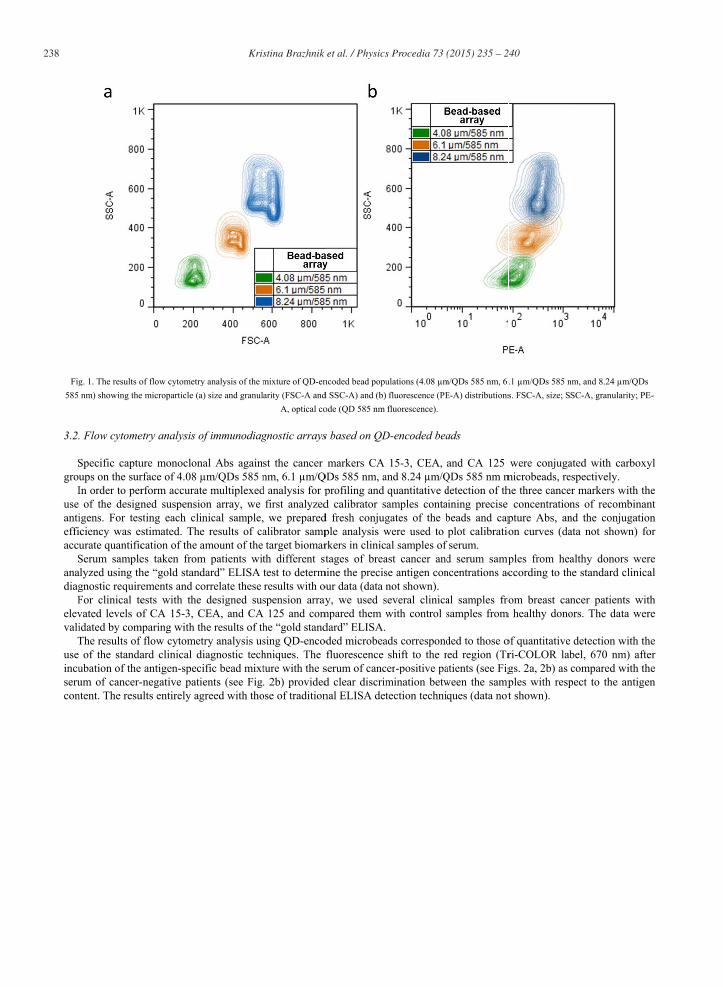
The adapted procedure of charged polymer deposition onto the surface of beads and incorporation of water-soluble carboxylated QDs was shown to be suitable for preparing optically encoded microparticles and using them in immunodiagnostic suspension arrays (Sukhanova et al. (2007); Brazhnik et al. (2014); Brazhnik et al. (2015)). The resultant single-color QD-encoded microbeads exhibited intense fluorescence with emission wavelengths that were nearly identical to those of the original QDs (data not shown). Flow cytometry analysis demonstrated that individual microbead populations (4.08 μm/QDs 585 nm, 6.1 μm/QDs 585 nm, and 8.24 μm/QDs 585 nm) were extremely bright and homogenous and were clearly distinguishable in the mixture by size and optical code (see Fig. 1).
238 Kristina Brazhnik et al. / Physics Procedia 73 ( 2015 ) 235 – 240
Fig585
3.2.
Sgrou
Iuse antieffiaccu
Sanadiag
Felevvali
Tuse incuserucon
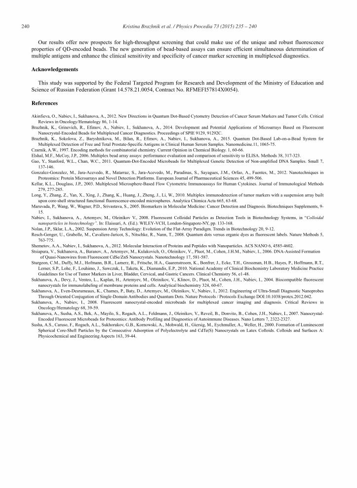
g. 1. The results onm) showing the
Flow cytome
Specific captuups on the surIn order to per
of the designigens. For tesciency was eurate quantificSerum samplelyzed using thgnostic requirFor clinical tevated levels oidated by comThe results of
of the standaubation of theum of cancer-ntent. The resu
of flow cytometrye microparticle (a
etry analysis of
ure monoclonrface of 4.08 μrform accuratned suspensiosting each clinstimated. Thecation of the aes taken fromhe “gold standements and coests with the
of CA 15-3, Cmparing with thf flow cytometard clinical d antigen-speci-negative patiults entirely ag
y analysis of the ma) size and granul
of immunodiag
nal Abs againsμm/QDs 585 ne multiplexedon array, we nical sample,e results of caamount of the
m patients witdard” ELISA orrelate these
designed susCEA, and CAhe results of thtry analysis usiagnostic techific bead mixtents (see Fig.
greed with tho
mixture of QD-enarity (FSC-A and
A, optical co
gnostic arrays
st the cancer nm, 6.1 μm/Qd analysis for p
first analyzed we preparedalibrator samptarget biomar
th different stest to determresults with ospension array125 and com
he “gold standsing QD-encohniques. The ture with the s. 2b) provided
ose of tradition
ncoded bead popud SSC-A) and (b)ode (QD 585 nm
s based on QD
markers CA QDs 585 nm, a
profiling and d calibrator sd fresh conjugple analysis wrkers in clinicstages of brea
mine the precisour data (data nay, we used smpared them wdard” ELISA. oded microbea
fluorescence serum of cancd clear discrimnal ELISA det
ulations (4.08 μm fluorescence (PEfluorescence).
D-encoded bea
15-3, CEA, and 8.24 μm/Qquantitative damples contagates of the bwere used to al samples of
ast cancer andse antigen connot shown). several clinicawith control s
ads correspondshift to the r
cer-positive pamination betwtection techniq
m/QDs 585 nm, 6.E-A) distributions
ads
and CA 125 QDs 585 nm mdetection of thining precise beads and capplot calibratioserum.
d serum sampncentrations ac
al samples frosamples from
ded to those oed region (Tr
atients (see Figween the sampques (data not
.1 μm/QDs 585 ns. FSC-A, size; S
were conjugamicrobeads, rehe three cance
concentrationpture Abs, anon curves (da
ples from heaccording to th
om breast cam healthy dono
of quantitativeri-COLOR labgs. 2a, 2b) as ples with respt shown).
nm, and 8.24 μm/SC-A, granularity
ated with carbespectively. er markers witns of recombnd the conjugata not shown
althy donors he standard cli
ncer patients ors. The data
detection witbel, 670 nm) compared witpect to the an
/QDs y; PE-
boxyl
th the binant gation n) for
were inical
with were
th the after
th the ntigen
Kristina Brazhnik et al. / Physics Procedia 73 ( 2015 ) 235 – 240 239
Frepro
dif
4. C
Mmaindiagin th
CcliniresuGon
Lfree a fasuspgene
Infor sdiffe125)bindto qucalibmult
Tthe din cl
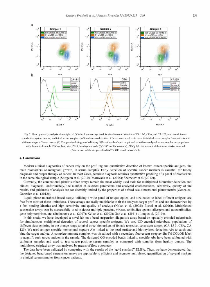
Fig. 2. Flow cytomoductive system t
fferent stages of bwith the control
onclusions
Modern clinican biomarkers
gnosis and prohe same biologCurrently, the ical diagnosislts, and quickn
nzalez et al. (2Liquid-phase m
from most ofst binding kin
pension arrayse polymorphisn this study, wsimultaneous erent sizes em). We used and the target anuantify each tbrator sampletiplexed (triple
The data have designed beadlinical serum s
metry analysis oftumors, in clinica
breast cancer. (b) sample. FSC-A,
al diagnostics of malignant
oper therapy ogical sample (conventional
s. Unfortunatekness of analys012)). microbead-basf these limitatinetics and hi
s can be succesm, etc. (Sukhwe have devemultiplexed
mitting in the ontigen-specificalyte. A comptarget antigen es and used ex) array was been validate
d-based suspensamples from
f multiplexed QDal serum samples.Comparative histbead size; PE-A,
(fluores
of cancer relt growth, in f cancer. In m
(Sturgeon et alplanar surfac
ely, the numbsis are conside
sed assays utiions. These asigh sensitivityessfully used hanova et al. (2eloped a novel
detection of orange range tc monoclonal plete immune in the sampleto test canceanalyzed by m
ed by comparinsion assays acancer patien
–bead microarray. (a) Simultaneoutograms indicatin, bead optical codscence of the strep
ly on the profserum sample
most cases, accl. (2010); Mare arrays remaber of selecteerably limited
ilizing a widessays are easily and quality to detect mult2007); Kellar l lab-on-a-beaseveral canceto label three capture Abs complex was
e. The designeer-positive sermeans of flowing with the rare applicablents.
ys used for simultus detection of thrng different levelsde (QD 585 nm flptavidin-Tri-COL
filing and quaes. Early detecurate diagnosruvada et al. (ain the most wed parametersd by the prope
e panel of unily modifiable of analysis
ltiple proteinset al. (2003);
ad suspensioner-specific anbiomarkers oflinked to the
s visualized wed QD-encoderum samples
w cytometry. results of the “e to efficient a
taneous detectionree cancer markers of each target mluorescence); PE-LOR visualization
antitative detecection of specsis requires qu2005); Sheme
widely used tos and analyzeerties of a fixe
ique optical anto fit the anal(Nolan et al. , viruses, antiGao et al. (20
n diagnostic asntigens. We uf female reprobead surface
with a secondared beads linke
as compared
“gold standardand accurate m
n of CA 15-3, CErs in three individ
marker in three ana-Cy5-A, the amoun label).
ction of knowcific cancer muantitative proetov et al. (201ools for multiped characteristed two-dimens
nd size codeslyzed target pr
(2002); Elshibodies agains011) ; Long et ssay based onsed QD-enco
oductive systeand biotinyla
ry fluorescented to specific d with sampl
d” ELISA. Thmultiplexed qu
EA, and CA 125, mdual serum sampl
nalyzed serum samunt of the cancer
wn cancer-spemarkers is esofiling of a pa12)). plexed biomartics, sensitivisional planar m
s to label diffrofiles and arehal et al. (20st allergens ant al. (2010)). n optically encoded microbeaem tumors (CAated detection t streptavidin- Abs have beeles from hea
hus, we have uantification o
markers of femalles from patients mples in comparismarker detected
cific antigenssential for tim
anel of biomar
rker detectionty, quality ofmatrix (Gonza
ferent antigense characterize06)). Multiplend autoantibod
coded microbad populationA 15-3, CEA,Abs to catch
Tri-COLOR len calibrated
althy donors.
demonstratedof several mar
e with son
, the mely rkers
n and f the alez-
s are ed by exed dies,
eads ns of , CA
h and label with The
d that rkers
240 Kristina Brazhnik et al. / Physics Procedia 73 ( 2015 ) 235 – 240
Our results offer new prospects for high-throughput screening that could make use of the unique and robust fluorescence properties of QD-encoded beads. The new generation of bead-based assays can ensure efficient simultaneous determination of multiple antigens and enhance the clinical sensitivity and specificity of cancer marker screening in multiplexed diagnostics.
Acknowledgements
This study was supported by the Federal Targeted Program for Research and Development of the Ministry of Education and Science of Russian Federation (Grant 14.578.21.0054, Contract No. RFMEFI57814X0054).
References
Akinfieva, O., Nabiev, I., Sukhanova, A., 2012. New Directions in Quantum Dot-Based Cytometry Detection of Cancer Serum Markers and Tumor Cells. Critical Reviews in Oncology/Hematology 86, 1-14.
Brazhnik, K., Grinevich, R., Efimov, A., Nabiev, I., Sukhanova, A., 2014. Development and Potential Applications of Microarrays Based on Fluorescent Nanocrystal-Encoded Beads for Multiplexed Cancer Diagnostics. Proceedings of SPIE 9129, 91292C.
Brazhnik, K., Sokolova, Z., Baryshnikova, M., Bilan, R., Efimov, A., Nabiev, I., Sukhanova, A., 2015. Quantum Dot-Based Lab-on-a-Bead System for Multiplexed Detection of Free and Total Prostate-Specific Antigens in Clinical Human Serum Samples. Nanomedicine.11, 1065-75.
Czarnik, A.W., 1997. Encoding methods for combinatorial chemistry. Current Opinion in Chemical Biology. 1, 60-66. Elshal, M.F., McCoy, J.P., 2006. Multiplex bead array assays: performance evaluation and comparison of sensitivity to ELISA. Methods 38, 317-323. Gao, Y., Stanford, W.L., Chan, W.C., 2011. Quantum-Dot-Encoded Microbeads for Multiplexed Genetic Detection of Non-amplified DNA Samples. Small 7,
137-146. Gonzalez-Gonzalez, M., Jara-Acevedo, R., Matarraz, S., Jara-Acevedo, M., Paradinas, S., Sayagues, J.M., Orfao, A., Fuentes, M., 2012. Nanotechniques in
Proteomics: Protein Microarrays and Novel Detection Platforms. European Journal of Pharmaceutical Sciences 45, 499-506. Kellar, K.L., Douglass, J.P., 2003. Multiplexed Microsphere-Based Flow Cytometric Immunoassays for Human Cytokines. Journal of Immunological Methods
279, 277-285. Long, Y., Zhang, Z., Yan, X., Xing, J., Zhang, K., Huang, J., Zheng, J., Li, W., 2010. Multiplex immunodetection of tumor markers with a suspension array built
upon core-shell structured functional fluorescence-encoded microspheres. Analytica Chimica Acta 665, 63-68. Maruvada, P., Wang, W., Wagner, P.D., Srivastava, S., 2005. Biomarkers in Molecular Medicine: Cancer Detection and Diagnosis. Biotechniques Supplements, 9-
15. Nabiev, I., Sukhanova, A., Artemyev, M., Oleinikov V., 2008. Fluorescent Colloidal Particles as Detection Tools in Biotechnology Systems, in “Colloidal
nanoparticles in biotechnology”. In: Elaissari, A. (Ed.). WILEY-VCH, London-Singapore-NY, pp. 133-168. Nolan, J.P., Sklar, L.A., 2002. Suspension Array Technology: Evolution of the Flat-Array Paradigm. Trends in Biotechnology 20, 9-12. Resch-Genger, U., Grabolle, M., Cavaliere-Jaricot, S., Nitschke, R., Nann, T., 2008. Quantum dots versus organic dyes as fluorescent labels. Nature Methods 5,
763-775. Shemetov, A.A., Nabiev, I., Sukhanova, A., 2012. Molecular Interaction of Proteins and Peptides with Nanoparticles. ACS NANO 6, 4585-4602. Stsiapura, V., Sukhanova, A., Baranov, A., Artemyev, M., Kulakovich, O., Oleinikov, V., Pluot, M., Cohen, J.H.M., Nabiev, I., 2006. DNA-Assisted Formation
of Quasi-Nanowires from Fluorescent CdSe/ZnS Nanocrystals. Nanotechnology 17, 581-587. Sturgeon, C.M., Duffy, M.J., Hofmann, B.R., Lamerz, R., Fritsche, H.A., Gaarenstroom, K., Bonfrer, J., Ecke, T.H., Grossman, H.B., Hayes, P., Hoffmann, R.T.,
Lerner, S.P., Lohe, F., Louhimo, J., Sawczuk, I., Taketa, K., Diamandis, E.P., 2010. National Academy of Clinical Biochemistry Laboratory Medicine Practice Guidelines for Use of Tumor Markers in Liver, Bladder, Cervical, and Gastric Cancers. Clinical Chemistry 56, e1-48.
Sukhanova, A., Devy, J., Venteo, L., Kaplan, H., Artemyev, M., Oleinikov, V., Klinov, D., Pluot, M., Cohen, J.H., Nabiev, I., 2004. Biocompatible fluorescent nanocrystals for immunolabeling of membrane proteins and cells. Analytical biochemistry 324, 60-67.
Sukhanova, A., Even-Desrumeaux, K., Chames, P., Baty, D., Artemyev, M., Oleinikov, V., Nabiev, I., 2012. Engineering of Ultra-Small Diagnostic Nanoprobes Through Oriented Conjugation of Single-Domain Antibodies and Quantum Dots. Nature Protocols / Protocols Exchange DOI:10.1038/protex.2012.042.
Sukhanova, A., Nabiev, I., 2008. Fluorescent nanocrystal-encoded microbeads for multiplexed cancer imaging and diagnosis. Critical Reviews in Oncology/Hematology 68, 39-59.
Sukhanova, A., Susha, A.S., Bek, A., Mayilo, S., Rogach, A.L., Feldmann, J., Oleinikov, V., Reveil, B., Donvito, B., Cohen, J.H., Nabiev, I., 2007. Nanocrystal-Encoded Fluorescent Microbeads for Proteomics: Antibody Profiling and Diagnostics of Autoimmune Diseases. Nano Letters 7, 2322-2327.
Susha, A.S., Caruso, F., Rogach, A.L., Sukhorukov, G.B., Kornowski, A., Mohwald, H., Giersig, M., Eychmuller, A., Weller, H., 2000. Formation of Luminescent Spherical Core-Shell Particles by the Consecutive Adsorption of Polyelectrolyte and CdTe(S) Nanocrystals on Latex Colloids. Colloids and Surfaces A: Physicochemical and Engineering Aspects 163, 39-44.