Embed Size (px)
Citation preview
Coleoid cephalopods through time (Warnke K., Keupp H., Boletzky S. v., eds)
Berliner Paläobiol. Abh. 03 141-162 Berlin 2003
MUSCLE ARRANGEMENT, FUNCTION AND SPECIALIZATION
IN RECENT COLEOIDS
W. M. Kier* & J. T. Thompson
Department of Biology, CB# 3280 Coker Hall, University of North Carolina, Chapel Hill, NC 27599-3280, USA
*corresponding author: [email protected]
ABSTRACT
The bodies of coleoid cephalopods are characterized by a dense musculature consisting of tightly packed bundles of
muscle fibers arranged in three mutually perpendicular planes. This arrangement of muscle, termed a ‘muscular
hydrostat’, generates force and also provides skeletal support. Muscle function during movement and locomotion thus
does not depend on rigid skeletal elements, even though many extant coleoids possess hard parts.
In this review, we describe the arrangement and microanatomy of the musculature and connective tissues from a
variety of coleoids and from a range of cephalopod organs and systems including the mantle, funnel, fins, arms,
tentacles, and suckers. We analyze the muscle and connective tissues from the standpoint of biomechanics in order to
describe their function in movement and locomotion. This analysis demonstrates that the same basic principles of
support and movement are shared by all of these structures. In addition, the crucial role played by fibrous collagenous
connective tissues in these systems is emphasized. Further work is required, however, to describe the mechanical
functions of the musculature of the diverse pelagic cephalopods, to understand neuromuscular control of these complex
systems, and to explore the mechanisms of specialization of coleoid cephalopod muscle.
INTRODUCTION
Although many recent coleoids retain hard parts, they
resemble the soft-bodied invertebrates because muscle
function during movement and locomotion generally
does not depend on hardened skeletal elements. The
shell of modern coleoids serves as a buoyancy control
device in the sepioids (Denton 1974, Denton & Gilpin-
Brown 1959, 1961), is reduced and has an uncertain
supportive role in the teuthoids (Donovan & Toll 1988,
Nigmatullin et al. 1991, Toll 1988), and is greatly
reduced and modified in the octopods (Wells 1978).
The transmission of force, antagonism of the
musculature, and the amplification of force,
displacement or velocity of muscle contraction in most
cases depend instead on a form of skeletal support that
resembles the hydrostatic skeleton of soft-bodied
invertebrates.
The coleoid body is characterized throughout by
dense musculature consisting of tightly packed bundles
of muscle fibers arranged in three mutually
perpendicular directions (see Budelmann et al. 1997
for a review of the microanatomy). Such an
arrangement of muscle, termed a ‘muscular hydrostat’,
serves both to generate the force and to provide the
skeletal support required for movement and locomotion
(Kier & Smith 1985, Smith & Kier 1989). The basic
principle of such a system of skeletal support is
straightforward. At physiological pressures, muscle
tissue is essentially incompressible and muscle
contraction results in no significant change in volume
[measurements suggest that the volume change is only
0.002% (Baskin 1967)]. Since the block of muscle is
essentially constant in volume, a decrease in one
dimension due to contraction of a given bundle of
muscle fibers must result in an increase in another
dimension. Since the muscle fibers are typically
arranged with bundles that can control each of the three

142
Fig. 1 Diagram of the ventral portion of the mantle of a
loliginid squid. Each tissue block is oriented with the mantle
cavity toward the top and the skin toward the bottom. The
upper block of the mantle illustrates the arrangement of the
muscle fibers. CM, circular muscle fibers; RM, radial muscle
fibers; CMP, central mitochondria-poor zone of circular
muscle fibers; SMR, superficial mitochondria-rich zones of
circular muscle fibers. The lower block illustrates the
organization of the connective tissue fibers (in black; muscle
in gray). Note that the collagen fibers in intramuscular fiber
system 1 (IM-1) are visible in both sagittal and frontal planes,
indicating that an individual IM-1 fiber follows an oblique
trajectory through the mantle. IT, inner tunic; IM-1, collagen
fibers in intramuscular fiber system 1; IM-2, collagen fibers in
intramuscular fiber system 2; IM-3, crimped collagen fibers in
intramuscular fiber system 3. OT, outer tunic. [Loliginid
sketch modified from a photograph in Norman 2000]
dimensions, a remarkably diverse array of
deformations and movements can be produced. In
addition to the complex arrangement of muscle fibers,
many of the muscular-hydrostatic systems in
cephalopods include arrays of collagenous connective
tissue fibers. These fibers serve an extremely important
role in control of shape change and in elastic energy
storage for restoration of shape following muscle
contraction.
In this review we will focus on the arrangement and
function of muscle and connective tissues in recent
coleoid cephalopods. In keeping with the theme of the
symposium for which this publication was prepared,
we hope that the insight provided from an
understanding of the recent forms will be of use in the
interpretation of fossil coleoid structure, function and
evolution.
MANTLE
Musculature
The mantle of coleoids serves important roles in
ventilation and locomotion by jet propulsion. The
structure and function of the mantle of loliginid and
ommastrephid squids has received the greatest
attention in the literature and will therefore be the focus
of the following description, although descriptions of
other coleoids will also be included. The mantle of
loliginid and ommastrephid squids includes two
predominant muscle orientations: circumferential
muscle fibers (known as circular muscles) that
constitute the bulk of the mantle wall and radial muscle
fibers that extend from the inner to the outer surface of
the mantle wall as partitions between the bundles of
circular muscle fibers (Fig. 1; Marceau 1905, Williams
1909, Young 1938). Each circular muscle fiber is
obliquely striated, uninucleate, 1 to 2 mm in length, up
to 10 mm in diameter, and is electrically coupled to
adjacent circular muscle fibers, presumably by gap
junctions (Bone et al. 1981, 1995, Milligan et al. 1997,
Young 1938). The radial muscle fibers are also
obliquely striated, uninucleate and may be up to 5 mm
in diameter (Bone et al. 1981, Mommsen et al. 1981).
The circular muscle fibers of a number of loliginid
and ommastrephid squid species are differentiated into
three zones: an outer zone adjacent to the external
surface of the mantle, a central zone, and an inner zone
adjacent to the inner surface of the mantle (Fig. 1). The
circular muscle fibers of the inner and outer zones,
known as superficial mitochondria rich (SMR) fibers
(Preuss et al. 1997), contain large cores occupied by
many mitochondria, show high succinic dehydrogenase
(SDH) activity and have a large ratio of oxidative to
glycolytic enzymes (Bone et al. 1981, Mommsen et al
1981). By contrast, the circular muscle fibers of the
143
central zone, termed central mitochondria poor (CMP)
fibers (Preuss et al. 1997), have few mitochondria, low
SDH activity and a low ratio of oxidative to glycolytic
enzymes (Bone et al. 1981, Mommsen et al. 1981).
The blood supply to these zones parallels these
differences and includes a dense capillary plexus in the
inner and outer zones compared with a sparse capillary
plexus in the central zone (Bone et al. 1981). The
radial muscle fibers are similar in structure and in
mitochondrial density to the CMP fibers (Bone et al.
1981, Mommsen et al. 1981) although Bone et al.
(1994) suggest that radial muscle fibers may be aerobic
given their repetitive activity in respiration. The
metabolic differentiation of the circular muscle fibers is
thought to be analogous to the subdivisions of red and
white muscle observed in the vertebrates (Bone et al.
1981, Mommsen et al. 1981, Rome et al. 1988). The
SMR circular muscle fibers, analogous to the red
muscles of vertebrates, power the constant ventilatory
movements and prolonged slow-speed swimming. The
CMP circular muscle fibers, analogous to the white
muscles of vertebrates, produce the brief escape jets
(Bartol 2001, Bone et al. 1981, Gosline et al. 1983,
Mommsen et al. 1981). In several species of loliginids,
the relative abundance of SMR circular muscle fibers
decreases substantially during growth (Preuss et al.
1997, Thompson & Kier 2001a, 2002).
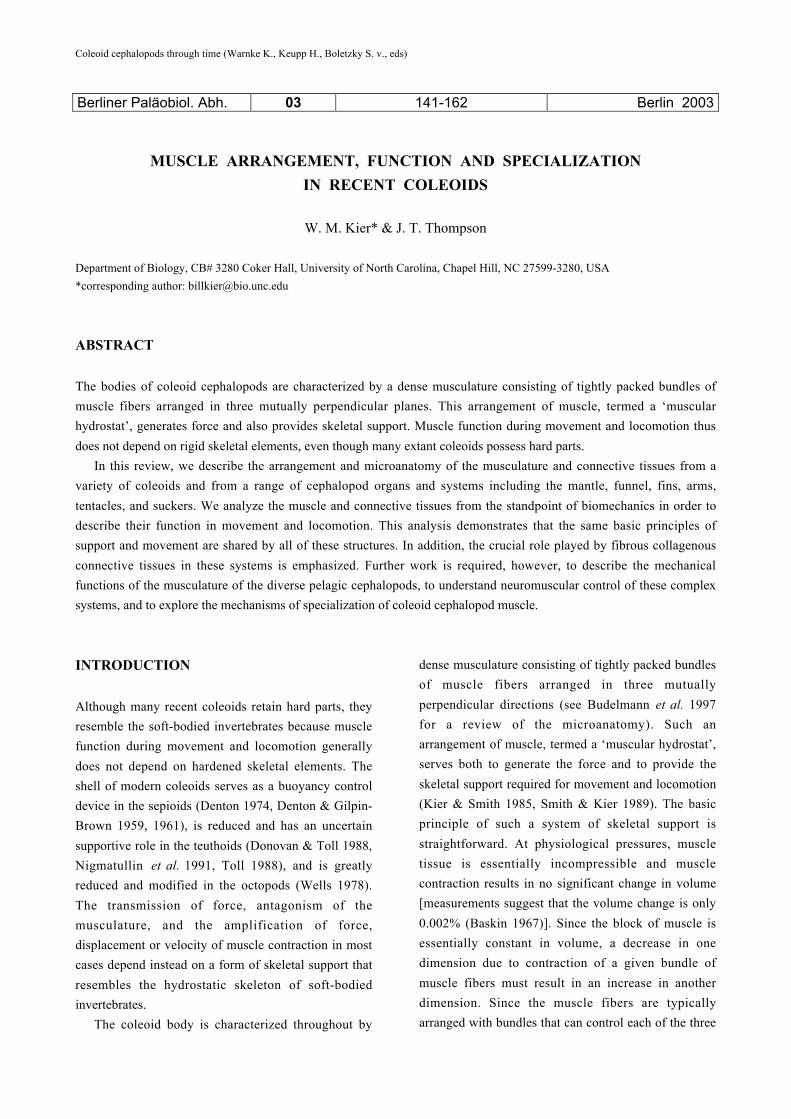
Unlike loliginid and ommastrephid squids, the
mantle of many octopodids contains two layers of
muscle fibers oriented parallel to the long axis of the
mantle, in addition to the circular and radial muscle
fibers. In Octopus vulgaris, for example, two layers of
longitudinal muscle fibers enclose densely packed
circular and radial muscles (Fig. 2, Wilson 1960).
Neither the longitudinal nor the circular muscle fibers
are differentiated into zones based on structural
characteristics (unpublished observation). Little has
been published about the vascular organization of the
octopodid mantle.
The morphology of the mantle musculature of
many gelatinous-bodied midwater and deepwater
squids and octopods differs from that of the shallow
water octopodids or the loliginid and ommastrephid
squids described above (unpublished observations). In
octopoteuthids, cycloteuthids, and some lepidoteuthids
there are three orientations of muscle fibers in the
mantle: a thin layer of longitudinal muscle fibers
adjacent to the outer surface of the mantle, two
Fig. 2 Diagram of the ventral region of the mantle of a
shallow-water octopus. Each tissue block is oriented with the
mantle cavity toward the top and the skin toward the bottom.
The upper block illustrates the organization of the muscle
fibers. CM, circular muscle fibers; LM, longitudinal muscle
fibers; RM, radial muscle fibers. The lower block illustrates
the arrangement of the connective tissue fibers (in black;
muscle in gray). CT, feltwork of connective tissue fibers; IM-
3, connective tissue fibers in intramuscular fiber system 3;
LCT, longitudinal connective tissue fibers. [Octopus sketch
modified from Nesis 1987]
relatively thin layers of circular muscle fibers near the
inner and outer surfaces of the mantle, and radial
muscle fibers (Fig. 3). In Cirrothauma murrayi Chun
the arrangement of the muscles is similar but an
additional layer of longitudinal muscle fibers is present
along the inner surface of the mantle (Aldred et al.
1983). In mastigoteuthids, chiroteuthids, histioteuthids,
and batoteuthids the arrangement of muscle layers is
similar to that described above but longitudinal muscle

144
3
Fig. 3 Diagram of the ventral portion of the mantle of a deep-
water squid. Each tissue block is oriented with the mantle
cavity toward the top and the skin toward the bottom. The
upper block indicates the arrangement of the muscle fibers.
CM, circular muscle fibers; LM, longitudinal muscle fibers;
RM, radial muscle fibers. The lower block illustrates the
organization of the connective tissues (in black; muscle in
gray). CT, feltwork of connective tissue fibers; FL, gelatinous,
fluid-filled layer; IM-3, connective tissue fibers in
intramuscular fiber system 3; LCT, longitudinal connective
tissue fibers; P, polyhedral connective tissue capsules. [Sketch
of a histioteuthid based on a photograph in Norman 2000]
fibers are absent. In all the families of gelatinous-
bodied cephalopods mentioned, the two layers of
circular muscle fibers enclose a gelatinous layer. The
gelatinous layer may constitute most of the mantle
wall , as in octopoteuthids, lepidoteuthids,
chiroteuthids, and Cirrothauma murrayi or may be a
small fraction, as in batoteuthids and histioteuthids. It
is unknown if the circular muscle is differentiated into
layers that differ in biochemical and morphological
characteristics as observed in the loliginid and
ommastrephid squids. Seibel et al. (2000) however,
demonstrated the presence of enzymes important for
both aerobic (citrate synthase) and anaerobic (octopine
dehydrogenase) metabolism in the mantle musculature
of more than 20 species of mid- and deep-water
gelatinous-bodied cephalopods. The blood supply to
the mantle muscles in these animals has not been
studied in detail.
Connective Tissue
The radial muscle fibers of loliginids and
ommastrephids have their origin and insertion on the
inner and outer tunics, robust layers of collagenous
connective tissue fibers that lie beneath the skin on the
inner and outer surface of the mantle (Fig. 1). The
fibers of the tunics are arranged in layers of closely
packed parallel fibers that are oriented either as right-
handed or left-handed helices. The handedness of each
layer in the tunic alternates and the fiber angle, the
angle that a fiber makes with the longitudinal axis of
the mantle, ranges from about 17° in Sepioteuthis
lessoniana to 27° in Loligo pealei and Lolliguncula
brevis (Thompson & Kier 2001a, Ward & Wainwright
1972). In S. lessoniana, the fiber angle of the tunics
decreases during ontogeny from 33° in hatchlings to
17° in subadult animals (Thompson & Kier 2001a).
In addition to the connective tissue fibers of the
tunics, the mantle also includes networks of connective
tissue fibers, termed “intramuscular connective tissue
fibers,” that span the mantle wall (Bone et al. 1981,
Gosline & Shadwick, 1983a, 1983b, Ward &
Wainwright 1972). Three groups of intramuscular
connective tissue fibers (IM) have been identified in
the cuttlefishes and loliginid squids, and are denoted
IM-1, IM-2 and IM-3 (Fig. 1). IM-1 fibers originate
and insert on the inner and outer tunics and follow
straight or slightly curved trajectories through the
mantle wall (Ward & Wainwright 1972). Viewed in
sagittal section, the IM-1 fibers are arranged at a low
angle (28° in L. brevis) relative to the long axis of the
mantle (Ward & Wainwright 1972) (Fig. 1). In sections
tangential to the surface of the mantle, the collagen
fibers in IM-1 are also arranged at low angles (10° to
15° in Alloteuthis subulata, 32° in S. lessoniana)
relative to the long axis of the mantle (Bone et al.

145
1981, Thompson & Kier 2001a) (Fig. 1). Thus, the IM-
1 fibers follow an oblique path through the mantle
wall, relative to both tangential and sagittal planes.
IM-2 fibers also originate and insert on the tunics
(Fig. 1). They are localized to the radial muscle bands,
follow straight trajectories, and are arranged at an angle
of about 55° to the mantle surface in A. subulata, Sepia
officinalis and S. lessoniana (Bone et al. 1981, Curtin
et al. 2000, Thompson & Kier 2001a).
IM-3 fibers, which are crimped or buckled in
histological sections, are arranged parallel to the
circular muscle fibers and are not attached to the tunics
(Bone et al. 1981) (Fig. 1).
Measurements of the birefringence of the
connective tissue fibers of IM-1 by Gosline and
Shadwick (1983a) suggest that they are collagen.
Transmission electron microscopy of IM-1, IM-2, and
IM-3 in L. pealei by MacGillivray et al. (1999)
revealed collagen fibers ranging from 1.5 to 4.5 mm in
diameter. Bone et al. (1981) reported that at least some
of the connective tissue fibers of the mantle of L .
vulgaris and A. subulata may be elastic fibers, based on
histochemical staining and absence of the characteristic
68-nm repeat pattern typical of collagen fibers
observed by transmission electron microscopy.
MacGillivray et al. (1999), however, did not observe
staining of intramuscular connective tissue fibers
indicative of elastic fibers in L. pealei.
Published descriptions of the connective tissue
organization of the mantle of octopodids are limited.
The mantle of Octopus bimaculatus does not contain
well-defined inner and outer tunics (unpublished
observation). Instead, the connective tissue layers that
enclose the mantle resemble a feltwork, with robust
collagen fibers arranged in various orientations. IM-3
connective tissue fibers are present in the mantle of O.
b i m a c u l a t u s (Fig. 2). The IM-3 fibers exhibit
histochemical staining properties consistent with
collagen. Robust connective tissue fibers aligned
parallel to the long axis of the mantle are also found in
the two longitudinal muscle layers in the mantle (Fig.
2). Connective tissue fibers similar to the IM-1 and IM-
2 fibers of squids are not observed in octopodids.
The mantle of many gelatinous-bodied squids and
octopods also differs in connective tissue morphology
from that described above (unpublished observations).
The central gelatinous portion of the mantle wall is
reinforced by a three dimensional meshwork of sheets
of robust connective tissue fibers (Fig. 3). These sheets
consist of connective tissue fibers that are embedded in
a matrix and form numerous small, polyhedral capsules
that may be filled with ammoniacal fluids, other low-
density fluids, or hemolymph. The fibers exhibit
birefringence and histochemical staining properties
consistent with collagen. Those gelatinous-bodied
squids that have longitudinal muscle fibers (e.g.
octopoteuthids, cycloteuthids, and some lepidoteuthids)
also lack well-defined tunics; inner and outer tunics are
present in gelatinous-bodied animals that lack
longitudinal muscle fibers. IM-1 and IM-2 connective
tissue fibers are absent in chiroteuthids, cycloteuthids,
s o m e lepidoteuthids, mastigoteuthids, and
octopoteuthids. IM-3 connective tissue fibers are
present in nearly all coleoids that have been examined.
Biomechanics
The basic function of the mantle musculature of
loliginids and ommastrephids depends on a muscular
hydrostatic mechanism. During ventilation of the
mantle cavity and jet locomotion, contraction of the
circular muscle fibers decreases the diameter of the
mantle, expelling water from the mantle cavity via the
funnel. Lengthening of the mantle is prevented by the
connective tissue fibers of the tunics (Ward &
Wainwright 1972). Because the mantle wall is
essentially constant in volume, decrease in mantle
diameter by circular muscle contraction must also
result in an increase in the thickness of the mantle wall,
thereby extending the radial muscle fibers and
stretching the connective tissue fibers of IM-1 and IM-
2. Following circular muscle contraction, elastic recoil
of the IM-1 and IM-2 connective tissue fibers and
contraction of the radial muscle fibers cause the mantle
wall to thin. This thinning results in an increase in the
diameter of the mantle and expansion of the mantle
cavity, thereby generating subambient pressure that
refills the mantle cavity with water (see Curtin et al.
2000, Gosline & Shadwick 1983a, 1983b, Gosline et
al. 1983, MacGillivray et al. 1999). Thinning of the
mantle wall also stretches the IM-3 connective tissue
fibers (Gosline et al. 1983).
During a locomotory jet, three phases are observed.
The first phase is termed “hyperinflation” and involves
146
contraction of the radial muscles only, expanding the
mantle cavity to a diameter that is larger than the
resting mantle diameter (Gosline et al. 1983). The
second phase, “the jet” occurs as the circular muscles
contract. Elastic recoil of the IM-3 connective tissue
fibers may aid the early stages of contraction of the
circular muscles (Gosline et al. 1983). The final phase,
“refilling” occurs as the mantle cavity re-expands,
primarily due to elastic recoil of the IM-1 and IM-2
connective tissue fiber networks, often with assistance
from radial muscle contraction (Gosline & Shadwick
1983a, Gosline et al. 1983). Additional assistance in
refilling of the mantle may be provided by flow-
induced pressure differentials when the animals are
swimming at high velocities (Vogel 1987). Respiratory
movements of the mantle are smaller than those of the
locomotory jet and typically occur in the anterior half
of the mantle only (Packard & Trueman 1974). Two
patterns of respiratory movement occur in loliginid
squids: 1) radial muscle contraction and water intake
by mantle expansion (i.e., hyperinflation) followed by
elastic recoil of the mantle and water expulsion; 2)
mantle contraction due to circular muscle activation
expels water from the mantle cavity followed by water
intake as the mantle recoils elastically (Gosline et al.
1983). In European cuttlefish Sepia officinalis and
possibly also in loliginid squids, contraction of the
radial muscles fills the mantle cavity and inward
movements of the muscular collar flaps of the mantle
expel water from the mantle cavity (Bone et al. 1994).
Coleoids exhibit remarkable control over the
mantle, from low amplitude respiratory movements to
large amplitude contractions during escape-jet
locomotion. In loliginids, the only squids in which
neuromuscular anatomy and physiology have been
well-studied, this fine control is likely the result of
innervation of the circular muscle fibers by both giant
and small diameter axons. The giant axons, which may
exceed 800 mm in diameter, activate the circular
muscles in an all-or-none response during escape-jet
locomotion (Young 1938). The small diameter axons
range from 0.5 to 50 mm (Bone et al. 1981) and
produce graded contractions of the mantle when
stimulated. They are hypothesized to control
respiratory movements of the mantle and low-speed jet
locomotion (Wilson 1960, Young 1938), although
some of the small diameter axons of the mantle may
also serve as sensory fibers. Otis and Gilly (1990)
found that the small diameter axons of L o l i g o
opalescens could also initiate delayed escape-jet
responses with or without giant axon activity.
Interaction of the giant and non-giant axon systems
may be important in modulating the magnitude of
escape responses (Otis & Gilly 1990). Such interaction,
however, may be limited to mature animals because the
small diameter axons of newly hatched L. opalescens
cannot elicit an escape response (Gilly et al. 1991).
The skeletal support of the mantle of gelatinous-
bodied coleoids does not depend on a muscular
hydrostatic mechanism. The gelatinous, fluid-filled
chamber in the center of the mantle wall implies that
skeletal support is analogous to the closed hydrostatic
skeletal support systems of annelids or the foot of
gastropod molluscs. Nevertheless, the principles of
mechanical function of the mantle are probably
conserved among all coleoids: the volume of the
mantle wall is essentially constant, circular and radial
muscle fibers act antagonistically, and the tunics
prevent lengthening of the mantle. In those animals that
lack well-defined tunics, longitudinal muscle fibers
control mantle length. One important exception to the
similarity in mechanical function of the mantle is
elastic energy storage. The arrangement of the IM-1
and IM-2 collagen fibers of loliginids, ommastrephids,
and cuttlefishes permits a substantial fraction of the
energy expended by the circular and radial muscle
fibers to be stored as elastic energy. The geometry of
IM-1 and IM-2 fibers makes the mantle mechanically
anisotropic and allows relatively small changes in the
thickness of the mantle wall (e.g. during contraction of
the circular muscles) to store significant elastic energy
(Curtin et al. 2000, Gosline & Shadwick 1983a,
MacGillivray et al. 1999). In contrast, the polyhedral
connective tissue capsules that support the fluid-filled
compartments of the mantle wall of gelatinous-bodied
coleoids probably make the mantle mechanically
isotropic. Although the geometry of the connective
tissue capsules are ideal for limiting deformation of the
mantle equally in three dimensions, we predict that the
collagen fibers within the capsules will store only a
minor component of the energy expended by the
circular and radial muscle fibers during jet locomotion.

147
FUNNEL
The funnel, also known as the siphon, directs the flow
of water from the mantle cavity during respiration and
jet locomotion. The tip and trunk of the funnel are
highly flexible and may be pointed in nearly any
direction (Tateno 1992, O’Dor 1988, Zuev 1966). The
aperture at the tip of the funnel of squids may be varied
during jet locomotion to permit fine modulation of the
thrust of the jet (O’Dor 1988, Zuev 1966).
Musculature and Connective Tissue
In loliginid squids, the funnel consists of obliquely
striated muscle fibers oriented in three mutually
perpendicular planes. Longitudinal muscle fibers are
located in bundles along the outer surface of the trunk
of the funnel. Circular muscle fibers compose the bulk
of the funnel, but thin bands of radial muscle extend
through the thickness of the funnel wall in an
arrangement analogous to the radial muscles of the
mantle (Williams 1909).
Thin layers of connective tissue fibers along the
outer and inner surfaces of the funnel enclose the
musculature. Other than Young’s (1938) observation
that connective tissue fibers are present in the
musculature of the funnel, information on the
connective tissues is limited.
Biomechanics
The organization of the muscle fibers and connective
tissue fibers of the funnel implies that its function
depends on a muscular hydrostatic mechanism.
Protrusion and retraction of the funnel trunk may be
accomplished via contraction of the circular and
longitudinal muscle fibers, respectively. Contraction of
the circular muscle fibers will also decrease the
diameter of the funnel trunk. An increase in the
diameter of the funnel trunk may be accomplished by
contraction of the radial muscle fibers and/or the
longitudinal muscle fibers of the funnel. Bending of the
funnel tip and trunk may be accomplished by
contraction of the radial muscle fibers while
longitudinal muscle fibers on the side of the bend
contract. The tonus of the longitudinal muscle fibers on
the side of the funnel opposite the bend may affect the
degree of bending.
FUNNEL, CEPHALIC, AND NUCHAL
RETRACTOR MUSCLES
Many coleoids possess muscles that control the
positions of the funnel and head relative to the mantle.
In loliginid squids, a pair of funnel retractor muscles
inserts on the dorsal portion of the funnel and
originates on the chitinous gladius of the mantle
(Williams 1909). Activity of the funnel retractor
muscles helps to support and position the funnel during
jet locomotion.
The cephalic and nuchal retractor muscles are tube-
or cone-shaped and enclose portions of the digestive
tract. The cephalic muscle originates on both the
gladius and nuchal cartilage, and inserts on the
posterior portion of the cephalic cartilage that encloses
the brain (Williams 1909). The nuchal retractor is a
robust muscle that originates on the pen (immediately
anterior to the cephalic retractor) and inserts on the
dorso-lateral edges and ventral surfaces of the nuchal
cartilage (Williams 1909). The functions of the
cephalic and nuchal retractor muscles of loliginid
squids include movement of the head in and out of the
mantle cavity.
Musculature
Longitudinal and transverse muscle fibers compose the
funnel retractor muscles of loliginid squids (Young
1938). Circular muscle fibers have not been reported.
The longitudinal muscle fibers of the funnel retractor
are continuous with the longitudinal muscle fibers of
the funnel itself (Williams 1909, Young 1938).
The orientation of muscle fibers in the nuchal and
cephalic retractors is similar. In each, a thin layer of
circular muscle fibers encloses a thick tube of
longitudinal muscle fibers. Transverse muscle fibers
whose orientation resembles that of the radial muscles
of the mantle are also present (Young 1938).
Connective Tissue
A thin layer of connective tissue fibers encloses the
funnel, cephalic, and nuchal retractor muscles. In

148
addition, sagittal sections of the funnel retractor
muscles of the loliginid squid, Sepioteuthis lessoniana,
reveal connective tissue fibers that span the entire
width of the muscle. The connective tissue fibers
exhibit staining reactions consistent with collagen, are
birefringent, and are arranged at an angle of about 55°
to the long axis of the muscle (unpublished
observations).
Biomechanics
The organization of the muscle and connective tissue
fibers in the funnel, cephalic, and nuchal retractors is
consistent with a muscular hydrostatic mechanism.
Shortening of the funnel retractors may occur via
contraction of the longitudinal muscle fibers and
relaxation of the transverse muscles. Elongation of the
funnel retractor muscles may be accomplished by
contraction of the transverse muscle fibers and
relaxation of the longitudinal muscles. The orientation
(i.e., about 55° to the long axis of the muscle) of the
connective tissue fibers spanning the width of each
funnel retractor is appropriate to resist elongation and
shortening of the muscle nearly equally (see Harris &
Crofton 1957). Therefore, the connective tissue fibers
may help antagonize both the transverse and
longitudinal muscle fibers and may also limit changes
in the shape of the muscle.
Shortening of the cephalic and nuchal retractors and
withdrawal of the head into the mantle cavity may
occur via contraction of the longitudinal muscle fibers
and relaxation of the circular and transverse muscle
fibers. Protrusion of the head out of the mantle cavity
may be accomplished by contraction of the circular or
the transverse muscle fibers and relaxation of the
longitudinal muscle fibers.
Although innervation of the funnel, cephalic, and
nuchal retractor muscles is complex, portions of all
three muscles are innervated by the same giant axon
(Young 1938). This highlights the close functional
relationship of the three muscles during jet locomotion.
For example, withdrawal of the head into the mantle
cavity occurs at the onset of escape-jet locomotion
(Thompson & Kier 2001b) as a result of simultaneous
activation of the cephalic and nuchal retractor muscles
(Young 1938).
FINS
The fins of cuttlefishes and squids produce rhythmic
undulatory waves that are used in locomotion and
hovering. Although much of the thrust for locomotion
is produced by contraction of the mantle during jet
propulsion, the fins aid in producing thrust at low
swimming speeds, in providing stability, and in
providing lift for hovering (Bidder & Boycott 1956,
Boycott 1958, Hoar et al. 1994, O’Dor & Webber
1986, Russel & Steven 1930). The fins of cuttlefishes,
loliginid and ommastrephid squids lack rigid
supportive elements and instead provide yet another
example of a three-dimensional array of musculature
termed a ‘muscular hydrostat’.
The profile and aspect ratio of the fins of coleoids
varies widely, and includes examples ranging from the
narrow fins of cuttlefishes that extend along the entire
length of the mantle to the more wing-like fins of many
pelagic squids (Hoar et al. 1994, Sweeney et al. 1992).
The structure and function of the fins of the cuttlefish
(Sepia officinalis) and of the loliginid squids (Loligo
forbesi and Sepioteuthis sepioidea) have received the
most attention previously and will therefore be
described here.
Musculature
The general arrangement of the musculature of the fins
of cuttlefishes and loliginid squids is similar and
includes bundles of obliquely striated muscle fibers
oriented in three mutually perpendicular directions
(Fig. 4). A dorsal and a ventral division of the
musculature is observed, separated by a median
connective tissue fascia. In both the dorsal and ventral
divisions of the fin, bundles of transverse muscle fibers
originate on the fin cartilage at the base of the fin and
extend laterally toward the fin margin, sending off
small bundles of fibers that insert on the median fascia.
The transverse muscle bundles in both the dorsal and
ventral portions of the fin are separated from one
another by sheets of dorsoventral muscle fibers that
originate on the median fascia. In the dorsal portion of
the fin these fibers extend to insert on an additional
fascia called the dorsal fascia that is located adjacent to
the dermis on the dorsal surface of the fin. An
analogous ventral fascia serves as the site of insertion

149
Fig. 4 Diagram of the fin of Sepia officinalis showing the arrangement of the muscle and connective tissue fibres. CT, crossed oblique
connective tissue fibres; DF, dorsal connective tissue fascia; DV, dorsoventral muscle; FC, fin cartilage; L, longitudinal muscle; MF,
median connective tissue fascia; T, transverse muscle; VF, ventral connective tissue fascia. The arrowheads delimit the dorsal and
ventral zones of transverse muscle fibres with more extensive mitochondrial cores (the oxidative fibres). The scale bar indicates the
scale for the lower portion of the diagram. [From Kier et al. (1989) with permission of the publisher]
of the dorsoventral fibers in the ventral portion of the
fin. In addition to the transverse and dorsoventral
muscle fibers, a layer of longitudinal muscle is present
adjacent to the dorsal and the ventral surface of the
median fascia. Examination of the fibers of the
transverse muscle bundles using electron microscopy
revealed that the mitochondrial content of the fibers
was not uniform across the muscle bundles (Kier
1989). Instead, transverse muscle fibers in narrow
zones adjacent to the dorsal and ventral fasciae include
a significantly larger mitochondrial core than the
remainder of the fibers and resemble the aerobic
muscle fibers observed in the mantle musculature
(Bone et al. 1981, Mommsen et al. 1981). The
dorsoventral and longitudinal muscle bundles lack
muscle fibers with a large mitochondrial core (Kier
1989).
Connective Tissues
The three fasciae described above include layers of
birefringent connective tissue fibers that exhibit

150
staining reactions typical of collagen. The fibers of the
dorsal and ventral fasciae show a slight degree of
preferred orientation in the transverse and longitudinal
directions, while those of the median fascia are
arranged as a feltwork and are embedded in an
amorphous matrix. Some of the fibers in the median
fascia exhibit staining reactions typical of elastin. In
addition to the three fasciae, birefringent connective
tissue fibers with staining reactions typical of collagen
are embedded the fin musculature. These fibers form a
delicate crossed fiber meshwork of obliquely oriented
fibers embedded in the dorsoventral and transverse
muscle bundles and extend between the fascia at an
angle of approximately 45º (Fig. 4; Kier 1989).
Biomechanics
Support and movement of the fin depends on a
muscular hydrostatic mechanism. The general form of
the movement of a given portion of the fin during the
passage of an undulatory wave involves sequential
bending dorsally and then ventrally. Dorsal bending
requires that the dorsal portion of the fin be reduced in
width (laterally compressed) relative to the ventral
portion and ventral bending requires an analogous
reduction in width of the ventral portion of the fin. The
transverse muscle bundles can provide this lateral
compression and thus their contraction is required for
active bending. This contraction, however, will cause
significant bending only if the lateral compressional
force on the opposite side of the fin is resisted. Without
this resistance, contraction of the transverse muscle
bundles will simply reduce the width of the fin by
pulling the lateral fin margin medially. Like other
muscular hydrostats, the fin is essentially constant in
volume and thus any reduction in the width of the fin
must result in either an increase in the thickness of the
fin or an increase in the length (or both). Increase in
length can be resisted by contractile activity of the
longitudinal muscles and the fin cartilage at the base of
the fin. Increase in thickness can be resisted by
contractile activity in the dorsoventral muscles or by
the array of crossed connective tissue fibers embedded
in the musculature (Johnsen & Kier 1993).
Based on an analysis of muscle activity patterns
during fin movement in the cuttlefish using
electromyography (Kier et al. 1989) and quantitative
modelling of the mechanical role of the crossed
connective tissue fibers (Johnsen & Kier 1993) the
following interpretation of fin movement has emerged.
During the gentle, low amplitude, low frequency fin
movements observed during hovering or when the
animals rest on the substratum, fin bending is caused
by contraction of the thin layer of aerobic fibers of the
transverse muscle bundles. Activity of the other muscle
orientations is not observed (Kier et al. 1989) and the
required resistance to lateral compression is provided
by the network of connective tissue fibers embedded in
the musculature (Johnsen & Kier 1993). During the
brief bursts of high amplitude, high frequency fin
beating observed during rapid locomotion and
maneuvering, the anaerobic transverse muscle fibers
are recruited and the required resistance to lateral
compression is provided by contractile activity of the
dorsoventral muscles on the opposite side of the fin,
perhaps with a contribution of the longitudinal muscles
for control of overall length of the fin. In addition to
providing resistance to lateral compression, the
quantitative modelling studies suggest that the crossed
connective tissue fibers may store elastic energy during
fin bending, allowing the fin to function as a harmonic
oscillator and thereby increasing the efficiency of the
fins during locomotion (Johnsen & Kier 1993).
Although little is known about neuromuscular control
mechanisms in the fins, there is evidence for
mechanoreceptors that may sense fin deformation and
in some way contribute to coordination of fin
movements (Kier et al. 1989).
ARMS AND TENTACLES
Coleoid cephalopods possess an array of appendages
encircling the mouth that serve in a remarkable variety
of functions. In the squids and cuttlefishes, five pairs of
appendages are present. One pair, termed ‘tentacles’, is
specialized for prey capture by remarkably rapid
elongation. For example, in Loligo pealei the tentacles
elongate by over 80% at peak extension velocities of
over 2 m s-1 and peak accelerations of 250 m s-2 (Kier
& van Leeuwen 1997, van Leeuwen & Kier 1997).
During a prey capture strike, the tentacles strike the
prey and attach to it with suckers that are present on an
expanded terminal portion termed the ‘club’. Twisting

151
Fig. 5 Diagram of arm of a loliginid squid. AN, axial nerve cord; ACT, aboral connective tissue (fibrous); AR, artery; BV, superficial
brachial vein; DCT, dermal connective tissue; EP, epithelium; IN, intramuscular nerve cord; LM, longitudinal muscle; OCT. oral
connective tissue (fibrous); OM, oblique muscle; PM, protective membrane; SKLM, swimming keel longitudinal muscle; SKTM,
swimming keel transverse muscle; SLM, superficial longitudinal muscle; SU, suckers; TM, transverse muscle; TR, trabeculae of
transverse muscle. [From Kier, (1982) with permission of the publisher]
of the tentacles about their long axis is frequently
observed during elongation. The tentacles then shorten
and the prey is withdrawn within reach of the 4 pairs of
arms, which subdue and manipulate it for ingestion
using primarily bending and twisting movements. The
arms of squids and cuttlefishes are also involved in
swimming and steering movements, behavioral
displays, and reproduction. Octopuses lack the
specialized prey capture tentacles observed in squids
and instead possess four pairs of arms, which are used
for locomotion, prey capture, exploring and
manipulating objects, grooming and behavioral
displays. The arms of octopuses are capable of a
remarkable diversity of movements including
elongation, shortening, bending and twisting.
Musculature and Connective Tissues
Three general groups of muscles are observed in the
arms and tentacles of coleoid cephalopods: transverse
muscle; longitudinal muscle; and helical or oblique
muscle. The arrangement observed in the arms of
squids is shown in Fig. 5. The transverse muscle mass
occupies the core of the arm surrounding the central
axial nerve cord, and consists of muscle fibers oriented
perpendicular to the long axis of the arm. In the
tentacles of squids, the transverse muscle fibers are
continuous with a thin layer of circular muscle fibers.
Groups of fibers termed ‘trabeculae’ (Graziadei 1965)
interdigitate with bundles of longitudinal muscle that
are oriented parallel to the long axis of the arm. The
transverse muscle fibers have their origin and insertion
on crossed fiber connective tissue layers both orally
(oral = side of arm facing the mouth) and aborally and
on the epimysial connective tissues surrounding a pair
of right- and left- handed oblique muscles on each side.
The oblique muscles in turn have their origin and
insertion on the oral and aboral crossed fiber
connective tissue layers. The fiber angle (angle that a
fiber makes with the long axis - approximately 72º in
Loligo pealei) of the connective tissue fibers is the
same as that of the associated oblique muscles.
Additional superficial layers of longitudinal muscle are
present surrounding the core of musculature described
above (Kier 1982). All of the muscles of the arms and
tentacles consist of obliquely striated fibers with the

152
Fig. 6 Diagram of tentacular stalk of a loliginid squid. AN, axial nerve cord; AR, artery; CM, circular muscle; DCT, dermal
connective tissue; EP, epithelium; HM, helical muscle; IN, intramuscular nerve cord; LM, Longitudinal muscle; SLM, superficial
longitudinal muscle; TM, transverse muscle; TR, trabeculae of transverse muscle; TV, superficial tentacular vein. [From Kier (1982)
with permission of the publisher]
exception of the transverse and circular muscles of the
tentacles, which are cross-striated (see below).
The arrangement of the musculature of the tentacles
of squids is shown in Fig. 6. As in the arm, the core is
occupied by a large mass of transverse muscle
surrounding a central axial nerve cord. Groups of
transverse muscle fibers extend peripherally and
interdigitate with bundles of longitudinal muscle
around the perimeter of the tentacle cross-section.
After passing between the longitudinal muscle bundles,
some of the transverse muscle fibers turn and become
part of a circular muscle layer and others extend to
insert on a connective tissue layer surrounding the
circular muscle layer. The circular muscle layer is in
turn wrapped by two thin helical muscle layers, one
with fibers arranged as a right-handed helix and the
other with fibers arranged as a left-handed helix [the
fiber angle is approximately 36º in an extended tentacle
and approximately 67º in a retracted tentacle, see Kier
(1982)]. Additional longitudinal muscle fibers are
observed in thin layers surrounding the circular muscle
layer (Guérin 1908, Kier 1982).
The arrangement of the musculature of the arms of
octopuses is shown in Fig. 7. As in the arms and
tentacles of squids, the core of the arm is occupied by a
mass of transverse muscle fibers. Groups of transverse
fibers extend toward the periphery as thin trabeculae
that interdigitate between longitudinal muscle bundles
and insert on a crossed-fiber connective tissue sheet
both orally and aborally and on epimysial connective
tissue of a median oblique muscle laterally. As in the
arms of squids, right- and left- handed oblique muscles
(the median oblique muscles and the external oblique
muscles) have their origin and insertion on these
crossed-fiber connective tissue sheets, although the two
muscles are separated by an additional longitudinal
muscle layer on each side. There is also an additional
obliquely arranged muscle pair, the internal oblique
muscle which is not found in the arms of squids and is
located between the transverse muscle and the

153
Fig. 7 Diagram of the arm of Octopus. AN, Axial nerve cord; AR, artery; CM, circumferential muscle; CT, connective tissue sheath;
DCT, dermal connective tissue; EP, epidermis; IN, intramuscular nerve; LM, longitudinal muscle; OME; external oblique muscle;
OMI, internal oblique muscle; OMM, median oblique muscle; SU, sucker; TM, transverse muscle; TR, trabeculae; V, vein. [From
Kier (1988) with permission of the publisher]
longitudinal muscle bundles laterally. An additional
thin layer of circumferential muscle wraps the outer
oblique muscle of the octopus arm (Colasanti 1876,
Graziadei 1971, Guérin 1908, Kier 1988, Matzner et al.
2000, Sumbre et al. 2001, Tittel 1961, 1964, Yekutieli
et al. 1998).
Biomechanics
Support and movement of the arms and tentacles of
coleoids again depends on a muscular hydrostatic
mechanism. Since the arms and tentacles consist
principally of essentially incompressible muscle, the
volume can be considered to be constant. In the case of
the tentacles of squids or the arms of octopuses, which
both show significant elongation, contraction of the
transverse muscle (and associated circular muscle in
the tentacles) decreases the cross-section, and because
the appendage is constant in volume, the length must
increase. The transverse muscle is antagonized by the
bundles of longitudinal muscle, which shorten the
appendage and reextend the transverse muscle fibers.
Because transverse muscle contraction decreases an
area (L2) and this results in an increase in length (L1)
the displacement is amplified. For instance, a 70%
elongation of the tentacles is produced by only a 23%
decrease in tentacle diameter (see Kier 1982, Kier &
Smith 1985, van Leeuwen & Kier 1997). In the arms of
squids and octopuses, the transverse and longitudinal
muscles also play a role in bending movements.
Bending results from selective contraction of
longitudinal muscle on one side of the arm. In order for
bending to occur, however, the longitudinal
compressional force must be resisted, otherwise
longitudinal muscle contraction will simply shorten the
arm. The transverse muscle fibers are arranged to resist
this compressional force because any shortening of the
essentially constant volume arm must result in an
increase in cross-section. Thus, bending movements
require simultaneous contraction of longitudinal
154
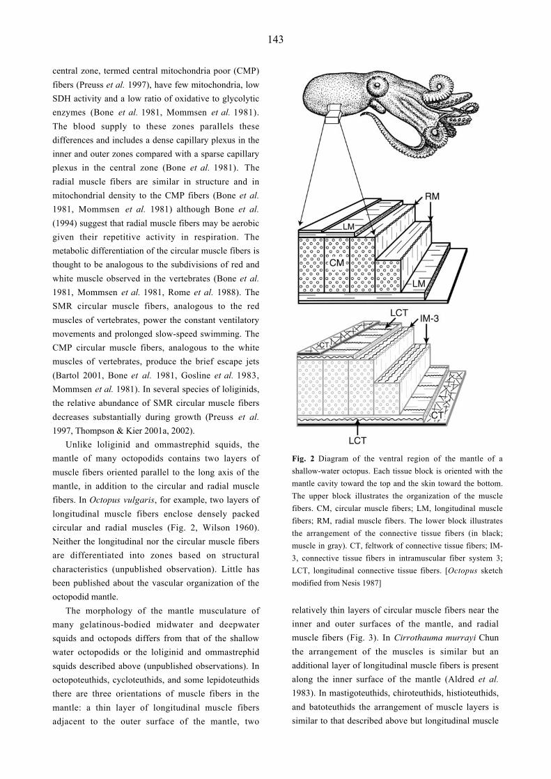
Fig. 8 Transmission electron micrographs of longitudinal
sections of the transverse muscle fibres of the tentacle (A) and
arm (B) of Loligo pealei. The extensive sarcoplasmic
reticulum (SR) of the cross-striated tentacle fibres is visible in
A. Note the short sarcomeres and short thick filaments of the
tentacle fibres in A compared with the much longer thick
filaments apparent in the obliquely striated arm fibres in B.
The scale bar length equals 1 µm. [From Kier & Curtin (2002)
with permission of the publisher]
muscle and transverse muscle (see Kier 1982, Kier &
Smith 1985, Smith & Kier 1989). As can be seen in
Figs 5, 6 and 7 the longitudinal muscles are typically
situated around the perimeter of the cross-section of the
arm or tentacle. Such a location away from the neutral
axis has mechanical significance since it results in a
greater bending moment than a more central location.
Torsion or twisting movements of the arms and
tentacles of coleoids are produced by contraction of the
helical or oblique muscle layers. The direction of
torsion (i.e., clockwise vs. counter clockwise) depends
on the handedness of the contracting helical or oblique
muscle layer. Torsion in both directions is observed in
the arms and tentacles of coleoids and both right- and
left-handed helical muscle layers are present in all
appendages examined thus far. The torsional moment
is greatest if the helical or oblique muscle layers are
located peripherally in the arm or tentacle, away from
the neutral axis and this is indeed where they are
typically located. In this regard the functional role of
the internal oblique muscle of the arms of octopuses is
perplexing, since it occupies a more central location
than the other two sets of oblique muscles.
Muscle Specialization in the Tentacles of Squids
The overall arrangement of the musculature of the arms
and tentacles of squids is quite similar (compare Figs 5
and 6), yet as described above, the functional role of
the musculature is dramatically different. In the arms,
the transverse muscle provides the support required for
the relatively slow bending movements while in the
tentacles, the transverse muscle is responsible for the
extremely rapid elongation that occurs during prey
capture.
What specializations of the tentacle muscle allow
for this dramatic difference in behavior? The most
significant difference in the transverse muscle of the
tentacles is the ultrastructure. Unlike the obliquely
striated muscle that characterizes virtually all of the
musculature of coleoids, the muscle fibers of the
transverse muscle of the tentacles are cross-striated
with unusually short sarcomeres and myofilaments
(Kier 1985, 1996, Kier & Curtin 2002) (Fig. 8). For
instance, in Loligo pealei, the thick myofilaments of
the obliquely striated muscle fibers of the arms are
approximately 7.4 mm while those in the cross striated
tentacle fibers are approximately 0.8 µm (Kier &
Curtin 2002). The remarkably short myofilaments and
sarcomeres of the tentacle fibers result in more
elements in series per unit length of fiber. Since
shortening velocities of elements in series are additive
(Huxley & Simmons 1972, Josephson 1975, van
Leeuwen 1991), this ultrastructural specialization
results in a dramatic increase in the shortening velocity
of the tentacle fibers. Indeed, recent measurements
(Kier & Curtin 2002) show the shortening velocity of
the tentacle fibers to be an order of magnitude higher
(approximately 15 L0 s-1 for the tentacle transverse
muscle versus 1.5 L0 s-1 for the arm transverse muscle
at 19ºC).
Although significant ultrastructural differences are
observed between the tentacle and arm transverse
muscle, comparison of samples of myofilament
preparations of the two muscle fiber types using
sodium dodecyl sulphate polyacrylamide gel
electrophoresis (SDS-PAGE) showed little evidence of
differences in contractile protein isoforms (Kier 1991,

155
Kier & Schachat 1992). In addition, peptide mapping
of myosin heavy chains from the two muscle fiber
types failed to demonstrate a difference between the
tentacle and arm fibers (Kier & Schachat 1992). These
biochemical studies, in conjunction with the
ultrastructural analyses provide an interesting contrast
to the situation observed in vertebrate skeletal muscle
fiber types, where the ultrastructural arrangement is
relatively invariant (Eisenberg 1983) but significant
biochemical heterogeneity is observed (see for example
Bandman 1985, Reiser et al. 1985, 1988, 1996,
Schiaffino et al. 1988, Sweeney et al. 1988). In the
squids, specialization for high shortening speed appears
to have occurred primarily through changes in the
dimensions and arrangement of the myofilament
lattice, rather than through changes in biochemistry.
An analysis of ultrastructural differentiation of the
transverse muscle of the arms and the tentacles in the
loliginid squid Sepioteuthis lessoniana showed that for
the first two weeks after hatching, the tentacle
transverse muscle fibers do not show the adult
ultrastructure and are indistinguishable from the
obliquely striated fibers of the transverse muscle of the
arms. Transverse striation of the tentacle fibers appears
at approximately 3 weeks and the adult ultrastructure is
finally present by 4-5 weeks after hatching. High-speed
video recordings of prey capture show correlated
behavioural changes. During the first 2-3 weeks after
hatching, the animals do not employ the rapid
tentacular strike; instead, they jet forward and capture
the prey with splayed arms and tentacles (Kier 1996).
This pattern of development is of interest in the context
of the presumed evolutionary history of the arms and
tentacles. Based on developmental and comparative
data, the ancestral coleoid is thought to have possessed
ten similar arm-like appendages (Naef 1921-1923,
Boletzky 1993). In the decapods (sepioids and
teuthoids) arm pair IV was modified and elaborated to
form the tentacles while in the octopods arm pair II is
believed to have been lost. The similarity in gross
arrangement between the arms and tentacles of squids
is thus not surprising in this light. In addition, the most
parsimonious hypothesis is that the ancestral muscle
striation pattern in cephalopods was obliquely striated
and the ontogenetic transition from obliquely striated to
cross-striated may reflect phylogeny (Kier 1996).
SUCKERS
The suckers of coleoid cephalopods are used for a
remarkable variety of tasks including locomotion,
anchoring the animal to the substratum, holding prey,
cleaning maneuvers, chemotactile recognition,
behavioural displays, and manipulating, sampling and
collecting objects (Packard 1988). The structure and
function of the suckers of octopuses has received the
most attention previously and so will be the focus of
the description that follows. Two major divisions of the
suckers of octopus are recognized. The exposed, disc-
like portion of the sucker, termed the infundibulum
(Girod 1884), is flattened against the surface of the
object when the sucker is attached. It is covered by a
chitinous cuticle or sucker lining and bears a series of
radial grooves and ridges (Naef 1921-1923, Nixon &
Dilly 1977, Packard 1988). A rim of loose and folded
dermis and epithelium encircles the infundibulum and
is separated from it by a circumferential groove. At the
center of the infundibulum is an orifice that opens into
a spherical cavity called the acetabulum, the second
major division of the sucker. The inner surface of the
acetabulum is covered by a continuation of the
chitinous cuticle (Hunt & Nixon 1981) that covers the
infundibulum. The radial grooves, in combination with
tiny denticles or pegs that cover the surface of the
chitinous lining of the infundibulum, may be important
in transmitting the reduced pressure in the acetabulum
underneath the infundibulum, establishing a pressure
differential over the entire area of the infundibulum
and pressing it against the substratum. This may be of
considerable importance in increasing the resistance to
shearing forces (for details see Kier & Smith 1990,
2002).
Musculature and Connective Tissues
As in the other coleoid cephalopod organs and
structures described above, a tightly packed three-
dimensional array of musculature characterizes the
suckers of octopuses (Girod 1884, Guérin 1908, Kier &
Smith 1990, Nachtigall 1974, Niemiec 1885, Tittel
1961, 1964). Three major obliquely striated muscle
fiber orientations are observed: 1) radial muscles that
traverse the wall; 2) circular muscles arranged

156
Fig. 9 Diagram of an octopus sucker. A, acetabulum; AR,
acetabular roof; AW, acetabular wall; C, circular muscle; CC,
crossed connective tissue fibers; D, dermis; E, extrinsic
muscle; EC, extrinsic circular muscle; EP, epithelium; IN,
infundibulum; IC, inner connective tissue layer; M, meridional
muscle; OC, outer connective tissue layer; R, radial muscle;
S1, primary sphincter muscle; S2, secondary sphincter muscle.
[From Kier & Smith (2002) with permission of the publisher]
circumferentially around the sucker; 3) meridional
muscles oriented perpendicular to the circular and
radial muscles (Fig 9). In the acetabulum, the radial
muscle fibers extend through the thickness of the wall,
perpendicular to the inner and the outer surface of the
acetabulum. They have their origin and insertion on
connective tissue capsules that cover the inner and
outer surfaces. The meridional muscle fibers radiate
out from a point on the apex of the acetabulum similar
to lines of longitude and extend down to a connective
tissue layer present at the junction between the
acetabulum and the infundibulum.
Circumferential muscle bundles are also present in
the acetabulum and are oriented parallel to the surface
of the infundibulum. Two robust bundles of
circumferential muscle fibers are observed adjacent to
the inner surface forming sphincter muscles
surrounding the orifice that connects the infundibulum
and acetabulum. The musculature of the wall of the
infundibulum is similar to that of the acetabulum. The
predominant feature is a robust array of radial muscle
fibers that extend between a series of circumferential
muscle bundles located adjacent to the inner surface of
the infundibular wall. Meridional muscle bundles
radiate out to the rim of the infundibulum from their
origin on a connective tissue layer between the
infundibulum and acetabulum (see Kier & Smith 1990,
2002).
The suckers are attached to the arm by a muscular
base consisting of a series of extrinsic muscle bundles
that originate on the connective tissue layer (see above)
surrounding the arm musculature. The extrinsic muscle
fibers extend down to converge on the sucker and
insert on the outer connective tissue capsule of the
acetabulum at the level of the sphincter muscle. A layer
of circumferentially arranged muscle encircles the
extrinsic muscle bundles. The extrinsic muscles orient
the sucker by selective contraction of a bundle or group
of bundles on one side, bending the muscular base.
Elongation of the base is created by the circumferential
muscle (Kier & Smith 1990, 2002).
Connective tissue capsules on the inner and outer
surface enclose the sucker musculature. The capsules
consist of fibers arranged in a crossed-fiber array (Kier
& Smith 1990). The fibers are highly birefringent and
have staining reactions typical of collagen. In addition,
the musculature of the roof of the acetabulum includes
intramuscular crossed connective tissue fibers that are
arranged obliquely to the radial muscle fibers, extend
from the outer to the inner connective tissue capsules
and are reminiscent of the intramuscular connective
tissue fibers of the mantle and fins described above.
These fibers are also highly birefringent and have
staining reactions typical of collagen.
Biomechanics of the Sucker
When a sucker attaches to an object a seal is formed at
the rim and the pressure in the acetabular cavity is
reduced. Reduction in pressure in the acetabular cavity
relies on a muscular-hydrostatic mechanism similar to
that described above for the other coleoid cephalopod
structures and organs. Contraction of the radial muscle
of the acetabulum generates a force that tends to
decrease the thickness of the wall. Since the wall
consists almost entirely of muscle and connective
tissue, its volume is essentially constant. Thinning of
the wall must therefore result in expansion of the
surface area of the acetabulum, increasing the volume
of the acetabular cavity in a manner analogous to
expansion and refilling of the mantle cavity of coleoids
by contraction of radial muscle fibers. If the sucker is
sealed to a surface however, the cohesiveness of water
157
in the acetabular cavity resists significant expansion
and a decrease in pressure in the cavity balances the
expansive force of the radial muscle of the acetabular
wall. Because of the high bulk modulus of water in the
acetabular cavity, it behaves mechanically like a solid
in tension. The radial muscles of the acetabulum are
antagonized by the meridional and circumferential
muscle bundles which, upon contraction, decrease the
circumference and thereby increase the thickness of the
acetabular wall (Kier & Smith 1990, 2002).
The infundibulum of the sucker is responsible for
formation of a watertight seal and must therefore be
flexible and dexterous in order to conform to the wide
variety of shapes and textures of objects to which the
suckers attach. As in the acetabulum, the movements
and deformations depend on a muscular-hydrostatic
mechanism. Radial muscle contraction thins the
infundibular wall and increases the diameter of the
infundibulum. The radial muscles are antagonized by
the meridional muscles, which upon contraction
decrease the diameter of the infundibulum. Contraction
of the circumferential muscle bundles constricts the
infundibulum to a conical shape. It is likely that
simultaneous contraction of the meridional and radial
muscles flattens the infundibulum and bends its rim
towards the acetabulum. A muscular-hydrostatic
mechanism may be particularly advantageous because
highly localized and complicated bends and
deformations may be produced at any location in the
infundibulum (Graziadei 1962, Graziadei & Gagne
1976a, b, Kier & Smith 1990, 2002). The infundibulum
can thus be deformed to closely match the contours of
the surface and provide a watertight seal.
The muscular-hydrostatic mechanism can generate
large pressure differentials between the ambient
pressure and pressure inside the sucker, but it appears
to require constant muscle contraction. It is common,
however, to observe an octopus remain attached for
many hours to an object or to the wall of an aquarium.
This may imply that the suckers possess a mechanism
of elastic energy storage that could be used to maintain
sub-ambient pressures without significant muscle
activity. Such a mechanism is suggested by the
presence of intramuscular crossed connective tissue
fibers in the roof of the acetabulum. As described
above for the mantle, the intramuscular connective
tissue fibers will be strained if force is applied to the
sucker roof that increases its thickness. The connective
tissue fibers are collagenous and thus they have high
resilience and can store significant strain energy. Once
the force that thickens the acetabular wall is removed,
the strain energy stored in the fibers exerts a force that
thins the roof and tends to expand the acetabular
cavity. Long-term attachment may therefore be
provided if the acetabular roof is first thickened by
contraction of the meridional and circular muscles,
storing elastic energy in the intramuscular fibers,
followed by sealing of the sucker to an object. When
the meridional and circular muscles relax, the energy
stored in intramuscular fibers will then exert a force
tending to thin the wall, reducing the pressure in the
acetabular cavity. Since the water filling the sucker has
a high bulk modulus, the volume change in the
acetabular cavity during this process is likely to be very
small. Indeed, the tensile properties of water have
important general implications for the attachment
forces of the suckers (see Smith 1991, 1996).
Decapod Suckers
The suckers of decapod coleoids differ from those of
the octopodids described above because they are
attached to the arms or tentacles by a muscular stalk or
pedicle. The stalk includes connective tissue and
longitudinal and transverse muscle fibers that allow
elongation, shortening and bending motions similar to
that described above for the arms (Guérin 1908). In
addition, the acetabulum is lined with a stiff inner
cylinder of chitin that is often equipped with tooth-like
projections that may aid attachment and prevent shear
(Hunt & Nixon 1981, Nixon & Dilly 1977). The wall
and roof of the acetabulum and infundibulum includes
radial, meridional and circular muscles (Guérin 1908)
that presumably operate in a manner analogous to that
proposed above for the octopodid suckers (Nixon &
Dilly 1977). In addition, the muscular roof of the
acetabulum is often arranged as “piston” within the
stiff cylinder such that tension on the stalk pulls the
piston back against the resistance of the water (Naef
1921-1923, Smith 1996). Thus, the greater the tension
on the stalk, the greater the pressure differential and the
greater the attachment force (Smith 1996). There is
considerable variation in the form of decapod suckers
including forms that show both radial and bilateral
symmetry, and in some groups (e.g. enoploteuthids and
onychoteuthids) the suckers may be replaced by

158
proteinaceous claws or teeth (Nixon & Dilly 1977).
SUMMARY AND PERSPECTIVES
A remarkable diversity of coleoid cephalopod
structures and organs are characterized by dense
musculature that includes tightly packed bundles and
sheets of muscle fibers arranged in three mutually
perpendicular directions. This arrangement of muscle,
termed a “muscular hydrostat” resembles the
hydrostatic skeleton of soft-bodied invertebrates and
provides both the force and the skeletal support that is
required for movement and locomotion. The overview
provided above also emphasizes the crucial role played
by fibrous collagenous connective tissues in these
systems. The connective tissues transmit the force of
muscular contraction, control shape change, and in
several cases store elastic energy in a manner that may
reduce the costs of locomotion, movement, or
adhesion.
Although we have a basic understanding of the
arrangement and function of the musculature of the
coleoid body in common near-shore forms such as the
loliginid and ommastrephid squids, some sepioids, and
some octopodids, we have relatively little detailed
information on the diverse pelagic cephalopods that are
less easily sampled. The range of mantle structure in
the pelagic forms summarized above serves to
emphasize the importance of future comparative
studies in providing us with an accurate picture of the
diversity of muscle structure and function in coleoids.
The complexity of the arrangement of the
musculature and the lack of rigid elements and joints
provides these structures with the potential to produce
more complicated and varied movements than
observed in animal structures that rely on more
conventional skeletal support systems. A more highly
subdivided and complicated neuromuscular control
system is required for a muscular-hydrostat that has the
potential to bend with multiple degrees of freedom at a
number of locations. We know relatively little,
however, about neural control of movement in these
structures, and this therefore also remains as an
important area for future research (see Matzner et al.
2000, Sumbre et al. 2001, Yekutieli et al. 1998).
Finally, the lack of rigid elements and joints implies
that different mechanisms must be used for mechanical
amplification of the force, displacement, and velocity
of muscle contraction. This may require different
mechanisms of muscle fiber specialization from that
observed previously in animals with rigid skeletons.
Indeed, muscle fiber specialization in the tentacles of
decapods (described above) is unusual because it
primarily involved changes in the dimensions and
arrangement of the myofilaments, rather than in their
biochemistry. It is unclear, however, whether this
represents a general mechanism of muscle
specialization for cephalopods and additional studies
on the ultrastructure, biochemistry and molecular
biology of cephalopod muscle are thus needed as well.
ACKNOWLEDGEMENTS
The authors thank the organizers of the symposium,
Coleoid Cephalopods Through Time: Neontological
Approaches to their Paleobiology in Light of the Fossil
Record for a stimulating interdisciplinary conference.
We thank S. Whitfield for assistance with the figures
and S. Guarda for technical assistance. We also
acknowledge support from NASA (NAG5-8759) to
WMK and NIH-SPIRE Postdoctoral Fellowship
(National Institute of General Medical Sciences –
Grant No. GM000678) to JTT.
REFERENCES
Aldred RG, Nixon M, Young JZ (1983) Cirrothauma
murrayi Chun, a finned octopod. Phil Trans R Soc
Lond, B 301: 1-54
Bandman E (1985) Myosin isoenzyme transitions in
muscle development, maturation, and disease. Int
Rev Cytol 97: 97-131
Bartol IK (2001) Role of aerobic and anaerobic circular
mantle muscle fibers in swimming squid:
electromyography. Biol Bull 200: 59-66
Baskin RJ (1967) Changes of volume in striated
muscle. Am Zool 7: 593-601
Bidder A, Boycott BB (1956) Pelagic Mollusca:
swimmers and drifters. Nature (Lond.) 177: 1023-
1025
159
Boletzky Sv (1993) The arm crown in cephalopod
development and evolution: a discussion of
morphological and behavioral homologies. Amer
Malac Bull 10: 61-69
Bone Q, Pulsford A, Chubb AD (1981) Squid mantle
muscle. J Mar Biol Assoc UK 61: 327-342
Bone Q, Brown ER, Travers G (1994) On the
respiratory flow in the cuttlefish Sepia officinalis. J
Exp Biol 194: 153-165
Bone Q, Brown ER, Usher M (1995) The structure and
physiology of cephalopod muscle fibres. In: Abbott
NJ, Williamson R, Maddock L (eds) Cephalopod
Neurobiology. Oxford University Press, New York,
pp 301-329
Boycott BB (1958) The cuttlefish-Sepia. In: Johnson
ML, Abercrombie M, Fogg GE (eds) New Biology.
The Campfield Press, St. Albans, pp 98-118
Budelmann BU, Schipp R, Boletzky Sv (1997)
Cephalopoda. In: Harrison FW, Kohn AJ (eds)
Microscopic Anatomy of Invertebrates. Mollusca II
(6A). Wiley-Liss, Inc, New York, pp 119-414
Colasanti G (1876) Anatomische und Physiologische
Untersuchungen über den Arm der Kephalopoden.
Arch Anat Physiol Wissenschaftliche Med, pp. 480-
500
Curtin NA, Woledge RC, Bone Q (2000) Energy
storage by passive elastic structures in the mantle of
Sepia officinalis. J Exp Biol 203: 869-878
Denton EJ (1974) On buoyancy and the lives of
modern and fossil cephalopods. Proc R Soc Lond B
185: 273-299
Denton EJ, Gilpin-Brown JB (1959) Buoyancy of the
cuttlefish. Nature 184: 1330-1331
Denton EJ, Gilpin-Brown JB (1961) The distribution of
gas and liquid within the cuttlebone. J Mar Biol
Assoc UK 41: 365-381
Donovan DT, Toll RB (1988) The gladius in coleoid
(Cephalopod) evolution. In: Clarke MR, Trueman
ER (eds) The Mollusca (12). Paleontology and
Neontology of Cephalopods. Academic Press, New
York, pp 89-101
Eisenberg BR (1983) Quantitative ultrastructure of
mammalian skeletal muscle. In: Peachey LD (ed.)
Handbook of Physiology, Section 10, Skeletal
Muscle. American Physiological Society, Bethesda,
Maryland, pp 73-112
Gilly WF, Hopkins B, Mackie GO (1991)
Development of giant motor axons and neural
control of escape responses in squid embryos and
hatchlings. Biol Bull 180: 209-220
Girod P (1884) Recherches sur la peau des
céphalopodes. La ventouse. Arch Zool Exp Gen 2:
379-401
Gosline JM, Shadwick RE (1983a) Molluscan collagen
and its mechanical organization in squid mantle. In:
Hochachka PW (ed.) The Mollusca (1). Metabolic
Biochemistry and Molecular Biomechanics.
Academic Press, New York, pp 371-398
Gosline JM, Shadwick RE (1983b) The role of elastic
energy storage mechanisms in swimming: an
analysis of mantle elasticity in escape jetting in the
squid, Loligo opalescens. Can J Zool 61: 1421-
1431
Gosline JM, Steeves JD, Harman AD, Demont ME
(1983) Patterns of circular and radial mantle muscle
activity in respiration and jetting of the squid Loligo
opalescens. J Exp Biol 104: 97-109
Graziadei P (1962) Receptors in the suckers of
Octopus. Nature 195: 57-59
Graziadei P (1965) Muscle receptors in cephalopods.
Proc R Soc Lond B Biol 161: 392-402
Graziadei P (1971) The nervous system of the arms. In:
Young JZ (ed.) The Anatomy of the Nervous
System of Octopus vulgaris. Oxford University
Press (Clarendon), London and New York, pp 45-
61
Graziadei PPC, Gagne HT (1976a) Sensory innervation
in the rim of the octopus sucker. J Morphol 150:
639-679
Graziadei PPC, Gagne HT (1976b) An unusual
receptor in the octopus. Tissue & Cell 8: 229-240
Guérin J (1908) Contribution a l'étude des systèmes
cutané, musculaire et nerveux de l'appareil
tentaculaire des céphalopodes. Arch Zool Exp Gén
8: 1-178
Harris JE, Crofton HD (1957) Structure and function in
the nematodes: Internal pressure and cuticular
structure in Ascaris. J Exp Biol 34: 116-130
Hoar JA, Sim E, Webber DM, O'Dor, RK (1994) The
role of fins in the competition between squid and
fish. In: Maddock L, Bone Q, Rayner JMV (eds)
Mechanics and Physiology of Animal Swimming.
Cambridge University Press, Cambridge, pp 27-43

160
Hunt S, Nixon M (1981) A comparative study of
protein composition in the chitin-protein complexes
of the beak, pen, sucker disc, radula and
oesophageal cuticle of cephalopods. Comp
Biochem Physiol 68B: 535-546
Huxley AF, Simmons RM (1972) Mechanical
transients and the origin of muscular force. Cold
Spring Harbor Symp Quant Biol 37: 669-680
Johnsen S, Kier WM (1993) Intramuscular crossed
connective tissue fibres: skeletal support in the
lateral fins of squid and cuttlefish (Mollusca:
Cephalopoda). J Zool Lond 231: 311-338
Josephson RK (1975) Extensive and intensive factors
determining the performance of striated muscle. J
Exp Zool 194: 135-154
Kier WM (1982) The functional morphology of the
musculature of squid (Loliginidae) arms and
tentacles. J Morphol 172: 179-192
Kier WM (1985) The musculature of squid arms and
tentacles: ultrastructural evidence for functional
differences. J Morphol 185: 223-239
Kier WM (1988) The arrangement and function of
molluscan muscle. In: Trueman ER, Clarke MR
(eds) The Mollusca (11). Form and Function.
Academic Press, New York, pp 211-252
Kier WM (1989) The fin musculature of cuttlefish and
squid (Mollusca, Cephalopoda): morphology and
mechanics. J Zool (Lond) 217: 23-38
Kier WM (1991) Squid cross-striated muscle: the
evolution of a specialized muscle fiber type. Bull
Mar Sci 49: 389-403
Kier WM (1996) Muscle development in squid:
ultrastructural differentiation of a specialized
muscle fiber type. J Morphol 229: 271-288
Kier WM, Curtin NA (2002) Fast muscle in squid
(Loligo pealei): contractile properties of a
specialized muscle fibre type. J Exp Biol 205:
1907-1916
Kier WM, Schachat FH (1992) Biochemical
comparison of fast- and slow-contracting squid
muscle. J Exp Biol 168: 41-56
Kier WM, Smith AM (1990) The morphology and
mechanics of octopus suckers. Biol Bull 178: 126-
136
Kier WM, Smith AM (2002) The structure and
adhesive mechanism of octopus suckers. Integr
Comp Biol 42: 1146-1153
Kier WM, Smith KK (1985) Tongues, tentacles and
trunks: the biomechanics of movement in muscular-
hydrostats. J Linn Soc Lond Zool 83: 307-324
Kier WM, Smith KK, Miyan JA (1989)
Electromyography of the fin musculature of the
cuttlefish Sepia officinalis. J Exp Biol 143: 17-31
Kier, WM, van Leeuwen JL (1997) A kinematic
analysis of tentacle extension in the squid Loligo
pealei. J Exp Biol 200: 41-53
MacGillivray PS, Anderson EJ, Wright GM, Demont
ME (1999) Structure and mechanics of the squid
mantle. J Exp Biol 202: 683-695
Marceau F (1905) Recherches sur la structure des
muscles du manteau des Céphalopodes en rapport
avec leur mode du contraction. Trav Lab Soc Sc
Arachon Ann 8: 48-65
Matzner H, Gutfreund Y, Hochner B (2000)
Neuromuscular system of the flexible arm of the
octopus: physiological characterization. J
Neurophysiol 83: 1315-1328
Milligan BJ, Curtin NA, Bone Q (1997) Contractile
properties of obliquely striated muscle from the
mantle of squid (Alloteuthis subulata) and cuttlefish
(Sepia officinalis). J Exp Biol 200: 2425-2436
Mommsen TP, Ballantyne J, MacDonald D, Gosline J,
Hochachka PW (1981) Analogues of red and white
muscle in squid mantle. Proc Nat Acad Sci USA
78: 3274-3278
Nachtigall W (1974) Biological Mechanisms of
Attachment: the comparative morphology and
bioengineering of organs for linkage, suction, and
adhesion. Springer-Verlag, New York
Naef A (1921-1923) Die Cephalopoden (Systematik).
Fauna Flora Golf Napoli, 35(I-1): 1-863 [English
translation, Jerusalem, Israel program for scientific
translations, available from Smithsonian Institution
Libaries, Washington, DC, 20560, USA]
Nesis KN (1987) Cephalopods of the World. TFH
Publications, Inc, Neptune City, New Jersey
Niemiec J (1885) Recherches mophologiques sur les
ventouses dans la règne animal. Rech Zool Suisse
2: 1-147
Nigmatullin ChM, Arkhipkin AI, Bizikov VA (1991)
Functional and evolutionary analysis of the
statolith, gladius and locomotor-apparatus
morphology in squids of the family
Ommastrephidae. Bull Mar Sci 49: 666

161
Nixon M, Dilly PN (1977) Sucker surfaces and prey
capture. Symp Zool Soc Lond 38: 447-511
Norman M (2000) Cephalopods: A World Guide.
Conch Books, Hackenheim, Germany
O’Dor RK (1988) The forces acting on swimming
squid. J Exp Biol 137: 421-442
O'Dor RK, Webber DM (1986) The constraints on
cephalopods: why squid aren't fish. Can J Zool 64:
1591-1605
Otis TS, Gilly WF (1990) Jet-propelled escape in the
squid Loligo opalescens: concerted control by giant
and non-giant motor axon pathways. Proc Nat Acad
Sci USA 87: 2911-2915
Packard A (1988) The skin of cephalopods (Coleoids):
general and special adaptations. In: Trueman ER,
Clarke MR (eds) The Mollusca (11). Form and
Function. Academic Press, Inc, San Diego, pp 37-
67
Packard A, Trueman ER (1974) Muscular activity of
the mantle of Sepia and Loligo (Cephalopoda)
during respiratory movements and jetting, and its
physiological interpretation. J Exp Biol 61: 411-419
Preuss T, Lebaric ZN, Gilly WF (1997) Post-hatching
development of circular mantle muscles in the
squid Loligo opalescens. Biol Bull 192: 375-387
Reiser PJ, Greaser ML, Moss RL (1988) Myosin heavy
chain composition of single cells from avian slow
skeletal muscle is strongly correlated with velocity
of shortening during development. Dev Biol 129:
400-407
Reiser PJ, Greaser ML, Moss RL (1996) Contractile
properties and protein isoforms of single fibres
from the chicken pectoralis red strip muscle. J
Physiol 493: 553-562
Reiser, PJ, Moss RL, Giulian GG, Greaser ML (1985)
Shortening velocity and myosin heavy chains of
developing rabbit muscle fibers. J Biol Chem 260:
14403-14405
Rome LC, Funke RP, Alexander RMcN, Lutz G,
Aldridge H, Scott F, Freadman M (1988) Why
animals have different muscle fibre types. Nature
(Lond) 335: 824-827
Russell FS, Steven GA (1930) The swimming of
cuttlefish. Nature (Lond) 125: 893
Schiaffino S, Ausoni S, Gorza L, Saggin L, Gundersen
K, Lomo T (1988) Myosin heavy chain isoforms
and velocity of shortening of type 2 skeletal muscle
fibres. Acta Physiol Scand 134: 575-576
Seibel BA, Thuesen EV, Childress JJ (2000) Light-
limitation on predator-prey interactions:
consequences for metabolism and locomotion of
deep-sea cephalopods. Biol Bull 198: 284-298
Smith AM (1991) Negative pressure generated by
octopus suckers: a study of the tensile strength of
water in nature. J Exp Biol 157: 257-271
Smith AM (1996) Cephalopod sucker design and the
physical limits to negative pressure. J Exp Biol 199:
949-958
Smith KK, Kier WM (1989) Trunks, tongues and
tentacles: moving with skeletons of muscle. Am Sci
77: 28-35
Sumbre G, Gutfreund Y, Fiorito G, Flash T, Hochner B
(2001) Control of octopus arm extension by a
peripheral motor program. Science 293: 1845-1848
Sweeney HL, Kushmerick MJ, Mabuchi K, Sréter FA,
Gergely J (1988) Myosin alkali light chain and
heavy chain variations correlate with altered
shortening velocity of isolated skeletal muscle
fibers. J Biol Chem 263: 9034-9039
Sweeney MJ, Roper CFE, Mangold KM, Clarke MR,
Boletzky Sv (eds) (1992) “Larval” and juvenile
cephalopods: A manual for their identification.
Smiths Contr Zool 513: 1-282
Tateno S, Tsuchiya Y, Katayama K, Hamada T (1992)
Fiberscopic observation of mantle-funnel complex
motion in Octopus vulgaris. Reprinted from
Scientific Papers of the College of Arts and
Sciences, The University of Tokyo 42: 161-178
Thompson JT, Kier WM (2001a) Ontogenetic changes
in fibrous connective tissue organization in the
Oval Squid, Sepioteuthis lessoniana Lesson, 1830.
Biol Bull 201: 136-153
Thompson JT, Kier WM (2001b) Ontogenetic changes
in mantle kinematics during escape-jet locomotion
in the Oval Squid, Sepioteuthis lessoniana Lesson,
1830. Biol Bull 201: 154-166
Thompson JT, Kier WM (2002) Ontogeny of squid
mantle function: Changes in the mechanics of
escape-jet locomotion in the Oval Squid,
Sepioteuthis lessoniana Lesson, 1830. Biol Bull
203: 14-26
Tittel K (1961) Der funktionelle Aufbau des
Tintenfischarmes im Vergleich mit dem
Muskelkörper der Säugerzungen. Verh Anat Ges
57: 264-275
162
Tittel K (1964) Saugnapf-, epi- und hypofasciale
Armmuskulatur der Kephalopoden - ein Beitrag zur
funktionellen Anatomie freibeweglicher
Skelettmuskelkörper. Gegenbaurs Morphol Jahrb
106: 90-115
Toll RB (1988) Functional morphology and adaptive
patterns of the teuthoid gladius. In: Trueman ER,
Clarke MR (eds) The Mollusca (11). Form and
Function. Academic Press, New York, pp 167-182
van Leeuwen JL (1991) Optimum power output and
structural design of sarcomeres. J Theor Biol 149:
229-256
van Leeuwen JL, Kier WM (1997) Functional design
of tentacles in squid: linking sarcomere
ultrastructure to gross morphological dynamics.
Philos Trans R Soc Lond B Biol Sci 352: 551-571
Vogel S (1987) Flow-assisted mantle cavity refilling in
jetting squid. Biol Bull 172: 61-68
Ward DV, Wainwright SA (1972) Locomotory aspects
of squid mantle structure. J Zool (Lond) 167: 437-
449
Wells MJ (1978) Octopus. Physiology and Behaviour
of an Advanced Invertebrate. Chapman and Hall,
London
Williams LW (1909) The Anatomy of the Common
Squid Loligo pealii, Lesueur. EJ Brill, Leiden
Wilson DM (1960) Nervous control of movement in
cephalopods. J Exp Biol 37: 57-72
Yekutieli Y, Hochner B, Flash T (1998) Three-
dimensional reconstruction of octopus arms during
motion as a tool for studying motion control.
Neurosci Lett Suppl 51: S44
Young JZ (1938) The functioning of the giant nerve
fibres of the squid. J Exp Biol 15: 170-185
Zuev GV (1966) Characteristic features of the structure
of cephalopod molluscs associated with controlled
movemen t s . Eko logo-Mor fo log ichesk ie
Issledovaniya Nektonnykh Zhivotnykh. Kiev,
Special Publication. (Canadian Fisheries and
Marine Services Translation series 1011, 1968)
Received: 13 March 2003 / Accepted: 23 July 2003