Embed Size (px)
Citation preview
Acta Musei Nationalis Pragae, Series B, Historia Naturalis, 50(1994)(1-4): 13-24 issued December 1995
MUSCLE ATTACHMENT AREAS IN THE SILURIANBELLEROPHONTACEAN GASTROPODS BELLEROPHON SCABER(PERNER) AND BUBOVICUS TARDUS (BARRANDE IN PERNER)
RADVAN J. HORNYDepartment of palaeontology, National Museum, 11579 Praha 1, Czech Republic
Horny, R. J. (1995): Muscle attachment areas in the Silurian bellerophontaceangastropods Bellerophon scaber (PERNER) and Bubovicus tardus (BARRANDE in PERNER). - Acta Mus. Nat. Pragae , Ser. B, Hist. Nat.,50(1994)(1 -4): 13-24. Praha. ISSN 0036-5343
Abstract. Two symmetrical umbilical muscle attachment areas are describedon internal moulds of the bellerophontaceans Bellerophon scaber (pERNER,1903) and Bubovicus tardus (BARRANDE in PERNER, 1903) from the Si-lurian of Bohemia. For the first time, myostracum is reported covering theattachment area, continuously increasing throughout the shell ontogeny . Someearlier records of muscle scars are discussed in this connection. Mode of fossilisation and later chemical processes are considered important for the shapeand appearance of the preserved muscle attachments.
• bellerophontacean gastropods, muscle attachment areas, myostracum, Silurian, Bellerophon, Bubovicus
Received May 06, 1993
The presence of a pair of retractor muscle scars in the umbilical area of a bellerophontiformshell seems to indicate most reliably its assignment to the real Bellerophontina and, therefore,to the Class Gastropoda. Knight (1947) was the first to describe these scars in BellerophonMONTFORT, 1808 and Sinuites KOKEN, 1896, pointing out their systematic importance.According to Peel (1982), scars of this type are frequently found on the internal moulds ofCarboniferous species of Bellerophon from Ireland and United Kingdom. All other finds arerare and exceptional. To date, retractor muscle insertions have been reported and describediu Sinuites (Knight 1947, Peel 1980, Runnegar 1981, Horny 1992a,b), Sylvestrosphaera PEEL,1980 (Peel 1980), Bellerophon (Knight 1947, Peel 1972, 1982), Salpingostoma ROEMER,1876 (Peel 1972), Tremanotus HALL, 1865 (Peel 1972), Megalomphala ULRICH in ULRICHet SCOFIELD, 1897 (Peel 1976; 1991b), Strangulites HORNY, 1962 (Horny 1990a, b), andCarinaropsis HALL, 1847 (Peel 1993). Following the previous descriptions of muscle scarsin Sinuiies and Strangulites from the Ordovician, the Silurian Bellerophon and BubovicusHORNY, 1962 are here added to the list of genera with ascertained circumbilical retractorscars from the Barrandian area (Central Bohemia).
Bellerophon scaber (PERNER, 1903) was originally described as Tremagyrus scaber PERNER, 1903. Tremagyrus PERNER, 1903 was later (Knight et al. 1960) synonymized withTremanotus HALL, 1865, but the type species of Tremagyrus was assigned to Bellerophon(Bellerophon) by Horny in 1963a. It is stratigraphically the oldest representative of Bellerophon from the Barrandian area.
Bubovicus, based on Cymbularia tarda BARRANDE in PERNER, 1903, is a rare Siluriangenus, a member of the Family Bellerophontidae McCOY , 1851. It is related to BellerophonMONTFORT, 1808 from which it distinguishes itself by the laterally compressed , high,moderately phaneromphalous shell with a narrow slit, generating a narrow raised selenizoneon a crest- Iike angulation.
With the exception of a few specimens of B. scaber from the Listice locality near Beroun,
13
both rare species have been collected to date at only one locality, the Barrande's excavationina pine-tree wood between the town Lodenice and the village Bubovice (a protected locality"Cernidla"). The excavation was situated in a local, about 20 m long, lens-like accumulationof benthic shelly fauna mixed with volcanic material, in a sequence of alternating limestonebeds and shales with volcanic admixture, respectively (uppermost Wenlockian; Homerian,Zone Cyrtograptus lundgreni, Sub-zone Testograptus testis). Some museum specimens bearBarrande's handwriting in black ink, indicating the locality (Bubowitz). The exact locationof the locality was lost for about a hundred years. It was rediscovered in the 1950s and sincethat time some more material has been collected, but only from a small old collectors' dump.The fossiliferous lens - or at least its favourably weathered subsurface part - seems to havebeen totally quarried and exploited.
At present, there are about 40 specimens of both species, including the types, availablein the collections of the Department of Palaeontology, National Museum, Prague. Morespecimens might be still found associated with the samples of other fauna, dispersed in thecollections. They are all to some extent fragmentary, either due to short transport beforedeposition or to not enough care during collection. The carbonatic, seemingly recrystallized,whitish to yellowish shell wall is rather fragile, cracking perpendicularly to the shell surface;it is easily removable from the internal mould by the use of a vibro-tool. Many specimenspreserve geopetal calcite fills. This is important for study of the muscle impression structureswhich cannot be easily observed on the normal coarse, grey-green carbonatic tuffaceousmatrix.
Environmental conditions and biology
Both species occur in a shallow marine assemblage. This high-energy environment wasstrongly influenced by contemporary volcanism. Tuffaceous shales with irregularly beddedlimestones containing variable amounts of volcanic products predominate . The fossiliferouslens represented a rapid accumulation of the remains of animals and plants which originallyinhabited a variety of environments near the volcanic islands - solid substrata in high -energyconditions to clayey volcanic deposits in local temporary shelters. Fossils are chaoticallydeposited, often fragmentary, adults with juvenile shells, and mixed with small unsortedshell fragments, tephra, and sharply angulated fragments or even small pebbles of tuffaceousshales. Gastropods predominate, with the variety of shell form indicating variable livingconditions: a high percentage of trochiform and lenticular pleurotomariins, bellerophontins,holopeids, platyceratids, oriostomatids, loxonematins, subulitids, and others. Most of theisostrophic shells have either the left or right side filled with matrix and the other side withcrystallized calcite, indicating their postmortal position on the bottom. Cyclotheca is a characteristic genus, and the problematic Ceratotheca adunca (BARRANDE, 1867) belongs tothe most frequent fossils. Brachiopods form the second abundant group (e. g. Leptaena,Pentlandina, Strophonella, Coolinia and others). Crinoid columnals , tabulate corals, bryozoans, and hyolithids are common, while trilobites are rare and nautiloids, bivalves and othergroups occur only sporadically. Algae are common, like Acanthochonia barrandei HINDE,1884 (previously known as Ischadites), and Pachytheca (see also KHz 1992).
Bellerophon scaber probably inhabited high energy environment as an active, perhapssemi-infaunal predator. A thick shell, with radial aperture and strong retractor muscle insertions, confirms this presumption. Linsley (1977, 1978), after discussing shell form andrates of locomotion, concluded that Bellerophon was a streamlined and quite fast gastropod.According to Peel (1984), the globose, compact shell of Bellerophon, with narrow or closedumbilici, may suggest a mobile, infaunal, perhaps predatory habit comparable to some livingbullomorphs.
Bubovicus tardus, possessing a laterally compressed, high, phaneromphalous shell withpseudo-tangential aperture could be probably classified as a slower, but still active epibenthicinhabitant following Linsley's scheme, possibly an algal grazer in less dynamic conditions .This presumed mode of life would be in accordance with the thinner shell and much weakermuscle insertions than in Bellerophon scaber.
14
Morphology
Genus BELLEROPHON Montfort, 1808
Type species: Bellerophon vasulites MONTFORT, 1808; Middle Devonian, Germany.
Bellerophon scaber (PERNER, 1903)PI. 1; 2; 3; 4, figs 1-5
1903 Tremagyrus scaber PERNER; Perner, Text-figs 91, 92 (pp. 129, 130) (holotype L 5790).1903 Cymbularia Bacchus PERNER; Perner, PI. 87:20 (paralectotype L 5793); 87:35, Text-figs 107a,f
(p. 153) (lectotype L 5792); Text-figs 107b,c,d (p. 153) (paralectotype L 5794); Text-fig. 107e(p. 153) (paralectotype L 30431); unfigured specimen, originally labelled as additional type toText-fig. 107 (p. 153) (paralectotype L 5795).
1903 Cymbularia verrucosa PERNER; Perner, PI. 87:15-18, Text-figs 106a,b (p. 152) (lectotypeL 5791); 87:19 (paralectotype L 5844).
1941 Tremagyrus scaber PERNER; Knight, p. 352, PI. 13:la,b.1960 Tremanotus scaber (PERNER); Knight et al., Treatise I, 1, p. /180 .1963a Bellerophon (B.) scaber (PERNER); Horny, pp. 125, 126, PI. 37: 1-6, PI. 46:5.
Description. A detailed description was published by Perner (1903), Knight (1941) and Horny(1963a). It should only be added that the species has a radial aperture and that the shells donot show the asymmetry pointed out by Perner (1903). All other additions concern structuresconnected with the muscle insertions, observed in six specimens: L 30427, L 30428, L 5794,L 5793, L 30429, and L 5795.
Specimen L 30427 (PI. 1, figs 3-6), an internal mould of a juvenile, has the left sidemostly filled with crystallized calcite, the right side with grey-green matrix. Length 13.8mm; height 10.2 mm.
On the left side, a shallow, narrow circumbilical groove, delimiting the dorsal margin ofthe muscle attachment area, is located just below the whorl angulation of the internal mouldpreserved in whitish crystallized calcite. The groove, regularly following the coiling spiral,is transversely asymmetrical, deepest near its addorsal, steepest slope. It is filled with brownish-grey myostracal deposit, observable as a narrow , dark, smooth, flat band, 0.05 - 0.25mmwide (adapically narrowest). The band-like deposit is locally exfoliated and the depth of thegroove is thus exposed. Adaperturally, the band widens into a small islet of dark grey myostracum, originally covering the whole insertion plane, and extending down onto the umbilicalwall of the whorl. The adapertural margin of the muscle area, where the myostracal depositwas thinnest, is indistinct. The whole observed muscle attachment structure occupies about310 degrees of a volution, the distance between the anterior margin of the insertion and theaperture being 210-220 degrees, i.e. slightly over a half of a whorl. The surface of theumbilical part of the internal mould near the aperture bears characteristic foliated structure,fading out addorsally and adapically towards a smooth area, about 1 mm long, in front ofthe muscle impression. A fibrillar structure is preserved on a small area adjacent addorsallyto the circumbilical groove. Structures on the right side of the shell are symmetrical but lesswell preserved.
Specimen L 30428 (PI. 3, figs 1-3), an internal mould of a not fully mature individual,lacks the apertural part; it is filled with dark grey limestone. Height 13.3 mm.
The left side shows the proximal part of the muscle area with a well exposed addorsalgroove. It is mostly empty and longitudinally striated, but with a patch of myostracum distally.Adaperturally, the groove deviates from the isometric spiral, the scar widens addorsaly andthe internal mould becomes slightly laterally inflated at its periphery. The adapertural partof the umbilical wall shows foliated structure, gradually fading out and disappearing about2 mm before the indistinct anterior margin of the muscle attachment area. A straight groove-like repaired injury obliquelly crosses the left dorsolateral area between the dorsal medianline and the muscle attachment area interrupting its addorsal ridge . The right side of thisspecimen was not prepared .
15
Specimen L 5794 (PI. 3, fig. 6) is a fragment of a not fully mature individual with damagedapertural part; the right side preserves shell , but the left side exposes the internal mould ofwhitish coarsely crystallized calcite. The right side is filled with greenish limestone witha volcanic component. Height 13.4mm. This specimen was figured by Perner (1903, Text-fig.107c, d).
The adapertural part of the muscle attachment area on the left side is damaged. Centraland distal parts are sharply delimited by a 0.2 mm wide circumbilical groove, which deviatesadaperturally such that the widened functional attachment area is located on the slightlyinflated portion of the lateral side of the internal mould.
Specimen L 5793 (PI. 3, fig. 5) represents an almost mature , incomplete specimen withoutaperture; the left side preserves the shell, while the right side exhibits the internal mould ofmainly crystallized calcite. Height 15.7 mm. This specimen was figured by Perner (1903,Text-fig. 107b).
The right side shows well developed inflation of the whorl in place of the functionalmuscle attachment area. The area itself was not prepared in order to preserve the originalappearance of the specimen.
Specimen L 30429 (PI 4, figs 1, 2) is an internal mould of an almost mature individual,filled with greenish limestone with a volcanic component; the spire is partly filled withcrystallized calcite. The narrow, 8 mm long slit is well exposed. Length 18.0 mm; height15.8 mm.
The muscle attachment area on the right side lacks myostracal deposits. The circumbilicalgroove is best preserved in the adapical part, where it is 0.30 mm wide. The anterior marginis indistinct, distant about 220 - 230 degrees of a volution back from the aperture. Thelocation of the functional part of the muscle scar is indicated by inflation of the internalmould. Foliated structure is visible on the surface of the internal mould near the aperture,locally obliterated by a layer with fibrillar structure. The area immediately adapertural ofthe muscle insertion area is smooth. The opposite side of the specimen was not prepared tosave the patches of shell.
Specimen L 5795 (PI. 2, figs 1-6; PI. 3, fig. 4; PI. 4, figs 3 - 5) represents an internalmould of a mature individual with patches of shell , mostly filled with green-grey limestonewith a volcanic component and fragments of fauna. The spire is partly filled with crystallizedcalcite. Length 24 mm; height 18 mm. This specimen represents an additional specimen toPerner (1903, Text-fig. 107).
The left side in this adult specimen has the best preserved muscle attachment area. Theadapertural part is mostly covered with dark grey myostracal deposits, maximum 0.05 mmthick, but these are lost in the narrower adapical part. The circumbilical groove, originallylocated below the whorl angulation, gradually deviates from its spiral course in a dorsoanteriordirection, extending over the inflated area of the whorl. The sharply delimited addorsalmargin of the myostracal deposit continues anteriorly to form a convex crescent-like edgelocally followed by a thin stria at a distance of about 0.1 mm. After turning adumbilically,down, it extends slightly obliquely over the umbilical wall as a darker grey film disappearingunder the unprepared shell wall, towards the median plane of the shell. The surface of themyostracal deposit, originally adherent to the shell wall, is uneven, irregular, locally bearingfibrillar structure; it is without crescentic structures but two short ridges follow the addorsalmargin. The deposit is slightly raised above the surrounding surface of the mould; its variablethickness reaching a maximum of about 0.05 mm. An area where the myostracal deposithas exfoliated shows an uneven, rough surface, with one incomplete crescentic structure.This surface seemingly represents a real adhering surface of the muscle attachment. Theadapical part of the insertion area, free of deposit, shows the circumbilical groove as wellas relics of an extremely thin, grey layer, preserved between the umbilical wall of the mouldand the remains of shell; this seemingly represents the abandoned parts of thinner , ontogeneticaly younger myostracal deposit. A smooth area adapertural to the muscle attachmentarea, about 3 mm long, gradually passes into foliated structure, well developed on the adapertural part of the internal mould. The distance between the anterior margin of the scar andthe aperture is about 250 - 260 degrees of a volution. The opposite side of the mould hasbilaterally symmetrical, but less well preserved, structures.
16
Recapitulation
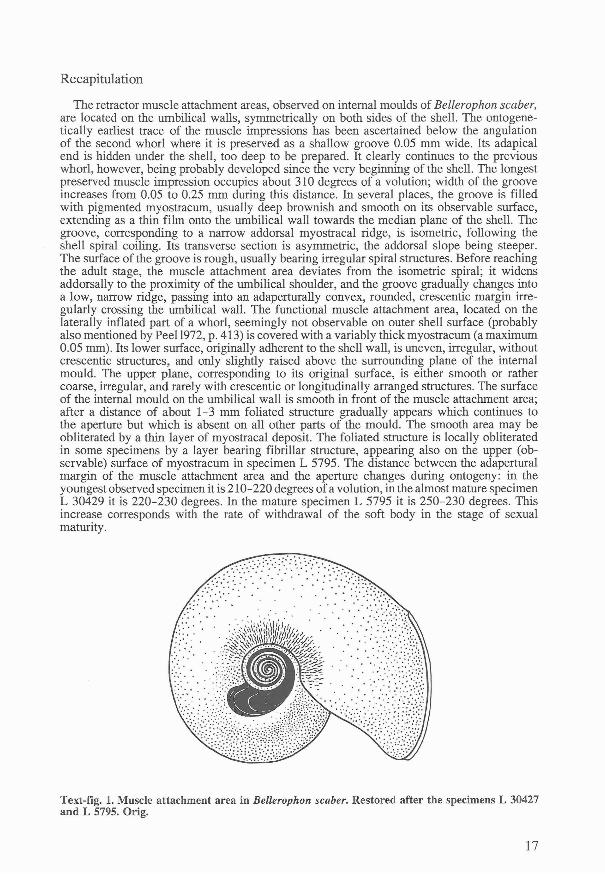
The retractor muscle attachment areas, observed on internal moulds of Bellerophon scaber,are located on the umbilical walls, symmetrically on both sides of the shell. The ontogenetically earliest trace of the muscle impressions has been ascertained below the angulationof the second whorl where it is preserved as a shallow groove 0.05 mm wide. Its adapicalend is hidden under the shell, too deep to be prepared. It clearly continues to the previouswhorl, however, being probably developed since the very beginning of the shell. The longestpreserved muscle impression occupies about 310 degrees of a volution; width of the grooveincreases from 0.05 to 0.25 mm during this distance. In several places, the groove is filledwith pigmented myostracum, usually deep brownish and smooth on its observable surface,extending as a thin film onto the umbilical wall towards the median plane of the shell. Thegroove, corresponding to a narrow addorsal myostracal ridge, is isometric, following theshell spiral coiling. Its transverse section is asymmetric, the addorsal slope being steeper.The surface of the groove is rough, usually bearing irregular spiral structures. Before reachingthe adult stage, the muscle attachment area deviates from the isometric spiral; it widensaddorsally to the proximity of the umbilical shoulder, and the groove gradually changes intoa low, narrow ridge, passing into an adaperturally convex, rounded, crescentic margin irregularly crossing the umbilical wall. The functional muscle attachment area, located on thelaterally inflated part of a whorl, seemingly not observable on outer shell surface (probablyalso mentioned by Peel 1972,p. 413) is covered with a variably thick myostracum (a maximum0.05 mm). Its lower surface, originally adherent to the shell wall, is uneven, irregular, withoutcrescentic structures, and only slightly raised above the surrounding plane of the internalmould. The upper plane, corresponding to its original surface, is either smooth or rathercoarse, irregular, and rarely with crescentic or longitudinally arranged structures. The surfaceof the internal mould on the umbilical wall is smooth in front of the muscle attachment area;after a distance of about 1-3 mm foliated structure gradually appears which continues tothe aperture but which is absent on all other parts of the mould . The smooth area may beobliterated by a thin layer of myostracal deposit. The foliated structure is locally obliteratedin some specimens by a layer bearing fibrillar structure, appearing also on the upper (observable) surface of myostracum in specimen L 5795. The distance between the adaperturalmargin of the muscle attachment area and the aperture changes during ontogeny: in theyoungest observed specimen it is 210-220 degrees of a volution, in the almost mature specimenL 30429 it is 220-230 degrees. In the mature specimen L 5795 it is 250-230 degrees. Thisincrease corresponds with the rate of withdrawal of the soft body in the stage of sexualmaturity.
Text-fig. 1. Muscle attachment area in Bellerophon scaber. Restored after the specimens L 30427and L 5795. Orig.
17
Genus BUBOVICUS Horny, 1962
Type species: Cymbularia tarda PERNER, 1903, Silurian, Bohemia.
Bubovicus tardus (BARRANDE in PERNER, 1903)PI. 4, fig. 6; PI. 5; PI. 6, figs 1-5
1903 Cymbularia tarda BARRANDE; Perner, PI. 87:32-34, 36, Text-fig. 105c (p. 150) (lectotypeL 5787); Text-fig. 105a (p. 150) (paralectotype L 5788); Text-fig. 105b (p. 150) (paralectotype30424); unfigured paralectotype L 30437.
1936 Cymbularia impressa n. sp.; Riha, p. 6, 7, PI. 1:9, 10 (holotype L 30438).1962 Bubovicus tardus (PERNER); Horny, p. 474.1963a Bubovicus tardus (PERNER); Horny, p. 121, 122, PI. 37:7, 8, PI. 38:6, 7.
Description. A detailed description was given by Perner (199~) and Horny (1963a). To thesynonomy of this species belongs also Cymbularia impressa RIHA, 1936, by error originallyreported from the Ordovician strata. A careful study of all available material proved thata weak spiral dorsolateral angulation exists in one specimen only, L 5789, figured by Horny(1963, PI. 37:7, 8). It probably represents an individual feature, not characteristic for thewhole population of the species. The position of the aperture is near to tangential. All otheradditions concern structures connected with the muscle insertions and were observed in sixspecimens: L 30432, L 30433, L 30434, L 30435, L 30436, and L 5789.
Specimen L 30432 (PI. 4, fig. 6; PI. 6, fig. 1) is an internal mould of a juvenile withpatches of shell; the left side was not prepared to preserve the shell. The mould is obliquelyfilled with crystallized calcite (the right side) and with tuffaceous limestone (the left side).Length 19.4 mm; height 16.2 mm.
The muscle attachment area on the right side is not delimited by a groove but can beobserved as a smooth area addorsally adjoining an otherwise coarse surface of the internalmould below the whorl angulation. Weak grooves accompany the addorsal margin. Theaddorsal circumbilical margin lies just below the umbilical shoulder, without deviation fromthe coiling spiral in its anterior part. The region between the indistinct, anterior margin ofthe attachment area and the aperture bears foliated structure which is less well observablein the proximity of the muscle scar. Estimated distance between the anterior margin of theattachment area and aperture is 180 degrees of a volution .
Specimen L 30433 (PI. 5, fig. 3) is a juvenile with the right side mostly with shell, theleft side is preserved as an internal mould filled with greenish tuffaceous limestone. Length24.0 mm; height 19.0 mm.
The surface of the internal mould on the left side is coarse but with observable structures.The adapical part of the attachment area is delimited addorsaly by an obscure, rounded, low,spiral ridge located more adumbilically below the angulation than in L 30345. The functionalpart of the attachment area is indistinctly delimited ; it is covered with a thin colourless shelllayer without details on its surface. The anterior margin is unclear, probably adaperturalyconvex; weak foliated structure is developed slightly in front of the margin. The distancebetween the anterior margin and aperture is estimated to about 180 degrees of a volution.
Specimen L 30434 (PI. 6, fig. 2) is a weathered internal mould with patches of shell ofa juvenile; the left side is partly filled with crystallized calcite while the right side is preservedin grey limestone. Height 14.3 mm. The functional part of the muscle attachment area onthe left side is located below the whorl angulation. The addorsal margin is delimited bya circumbilical groove, exposed in the adapical part. Most of the attachment area is coveredwith a thin layer bearing reflected, transversely oriented, foliated structure on its observablesurface. The layer covers the whole exposed umbilical wall, extending to near the medianshell plane. The anterior margin of the attachment area is almost indistinct, but is locatedat about 200 degrees of a volution from the aperture; the area in front of the scar is smooth.The traces of myostracum attached to the area are brown due to higher concentration oflimonite.
18
Specimen L 30435 (PI. 5, figs 4-6) represents the internal mould of an immature specimen,with small patches of shell. The left side is partly filled with crystallized calcite, the rightside with greenish tuffaceous limestone. Length 26.0 mm; height 21 mm.
On the left side, a shallow circumbilical groove is visible below the shoulder in the exposedadapical part; it approaches the whorl angulation adaperturally and fades away in the functionalpart of the muscle attachment area, the anterior part of which is not clearly delimited. Thesurface of the area is covered with scattered traces of a thin myostracal layer, locally showingtransversely arranged foliated structure on its surface. Below this layer obscure, irregular ,fine spiral striae are locally visible. The myostracal layer extends over the umbilical wall,where it is preserved as small islets even in the adapica l part, locally with fine and weakfibrillar structure on its surface. In the most adapically exposed part, a short oblique linecrosses the circumbilical groove (seemingly not a part of the scar). The whole muscle attachment area is slightly brown coloured, especially the islets of myostracum.
Specimen L 30436 (PI. 6, figs 3-5) is the internal mould of a mature specimen with patchof shell showing the selenizone on a crest. The left side of the mould is partly filled withcrystallized calcite, the right side with greenish tuffaceous limestone. The specimen carriesBarrande's inscription in black ink: Bubow. (= loco Bubovice). Length 26.0 mm; height 23.0mm.
The muscle attachment area on the left side is well marked by brownish colouration, butrather weakly in terms of relief. The circumbilical ridge is weak, just observable on theaddorsal margin of the functional attachment, where it is located well below the whorl angulation. The anterior margin is only slightly marked by the extent of thin myostracal deposit,extending also deeply down onto the umbilical wall. The area in front of the attachmentarea is smooth over a distance of about 3 mm, after which fine, dense, foliated structuregradually appears. The anterior margin of the attachment area is about 270 degrees of a volution back from the aperture . The umbilical area of the left side is filled with shell.
Specimen L 5789 (PI. 5, figs 1-2) represents the largest specimen with an observablemuscle attachment area. It is an internal mould with a large proportion of preserved shell;the left side with a visible muscle attachment area is partly filled with crystallized calcite,other parts with 'greenish tuffaceous limestone. Length 28,6 mm; height 26.5 mm. Specimenfigured by Horny (1963, PI. 37, figs 7, 8).
The adapical part of the muscle attachment area is not exposed. The extent of the musclearea is not sharply delimited, but it is observable as a structure deviating from the circumbilicalspiral and extending to the shoulder. Rather small islets of very thin myostracal layer remainon its surface; the whole area bears dense, fine transverse foliated structure which continuesadaperturally without interruption. The inferred anterior margin is about 270 degrees ofa volution back from the aperture.
Recapitulation
Fundamentally, almost all morphological features of Bubovicus tardus connected with themuscle attachment are similar to those described in Bellerophon scaber, but they are weakerand less well marked. This undoubtedly reflects the different shape of the shell, its thinnerwall and assumed different mode of life. In immature specimens, the position of the isometricspiral groove on the upper part of the umbil ical wall varies slightly in different specimensbut always lies below the umbilical angulation. At maturity, the groove deviates, and themuscle attachment area widens, almost reaching the rounded umbilical shoulder. The functional part of the attachment area is indistinctly delimited, mostly definable only by theextent of rather thin myostracum, either colourless or brownish or grey according to highercontent of limonite or carbon. The myostracum covers the whole exposed umbilical wall,extending adumbilically to the proximity of the median shell plane. Its lower surface, originallyadherent to the shell wall, is seemingly smooth but often bears the impression of transverselyoriented foliated structure from the originally underlying shell layer. The upper plane of themyostracum, corresponding to its original surface, bears weak, irregular, spiral grooves. Noinflation of the internal mould connected with the location of the final mature attachmentwas observed . The surface of the umbilical wall of the internal mould between the attachment
19
area and the aperture bears fine, dense, transversely oriented, foliated structure, seeminglybecoming finer with age, absent on other parts of the internal mould and, in some specimens,probably obliterated with thin myostracal deposit for a short distance (1-3 mm) adaperturalto the attachment area. Fibrillar structure is weak and rarely preserved. The distance betweenthe adapertural margin of the muscle attachment area and aperture varies during ontogeny:it is rather small in the juveniles, about 180 degrees but increases to 270 degrees in maturity.
Discussion
Muscle scars in bellerophontacean gastropods have been described previously only oninternal moulds. Their preservation and study are, however, influenced by many factors,ranging from the mode of life of the animal , its age, shell composition and structure to thegeological processes itself. Finally, we must also take account of the methods of preparation .
Knight (1947) characterized molluscan muscle scars as depressions in the inner layer ofthe shell. Common features of bellerophontacean muscle scars, as we can observe them oninternal moulds (i. e., with opposite relief to the shell interior) are usually raised areas orridges, or depressed areas and grooves, or, rarely, both possibilities combined. All to datedescribed muscle attachment areas in four specimens of Bellerophon (Knight 1947, Peel1972, 1982) are, as a matter of fact, preserved as depressions associated with raised structures,like ridges, swellings etc. This mode of preservation is also well demonstrated on severalspecimens of the Bohemian Bellerophon scaber. The depressed area located below (adumbilically) the addorsal ridge was originally filled with myostracum. In immature shells, thislayer was provided with an asymmetric spiral crest generating a circumbilical groove on theinternal mould near the addorsal scar periphery. In maturity, the peripheral crest deviatedfrom the isometric spiral, passing into the thickest addorsal margin of the myostracal deposit.The circumbilical groove on the internal mould is thus gradually replaced by a step or a ridge(or other associated structures observed in the Upper Palaeozoic bellerophonts by Peel 1972,equating with channels or ridges on the shell interior). The myostracum deposit in matureBellerophon scaber is about 0.05 mm thick; it was slightly raised above the shell interiorand similarly slightly depressed in the underlying shell. Muscle fibres were thus attached toa slightly raised area and not to myostracum in a depression like in many other molluscs(e.g., bivalves or tergomyans) . Evaluation of the implication of these observations and associated shell microstructure in terms of functional anatomy and perhaps even bellerophontacean systematics remains for future study. Characteristic is the transversely striated layerwhich seemingly served for fixing the myostracum on the shell wall; its presence on theumbilical wall is probably the most important criterion for recognizing bellerophontaceangastropods among the bellerophontiform molluscs in cases when the muscle scars are notobservable.
Myostracum in the Bohemian material is often coloured. This could probably be connectedwith its complex-prismatic structure or organic admixture. Brown colour testifies to a highercontent of limonite (originally pyrite) and grey colour to carbon (probably bitumen). Similardark grey or even black structures have been reported in the Silurian Archaeopraga pinnaeformis (PERNER, 1903) (Horny 1963) and are well known in the Ordovician tergomyanArchaeophiala antiquissima (HISINGER, 1837). Rosov (1975) reported pigmented calciticlayer covering the surface of the muscle scars in Nyuella bjalyi ROSOV, 1975.
Myostracum, reported here in the Silurian species of Bellerophon (and Bubovicus), wasdefinitely developed also in Carboniferous species of the genus. This is in accordance withthe statement of MacClintock (1967, p. 50), that "For each isolated accessory scar in themollusk shell, there is a lath-shaped blade of shell material starting at the apex of the shelland gradually expanding to the outcrop area on the inner surface of the shell." This can bewell demonst rated in a juvenile specimen of Bellerophon scaber (L 30427, PI. 1, figs 3-6),where a narrow band of dark brown myostracum extends and gradually widens adaperturallyfor a visible distance of almost a whorl.
Preservation of myostracal deposits is rather rare. It could have happened in chemicallyfavourable conditions where the shells were quickly buried in shallow-water deposit andrapid crystallization of calcium carbonate took place. In the majority of cases, the innermostlayers of the shell, together with myostracum, were the first to undergo dissolution . Different
20
phases of this process were described by Horny (1992b) in Sinuites from the Ordovician ofBohemia (Horny, however, did not explain the process of destruction of the continuousmigration tracks as connected with dissolvement of myostracum). Peel (1991a) suggestedthat differences in the shape of muscle scars in sinuitids and those in Bellerophon may beillusory, since described specimens of the latter genus with muscle scars are of Carboniferousage and therefore significantly younger than the sinuitids (p. 25). The find of muscle attachment scars in a Silurian representative of Bellerophon, similar to those in the Carboniferousspecimens, cannot help to solve the problem. In my opinion, attachment areas in both ofthese different, but related, groups of bellerophontaceans are fundamentally similar (see alsoPeel 1991, p. 25) but selective dissolution of myostracum contributed in their apparent dissimilarity (Horny 1992b, pp. 97, 98; Text-fig. 5, p. 85; PI. 1, figs 6-11).
The muscle attachment area of Bellerophon is principially similar to that of some pleurotomariaceans (see also Peel, 1986). In an undetermined Silurian specimen of .Euryzone"sp., labelled as Pleurotomaria (Euryzone) consolans BARR. by Perner, the final attachmentarea lies at the rounded circumbilical angulation, addorsally accompanied by narrow, raisedstructures deviating from the isometric spiral (PI. 6, fig. 6). The distance between the adapertural margin of the muscle attachment area and the aperture is about 180 degrees in thisspecimen (like in the juveniles of Bubovicus tardus or in the Upper Palaeozoic representativesof Bellerophon).
The recognition of the muscle attachment area in the Silurian Bellerophon offers a possibility for comparison with its Upper Palaeozoic representatives. Unfortunately, our knowledgeof this most popular genus is rather poor and we could easily find that we are comparingadmittedly related, but different, genera with similar shell morphology, representinga well-tested form that existed for considerable time span, more than 200 million years. Thetype species, B. vasulites from the Middle Devonian of Germany, seems to be not too farmorphologically from the Bohemian Silurian species. On the contrary, the Upper Palaeozoicheavy thick-shelled species with wide selenizone could represent another genus . Main differences include the depth of location of the muscle attachment areas within the shell, whichis 230 degrees back from the aperture in the Silurian species but about 180 degrees in theUpper Palaeozoic material. The position of the circumbilical groove is below the umbilicalshoulder in the Silurian species, but at the shoulder in the Upper Palaeozoic material. Abapertural projecting ridges are not developed in the Silurian material, but are well developedin the Upper Palaeozoic specimens. Associated addorsal structures, almost absent in theSilurian, are well developed in the Upper Palaeozoic material. Some of these characters maybe connected with mode of life but, in any case, available data are still too limited to evaluatetheir possible systematic significance.
Acknowledgements
Professor John S. Peel (University of Uppsala) is thanked for reviewing the manuscriptprior to publication. Jifi KHz and Ladislav Marek kindly extended their material for study.
UPONY RETRAKTORU U SILURSKYCH GASTROPODU BELLEROPHON SCABER (PERNER) ABUBOVICUS TARDUS (BARRANDE in PERNER) (SOUHRN)
V praci jsou popsany upony retraktoni u dvou silurskych zastupcu podradu Bellerophontina,Bellerophon scaber (PERNER, 1903) a Bubovicus tardus (BARRANDE in PERNER, 1903).U rodu Bellerophon MONTFORT, 1808 byly dosud znamy pouze u jedincu z rnladsihopaleozoika Velke Britanie a Severni Ameriky, u rodu Bubovicus HORNY, 1962 jsou novezjisteny. Jsou umisteny na periferii umbilikalni oblasti po obou stranach ulity. Poprve jeu techto gastropodu objeveno myostrakum, pokryvajici svalovou inserci a souvisle pfinistajiciv prubehu ontogeneze. Oba nalezy pochazeji ze svrchnowenlockych melkovodnich tufitickychvapencu z odkryvu u silnice mezi Lodenici a Bubovicemi, ve kterych bylo mimoradne vhodneprostredi pro fosilizaci doposud neznamych struktur.
21
REFERENCES
Horny, R. J. (1962): New genera of Bohemian Lower Paleozoic Bellerophontina. - Vest. Ustf. Ust.Geol., 37(6): 463 - 466.
Horny, R. J.(1963a): Lower Paleozoic Bellerophontina (Gastropoda) of Bohemia. - Sbor . geol. Ved,Paleont., 2: 57-164.
Horny, R. J. (1963b): Archaeopraga, a new problematic genus of monoplacophoran molluscs from theSilurian of Bohemia. - J. Paleont., 37(5): 1071-1073.
Horny, R. J. (1990a)~ Svalove vtisky u Sinuites (Strangulites) (Mollusca) z ceskeho ordoviku. - Cas.Nar. Muz . Praze, Rada pfirodoved., 155(1-4): 109-118.
Horny, R. J. (1990b)~ Dalsi poznatky 0 svalovych vtiscich u Sinuites (Strangulites) (Mollusca). - Cas.Nar. Muz . Praze, Rada prirodoved., 156(1-4): 46-48.
Horny, R. J. (1992a): Shell morphology and muscle scars of Sinuitopsis neglecta Perner (Mollusca,Monoplacophora) . - Cas. Nar. Muz. Praze, Rada prirodoved., 157(1-4): 81- 105.
Horny, R. J. (1992b): Svalove vtisky u rodu Sinuites (Mollusca, Gastropoda) z ceskeho spodniho ordoviku.- Cas. Nar . Muz. Praze, Rada prirodoved., 158(1-4): 79-100.
Horny, R. J. (1993): Retractor muscle scars in the Bohemian Silurian bellerophontacean gastropodsBellerophon and Bubovicus; a comparison with the muscle scar patterns in the sinuitids and thecyrtonellid tergomyans. - Palaont. Ges., 63. Jahrestagung, Abstr., Praha. 13, 14 pp.
Knight, J. B. (1941): Paleozoic gastropod genotypes. - Geol. Soc . Am., Spec . Pap. , 32: 1-510.Knight, J. B. (1947) : Bellerophont muscle scars. - J. Paleont., 21: 264-267.Knight, J. B. et al. (1960): Systematic descriptions (Archaeogastropoda), Supplement (Paleozoic and
some Mesozoic Caenogastropoda and Opisthobranchia) . - In: Moore, R. C. (ed.), Treatise on Invertebrate Paleontology, Part I (Mollusca 1). Geol. Soc. Am. and Univ. of Kansas Press. Lawrence,Il69-I332 pp.
Kfiz, J. (1992) : Silurian field excursions: Prague Basin (Barrandian), Bohemia. - Nat. Mus. Wales,Geol. Ser., 13: 1-111.
Linsley, R. M. (1977): Some "laws" of gastropod shell form. - Paleobiology, 3: 196-206.Linsley, R. M. (1978): Locomotion rates and shell form in the Gastropoda. - Malacologia, 17: 193-206.MacClintock, C. (1967): Shell structure of patelloid and bellerophontid gastropods (Mollusca). - Peabody
Mus. Nat. Hist. Bull., 22: 1-140.Peel, J. S. (1972): Observations on some Lower Palaeozoic tremanotiform Bellerophontacea (Gastropoda)
from North America. - Palaeontology, 15: 412 -422.Peel, J. S. (1976): Musculature and systematic position of Megalomphala taenia (Bellerophontacea,
Gastropoda) from the Silurian of Gotland. - Bull. geol. Soc. Denmark, 25: 49- 55.Peel, J. S. (1980): A new Silurian retractile monoplacophoran and the origin of the gastropods. - Proc.
Geol. Ass., 91: 91 -97.Peel, J. S. (1982): Muscle scars in Bellerophon recticostatus (Mollusca) from the Carboniferous of
Ireland. - J. Paleont., 56: 1307-1310.Peel, J. S. (1984): Autecology of Silurian gastropods and monoplacophorans. - Spec. Pap. Palaeontology,
32: 165-182.Peel, J. S. (1986): Muscle scars in Porcellia (Gastropoda; Pleurotomariacea) from the Carboniferous of
England. - Bull . geol. Soc. Denmark, 35: 53-58.Peel, J. S. (1991a): The Classes Tergomya and He1cionelloida, and early molluscan evolution. - Bull.
Grcnlands geol. Unders., 161: 11-65.Peel, J. S. (1991b): Salpingostomatiform and related bellerophontacean gastropods from Greenland and
the Baltic region. - Bull. Gre nlands geol. Unders., 161: 67-116.Peel, J. S. (1993) : Muscle scars and mode of life of Carinaropsis (Bellerophontoidea, Gastropoda) from
the Ordovician of Termessee . - J. Paleont., 67(4) : 528-534.Perner, J. (1903): Gasteropodes, 1. - In: Barrande, J.: Systeme silurien du centre de la Boheme, 4.
Prague, 164 pp .Rosov, S. N. (1975): Novyj otrjad monoplakofor. - Paleont. Z., 1975(1) : 41-45.Runnegar, B. (1981): Muscle scars, shell form and torsion in Cambrian and Ordovician univalved mo
lluscs. - Lethaia, 14: 311-322.Riha, A. (1936): 0 novych bellerophontidech z ceskeho paleozoika. - Rozpr. Ces. Akad. Ved Urn. (II. Tf.),
46(12): 1-13.
22
EXPLANATION OF THE PLATES
All specimens are deposited in the collections of the Department of Palaeontology, Museum of NaturalHistory, National Museum, Prague . If not otherwise stated, covered with ammonium chloride sublimate.
PLATE 1
Bellerophon scaber (PERNER, 1903)
1-2. L 5791, adult specimen, dorsal and left umbilical views. Note the thick shell, narrow umbilicusand the periodical increments, otherwise uncommon in this species. x 2.5.
3-6. L 30427, internal mould of an initial part of a juvenile with partly prepared spire to show thenarrow circumbilical groove, locally filled with brownish-grey myostracum. 3, 4 - not whitenedto show the distinct colouring, 3 - x 3, 4 - x 13; 5 - whitened to show the relief, x 13; 6 - enlargedgroove with preserved parts of myostracum, x 20.
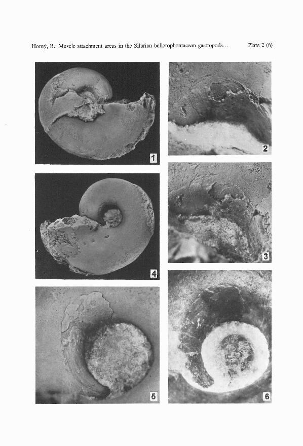
PLATE 2
Bellerophon scaber (PERNER, 1903)
1-6. L 5795, adult specimen, mostly internal mould. 1 - left side, x 2.5. 2, 3 - two views of the rightmuscle attachment area, mostly covered with myostracum, x 10; 4 - right side, x 2.5; 5 - leftmuscle attachment area, adapically without myostracum but with well exposed groove, x 10;6 - the same, not whitened to show the dark grey coloured myostracum, x 10.
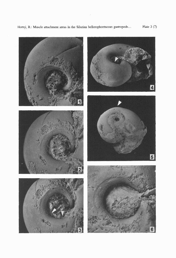
PLATE 3
Bellerophon scaber (PERNER, 1903)
1-3. L 30428, immature specimen, internal mould with a straight injury in the dorsal region. 1 - dorsolateral, 2; 3 - right umblical views in different light; a smooth area (arrowed in 3) is the placeof the muscle attachment. Note the injury crossing the addorsal muscle attachment margin . All x 6.
4. L 5795, internal mould with an inflated whorl above the muscle attachment area (arrowed), x 2.5.5. L 5793, internal mould of an immature specimen with an inflated whorl above the muscle attachment
area (arrowed). x 2.5.6. L 5794, internal mould with a deep, striated, circumbilical groove and patches of myostracum
partly covering the muscle attachment area. x 8.
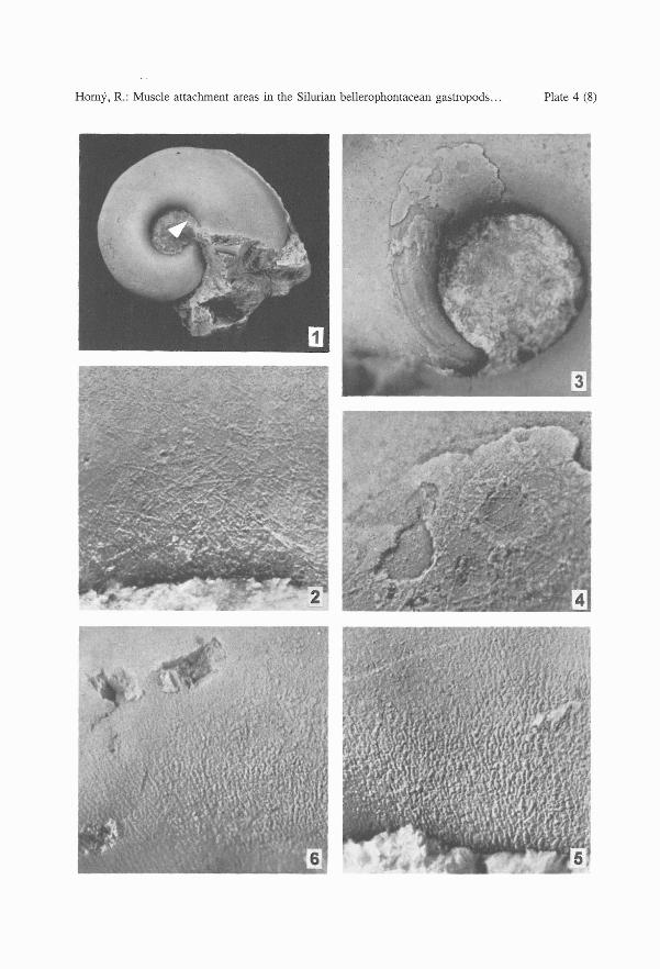
PLATE 4
Bellerophon scaber (PERNER, 1903)
1,2. L 30429, internal mould of an immature specimen. 1 - right side with a long slit, x 2.5; 2 - umbilicalwall near the aperture (arrowed in fig. 1). Lower side of the layer with fibrillar structure whichobliterate the internal shell surface with foliated structure. x 35.
3-5. L 5795. 3, internal mould, muscle attachment area mostly with preserved myostracum near theadapertural margin, x 10; 4, the same, enlarged to show the myostracum covered with fibrillarlayer on its lower irregular surface (originally adherent to the shell wall), x 35; 5 - umbilical wallbetween the muscle impression area and the aperture, with an impression of a coarse foliatedstructure. x 35
Bubovicus tardus (BARRANDE in PERNER, 1903)
6. 30432, right umbilical wall of a juvenile with an impression of foliated structure, x 25.
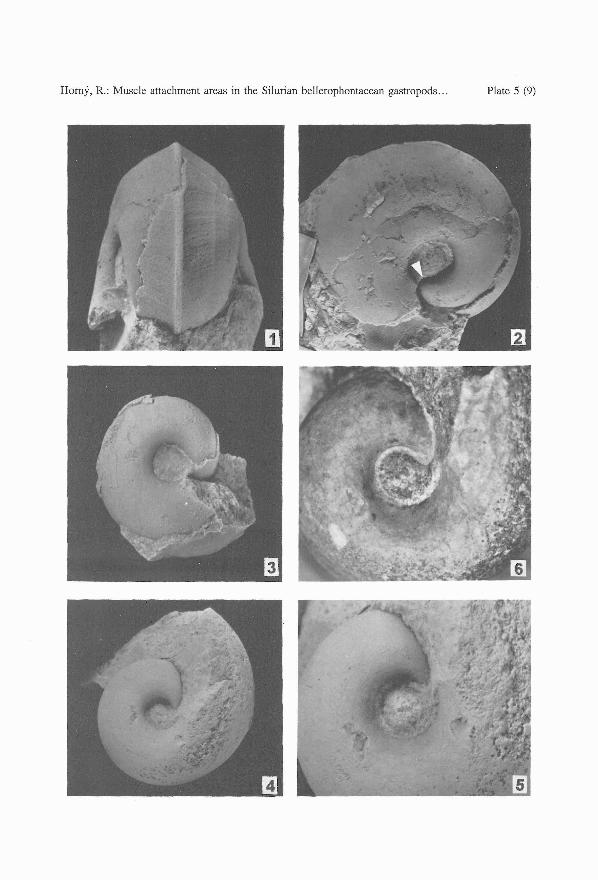
PLATE 5
Bubovicus tardus (BARRANDE in PERNER, 1903)
1-2. L 5789, a mature specimen with patches of shell. 1 - posterodorsal view; 2 - left umbilical view;the circumbilical ridge is visible at the prepared apical end of the whorl (arrowed). x 2.
3. L 30433, left umbilical view of an internal mould of a juvenile, with a low circumbilical ridge(to the left of the umbilicus) . x 2.
4-6. L 30435, left umbilical view of an immature specimen (4, x 2), with a well visible sharp circumbilicalgroove (5, x 4), and with scattered traces of dark brown myostracum delimiting the muscle attachment area (6, x 4, not whitened) .
23
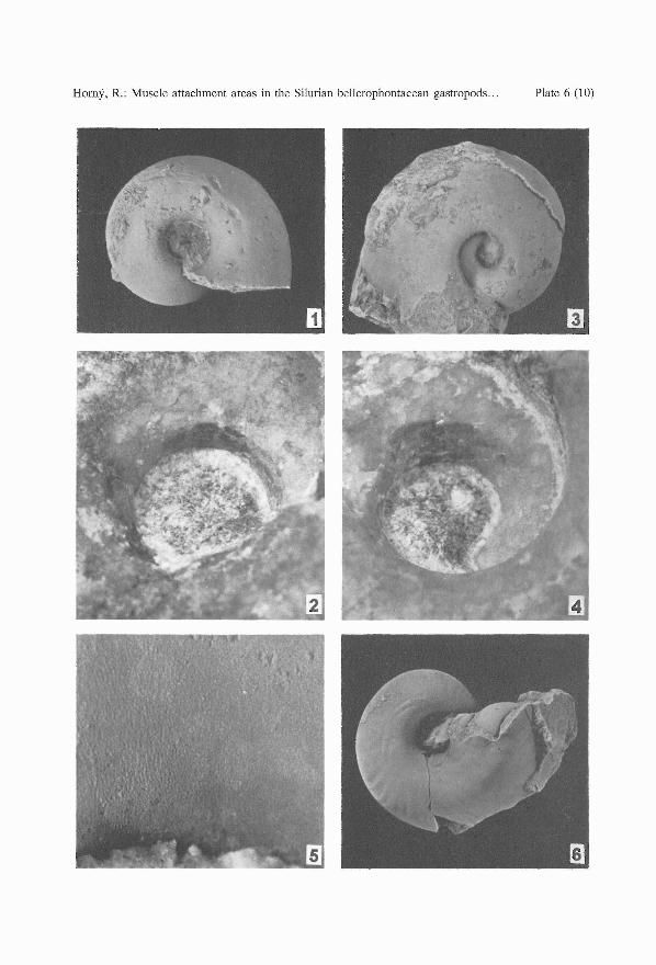
PLATE 6
Bubovicus tardus (BARRANDE in PERNER, 1903)
1. L 30432, right umbilical view of a juvenile, internal mould with ill-defined muscle attachmentstructures (to the left of the umbilicus), x 3.
2. L 30434, umbilical wall of an internal mould of a juvenile with dark patches of myostracum atthe attachment area, x 10, not whitened.
3-5. L 30436, a mature specimen, mostly internal mould, left umbilical view. 3 - x 2; 4 - muscleattachment area, well marked by brownish colouring, x 8, not whitened; 5 - foliated shell structureon the umbilical wall between the anterior margin of the muscle attachment area and the aperturalmargin, x 25.
"Euryzone" sp., labelled by Perner as Pleurotomaria (Euryzone) consolans BARR.; Silurian, probablyKopanina Formation, Lochkov
6. L 29618, basal view of an internal mould with preserved muscle attachment macrostructures (tothe left of the umbilicus), x 3.
24
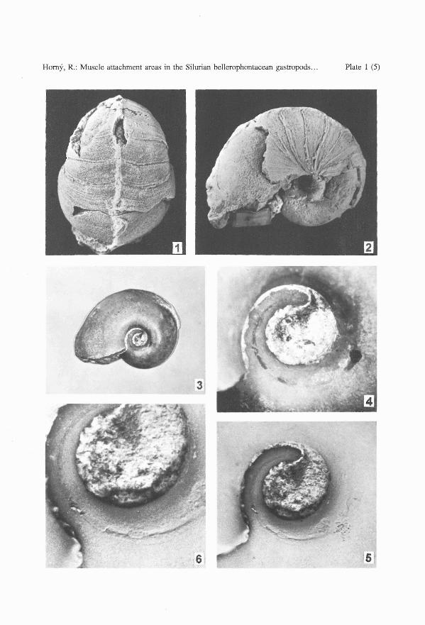
Horny, R.: Muscle attachment areas in the Silurian bellerophontacean gastropods . .. Plate 1 (5)
3
5
Horny, R.: Muscle attachment areas in the Silurian bellerophontacean gastropods .. . Plate 2 (6)
Horny, R.: Muscle attachment areas in the Silurian bellerophontacean gastropods . .. Plate 3 (7)
Horny , R.: Muscle attachment areas in the Silurian bellerophontacean gastropods . . . Plate 4 (8)
Horny, R.: Muscle attachment areas in the Silurian bellerophontacean gastropods .. . Plate 5 (9)
Horny, R.: Muscle attachment areas in the Silurian bellerophontacean gastropods ... Plate 6 (10)