Embed Size (px)
Citation preview
27/08/2013
1
Learning Outcome B4
Is the most abundant tissue type is widely
distributed throughout the body.
Found in blood, under skin, in bone and
around many organs.
Connects, binds together parts of the
body.
Also functions in support and protection,
allows the storage of fat and the transport
of substances.
Most connective tissues share two similar
characteristics:
• Both have a good blood supply.
Ligaments, tendons and cartilage are also types of
connective tissue but they do not have a blood
supply.
This is the reason why injuries to these areas heal
very slowly.
Have an abundance of intracellular matrix.
Intercellular Matrix – is what makes types
of connective tissue so different.
Located outside cells and fills spaces
between cells.
Cells secrete matrix and it fills
intercellular spaces.
Hardness of intercellular matrix varies
from cell to cell.
Matrix may be liquid as in blood, gel-like as
in fat tissue or hard as in bone.
Amount of matrix also varies between cell
types.
In fat tissue the cells are close together;
with very little intercellular matrix.
Bone and cartilage have very few cells and
large amounts of intercellular matrix.
Also in the matrix of most connective tissue
is protein fibers:
Collagen – strong and flexible but only
slightly elastic, white in color.
Elastin – not very strong but stretchy,
yellow in color.
Reticular fibers (fine collagen) – also
strong and very flexible
27/08/2013
2
Used to remove unwanted wrinkles and
lines
Use cattle collagen or patient’s collagen
from hips, thighs or abdomen.
Injected under skin and fills unwanted
wrinkles
Loose (areolar) Connective Tissue
Is beneath skin and most epithelial tissue
Also found between muscle
Functions to bind together, cushions,
protects and acts as tissue glue.
Made up of collagen and elastic fibers in an
intercellular matrix.
Soft and protects and cushions many organs.
Holds organs in position.
A layer underlies all mucous membranes.
Adipose Connective Tissue Is located beneath skin (subcutaneous
layer), around the heart and kidneys, behind the eyeballs.
Functions to cushion insulate and stores fat.
A type of loose connective tissue that supports fat.
Can insulate body and prevent heat loss. Deposited around certain organs.
• Example holds kidneys in place.
Dense fibrous Connective Tissue Makes up tendons, ligaments, and skin
(dermis). Functions to bind structures together. Contains many collagen and elastic fibers. The main type of fiber is collagen; it forms
strong supporting structures: tendons, ligaments and fascia. • Tendons are cord-like structures that attach
muscle to bone. • Ligaments are dense fibrous connective tissue
that cross joints and attach bone to bone.
Ligaments contain more elastic fibers than tendons; therefore have a greater ability to stretch.
Ability to stretch: prevents tearing of ligaments when joints bend. • Fascia are bands and/or sheets of dense
fibrous connective tissue.
Covers muscle, blood vessels and nerves.
Functions to cover, support and anchors the organs to nearby structures.
27/08/2013
3
Tendons and ligaments can tear and
stretch with excessive stretching.
Example a torn Achilles tendon causes
loss of movement because tendons attach
the leg muscle to the heel.
Reticular Connective Tissue
Makes lymphoid tissue; make up lymph
nodes, spleen and bone marrow.
Functions to form internal framework of
lymphoid organs.
Cartilage
Formed by cells known as chondrocytes.
Chondrocytes secrete a protein that is
put into the intercellular matrix, this
allows the matrix to be firm, smooth and
flexible.
Although the matrix is solid it is not as
hard as the matrix of bone.
Most cartilage is covered by a
perchondrium.
Perichondrium is a layer of connective
tissue that carries blood vessels to the
cartilage.
The perichondrium supplies oxygen and
nutrients to the cartilage.
Cartilage does not have its own blood
supply.
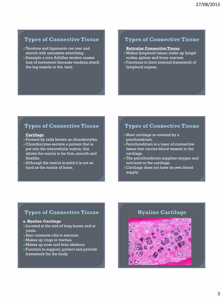
a. Hyaline Cartilage
Located at the end of long bones and at
joints.
Also connects ribs to sternum
Makes up rings in trachea
Makes up nose and fetal skeleton
Function to support, protect and provide
framework for the body.
27/08/2013
4
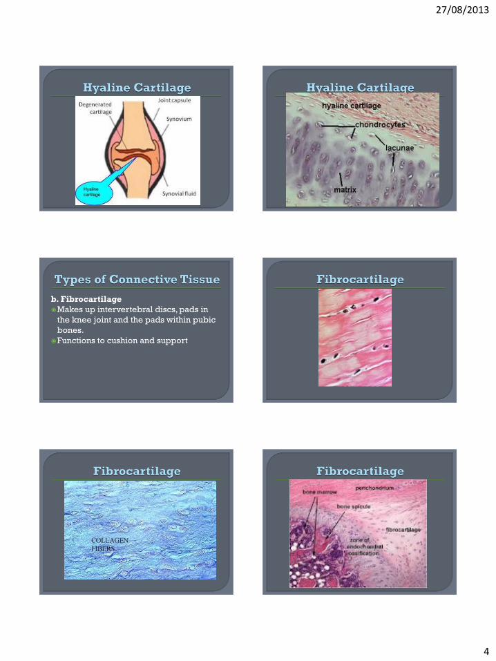
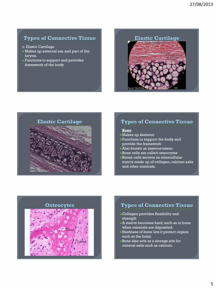
b. Fibrocartilage
Makes up intervertebral discs, pads in
the knee joint and the pads within pubic
bones.
Functions to cushion and support
27/08/2013
5
c. Elastic Cartilage
Makes up external ear and part of the
larynx.
Functions to support and provides
framework of the body
Bone
Makes up skeleton
Functions to support the body and
provide the framework
Also known as osseous tissue.
Bone cells are called osteocytes
Bones cells secrete an intercellular
matrix made up of collagen, calcium salts
and other minerals.
Collagen provides flexibility and
strength
A matrix becomes hard, such as in bone
when minerals are deposited.
Hardness of bone lets it protect organs
such as the brain
Bone also acts as a storage site for
mineral salts such as calcium.
27/08/2013
6
When mineralization of bone tissue is
diminished, the bone is weakened and
tends to break easily.
• This is known as osteoporosis
Calcium is the most important mineral
especially through childhood and
menopause due to lack of estrogen.
Estrogen helps calcium deposit into
bone; so do weight bearing activities
Blood
Function to transport nutrients,
hormones, respiratory gases (O2 and
CO2) and wastes.
Lymph Makes up all lymphatic vessels Functions to drain all interstitial fluid and
functions in immunity. Blood and lymph are two type so connective
tissue that have a watery intercellular matrix.
Blood is surrounded by and intercellular matrix is known as plasma
Plasma does not contain elastin or collagen; it contains a non fibrous plasma protein.
Lymph is found in lymphatic tissue.
Makes up the brain, spinal cord and
nerves
Made up of two types of cells: neurons
and neuroglia
Neurons transmit electrical signals to the
brain and spinal cord
Neurons have many shapes and sizes.
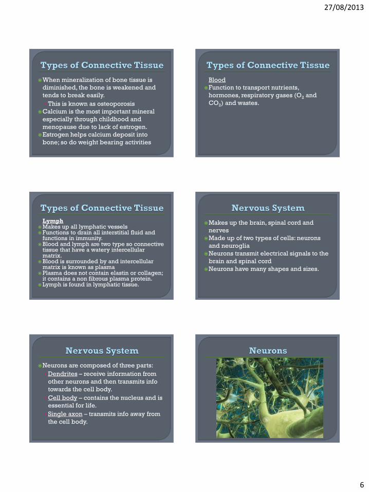
Neurons are composed of three parts:
• Dendrites – receive information from
other neurons and then transmits info
towards the cell body.
• Cell body – contains the nucleus and is
essential for life.
• Single axon – transmits info away from
the cell body.
27/08/2013
7
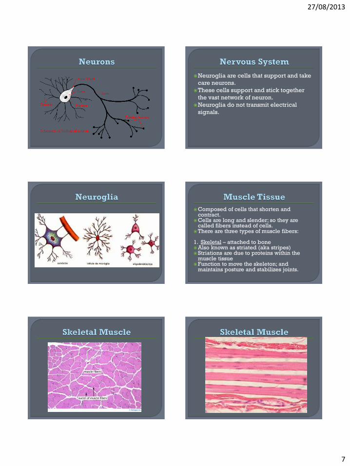
Neuroglia are cells that support and take
care neurons.
These cells support and stick together
the vast network of neuron.
Neuroglia do not transmit electrical
signals.
Composed of cells that shorten and contract.
Cells are long and slender; so they are called fibers instead of cells.
There are three types of muscle fibers: 1. Skeletal – attached to bone Also known as striated (aka stripes) Striations are due to proteins within the
muscle tissue Function to move the skeleton; and
maintains posture and stabilizes joints.
27/08/2013
8
2. Smooth muscle – found in the walls of viscera and organs.
Example – stomach, intestine, urinary bladder. Also make up the walls of bronchioles and blood
vessels. Function is related to location
• Small muscle in stomach helps mash and turn food
• Small muscle in bladder helps expel urine
Smooth muscle is not voluntarily controlled, so it is known as involuntary muscle.
Not striated; therefore known as non-striated muscle
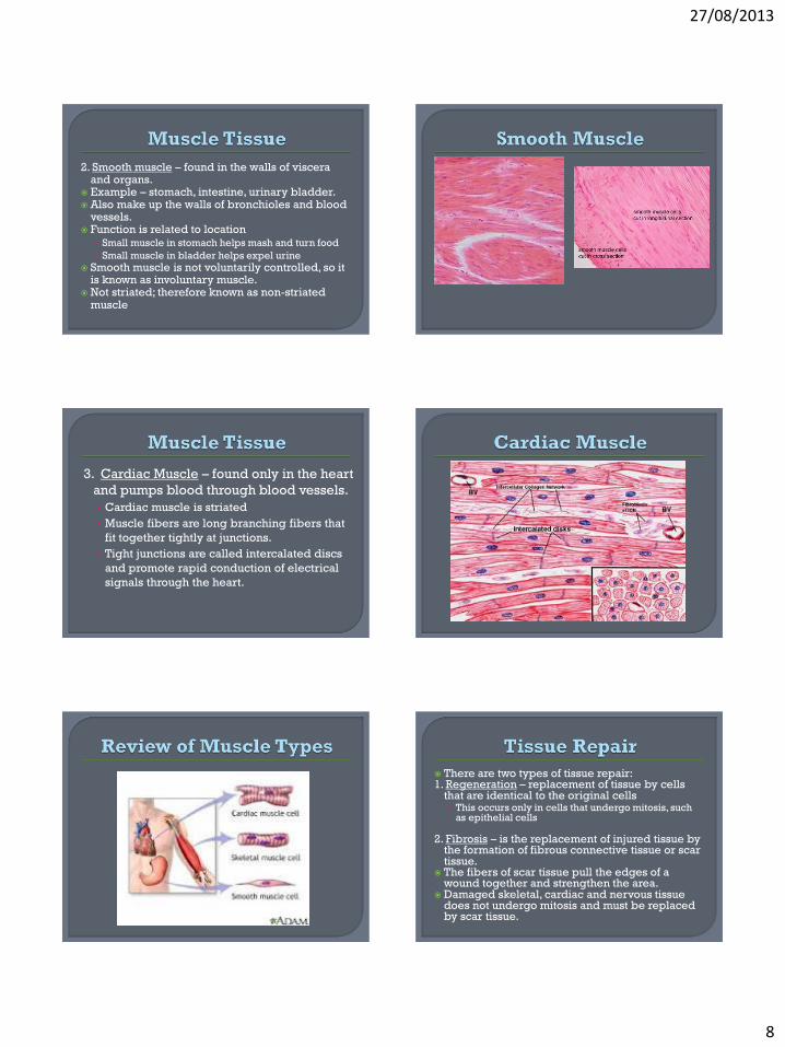
3. Cardiac Muscle – found only in the heart
and pumps blood through blood vessels.
• Cardiac muscle is striated
• Muscle fibers are long branching fibers that
fit together tightly at junctions.
• Tight junctions are called intercalated discs
and promote rapid conduction of electrical
signals through the heart.
There are two types of tissue repair: 1. Regeneration – replacement of tissue by cells
that are identical to the original cells • This occurs only in cells that undergo mitosis, such
as epithelial cells
2. Fibrosis – is the replacement of injured tissue by the formation of fibrous connective tissue or scar tissue.
The fibers of scar tissue pull the edges of a wound together and strengthen the area.
Damaged skeletal, cardiac and nervous tissue does not undergo mitosis and must be replaced by scar tissue.
27/08/2013
9
1. Deep wound in the skin severs blood vessels; causing blood to fill the wound.
2. A blood clot forms and as it dries a scab is formed.
3. Tissue repair begins; scar tissue forms in the deep layer.
4. At the same time surface epithelial cells multiply and fill the area between the scar tissue and the scab.
5. When the epithelium is completed the scab detaches.
6. A fully generated layer of epithelium over an underlying area of scar tissue.
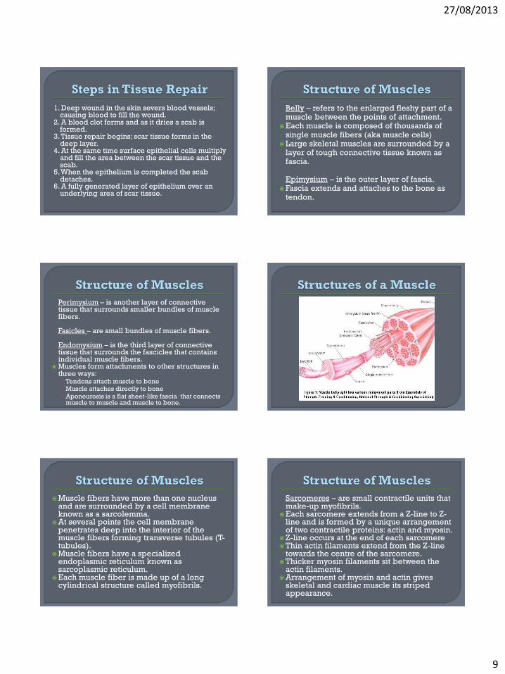
Belly – refers to the enlarged fleshy part of a muscle between the points of attachment.
Each muscle is composed of thousands of single muscle fibers (aka muscle cells)
Large skeletal muscles are surrounded by a layer of tough connective tissue known as fascia.
Epimysium – is the outer layer of fascia. Fascia extends and attaches to the bone as
tendon.
Perimysium – is another layer of connective tissue that surrounds smaller bundles of muscle fibers.
Fasicles – are small bundles of muscle fibers.
Endomysium – is the third layer of connective tissue that surrounds the fascicles that contains individual muscle fibers.
Muscles form attachments to other structures in three ways: • Tendons attach muscle to bone • Muscle attaches directly to bone • Aponeurosis is a flat sheet-like fascia that connects
muscle to muscle and muscle to bone.
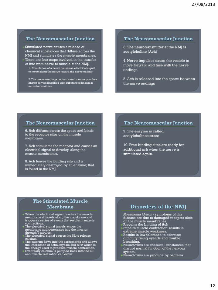
Muscle fibers have more than one nucleus and are surrounded by a cell membrane known as a sarcolemma.
At several points the cell membrane penetrates deep into the interior of the muscle fibers forming transverse tubules (T-tubules).
Muscle fibers have a specialized endoplasmic reticulum known as sarcoplasmic reticulum.
Each muscle fiber is made up of a long cylindrical structure called myofibrils.
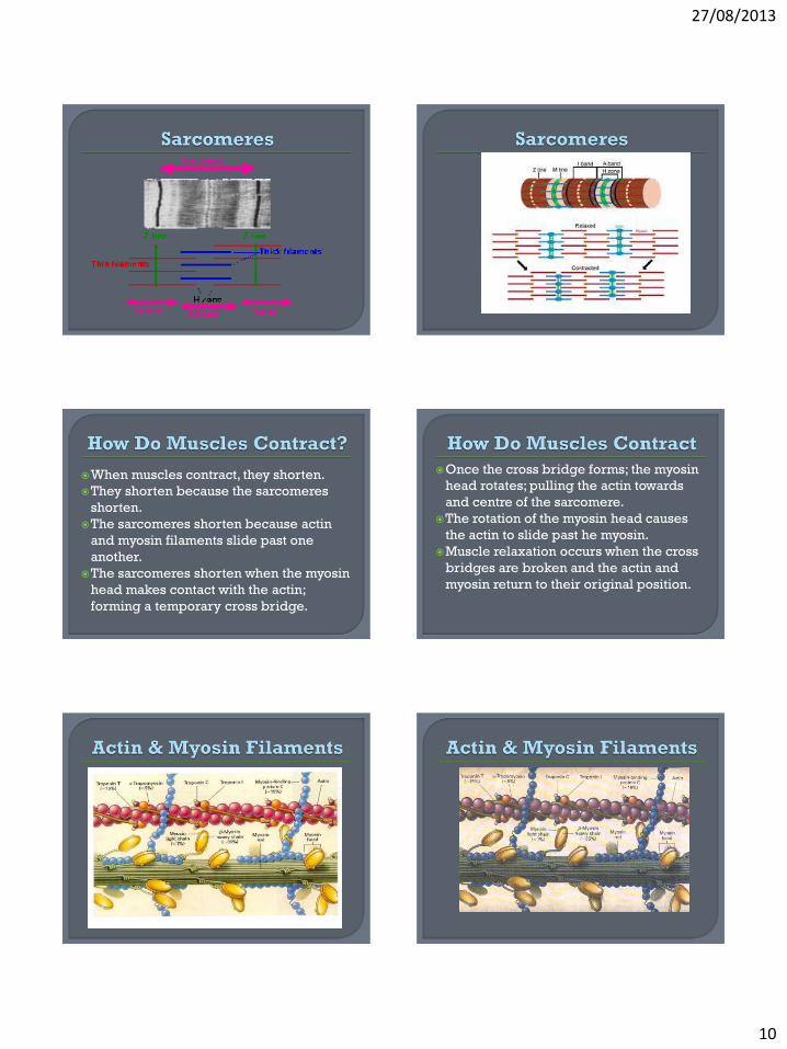
Sarcomeres – are small contractile units that make-up myofibrils.
Each sarcomere extends from a Z-line to Z-line and is formed by a unique arrangement of two contractile proteins: actin and myosin.
Z-line occurs at the end of each sarcomere Thin actin filaments extend from the Z-line
towards the centre of the sarcomere. Thicker myosin filaments sit between the
actin filaments. Arrangement of myosin and actin gives
skeletal and cardiac muscle its striped appearance.
27/08/2013
10
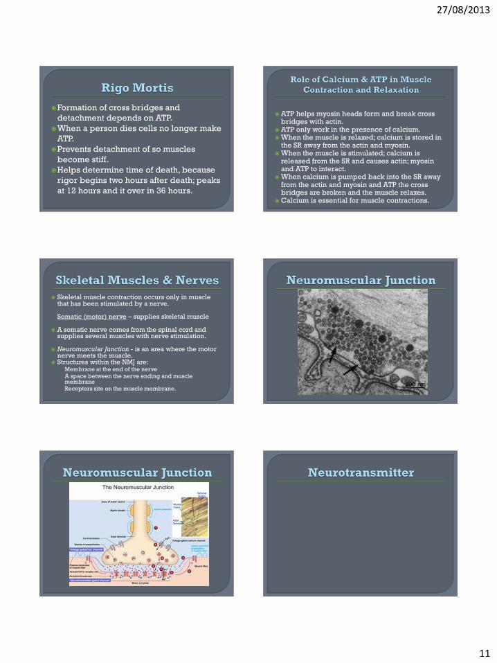
When muscles contract, they shorten.
They shorten because the sarcomeres
shorten.
The sarcomeres shorten because actin
and myosin filaments slide past one
another.
The sarcomeres shorten when the myosin
head makes contact with the actin;
forming a temporary cross bridge.
Once the cross bridge forms; the myosin
head rotates; pulling the actin towards
and centre of the sarcomere.
The rotation of the myosin head causes
the actin to slide past he myosin.
Muscle relaxation occurs when the cross
bridges are broken and the actin and
myosin return to their original position.
27/08/2013
11
Formation of cross bridges and
detachment depends on ATP.
When a person dies cells no longer make
ATP.
Prevents detachment of so muscles
become stiff.
Helps determine time of death, because
rigor begins two hours after death; peaks
at 12 hours and it over in 36 hours.
ATP helps myosin heads form and break cross
bridges with actin. ATP only work in the presence of calcium. When the muscle is relaxed; calcium is stored in
the SR away from the actin and myosin. When the muscle is stimulated; calcium is
released from the SR and causes actin; myosin and ATP to interact.
When calcium is pumped back into the SR away from the actin and myosin and ATP the cross bridges are broken and the muscle relaxes.
Calcium is essential for muscle contractions.
Skeletal muscle contraction occurs only in muscle that has been stimulated by a nerve.
Somatic (motor) nerve – supplies skeletal muscle
A somatic nerve comes from the spinal cord and supplies several muscles with nerve stimulation.
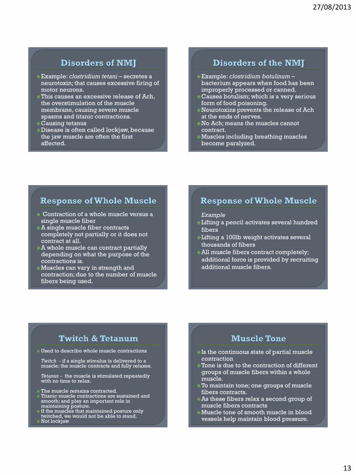
Neuromuscular Junction - is an area where the motor nerve meets the muscle.
Structures within the NMJ are: • Membrane at the end of the nerve
• A space between the nerve ending and muscle membrane
• Receptors site on the muscle membrane.
27/08/2013
12
Stimulated nerve causes a release of
chemical substances that diffuse across the
NMJ and stimulates the muscle membranes.
There are four steps involved in the transfer
of info from nerve to muscle at the NMJ. 1. Stimulation of a nerve causes an electrical signal
to move along the nerve toward the nerve ending.
2. The nerves endings contain membranous pouches
known as vesicles filled with substances known as
neurotransmitters.
3. The neurotransmitter at the NMJ is
acetylcholine (Ach)
4. Nerve impulses cause the vesicle to
move forward and fuse with the nerve
endings
5. Ach is released into the space between
the nerve endings
6. Ach diffuses across the space and binds to the receptor sites on the muscle membrane.
7. Ach stimulates the receptor and causes an
electrical signal to develop along the muscle membranes.
8. Ach leaves the binding site and is
immediately destroyed by an enzyme; that is found in the NMJ.
9. The enzyme is called
acetylcholinesterase
10. Free binding sites are ready for
additional ach when the nerve is
stimulated again.
When the electrical signal reaches the muscle membrane it travels along the membrane and triggers a series of events that results in muscle contractions.
The electrical signal travels across the membrane and penetrates into the interior through T-tubules.
The electrical signal causes the SR to release calcium.
The calcium flows into the sarcomeres and allows the interaction of actin, myosin and ATP, which is the energy used to produce muscle contraction.
Eventually calcium is pumped back into the SR and muscle relaxation can occur.
Myasthenia Gravis - symptoms of this disease are due to damaged receptor sites on the muscle membranes.
Prevents the binding of Ach Impairs muscle contraction; results in
extreme muscle weakness. Results in low tolerance to exercise;
difficulty rising eyelids and trouble breathing.
Neurotoxins are chemical substances that disrupt normal function of the nervous system.
Neurotoxins are produce by bacteria.
27/08/2013
13
Example: clostridium tetani – secretes a neurotoxin; that causes excessive firing of motor neurons.
This causes an excessive release of Ach, the overstimulation of the muscle membrane, causing severe muscle spasms and titanic contractions.
Causing tetanus Disease is often called lockjaw, because
the jaw muscle are often the first affected.
Example: clostridium botulinum – bacterium appears when food has been improperly processed or canned.
Causes botulism; which is a very serious form of food poisoning.
Neurotoxins prevents the release of Ach at the ends of nerves.
No Ach; means the muscles cannot contract.
Muscles including breathing muscles become paralyzed.
Contraction of a whole muscle versus a single muscle fiber
A single muscle fiber contracts completely not partially or it does not contract at all.
A whole muscle can contract partially depending on what the purpose of the contractions is.
Muscles can vary in strength and contraction; due to the number of muscle fibers being used.
Example
Lifting a pencil activates several hundred
fibers
Lifting a 100lb weight activates several
thousands of fibers
All muscle fibers contract completely;
additional force is provided by recruiting
additional muscle fibers.
Used to describe whole muscle contractions
Twitch - if a single stimulus is delivered to a muscle; the muscle contracts and fully relaxes.
Tetanus - the muscle is stimulated repeatedly with no time to relax.
The muscle remains contracted. Titanic muscle contractions are sustained and
smooth; and play an important role in maintaining posture.
If the muscles that maintained posture only twitched, we would not be able to stand.
Not lockjaw
Is the continuous state of partial muscle contraction
Tone is due to the contraction of different groups of muscle fibers within a whole muscle.
To maintain tone; one groups of muscle fibers contracts.
As these fibers relax a second group of muscle fibers contracts
Muscle tone of smooth muscle in blood vessels help maintain blood pressure.
27/08/2013
14
ATP is consumed by the contracting of muscle and it is replaced in three ways • Aerobic metabolism is the presence of oxygen; fuels
such as glycogen, glucose and fasts can be completely broken down to yield energy (ATP)
• Anaerobic metabolism is when the body can metabolize fuels in the absence of oxygen.
Without oxygen the complete breakdown of fuel is not possible and lactic acid is produced.
Accumulation of lactic acid is the cause of muscle soreness after heavy exercise.
Creatine phosphate contains energy that
the body can use to replenish ATP
quickly during muscle contraction.
Storage form of energy
Origin and insertion refers to the site of
muscle attachment
When a muscle contracts across a joint;
one bone remains stationary.
Origin – muscle that attaches to a
stationary bone
Insertion muscle that attaches to the more
moveable bone
Example - the origin of the biceps brachii is the scapula while the insertion is on the radius
One contraction of the biceps branchii the radius is pulled toward the scapula
Movement is generally accomplished by the group of muscles, but a single muscle is responsible for most of the movement.
Prime movers are the “chief muscle” responsible for most of the movement.
Synergists are the “helper muscles” that help move the prime mover.
Antagonists are muscles that oppose the motion of another muscle.
Example - the contraction of the biceps
brachii; the prime mover, pulls the lower
arm towards the shoulder. The triceps
brachii is the antagonists. it opposes the
action of the biceps brachii by pulling the
lower arm away from the scapula.
Hypertrophy
Overused muscles will increase in size.
Cardiac muscle can also undergo
hypertrophy; this generally indicates the
heart is working too hard.
Example - hypertension • Causes the heart to push blood into blood
vessels that are resistant to the flow of blood
• This extra work causes the heart to enlarge.
27/08/2013
15
Atrophy
Occurs if muscles are not used.
They decrease in size.
Example - a person with a broken leg
Contracture
Occurs if the muscle is immobilized for a
long period of time
Is an abnormal formation of fibrous tissue
within a muscle, causes muscles to freeze
in flexed position and severely restricts
joint mobility.
Name is based on the following
characteristics:
• Shape
• Size
• Direction of fibers
• Location of fibers
• Number of origins
• Origin and insertion
• Muscle action.
Deltoid - triangular
Latissimus – wide
Trapeziums - trapezoid
Rhomboideus - rhomboid
Teres - round
Vastus - huge
Maximum - large
Longus - long
Minimus - small
Brevis - short
Rectus - straight
Oblique - diagonal
Transverse - across
Circularis - circular
27/08/2013
16
Pectoralis - chest
Gluteus - buttocks
Brachii - arm
Supra - above
Infra - below
Sub - underneath
Lateralis - lateral
Number of sites at which the muscle is
anchored
2 biceps
3triceps
4 quadricep
Number of sites at which the muscle is
anchored
• 2 biceps
• 3triceps
• 4 quadricep
Abductor muscle moves away from
midline
Adductor muscle moves toward midline
Flexor causes flexion
Extension straightens limb
Levator elevates a structure
Massetter enables to chew
There are two categories: • Facial muscles
• Chewing muscles
Facial Muscles
Inserted directly into the soft tissue of
skin and other muscles of skin
When facial muscles contract; they pull of
the soft tissue.
Frontalis
Is a flat muscle that covers the frontal
bone
Extends from the cranial aponeurosis to
the skin of the eyebrows
Function is contraction, which raises the
eyebrow and wrinkles the forehead

27/08/2013
17