Embed Size (px)
Citation preview
Muscle Tissue 11/14
•Nearly half of body's mass•Three types
– Skeletal– Cardiac– Smooth
•Differ in structure, location, function and activation•Muscle terminology:
– Sarcolemma: muscle plasma membrane– Sarcoplasm: cytoplasm of a muscle cell– Prefixes for muscle: Myo, mys, and sarco
BIO lab 105--Lab 8A-muscle histol 1
1/46
Lab 8A, BIO 105
• Skeletal muscles – muscles attached to skin and bones and covers
bones– Responsible for locomotion and manipulation– Elongated cells called muscle fibers– Striated (striped) – Voluntary (i.e., conscious control)– Contract rapidly; tire easily; powerful– Require nervous system stimulation
BIO lab 105--Lab 8-muscle histol 2
• Cardiac muscle– Only in heart; bulk of heart walls – Striated, but involuntary
• we can’t control rate and pace of contraction however neural controls allow heart to speed up for short periods
– Can contract without nervous system stimulation; rate set by the pacemaker of the heart (group of special cells)
– Intercalated discs are gap junctions and allow innervation to spread in coordinated fashion
– Muscle is highly resistant to fatigue
BIO lab 105--Lab 8-muscle histol 3
• Smooth muscle –– spindle shaped (fusiform) cells; 1 nucleus– Not striated– Involuntary– Can contract without nervous system stimulation
—hormones, local chemical changes and Autonomic Nervous System can stimulate contraction
– Muscle contractions are slow and sustained– Role is to force substances through body channels– In walls of hollow organs, e.g., stomach, urinary
bladder, and respiratory passageways
BIO lab 105--Lab 8-muscle histol 4
Skeletal Muscle•Each muscle is an organ composed of muscle tissue, blood vessels, nerves and connective tissue
– Every skeletal muscle fiber supplied by nerve ending that controls its activity
– Huge nutrient and oxygen need; generates large amount of waste
BIO lab 105--Lab 8-muscle histol 5
• Connective tissue sheaths of skeletal muscle– Support cells; reinforce whole muscle– Internal to external
• Endomysium: areolar connective tissue surrounding each muscle fiber; located immediately superior to sarcolemma
• Perimysium: fibrous connective tissue surrounding groups of muscle fibers called fascicles
• Epimysium: dense irregular connective tissue surrounding entire muscle; blends with tendons
BIO lab 105--Lab 8-muscle histol 6
Muscle Attachments•Attach in at least two places
– Insertion – movable bone– Origin – immovable (less movable) bone
•Attachments– fused to periosteum of bone or cartilage– connective tissue wrappings extend beyond
muscle as tendon or aponeurosis (sheetlike connective tissue layer where muscle attaches)
•Don’t need to know terms direct or indirect
BIO lab 105--Lab 8-muscle histol 7
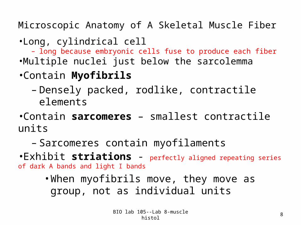
Microscopic Anatomy of A Skeletal Muscle Fiber
•Long, cylindrical cell – long because embryonic cells fuse to produce each fiber
•Multiple nuclei just below the sarcolemma•Contain Myofibrils
– Densely packed, rodlike, contractile elements •Contain sarcomeres – smallest contractile units
– Sarcomeres contain myofilaments•Exhibit striations - perfectly aligned repeating series of dark A bands and light I bands
• When myofibrils move, they move as group, not as individual units
BIO lab 105--Lab 8-muscle histol 8
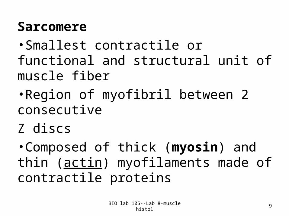
Sarcomere•Smallest contractile or functional and structural unit of muscle fiber•Region of myofibril between 2 consecutive Z discs•Composed of thick (myosin) and thin (actin) myofilaments made of contractile proteins
BIO lab 105--Lab 8-muscle histol 9
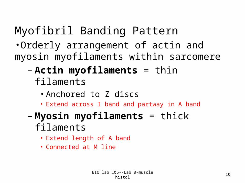
Myofibril Banding Pattern•Orderly arrangement of actin and myosin myofilaments within sarcomere
– Actin myofilaments = thin filaments• Anchored to Z discs• Extend across I band and partway in A band
– Myosin myofilaments = thick filaments• Extend length of A band• Connected at M line
BIO lab 105--Lab 8-muscle histol 10
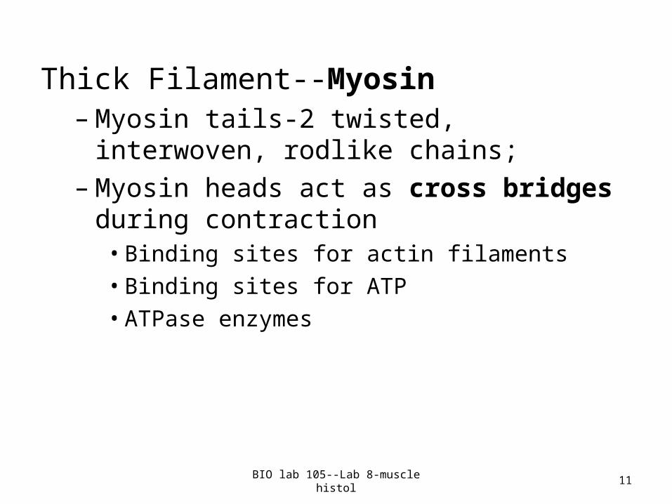
Thick Filament--Myosin– Myosin tails-2 twisted, interwoven, rodlike chains; – Myosin heads act as cross bridges during
contraction • Binding sites for actin filaments• Binding sites for ATP• ATPase enzymes
BIO lab 105--Lab 8-muscle histol 11
Thin Filament--Actin•Twisted double strand of fibrous protein •Contains sites for myosin head attachment during contraction•Tropomyosin and troponin - proteins bound to actin
– Tropomyosin in a relaxed fiber blocks the myosin binding sites• Twists around actin fibers and help stiffen and
stabilize it– Troponin binds calcium ions
BIO lab 105--Lab 8-muscle histol 12
Sarcoplasmic Reticulum (SR)•Network of smooth endoplasmic reticulum surrounding each myofibril•Pairs of terminal cisternae form perpendicular cross channels throughout SR•Functions in regulation of intracellular calcium levels
– Stores and releases Ca2+
BIO lab 105--Lab 8-muscle histol 13
26/46
T Tubules•Continuations of sarcolemma--pushes deeply into interior of cell•Increase muscle fiber's surface area•Conduct nerve impulses to deepest areas of muscle fiber•T tubules run between cross channels called terminal cisternae (cisterns) of the sarcoplasmic reticulum (smooth endoplasmic reticulum)•Impulses signal calcium to be released from adjacent terminal cisternae•Are associated with paired terminal cisterns to form triads that circle each sarcomere;
– triads are where the terminal cisterns border a T tubule
BIO lab 105--Lab 8-muscle histol 14
Physiology of Skeletal Muscle Fibers•For skeletal muscle to contract
– Activation (at neuromuscular junction)• Requires nervous system stimulation• AND an electric current or action potential
along sarcolemma• Intracellular Ca2+ levels must rise (final
requirement for contraction to begin)
BIO lab 105--Lab 8-muscle histol 15
Nerve Stimulus and the Neuromuscular Junction •Skeletal muscles stimulated by motor neurons of voluntary nervous system•Axons of motor neurons travel via nerves to skeletal muscle cells•Each axon forms several branches as it enters muscle •Each axon ending or branch, forms a neuromuscular junction with a single muscle fiber
BIO lab 105--Lab 8-muscle histol 16
Neuromuscular Junction (NMJ) is where axon and muscle fiber meet.•Axon terminal and muscle fiber separated by space called synaptic cleft•Synaptic vesicles from axon terminal contain neurotransmitter acetylcholine (ACh) •Sarcolemma in NMJ contain ACh receptors •ACh diffuses across synaptic cleft and attaches to ACh receptors in sarcolemma•ACh binding triggers electrical events to generate an action potential
BIO lab 105--Lab 8-muscle histol 17
• Action potential causes changes in properties of cell membrane channels
• Ca2+ channels open Ca2+ moves into nerve axon causing release of ACh into synaptic cleft
• ACh diffuses across cleftsarcolemma initiates action potential in muscle
BIO lab 105--Lab 8-muscle histol 18
Contraction of Skeletal Muscles•Contraction produces muscle tension, force exerted on load or object to be moved
– Refers to activation of sliding filaments and forming of cross bridges between actin and myosin
•Contraction ends when cross bridges are deactivated because of lack of nerve stimulation or not enough calcium present
BIO lab 105--Lab 8-muscle histol 19
Sliding Filament Model of Contraction•In relaxed state, thin and thick filaments overlap only at ends of A band•Sliding filament model of contraction
– Upon muscle stimulation, thin filaments slide past thick filaments actin and myosin overlap to a greater degree
•When myosin heads bind to actin cross bridges form and sliding begins
– Cross bridges form and break several times, moving thin filaments toward center of sarcomere
• Causes shortening of muscle fiber
BIO lab 105--Lab 8-muscle histol 20
Motor Unit: The Nerve-Muscle Functional Unit
•Each muscle is served by at least one motor nerve– A motor nerve contains axons of many motor
neurons– Axons branch into terminals, each of which form a
NMJ with single muscle fiber•Motor unit = motor neuron and all muscle fibers it supplies
– For fine control—each motor neuron supplies a smaller number of fibers
– For large, weight bearing muscles, each motor neuron supplies a lot of muscle fibers
•Muscle’s response to single threshold stimulus called a Muscle Twitch
BIO lab 105--Lab 8-muscle histol 21
LABWORK
1. Identify and describe the three kinds of muscle tissue (3 microscope slides).
2. Identify and describe all components of a muscle, including connective tissue wrappings around each part (models and muscle cross section slide).
3. Identify and describe microstructure of skeletal
muscle cells, and basics involved in contraction
mechanism (models and neuromuscular junction slide).
4. Explain the concepts covered about contraction and muscle physiology
BIO lab 105--Lab 8-muscle histol 22