Embed Size (px)
Citation preview

Musclin is an activity-stimulated myokine thatenhances physical enduranceEkaterina Subbotinaa, Ana Sierraa, Zhiyong Zhua, Zhan Gaoa, Siva Rama Krishna Kogantia, Santiago Reyesb,Elizabeth Stepniaka, Susan A. Walshc, Michael R. Acevedoc, Carmen M. Perez-Terzicd, Denice M. Hodgson-Zingmana,e,1,2,and Leonid V. Zingmana,e,f,1,2
aDepartment of Internal Medicine, University of Iowa Carver College of Medicine, Iowa City, IA 52242; bDepartment of Internal Medicine, Mayo Clinic,Rochester, MN 55905; cDepartment of Radiology, University of Iowa Carver College of Medicine, Iowa City, IA 52242; dDepartment of Physical Medicine andRehabilitation, Mayo Clinic, Rochester, MN 55905; eFraternal Order of Eagles Diabetes Research Center, University of Iowa Carver College of Medicine, IowaCity, IA 52242; and fDepartment of Medicine, Medical Center, Department of Veterans Affairs, Iowa City, IA 52242
Edited by Bruce M. Spiegelman, Dana-Farber Cancer Institute and Harvard Medical School, Boston, MA, and approved November 6, 2015 (received for reviewJuly 20, 2015)
Exercise remains the most effective way to promote physical andmetabolic wellbeing, but molecular mechanisms underlying exer-cise tolerance and its plasticity are only partially understood. Inthis study we identify musclin—a peptide with high homology tonatriuretic peptides (NP)—as an exercise-responsive myokine thatacts to enhance exercise capacity in mice. We use human primarymyoblast culture and in vivo murine models to establish that theactivity-related production of musclin is driven by Ca2+-dependentactivation of Akt1 and the release of musclin-encoding gene (Ostn)transcription from forkhead box O1 transcription factor inhibition.Disruption of Ostn and elimination of musclin secretion in miceresults in reduced exercise tolerance that can be rescued by treat-ment with recombinant musclin. Reduced exercise capacity in micewith disrupted musclin signaling is associated with a trend towardlower levels of plasma atrial NP (ANP) and significantly smallerlevels of cyclic guanosine monophosphate (cGMP) and peroxisomeproliferator-activated receptor gamma coactivator 1-α in skeletalmuscles after exposure to exercise. Furthermore, in agreementwith the established musclin ability to interact with NP clearancereceptors, but not with NP guanyl cyclase-coupled signaling recep-tors, we demonstrate that musclin enhances cGMP production incultured myoblasts only when applied together with ANP. Elim-ination of the activity-related musclin-dependent boost of ANP/cGMP signaling results in significantly lower maximum aerobiccapacity, mitochondrial protein content, respiratory complex pro-tein expression, and succinate dehydrogenase activity in skeletalmuscles. Together, these data indicate that musclin enhancesphysical endurance by promoting mitochondrial biogenesis.
osteocrin | mitochondria | skeletal muscle | exercise | natriuretic peptide
The ability to sustain physical activity is necessary for bothquality and longevity of life. Regular exposure to exercise is
associated with reduced rates of all-cause mortality (1). Thereare multiple mechanisms by which physical activity promoteshealth; however, recently there has been an interest in definingthe contribution of circulating proteins secreted by skeletalmuscle, termed myokines (2, 3). Myokines are autocrine, para-crine, or endocrine stimuli that may guide local skeletal muscleremodeling, repair, and maintenance or steer systemic adapta-tion related to physical activity (2). Understanding the functionalrole and the signaling pathways of myokines, particularly as theyrelate to exercise, may reveal new therapeutic targets to promotehealth and augment the benefits of physical activity.This study is focused on the recently discovered myokine
musclin (4, 5). Two groups initially identified this peptide: one asbone-derived osteocrin (5) and the second as muscle-secretedmusclin (4). Musclin mRNA expression has been linked toinsulin-induced activation of protein kinase B (Akt) thatphosphorylates forkhead box O1 transcription factor (FOXO1),causing it to be exported from the nucleus and thus releasingthe musclin-encoding gene from transcriptional inhibition
(4, 6). This pathway has been demonstrated to regulate musclintranscription in both cell culture and skeletal muscles (4, 6).Musclin contains two KKKR putative serine protease cleavagesites and a region homologous to members of the natriureticpeptide (NP) family (4, 5). However, musclin does not have twocysteine residues needed to form the Ω-like structure characteristicfor NPs (4, 5). In line with these structural characteristics, it hasbeen demonstrated that musclin binds to the NP clearance re-ceptor, NPRC, with affinity comparable to NPs, but exhibits onlyweak binding to NPRA and NPRB without activating the linkedguanyl cyclase that is the primary effector of NP physiologic ac-tions (7–9). Thus, it has been suggested that musclin function maybe due to modulation of the action of NPs by competition withthem for clearance via NPRC binding (8, 9). Indeed, musclinoverexpression in osteoblast-lineage cells has been shown to resultin elongated bones and marked kyphosis (9), which is similar to thephenotype of mice transgenically overexpressing BNP (10) or CNP(11) or lacking NPRC (12, 13). However, the physiological role ofmusclin production in skeletal muscles has remained elusive.In this work, we demonstrate that musclin production by
skeletal muscle is stimulated by physical activity and is paralleledby increased systemic musclin levels. Disruption of normal musclin
Significance
Skeletal muscle is increasingly recognized as a secretory organ.Revealing the identity and function of myokines can improveour understanding of skeletal muscle function under sedentaryor exercise conditions, as well as its coordination with otherorgans, tissues, and overall body metabolism. This studyidentifies musclin as an exercise-responsive myokine critical forskeletal muscle adaptation to physical activity. We develop amusclin-encoding gene (Ostn) knockout mouse, which allowsus to determine a previously unrecognized physiologic func-tion of musclin in regulation of skeletal muscle mitochondrialbiogenesis and physical endurance. The demonstrated molec-ular mechanism for musclin-dependent skeletal muscle adap-tation to exercise also transforms the perspective on natriureticpeptide signaling, particularly as it relates to physical activityand exercise-induced remodeling in different tissues.
Author contributions: D.M.H.-Z. and L.V.Z. designed research; E. Subbotina, A.S., Z.Z., Z.G.,S.R.K.K., S.R., E. Stepniak, S.A.W., M.R.A., C.M.P.-T., D.M.H.-Z., and L.V.Z. performed re-search; D.M.H.-Z. and L.V.Z. contributed new reagents/analytic tools; E. Subbotina, A.S.,Z.Z., Z.G., S.R.K.K., S.R., S.A.W., M.R.A., D.M.H.-Z., and L.V.Z. analyzed data; and E. Subbotina,D.M.H.-Z., and L.V.Z. wrote the paper.
The authors declare no conflict of interest.
This article is a PNAS Direct Submission.1D.M.H.-Z. and L.V.Z. contributed equally to this work.2To whom correspondence may be addressed. Email: [email protected] [email protected].
This article contains supporting information online at www.pnas.org/lookup/suppl/doi:10.1073/pnas.1514250112/-/DCSupplemental.
16042–16047 | PNAS | December 29, 2015 | vol. 112 | no. 52 www.pnas.org/cgi/doi/10.1073/pnas.1514250112

signaling in mice by knockout of the musclin-encoding gene, Ostn(Ostn-KO), results in diminished exercise tolerance coupled withdowngraded activity-related atrial NP (ANP)/cyclic guanosinemonophosphate (cGMP)/PGC-1α–dependent skeletal musclemitochondrial biogenesis. Thus, this work identifies a physiologicalrole of musclin in enhancing skeletal muscle oxidative capacity andphysical endurance.
ResultsMusclin Production and Secretion into the Systemic Circulation AreStimulated by Exercise. Normal skeletal muscle function requirestight coordination with the operation of other organs and systems.Such coordination has been attributed in part to the action of myo-kines. Specifically, “exercise factors,” a subset of myokines whoseproduction and secretion into systemic circulation are stimulated byphysical activity, have been shown to modulate skeletal muscle andsystemic metabolism, angiogenesis, growth, and inflammation (14).To determine whether musclin is an exercise factor, the level
of musclin peptide in skeletal muscle was probed in two groupsof wild-type (WT) mice: one group that exercised on a movingtreadmill for 45 min daily (exercise) and a sedentary controlgroup. After 5 d of exercise or control treadmill exposure, micewere euthanized, and their tissues were harvested by rapid ex-cision and freeze clamp. Proteins were extracted from gastroc-nemius muscles and segregated by Western blot (Fig. 1A),showing that exercise was associated with a nearly 100% increasein skeletal muscle musclin over control conditions [1.15 ± 0.2 vs.0.60 ± 0.1 arbitrary units (AU), n = 5 each, P < 0.05; Fig. 1B]. Asimilar increase was demonstrated in skeletal muscle musclinmRNA from tibialis anterior samples (0.75 ± 0.05 vs. 0.49 ±0.05 AU, n = 5 each, P < 0.05; Fig. 1C), whereas musclinmRNA levels in femur were markedly (96–98%) lower and
were unresponsive to exercise [0.02 ± 0.002 vs. 0.02 ± 0.003AU, respectively, n = 4 each, P = not significant (NS) betweenexercise and sedentary, P < 0.05 compared with skeletal musclemRNA]. The increased musclin production by skeletal musclewas paralleled by an increase in the plasma musclin level from27.71 ± 5.54 pg/mL (n = 3) in sedentary control WT mice to46.24 ± 4.69 pg/mL in WT mice after exercise (n = 6, P < 0.05;Fig. 1D). Furthermore, immunohistochemistry of gastrocne-mius cross-sections demonstrated more intense staining formusclin when mice were after exercise vs. sedentary (Fig. 1E).Thus, musclin production and secretion into the systemic cir-culation are up-regulated in response to exercise, establishingmusclin as an exercise factor.
Activity-Induced Musclin Production Is Linked to Ca2+-DependentActivation of Akt. Regulation of musclin transcription has beenlinked to Akt activation (4, 6, 15, 16). Akt is a serine/threoninekinase that has emerged as a critical signaling component for theregulation of cellular metabolism, growth, and survival in multiplesystems (17). Akt activity is increased in response to numerousstimuli, including a wide variety of growth factors and hormonesactivating phosphatidylinositol 3-kinase (PI3-kinase) (18–20). Aktcan also be activated by mechanisms independent of PI3-kinase—for example, in response to increases in intracellular Ca2+ or cAMP,as occurs with increased muscle contractile activity (21–25).Here we confirm Akt activation in our model of treadmill-ex-
ercised mice. Specifically, the levels of phosphorylated Akt (S473and T308) and total Akt from gastrocnemius of WT mice werecompared by Western blot (Fig. 2 A–C), showing a significantincrease in phosphorylated Akt (0.48 ± 0.09 vs. 0.28 ± 0.01 AU, n= 5 each, P < 0.05 for S473; Fig. 2E; and 1.14 ± 0.02 vs. 0.68 ± 0.07AU, n = 5 each, P < 0.05 for T308; Fig. 2F), but not total Akt (1.02± 0.05 vs. 1.21 ± 0.06 AU, n = 5 each, P = NS; Fig. 2G), in re-sponse to exercise vs. sedentary conditions. Akt is a known regu-lator of FOXO1 nuclear export (15, 26). Here, we find a significantincrease in phosphorylated FOXO1 (1.473 ± 0.047 vs. 1.185 ±0.056 AU, n = 3 each, P < 0.05), but not total FOXO1 (1.380 ±0.046 vs. 1.337 ± 0.103 AU, n = 3 each, P = NS) normalized toGAPDH in gastrocnemius muscle from exercised vs. sedentarymuscle (Fig. S1). Also, FOXO1 nuclear content quantification byWestern blot (Fig. 2D) shows a dramatic reduction in response toexercise (0.16 ± 0.07 vs. 0.95 ± 0.29 AU, n = 5 each, P < 0.05; Fig.2H). Because FOXO1 is known to inhibit musclin-encoding genetranscription in skeletal muscle (6), this exercise-related reductionin nuclear FOXO1 is consistent with our finding of increasedmusclin mRNA after exercise. Furthermore, we found no signifi-cant exercise-induced changes in musclin mRNA/hypoxanthineguanine phosphoribosyl transferase (HPRT) in Akt1-KO mice(0.47 ± 0.10 AU, n = 5 vs. 0.45 ± 0.06 AU, n = 4, P = NS).This molecular cascade was verified in a cell culture model of
primary skeletal myoblasts isolated from WT mice in whichphosphorylation of Akt and FOXO1 were induced by applicationof a Ca2+ ionophore (A23187; Sigma Aldrich; Fig. 3 A–C; p-Akt/GAPDH 0.26 ± 0.008, n = 2 vs. 0.91 ± 0.05, n = 4, pFOXO1/GAPDH 0.12 ± 0.02, n = 2 vs. 0.39 ± 0.04, n = 4, pAkt/totalAkt 0.21 ± 0.01, n = 2 vs. 0.66 ± 0.03, n = 4, pFOXO1/totalFOXO1 0.07 ± 0.01, n = 2 vs. 0.27 ± 0.03, n = 4, for control andionophore conditions, respectively, all in the presence of 1 mMCa2+; P < 0.05 for all comparisons). This activation of Akt byCa2+ ionophore was paralleled by augmented musclin pro-duction (Fig. 3 D and E). Specifically, an increase in musclinmRNA was induced by Ca2+ ionophore in a dose-dependentmanner (1.00 ± 0.08 AU no ionophore vs. 2.20 ± 0.06 AU for0.5 μM ionophore vs. 3.60 ± 0.81 AU for 1 μM ionophore, all inpresence of 1.0 mM Ca2+, n = 3 each, P < 0.05 for each iono-phore concentration vs. no ionophore or control; Fig. 3D)—aresponse that was eliminated when myoblasts were pretreatedwith Akt inhibitor-viii (Sigma-Aldrich; 1.09 ± 0.03 AU, n = 3,P = NS vs. control; Fig. 3D), or by removal of extracellular Ca2+
from the medium (0.96 ± 0.012 AU, n = 3 each, P = NS vs.control; Fig. 3D). The same musclin response to Ca2+ was
A
E
B C D
Fig. 1. Musclin expression is exercise-responsive. Musclin expression was testedin muscles of WT mice after 5 d of treadmill exercise vs. no exercise (control).(A) Representative Western blots for musclin and GAPDH in protein extractsfrom gastrocnemius. (B–D) Summary statistics for musclin protein expressionnormalized to GAPDH by densitometry of Western blots of protein extracts fromgastrocnemius (B), tibialis anterior musclin mRNA normalized to hypoxanthineguanine phosphoribosyl transferase (HPRT) by quantitative RT-PCR (qRT-PCR) (C),and musclin peptide expression in plasma by custom ELISA (the y axis rangebegins at the lower limit for detection for this assay of 20 pg/mL; D). (E) Rep-resentative immunohistochemical stains of gastrocnemius cross-sections imagedby confocal microscopy. Red, musclin; green, nuclei. *P < 0.05 vs. control.
Subbotina et al. PNAS | December 29, 2015 | vol. 112 | no. 52 | 16043
PHYS
IOLO
GY

observed in a primary culture of myoblasts isolated from healthyhuman subjects (ZenBio; 1.0 ± 0.03 AU for no ionophore vs.3.02 ± 0.23 AU for 0.5 μM ionophore vs. 3.95 ± 0.83 AU for1 μM ionophore, all in presence of 1.0 mM Ca2+, n = 3 each; P <0.05 for each ionophore concentration vs. no ionophore orcontrol, values in Ca2+-free buffer were 1.0 ± 0.05, 0.74 ± 0.18,and 0.79 ± 0.18 AU, respectively; n = 3 each, P = NS; Fig. 3E),confirming that this mechanism is not specific to mice. Thesefindings all indicate that exercise-related musclin production isdriven by the Ca2+–Akt–FOXO1 signaling cascade.
Genetic Disruption of Musclin Production Causes Reduced PhysicalEndurance. To investigate the physiological significance andfunction of a physical-activity-induced increase in musclin pro-duction, we generated a mouse model with ubiquitous disruptionof the musclin-encoding gene, Ostn (genOway) and confirmedthe absence of musclin production in skeletal muscle of Ostn-KOmice vs. WT controls by Western blot (Fig. 4 A and B). TheOstn-KOmice, housed normally and fed standard chow, exhibited noskeletal deformities or differences in bone density and no growthabnormalities, blood pressure, or body composition changes(Fig. S2 and Table S1) compared with controls at 7–8 wk of age;however, they do demonstrate lower exercise tolerance thancontrols. Specifically, when challenged with a program oftreadmill exercise with progressive increase in speed and incline(Fig. 4C), Ostn-KO mice demonstrate a significant deficit inexertional tolerance with respect to duration (71 ± 6 vs. 91 ± 6min., n = 6 each, P < 0.05; Fig. 4D), distance (769 ± 102 vs. 1147± 121 m, n = 6 each, P < 0.05; Fig. 4E), and overall workload (34± 5 vs. 53 ± 4 J, n = 6 each, P < 0.05; Fig. 4F). Similarly, whenmice were offered the opportunity for voluntary exercise onrunning wheels (Fig. 4G), Ostn-KO mice demonstrated signifi-cantly lower mean velocity (35 ± 4 vs. 52 ± 4 rotations per 5min, n = 6 each, P < 0.05; Fig. 4H), duration (303 ± 38 vs. 383
± 64 min., n = 6 each, P < 0.05; Fig. 4I), and distance (1,505 ±231 vs. 2,218 ± 253 m, n = 6 each, P < 0.05; Fig. 4J) during thenight when the vast majority of activity was recorded. To confirmthat the observed phenotype is related to the absence of musclinin the systemic circulation, mice were implanted with osmoticpumps (Alzet Durect) loaded with saline or 50 μg of musclin.This dose resulted in musclin plasma levels of 69.7 ± 8.8 pg/mL(n = 3), comparable with levels observed in mice following ex-ercise as presented in Fig. 1. Voluntary exercise on runningwheels (Fig. 4K) was significantly increased in WT mice treatedwith musclin (n = 5) compared with WT mice treated with saline(n = 4), with respect to mean velocity (51 ± 7 vs. 16 ± 4 rotationsper 5 min, P < 0.05; Fig. 4L), duration (336 ± 39 vs. 147 ± 10min, P < 0.05; Fig. 4M), and distance (2,202 ± 280 vs. 696 ± 179 m,P < 0.05; Fig. 4N). Furthermore, treatment with musclin“rescued” the Ostn-KO mice (n = 4) because their exerciseactivity was equalized to that of musclin-treated WT mice (n = 5)in terms of night-time mean velocity (53 ± 15 vs. 51 ± 7 rotationsper 5 min, P =NS; Fig. 4L), duration of running (333 ± 53 vs. 336 ±39 min, P = NS; Fig. 4M), and distance run (2,290 ± 632 vs. 2,202 ±280 m, P = NS; Fig. 4N). Thus, intact musclin production is criticalfor optimal exercise performance.
Musclin Boosts Activity-Related cGMP Production in Skeletal Muscle.To address the relationship between exercise, musclin, and ANP,we examined WT and Ostn-KO mice after exercise using thesame protocol as in Fig. 1, which established a significant exer-cise-related musclin response in WT mice. We found a trendtoward higher-plasma ANP levels in exercised WT (n = 18 micein six groups) compared with Ostn-KO (n = 15 mice in fivegroups) mice, although it did not achieve statistical significance(140.4 ± 19.9 pg/mL vs. 98.2 ± 4.5, P = 0.09; Fig. 5A). Fur-thermore, when gastrocnemius muscles were assayed after
p-A
kt47
3/G
APD
H
0.0
0.1
0.2
0.3
0.4
0.5
0.6
*
ExerciseControl
p-A
kt30
8/G
APD
H
0.0
0.2
0.4
0.6
0.8
1.0
1.2
1.4
*
ExerciseControl
Tota
l Akt
/GA
PDH
0.0
0.2
0.4
0.6
0.8
1.0
1.2
1.4
ExerciseControl
p-Akt473GAPDH
Control Exercise
Total AktGAPDH
ExerciseControl
p-Akt308GAPDH
ExerciseControl
FOXO1TBP
ExerciseControl
FOXO
1/TB
P
0.0
0.2
0.4
0.6
0.8
1.0
1.2
1.4
ExerciseControl
*
A
C
B
D
E F G H
Fig. 2. Exercise promotes skeletal muscle Akt phosphorylation and FOXO1nuclear export. Gastrocnemius of WT mice were assayed after 5 d oftreadmill exercise vs. no exercise (control). (B–D) Representative Westernblots of GAPDH and Akt phosphorylated at residue 473 (A), residue 308 (B),and total Akt (C) in muscle and representative Western blots of TBP andFOXO1 in nuclear extracts from muscle (D). (E–H) Summary statistics forexpression of Akt phosphorylated at residue 473 (E), residue 308 (F), andtotal Akt normalized to GAPDH in muscle (G) and FOXO1 normalized to TBPin nuclear extracts from muscle by densitometry of Western blots (H). TBP,anti-TATA binding protein. *P < 0.05 vs. control.
pAkt
Total Akt
pFOXO1
Total FOXO1
GAPDH
mR
NA
Mus
clin
/HPR
T
0
1
2
3
4
5
ionophore ( M)calcium (mM)Akt inhibitor
1.0 1.0 1.0 1.00- - - +-
0 0.5 1.0 0.50.5
*
*
mR
NA
Mus
clin
/HPR
T
0
1
2
3
4
5
6
CalciumCalcium-free
control ionophore 0.5 M
ionophore 1 M
**
prot
ein/
tota
l0.0
0.2
0.4
0.6
0.8
pAktpFOXO1
*
*
prot
ein/
GAP
DH
0.0
0.2
0.4
0.6
0.8
1.0
pAktpFOXO1
*
*
Human primary myoblasts
GAPDH
control ionophore
A
D E
B C
Fig. 3. Musclin production is stimulated by Ca2+-dependent Akt phos-phorylation. (A) Representative Western blots of Akt, FOXO1, and GAPDHfrom cultured murine primary myoblasts in 1 mM Ca2+ without ionophore(control) vs. with 1 μM ionophore A23187 (Sigma-Aldrich). (B and C) Sum-mary statistics for phosphorylated Akt (pAkt) and phosphorylated FOXO1(pFOXO1) normalized to GAPDH (B) and total Akt and total FOXO1 (C), re-spectively, with (gray) and without (white; control) 1 μM ionophore. *P < 0.05 vs.control. (D and E) Summary statistics for musclin mRNA normalized to HPRT inmurine cultured primary myoblasts exposed to various concentrations of iono-phore, Ca2+ and Akt inhibitor-viii (D) and human cultured primary myoblastsexposed to no Ca2+ vs. 1.0 mM Ca2+ and various doses of ionophore, by densi-tometry of Western blots (E). *P < 0.05 vs. no ionophore (D) or vs. Ca2+-free (E).
16044 | www.pnas.org/cgi/doi/10.1073/pnas.1514250112 Subbotina et al.
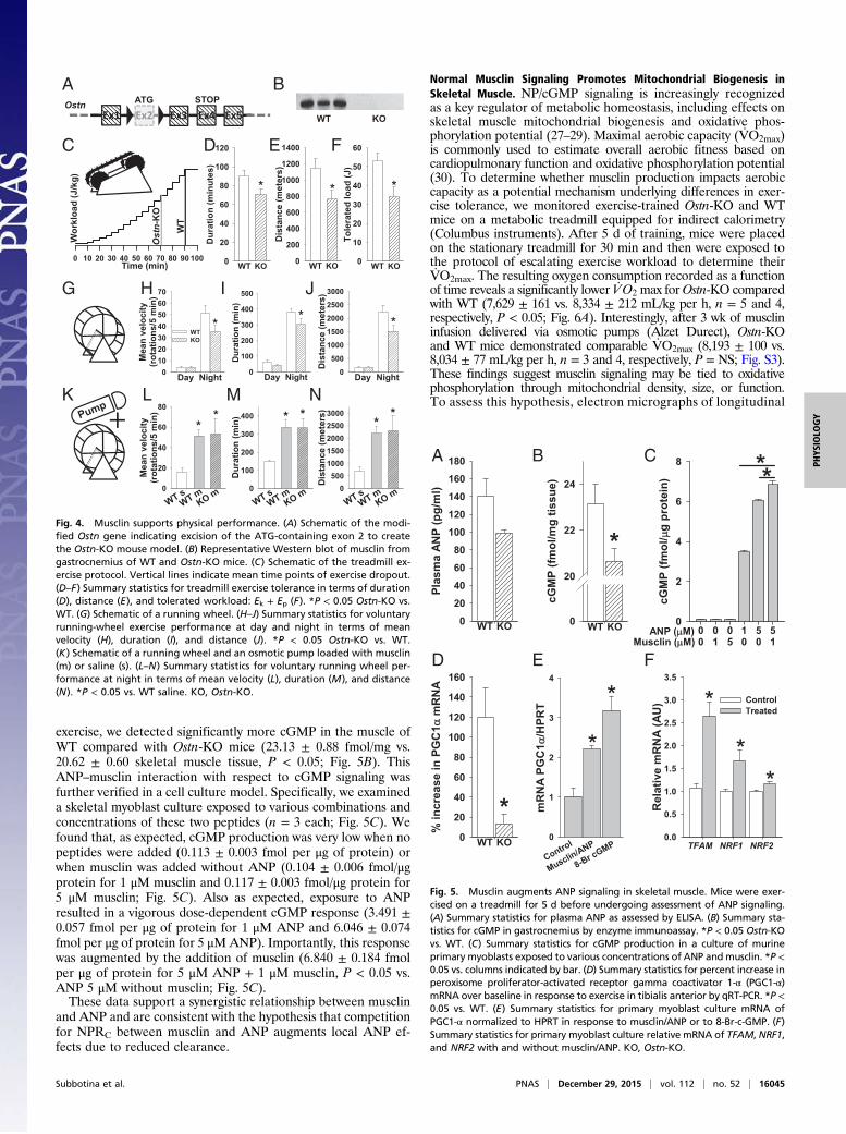
exercise, we detected significantly more cGMP in the muscle ofWT compared with Ostn-KO mice (23.13 ± 0.88 fmol/mg vs.20.62 ± 0.60 skeletal muscle tissue, P < 0.05; Fig. 5B). ThisANP–musclin interaction with respect to cGMP signaling wasfurther verified in a cell culture model. Specifically, we examineda skeletal myoblast culture exposed to various combinations andconcentrations of these two peptides (n = 3 each; Fig. 5C). Wefound that, as expected, cGMP production was very low when nopeptides were added (0.113 ± 0.003 fmol per μg of protein) orwhen musclin was added without ANP (0.104 ± 0.006 fmol/μgprotein for 1 μM musclin and 0.117 ± 0.003 fmol/μg protein for5 μM musclin; Fig. 5C). Also as expected, exposure to ANPresulted in a vigorous dose-dependent cGMP response (3.491 ±0.057 fmol per μg of protein for 1 μM ANP and 6.046 ± 0.074fmol per μg of protein for 5 μMANP). Importantly, this responsewas augmented by the addition of musclin (6.840 ± 0.184 fmolper μg of protein for 5 μM ANP + 1 μM musclin, P < 0.05 vs.ANP 5 μM without musclin; Fig. 5C).These data support a synergistic relationship between musclin
and ANP and are consistent with the hypothesis that competitionfor NPRC between musclin and ANP augments local ANP ef-fects due to reduced clearance.
Normal Musclin Signaling Promotes Mitochondrial Biogenesis inSkeletal Muscle. NP/cGMP signaling is increasingly recognizedas a key regulator of metabolic homeostasis, including effects onskeletal muscle mitochondrial biogenesis and oxidative phos-phorylation potential (27–29). Maximal aerobic capacity (V·O2max)is commonly used to estimate overall aerobic fitness based oncardiopulmonary function and oxidative phosphorylation potential(30). To determine whether musclin production impacts aerobiccapacity as a potential mechanism underlying differences in exer-cise tolerance, we monitored exercise-trained Ostn-KO and WTmice on a metabolic treadmill equipped for indirect calorimetry(Columbus instruments). After 5 d of training, mice were placedon the stationary treadmill for 30 min and then were exposed tothe protocol of escalating exercise workload to determine theirV·O2max. The resulting oxygen consumption recorded as a functionof time reveals a significantly lower V·O2 max forOstn-KO comparedwith WT (7,629 ± 161 vs. 8,334 ± 212 mL/kg per h, n = 5 and 4,respectively, P < 0.05; Fig. 6A). Interestingly, after 3 wk of musclininfusion delivered via osmotic pumps (Alzet Durect), Ostn-KOand WT mice demonstrated comparable V·O2max (8,193 ± 100 vs.8,034 ± 77 mL/kg per h, n = 3 and 4, respectively, P = NS; Fig. S3).These findings suggest musclin signaling may be tied to oxidativephosphorylation through mitochondrial density, size, or function.To assess this hypothesis, electron micrographs of longitudinal
0 10 20 30 40 50 60 70 80 90 100
Dur
atio
n (m
inut
es)
0
20
40
60
80
100
120
WT KO
*
Wor
kloa
d (J
/kg)
Ostn-
KO
WT
Time (min)
ATG
WT KOOstn
Ex1 Ex3 Ex4 Ex5Ex2STOP
Dis
tanc
e (m
eter
s)
0
200
400
600
800
1000
1200
1400
WT KO
*
Tole
rate
d lo
ad (J
)
0
10
20
30
40
50
60
WT KO
*
Dur
atio
n (m
in)
0
100
200
300
400
500
*
Day Night
Mea
n ve
loci
ty(r
otat
ions
/5 m
in)
010203040506070
WTKO
*
Day Night
Dis
tanc
e (m
eter
s)
0
500
1000
1500
2000
2500
3000
*
Day Night
Mea
n ve
loci
ty(r
otat
ions
/5 m
in)
0
20
40
60
80*
*
Dur
atio
n (m
in)
0
100
200
300
400 **
Dis
tanc
e (m
eter
s)
0500
10001500200025003000
**
A
C
G
K L M N
H I J
D E F
B
Fig. 4. Musclin supports physical performance. (A) Schematic of the modi-fied Ostn gene indicating excision of the ATG-containing exon 2 to createthe Ostn-KO mouse model. (B) Representative Western blot of musclin fromgastrocnemius of WT and Ostn-KO mice. (C) Schematic of the treadmill ex-ercise protocol. Vertical lines indicate mean time points of exercise dropout.(D–F) Summary statistics for treadmill exercise tolerance in terms of duration(D), distance (E), and tolerated workload: Ek + Ep (F). *P < 0.05 Ostn-KO vs.WT. (G) Schematic of a running wheel. (H–J) Summary statistics for voluntaryrunning-wheel exercise performance at day and night in terms of meanvelocity (H), duration (I), and distance (J). *P < 0.05 Ostn-KO vs. WT.(K) Schematic of a running wheel and an osmotic pump loaded with musclin(m) or saline (s). (L–N) Summary statistics for voluntary running wheel per-formance at night in terms of mean velocity (L), duration (M), and distance(N). *P < 0.05 vs. WT saline. KO, Ostn-KO.
cGM
P (fm
ol/m
g tis
sue)
0
20
22
24
WT KO
*
cGM
P (fm
ol/
g pr
otei
n)
0
2
4
6
8 **
5Musclin ( M)ANP ( M) 1 5 5
1100
0 00 0
Plas
ma
ANP
(pg/
ml)
0
20
40
60
80
100
120
140
160
180
WT KO
% in
crea
se in
PG
C1
mR
NA
0
20
40
60
80
100
120
140
160
*WT KO
Rel
ativ
e m
RN
A (A
U)
0.0
0.5
1.0
1.5
2.0
2.5
3.0
3.5
ControlTreated
TFAM NRF1 NRF2
*
**
mR
NA
PGC
1/H
PRT
0
1
2
3
4
Control
Musclin/ANP
*
8-Br cGMP
*
A
D E F
B C
Fig. 5. Musclin augments ANP signaling in skeletal muscle. Mice were exer-cised on a treadmill for 5 d before undergoing assessment of ANP signaling.(A) Summary statistics for plasma ANP as assessed by ELISA. (B) Summary sta-tistics for cGMP in gastrocnemius by enzyme immunoassay. *P < 0.05 Ostn-KOvs. WT. (C) Summary statistics for cGMP production in a culture of murineprimary myoblasts exposed to various concentrations of ANP and musclin. *P <0.05 vs. columns indicated by bar. (D) Summary statistics for percent increase inperoxisome proliferator-activated receptor gamma coactivator 1-α (PGC1-α)mRNA over baseline in response to exercise in tibialis anterior by qRT-PCR. *P <0.05 vs. WT. (E) Summary statistics for primary myoblast culture mRNA ofPGC1-α normalized to HPRT in response to musclin/ANP or to 8-Br-c-GMP. (F)Summary statistics for primary myoblast culture relative mRNA of TFAM, NRF1,and NRF2 with and without musclin/ANP. KO, Ostn-KO.
Subbotina et al. PNAS | December 29, 2015 | vol. 112 | no. 52 | 16045
PHYS
IOLO
GY
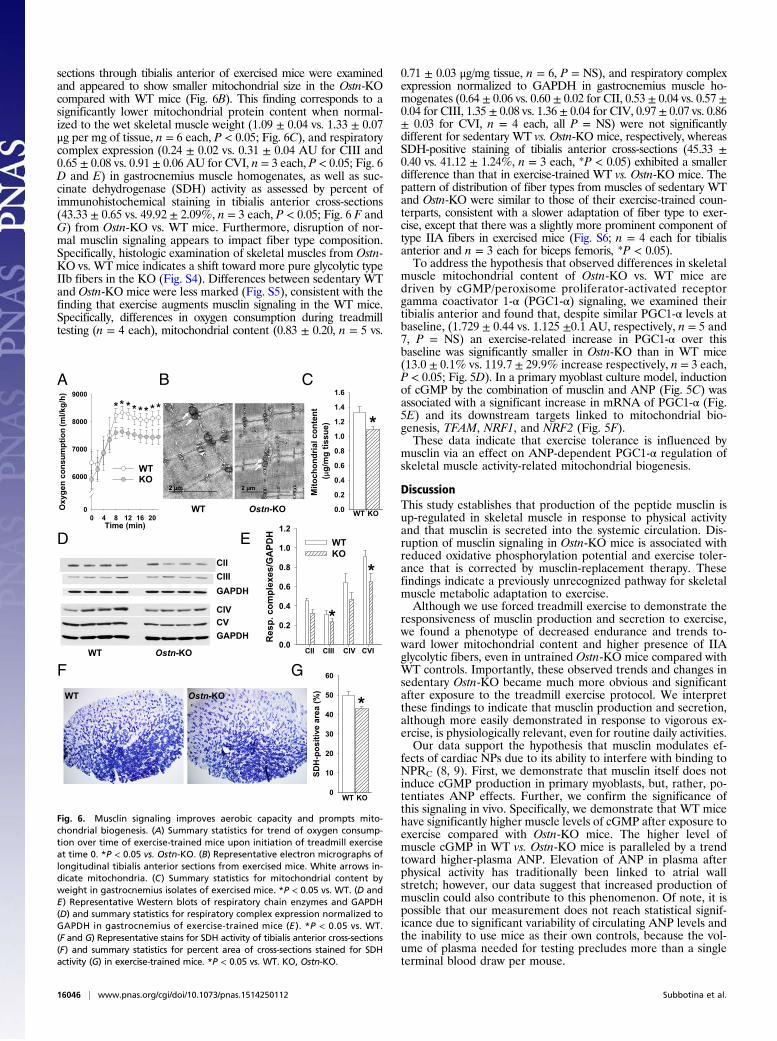
sections through tibialis anterior of exercised mice were examinedand appeared to show smaller mitochondrial size in the Ostn-KOcompared with WT mice (Fig. 6B). This finding corresponds to asignificantly lower mitochondrial protein content when normal-ized to the wet skeletal muscle weight (1.09 ± 0.04 vs. 1.33 ± 0.07μg per mg of tissue, n = 6 each, P < 0.05; Fig. 6C), and respiratorycomplex expression (0.24 ± 0.02 vs. 0.31 ± 0.04 AU for CIII and0.65 ± 0.08 vs. 0.91 ± 0.06 AU for CVI, n = 3 each, P < 0.05; Fig. 6D and E) in gastrocnemius muscle homogenates, as well as suc-cinate dehydrogenase (SDH) activity as assessed by percent ofimmunohistochemical staining in tibialis anterior cross-sections(43.33 ± 0.65 vs. 49.92 ± 2.09%, n = 3 each, P < 0.05; Fig. 6 F andG) from Ostn-KO vs. WT mice. Furthermore, disruption of nor-mal musclin signaling appears to impact fiber type composition.Specifically, histologic examination of skeletal muscles from Ostn-KO vs. WT mice indicates a shift toward more pure glycolytic typeIIb fibers in the KO (Fig. S4). Differences between sedentary WTand Ostn-KO mice were less marked (Fig. S5), consistent with thefinding that exercise augments musclin signaling in the WT mice.Specifically, differences in oxygen consumption during treadmilltesting (n = 4 each), mitochondrial content (0.83 ± 0.20, n = 5 vs.
0.71 ± 0.03 μg/mg tissue, n = 6, P = NS), and respiratory complexexpression normalized to GAPDH in gastrocnemius muscle ho-mogenates (0.64 ± 0.06 vs. 0.60 ± 0.02 for CII, 0.53 ± 0.04 vs. 0.57 ±0.04 for CIII, 1.35 ± 0.08 vs. 1.36 ± 0.04 for CIV, 0.97 ± 0.07 vs. 0.86± 0.03 for CVI, n = 4 each, all P = NS) were not significantlydifferent for sedentary WT vs. Ostn-KO mice, respectively, whereasSDH-positive staining of tibialis anterior cross-sections (45.33 ±0.40 vs. 41.12 ± 1.24%, n = 3 each, *P < 0.05) exhibited a smallerdifference than that in exercise-trained WT vs. Ostn-KO mice. Thepattern of distribution of fiber types from muscles of sedentary WTand Ostn-KO were similar to those of their exercise-trained coun-terparts, consistent with a slower adaptation of fiber type to exer-cise, except that there was a slightly more prominent component oftype IIA fibers in exercised mice (Fig. S6; n = 4 each for tibialisanterior and n = 3 each for biceps femoris, *P < 0.05).To address the hypothesis that observed differences in skeletal
muscle mitochondrial content of Ostn-KO vs. WT mice aredriven by cGMP/peroxisome proliferator-activated receptorgamma coactivator 1-α (PGC1-α) signaling, we examined theirtibialis anterior and found that, despite similar PGC1-α levels atbaseline, (1.729 ± 0.44 vs. 1.125 ±0.1 AU, respectively, n = 5 and7, P = NS) an exercise-related increase in PGC1-α over thisbaseline was significantly smaller in Ostn-KO than in WT mice(13.0 ± 0.1% vs. 119.7 ± 29.9% increase respectively, n = 3 each,P < 0.05; Fig. 5D). In a primary myoblast culture model, inductionof cGMP by the combination of musclin and ANP (Fig. 5C) wasassociated with a significant increase in mRNA of PGC1-α (Fig.5E) and its downstream targets linked to mitochondrial bio-genesis, TFAM, NRF1, and NRF2 (Fig. 5F).These data indicate that exercise tolerance is influenced by
musclin via an effect on ANP-dependent PGC1-α regulation ofskeletal muscle activity-related mitochondrial biogenesis.
DiscussionThis study establishes that production of the peptide musclin isup-regulated in skeletal muscle in response to physical activityand that musclin is secreted into the systemic circulation. Dis-ruption of musclin signaling in Ostn-KO mice is associated withreduced oxidative phosphorylation potential and exercise toler-ance that is corrected by musclin-replacement therapy. Thesefindings indicate a previously unrecognized pathway for skeletalmuscle metabolic adaptation to exercise.Although we use forced treadmill exercise to demonstrate the
responsiveness of musclin production and secretion to exercise,we found a phenotype of decreased endurance and trends to-ward lower mitochondrial content and higher presence of IIAglycolytic fibers, even in untrained Ostn-KO mice compared withWT controls. Importantly, these observed trends and changes insedentary Ostn-KO became much more obvious and significantafter exposure to the treadmill exercise protocol. We interpretthese findings to indicate that musclin production and secretion,although more easily demonstrated in response to vigorous ex-ercise, is physiologically relevant, even for routine daily activities.Our data support the hypothesis that musclin modulates ef-
fects of cardiac NPs due to its ability to interfere with binding toNPRC (8, 9). First, we demonstrate that musclin itself does notinduce cGMP production in primary myoblasts, but, rather, po-tentiates ANP effects. Further, we confirm the significance ofthis signaling in vivo. Specifically, we demonstrate that WT micehave significantly higher muscle levels of cGMP after exposure toexercise compared with Ostn-KO mice. The higher level ofmuscle cGMP in WT vs. Ostn-KO mice is paralleled by a trendtoward higher-plasma ANP. Elevation of ANP in plasma afterphysical activity has traditionally been linked to atrial wallstretch; however, our data suggest that increased production ofmusclin could also contribute to this phenomenon. Of note, it ispossible that our measurement does not reach statistical signif-icance due to significant variability of circulating ANP levels andthe inability to use mice as their own controls, because the vol-ume of plasma needed for testing precludes more than a singleterminal blood draw per mouse.
Time (min)0 4 8 12 16 20
Oxy
gen
cons
umpt
ion
(ml/k
g/h)
0
6000
7000
8000
9000
WTKO
********
CIICIII
CV
GAPDH
CIV
GAPDH
WT Ostn-KO
Res
p. c
ompl
exes
/GA
PDH
0.0
0.2
0.4
0.6
0.8
1.0
1.2WTKO
CII CIII CIV CVI
*
*
Mito
chon
dria
l con
tent
(
g/m
g tis
sue)
0.0
0.2
0.4
0.6
0.8
1.0
1.2
1.4
1.6
WT KO
*
Ostn-KOWT
WT Ostn-KO
SDH
-pos
itive
are
a (%
)
0
10
20
30
40
50
60
WT KO
*
A
D
F G
E
B C
Fig. 6. Musclin signaling improves aerobic capacity and prompts mito-chondrial biogenesis. (A) Summary statistics for trend of oxygen consump-tion over time of exercise-trained mice upon initiation of treadmill exerciseat time 0. *P < 0.05 vs. Ostn-KO. (B) Representative electron micrographs oflongitudinal tibialis anterior sections from exercised mice. White arrows in-dicate mitochondria. (C) Summary statistics for mitochondrial content byweight in gastrocnemius isolates of exercised mice. *P < 0.05 vs. WT. (D andE) Representative Western blots of respiratory chain enzymes and GAPDH(D) and summary statistics for respiratory complex expression normalized toGAPDH in gastrocnemius of exercise-trained mice (E ). *P < 0.05 vs. WT.(F and G) Representative stains for SDH activity of tibialis anterior cross-sections(F) and summary statistics for percent area of cross-sections stained for SDHactivity (G) in exercise-trained mice. *P < 0.05 vs. WT. KO, Ostn-KO.
16046 | www.pnas.org/cgi/doi/10.1073/pnas.1514250112 Subbotina et al.

We detected an order-of-magnitude lower production of musclinin the bone of our adult mice compared with that from skeletalmuscle. This finding is consistent with original reports that indicatethat high musclin expression during early bone development sharplydeclines in a time- and maturity-dependent manner in both mice (5)and humans (31). Furthermore, in contrast to skeletal muscle, noexercise-responsive increase in mRNA levels of musclin was ob-served in bones. Thus, it seems likely that the elevated systemiccirculating musclin levels after exercise are largely supported byaugmented production and secretion by skeletal muscle.Identification of the exercise-responsive nature of musclin sig-
naling, along with its systemic circulation, has important implica-tions for our understanding of exercise-dependent NP signaling.Cardiac NPs are increasingly recognized as hormones with a widespectrum of targets: In addition to traditional targets of vascula-ture and kidney, recently their effects on skeletal muscle mito-chondrial biogenesis, angiogenesis, lipolysis, and adipose tissueremodeling (browning) have been reported (27–29, 32). Althoughthe present study focuses on one aspect of this signaling network,intramuscular cGMP signaling and mitochondrial biogenesis, it ispossible that other cardiac NP signaling targets are similarly af-fected. For example, musclin may be at least partially responsiblefor the beneficial effect of exercise on cardiac remodeling. Suchtargets should be the subject of future investigation.cGMP signaling has been linked to PGC1-α–dependent mito-
chondrial biogenesis in many studies in different tissues and or-gans (33–35). cGMP production in skeletal muscles is typicallylinked to nitric oxide signaling (34–36), although recently a rolefor NPs in this process has been established (28). Our data supportthe importance NP signaling and its regulation by musclin incGMP/PGC1-α–driven mitochondrial biogenesis: We confirmsignificantly greater mitochondrial quantity and function by mul-tiple methods and demonstrate the in vivo functional importance by
revealing a meaningful increase in the V·O2max, a parameter thatreflects many factors, including oxidative phosphorylation potentialand cardiovascular and pulmonary functions critical for physicalendurance, of mice with intact vs. disrupted musclin signaling.Finally, our demonstration that musclin infusion rescues ex-
ercise and oxidative capacity in Ostn-KO mice, as well as en-hances exercise and oxidative capacity in untrained WT mice,suggests a potential therapeutic role for musclin. Overexpressionof musclin in chondrocytes has been linked to abnormal skeletalgrowth (9), but such changes may or may not occur with sys-temically delivered musclin. Thus, to better understand musclin’stherapeutic potential, further studies will be required to de-termine the extent and durability of beneficial effects andwhether there are accompanying long-term deleterious conse-quences. Such studies may be aided by conditional models.In summary, this study defines musclin as an exercise-responsive
factor promoting skeletal muscle mitochondrial biogenesis andexercise endurance.
Materials and MethodsAll animal protocols conform to the Guide for the Care and Use of Labo-ratory Animals (37) generated by the Institute for Laboratory Animal Re-search, National Research Council of the National Academies. All animalprotocols were approved by the University of Iowa Institutional Animal Careand Use Committee. See SI Materials and Methods for detailed methods.Results are expressed as mean ± SEM.
ACKNOWLEDGMENTS. We thank Chantal Allamargot, PhD, for her assis-tance with electron microscopy. This work was supported by NationalInstitutes of Health Grants HL113089 (to D.H-Z.) and HL093368 andDK092412 (to L.V.Z.); Veterans Affairs Merit Review Program 1I0BX000718(to L.V.Z.); and the Fraternal Order of Eagles Diabetes Research Center.
1. Lee IM, et al.; Lancet Physical Activity Series Working Group (2012) Effect of physicalinactivity on major non-communicable diseases worldwide: An analysis of burden ofdisease and life expectancy. Lancet 380(9838):219–229.
2. Pedersen BK, Akerström TC, Nielsen AR, Fischer CP (2007) Role of myokines in exerciseand metabolism. J Appl Physiol (1985) 103(3):1093–1098.
3. Pedersen BK, et al. (2004) The metabolic role of IL-6 produced during exercise: Is IL-6an exercise factor? Proc Nutr Soc 63(2):263–267.
4. Nishizawa H, et al. (2004) Musclin, a novel skeletal muscle-derived secretory factor.J Biol Chem 279(19):19391–19395.
5. Thomas G, et al. (2003) Osteocrin, a novel bone-specific secreted protein that mod-ulates the osteoblast phenotype. J Biol Chem 278(50):50563–50571.
6. Yasui A, et al. (2007) Foxo1 represses expression of musclin, a skeletal muscle-derivedsecretory factor. Biochem Biophys Res Commun 364(2):358–365.
7. Potter LR, Abbey-Hosch S, Dickey DM (2006) Natriuretic peptides, their receptors, and cyclicguanosine monophosphate-dependent signaling functions. Endocr Rev 27(1):47–72.
8. Kita S, et al. (2009) Competitive binding of musclin to natriuretic peptide receptor 3with atrial natriuretic peptide. J Endocrinol 201(2):287–295.
9. Moffatt P, et al. (2007) Osteocrin is a specific ligand of the natriuretic Peptide clear-ance receptor that modulates bone growth. J Biol Chem 282(50):36454–36462.
10. Suda M, et al. (1998) Skeletal overgrowth in transgenic mice that overexpress brainnatriuretic peptide. Proc Natl Acad Sci USA 95(5):2337–2342.
11. Yasoda A, et al. (2004) Overexpression of CNP in chondrocytes rescues achondroplasiathrough a MAPK-dependent pathway. Nat Med 10(1):80–86.
12. Jaubert J, et al. (1999) Three new allelic mouse mutations that cause skeletal over-growth involve the natriuretic peptide receptor C gene (Npr3). Proc Natl Acad Sci USA96(18):10278–10283.
13. Matsukawa N, et al. (1999) The natriuretic peptide clearance receptor locally modu-lates the physiological effects of the natriuretic peptide system. Proc Natl Acad SciUSA 96(13):7403–7408.
14. CatoireM, Kersten S (2015) The search for exercise factors in humans. FASEB J 29(5):1615–1628.15. Gross DN, Wan M, Birnbaum MJ (2009) The role of FOXO in the regulation of me-
tabolism. Curr Diab Rep 9(3):208–214.16. Stitt TN, et al. (2004) The IGF-1/PI3K/Akt pathway prevents expression of muscle atrophy-
induced ubiquitin ligases by inhibiting FOXO transcription factors. Mol Cell 14(3):395–403.17. Wu M, Falasca M, Blough ER (2011) Akt/protein kinase B in skeletal muscle physiology
and pathology. J Cell Physiol 226(1):29–36.18. Shaw M, Cohen P, Alessi DR (1998) The activation of protein kinase B by H2O2 or heat
shock is mediated by phosphoinositide 3-kinase and not by mitogen-activated proteinkinase-activated protein kinase-2. Biochem J 336(Pt 1):241–246.
19. Coffer PJ, Jin J, Woodgett JR (1998) Protein kinase B (c-Akt): A multifunctional me-diator of phosphatidylinositol 3-kinase activation. Biochem J 335(Pt 1):1–13.
20. Vanhaesebroeck B, Alessi DR (2000) The PI3K-PDK1 connection: More than just a roadto PKB. Biochem J 346(Pt 3):561–576.
21. Sable CL, Filippa N, Hemmings B, Van Obberghen E (1997) cAMP stimulates proteinkinase B in a Wortmannin-insensitive manner. FEBS Lett 409(2):253–257.
22. Yano S, Tokumitsu H, Soderling TR (1998) Calcium promotes cell survival throughCaM-K kinase activation of the protein-kinase-B pathway. Nature 396(6711):584–587.
23. Nader GA, Esser KA (2001) Intracellular signaling specificity in skeletal muscle in re-sponse to different modes of exercise. J Appl Physiol (1985) 90(5):1936–1942.
24. Sakamoto K, Hirshman MF, Aschenbach WG, Goodyear LJ (2002) Contraction regu-lation of Akt in rat skeletal muscle. J Biol Chem 277(14):11910–11917.
25. Turinsky J, Damrau-Abney A (1999) Akt kinases and 2-deoxyglucose uptake in rat skel-etal muscles in vivo: Study with insulin and exercise. Am J Physiol 276(1 Pt 2):R277–R282.
26. Brunet A, et al. (1999) Akt promotes cell survival by phosphorylating and inhibiting aForkhead transcription factor. Cell 96(6):857–868.
27. Zois NE, et al. (2014) Natriuretic peptides in cardiometabolic regulation and disease.Nat Rev Cardiol 11(7):403–412.
28. Miyashita K, et al. (2009) Natriuretic peptides/cGMP/cGMP-dependent protein kinasecascades promote muscle mitochondrial biogenesis and prevent obesity. Diabetes58(12):2880–2892.
29. Engeli S, et al. (2012) Natriuretic peptides enhance the oxidative capacity of humanskeletal muscle. J Clin Invest 122(12):4675–4679.
30. Shephard RJ (1984) Tests of maximum oxygen intake. A critical review. Sports Med1(2):99–124.
31. Bord S, Ireland DC, Moffatt P, Thomas GP, Compston JE (2005) Characterization ofosteocrin expression in human bone. J Histochem Cytochem 53(10):1181–1187.
32. Gruden G, Landi A, Bruno G (2014) Natriuretic peptides, heart, and adipose tissue:New findings and future developments for diabetes research. Diabetes Care 37(11):2899–2908.
33. Brown GC (2007) Nitric oxide and mitochondria. Front Biosci 12:1024–1033.34. Nisoli E, et al. (2003) Mitochondrial biogenesis in mammals: The role of endogenous
nitric oxide. Science 299(5608):896–899.35. Nisoli E, et al. (2004) Mitochondrial biogenesis by NO yields functionally active mi-
tochondria in mammals. Proc Natl Acad Sci USA 101(47):16507–16512.36. Haas B, et al. (2009) Protein kinase G controls brown fat cell differentiation and mi-
tochondrial biogenesis. Sci Signal 2(99):ra78.37. Committee on Care and Use of Laboratory Animals (1996) Guide for the Care and Use
of Laboratory Animals (Natl Inst Health, Bethesda), DHHS Publ No (NIH) 85-23.38. He BJ, et al. (2011) Oxidation of CaMKII determines the cardiotoxic effects of aldo-
sterone. Nat Med 17(12):1610–1618.39. Alekseev AE, et al. (2010) Sarcolemmal ATP-sensitive K(+) channels control energy
expenditure determining body weight. Cell Metab 11(1):58.40. Zhu Z, et al. (2014) Sarcolemmal ATP-sensitive potassium channels modulate skeletal
muscle function under low-intensity workloads. J Gen Physiol 143(1):119–134.41. Zingman LV, et al. (2002) Kir6.2 is required for adaptation to stress. Proc Natl Acad Sci
USA 99(20):13278–13283.
Subbotina et al. PNAS | December 29, 2015 | vol. 112 | no. 52 | 16047
PHYS
IOLO
GY