Embed Size (px)
Citation preview
Musculoskeletal Anatomy
Chapter 1
1
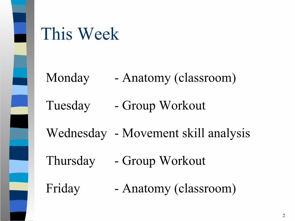
This Week
Monday - Anatomy (classroom)
Tuesday - Group Workout
Wednesday - Movement skill analysis
Thursday - Group Workout
Friday - Anatomy (classroom)
2
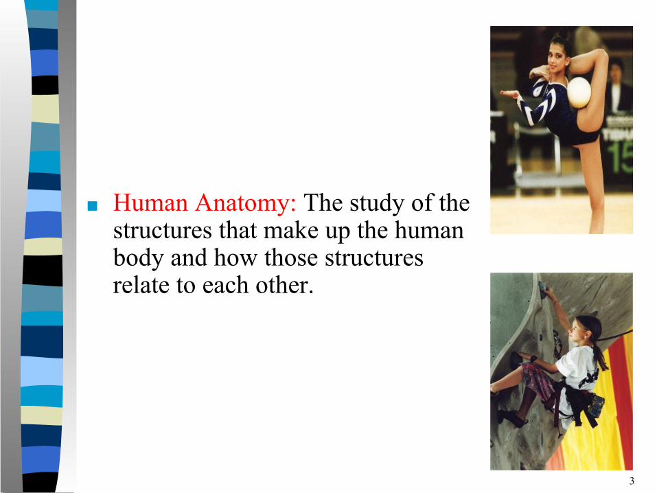
■ Human Anatomy: The study of the structures that make up the human body and how those structures relate to each other.
3

The Musculoskeletal System
■ The Musculoskeletal System is made up of–the skeletal system which includes bones and joints–the muscular system which contains the muscles
When bones come together, they form joints. Muscles cross these joints and pull on bones causing movement at the joints.
4
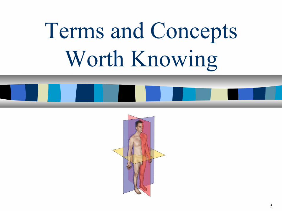
Terms and Concepts Worth Knowing
5

Anatomical Position
6
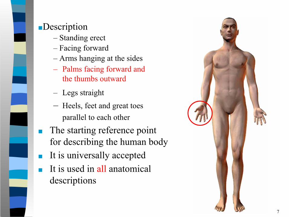
– Legs straight– Heels, feet and great toes
parallel to each other ■ The starting reference point
for describing the human body■ It is universally accepted ■ It is used in all anatomical
descriptions
– Palms facing forward and the thumbs outward
7
■Description– Standing erect– Facing forward– Arms hanging at the sides
Directional Terms
8
Reminder:
All directional terms are based on the assumption that the body is in the
anatomical position.
9
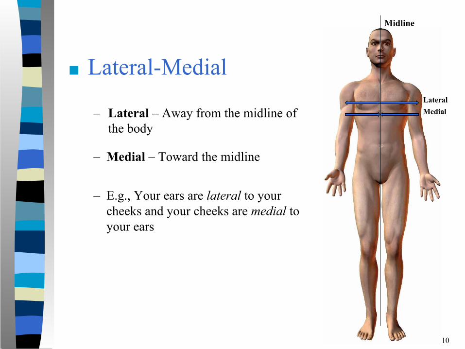
■ Lateral-Medial
– Lateral – Away from the midline of the body
10
Midline
LateralMedial
– Medial – Toward the midline
– E.g., Your ears are lateral to your cheeks and your cheeks are medial to your ears
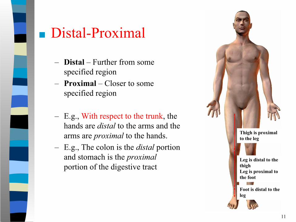
■ Distal-Proximal
– Distal – Further from some specified region
– Proximal – Closer to some specified region
– E.g., With respect to the trunk, the hands are distal to the arms and the arms are proximal to the hands.
– E.g., The colon is the distal portion and stomach is the proximal portion of the digestive tract
11
Thigh is proximal to the leg
Leg is distal to the thighLeg is proximal to the foot
Foot is distal to the leg
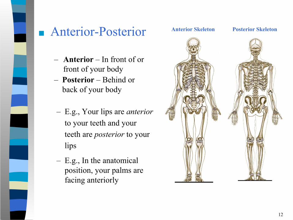
■ Anterior-Posterior
– Anterior – In front of or front of your body
12
Anterior Skeleton
– Posterior – Behind or back of your body
– E.g., Your lips are anterior to your teeth and your teeth are posterior to your lips
Posterior Skeleton
– E.g., In the anatomical position, your palms are facing anteriorly
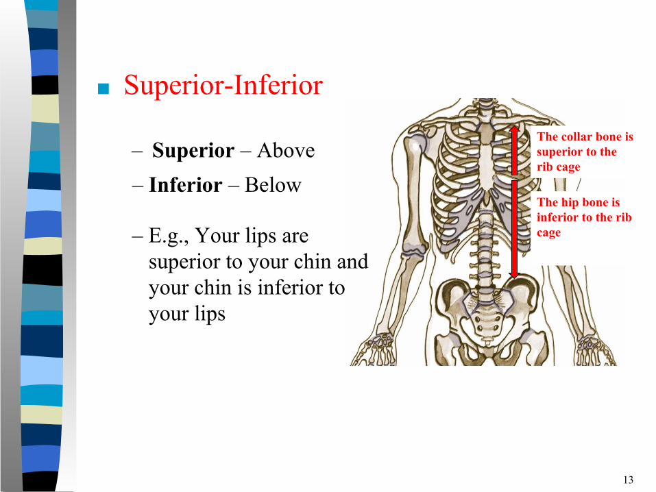
■ Superior-Inferior
– Superior – Above
13
The collar bone is superior to the rib cage
– Inferior – Below
– E.g., Your lips are superior to your chin and your chin is inferior to your lips
The hip bone isinferior to the ribcage
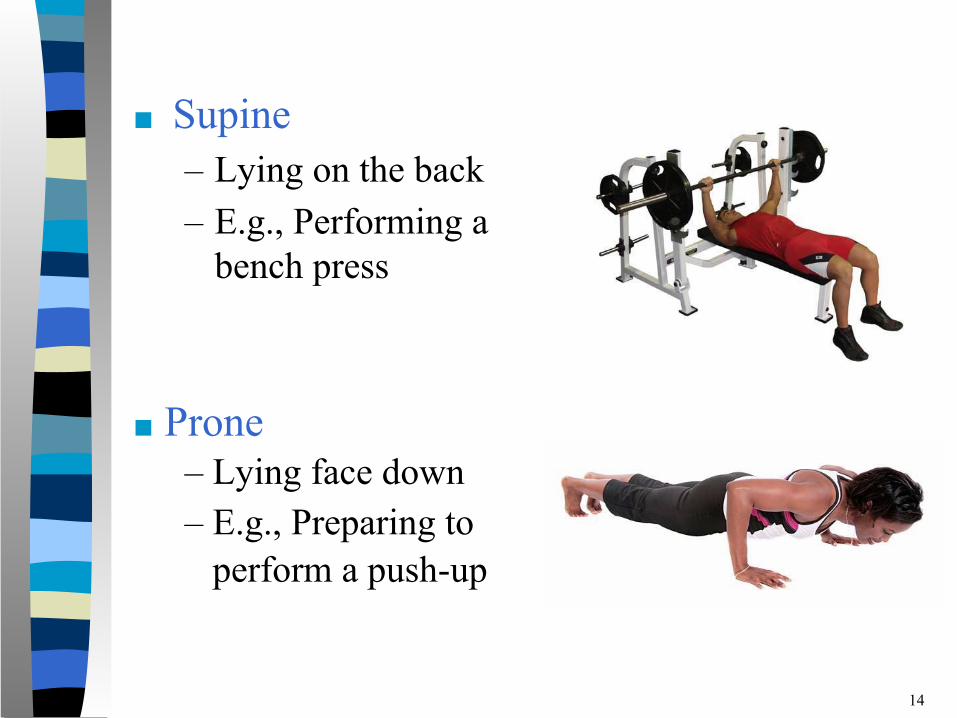
■ Supine– Lying on the back – E.g., Performing a
bench press
14
■ Prone – Lying face down – E.g., Preparing to perform a push-up
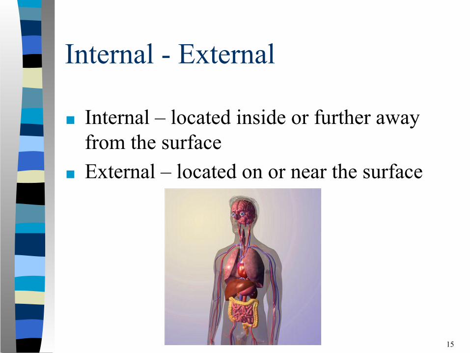
Internal - External
■ Internal – located inside or further away from the surface
■ External – located on or near the surface
15
Planes of the Body
16
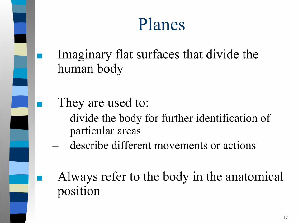
Planes■ Imaginary flat surfaces that divide the
human body
■ They are used to:– divide the body for further identification of
particular areas– describe different movements or actions
■ Always refer to the body in the anatomical position
17
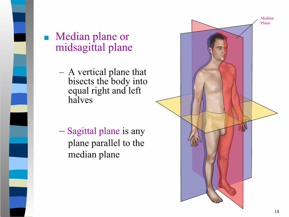
■ Median plane or midsagittal plane
– A vertical plane that bisects the body into equal right and left halves
18
– Sagittal plane is any plane parallel to the median plane
MedianPlane
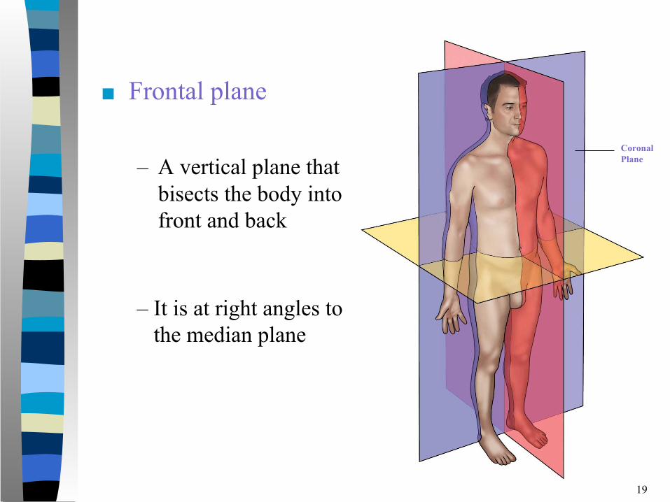
■ Frontal plane
– A vertical plane that bisects the body into front and back
19
– It is at right angles to the median plane
Coronal Plane
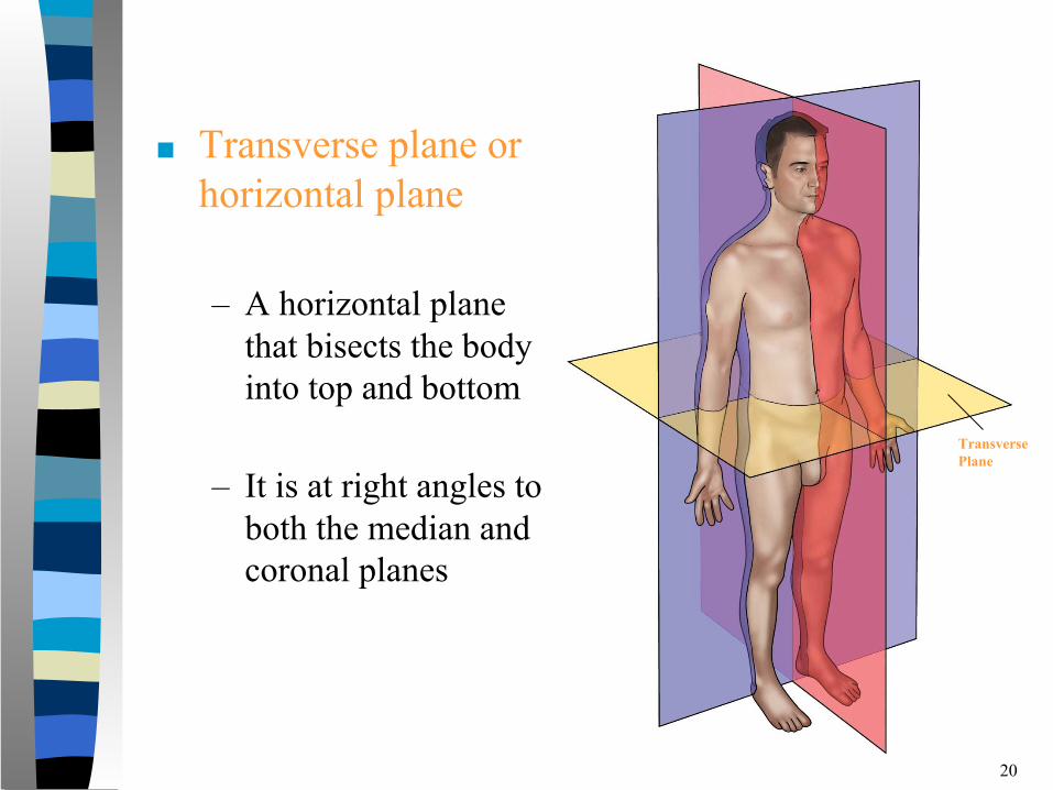
■ Transverse plane or horizontal plane
– A horizontal plane that bisects the body into top and bottom
– It is at right angles to both the median and coronal planes
20
Transverse Plane
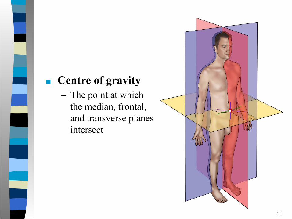
■ Centre of gravity– The point at which
the median, frontal, and transverse planes intersect
21

Movements
22
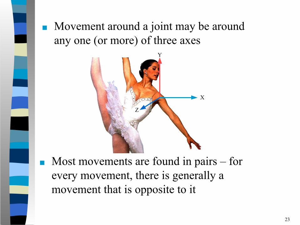
■ Movement around a joint may be around any one (or more) of three axes
■ Most movements are found in pairs – for every movement, there is generally a movement that is opposite to it
23
X
Y
Z
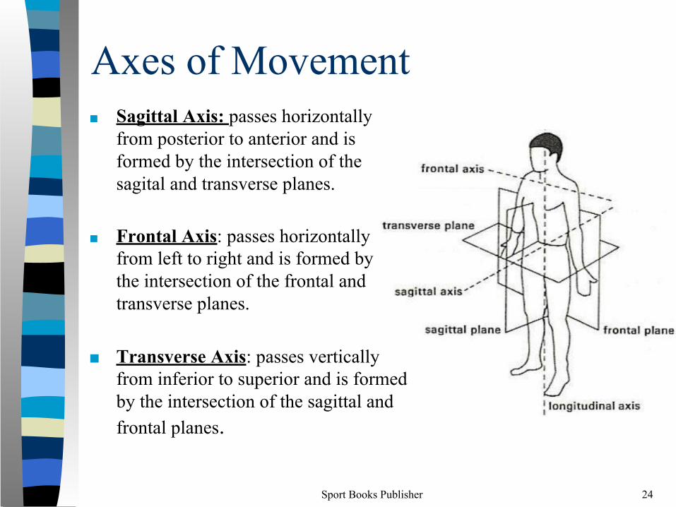
Axes of Movement■ Sagittal Axis: passes horizontally
from posterior to anterior and is formed by the intersection of the sagital and transverse planes.
■ Frontal Axis: passes horizontally from left to right and is formed by the intersection of the frontal and transverse planes.
■ Transverse Axis: passes vertically from inferior to superior and is formed by the intersection of the sagittal and frontal planes.
Sport Books Publisher 24
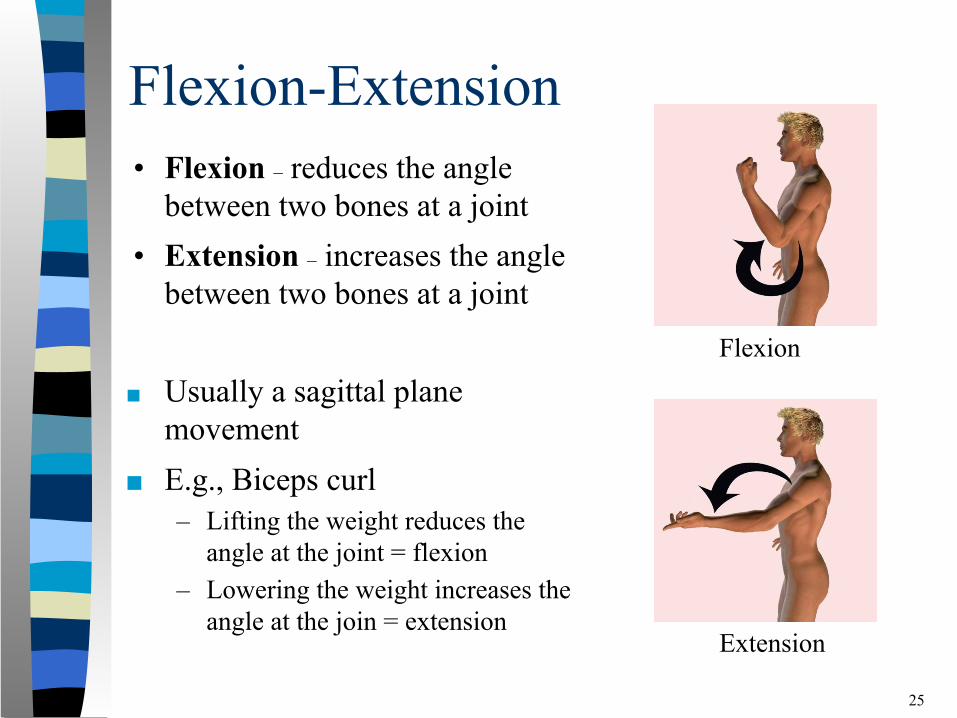
Flexion-Extension
■ Usually a sagittal plane movement
■ E.g., Biceps curl – Lifting the weight reduces the
angle at the joint = flexion– Lowering the weight increases the
angle at the join = extension
25
• Flexion – reduces the angle between two bones at a joint
• Extension – increases the angle between two bones at a joint
Flexion
Extension
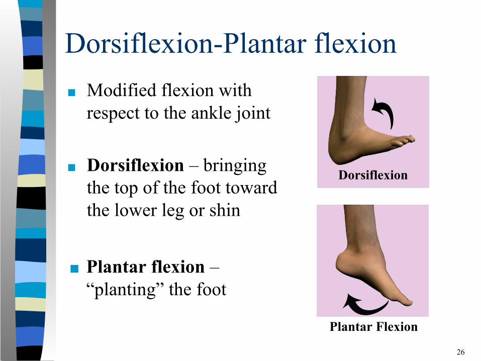
Dorsiflexion-Plantar flexion■ Modified flexion with
respect to the ankle joint
■ Dorsiflexion – bringing the top of the foot toward the lower leg or shin
26
■ Plantar flexion – “planting” the foot
Dorsiflexion
Plantar Flexion
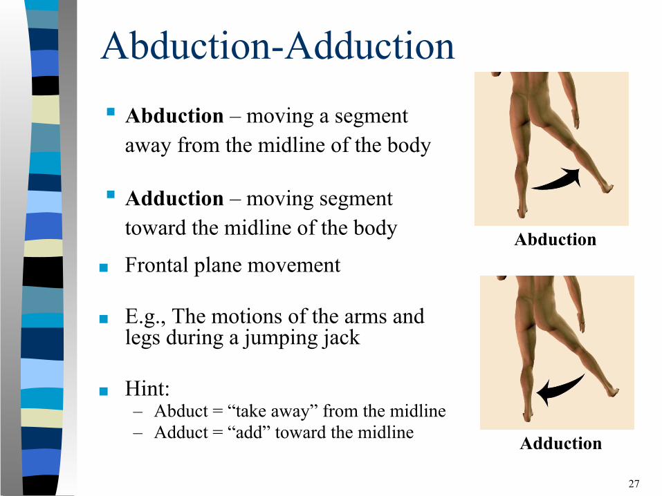
Abduction-Adduction
■ Frontal plane movement
■ E.g., The motions of the arms and legs during a jumping jack
■ Hint: – Abduct = “take away” from the midline– Adduct = “add” toward the midline
27
Abduction
Adduction
▪ Abduction – moving a segment away from the midline of the body
▪ Adduction – moving segment toward the midline of the body
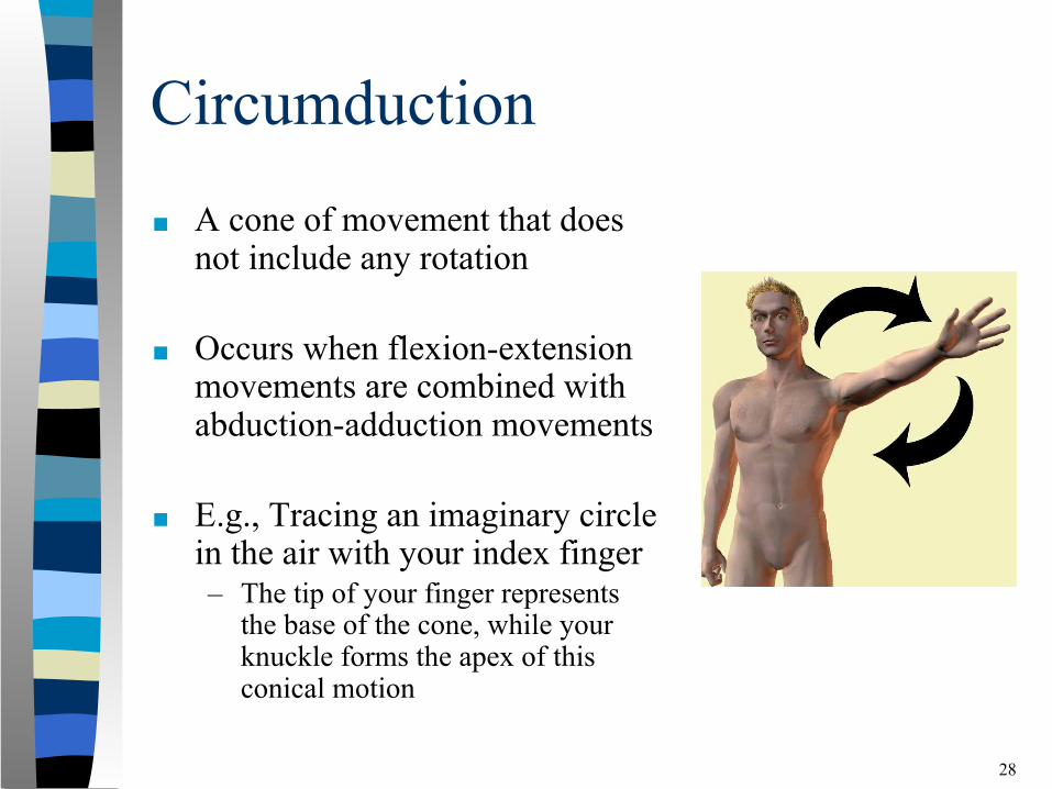
Circumduction
■ A cone of movement that does not include any rotation
■ Occurs when flexion-extension movements are combined with abduction-adduction movements
■ E.g., Tracing an imaginary circle in the air with your index finger – The tip of your finger represents
the base of the cone, while your knuckle forms the apex of this conical motion
28
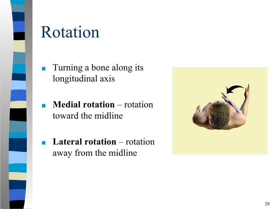
Rotation
■ Turning a bone along its longitudinal axis
■ Medial rotation – rotation toward the midline
■ Lateral rotation – rotation away from the midline
29
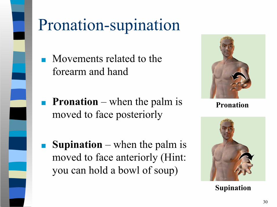
Pronation-supination
■ Movements related to the forearm and hand
■ Pronation – when the palm is moved to face posteriorly
■ Supination – when the palm is moved to face anteriorly (Hint: you can hold a bowl of soup)
30
Pronation
Supination
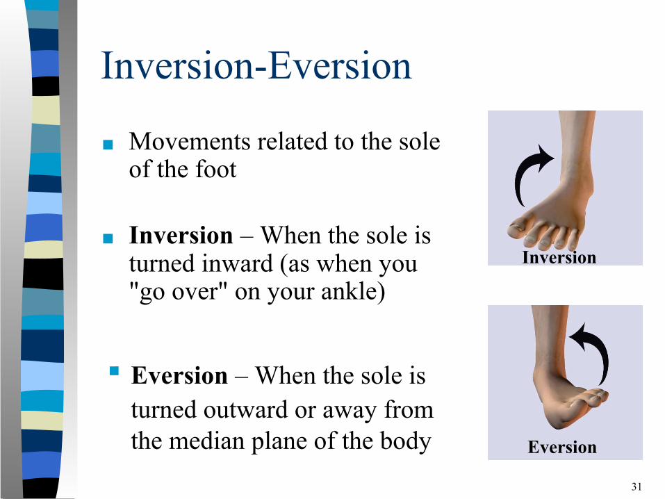
Inversion-Eversion
■ Movements related to the sole of the foot
■ Inversion – When the sole is turned inward (as when you "go over" on your ankle)
31
Eversion
Inversion
▪ Eversion – When the sole is turned outward or away from the median plane of the body
The Musculoskeletal System
32
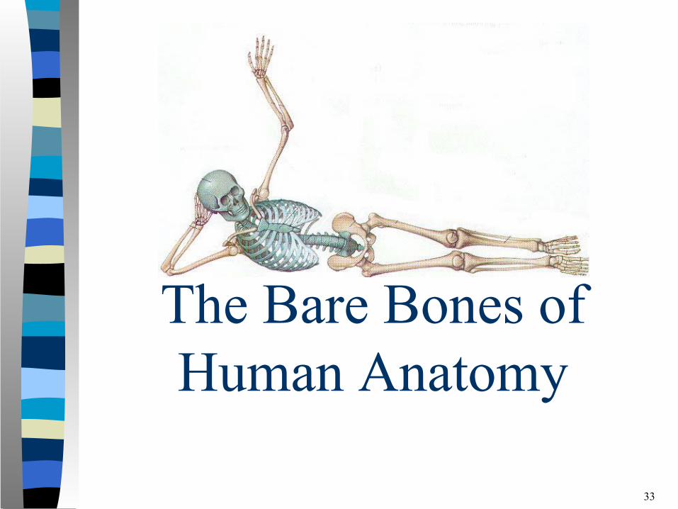
The Bare Bones ofHuman Anatomy
33
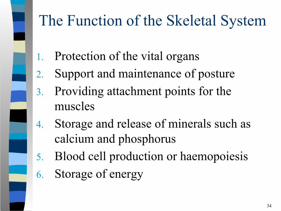
The Function of the Skeletal System
1. Protection of the vital organs2. Support and maintenance of posture3. Providing attachment points for the
muscles4. Storage and release of minerals such as
calcium and phosphorus5. Blood cell production or haemopoiesis6. Storage of energy
34
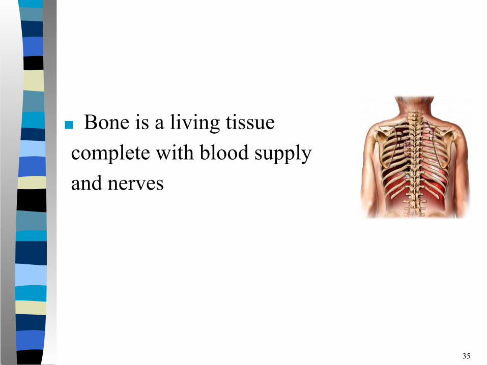
■ Bone is a living tissue complete with blood supply and nerves
35
■ The skeletal system is made up of the bones, cartilage, ligaments and joints of the body, and accounts for approximately 20% of body weight.
■ The skeleton roughly determines the shape and size of the body.
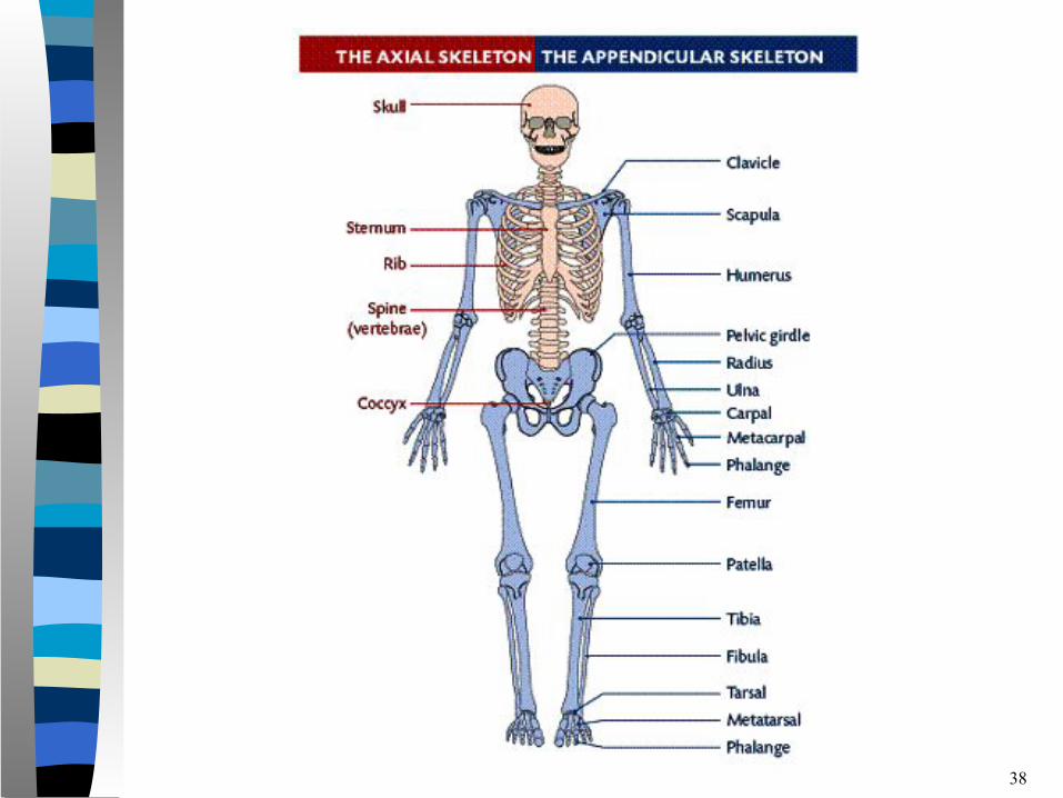
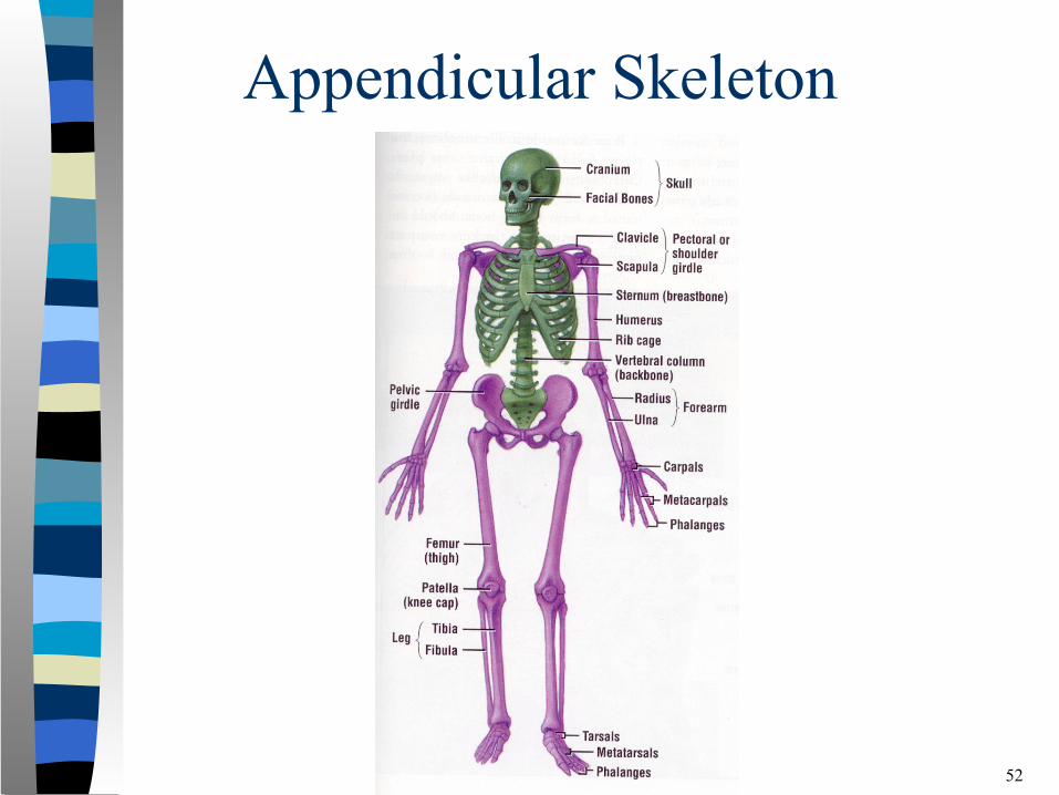
■ There are 206 bones in the skeleton and it is divided into two parts: the axial skeleton and the appendicular skeleton.
36
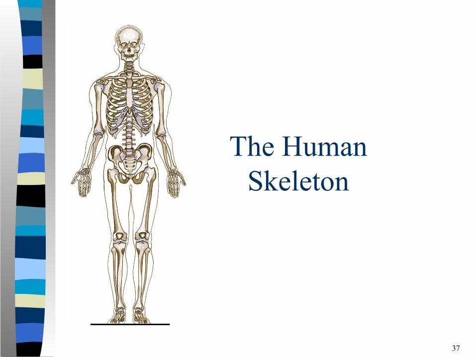
The Human Skeleton
37
38
Axial Skeleton
39
40
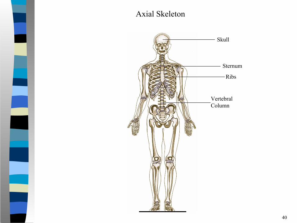
Skull
Sternum
Ribs
Vertebral Column
Axial Skeleton
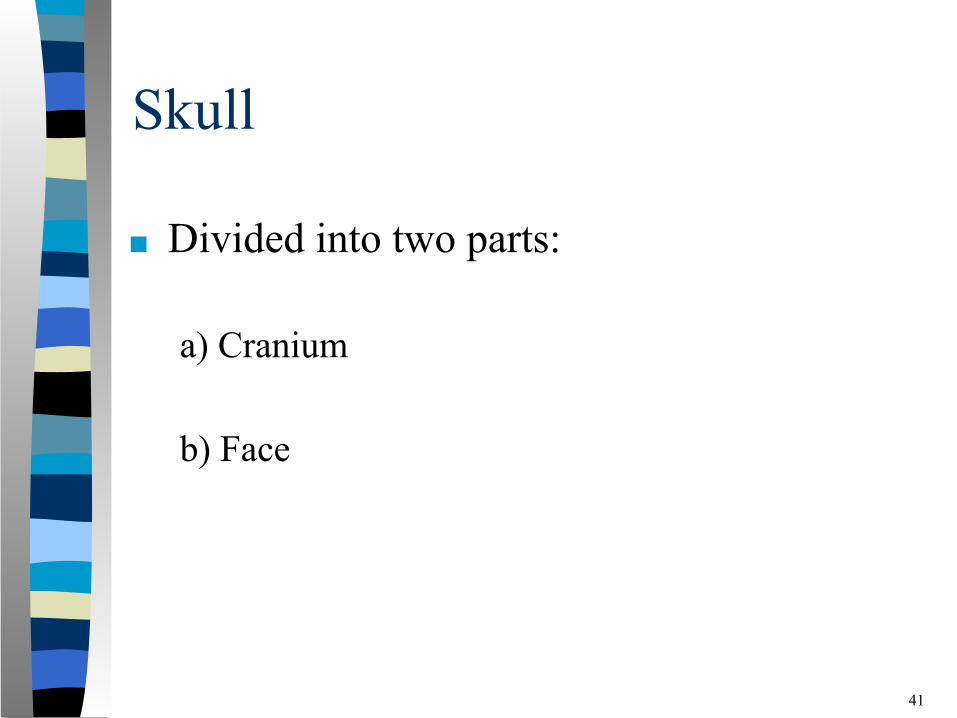
Skull
■ Divided into two parts:
a) Cranium
b) Face
41
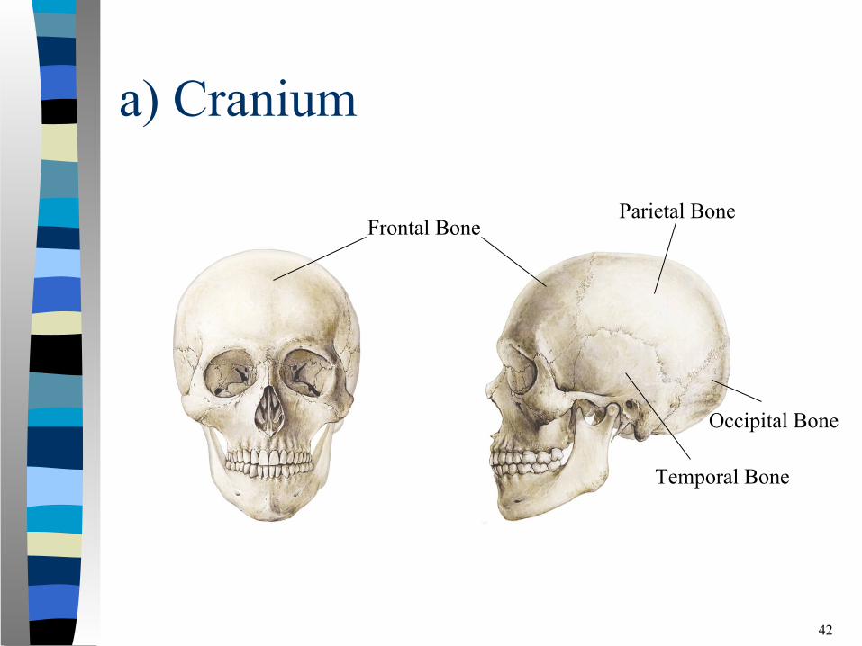
a) Cranium
42
Frontal BoneParietal Bone
Temporal Bone
Occipital Bone
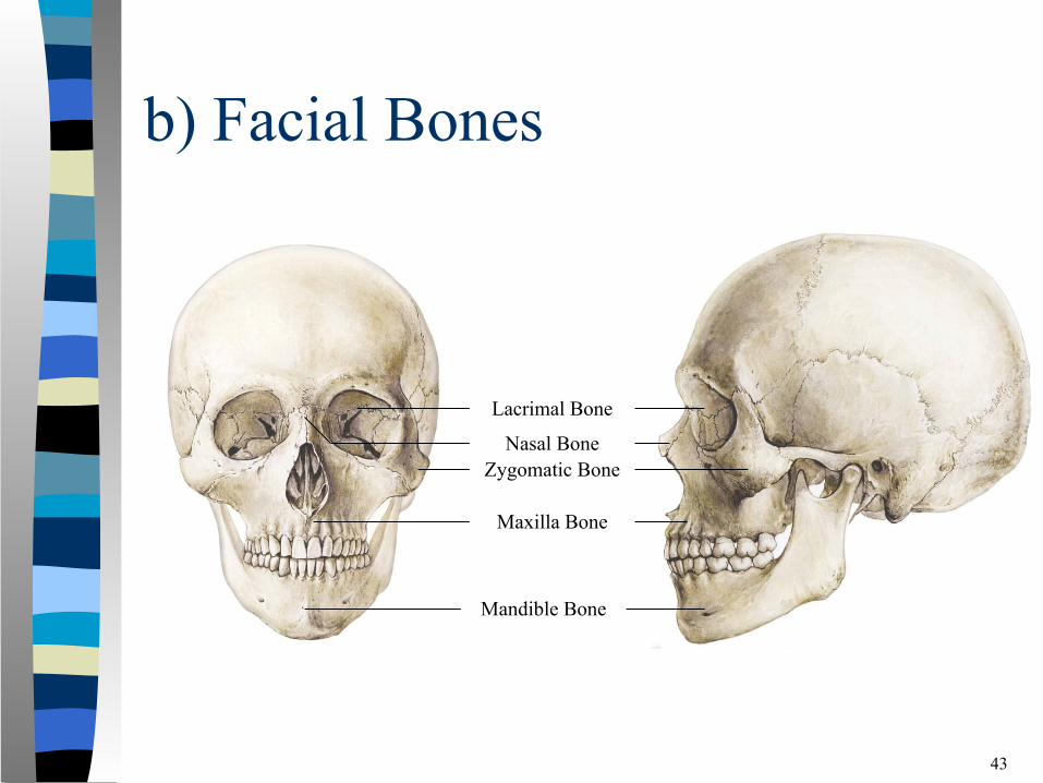
b) Facial Bones
43
Lacrimal Bone
Nasal Bone
Maxilla Bone
Mandible Bone
Zygomatic Bone
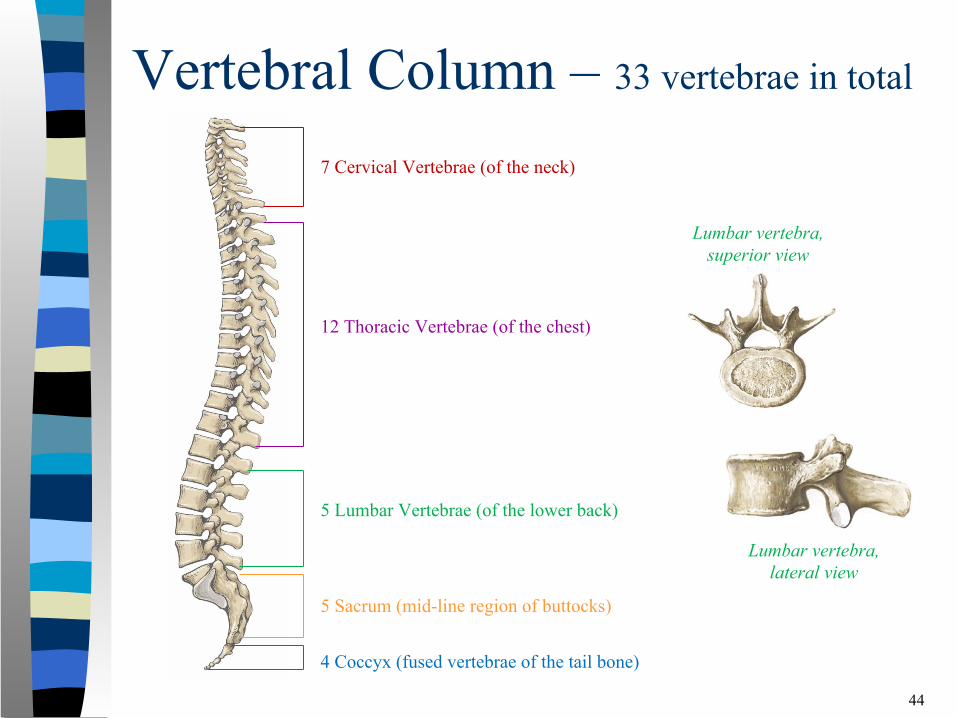
Vertebral Column – 33 vertebrae in total
44
5 Sacrum (mid-line region of buttocks)
4 Coccyx (fused vertebrae of the tail bone)
7 Cervical Vertebrae (of the neck)
12 Thoracic Vertebrae (of the chest)
5 Lumbar Vertebrae (of the lower back)
Lumbar vertebra, lateral view
Lumbar vertebra, superior view
Vertebral Column
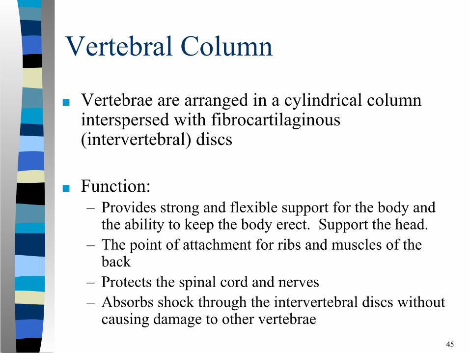
■ Vertebrae are arranged in a cylindrical column interspersed with fibrocartilaginous (intervertebral) discs
■ Function:– Provides strong and flexible support for the body and
the ability to keep the body erect. Support the head.– The point of attachment for ribs and muscles of the
back– Protects the spinal cord and nerves– Absorbs shock through the intervertebral discs without
causing damage to other vertebrae 45
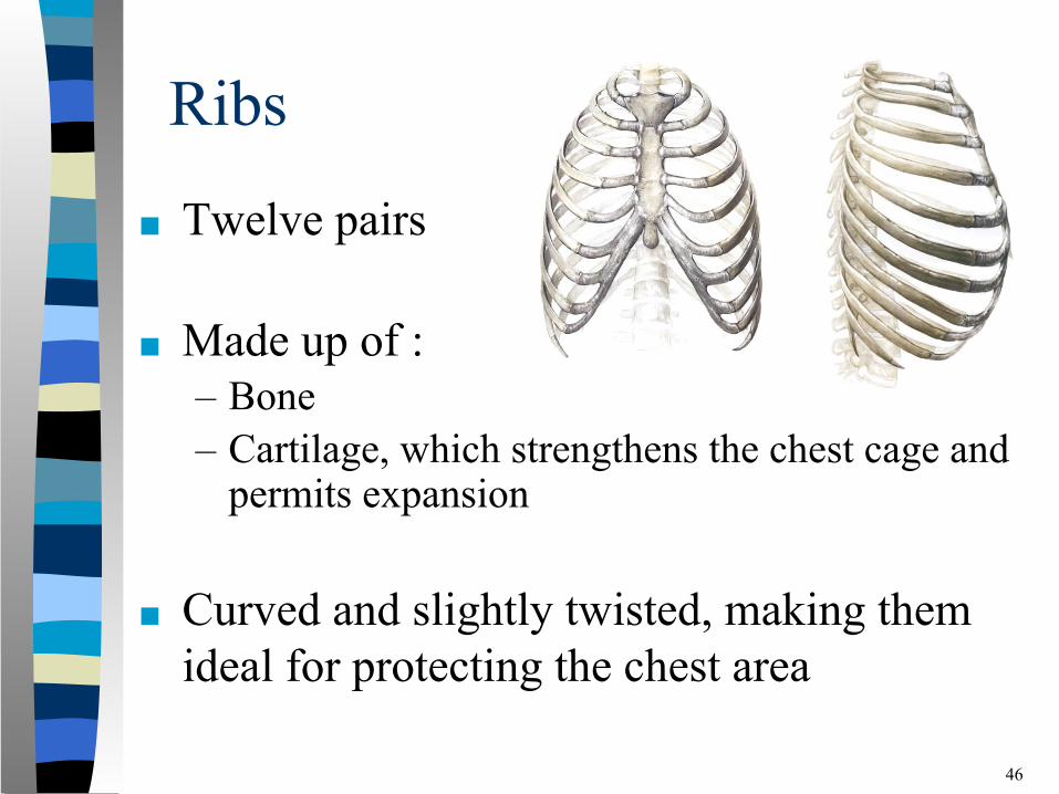
Ribs
■ Twelve pairs
■ Made up of :– Bone– Cartilage, which strengthens the chest cage and
permits expansion
46
■ Curved and slightly twisted, making them ideal for protecting the chest area
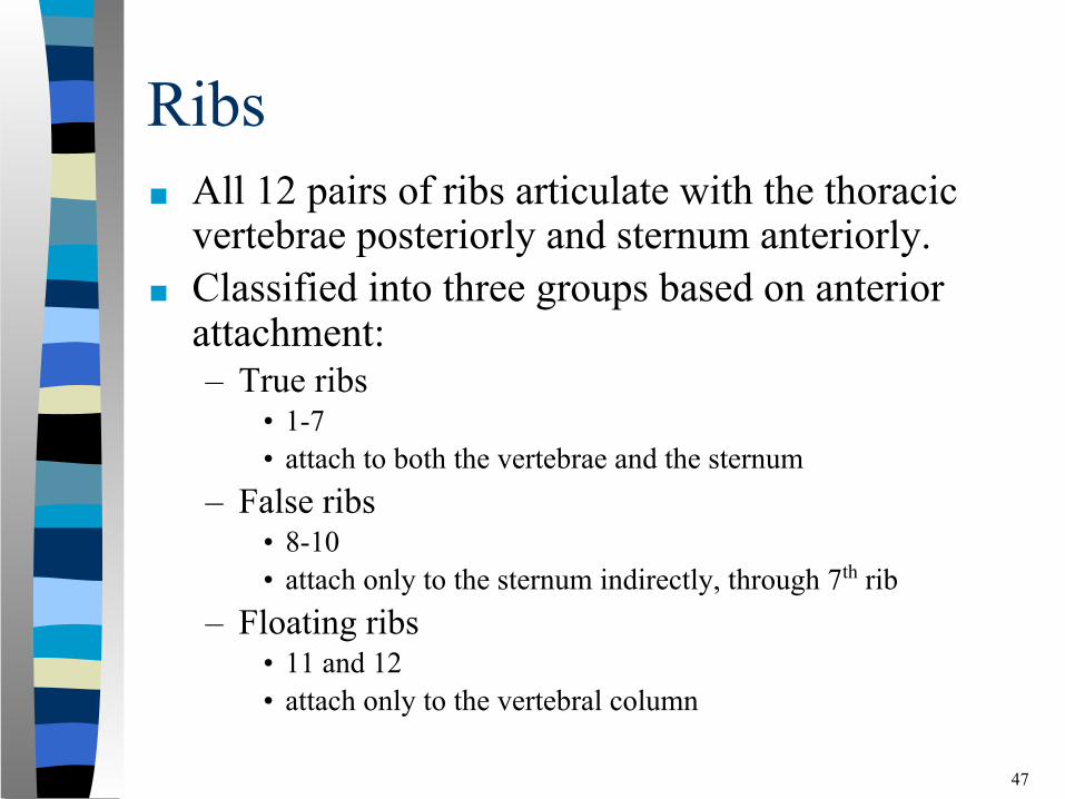
Ribs■ All 12 pairs of ribs articulate with the thoracic
vertebrae posteriorly and sternum anteriorly.■ Classified into three groups based on anterior
attachment:– True ribs
• 1-7• attach to both the vertebrae and the sternum
– False ribs • 8-10 • attach only to the sternum indirectly, through 7th rib
– Floating ribs• 11 and 12 • attach only to the vertebral column
47
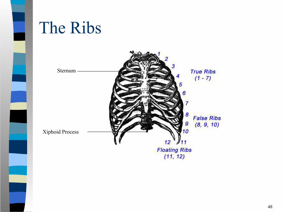
The Ribs
48
Sternum
Xiphoid Process
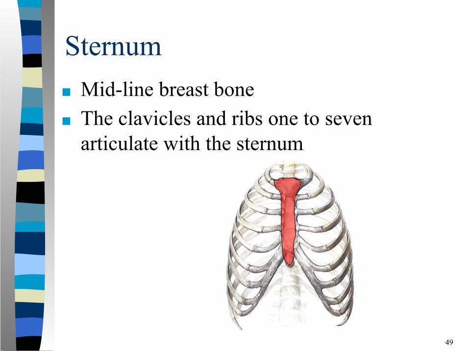
Sternum■ Mid-line breast bone ■ The clavicles and ribs one to seven
articulate with the sternum
49

Appendicular Skeleton
50
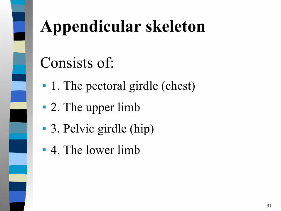
Consists of:▪ 1. The pectoral girdle (chest)
▪ 2. The upper limb
▪ 3. Pelvic girdle (hip)
▪ 4. The lower limb
51
Appendicular skeleton
Appendicular Skeleton
52
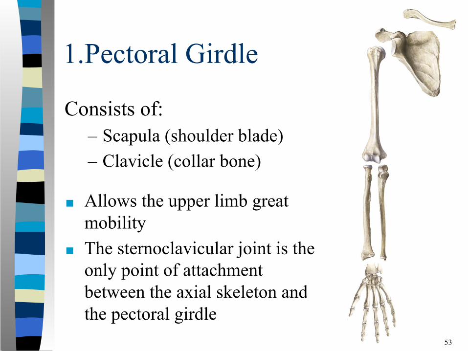
1.Pectoral Girdle
Consists of: – Scapula (shoulder blade) – Clavicle (collar bone)
53
■ Allows the upper limb great mobility
■ The sternoclavicular joint is the only point of attachment between the axial skeleton and the pectoral girdle
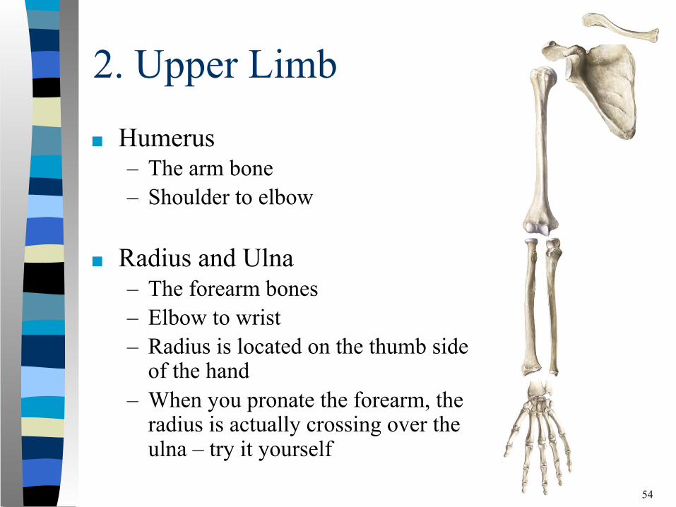
2. Upper Limb
■ Humerus– The arm bone – Shoulder to elbow
■ Radius and Ulna– The forearm bones– Elbow to wrist– Radius is located on the thumb side
of the hand– When you pronate the forearm, the
radius is actually crossing over the ulna – try it yourself
54
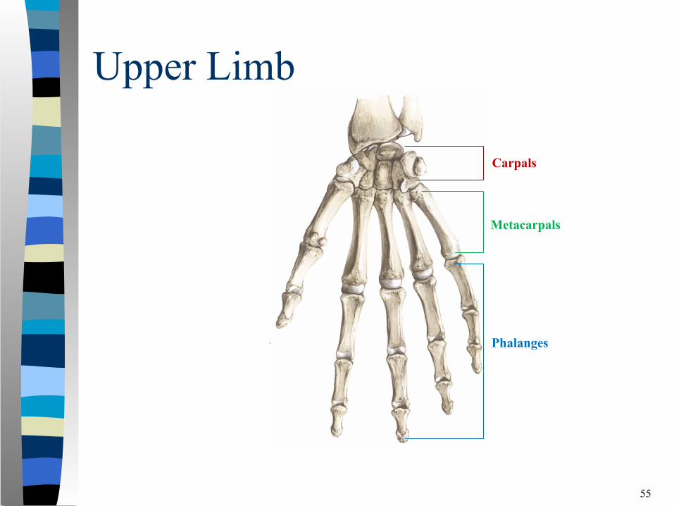
Upper Limb
55
Carpals
Phalanges
Metacarpals
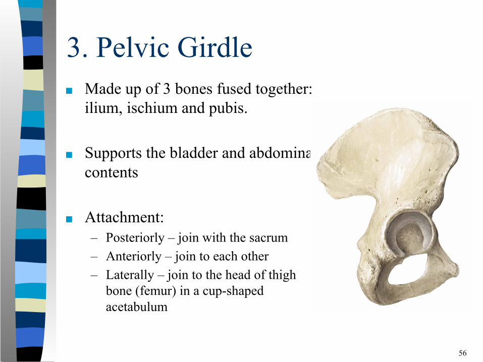
3. Pelvic Girdle■ Made up of 3 bones fused together:
ilium, ischium and pubis.
■ Supports the bladder and abdominal contents
■ Attachment:– Posteriorly – join with the sacrum – Anteriorly – join to each other– Laterally – join to the head of thigh
bone (femur) in a cup-shaped acetabulum
56
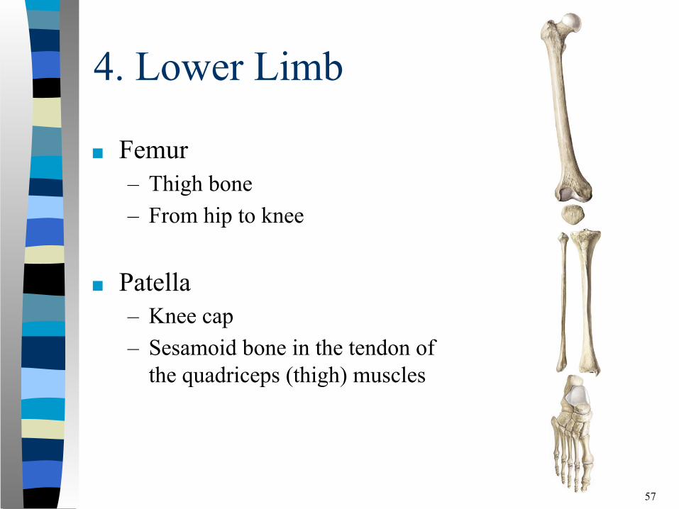
4. Lower Limb
■ Femur – Thigh bone – From hip to knee
■ Patella – Knee cap– Sesamoid bone in the tendon of
the quadriceps (thigh) muscles
57
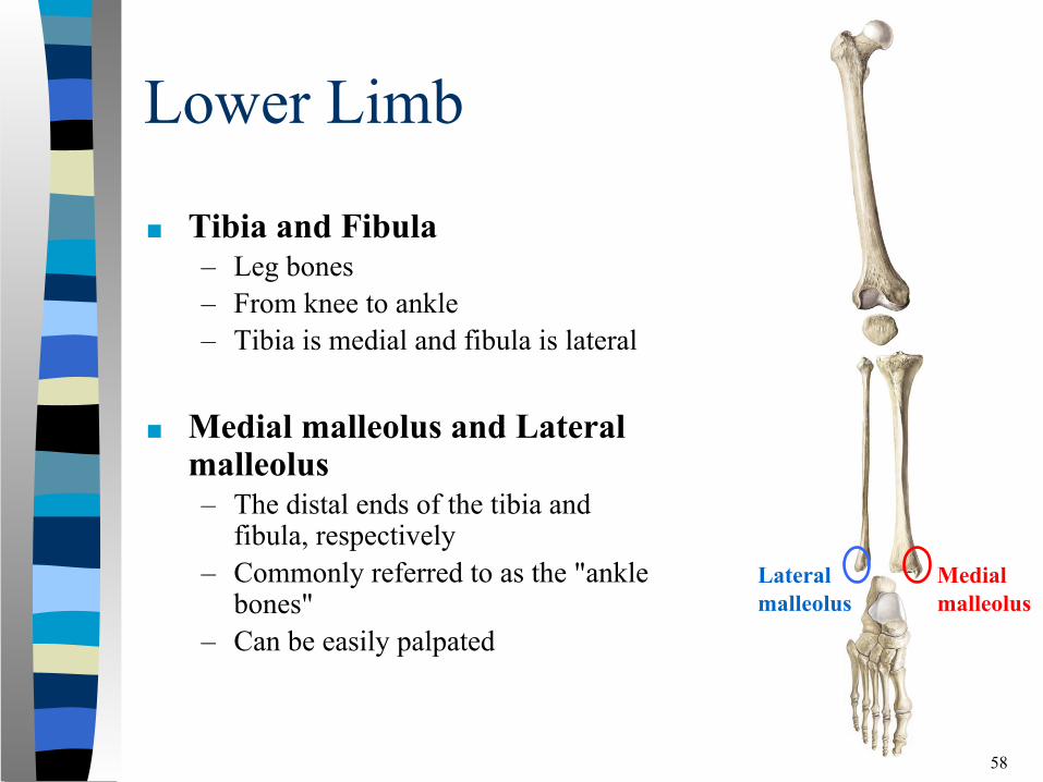
Lower Limb
■ Tibia and Fibula– Leg bones– From knee to ankle– Tibia is medial and fibula is lateral
■ Medial malleolus and Lateral malleolus– The distal ends of the tibia and
fibula, respectively– Commonly referred to as the "ankle
bones"– Can be easily palpated
58
Medialmalleolus
Lateralmalleolus
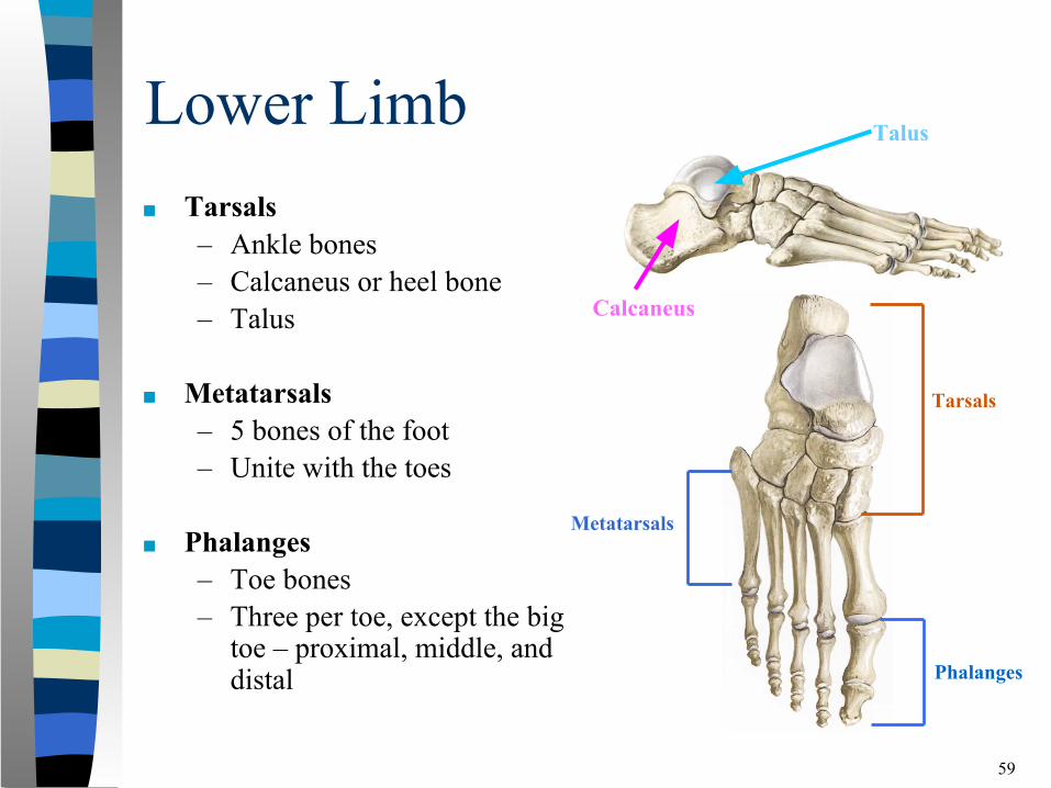
Lower Limb■ Tarsals
– Ankle bones– Calcaneus or heel bone– Talus
■ Metatarsals – 5 bones of the foot – Unite with the toes
■ Phalanges– Toe bones– Three per toe, except the big
toe – proximal, middle, and distal
59
Calcaneus
Talus
Phalanges
Metatarsals
Tarsals
60

61http://www.softschools.com/science/human_body/skeletal_system/
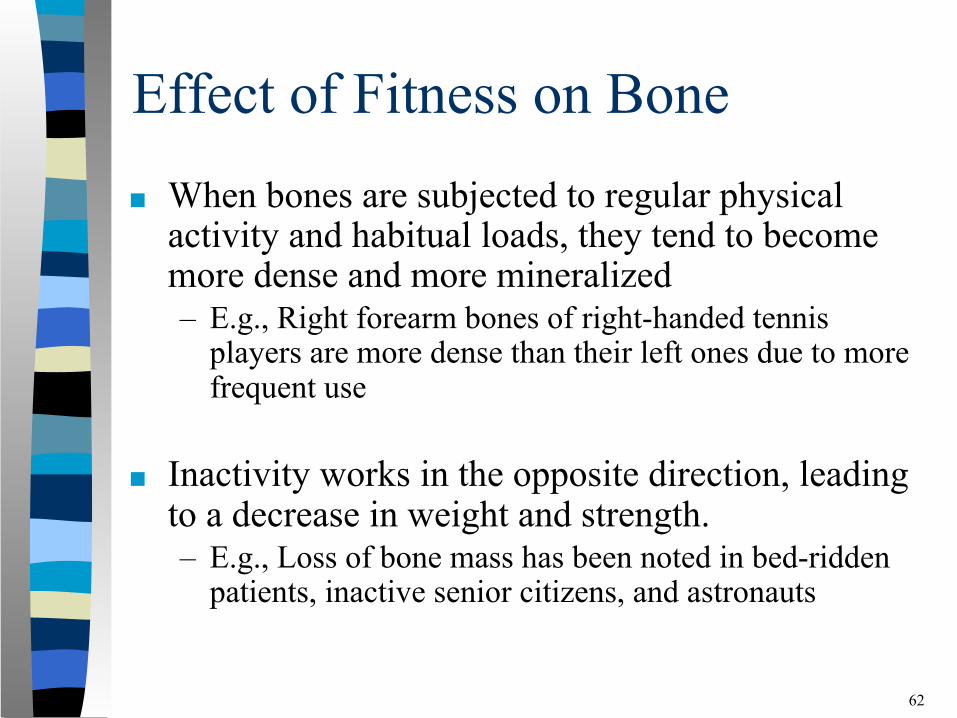
Effect of Fitness on Bone■ When bones are subjected to regular physical
activity and habitual loads, they tend to become more dense and more mineralized– E.g., Right forearm bones of right-handed tennis
players are more dense than their left ones due to more frequent use
■ Inactivity works in the opposite direction, leading to a decrease in weight and strength. – E.g., Loss of bone mass has been noted in bed-ridden
patients, inactive senior citizens, and astronauts
62
Joints of the Human Body
63
A joint or articulation is where two or more bones come into contact or articulate with each other.
What is the main function of joints?
Joint Movement and Stability■ Joint movement is linked to joint stability.
The more movement a joint has, the less stability it has and the greater the risk of injury.
■ Discuss a high risk joint.
64
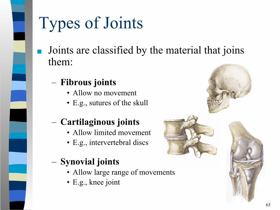
Types of Joints■ Joints are classified by the material that joins
them:
– Fibrous joints• Allow no movement• E.g., sutures of the skull
– Cartilaginous joints• Allow limited movement• E.g., intervertebral discs
– Synovial joints• Allow large range of movements• E.g., knee joint
65
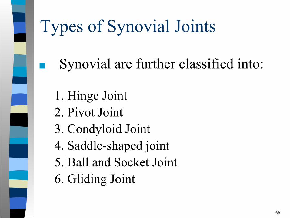
Types of Synovial Joints
■ Synovial are further classified into:
1. Hinge Joint2. Pivot Joint3. Condyloid Joint4. Saddle-shaped joint5. Ball and Socket Joint6. Gliding Joint
66
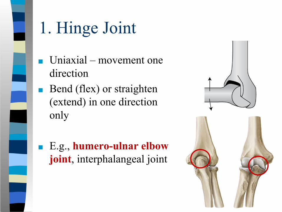
1. Hinge Joint
■ Uniaxial – movement one direction
■ Bend (flex) or straighten (extend) in one direction only
■ E.g., humero-ulnar elbow joint, interphalangeal joint
67
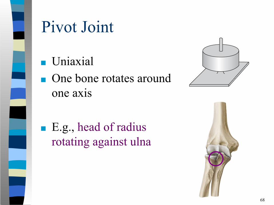
Pivot Joint
■ Uniaxial■ One bone rotates around
one axis
■ E.g., head of radius rotating against ulna
68
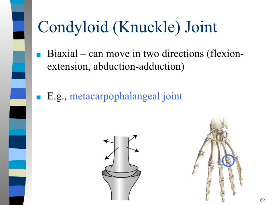
Condyloid (Knuckle) Joint■ Biaxial – can move in two directions (flexion-
extension, abduction-adduction)
■ E.g., metacarpophalangeal joint
69
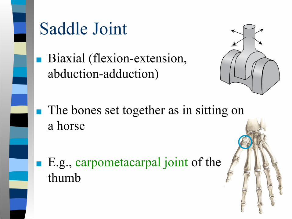
Saddle Joint■ Biaxial (flexion-extension,
abduction-adduction)
■ The bones set together as in sitting on a horse
■ E.g., carpometacarpal joint of the thumb
70
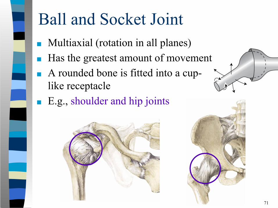
Ball and Socket Joint■ Multiaxial (rotation in all planes)■ Has the greatest amount of movement■ A rounded bone is fitted into a cup-
like receptacle■ E.g., shoulder and hip joints
71
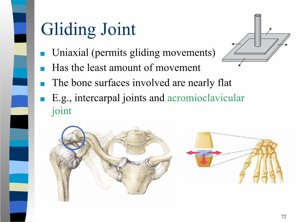
Gliding Joint■ Uniaxial (permits gliding movements)■ Has the least amount of movement■ The bone surfaces involved are nearly flat■ E.g., intercarpal joints and acromioclavicular
joint
72
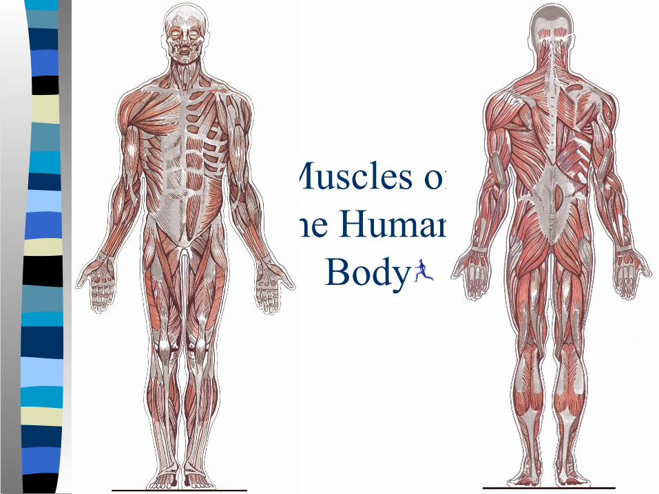
Muscles of the Human
Body
73
■ Over 600 muscles in the human body
■ Allow the skeleton to move
■ Vary in size, shape and structure
■ Make up about 40-50% of the weight of the body
74
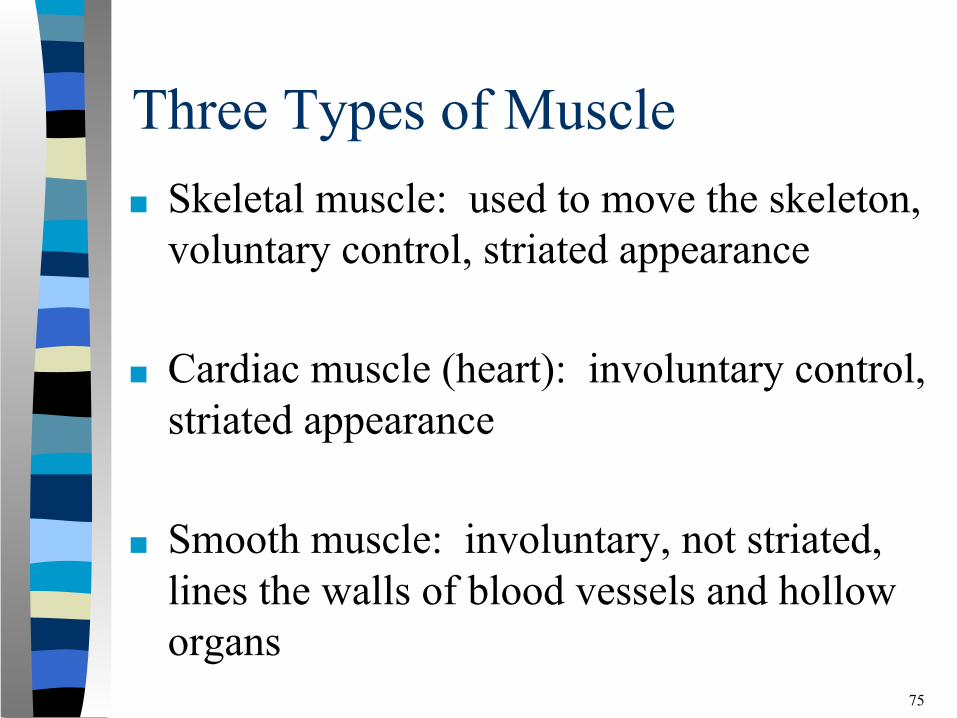
Three Types of Muscle■ Skeletal muscle: used to move the skeleton,
voluntary control, striated appearance
■ Cardiac muscle (heart): involuntary control, striated appearance
■ Smooth muscle: involuntary, not striated, lines the walls of blood vessels and hollow organs
75
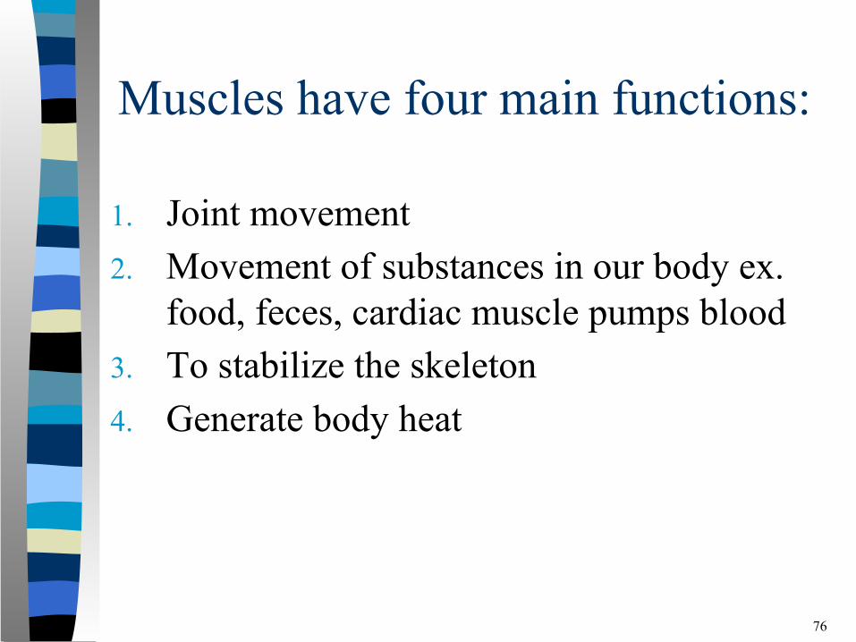
Muscles have four main functions:
1. Joint movement2. Movement of substances in our body ex.
food, feces, cardiac muscle pumps blood3. To stabilize the skeleton4. Generate body heat
76
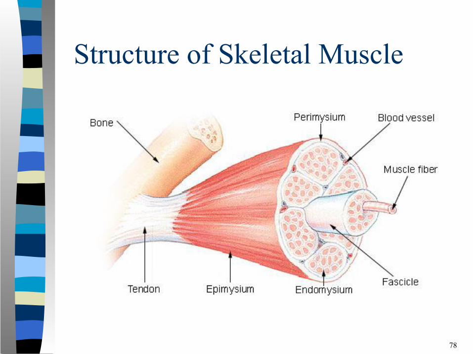
Structure of Skeletal Muscle
■ Fascia is a type of connective tissue that is located in-between and surrounding other tissues of the body such as muscles and bones.
■ Fascia is made up of fibrous tissue, adipose tissue and fluid.
77
Structure of Skeletal Muscle
78
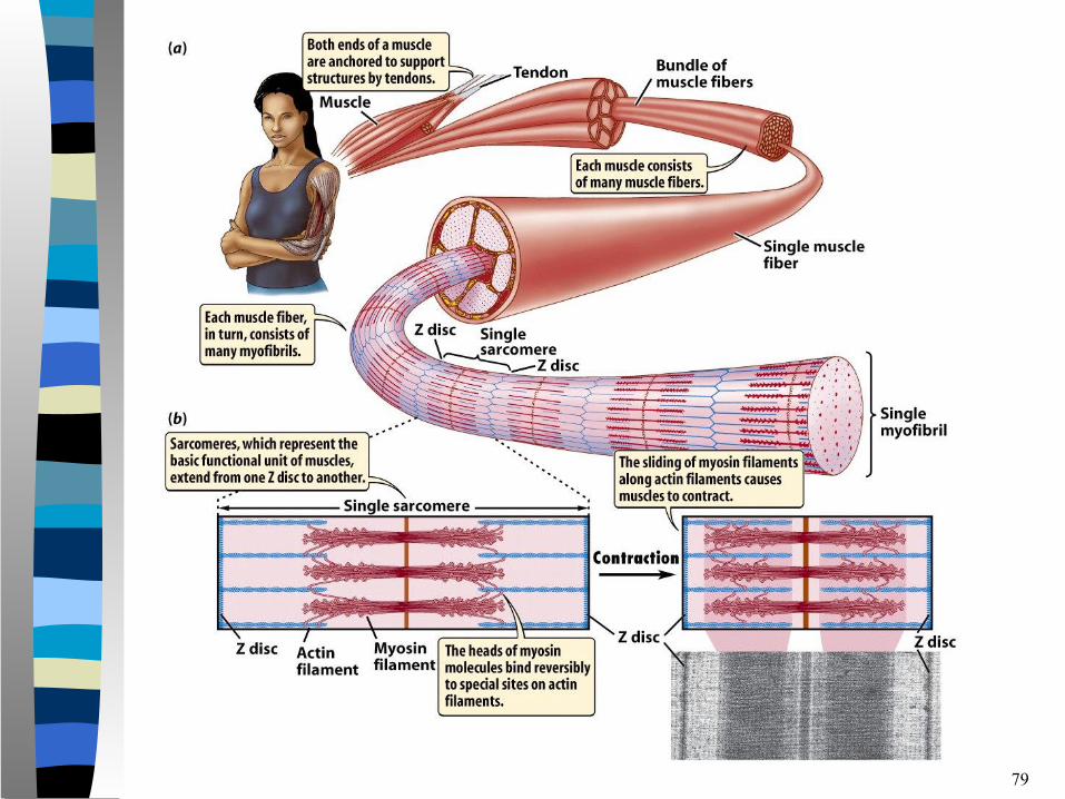
79
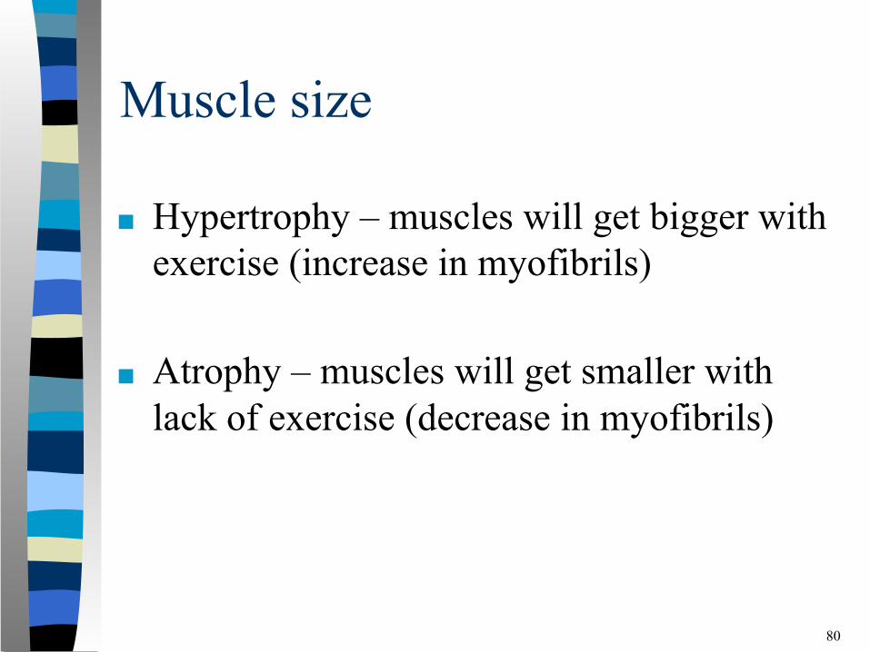
Muscle size
■ Hypertrophy – muscles will get bigger with exercise (increase in myofibrils)
■ Atrophy – muscles will get smaller with lack of exercise (decrease in myofibrils)
80
Origin and Insertion of Muscles■ Two attachment points for muscles:1. Origin is usually the more proximal
attachment (the end that is closest to the centre of the body). This is the bone that usually stays fixed.
2. Insertion is usually the more distal attachment (the end furthest away from the centre of the body). This is usually the moveable bone.
81
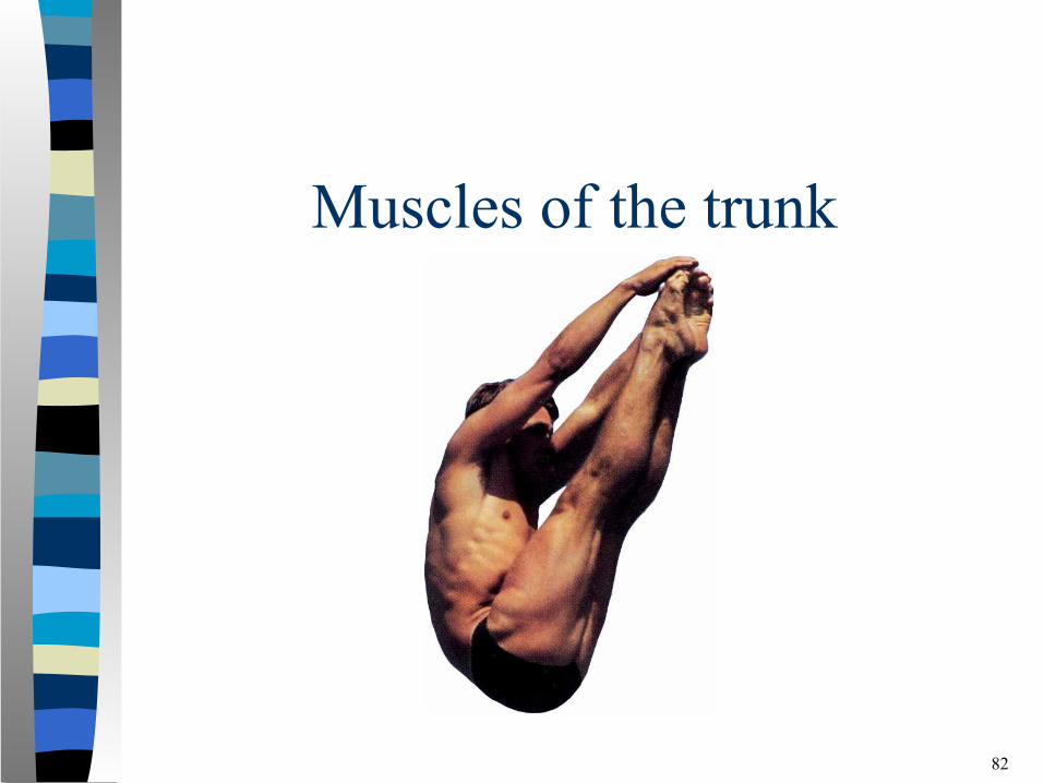
Muscles of the trunk
82
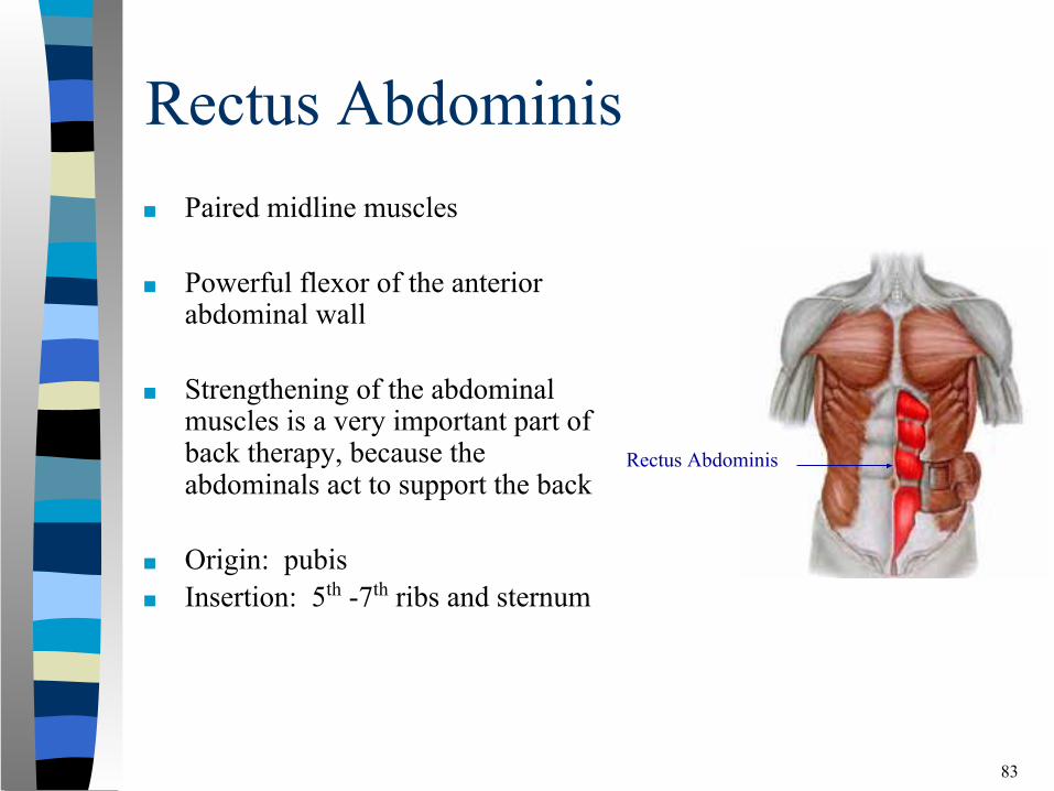
Rectus Abdominis■ Paired midline muscles
■ Powerful flexor of the anterior abdominal wall
■ Strengthening of the abdominal muscles is a very important part of back therapy, because the abdominals act to support the back
■ Origin: pubis■ Insertion: 5th -7th ribs and sternum
83
Rectus Abdominis
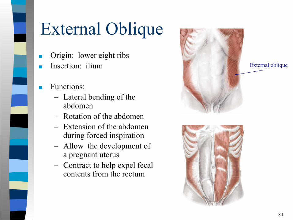
External Oblique■ Origin: lower eight ribs■ Insertion: ilium
■ Functions:– Lateral bending of the
abdomen– Rotation of the abdomen – Extension of the abdomen
during forced inspiration– Allow the development of
a pregnant uterus– Contract to help expel fecal
contents from the rectum
84
External oblique
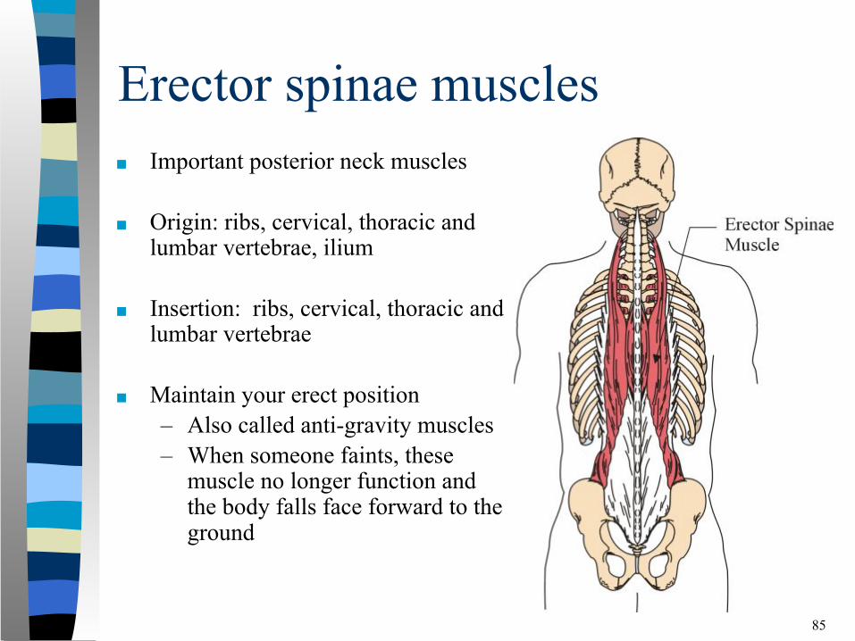
Erector spinae muscles■ Important posterior neck muscles
■ Origin: ribs, cervical, thoracic and lumbar vertebrae, ilium
■ Insertion: ribs, cervical, thoracic and lumbar vertebrae
■ Maintain your erect position – Also called anti-gravity muscles– When someone faints, these
muscle no longer function and the body falls face forward to the ground
85
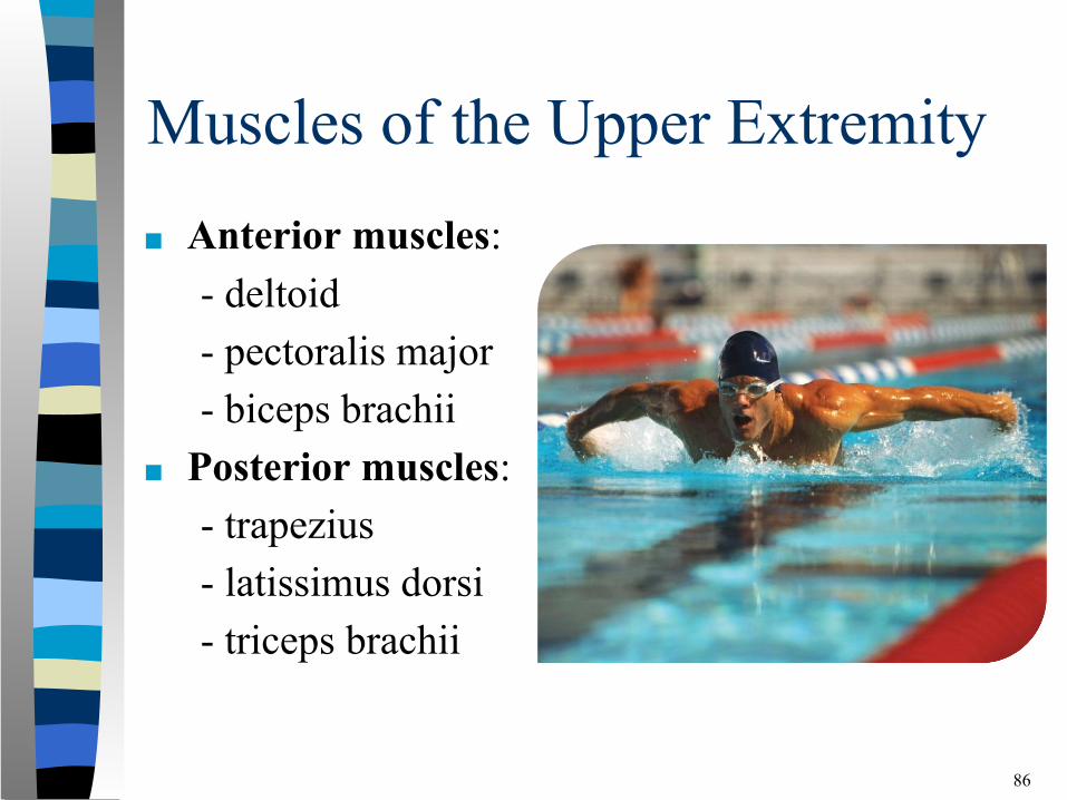
Muscles of the Upper Extremity■ Anterior muscles:
- deltoid- pectoralis major- biceps brachii
■ Posterior muscles: - trapezius
- latissimus dorsi- triceps brachii
86
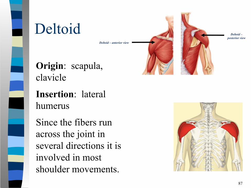
Deltoid
87
Deltoid – anterior view
Deltoid – posterior view
Origin: scapula, clavicle
Insertion: lateral humerus
Since the fibers run across the joint in several directions it is involved in most shoulder movements.
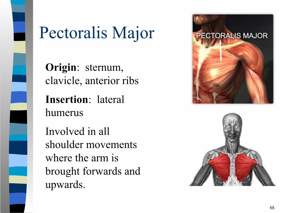
Pectoralis Major
88
Origin: sternum, clavicle, anterior ribs
Insertion: lateral humerus
Involved in all shoulder movements where the arm is brought forwards and upwards.
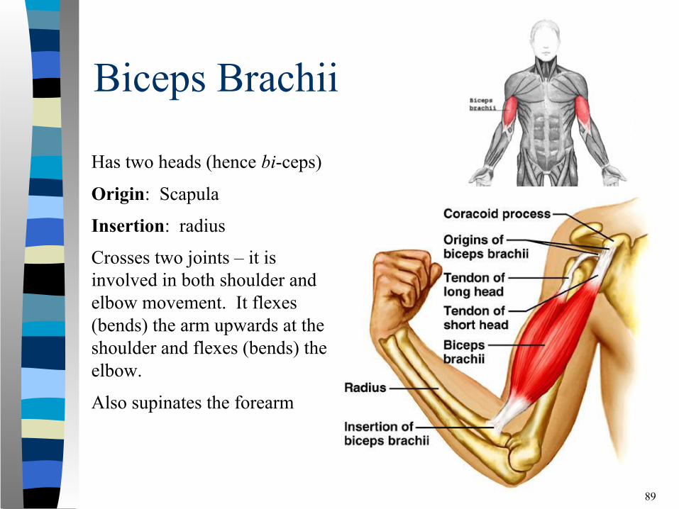
Biceps Brachii
89
Has two heads (hence bi-ceps)
Origin: Scapula
Insertion: radius
Crosses two joints – it is involved in both shoulder and elbow movement. It flexes (bends) the arm upwards at the shoulder and flexes (bends) the elbow.
Also supinates the forearm
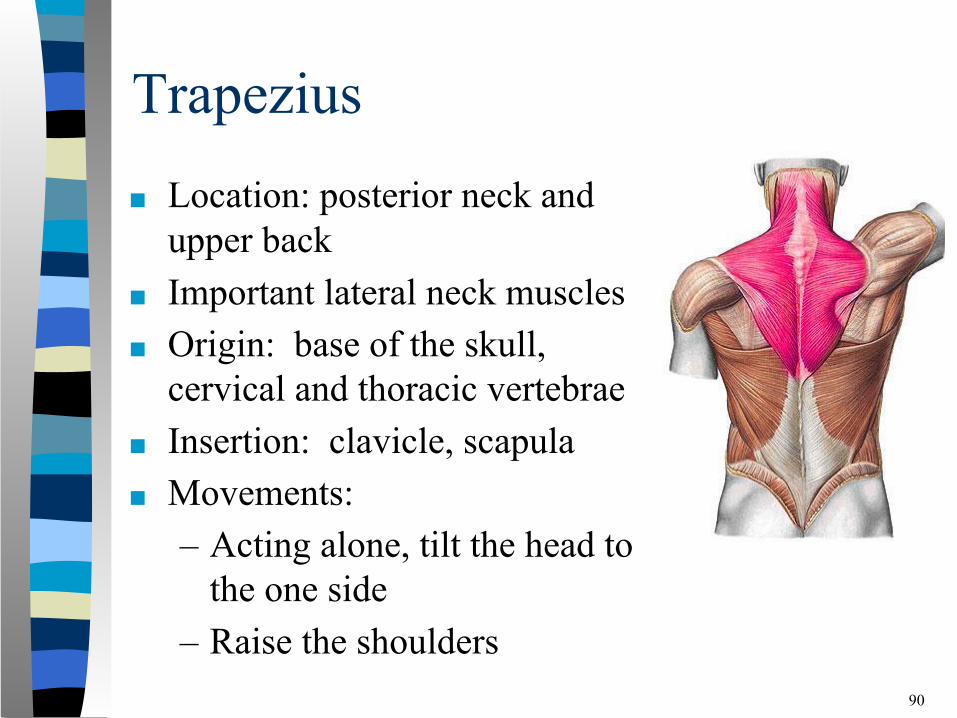
Trapezius■ Location: posterior neck and
upper back■ Important lateral neck muscles■ Origin: base of the skull,
cervical and thoracic vertebrae■ Insertion: clavicle, scapula■ Movements:
– Acting alone, tilt the head to the one side
– Raise the shoulders90
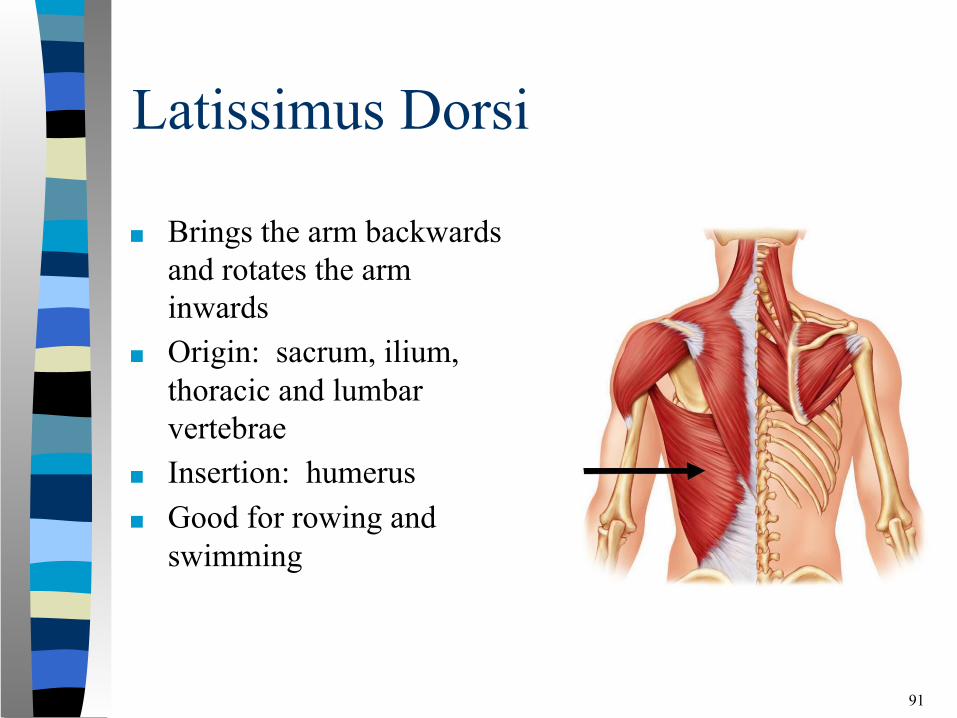
Latissimus Dorsi
■ Brings the arm backwards and rotates the arm inwards
■ Origin: sacrum, ilium, thoracic and lumbar vertebrae
■ Insertion: humerus■ Good for rowing and
swimming
91
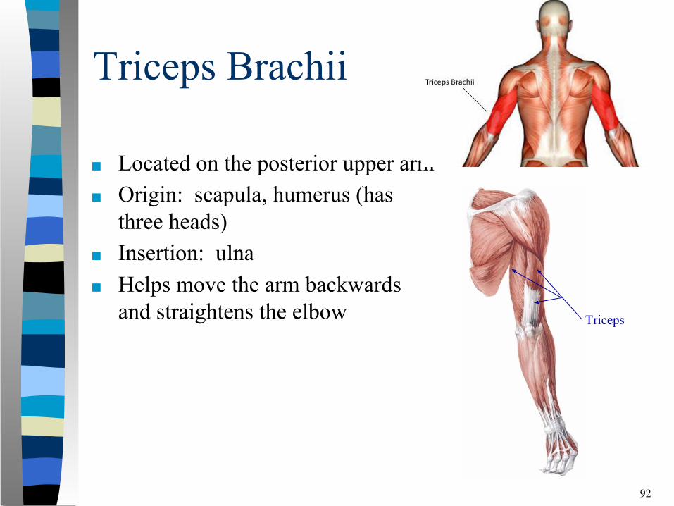
Triceps Brachii
■ Located on the posterior upper arm■ Origin: scapula, humerus (has
three heads)■ Insertion: ulna■ Helps move the arm backwards
and straightens the elbow
92
Triceps

Muscles of the Lower Extremity
93
Muscles of Lower Extremity
■ Generally bigger than the upper extremity muscles.
■ They bear the weight of the entire body and forcefully push off the ground to move forwards and upwards when walking.
94
Muscles of the Lower Extremity
■ Anterior Muscles- iliopsoas- sartorius- quadriceps- tibialis anterior
■ Posterior Muscles- gluteus maximus- hamstrings- gastrocnemius- soleus
95
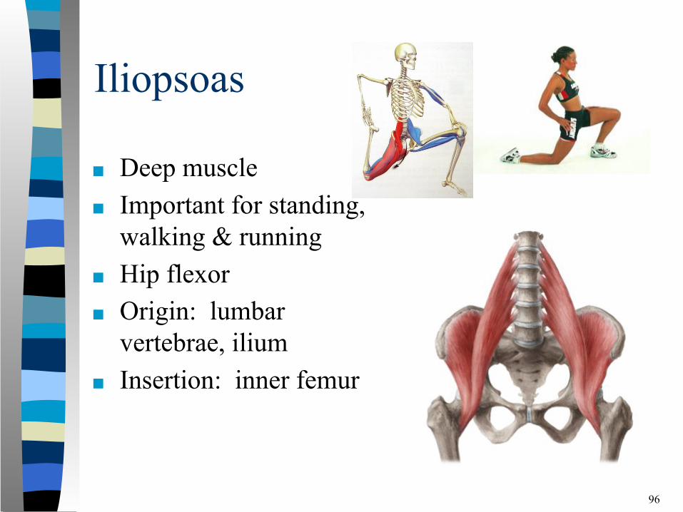
Iliopsoas
■ Deep muscle■ Important for standing,
walking & running■ Hip flexor■ Origin: lumbar
vertebrae, ilium■ Insertion: inner femur
96
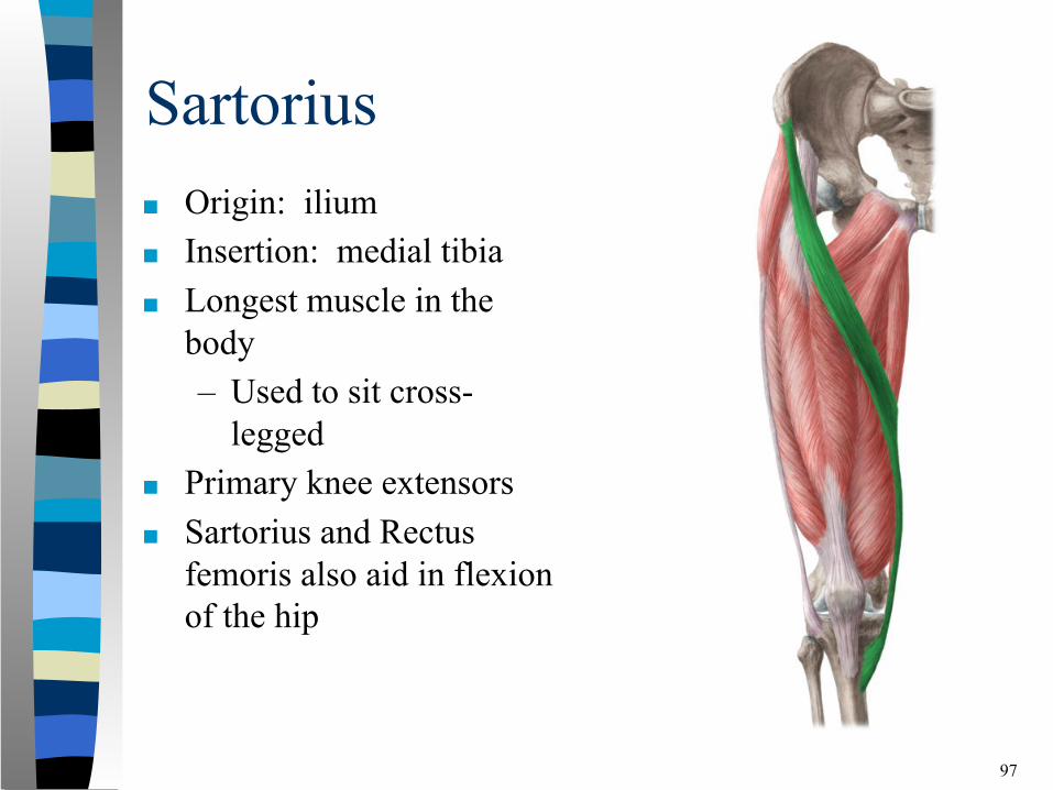
Sartorius■ Origin: ilium■ Insertion: medial tibia■ Longest muscle in the
body– Used to sit cross-
legged■ Primary knee extensors ■ Sartorius and Rectus
femoris also aid in flexion of the hip
97
Quadriceps
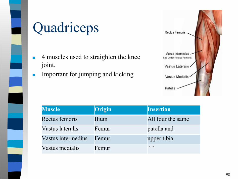
■ 4 muscles used to straighten the knee joint.
■ Important for jumping and kicking
98
Muscle Origin InsertionRectus femoris Ilium All four the sameVastus lateralis Femur patella and Vastus intermedius Femur upper tibiaVastus medialis Femur “ “
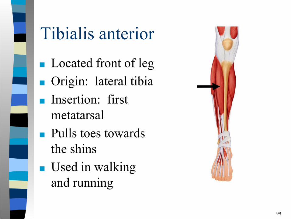
Tibialis anterior■ Located front of leg■ Origin: lateral tibia■ Insertion: first
metatarsal ■ Pulls toes towards
the shins■ Used in walking
and running
99

Gluteus Maximus
■ Largest of the three buttock muscles
■ Moves thigh backwards and principal extensor of the hip
■ Origin: posterior ilium, sacrum and coccyx
■ Insertion: lateral femur
100
Gluteus Maximus
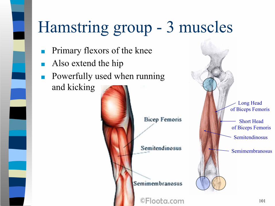
Hamstring group - 3 muscles■ Primary flexors of the knee■ Also extend the hip ■ Powerfully used when running
and kicking
101
Short Head of Biceps Femoris
Long Head of Biceps Femoris
Semitendinosus
Semimembranosus
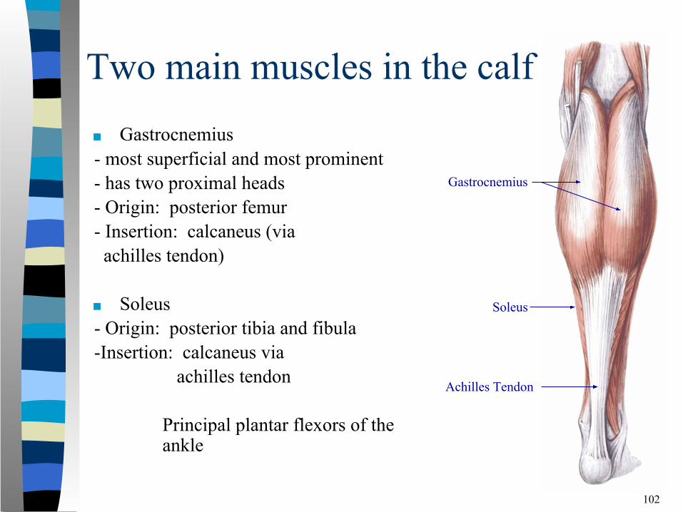
Two main muscles in the calf■ Gastrocnemius- most superficial and most prominent- has two proximal heads- Origin: posterior femur- Insertion: calcaneus (via achilles tendon)
■ Soleus - Origin: posterior tibia and fibula-Insertion: calcaneus via
achilles tendon
Principal plantar flexors of the ankle
102
Gastrocnemius
Soleus
Achilles Tendon
Summary■ Human anatomy deals with the structures that make up the
human body (structure determines function)■ The bones, joints, and muscles that make up the
musculoskeletal system allow numerous movements to occur, with varying degrees of:– Motion capabilities– Strength– Flexibility
103
▪ Bones provide the structural framework necessary for support
▪ Muscles supply the power
▪ Joints supply the mechanism that allows human movement to occur