Embed Size (px)
Citation preview
“Musculoskeletal Overdose Injuries”Sarah E. Hagerty, DO
POMA District VIII 31st Annual Educational Winter SeminarJanuary 25‐28, 2018 1
Musculoskeletal Overuse Injuries
Dr. Sarah Hagerty, DO
Sports and Spine Physiatrist
Allegheny Health Network
Thursday, January 25, 2018
Disclosures
None relevant to lecture
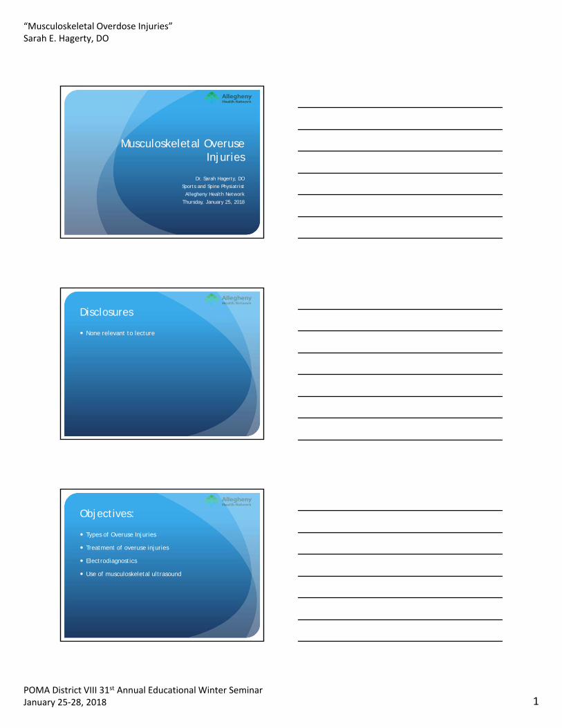
Objectives:
Types of Overuse Injuries
Treatment of overuse injuries
Electrodiagnostics
Use of musculoskeletal ultrasound
“Musculoskeletal Overdose Injuries”Sarah E. Hagerty, DO
POMA District VIII 31st Annual Educational Winter SeminarJanuary 25‐28, 2018 2
Injuries: Tendinopathies
Medial and Lateral Epicondylitis
IT band syndrome
Bursitis
Medial Tibial Stress Syndrome (Shin Splints)
Carpal and Cubital Tunnel Syndromes
Stress fracture (not included in lecture)
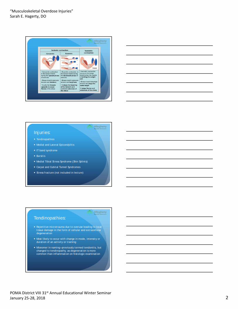
Tendinopathies:
Repetitive microtrauma due to overuse leading to local tissue damage in the form of cellular and extracellular degeneration
Most likely to occur with change in mode, intensity or duration of an activity or training
Misnomer in naming—previously termed tendonitis, but changed to tendinopathy, as degeneration is more common than inflammation on histologic examination
“Musculoskeletal Overdose Injuries”Sarah E. Hagerty, DO
POMA District VIII 31st Annual Educational Winter SeminarJanuary 25‐28, 2018 3
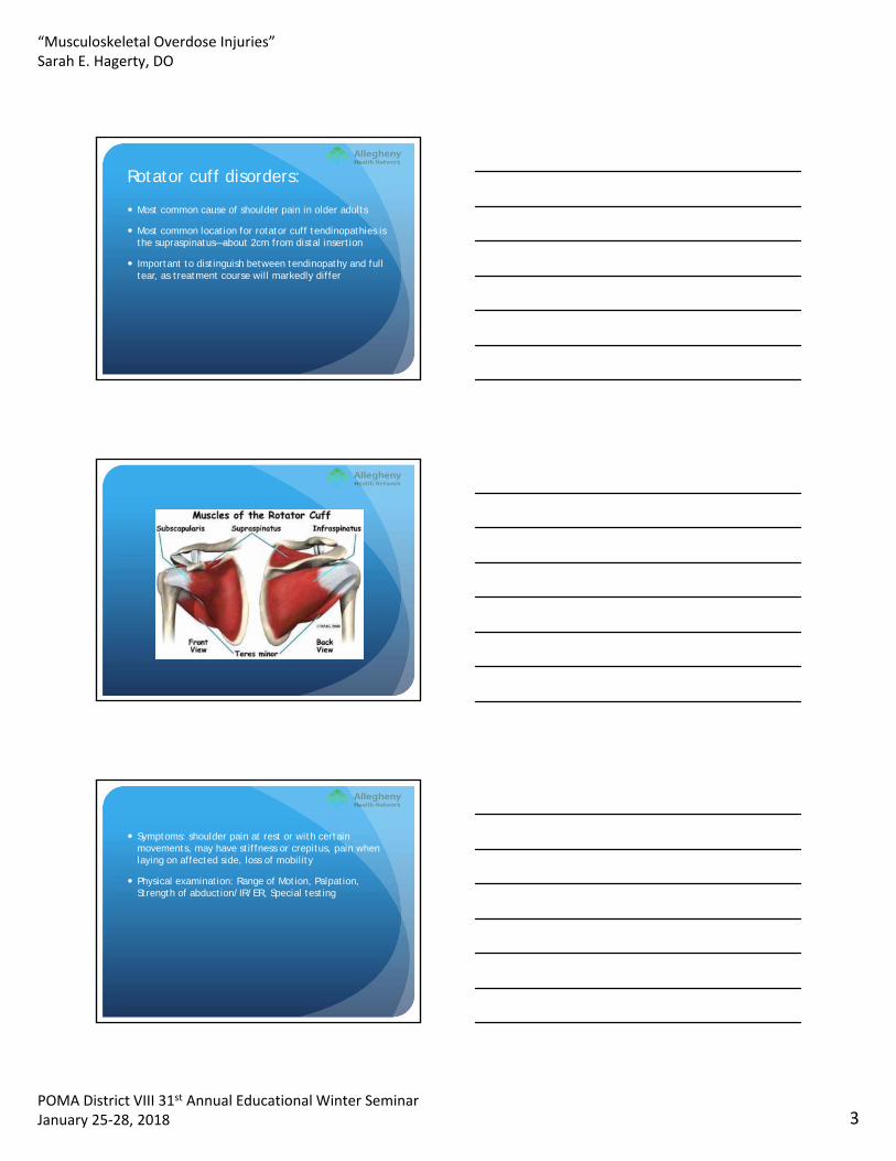
Rotator cuff disorders:
Most common cause of shoulder pain in older adults
Most common location for rotator cuff tendinopathies is the supraspinatus—about 2cm from distal insertion
Important to distinguish between tendinopathy and full tear, as treatment course will markedly differ
Symptoms: shoulder pain at rest or with certain movements, may have stiffness or crepitus, pain when laying on affected side, loss of mobility
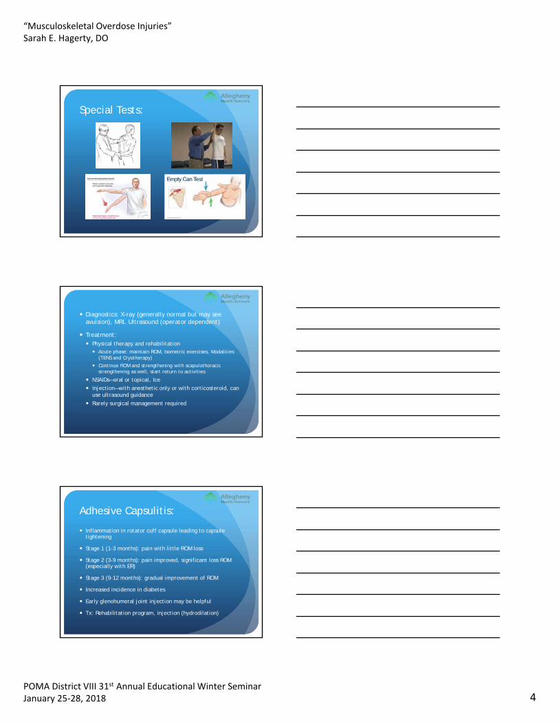
Physical examination: Range of Motion, Palpation, Strength of abduction/IR/ER, Special testing
“Musculoskeletal Overdose Injuries”Sarah E. Hagerty, DO
POMA District VIII 31st Annual Educational Winter SeminarJanuary 25‐28, 2018 4
Special Tests:
Diagnostics: X-ray (generally normal but may see avulsion), MRI, Ultrasound (operator dependent)
Treatment: Physical therapy and rehabilitation Acute phase: maintain ROM, Isometric exercises, Modalities
(TENS and Cryotherapy)
Continue ROM and strengthening with scapulothoracic strengthening as well, start return to activities
NSAIDs—oral or topical, Ice
Injection—with anesthetic only or with corticosteroid, can use ultrasound guidance
Rarely surgical management required
Adhesive Capsulitis:
Inflammation in rotator cuff capsule leading to capsule tightening
Stage 1 (1-3 months): pain with little ROM loss
Stage 2 (3-9 months): pain improved, significant loss ROM (especially with ER)
Stage 3 (9-12 months): gradual improvement of ROM
Increased incidence in diabetes
Early glenohumeral joint injection may be helpful
Tx: Rehabilitation program, injection (hydrodilation)
“Musculoskeletal Overdose Injuries”Sarah E. Hagerty, DO
POMA District VIII 31st Annual Educational Winter SeminarJanuary 25‐28, 2018 5
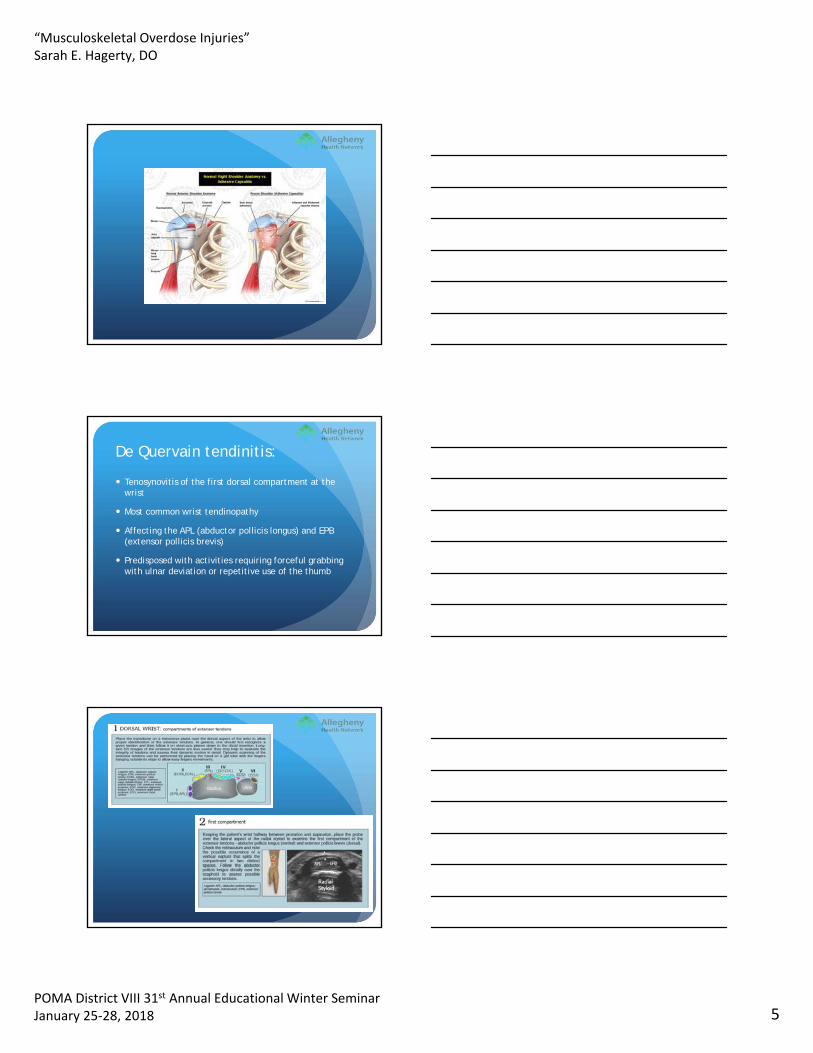
De Quervain tendinitis:
Tenosynovitis of the first dorsal compartment at the wrist
Most common wrist tendinopathy
Affecting the APL (abductor pollicis longus) and EPB (extensor pollicis brevis)
Predisposed with activities requiring forceful grabbing with ulnar deviation or repetitive use of the thumb
“Musculoskeletal Overdose Injuries”Sarah E. Hagerty, DO
POMA District VIII 31st Annual Educational Winter SeminarJanuary 25‐28, 2018 6
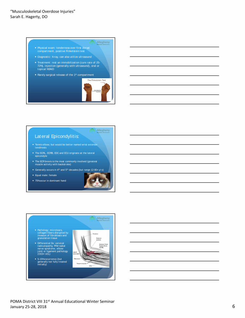
Physical exam: tenderness over first dorsal compartment, positive Finkelstein test
Diagnostic: X-ray, can also utilize ultrasound
Treatment: rest an immobilization (cure rate of 25-72%), injection (generally with ultrasound), oral or topical NSAID
Rarely surgical release of the 1st compartment
Lateral Epicondylitis:
Tennis elbow, but would be better named wrist extensor tendinosis
The ECRL, ECRB, EDC and ECU originate at the lateral epicondyle
The ECR brevis is the most commonly involved (greatest muscle activity with backstroke)
Generally occurs in 4th and 5th decades (but range 12-80+ y/o)
Equal male: female
75% occur in dominant hand
Pathology: microtears, collagen fibers disrupted by invasion of fibroblasts and granulation tissue
Differential Dx: cervical radiculopathy, PIN/radial nerve syndrome, elbow joint or ligament pathology (OCD< UCL)
5-15% recurrence (but generally not fully treated initially)
“Musculoskeletal Overdose Injuries”Sarah E. Hagerty, DO
POMA District VIII 31st Annual Educational Winter SeminarJanuary 25‐28, 2018 7
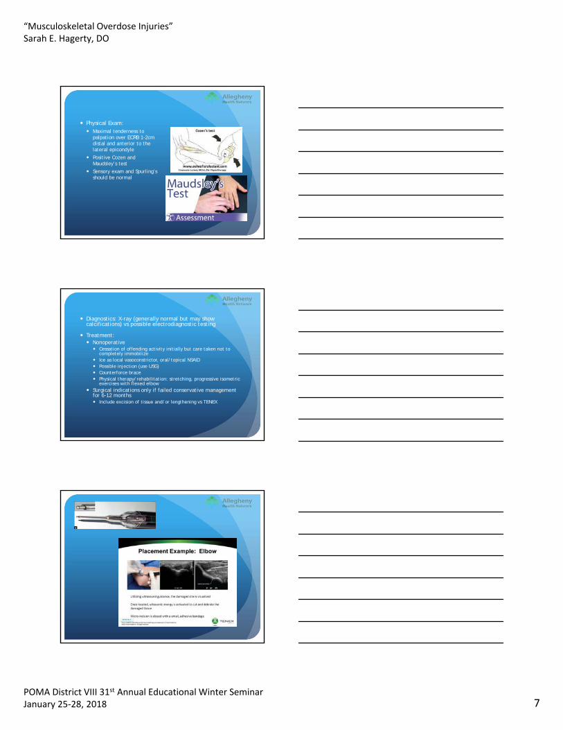
Physical Exam: Maximal tenderness to
palpation over ECRB 1-2cm distal and anterior to the lateral epicondyle
Positive Cozen and Maudsley’s test
Sensory exam and Spurling’s should be normal
Diagnostics: X-ray (generally normal but may show calcifications) vs possible electrodiagnostic testing
Treatment: Nonoperative
Cessation of offending activity initially but care taken not to completely immobilize
Ice as local vasoconstrictor, oral/topical NSAID Possible injection (use USG) Counterforce brace Physical therapy/rehabilitation: stretching, progressive isometric
exercises with flexed elbow Surgical indications only if failed conservative management
for 6-12 months Include excision of tissue and/or lengthening vs TENEX
“Musculoskeletal Overdose Injuries”Sarah E. Hagerty, DO
POMA District VIII 31st Annual Educational Winter SeminarJanuary 25‐28, 2018 8
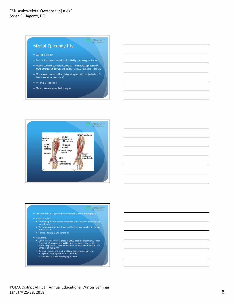
Medial Epicondylitis:
Golfer’s elbow
Due to increased overhead activity and valgus stress
Musculotendinous structures at the medial epicondyle: FCR, pronator teres, palmaris longus, FDS and the FCU
Much less common than lateral epicondylitis (which is 7-20 times more frequent)
4th and 5th decade
Male: female essentially equal
Differential Dx: ligamentous instability, ulnar neuropathy
Physical Exam: Pain along medial elbow worsened with forearm pronation or
wrist flexion Tenderness increased distal and lateral to medial epicondyle
at FCR or PT Normal strength and sensation
Treatment: Conservative: Phase 1 (rest, NSAID, possible injection), Phase
2 (activity/equipment modification, rehabilitation with stretching and progressive isometrics), can add eccentric and concentric exercises
Surgical: persistent medial elbow pain unresponsive to nonoperative program for 6-12 months Can perform traditonal surgery vs TENEX
“Musculoskeletal Overdose Injuries”Sarah E. Hagerty, DO
POMA District VIII 31st Annual Educational Winter SeminarJanuary 25‐28, 2018 9
Patellar Tendinopathy: Results from repeated loading of the knee extensor mechanism
Prevalent in jumping activities/sports
Similar to other tendinopathies, histologic studies show abnormal collagen, tenocytes and abundant small vessel growth (neovascularization)
Degenerative rather than inflammatory condition
Classic site is at the low pole of the patella
If located at insertion onto tibial tuberosity=jumper’s knee
Worse with strenuous activity
Complaints of pain after sitting for prolonged periods and with use of stairs
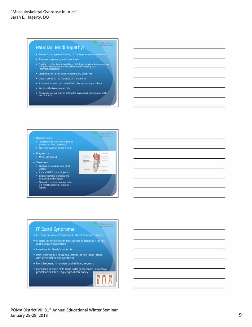
Physical exam: Tenderness of the inferior pole of
patella or tibial tuberosity
Pain improved with knee flexion
Diagnostics: MRI or ultrasound
Treatment: Relative or absolute rest (4-12
weeks)
Ice and NSAID, rarely injection
Begin eccentric exercises and stretching and progress
Surgical if no improvement after 3-6 months (drilling, excision, repair)
IT Band Syndrome: Friction between IT band and lateral femoral condyle
IT band originates from confluence of fascia of the TFL and gluteal musculature
Inserts onto Gerdy’s tubercle
Pain/burning of the lateral aspect of the knee (about 3cm proximal to the jointline)
More frequent in runners and military recruits
Increased tension of IT band with genu varum, excessive pronation of tibia, leg length discrepancy
“Musculoskeletal Overdose Injuries”Sarah E. Hagerty, DO
POMA District VIII 31st Annual Educational Winter SeminarJanuary 25‐28, 2018 10
Physical Exam: Tenderness of lateral femoral condyle
(worse with knee flexed at 30°) Positive Ober test indicates IT band
tightness
Diagnostics: X-ray (generally normal), MRI if consider surgery
Differential Dx: tendinopathy hamstring, lateral meniscus pathology, stress fx, early DJD
Treatment: Nonoperative (gen improve in 3-6
weeks): activity modification, NSAIDs, gluteal stretching and strengthening, possible orthotics
Rarely surgical (fail >6 months tx)
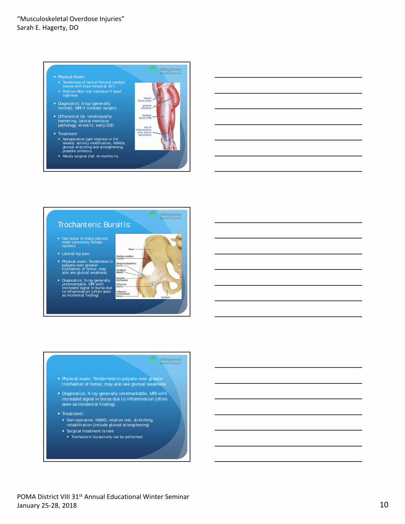
Trochanteric Bursitis:
Can occur in many patient, most commonly female runners
Lateral hip pain
Physical exam: Tenderness to palpate over greater trochanter of femur, may also see gluteal weakness
Diagnostics: X-ray generally unremarkable, MRI with increased signal in bursa due to inflammation (often seen as incidental finding)
Physical exam: Tenderness to palpate over greater trochanter of femur, may also see gluteal weakness
Diagnostics: X-ray generally unremarkable, MRI with increased signal in bursa due to inflammation (often seen as incidental finding)
Treatment: Non-operative: NSAID, relative rest, stretching,
rehabilitation (include gluteal strengthening)
Surgical treatment is rare Trochanteric bursectomy can be performed
“Musculoskeletal Overdose Injuries”Sarah E. Hagerty, DO
POMA District VIII 31st Annual Educational Winter SeminarJanuary 25‐28, 2018 11
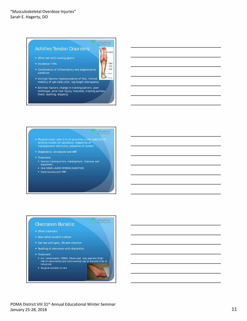
Achilles Tendon Disorders:
Often see with running sports
Incidence 7-9%
Combination of inflammatory and degenerative condition
Intrinsic factors: hyperpronation of foot, limited mobility of sub-talar joint, leg length discrepancy
Extrinsic factors: change in training pattern, poor technique, prior foot injury, footwear, training surface (hard, slanting, slippery)
Physical exam: pain 2-6 cm proximal to the insertion of Achilles tendon on calcaneus, inspection of malalignment/deformity, palpation of tendon
Diagnostics: ultrasound and MRI
Treatment: Correct training errors, malalignment, footwear and
equipment
Oral NSAID—AVOID STEROID INJECTION
Good success with PRP
Olecranon Bursitis: Often traumatic
Also called student’s elbow
Can see with gout, RA and infection
Swelling of olecrenon with discomfort
Treatment: Ice, compression, NSAID, elbow pad, may aspirate (high
risk of recurrence) and controversial use of steroids (risk of infection)
Surgical excision is rare
“Musculoskeletal Overdose Injuries”Sarah E. Hagerty, DO
POMA District VIII 31st Annual Educational Winter SeminarJanuary 25‐28, 2018 12
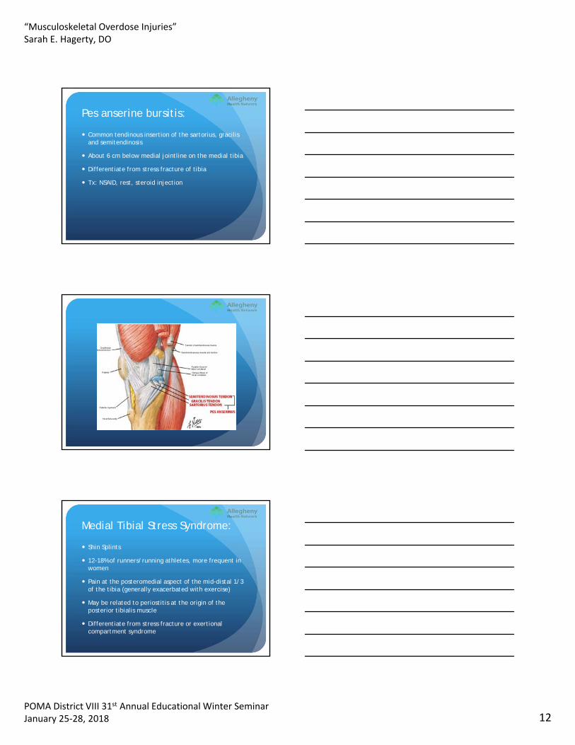
Pes anserine bursitis:
Common tendinous insertion of the sartorius, gracilisand semitendinosis
About 6 cm below medial jointline on the medial tibia
Differentiate from stress fracture of tibia
Tx: NSAID, rest, steroid injection
Medial Tibial Stress Syndrome:
Shin Splints
12-18% of runners/running athletes, more frequent in women
Pain at the posteromedial aspect of the mid-distal 1/3 of the tibia (generally exacerbated with exercise)
May be related to periostitis at the origin of the posterior tibialis muscle
Differentiate from stress fracture or exertionalcompartment syndrome
“Musculoskeletal Overdose Injuries”Sarah E. Hagerty, DO
POMA District VIII 31st Annual Educational Winter SeminarJanuary 25‐28, 2018 13
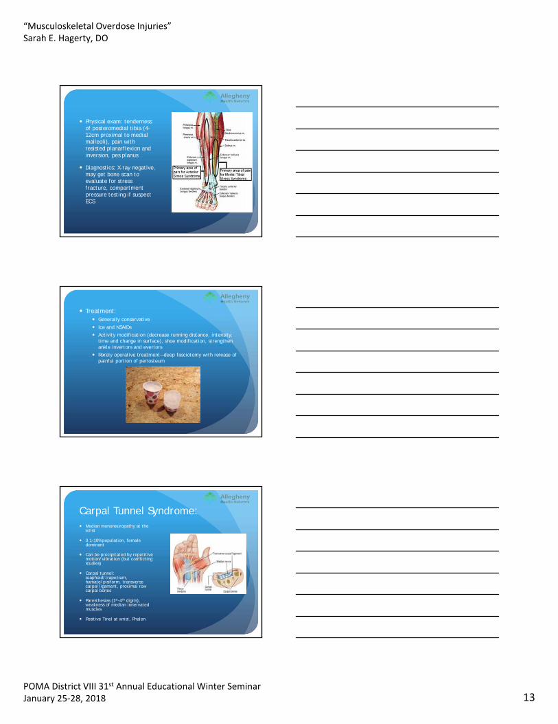
Physical exam: tenderness of posteromedial tibia (4-12cm proximal to medial malleoli), pain with resisted planarflexion and inversion, pes planus
Diagnostics: X-ray negative, may get bone scan to evaluate for stress fracture, compartment pressure testing if suspect ECS
Treatment: Generally conservative
Ice and NSAIDs
Activity modification (decrease running distance, intensity, time and change in surface), shoe modification, strengthen ankle invertors and evertors
Rarely operative treatment—deep fasciotomy with release of painful portion of periosteum
Carpal Tunnel Syndrome: Median mononeuropathy at the
wrist
0.1-10% population, female dominant
Can be precipitated by repetitive motion/vibration (but conflicting studies)
Carpal tunnel: scaphoid/trapezium, hamate/pisiform, transverse carpal ligament, proximal row carpal bones
Paresthesias (1st-4th digits), weakness of median innervated muscles
Positive Tinel at wrist, Phalen
“Musculoskeletal Overdose Injuries”Sarah E. Hagerty, DO
POMA District VIII 31st Annual Educational Winter SeminarJanuary 25‐28, 2018 14
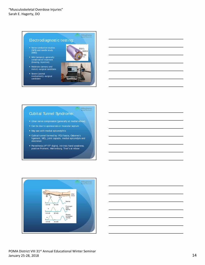
Electrodiagnostic testing:
Nerve conduction studies (NCS) and needle study (EMG)
Mild (sensory)—generally conservative treatment (bracing, injection)
Moderate (sensory and motor)—surgical candidate
Severe (axonal involvement)—surgical candidate
Cubital Tunnel Syndrome:
Ulnar nerve compression (generally at medial elbow)
Can be due to aponeurosis or muscular septum
May see with medial epicondylitis
Cubital tunnel formed by: FCU fascia, Osborne’s ligament, MCL, joint capsule, medial epicondyle and olecrenon
Paresthesia (4th/5th digits), intrinsic hand weakness, positive Froment, Wartenburg, Tinel’s at elbow
“Musculoskeletal Overdose Injuries”Sarah E. Hagerty, DO
POMA District VIII 31st Annual Educational Winter SeminarJanuary 25‐28, 2018 15
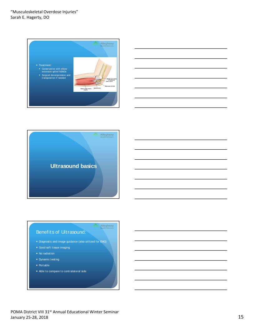
Treatment: Conservative with elbow
extension splint/NSAIDs
Surgical decompression and transposition if needed
Ultrasound basics
Benefits of Ultrasound:
Diagnostic and image guidance (also utilized for EMG)
Good soft tissue imaging
No radiation
Dynamic testing
Portable
Able to compare to contralateral side
“Musculoskeletal Overdose Injuries”Sarah E. Hagerty, DO
POMA District VIII 31st Annual Educational Winter SeminarJanuary 25‐28, 2018 16
Therapeutic Benefits:
Can visualize response to treatment: effusion side, pathology/improvement of muscle, tendons, nerve
Image guidance for diagnostic and therapeutic injections
Limitations of Ultrasound:
Operator dependent
Unable to penetrate bone (tumors, stress fracture, intra-articular pathologies)
Cost of machine
Coverage/billing, lack of universal certification/organization
If sound wave is reflected, it produces a bright echo with a brighter “white” image (bone)
If sound absorbed, a lighter/fainter white color (muscle, tendon, nerve) or black (effusion, blood)
“Musculoskeletal Overdose Injuries”Sarah E. Hagerty, DO
POMA District VIII 31st Annual Educational Winter SeminarJanuary 25‐28, 2018 17
Ultrasound “lingo”
Echogenicity: Capacity of a structure in the path of an ultrasound beam to reflect back sound waves
Anechoic: no internal echoes (black); fluid, artery, vein
Hyperechoic: High reflective pattern, appears brighter than the surrounding tissue
Hypoechoic: Low reflective pattern, manifesting as an area where the echoes are not as bright as the surrounding tissue
Anisotropy: artifact that occurs when the beams exiting the transducer are not 90° to the target
Tendinosis
Thickening
Focal hypoechogenicity
Calcification
Partial tears
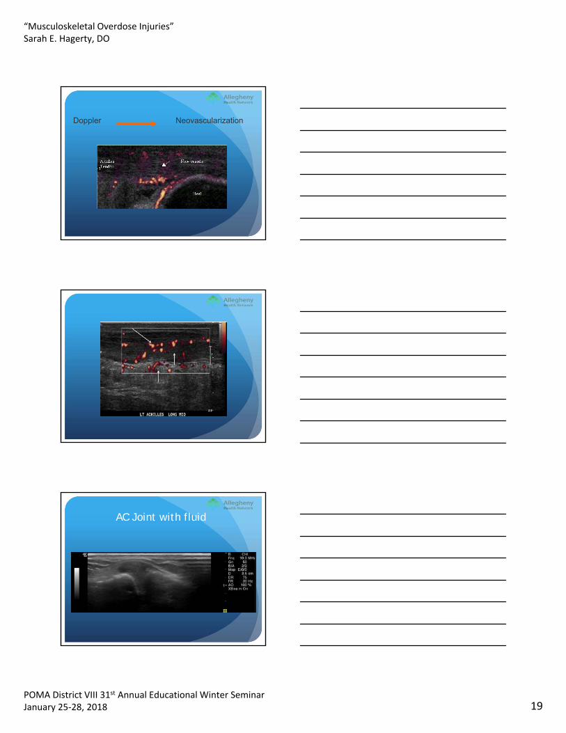
May see “inflammation” due to neovascularization with Doppler
“Musculoskeletal Overdose Injuries”Sarah E. Hagerty, DO
POMA District VIII 31st Annual Educational Winter SeminarJanuary 25‐28, 2018 18
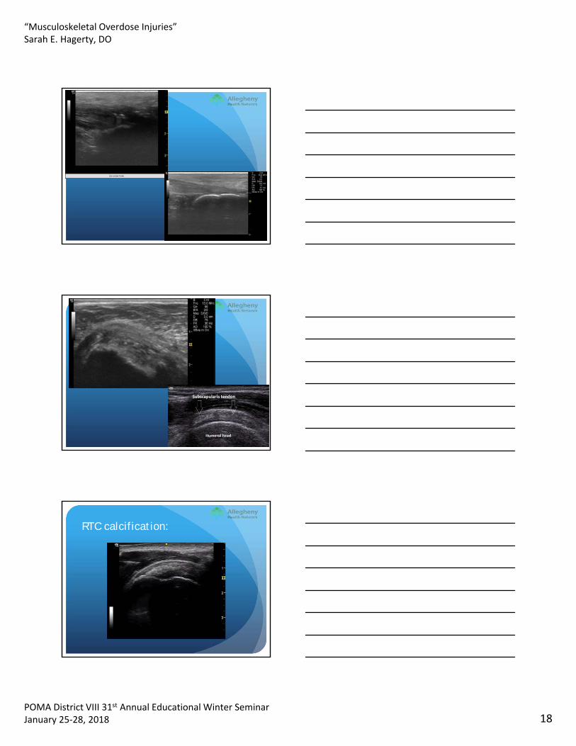
RTC calcification:
“Musculoskeletal Overdose Injuries”Sarah E. Hagerty, DO
POMA District VIII 31st Annual Educational Winter SeminarJanuary 25‐28, 2018 19
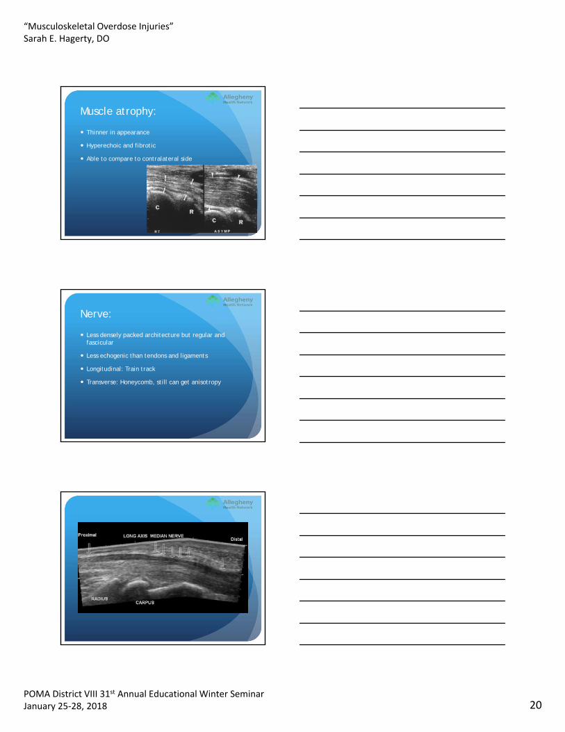
Doppler Neovascularization
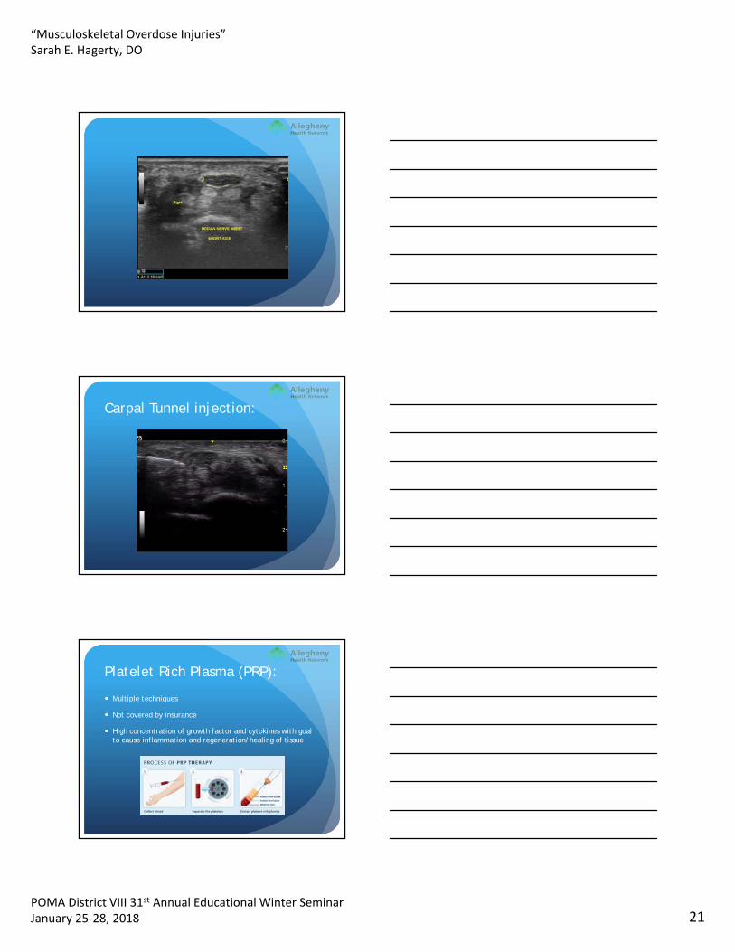
AC Joint with fluid
“Musculoskeletal Overdose Injuries”Sarah E. Hagerty, DO
POMA District VIII 31st Annual Educational Winter SeminarJanuary 25‐28, 2018 20
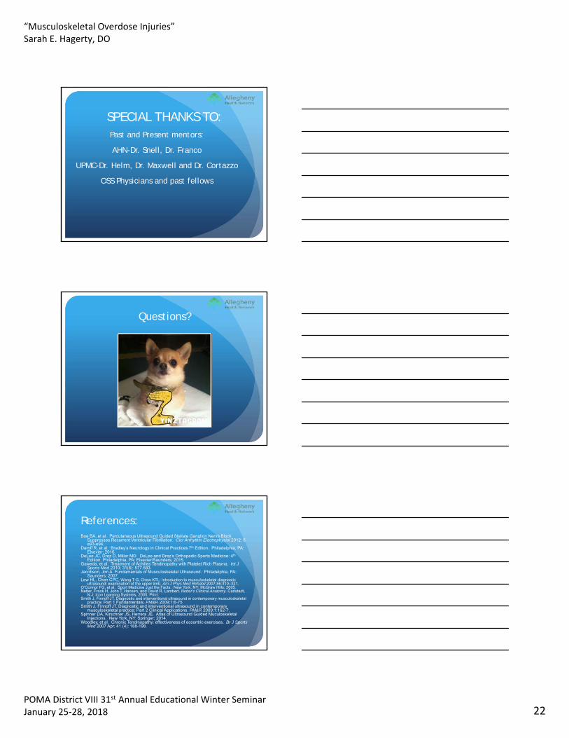
Muscle atrophy:
Thinner in appearance
Hyperechoic and fibrotic
Able to compare to contralateral side
Nerve:
Less densely packed architecture but regular and fascicular
Less echogenic than tendons and ligaments
Longitudinal: Train track
Transverse: Honeycomb, still can get anisotropy
“Musculoskeletal Overdose Injuries”Sarah E. Hagerty, DO
POMA District VIII 31st Annual Educational Winter SeminarJanuary 25‐28, 2018 21
Carpal Tunnel injection:
Platelet Rich Plasma (PRP):
Multiple techniques
Not covered by insurance
High concentration of growth factor and cytokines with goal to cause inflammation and regeneration/healing of tissue
“Musculoskeletal Overdose Injuries”Sarah E. Hagerty, DO
POMA District VIII 31st Annual Educational Winter SeminarJanuary 25‐28, 2018 22
SPECIAL THANKS TO:Past and Present mentors:
AHN-Dr. Snell, Dr. Franco
UPMC-Dr. Helm, Dr. Maxwell and Dr. Cortazzo
OSS Physicians and past fellows
Questions?
References:Boe BA, et al. Percutaneous Ultrasound Guided Stellate Ganglion Nerve Block
Suppresses Recurrent Ventricular Fibrillation. Cicr Arrhythm Electrophysiol 2012; 5 e93-e94.
Daroff R, et al. Bradley’s Neurology in Clinical Practices 7th Edition. Philadelphia, PA: Elsevier; 2016.
DeLee JC, Drez D, Miller MD. DeLee and Drez’s Orthopedic Sports Medicine: 4th
Edition. Philadelphia, PA: Elsevier/Saunders; 2015.Gaweda, et al. Treatment of Achilles Tendinopathy with Platelet Rich Plasma. Int J
Sports Med 2010; 31(8): 577.583. Jacobson, Jon A. Fundamentals of Musculoskeletal Ultrasound. Philadelphia, PA:
Saunders; 2007.Lew HL, Chen CPC, Wang T-G, Chew KTL: Introduction to musculoskeletal diagnostic
ultrasound: examination of the upper limb. Am J Phys Med Rehabil 2007;86:310–321.O’Connor FG, et al. Sport Medicine Just the Facts. New York, NY: McGraw Hills; 2005.Netter, Frank H, John T. Hansen, and David R. Lambert. Netter's Clinical Anatomy. Carlstadt,
N.J: Icon Learning Systems, 2005. Print.Smith J, Finnoff JT. Diagnostic and interventional ultrasound in contemporary musculoskeletal
practice: Part 1 Fundamentals. PM&R 2009;1:6-75Smith J, Finnoff JT. Diagnostic and interventional ultrasound in contemporary
musculoskeletal practice: Part 2 Clinical Applications. PM&R 2009;1:162-7.Spinner DA, Kirschner JS, Herrera JE. Atlas of Ultrasound Guided Muculoskeletal
Injections. New York, NY: Springer; 2014.Woodley, et al. Chronic Tendinopathy: effectiveness of eccentric exercises. Br J Sports
Med 2007 Apr; 41 (4): 188-198.
“Musculoskeletal Overdose Injuries”Sarah E. Hagerty, DO
POMA District VIII 31st Annual Educational Winter SeminarJanuary 25‐28, 2018 23
Advancing the Science of Ultrasound Guided Regional Anesthesia and Pain Medicine. http://www.usra.ca/
Carpal Tunnel Syndrome. https://radiopaedia.org/articles/carpal-tunnel-syndrome-1
ESSR: Ultrasound. https://essr.org/
Research Gate. https://www.researchgate.net/figure/236457357_fig14_Fig-17-Thickening-and-altered-echo-texture-in-the-mid-substance-of-the-Achilles-arrow
Ivanoski SP. Ultrasound assessment of most frequent shoulder disorders. Educational Exhibits. http://dx.doi.org/10.1594/ecr2014/C-2026
TENEX. https://www.tenexhealth.com