Embed Size (px)
Citation preview
8/8/2013
1
Diagnosis and Management of Common Shoulder and Hip
Complaints
UCSF Essentials of Primary Care August 8, 2013Carlin Senter, M.D.
At the end of this hour you will know1. The differential diagnosis for patients with
decreased AROM and PROM of shoulder.2. The key difference between impingement
syndrome and rotator cuff tear.3. How to diagnose a shoulder labral tear.4. The key exam finding in hip OA.5. The 2 exam maneuvers to bring out hip
impingement and/or labral tear.
Musculoskeletal work-up•History• Inspection•Palpation•Range of motion•Other Tests
Shoulder Problems
8/8/2013
2
Shoulder keys• History
– Hand dominance– Occupation– H/o dislocation– Pain that wakes patient from sleep
• Exam– Always perform neck exam with shoulder– Inspection: gown tied under arms or shirt off– Always examine unaffected side first
Shoulder examination• Inspection• Palpation• ROM
– Abduction– Forward flexion– ER– IR
• Strength– Supra– Infra and teres minor– Subscapularis
• Other testshttp://www.aafp.org/afp/20000515/3079.html
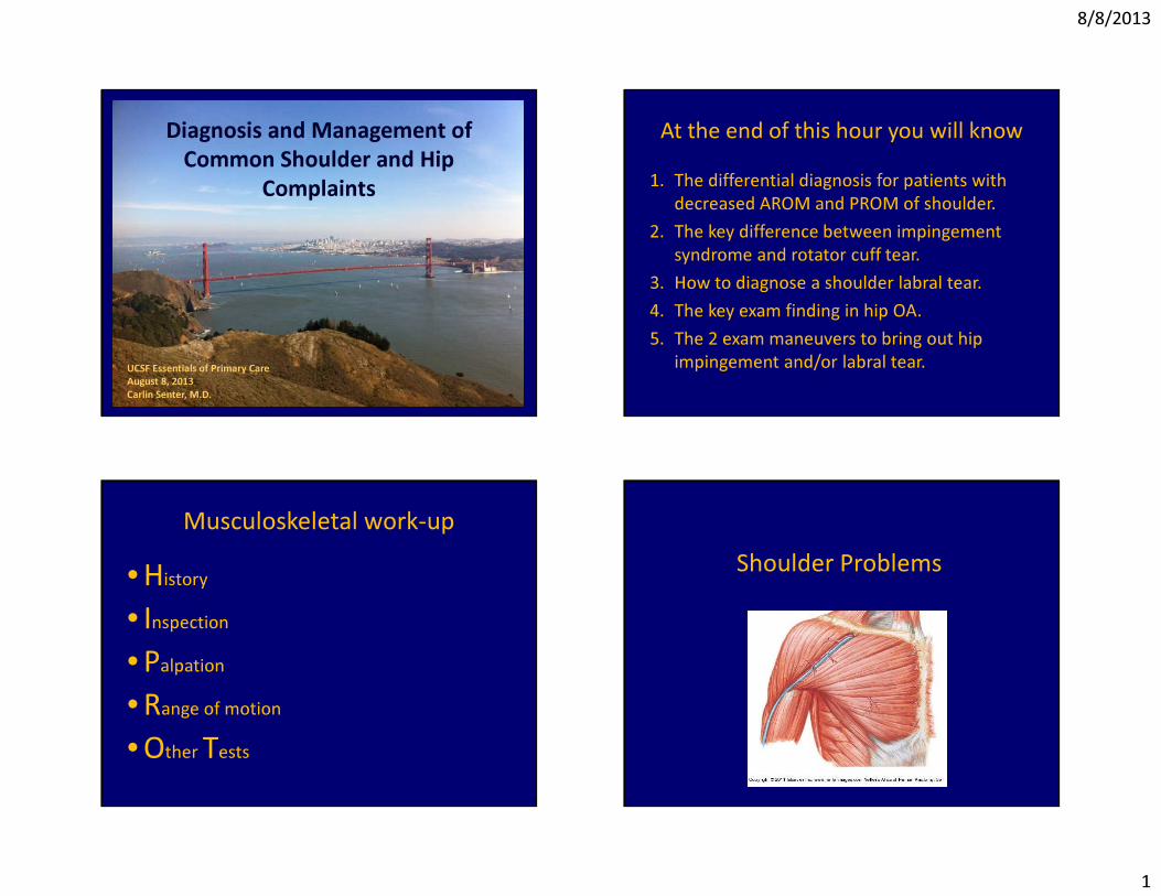
Shoulder: diagnosis driven examActive ROM
DecreasedNormal
Passive ROMNormal
Decreased
XrayFrozen shoulder
Normal
GH joint OA
Abnormal
ImpingementRC tear
Labral tearBiceps tendinitis
AC joint OA
Case #1• 50 y/o RHD woman with DM2 and
hypothyroidism presenting with R shoulder pain. No injury. Waking up at night during sleep. Shoulder feels very stiff, having trouble reaching behind and raising above head.
8/8/2013
3
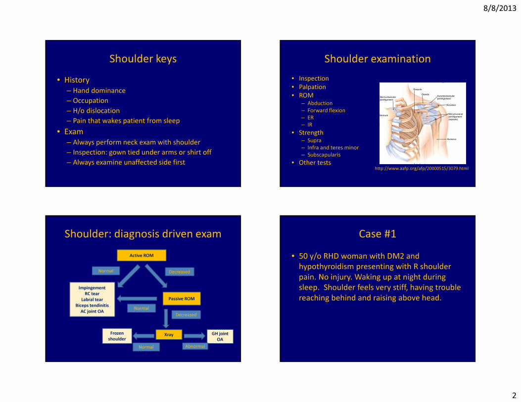
Range of motion
AbductionFlexion
Range of motion
External rotation
Internal rotation
Supine shoulder PROM
External rotation
Internal rotation
Physical exam: AROM
http://www.belmarpt.com/newwordpress/wp-content/uploads/2009/03/img_0294.jpg
Unable to lift the shoulder so uses entire shoulder girdle to abduct and FF.
8/8/2013
4
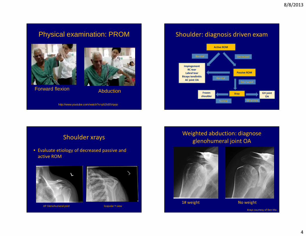
Physical examination: PROM
Forward flexion Abduction
http://www.youtube.com/watch?v=p52IdSVqvjo
Shoulder: diagnosis driven examActive ROM
DecreasedNormal
Passive ROMNormal
Decreased
XrayFrozen shoulder
Normal
GH joint OA
Abnormal
ImpingementRC tear
Labral tearBiceps tendinitis
AC joint OA
Shoulder xrays• Evaluate etiology of decreased passive and
active ROM
AP Glenohumeral joint Scapular Y view
Weighted abduction: diagnose glenohumeral joint OA
1# weight No weightXrays courtesy of Ben Ma.
8/8/2013
5
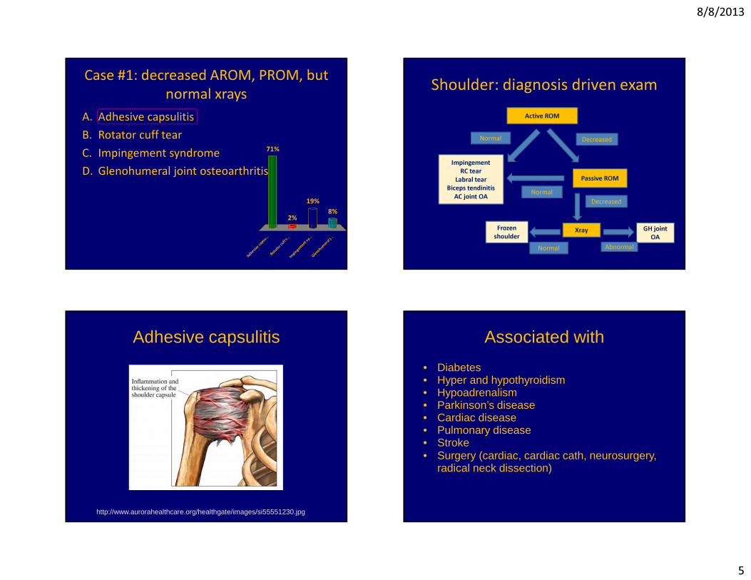
Case #1: decreased AROM, PROM, but normal xrays
A dh e s
i v e c a p
s u . ..
R ot a t o
r c uf f t .
. . I m
p i ng e m
e n t s y .
. . G l e
n o hu m
e r al j . . .
71%
8%19%
2%
A. Adhesive capsulitisB. Rotator cuff tearC. Impingement syndromeD. Glenohumeral joint osteoarthritis
Shoulder: diagnosis driven examActive ROM
DecreasedNormal
Passive ROMNormal
Decreased
XrayFrozen shoulder
Normal
GH joint OA
Abnormal
ImpingementRC tear
Labral tearBiceps tendinitis
AC joint OA
Adhesive capsulitis
http://www.aurorahealthcare.org/healthgate/images/si55551230.jpg
Associated with
• Diabetes• Hyper and hypothyroidism• Hypoadrenalism• Parkinson’s disease• Cardiac disease• Pulmonary disease• Stroke• Surgery (cardiac, cardiac cath, neurosurgery,
radical neck dissection)
8/8/2013
6
Adhesive capsulitis is a clinical diagnosis
• No need for MRI• Xrays helpful to r/o GH joint OA
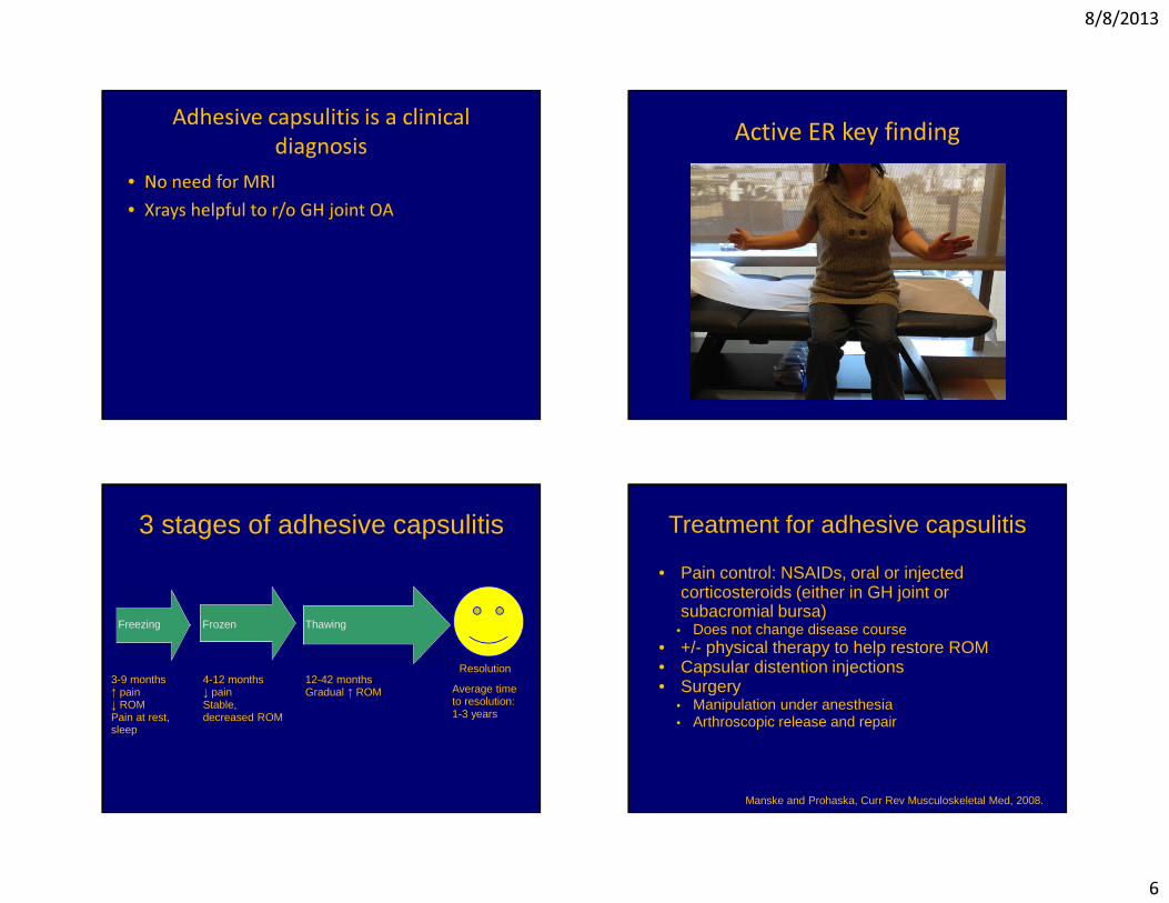
Active ER key finding
3 stages of adhesive capsulitis
Freezing Frozen Thawing
3-9 months↑ pain↓ ROMPain at rest, sleep
4-12 months↓ painStable, decreased ROM
12-42 monthsGradual ↑ ROM
Resolution
Average time to resolution: 1-3 years
Treatment for adhesive capsulitis
• Pain control: NSAIDs, oral or injected corticosteroids (either in GH joint or subacromial bursa)
• Does not change disease course• +/- physical therapy to help restore ROM• Capsular distention injections• Surgery
• Manipulation under anesthesia• Arthroscopic release and repair
Manske and Prohaska, Curr Rev Musculoskeletal Med, 2008.
8/8/2013
7
Case #2• 57 y/o RHD man presents with R shoulder pain
that started after he fell 3 months ago. Pain at R deltoid. He tried physical therapy without benefit. Waking at night from sleep due to pain.
Case #2 Exam• I: no atrophy• P: mild ttp deltoid, nontender biceps and AC
joint• ROM: Unable to actively abduct past 120
degrees 2/2 pain. Full PROM.
Shoulder: diagnosis driven examActive ROM
DecreasedNormal
Passive ROMNormal
Decreased
XrayFrozen shoulder
Normal
GH joint OA
Abnormal
ImpingementRC tear
Labral tearBiceps tendinitis
AC joint OA
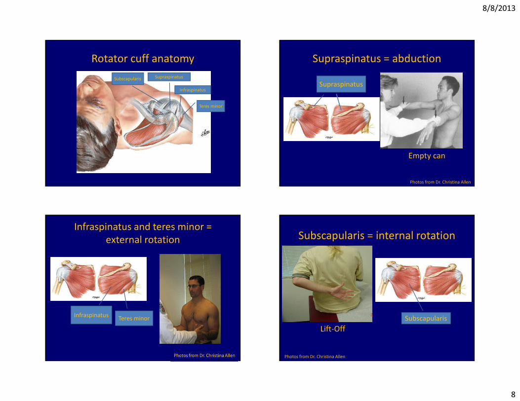
Rotator cuff anatomy
8/8/2013
8
Rotator cuff anatomySubscapularis Supraspinatus
Infraspinatus
Teres minor
Supraspinatus = abduction
Empty can
Photos from Dr. Christina Allen
Supraspinatus
Infraspinatus and teres minor = external rotation
Infraspinatus Teres minor
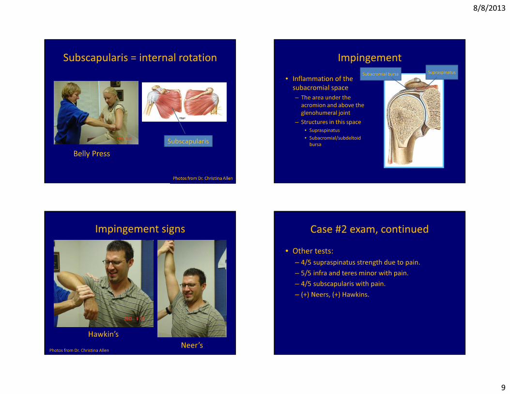
Subscapularis = internal rotation
Lift-Off
Photos from Dr. Christina Allen
Subscapularis
8/8/2013
9
Subscapularis = internal rotation
Subscapularis
Impingement• Inflammation of the
subacromial space– The area under the
acromion and above the glenohumeral joint
– Structures in this space• Supraspinatus• Subacromial/subdeltoid
bursa
Subacromial bursa Supraspinatus
Impingement signs
Hawkin’sNeer’sPhotos from Dr. Christina Allen
Case #2 exam, continued• Other tests:
– 4/5 supraspinatus strength due to pain.– 5/5 infra and teres minor with pain.– 4/5 subscapularis with pain. – (+) Neers, (+) Hawkins.
8/8/2013
10
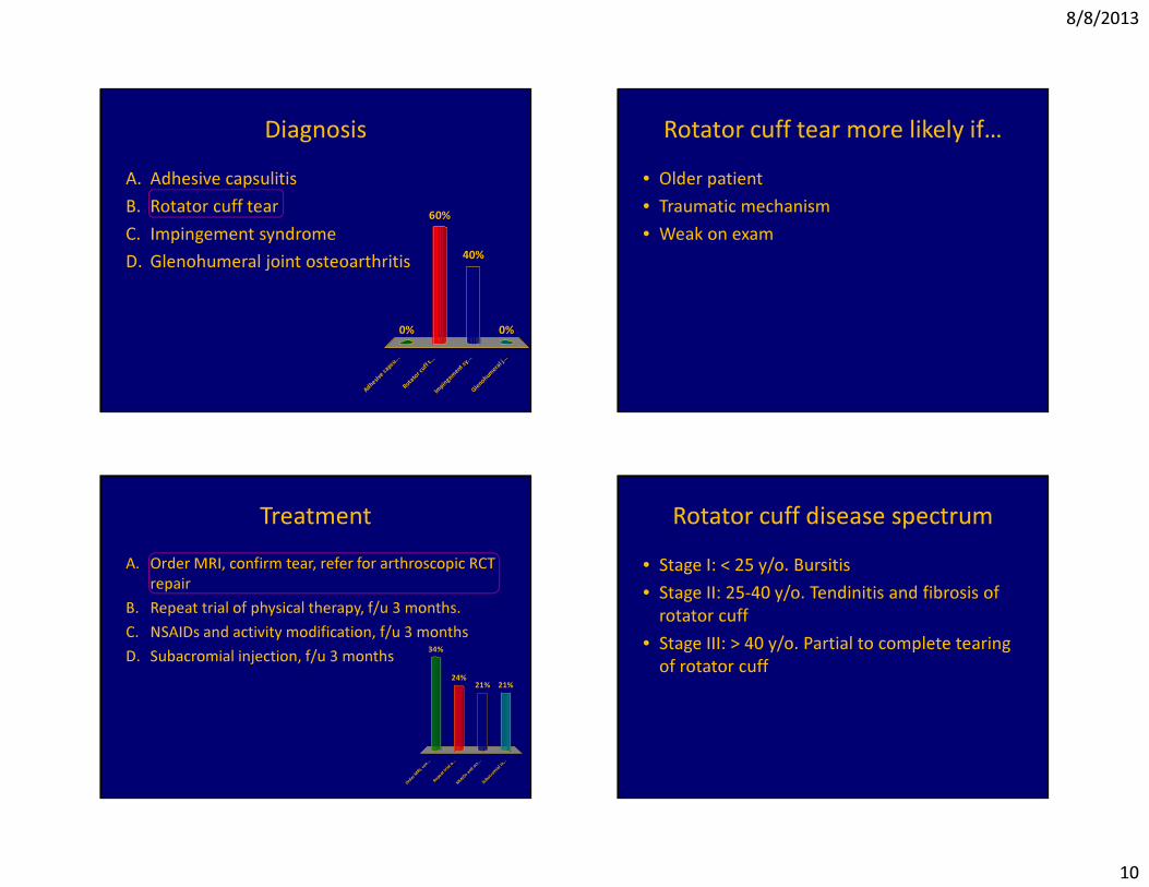
Diagnosis
A dh e s
i v e c a p
s u . ..
R ot a t o
r c uf f t
. . . I m
p i ng e m
e n t s y .
. . G l e
n o hu m
e r al j . .
.
0% 0%
40%
60%
A. Adhesive capsulitisB. Rotator cuff tearC. Impingement syndromeD. Glenohumeral joint osteoarthritis
Rotator cuff tear more likely if…• Older patient• Traumatic mechanism• Weak on exam
Treatment
O rd e r
M RI , c o
n . . . R e
p e at t r
i a l o. . .
N SA I D
s a nd a
c t . ..
S ub a c
r o mi a l
i n . ..
34%
21%21%24%
A. Order MRI, confirm tear, refer for arthroscopic RCT repair
B. Repeat trial of physical therapy, f/u 3 months.C. NSAIDs and activity modification, f/u 3 monthsD. Subacromial injection, f/u 3 months
Rotator cuff disease spectrum• Stage I: < 25 y/o. Bursitis • Stage II: 25-40 y/o. Tendinitis and fibrosis of
rotator cuff• Stage III: > 40 y/o. Partial to complete tearing
of rotator cuff
8/8/2013
11
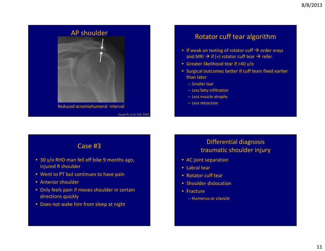
AP shoulder
Reduced acromiohumeral intervalSaupe N, et al. AJR, 2006.
Rotator cuff tear algorithm• If weak on testing of rotator cuff � order xrays
and MRI � if (+) rotator cuff tear � refer.• Greater likelihood tear if >40 y/o• Surgical outcomes better if cuff tears fixed earlier
than later – Smaller tear– Less fatty infiltration– Less muscle atrophy– Less retraction
Case #3• 30 y/o RHD man fell off bike 9 months ago,
injured R shoulder• Went to PT but continues to have pain• Anterior shoulder• Only feels pain if moves shoulder in certain
directions quickly• Does not wake him from sleep at night
Differential diagnosistraumatic shoulder injury
• AC joint separation• Labral tear• Rotator cuff tear• Shoulder dislocation• Fracture
– Humerus or clavicle
8/8/2013
12
Physical examination• No atrophy• Tender biceps tendon, nontender AC joint• AROM R shoulder
– FF 0-170 with pain at top– Abd 0-170 with pain at top– ER 45, IR L1 (Same as L shoulder)
• Strength 5/5 rotator cuff• (-) Neers and Hawkins• (+) O’Brien’s test
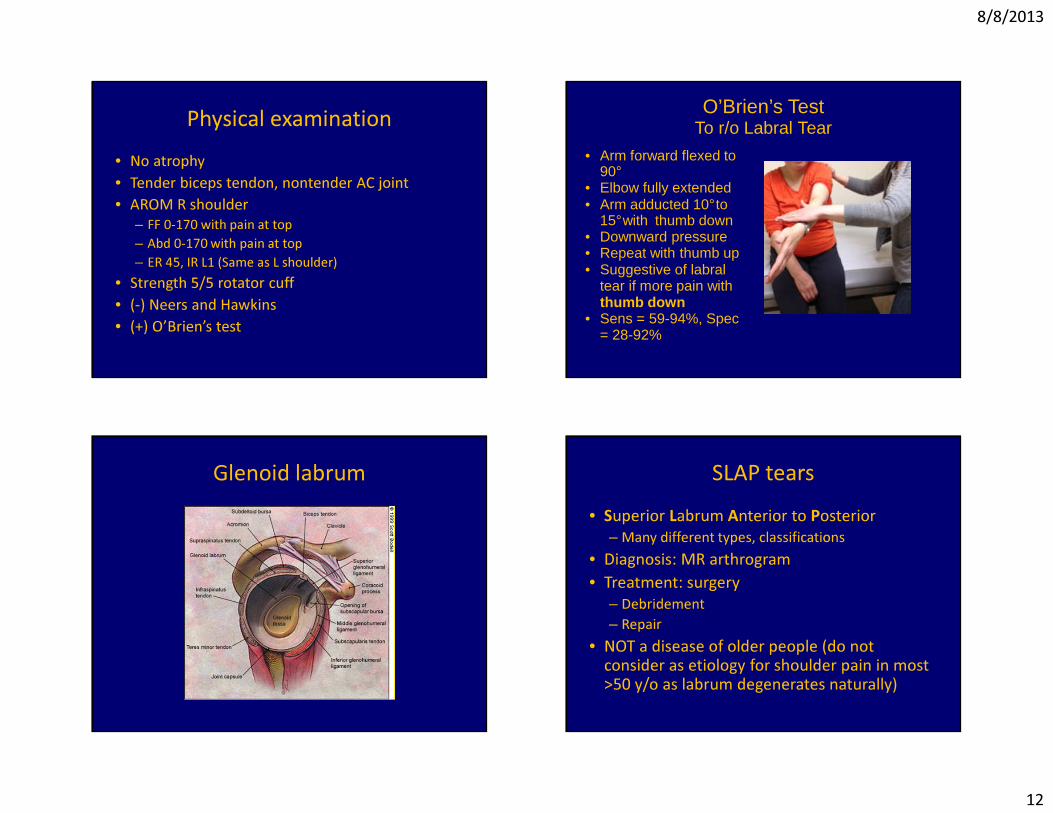
O’Brien’s TestTo r/o Labral Tear
• Arm forward flexed to 90°
• Elbow fully extended• Arm adducted 10°to
15°with thumb down• Downward pressure• Repeat with thumb up• Suggestive of labral
tear if more pain with thumb down
• Sens = 59-94%, Spec = 28-92%
Glenoid labrum SLAP tears• Superior Labrum Anterior to Posterior
– Many different types, classifications• Diagnosis: MR arthrogram• Treatment: surgery
– Debridement– Repair
• NOT a disease of older people (do not consider as etiology for shoulder pain in most >50 y/o as labrum degenerates naturally)
8/8/2013
13
Hip Problems
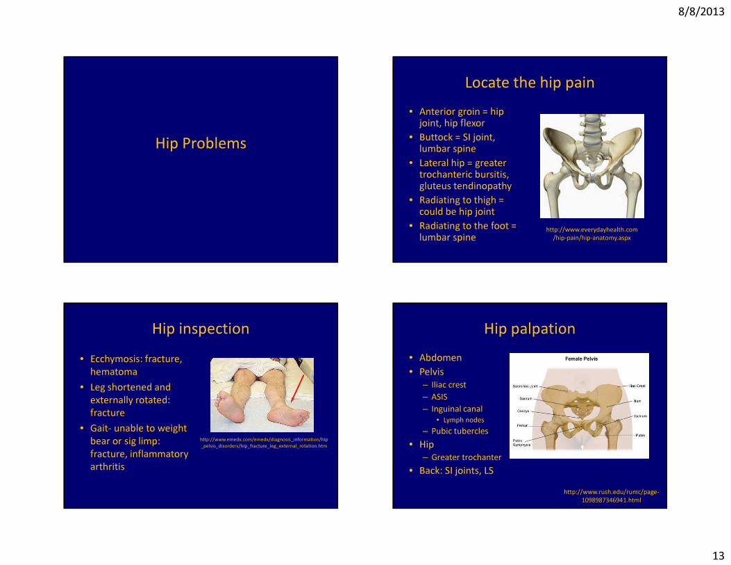
Locate the hip pain• Anterior groin = hip
joint, hip flexor• Buttock = SI joint,
lumbar spine• Lateral hip = greater
trochanteric bursitis, gluteus tendinopathy
• Radiating to thigh = could be hip joint
• Radiating to the foot = lumbar spine http://www.everydayhealth.com
/hip-pain/hip-anatomy.aspx
Hip inspection• Ecchymosis: fracture,
hematoma• Leg shortened and
externally rotated: fracture
• Gait- unable to weight bear or sig limp: fracture, inflammatory arthritis
http://www.emedx.com/emedx/diagnosis_information/hip_pelvis_disorders/hip_fracture_leg_external_rotation.htm
Hip palpation• Abdomen• Pelvis
– Iliac crest– ASIS– Inguinal canal
• Lymph nodes– Pubic tubercles
• Hip– Greater trochanter
• Back: SI joints, LShttp://www.rush.edu/rumc/page-
1098987346941.html
8/8/2013
14
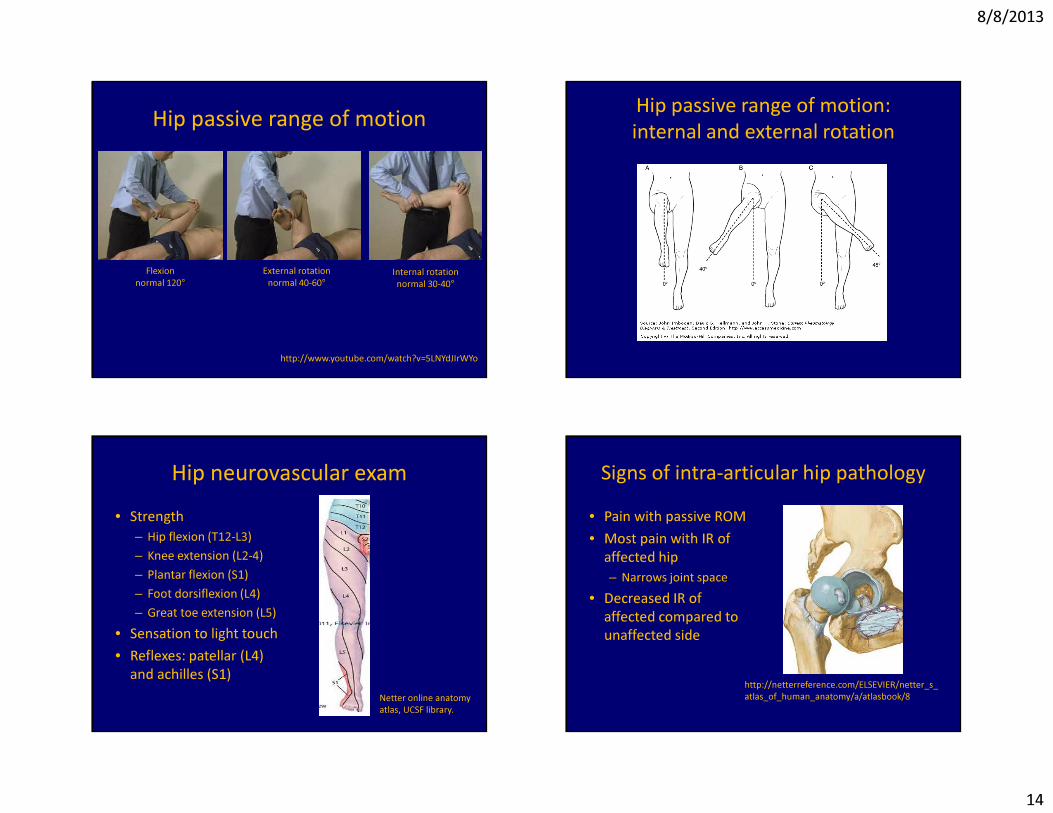
Hip passive range of motion
Flexionnormal 120°
External rotationnormal 40-60°
Internal rotationnormal 30-40°
http://www.youtube.com/watch?v=5LNYdJIrWYo
Hip passive range of motion:internal and external rotation
Hip neurovascular exam• Strength
– Hip flexion (T12-L3)– Knee extension (L2-4)– Plantar flexion (S1)– Foot dorsiflexion (L4)– Great toe extension (L5)
• Sensation to light touch• Reflexes: patellar (L4)
and achilles (S1)Netter online anatomy atlas, UCSF library.
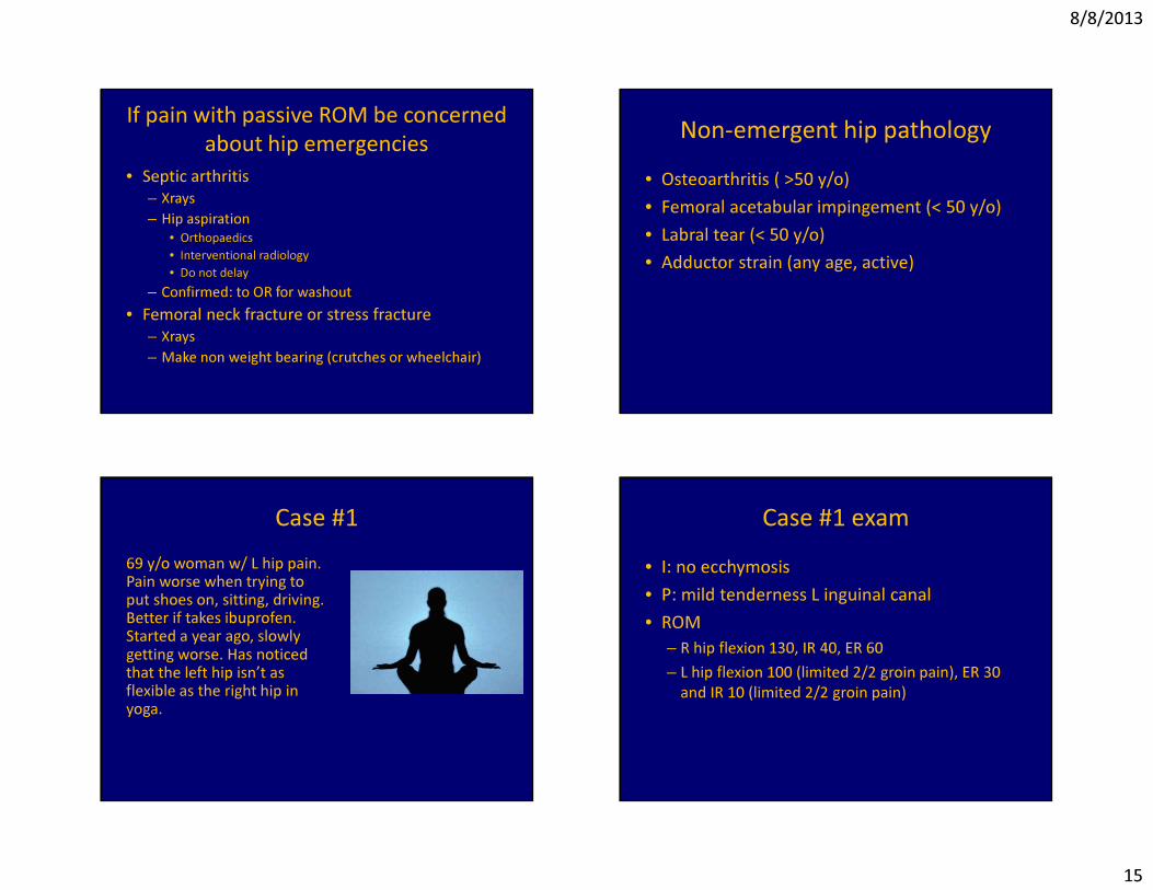
Signs of intra-articular hip pathology• Pain with passive ROM• Most pain with IR of
affected hip– Narrows joint space
• Decreased IR of affected compared to unaffected side
http://netterreference.com/ELSEVIER/netter_s_atlas_of_human_anatomy/a/atlasbook/8
8/8/2013
15
If pain with passive ROM be concerned about hip emergencies
• Septic arthritis– Xrays– Hip aspiration
• Orthopaedics• Interventional radiology• Do not delay
– Confirmed: to OR for washout• Femoral neck fracture or stress fracture
– Xrays– Make non weight bearing (crutches or wheelchair)
Non-emergent hip pathology• Osteoarthritis ( >50 y/o)• Femoral acetabular impingement (< 50 y/o)• Labral tear (< 50 y/o)• Adductor strain (any age, active)
Case #169 y/o woman w/ L hip pain. Pain worse when trying to put shoes on, sitting, driving. Better if takes ibuprofen. Started a year ago, slowly getting worse. Has noticed that the left hip isn’t as flexible as the right hip in yoga.
Case #1 exam• I: no ecchymosis• P: mild tenderness L inguinal canal• ROM
– R hip flexion 130, IR 40, ER 60– L hip flexion 100 (limited 2/2 groin pain), ER 30
and IR 10 (limited 2/2 groin pain)
8/8/2013
16
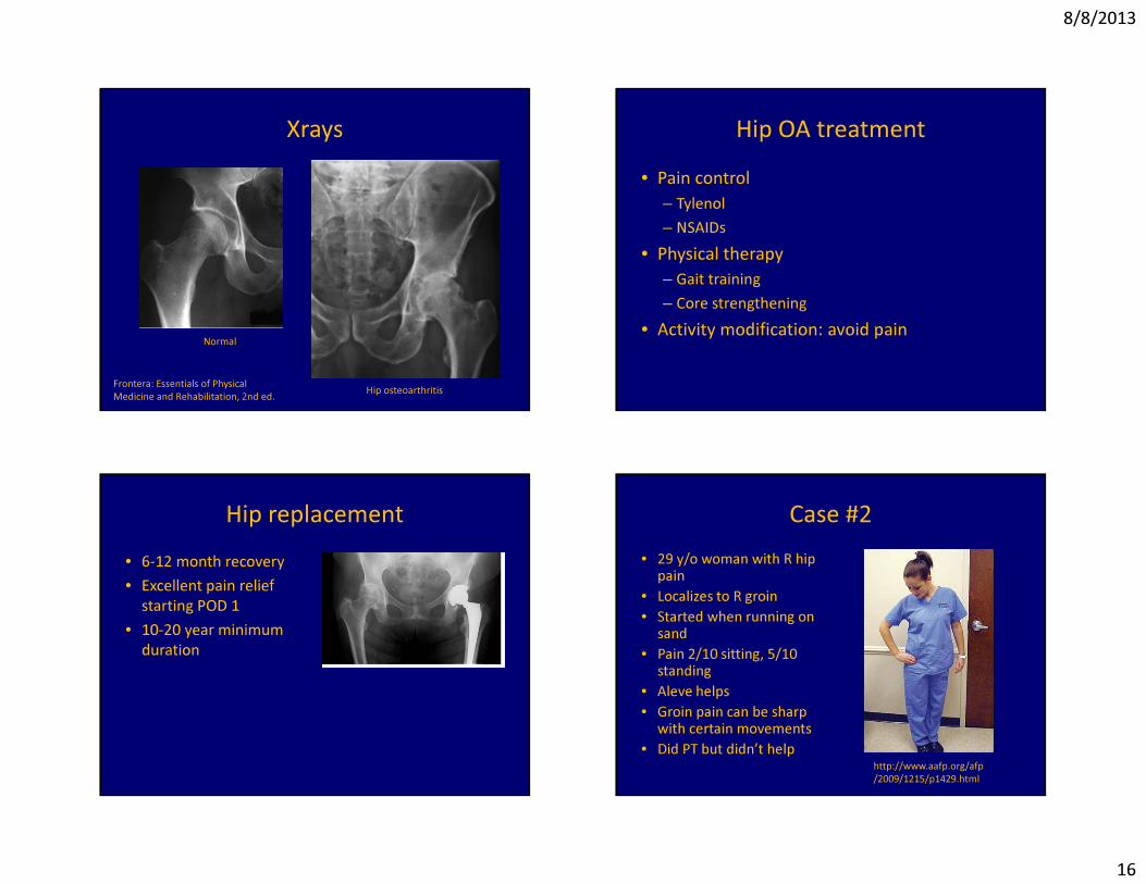
Xrays
Normal
Hip osteoarthritisFrontera: Essentials of Physical Medicine and Rehabilitation, 2nd ed.
Hip OA treatment• Pain control
– Tylenol– NSAIDs
• Physical therapy– Gait training– Core strengthening
• Activity modification: avoid pain
Hip replacement• 6-12 month recovery• Excellent pain relief
starting POD 1• 10-20 year minimum
duration
Case #2• 29 y/o woman with R hip
pain• Localizes to R groin• Started when running on
sand • Pain 2/10 sitting, 5/10
standing• Aleve helps• Groin pain can be sharp
with certain movements• Did PT but didn’t help
http://www.aafp.org/afp/2009/1215/p1429.html
8/8/2013
17
5 questions for every athlete with hip pain
1. Training: increased mileage?2. Nutrition: Calories in versus calories out?
History of eating d/o? Dietary restrictions?3. History of stress fractures?4. Family history of osteoporosis?5. Menstrual history?
Case #2 exam• I: no ecchymosis• P: ttp R inguinal canal• ROM: bilateral flexion 130, IR 40 and ER 60
but R groin pain with flexion and IR.• OT:
– FADIR and FABER R hip cause R groin pain– No pain with FADIR and FABER L hip
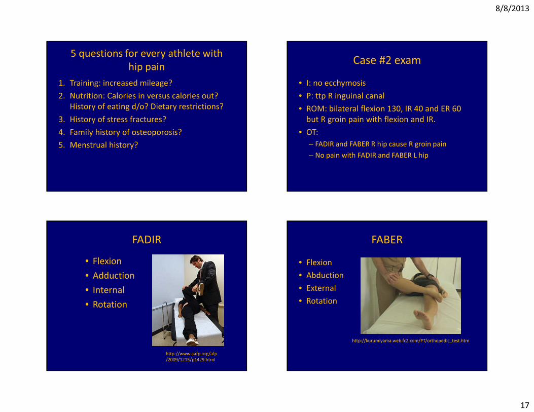
FADIR• Flexion• Adduction• Internal• Rotation
http://www.aafp.org/afp/2009/1215/p1429.html
FABER• Flexion• Abduction• External• Rotation
http://kurumiyama.web.fc2.com/PT/orthopedic_test.htm
8/8/2013
18
Case #2 differential diagnosis1. Hip labral tear2. Hip impingement3. Labral tear and impingement4. Femoral neck stress fracture
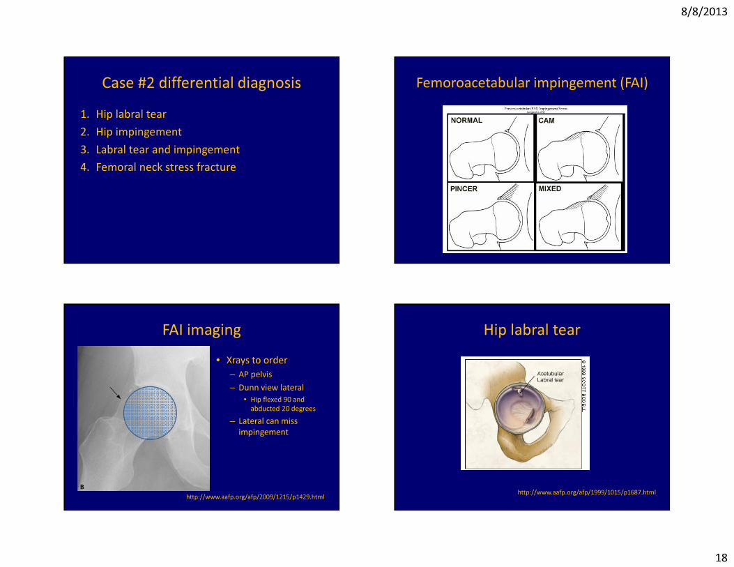
Femoroacetabular impingement (FAI)
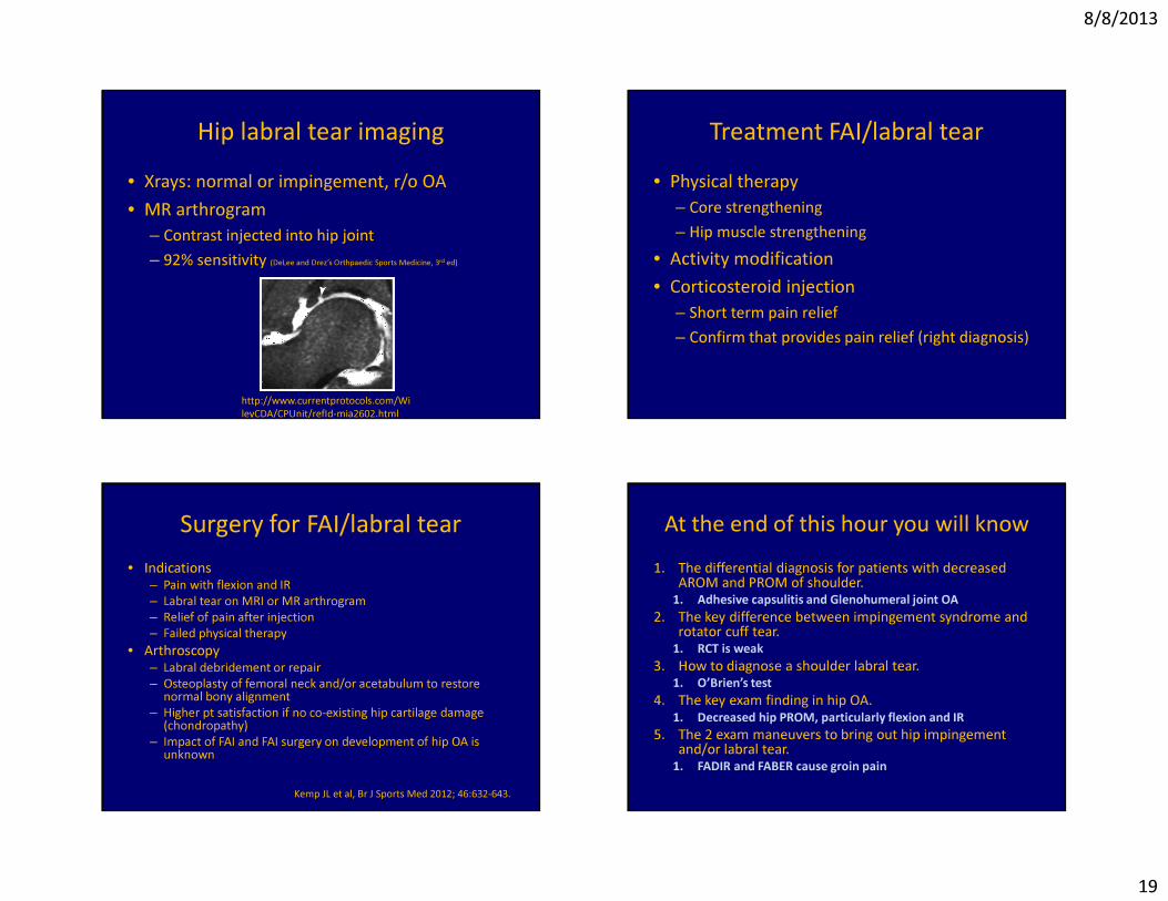
FAI imaging• Xrays to order
– AP pelvis– Dunn view lateral
• Hip flexed 90 and abducted 20 degrees
– Lateral can miss impingement
http://www.aafp.org/afp/2009/1215/p1429.html
Hip labral tear
http://www.aafp.org/afp/1999/1015/p1687.html
8/8/2013
19
Hip labral tear imaging• Xrays: normal or impingement, r/o OA• MR arthrogram
– Contrast injected into hip joint– 92% sensitivity (DeLee and Drez’s Orthpaedic Sports Medicine, 3rd ed)
http://www.currentprotocols.com/WileyCDA/CPUnit/refId-mia2602.html
Treatment FAI/labral tear• Physical therapy
– Core strengthening– Hip muscle strengthening
• Activity modification• Corticosteroid injection
– Short term pain relief– Confirm that provides pain relief (right diagnosis)
Surgery for FAI/labral tear• Indications
– Pain with flexion and IR– Labral tear on MRI or MR arthrogram– Relief of pain after injection– Failed physical therapy
• Arthroscopy– Labral debridement or repair– Osteoplasty of femoral neck and/or acetabulum to restore
normal bony alignment– Higher pt satisfaction if no co-existing hip cartilage damage
(chondropathy)– Impact of FAI and FAI surgery on development of hip OA is
unknown
Kemp JL et al, Br J Sports Med 2012; 46:632-643.
At the end of this hour you will know1. The differential diagnosis for patients with decreased
AROM and PROM of shoulder.1. Adhesive capsulitis and Glenohumeral joint OA
2. The key difference between impingement syndrome and rotator cuff tear.
1. RCT is weak3. How to diagnose a shoulder labral tear.
1. O’Brien’s test4. The key exam finding in hip OA.
1. Decreased hip PROM, particularly flexion and IR5. The 2 exam maneuvers to bring out hip impingement
and/or labral tear.1. FADIR and FABER cause groin pain


![1810 Federal Census · NORTON, Ifse 2 BOWLES, Evan 6 CARLIN, Alexis French/Kouri-Vini 5 CARLIN, Honoré 12 CARLIN, Célestin 6 CARLIN, Denis 7 CARLIN, Widow [of] 2 CARLIN, Eugêne](https://img.pdfslide.net/doc/110x75/5e6b107934ce1567772964a1/1810-federal-census-norton-ifse-2-bowles-evan-6-carlin-alexis-frenchkouri-vini.jpg)