Embed Size (px)
Citation preview
Mutants of Arabidopsis Lacking Starch Branching Enzyme IISubstitute Plastidial Starch Synthesis by CytoplasmicMaltose Accumulation W
Sylvain Dumez,a Fabrice Wattebled,a,1 David Dauvillee,a David Delvalle,a Veronique Planchot,b Steven G. Ball,a
and Christophe D’Hulsta,2
a Unite de Glycobiologie Structurale et Fonctionnelle, Unite Mixte de Recherche 8576, Centre National de la
Recherche Scientifique, Universite des Sciences et Technologies de Lille,
59655 Villeneuve d’Ascq Cedex, Franceb Unite de Recherche Biopolymeres, Interactions, Assemblages, Centre de Nantes, Institut National de la Recherche
Agronomique, 44316 Nantes Cedex 3, France
Three genes, BE1, BE2, and BE3, which potentially encode isoforms of starch branching enzymes, have been found in the
genome of Arabidopsis thaliana. Although no impact on starch structure was observed in null be1 mutants, modifications in
amylopectin structure analogous to those of other branching enzyme II mutants were detected in be2 and be3. No impact on
starch content was found in any of the single mutant lines. Moreover, three double mutant combinations were produced
(be1 be2, be1 be3, and be2 be3), and the impact of the mutations on starch content and structure was analyzed. Our results
suggest that BE1 has no apparent function for the synthesis of starch in the leaves, as both be1 be2 and be1 be3 double
mutants display the same phenotype as be2 and be3 separately. However, starch synthesis was abolished in be2 be3, while
high levels of a-maltose were assayed in the cytosol. This result indicates that the functions of both BE2 and BE3, which
belong to class II starch branching enzymes, are largely redundant in Arabidopsis. Moreover, we demonstrate that maltose
accumulation depends on the presence of an active ADP-glucose pyrophosphorylase and that the cytosolic transgluco-
sidase DISPROPORTIONATING ENZYME2, required for maltose metabolization, is specific for b-maltose.
INTRODUCTION
Starch is a polymer of D-glucose that accumulates in the plastids
of plants as huge water-insoluble granules. Glucose residues are
linked together by a-1,4 and a-1,6 O-glycosidic bonds. The
a-1,6 linkages are commonly referred to as the branch points,
while the a-1,4 O-glycosidic bonds compose the linear back-
bone of the polymer. The starch granule is composed of two
structurally distinct homopolymers: amylose, which is essentially
linear, and amylopectin, which is a moderately branched mac-
romolecule (usually 6% of a-1,6 bonds within the polymer). The
asymmetrical distribution of the branch points within amylopec-
tin is responsible for the cluster organization of the molecule that
further adopts a semicrystalline structure (Hizukuri, 1986; for re-
view, see Buleon et al., 1998).
The synthesis pathway of amylopectin is rather complex and
involves different enzymatic activities usually supported by mul-
tiple and genetically distinct forms of enzymes. This pathway
includes starch synthases (elongation step), starch branching
enzymes (introduction of branches), starch debranching en-
zymes, and a-1,4-glucanotransferases (maturation of the struc-
ture required for further growth and crystallization of the granule)
(for review, see Myers et al., 2000; Ball and Morell, 2003).
Starch branching enzymes (SBEs; EC 2.4.1.18; CAZy family
GH13) catalyze the formation of the a-1,6 linkages within the
polymer. SBEs first cleave a preexisting a-1,4 linkage and trans-
fer the fragment initially positioned at the nonreducing end of the
cleaved glucan to an a-1,6 position through an intra- or inter-
molecular mechanism (for review, see Sivak and Preiss, 1998).
The consequence of this reaction is to increase the number of
nonreducing ends within the molecule, thus facilitating the elon-
gation by the starch synthases. Two to three genetically inde-
pendent isoforms of SBEs were observed in most plants (Fisher
et al., 1996a; Larsson et al., 1996, 1998; Morell et al., 1997;
Mizuno et al., 1992; Nakamura et al., 1992). Depending on their
peptide sequence, these SBEs were categorized into two clas-
ses named SBEI (or B family) and SBEII (or A family). Some of
these forms, essentially maize (Zea mays) and potato (Solanum
tuberosum) SBEs, were kinetically characterized after their pu-
rification from plant tissues or their heterologous expression in
Escherichia coli (Guan and Preiss, 1993; Takeda et al., 1993;
Guan et al., 1994; Rydberg et al., 2001). From these studies, it
appears that SBEI is essentially active on long linear chains (of
amylose type), while SBEII is more active on the shorter chains of
amylopectin. Moreover, SBEI preferentially transfers longer
chains than SBEII.
1 Current address: Centre d’Angers, Institut National de la RechercheAgronomique, Unite Mixte de Recherche 1259, Genetique et Horticul-ture (GenHort), B.P. 60057, 49071 Beaucouze Cedex, France.2 To whom correspondence should be addressed. E-mail [email protected]; fax 33-3-20-43-6555.The author responsible for distribution of materials integral to thefindings presented in this article in accordance with the policy describedin the Instructions for Authors (www.plantcell.org) is: Christophe D’Hulst([email protected]).W Online version contains Web-only data.www.plantcell.org/cgi/doi/10.1105/tpc.105.037671
The Plant Cell, Vol. 18, 2694–2709, October 2006, www.plantcell.org ª 2006 American Society of Plant Biologists
Mutant lines defective for SBEI, IIa, and IIb in maize (Stinard
et al., 1993; Blauth et al., 2001, 2002; Yao et al., 2004) and rice
(Oryza sativa; Nishi et al., 2001; Nakamura, 2002; Satoh et al.,
2003) were isolated and analyzed for their ability to synthesize
starch. The absence of SBEI in both plants has little or no impact
on starch that accumulates in the endosperm, while the absence
of SBEIIb leads to the synthesis of a modified amylopectin with
a strong decrease in the number of short chains and an increase
in the number of long chains. On the other hand, the absence of
SBEIIa in maize leads only to the modification of the structure of
starch that accumulates in the leaf (transitory starch) without
impact on the endosperm starch (storage starch). In Arabidopsis
thaliana, two SBEs belonging to class II were cloned (Fisher
et al., 1996b), and their expression levels were characterized
(Khoshnoodi et al., 1998). The precise function of these two
forms of SBEII remains unknown. A third gene putatively encod-
ing an SBE was observed in the nuclear genome of this plant.
However, the corresponding protein does not belong to a type I
class of SBEs as described in other plants.
The objective of this work was to investigate the function of the
three SBEs (BE1, BE2, and BE3) in the leaves of Arabidopsis
through the selection and phenotypic characterization of mutants
specifically defective in each of the three isoforms. Moreover, we
have constructed double mutant combinations defective in two
out of the three SBEs. Our results indicate that (1) BE1 has no ap-
parent function in the metabolism of starch in Arabidopsis leaves;
(2) BE2 and BE3, which belong to class II of SBEs, are required for
the synthesis of normal starch, and their activities are largely
redundant; (3) the double mutant line simultaneously defective in
both BE2 and BE3 substitutes starch by the accumulation of
a-maltose in the cytosol; (4) the latter is generated through de-
gradation of abnormal products of the starch synthesis pathway;
and (5) DISPROPORTIONATING ENZYME2 (DPE2) is specific for
b-maltose, thereby explaining the rationale of b-amylolysis as the
major route of starch degradation in plant leaves.
RESULTS
The Nuclear Genome of Arabidopsis Contains Three
Candidate Genes Putatively Encoding SBEs
The three genes putatively encoding SBEs (EC 2.4.1.18; CAZy
family GH13) in the Arabidopsis genome are BE1, BE2, and BE3
(Table 1). These genes are expressed in the leaves of Arabidop-
sis during either short (8 h) or normal (12 h) daylengths (http://
www.starchmetnet.org/Datapages/AtBE2/AtBE2Frameset.htm),
although the expression of BE1 is very low and rather constant
throughout the cycle when compared with those of BE2 and BE3.
The transcript level of BE3 increases during the illuminated
period to reach a maximum at the transition between day and
night. It then regularly falls to a minimum at the end of the dark
period. BE2 transcript level is maximal 4 h after light onset and
slowly decreases to a minimum near the end of the dark phase.
The proteins corresponding to the BE1, BE2, and BE3 genes
contain a predicted chloroplast-targeting peptide of 49, 61, and
37 amino acids, respectively (ChloroP, http://www.cbs.dtu.dk/
services/ChloroP/; Emanuelsson et al., 1999), thereby suggest-
ing that these proteins are likely to be localized within the
chloroplast. BE2 and BE3 are two highly conserved proteins
with >75% amino acid identity. The relatively well-conserved
intron-exon organization in 59 parts of both BE2 and BE3 genes
(Figure 1) suggests that these two genes probably arose from
a duplication event. By comparison, the sequence homology
between BE1 and both BE2 and BE3 can be considered as low
(only 28% and 27% amino acid identity, respectively), suggest-
ing a different ancestral origin of the corresponding gene. Protein
sequence comparisons with SBEs from other plant sources in-
dicate that BE2 and BE3 belong to the class A of SBEs, while BE1
cannot be easily classified within plant SBEs. BE1 is not related
to the standard plant A- or B-type SBE families but shows more
relatedness to the glycogen-branching enzymes from fungi and
animals, although the protein sequence similarity is quite low in
that case too (<30%). The situation in Arabidopsis is thus
different from that observed in other dicots, such as potato,
pea (Pisum sativum), and bean (Phaseolus vulgaris), where only
one type A of SBE can be found along with one type B of SBE (see
dendrogram of branching enzyme families in Supplemental
Figure 1 online; for review, see Ball and Morell, 2003).
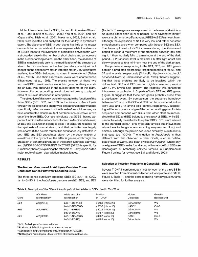
Selection of Insertion Mutations in Genes BE1, BE2, and BE3
Several T-DNA insertion mutant lines for each of the three SBEs
were selected from different collections (Genoplante and SALK;
Figure 1, Table 1), and the corresponding homozygous mutants
were identified for further analysis.
Table 1. Description of the Different Arabidopsis Mutant Alleles of SBEs Used in This Work
Gene
AGI Gene
Identificationa
Allele and Line
Identification
Position
of T-DNAb
Mutant
Collection
Genetic
Background
BE1 At3g20440 be1-1 (DYK140) þ6461 (intron 20) Genoplantec Ws
be1-2 (N637880) þ5592 (intron 15) NASCd Col-0
BE2 At5g03650 be2-1 (EFH20) þ5022 (intron 19) Genoplante Ws
be2-2 (DSA16) þ5487 (exon 22) Genoplante Ws
BE3 At2g36390 be3-1 (N548089) þ3262 (exon 10) NASC Col-0
be3-2 (EQJ13) þ4485 (exon 15) Genoplante Ws
a AGI, Arabidopsis Genome Initiative.b Position of T-DNA is given from the start codon.c Genoplante: http://genoplante-info.infobiogen.fr/FLAGdb/.d Nottingham Arabidopsis Stock Centre: http://nasc.nott.ac.uk/.
SBE Mutants of Arabidopsis 2695
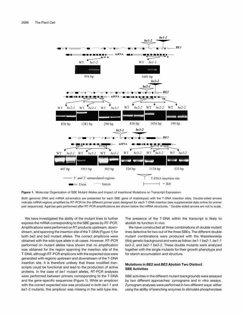
We have investigated the ability of the mutant lines to further
express the mRNA corresponding to the SBE genes by RT-PCR.
Amplifications were performed on RT products upstream, down-
stream, and spanning the insertion site of the T-DNA (Figure 1) for
both be2 and be3 mutant alleles. The correct amplicons were
obtained with the wild-type allele in all cases. However, RT-PCR
performed on mutant alleles have shown that no amplification
was obtained for the region spanning the insertion site of the
T-DNA, although RT-PCR amplicons with the expected size were
generated with regions upstream and downstream of the T-DNA
insertion site. It is therefore unlikely that these modified tran-
scripts could be functional and lead to the production of active
proteins. In the case of be1 mutant alleles, RT-PCR analyses
were performed between primers corresponding to the T-DNA
and the gene-specific sequences (Figure 1). While an amplicon
with the correct expected size was produced in both be1-1 and
be1-2 mutants, this amplicon was missing in the wild-type line.
The presence of the T-DNA within the transcript is likely to
abolish its function in vivo.
We have constructed all three combinations of double mutant
lines defective for two out of the three SBEs. The different double
mutant combinations were produced with the Wassilewskija
(Ws) genetic background and were as follow: be1-1 be2-1, be1-1
be3-2, and be2-1 be3-2. These double mutants were analyzed
together with the single mutants for their growth phenotype and
for starch accumulation and structure.
Mutations in BE2 and BE3 Abolish Two Distinct
SBE Activities
SBE activities in the different mutant backgrounds were assayed
by two different approaches: zymograms and in vitro assays.
Zymogram analyses were performed in two different ways: either
using the ability of branching enzymes to stimulate phosphorylase
Figure 1. Molecular Organization of SBE Mutant Alleles and Impact of Insertional Mutations on Transcript Expression.
Both genomic DNA and mRNA schematics are presented for each SBE gene of Arabidopsis with the T-DNA insertion sites. Double-sided arrows
indicate mRNA regions amplified by RT-PCR for the different primer pairs designed for each T-DNA insertion (see supplemental data online for primer
pair sequences). Agarose gels performed after RT-PCR amplifications are shown below the mRNA structures. * Double-sided arrows are not to scale.
2696 The Plant Cell
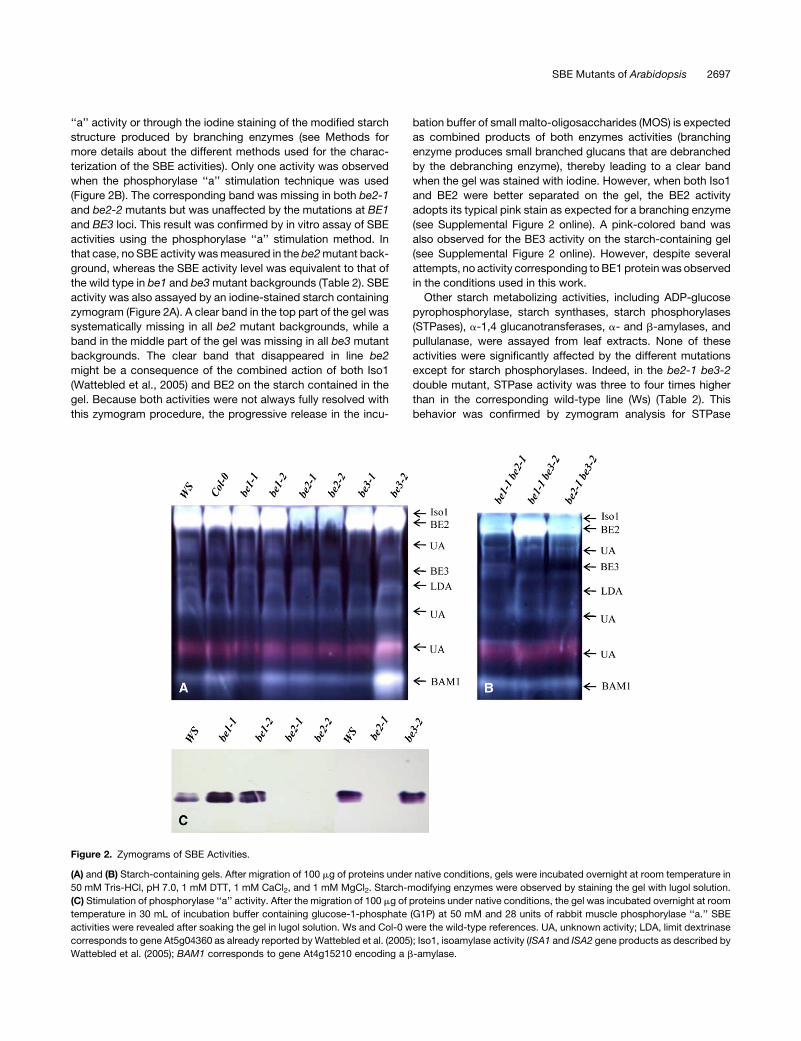
‘‘a’’ activity or through the iodine staining of the modified starch
structure produced by branching enzymes (see Methods for
more details about the different methods used for the charac-
terization of the SBE activities). Only one activity was observed
when the phosphorylase ‘‘a’’ stimulation technique was used
(Figure 2B). The corresponding band was missing in both be2-1
and be2-2 mutants but was unaffected by the mutations at BE1
and BE3 loci. This result was confirmed by in vitro assay of SBE
activities using the phosphorylase ‘‘a’’ stimulation method. In
that case, no SBE activity was measured in the be2 mutant back-
ground, whereas the SBE activity level was equivalent to that of
the wild type in be1 and be3 mutant backgrounds (Table 2). SBE
activity was also assayed by an iodine-stained starch containing
zymogram (Figure 2A). A clear band in the top part of the gel was
systematically missing in all be2 mutant backgrounds, while a
band in the middle part of the gel was missing in all be3 mutant
backgrounds. The clear band that disappeared in line be2
might be a consequence of the combined action of both Iso1
(Wattebled et al., 2005) and BE2 on the starch contained in the
gel. Because both activities were not always fully resolved with
this zymogram procedure, the progressive release in the incu-
bation buffer of small malto-oligosaccharides (MOS) is expected
as combined products of both enzymes activities (branching
enzyme produces small branched glucans that are debranched
by the debranching enzyme), thereby leading to a clear band
when the gel was stained with iodine. However, when both Iso1
and BE2 were better separated on the gel, the BE2 activity
adopts its typical pink stain as expected for a branching enzyme
(see Supplemental Figure 2 online). A pink-colored band was
also observed for the BE3 activity on the starch-containing gel
(see Supplemental Figure 2 online). However, despite several
attempts, no activity corresponding to BE1 protein was observed
in the conditions used in this work.
Other starch metabolizing activities, including ADP-glucose
pyrophosphorylase, starch synthases, starch phosphorylases
(STPases), a-1,4 glucanotransferases, a- and b-amylases, and
pullulanase, were assayed from leaf extracts. None of these
activities were significantly affected by the different mutations
except for starch phosphorylases. Indeed, in the be2-1 be3-2
double mutant, STPase activity was three to four times higher
than in the corresponding wild-type line (Ws) (Table 2). This
behavior was confirmed by zymogram analysis for STPase
Figure 2. Zymograms of SBE Activities.
(A) and (B) Starch-containing gels. After migration of 100 mg of proteins under native conditions, gels were incubated overnight at room temperature in
50 mM Tris-HCl, pH 7.0, 1 mM DTT, 1 mM CaCl2, and 1 mM MgCl2. Starch-modifying enzymes were observed by staining the gel with lugol solution.
(C) Stimulation of phosphorylase ‘‘a’’ activity. After the migration of 100 mg of proteins under native conditions, the gel was incubated overnight at room
temperature in 30 mL of incubation buffer containing glucose-1-phosphate (G1P) at 50 mM and 28 units of rabbit muscle phosphorylase ‘‘a.’’ SBE
activities were revealed after soaking the gel in lugol solution. Ws and Col-0 were the wild-type references. UA, unknown activity; LDA, limit dextrinase
corresponds to gene At5g04360 as already reported by Wattebled et al. (2005); Iso1, isoamylase activity (ISA1 and ISA2 gene products as described by
Wattebled et al. (2005); BAM1 corresponds to gene At4g15210 encoding a b-amylase.
SBE Mutants of Arabidopsis 2697
activities where both the cytosolic and the plastidial forms of
STPase were increased (see Supplemental Figure 4 online).
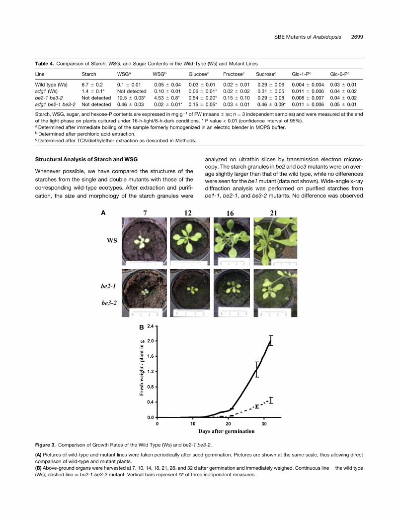
Impact of the Mutations on Starch Accumulation Levels
and the Growth Phenotype
We measured the leaf starch and water-soluble glucan (WSGs)
contents at the end of the light period (16 h day/8 h night) in the
different single and double mutants and compared them with that
of the corresponding wild-type ecotypes (Table 3). We did not
detect any significant alteration of the starch content in the single
mutants or in both be1-1 be2-1 and be1-1 be3-2 double mutants.
However, the be2-1 be3-2 double mutant was free of starch
(see Supplemental Figure 6 online). The absence of starch was
coupled with the accumulation of very high levels of WSGs (what-
ever extraction method was used) that were not observed in other
lines (Tables 3 and 4). Moreover, this double mutant displayed a
lower growth rate, a reduced size of the mature plant, a pale color,
and a general wilting of the inflorescence (Figure 3; see Supple-
mental Figure 5 online). Thirty days after seed germination, the
fresh weight of the above-ground organs of the double mutant
was only one-fifth of the wild type (Figure 3) under the 16-h-day/
8-h-night growth conditions used during this work. The same
lower growth rate was observed under the 12-h-day/12-h-night
regime, although it was less pronounced. Both dpe2 and mex1
mutants of Arabidopsis that have lost their ability to metabolize
maltose display such a dwarf-like phenotype. However, both
mutants exhibit a starch excess phenotype and are not affected
for polysaccharide biosynthesis. Despite the growth phenotype,
the be2-1 be3-2 double mutant was still able to produce siliques
and viable seeds after self-pollination, although the flowering rate
was low when compared with that of other lines.
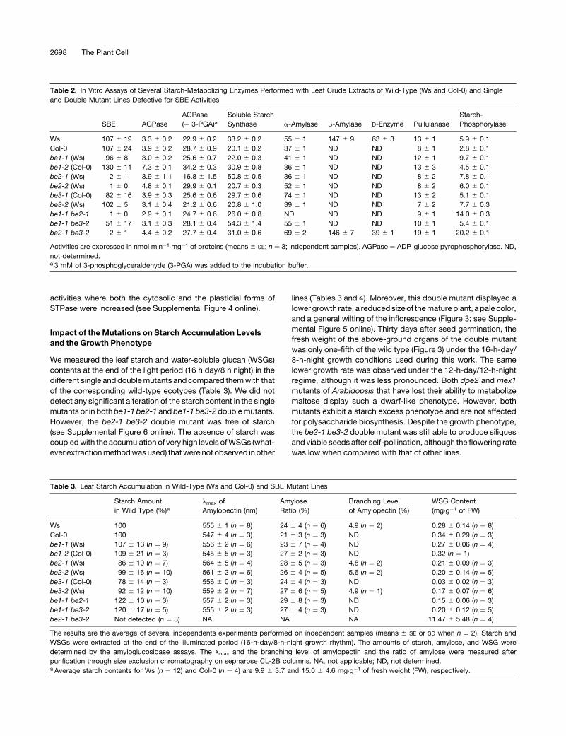
Table 2. In Vitro Assays of Several Starch-Metabolizing Enzymes Performed with Leaf Crude Extracts of Wild-Type (Ws and Col-0) and Single
and Double Mutant Lines Defective for SBE Activities
SBE AGPase
AGPase
(þ 3-PGA)aSoluble Starch
Synthase a-Amylase b-Amylase D-Enzyme Pullulanase
Starch-
Phosphorylase
Ws 107 6 19 3.3 6 0.2 22.9 6 0.2 33.2 6 0.2 55 6 1 147 6 9 63 6 3 13 6 1 5.9 6 0.1
Col-0 107 6 24 3.9 6 0.2 28.7 6 0.9 20.1 6 0.2 37 6 1 ND ND 8 6 1 2.8 6 0.1
be1-1 (Ws) 96 6 8 3.0 6 0.2 25.6 6 0.7 22.0 6 0.3 41 6 1 ND ND 12 6 1 9.7 6 0.1
be1-2 (Col-0) 130 6 11 7.3 6 0.1 34.2 6 0.3 30.9 6 0.8 36 6 1 ND ND 13 6 3 4.5 6 0.1
be2-1 (Ws) 2 6 1 3.9 6 1.1 16.8 6 1.5 50.8 6 0.5 36 6 1 ND ND 8 6 2 7.8 6 0.1
be2-2 (Ws) 1 6 0 4.8 6 0.1 29.9 6 0.1 20.7 6 0.3 52 6 1 ND ND 8 6 2 6.0 6 0.1
be3-1 (Col-0) 82 6 16 3.9 6 0.3 25.6 6 0.6 29.7 6 0.6 74 6 1 ND ND 13 6 2 5.1 6 0.1
be3-2 (Ws) 102 6 5 3.1 6 0.4 21.2 6 0.6 20.8 6 1.0 39 6 1 ND ND 7 6 2 7.7 6 0.3
be1-1 be2-1 1 6 0 2.9 6 0.1 24.7 6 0.6 26.0 6 0.8 ND ND ND 9 6 1 14.0 6 0.3
be1-1 be3-2 51 6 17 3.1 6 0.3 28.1 6 0.4 54.3 6 1.4 55 6 1 ND ND 10 6 1 5.4 6 0.1
be2-1 be3-2 2 6 1 4.4 6 0.2 27.7 6 0.4 31.0 6 0.6 69 6 2 146 6 7 39 6 1 19 6 1 20.2 6 0.1
Activities are expressed in nmol�min�1�mg�1 of proteins (means 6 SE; n ¼ 3; independent samples). AGPase ¼ ADP-glucose pyrophosphorylase. ND,
not determined.a 3 mM of 3-phosphoglyceraldehyde (3-PGA) was added to the incubation buffer.
Table 3. Leaf Starch Accumulation in Wild-Type (Ws and Col-0) and SBE Mutant Lines
Starch Amount
in Wild Type (%)almax of
Amylopectin (nm)
Amylose
Ratio (%)
Branching Level
of Amylopectin (%)
WSG Content
(mg�g�1 of FW)
Ws 100 555 6 1 (n ¼ 8) 24 6 4 (n ¼ 6) 4.9 (n ¼ 2) 0.28 6 0.14 (n ¼ 8)
Col-0 100 547 6 4 (n ¼ 3) 21 6 3 (n ¼ 3) ND 0.34 6 0.29 (n ¼ 3)
be1-1 (Ws) 107 6 13 (n ¼ 9) 556 6 2 (n ¼ 6) 23 6 7 (n ¼ 4) ND 0.27 6 0.06 (n ¼ 4)
be1-2 (Col-0) 109 6 21 (n ¼ 3) 545 6 5 (n ¼ 3) 27 6 2 (n ¼ 3) ND 0.32 (n ¼ 1)
be2-1 (Ws) 86 6 10 (n ¼ 7) 564 6 5 (n ¼ 4) 28 6 5 (n ¼ 3) 4.8 (n ¼ 2) 0.21 6 0.09 (n ¼ 3)
be2-2 (Ws) 99 6 16 (n ¼ 10) 561 6 2 (n ¼ 6) 26 6 4 (n ¼ 5) 5.6 (n ¼ 2) 0.20 6 0.14 (n ¼ 5)
be3-1 (Col-0) 78 6 14 (n ¼ 3) 556 6 0 (n ¼ 3) 24 6 4 (n ¼ 3) ND 0.03 6 0.02 (n ¼ 3)
be3-2 (Ws) 92 6 12 (n ¼ 10) 559 6 2 (n ¼ 7) 27 6 6 (n ¼ 5) 4.9 (n ¼ 1) 0.17 6 0.07 (n ¼ 6)
be1-1 be2-1 122 6 10 (n ¼ 3) 557 6 2 (n ¼ 3) 29 6 8 (n ¼ 3) ND 0.15 6 0.06 (n ¼ 3)
be1-1 be3-2 120 6 17 (n ¼ 5) 555 6 2 (n ¼ 3) 27 6 4 (n ¼ 3) ND 0.20 6 0.12 (n ¼ 5)
be2-1 be3-2 Not detected (n ¼ 3) NA NA NA 11.47 6 5.48 (n ¼ 4)
The results are the average of several independents experiments performed on independent samples (means 6 SE or SD when n ¼ 2). Starch and
WSGs were extracted at the end of the illuminated period (16-h-day/8-h-night growth rhythm). The amounts of starch, amylose, and WSG were
determined by the amyloglucosidase assays. The lmax and the branching level of amylopectin and the ratio of amylose were measured after
purification through size exclusion chromatography on sepharose CL-2B columns. NA, not applicable; ND, not determined.a Average starch contents for Ws (n ¼ 12) and Col-0 (n ¼ 4) are 9.9 6 3.7 and 15.0 6 4.6 mg�g�1 of fresh weight (FW), respectively.
2698 The Plant Cell
Structural Analysis of Starch and WSG
Whenever possible, we have compared the structures of the
starches from the single and double mutants with those of the
corresponding wild-type ecotypes. After extraction and purifi-
cation, the size and morphology of the starch granules were
analyzed on ultrathin slices by transmission electron micros-
copy. The starch granules in be2 and be3 mutants were on aver-
age slightly larger than that of the wild type, while no differences
were seen for the be1 mutant (data not shown). Wide-angle x-ray
diffraction analysis was performed on purified starches from
be1-1, be2-1, and be3-2 mutants. No difference was observed
Table 4. Comparison of Starch, WSG, and Sugar Contents in the Wild-Type (Ws) and Mutant Lines
Line Starch WSGa WSGb Glucosec Fructosec Sucrosec Glc-1-Pc Glc-6-Pc
Wild type (Ws) 6.7 6 0.2 0.1 6 0.01 0.05 6 0.04 0.03 6 0.01 0.02 6 0.01 0.29 6 0.06 0.004 6 0.004 0.03 6 0.01
adg1 (Ws) 1.4 6 0.1* Not detected 0.10 6 0.01 0.06 6 0.01* 0.02 6 0.02 0.31 6 0.05 0.011 6 0.006 0.04 6 0.02
be2-1 be3-2 Not detected 12.5 6 0.03* 4.53 6 0.8* 0.54 6 0.20* 0.15 6 0.10 0.29 6 0.08 0.008 6 0.007 0.04 6 0.02
adg1 be2-1 be3-2 Not detected 0.46 6 0.03 0.02 6 0.01* 0.15 6 0.05* 0.03 6 0.01 0.46 6 0.09* 0.011 6 0.006 0.05 6 0.01
Starch, WSG, sugar, and hexose-P contents are expressed in mg�g�1 of FW (means 6 SE; n ¼ 3 independent samples) and were measured at the end
of the light phase on plants cultured under 16-h-light/8-h-dark conditions. * P value < 0.01 (confidence interval of 95%).a Determined after immediate boiling of the sample formerly homogenized in an electric blender in MOPS buffer.b Determined after perchloric acid extraction.c Determined after TCA/diethylether extraction as described in Methods.
Figure 3. Comparison of Growth Rates of the Wild Type (Ws) and be2-1 be3-2.
(A) Pictures of wild-type and mutant lines were taken periodically after seed germination. Pictures are shown at the same scale, thus allowing direct
comparison of wild-type and mutant plants.
(B) Above-ground organs were harvested at 7, 10, 14, 18, 21, 28, and 32 d after germination and immediately weighed. Continuous line ¼ the wild type
(Ws); dashed line ¼ be2-1 be3-2 mutant. Vertical bars represent SE of three independent measures.
SBE Mutants of Arabidopsis 2699
when compared with the wild type. All starch samples were of the
B crystalline-type and displayed ;37% level of crystallinity.
Starches purified from the different mutant backgrounds were
submitted to size exclusion chromatography on a sepharose
CL-2B column to purify amylopectin and measure the amount of
amylose. The amount of amylose and the branching level of
amylopectin were not significantly different in the mutants when
compared with the wild type (Table 3).
We have established the chain length (CL) distribution profiles
of the amylopectin from each single and double mutant. Average
profiles produced from several independent analyses are pre-
sented in Figure 4. For better clarity, we have combined profiles
from mutant lines in both Ws and Columbia (Col-0) genetic
backgrounds since individual profiles were the same whatever
the genetic background (CL profiles were the same for Ws and
Col-0 samples). Both be2 and be3 mutants displayed more or
less the same profiles that were slightly different from those of the
wild type (Figures 4A, 4C, and 4D). DP 6-7 and DP 5-9 chains
were slightly decreased in be2 and be3, respectively, while DP
10-16 chains were slightly increased. No difference was evi-
denced for be1 in comparison with the wild type (Figure 4B).
be1-1 be2-1 (Figure 4E) and be1-1 be3-2 (Figure 4F) double
mutants showed the same profiles as the corresponding be2 and
be3 single mutants, respectively.
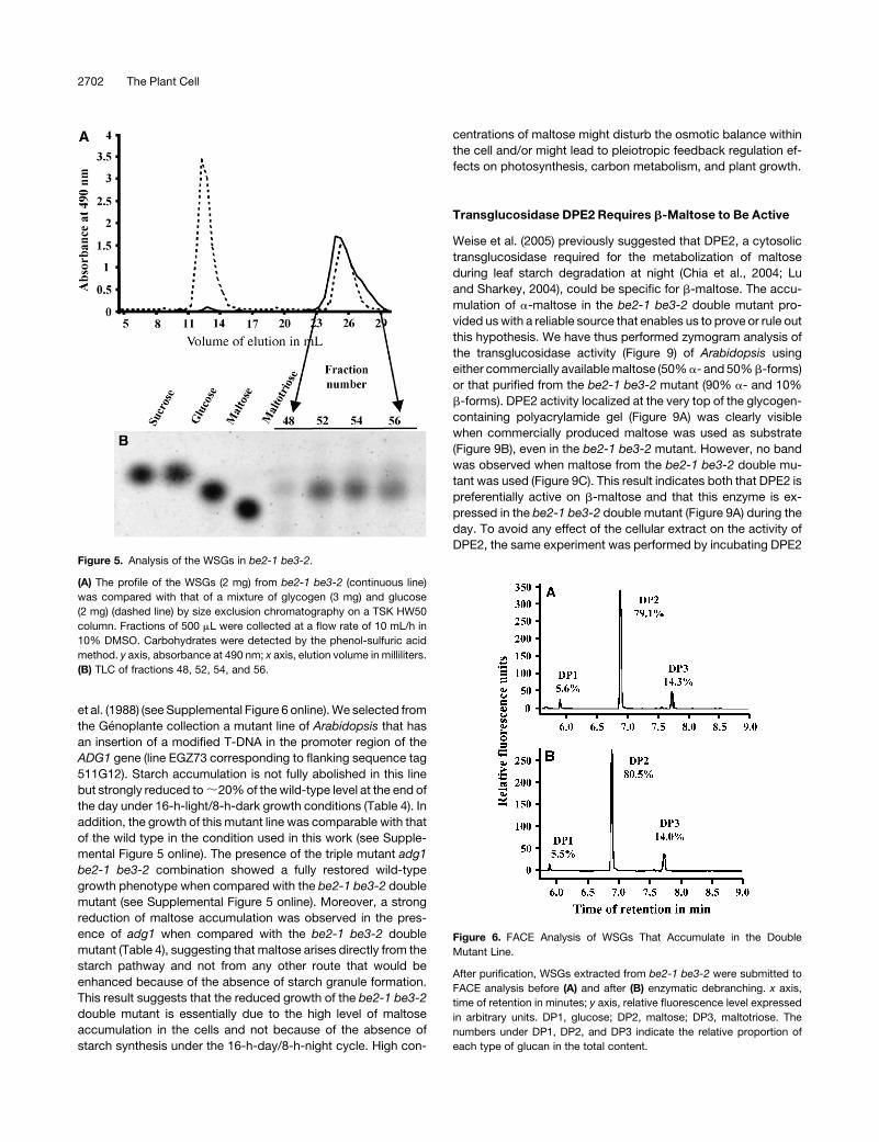
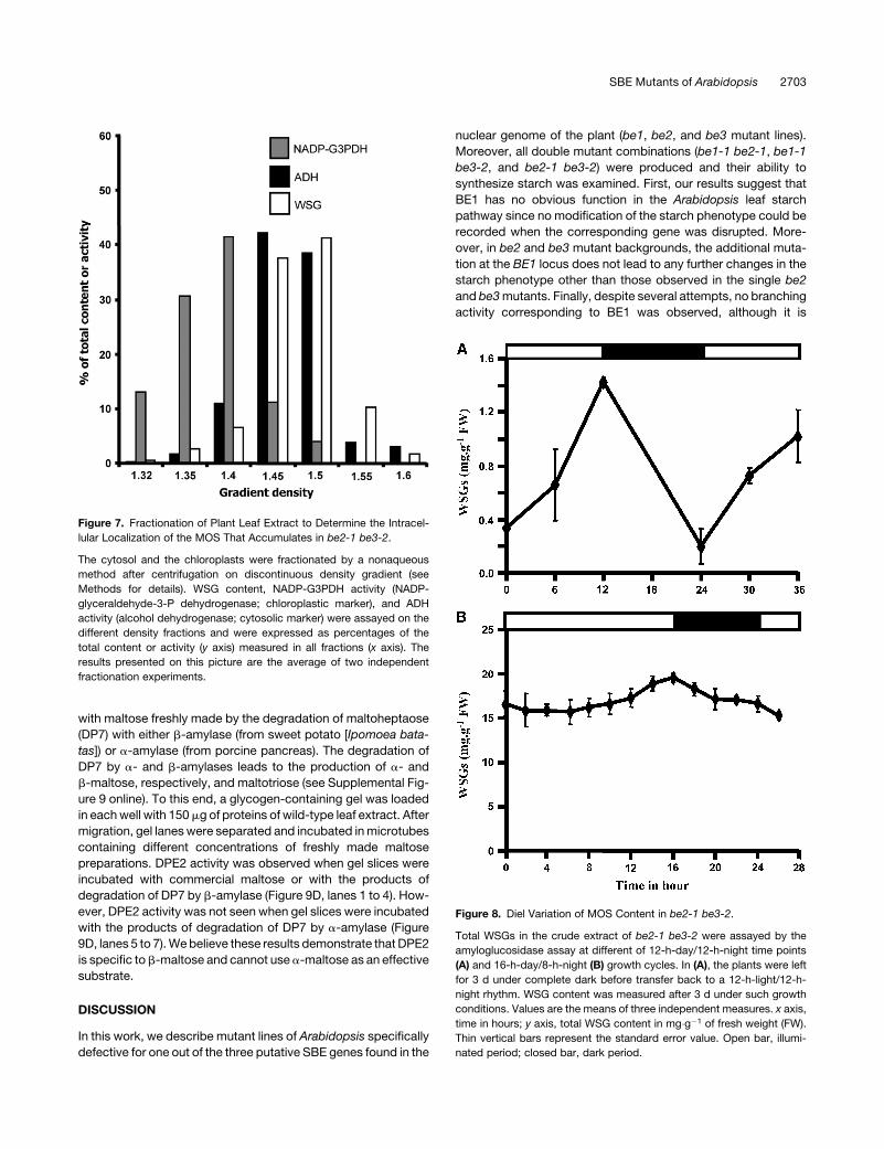
We characterized the structure of the accumulating WSGs in
the be2-1 be3-2 mutant. After extraction, the WSGs were sub-
mitted to size exclusion chromatography on a TSK HW50 col-
umn. The elution profile was compared with that of a mix of
glycogen and glucose after the detection of the corresponding
sugars by the phenol-sulfuric method (Figure 5A). Only one peak
of material that comigrates with glucose was observed for the
be2-1 be3-2 double mutant. The fractions corresponding to this
peak were analyzed by thin layer chromatography (TLC) (Figure
5B) and fluorophore-assisted capillary electrophoresis (FACE)
before and after enzymatic debranching (Figure 6). Our results
indicate that WSGs accumulating in the double mutant are com-
posed of very short MOS made of maltose (80%), maltotriose
(14%), and glucose (6%). Similar results were obtained whatever
the method used for the extraction of WSGs (nonacid or per-
chloric acid methods; see Supplemental Figures 7 and 8 online).
The sucrose, glucose, fructose, Glc-6-P, and Glc-1-P levels
were determined in the different lines under study after extraction
with 16% trichloroacetic acid (TCA) (w/v) in diethylether (Table 4).
Sucrose, fructose, Glc-6-P, and Glc-1-P levels were not signif-
icantly changed in the double mutant when compared with that of
the wild type and other mutants. However, an increase in glucose
content was observed in the be2-1 be3-2 mutant as expected
from the previous analysis of the composition of the WSGs.
Maltose in be2-1 be3-2 Essentially Accumulates
in the Cytosol
We have performed cell fractionation experiments (nonaqueous
method) to determine the intracellular location of the maltose
contained in the be2-1 be3-2 mutant. Our results (based on two
independent fractionation experiments) indicate that it essen-
tially accumulates in the cytosol (;80% of WSG is localized in
the cytosol) 10 h after the beginning of the 16-h light phase
(Figure 7). This result is not surprising since the maltose catab-
olism is expected to occur in the cytosol after its translocation
from the stroma by MEX1, the maltose exporter of the chloro-
plast envelop. Day/night evolution of MOS content in the be2-1
be3-2 mutant was performed under 12-h-day/12-h-night growth
conditions. The plants were left in the dark for 3 d to remove all
residual glucans (including starch and MOS) and then transferred
to 12-h-day/12-h-night growth rhythms. Leaf samples were
harvested 3 d after reilluminating the plants. This experiment
shows that MOS accumulates during the day to reach a maxi-
mum at the day/night transition and is degraded at night to reach
a minimum at the night/day transition (Figure 8A). However, when
plants were kept for several days under a 16-h-day/8-h-night
rhythm, the MOS concentration was always very high in the
double mutant compared with the wild type whatever the time
of the cycle (Figure 8B). Nonetheless, an increase was observed
during the day, while a decrease was observed during the night.
This result suggests that MOS removal is not complete under
short nights, leading to an overaccumulation of these molecules
after several days of culture in such conditions.
Finally, we have determined that a-maltose represents >90%
of the total pool of maltose present in the double mutant (the level
of a-maltose was determined by the enzymatic method de-
scribed in Shirokane et al., 2000) at the end of the light period
when plants were cultured under a 16-h-light/8-h-dark regime.
This test was performed on fresh material immediately after
extraction of the MOS to limit the impact of mutarotation in the
samples and after immediate boiling to inactivate enzymes.
Mutarotation occurs spontaneously when molecules are con-
served in solution, although its rate is quite low according to
Weise et al. (2005) and based on our own observations. Muta-
rotation can be defined as the spontaneous conversion of either
a- to b-maltose or b- to a-maltose. Indeed, after leaving freshly
extracted MOS on melting ice for 1 week, we determined that the
b-maltose content had reached 40% of the total maltose, indi-
cating that, in the conditions we used during the purification
procedure of the MOS, the mutarotation process is very slow
(spontaneous anomerization of maltose may occur at a higher
rate at room temperature and is strongly dependent upon the
incubation conditions).
We have also determined the maltose anomery at the end of
the light period on be2-1 be3-2 plants cultivated under a
12-h-light/12-h-night regime. a-Maltose represents 65% of the
total pool of maltose at the end of the light phase in these
conditions.
Production of Maltose Is Strongly Reduced in the adg1
Mutant Background
To ensure that the production of maltose in the be2-1 be3-2
double mutant actually occurs through a modified starch path-
way, we produced a triple mutant line that is defective for be2,
be3, and adg1 (or APS1). The ADG1 locus encodes one of the
two small subunits of ADP-glucose pyrophosphorylase. ADP-
glucose pyrophosphorylase is responsible for the synthesis of
the unique precursor molecule of starch synthesis: ADP-glucose.
A mutation at the ADG1 locus leads to a strong reduction of
starch accumulation in Arabidopsis leaves as described by Lin
2700 The Plant Cell
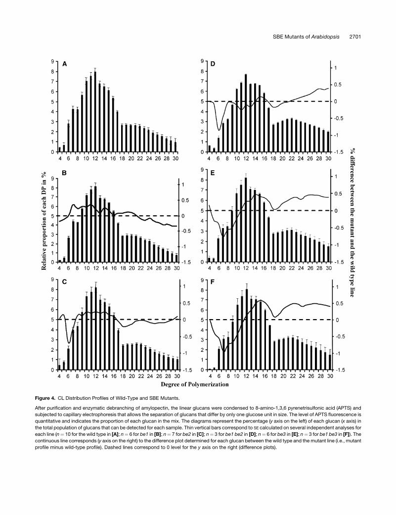
Figure 4. CL Distribution Profiles of Wild-Type and SBE Mutants.
After purification and enzymatic debranching of amylopectin, the linear glucans were condensed to 8-amino-1,3,6 pyrenetrisulfonic acid (APTS) and
subjected to capillary electrophoresis that allows the separation of glucans that differ by only one glucose unit in size. The level of APTS fluorescence is
quantitative and indicates the proportion of each glucan in the mix. The diagrams represent the percentage (y axis on the left) of each glucan (x axis) in
the total population of glucans that can be detected for each sample. Thin vertical bars correspond to SE calculated on several independent analyses for
each line (n¼ 10 for the wild type in [A]; n¼ 6 for be1 in [B]; n¼ 7 for be2 in [C]; n¼ 3 for be1 be2 in [D]; n¼ 6 for be3 in [E]; n¼ 3 for be1 be3 in [F]). The
continuous line corresponds (y axis on the right) to the difference plot determined for each glucan between the wild type and the mutant line (i.e., mutant
profile minus wild-type profile). Dashed lines correspond to 0 level for the y axis on the right (difference plots).
SBE Mutants of Arabidopsis 2701
et al. (1988) (see Supplemental Figure 6 online). We selected from
the Genoplante collection a mutant line of Arabidopsis that has
an insertion of a modified T-DNA in the promoter region of the
ADG1 gene (line EGZ73 corresponding to flanking sequence tag
511G12). Starch accumulation is not fully abolished in this line
but strongly reduced to ;20% of the wild-type level at the end of
the day under 16-h-light/8-h-dark growth conditions (Table 4). In
addition, the growth of this mutant line was comparable with that
of the wild type in the condition used in this work (see Supple-
mental Figure 5 online). The presence of the triple mutant adg1
be2-1 be3-2 combination showed a fully restored wild-type
growth phenotype when compared with the be2-1 be3-2 double
mutant (see Supplemental Figure 5 online). Moreover, a strong
reduction of maltose accumulation was observed in the pres-
ence of adg1 when compared with the be2-1 be3-2 double
mutant (Table 4), suggesting that maltose arises directly from the
starch pathway and not from any other route that would be
enhanced because of the absence of starch granule formation.
This result suggests that the reduced growth of the be2-1 be3-2
double mutant is essentially due to the high level of maltose
accumulation in the cells and not because of the absence of
starch synthesis under the 16-h-day/8-h-night cycle. High con-
centrations of maltose might disturb the osmotic balance within
the cell and/or might lead to pleiotropic feedback regulation ef-
fects on photosynthesis, carbon metabolism, and plant growth.
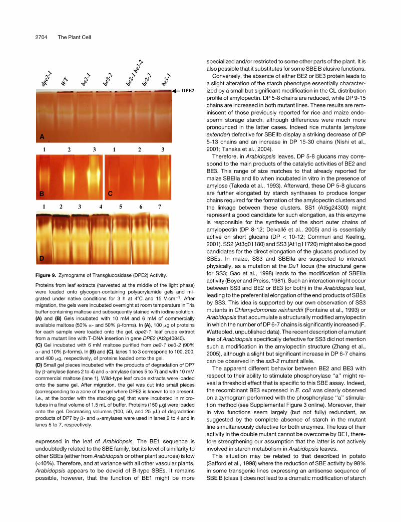
Transglucosidase DPE2 Requires b-Maltose to Be Active
Weise et al. (2005) previously suggested that DPE2, a cytosolic
transglucosidase required for the metabolization of maltose
during leaf starch degradation at night (Chia et al., 2004; Lu
and Sharkey, 2004), could be specific for b-maltose. The accu-
mulation of a-maltose in the be2-1 be3-2 double mutant pro-
vided us with a reliable source that enables us to prove or rule out
this hypothesis. We have thus performed zymogram analysis of
the transglucosidase activity (Figure 9) of Arabidopsis using
either commercially available maltose (50% a- and 50% b-forms)
or that purified from the be2-1 be3-2 mutant (90% a- and 10%
b-forms). DPE2 activity localized at the very top of the glycogen-
containing polyacrylamide gel (Figure 9A) was clearly visible
when commercially produced maltose was used as substrate
(Figure 9B), even in the be2-1 be3-2 mutant. However, no band
was observed when maltose from the be2-1 be3-2 double mu-
tant was used (Figure 9C). This result indicates both that DPE2 is
preferentially active on b-maltose and that this enzyme is ex-
pressed in the be2-1 be3-2 double mutant (Figure 9A) during the
day. To avoid any effect of the cellular extract on the activity of
DPE2, the same experiment was performed by incubating DPE2Figure 5. Analysis of the WSGs in be2-1 be3-2.
(A) The profile of the WSGs (2 mg) from be2-1 be3-2 (continuous line)
was compared with that of a mixture of glycogen (3 mg) and glucose
(2 mg) (dashed line) by size exclusion chromatography on a TSK HW50
column. Fractions of 500 mL were collected at a flow rate of 10 mL/h in
10% DMSO. Carbohydrates were detected by the phenol-sulfuric acid
method. y axis, absorbance at 490 nm; x axis, elution volume in milliliters.
(B) TLC of fractions 48, 52, 54, and 56.
Figure 6. FACE Analysis of WSGs That Accumulate in the Double
Mutant Line.
After purification, WSGs extracted from be2-1 be3-2 were submitted to
FACE analysis before (A) and after (B) enzymatic debranching. x axis,
time of retention in minutes; y axis, relative fluorescence level expressed
in arbitrary units. DP1, glucose; DP2, maltose; DP3, maltotriose. The
numbers under DP1, DP2, and DP3 indicate the relative proportion of
each type of glucan in the total content.
2702 The Plant Cell
with maltose freshly made by the degradation of maltoheptaose
(DP7) with either b-amylase (from sweet potato [Ipomoea bata-
tas]) or a-amylase (from porcine pancreas). The degradation of
DP7 by a- and b-amylases leads to the production of a- and
b-maltose, respectively, and maltotriose (see Supplemental Fig-
ure 9 online). To this end, a glycogen-containing gel was loaded
in each well with 150 mg of proteins of wild-type leaf extract. After
migration, gel lanes were separated and incubated in microtubes
containing different concentrations of freshly made maltose
preparations. DPE2 activity was observed when gel slices were
incubated with commercial maltose or with the products of
degradation of DP7 by b-amylase (Figure 9D, lanes 1 to 4). How-
ever, DPE2 activity was not seen when gel slices were incubated
with the products of degradation of DP7 by a-amylase (Figure
9D, lanes 5 to 7). We believe these results demonstrate that DPE2
is specific to b-maltose and cannot use a-maltose as an effective
substrate.
DISCUSSION
In this work, we describe mutant lines of Arabidopsis specifically
defective for one out of the three putative SBE genes found in the
nuclear genome of the plant (be1, be2, and be3 mutant lines).
Moreover, all double mutant combinations (be1-1 be2-1, be1-1
be3-2, and be2-1 be3-2) were produced and their ability to
synthesize starch was examined. First, our results suggest that
BE1 has no obvious function in the Arabidopsis leaf starch
pathway since no modification of the starch phenotype could be
recorded when the corresponding gene was disrupted. More-
over, in be2 and be3 mutant backgrounds, the additional muta-
tion at the BE1 locus does not lead to any further changes in the
starch phenotype other than those observed in the single be2
and be3 mutants. Finally, despite several attempts, no branching
activity corresponding to BE1 was observed, although it is
Figure 7. Fractionation of Plant Leaf Extract to Determine the Intracel-
lular Localization of the MOS That Accumulates in be2-1 be3-2.
The cytosol and the chloroplasts were fractionated by a nonaqueous
method after centrifugation on discontinuous density gradient (see
Methods for details). WSG content, NADP-G3PDH activity (NADP-
glyceraldehyde-3-P dehydrogenase; chloroplastic marker), and ADH
activity (alcohol dehydrogenase; cytosolic marker) were assayed on the
different density fractions and were expressed as percentages of the
total content or activity (y axis) measured in all fractions (x axis). The
results presented on this picture are the average of two independent
fractionation experiments.
Figure 8. Diel Variation of MOS Content in be2-1 be3-2.
Total WSGs in the crude extract of be2-1 be3-2 were assayed by the
amyloglucosidase assay at different of 12-h-day/12-h-night time points
(A) and 16-h-day/8-h-night (B) growth cycles. In (A), the plants were left
for 3 d under complete dark before transfer back to a 12-h-light/12-h-
night rhythm. WSG content was measured after 3 d under such growth
conditions. Values are the means of three independent measures. x axis,
time in hours; y axis, total WSG content in mg�g�1 of fresh weight (FW).
Thin vertical bars represent the standard error value. Open bar, illumi-
nated period; closed bar, dark period.
SBE Mutants of Arabidopsis 2703
expressed in the leaf of Arabidopsis. The BE1 sequence is
undoubtedly related to the SBE family, but its level of similarity to
other SBEs (either from Arabidopsis or other plant sources) is low
(<40%). Therefore, and at variance with all other vascular plants,
Arabidopsis appears to be devoid of B-type SBEs. It remains
possible, however, that the function of BE1 might be more
specialized and/or restricted to some other parts of the plant. It is
also possible that it substitutes for some SBE B elusive functions.
Conversely, the absence of either BE2 or BE3 protein leads to
a slight alteration of the starch phenotype essentially character-
ized by a small but significant modification in the CL distribution
profile of amylopectin. DP 5-8 chains are reduced, while DP 9-15
chains are increased in both mutant lines. These results are rem-
iniscent of those previously reported for rice and maize endo-
sperm storage starch, although differences were much more
pronounced in the latter cases. Indeed rice mutants (amylose
extender) defective for SBEIIb display a striking decrease of DP
5-13 chains and an increase in DP 15-30 chains (Nishi et al.,
2001; Tanaka et al., 2004).
Therefore, in Arabidopsis leaves, DP 5-8 glucans may corre-
spond to the main products of the catalytic activities of BE2 and
BE3. This range of size matches to that already reported for
maize SBEIIa and IIb when incubated in vitro in the presence of
amylose (Takeda et al., 1993). Afterward, these DP 5-8 glucans
are further elongated by starch synthases to produce longer
chains required for the formation of the amylopectin clusters and
the linkage between these clusters. SS1 (At5g24300) might
represent a good candidate for such elongation, as this enzyme
is responsible for the synthesis of the short outer chains of
amylopectin (DP 8-12; Delvalle et al., 2005) and is essentially
active on short glucans (DP < 10-12; Commuri and Keeling,
2001). SS2 (At3g01180) and SS3 (At1g11720) might also be good
candidates for the direct elongation of the glucans produced by
SBEs. In maize, SS3 and SBEIIa are suspected to interact
physically, as a mutation at the Du1 locus (the structural gene
for SS3; Gao et al., 1998) leads to the modification of SBEIIa
activity (Boyer and Preiss, 1981). Such an interaction might occur
between SS3 and BE2 or BE3 (or both) in the Arabidopsis leaf,
leading to the preferential elongation of the end products of SBEs
by SS3. This idea is supported by our own observation of SS3
mutants in Chlamydomonas reinhardtii (Fontaine et al., 1993) or
Arabidopsis that accumulate a structurally modified amylopectin
in which the number of DP 6-7 chains is significantly increased (F.
Wattebled, unpublished data). The recent description of a mutant
line of Arabidopsis specifically defective for SS3 did not mention
such a modification in the amylopectin structure (Zhang et al.,
2005), although a slight but significant increase in DP 6-7 chains
can be observed in the ss3-2 mutant allele.
The apparent different behavior between BE2 and BE3 with
respect to their ability to stimulate phosphorylase ‘‘a’’ might re-
veal a threshold effect that is specific to this SBE assay. Indeed,
the recombinant BE3 expressed in E. coli was clearly observed
on a zymogram performed with the phosphorylase ‘‘a’’ stimula-
tion method (see Supplemental Figure 3 online). Moreover, their
in vivo functions seem largely (but not fully) redundant, as
suggested by the complete absence of starch in the mutant
line simultaneously defective for both enzymes. The loss of their
activity in the double mutant cannot be overcome by BE1, there-
fore strengthening our assumption that the latter is not actively
involved in starch metabolism in Arabidopsis leaves.
This situation may be related to that described in potato
(Safford et al., 1998) where the reduction of SBE activity by 98%
in some transgenic lines expressing an antisense sequence of
SBE B (class I) does not lead to a dramatic modification of starch
Figure 9. Zymograms of Transglucosidase (DPE2) Activity.
Proteins from leaf extracts (harvested at the middle of the light phase)
were loaded onto glycogen-containing polyacrylamide gels and mi-
grated under native conditions for 3 h at 48C and 15 V�cm�1. After
migration, the gels were incubated overnight at room temperature in Tris
buffer containing maltose and subsequently stained with iodine solution.
(A) and (B) Gels incubated with 10 mM and 6 mM of commercially
available maltose (50% a- and 50% b-forms). In (A), 100 mg of proteins
for each sample were loaded onto the gel. dpe2-1: leaf crude extract
from a mutant line with T-DNA insertion in gene DPE2 (At2g40840).
(C) Gel incubated with 6 mM maltose purified from be2-1 be3-2 (90%
a- and 10% b-forms). In (B) and (C), lanes 1 to 3 correspond to 100, 200,
and 400 mg, respectively, of proteins loaded onto the gel.
(D) Small gel pieces incubated with the products of degradation of DP7
by b-amylase (lanes 2 to 4) and a-amylase (lanes 5 to 7) and with 10 mM
commercial maltose (lane 1). Wild-type leaf crude extracts were loaded
onto the same gel. After migration, the gel was cut into small pieces
(corresponding to a zone of the gel where DPE2 is known to be present;
i.e., at the border with the stacking gel) that were incubated in micro-
tubes in a final volume of 1.5 mL of buffer. Proteins (150 mg) were loaded
onto the gel. Decreasing volumes (100, 50, and 25 mL) of degradation
products of DP7 by b- and a-amylases were used in lanes 2 to 4 and in
lanes 5 to 7, respectively.
2704 The Plant Cell
structure. The remaining SBE A activity (measured through the
phosphorylase stimulation assay) accounts only for a minor part
of the total SBE activity in potato tuber but is sufficient to sustain
normal starch synthesis. However, reduction of SBE A expres-
sion by antisense technology leads to a much bigger modifica-
tion in starch structure (Jobling et al., 1999) that is somewhat
different to the situation observed in Arabidopsis where the con-
tribution of each SBE II (family A) by itself is low, as shown by the
marginal phenotype described in each single mutant. This in turn
might reveal some basic differences between transitory and
storage starches. Only the antisense inhibition of both SBE A and
B leads to a strongly altered structure of tuber starch, although
starch synthesis was not abolished in these lines and was only
reduced by 50% (Schwall et al., 2000). This could be due to the
presence of low, albeit significant amounts of residual BE activity.
The origin of maltose accumulation in the double mutant line
cannot yet be completely addressed, although we have definitely
shown with the analysis of the adg1 be2-1 be3-2 triple mutant
that the maltose derives from the starch pathway. The absence
of maltose in the starch-free pgm1 mutant (Caspar et al., 1985)
and in the very-low-starch ss2 ss3 double mutant (M. James and
A. Myers, personal communication) confirms this conclusion.
The amount of maltose in the be2-1 be3-2 mutant is 300- to
600-fold higher than that assayed at midcourse of the light phase
in the wild type but is more or less equivalent to that measured in
dpe2 mutants defective for cytosolic transglucosidase (Chia
et al., 2004; Lu and Sharkey, 2004; Weise et al., 2005). The
stunted growth phenotype of the double mutant is equally
reminiscent of that observed in the dpe2 mutants. The lack of
maltose assimilation in the dpe2 mutant can explain why such a
high level of maltose accumulates in the cell. Spontaneous
mutarotation of maltose could explain why equivalent amounts
of both anomers are assayed in the mutant (Weise et al., 2005). In
our study, >90% of maltose assayed in the be2-1 be3-2 double
mutant was in the a-form. Although no definitive explanation can
yet address this difference between our results and those of
Weise et al. (2005), several hypotheses may be raised to explain
the origin of a-maltose accumulation in the Arabidopsis be2-1
be3-2 double mutant. In this line, despite the absence of SBE
activity, the starch synthases could still be able to produce linear
a-glucans from ADP-glucose (ADP-glucose pyrophosphorylase
and other starch metabolizing enzymes remain unaffected, with
the exception of STPases; Table 1). We propose that these linear
glucans are rapidly degraded (even during the light phase) by
amylases to produce maltose in great majority and some other
short MOS (glucose and DP 3 are side products of degradation).
After its translocation to the cytosol by the MEX1 maltose ex-
porter (Niittyla et al., 2004; Weise et al., 2004), maltose is de-
graded by a cytosolic transglucosidase (DPE2; Chia et al., 2004;
Lu and Sharkey, 2004) with the help of an acceptor molecule
whose nature is not yet known, although the existence of a
cytosolic complex soluble heteroglycan was recently reported
in Arabidopsis leaves (Fettke et al., 2005a, 2005b). Plastidial
b-amylases were shown to interact directly with semicrystalline
starch granules and were suggested to be important factors for
leaf starch degradation in the potato leaf (Scheidig et al., 2002).
However, the situation is obviously different in the be2-1 be3-2
double mutant since no granular starch was observed in this line.
The abnormal products of the starch pathway may thus be
comparatively more accessible to other enzymes of the starch
pathway. In such a modified situation, a plastidial a-amylase
might become the most effective enzyme of degradation of those
glucans produced by starch synthases in the be2-1 be3-2 line.
Since a-amylases exhibit a retaining mechanism (for more de-
tails, see http://afmb.cnrs-mrs.fr/CAZY/GH_13.html), the main
end product of a-amylases on linear a-1,4–linked glucans is
a-maltose, with some other side products like maltotriose and
other short MOS (Robyt and French, 1970). a-Maltose is then
exported to the cytosol by MEX1 where it will be metabolized by
DPE2 after mutarotation. Abnormal accumulation of a-maltose in
be2-1 be3-2 may be understood because of the substrate spe-
cificity that we now demonstrate for DPE2 (Figures 9B and 9C).
Indeed, the latter seems to be significantly active on b-maltose
only. b-Maltose is slowly generated by spontaneous mutarota-
tion of a-maltose and is subsequently metabolized by DPE2
while accumulating in the cytosol. The rate of spontaneous
mutarotation being quite low (Weise et al., 2005; this work), this
could explain why accumulation of a-maltose is observed during
the illuminated period (Figure 8). At night (in 12-h-day/12-h-night
growth conditions), because de novo synthesis of maltose is
turned off, maltose content progressively decreases because of
the degradation of the b-form by DPE2. However, under long-
day (16 h) growth conditions, maltose cannot be completely
degraded since mutarotation is too slow to convert all the
a-maltose to b-maltose during the short night. This might explain
why maltose progressively accumulates in the leaf cells even at
the end of the dark phase when the plants are subjected to a
16-h-day/8-h-night growth regime.
An alternative to this hypothesis is that maltose is produced to
the same extent by both a- and b-amylases, leading to the
production of both a- and b- maltose. After its transfer to cytosol,
b-maltose is rapidly metabolized by DPE2, while a-maltose is
only slowly degraded after its spontaneous anomerization to the
b-form. Therefore, under long-day/short-night growth condi-
tions, a-maltose concentration gradually increases in the leaf of
the plant because it is not a suitable substrate for DPE2.
METHODS
Materials
ADP [U-14C] glucose, [U-14C] glucose-1-phosphate, the CL-2B sephar-
ose column, and Percoll were obtained from Amersham Biosciences.
TSK HW50 Toyopearl matrix was purchased from Tosoh Bioscience.
ADP-glucose, G1P, and enzymes (a- and b-amylases [from porcine
pancreas and sweet potato, respectively], maltose epimerase, and malt-
ose phosphorylase) were from Sigma-Aldrich (unless specified). The
starch assay kit was purchased from Enzytec. The fructose and glucose
assay kit was obtained from Megazyme.
Arabidopsis Lines, Growth Conditions, and Media
Wild-type (Ws and Col-0) and mutant lines of Arabidopsis thaliana were
obtained from the T-DNA mutant collections generated at Genoplante (Unite
de Recherche en Genomique Vegetale, Institut National de la Recherche
Agronomique of Versailles; Bechtold et al., 1993; Bouchez et al., 1993) and
the Nottingham Arabidopsis Stock Centre (Alonso et al., 2003). Standard
SBE Mutants of Arabidopsis 2705
procedures were employed for plant germination and growth. The plants
were grown on peat-based compost (seeds were previously incubated at
48C before sowing) under a 16-h-light/8-h-dark cycle, with temperature
ranging from 168C during the night to 218C during the day.
RT-PCR Amplifications
Approximately 100 mg of fresh tissue was harvested at the middle of the
light phase (see culture conditions described above) for total RNA
extraction with the Plant RNeasy kit (Qiagen) following the supplier’s
instructions. Twenty nanograms of purified total RNA were used to
perform RT-PCR amplifications using the One-Step RT-PCR kit (Qiagen).
Three different regions surrounding the T-DNA insertion site were targeted
for amplification: upstream, downstream, and spanning the T-DNA inser-
tion site (see Supplemental Table 1 online for detailed primer sequences).
Extraction and Purification of Starch
Starch from ;10 g of leaves was extracted and purified as described by
Delvalle et al. (2005).
Extraction of Soluble Carbohydrates (WSGs)
Extraction with Perchloric Acid
All leaves of individuals were harvested at the end of the light period and
immediately frozen in liquid nitrogen. Leaves were broken down to rough
pieces with a pestle directly in a microtube. Five hundred microliters of
0.7 M perchloric acid was added, and the samples were immediately
homogenized with a polytron blender. The samples were then centrifuged
for 15 min at 3000g at 48C. The soluble phase was collected and
subsequently neutralized with 2 M KOH, 0.4 M MES, and 0.4 M KCl.
The potassium perchlorate precipitate was then removed by centrifuga-
tion at 16,000g for 15 min at 48C. The supernatant was conserved at
�808C before use.
Nonacid Extraction of WSGs
Leaves harvested at the same time as for starch extraction were imme-
diately soaked into liquid nitrogen, broken down to fine powder with pestle
and mortar, and placed for 10 min in a boiling water bath to inactivate
enzymes. The corresponding samples were then extracted in an ice-
cooled buffer (100 mM MOPS, pH 7.2, 5 mM EDTA, 10% [v/v] ethanediol)
and further homogenized using a polytron blender. The tubes were always
kept on ice during the whole extraction procedure and processed as
quickly as possible for analysis to limit the impact of spontaneous
mutarotation on maltose anomery. After centrifugation of the homogenate
(10 min at 10,000g at 48C), the supernatant was directly used for the assay
of the glucan polymers (amyloglucosidase assay) and the determination of
maltose anomery but was lyophilized prior to chromatography by size
exclusion on sephadex TSK HW50 matrix (see below).
Determination of Starch and WSG Contents
and Spectral Properties of the Iodine-Starch Complex
A full account of lmax (maximal absorbance wavelength of the iodine
polysaccharide complex) measure can be found in Delrue et al. (1992).
Starch and WSG contents in leaves were determined after extraction (as
described above) by the amyloglucosidase assay as described by
Delvalle et al. (2005).
Extraction of Soluble Sugars (Glucose, Sucrose, Fructose,
and Hexose-P)
Sugars were extracted from 200 to 300 mg of rosette leaves harvested at
the end of the light phase and immediately frozen in liquid nitrogen. The
leaves were homogenized with an electric polytron blender (the metallic
device was replaced by a plastic pestle adapted for 1.5-mL microfuge
tubes) in 1 mL of ice-cold 16% TCA (w/v) in diethylether. During the
procedure, the samples were kept on ice. EGTA was then added to 5 mM
final concentration, and the samples were further homogenized with the
polytron blender. Samples were left for 2 h on ice before centrifugation at
16,000g for 5 min at 48C. The nonaqueous phase was removed, and the
aqueous phase was washed four times with diethylether (centrifugation
for 5 min at 16,000g at 48C between each wash). The aqueous phase is
transferred to a new tube, neutralized by the addition of 50 mM KOH and
10 mM triethanolamine buffer, and conserved at �808C.
Determination of Glucose, Sucrose, Fructose,
and Hexose-P Contents
Glucose and fructose contents were determined from leaf extract by the
use of a specific kit (Megazyme) following the supplier’s instructions.
Sucrose was assayed as follows: 30 mL of leaf extract was added to 60 mL
of 20 mM NaH2PO4, pH 4.5, and 10 units of invertase (Sigma-Aldrich).
After 20 min at 558C, 300 mL of water, 300 mL of triethanolamine buffer, pH
7.6, 1.5 mM NADP, 5 mM ATP, 11 mM MgSO4, 2 units of hexokinase, and
1 unit of G-6-P dehydrogenase (Enzytec) were added to the sample. The
reaction was performed at room temperature until absorbance remained
stable at 365 nm. The amount of glucose determined previously was
deducted from the value obtained in that case. G-6-P content was
determined as follow: 30 mL of leaf extract was added to 360 mL of water,
and 300 mL of triethanolamine buffer, pH 7.6, 1.5 mM NADP, 5 mM ATP,
11 mM MgSO4, and 1 unit of G-6-P dehydrogenase (Enzytec) were added
to the sample. The reaction was performed at room temperature until
absorbance remained stable at 365 nm. To measure G-1-P content,
2 units of phosphoglucomutase (Sigma-Aldrich) was added to the pre-
vious sample, and absorbance was read at 365 nm at room temperature
until it remained stable. G-1-P level was deduced after the subtraction of
the amount of G-6-P measured previously.
Separation of Starch Polysaccharides and Water-Soluble
Polysaccharides by Size Exclusion Chromatography
Amylopectin and amylose were separated from 1.5 to 2.0 mg of starch by
size exclusion chromatography on a CL-2B column as fully described by
Delvalle et al. (2005).
Two milligrams of purified and lyophilized WSGs were dissolved in
500 mL of 10% DMSO (v/v) and loaded on a sephadex TSK HW50 column
(1 cm i.d. 3 50 cm), equilibrated, and eluted with 10% DMSO (v/v).
Fractions of 500 mL were collected at a flow rate of 10 mL/h. Carbohy-
drates in the collected fractions were detected by the phenol-sulfuric acid
method. Twenty microliters of 5% phenol were added to 20 mL of sample.
After thorough shaking, 100 mL of concentrated sulfuric acid were added,
and after gentle shaking, the samples were incubated for 30 min at 808C.
Absorbance was determined at 490 nm and compared with that of a
glucose solution of known concentration.
CL Distribution of Amylopectin and WSGs, and
Branching Level Determination
Full procedures for the determination of amylopectin CL distribution after
enzymatic debranching can be found in Wattebled et al. (2005). CL profile
of WSGs was established after FACE using APTS as fluorophore (the
protocol for derivatization of the soluble glucans is the same as the one
described for amylopectin). Only glucans or sugars with a reducing end
can be labeled with APTS.
Branching degree of amylopectin was determined by methylation of
glucans as described by Delvalle et al. (2005). Five hundred micrograms
of purified amylopectin was used to this end.
2706 The Plant Cell
TLC
Four microliters of different fractions from the TSK HW50 column (see
above) were loaded on silica gel 60 (Merck). Sucrose, glucose, maltose,
and maltotriose (2 mg each) were loaded on the sheet as controls. The
sheet was developed for 4 h in the following solution: butanol/ethanol/
water in ratio 1/1/1 (v/v/v). After migration, the sheet was dried before
spraying sulfuric orcinol solution and heating for several minutes at 1008C
until sugars appeared as brown bands on the TLC.
Production of Fresh Maltose by a- and b-Amylases
Twenty milligrams of maltoheptaose (Sigma-Aldrich) were incubated for
2 h at 308C in a final volume of 500 mL with 20 units of a-amylase (from
porcine pancreas) or 20 units of b-amylase (from sweet potato). Incuba-
tion was extended by 2 h after the addition of an additional 20 units of
each enzyme to ensure a complete degradation of DP7. For incubation
with a-amylase, the buffer was composed of 25 mM sodium acetate, pH
4.8, 5 mM DTT, and 2 mM CaCl2. For incubation with b-amylase, the
buffer was composed of 25 mM sodium acetate, pH 4.8, 5 mM DTT, and
5 mM EDTA. The reaction was stopped by the addition of 500 mL of
phenol/chloroform (1/1; v/v). After a thorough mixing, the samples were
centrifuged for 10 min at 16,000g at 48C, and the supernatant was
extracted three times with chloroform to remove traces of phenol. The
degradation of DP7 to maltose and maltotriose was checked by TLC on
silica gel 60 (see above and Supplemental Figure 9 online) and subse-
quently used at different concentrations for incubation with small pieces
of gel containing glycogen and DPE2 activity (Figure 9D; see below for
DPE2 zymogram).
Determination of Maltose Anomery
Anomery of maltose was determined using the procedure described by
Shirokane et al. (2000). Twelve nanomoles of total purified WSGs were
incubated in the following buffer: triethanolamine, pH 7.6, 155 mM
NaH2PO4, 1.5 mM NADP, 5 mM ATP, and 11 mM MgSO4 at 308C in the
presence of 2 units of hexokinase and 1 unit of G-6-P dehydrogenase.
The reaction was started by the addition of 5 units of maltose phospho-
rylase alone (to determine the proportion of a-maltose in the sample) or by
the addition of both 5 units of maltose phosphorylase and 5 units of
maltose epimerase (for a- and b-maltose determination). Absorbance
was read at 365 nm in a spectrophotometer (U 2000; Hitachi).
Zymogram Techniques
For a complete description of soluble starch synthase activities, refer to
Delvalle et al. (2005).
Starch modifying activities on starch-containing gel (hydrolases and
branching enzymes) were tested as follows: 100 mg of leaf extract proteins
were loaded onto a native PAGE (7.5% acrylamide) containing potato-
soluble starch (Sigma-Aldrich) at 0.3% final concentration and separated
for 3 h at 48C and 15 V�cm�1. The gels were incubated overnight at room
temperature in the following buffer: 50 mM Tris-HCl, pH 7.0, 1 mM DTT,
1 mM CaCl2, and 1 mM MgCl2. The hydrolytic activities were revealed by
soaking the gel into iodine solution (0.2% I2 [w/v] and 2% KI [w/v]).
For phosphorylase ‘‘a’’ stimulation, 100 mg of proteins from a leaf crude
extract were loaded on a native PAGE (6.5% acrylamide). After electro-
phoresis at 48C for 2 h and 30 min, the gel was incubated overnight at
room temperature in 30 mL of the following buffer: 50 mM HEPES/NaOH,
pH 7.0, 10% glycerol (v/v); 50 mM G1P, 2.5 mM AMP, and 28 units of
phosphorylase ‘‘a’’ from rabbit muscle. Branching activities were re-
vealed by iodine staining.
For transglucosidase DPE2, 100 mg of proteins from a leaf crude ex-
tract were loaded on a native PAGE (7.5% acrylamide) containing 0.3% of
rabbit liver glycogen. After migration (under native conditions for 3 h at
48C at 15 V�cm�1), the gel was preincubated for 1 h in 50 mL of 100 mM
Tris-HCl, pH 7.0, 1 mM MgCl2, and 1 mM DTT and incubated overnight at
room temperature in the same buffer. Unless otherwise indicated in the
text, the maltose concentration was 10 mM. To test for DPE2 activity with
degradation products of DP7 by a- or b-amylases (Figure 9D), 150 mg of
proteins were loaded onto the gel.
In Vitro Assays of Starch Metabolizing Enzymes
For SBEs, proteins were extracted in 50 mM NaH2PO4, pH 8.0, and
500 mM NaCl. One hundred micrograms of protein were incubated at
308C for 2 h in 250 mL of the following: 50 mM HEPES/NaOH, pH 7.0, 1 mM
AMP, 10% glycerol (v/v), 8 mM [U-14C] G1P (150 mCi/mmol), 50 mM G1P,
and 2 units of phosphorylase ‘‘a’’ from rabbit muscle. Reaction was
stopped by boiling the sample for 10 min. One hundred microliters from a
100-mg/mL stock solution of glycogen and 1 mL of 75% methanol (v/v)/
1% KCl (w/v) were added. The resulting precipitate was then filtered on a
glass-fiber filter (Whatmann), rinsed two times with 15 mL of deionized
water, two times with 70% ethanol, and dried. The sample was finally
counted in a scintillation counter after the addition of 3 mL of counting
solution (UltimaGold; Perkin-Elmer).
Soluble starch synthases and ADP-glucose pyrophosphorylase were
assayed according to Delvalle et al. (2005).
The a- and b-amylases, pullulanase, a-1,4 glucanotransferase, and
starch phosphorylase activities were assayed as described by Delvalle
et al. (2005).
Nonaqueous Fractionation of Chloroplasts
The method was previously described (Gerhardt and Heldt, 1984) and
modified (Weise et al., 2005). The procedure is briefly described here.
Seven to eight grams of fresh leaves were harvested 10 h after the be-
ginning of the 16-h-light period and immediately frozen in liquid nitrogen.
Leaves were ground with a mortar and pestle and were continuously kept
frozen by the addition of liquid nitrogen. The ground material was then
poured into a 50-mL Falcon tube and dried by lyophilization. The powder
was resuspended in 20 mL of dry C2Cl4/n-heptane (density ¼1.32 g�mL�1) and homogenized by ultrasonication at a power level of
5.5 and 40% duty cycle. The sample was sonicated for 3 min in 10-s
periods followed by a pause of 10 s on ice. The homogenate was filtered
through two layers of Miracloth (Calbiochem, EMD Biosciences) into a
new 50-mL Falcon tube. The retentate was rinsed by 10 mL of dry C2Cl4/
n-heptane (density ¼ 1.32 g�mL�1) and then by 10 mL of dry n-heptane.
The filtrate was centrifuged for 7 min at 1800g. The supernatant was
discarded, and the pellet was resuspended in 4 mL of dry C2Cl4/n-
heptane (density ¼ 1.32 g.mL�1) and layered on top of a discontinuous
gradient consisting of six density steps, 4 mL each, from 1.60 to 1.35
g�mL�1. It was then centrifuged at 3200g for 1.5 h. The content of the
centrifugation was collected in 4-mL fractions. Two volumes of n-heptane
were then added to each fraction and then tubes were centrifuged for
10 min at 2200g. The supernatant was discarded, and the pellets were
dried in a dessicator for 18 h at room temperature.
Sample Preparation for Assay of Marker Enzyme Activities
and MOS Measurement
Two hundred microliters of 50 mM NaH2PO4, pH 8.0, and 500 mM NaCl
were added to the dried sample. The suspension was ultrasonicated at a
power level of 7 and 70% duty cycle for 30 s in 10-s periods followed by a
pause of 20 s on ice. The sample was then centrifuged for 5 min at
10,000g, and the resulting supernatant was used for assaying the marker
enzyme activities and the determination of the MOS content.
SBE Mutants of Arabidopsis 2707
Assay of Marker Enzyme Activities
The following marker enzymes were assayed at 258C as described here:
for the chloroplastic marker NADP-glyceraldehyde-3-P dehydrogenase,
20 mL of sample was added to 180 mL of the following buffer: 50 mM
GlyGly/NaOH, pH 8.2, 80 mM 3-phosphoglyceric acid, 20 mM MgSO4,
10 mM cysteine, 5 mM gluthatione, 0.4 mM NADPH, and 2 units of phos-
phoglycerat kinase. Oxidation of NADPH was followed in a spectropho-
tometer at 340 nm for 10 min. The result for each sample is given by the
slope of the corresponding curve that is related to the activity rate of
NADP-glyceraldehyde-3-P dehydrogenase.
For the cytosolic marker alcohol dehydrogenase, 50 mL of sample was
added to 950 mL of 50 mM GlyGly/NaOH, pH 8.7, 2 mM NAD, and 30%
(v/v) ethanol. Reduction of NAD was followed at 340 nm for 5 min.
Transmission Electron Microscopy and
X-Ray Diffraction Measurements
A full account of these techniques can be found in Delvalle et al. (2005).
Accession Numbers
Arabidopsis Genome Initiative accession numbers for BE1, BE2, and BE3
can be found in Table 1. The Arabidopsis Genome Initiative accession
number for ADG1 is At5g48300.
Supplemental Data
The following materials are available in the online version of this article.
Supplemental Table 1. Primer Pairs Used for the Selection of the
Mutant Alleles by RT-PCR Amplification.
Supplemental Figure 1. Dendrogram of Branching Enzyme Amino
Acid Sequences from Various Sources.
Supplemental Figure 2. Zymogram for Starch Modifying/Degrading
Enzymes.
Supplemental Figure 3. Zymogram Analysis of Recombinant BE3
Expressed in E. coli.
Supplemental Figure 4. Zymogram of Starch Phosphorylases.
Supplemental Figure 5. Comparison of Growth Phenotypes.
Supplemental Figure 6. Iodine Staining of Arabidopsis Leaves from
Different Lines.
Supplemental Figure 7. Fluorophore-Assisted Capillary Electropho-
resis of WSGs Extracted from be2-1 be3-2.
Supplemental Figure 8. Thin Layer Chromatography of WSGs
Extracted from be2-1 be3-2.
Supplemental Figure 9. Thin Layer Chromatography of Products
Obtained after the Degradation of Maltoheptaose (DP7) by a-Amylase
or b-Amylase.
ACKNOWLEDGMENTS
This work was supported by Genoplante (program Af2001030), the
Centre National de la Recherche Scientifique, the Institut National de la
Recherche Agronomique, the Region Nord-Pas de Calais, the European
Union FEDER (grant ARCir to C.D.), and the Ministere Delegue a la
Recherche (Grant JC5145 Action Concertee Incitative Jeunes-Chercheurs).
We thank Alan Myers and Martha James (Iowa State University) for
sharing unpublished results and for helpful discussions. We are grateful
to Frederic Chirat for his assistance with FACE and Yves Leroy for his
assistance with gas chromatography.
Received September 14, 2005; revised June 30, 2006; accepted
September 11, 2006; published October 6, 2006.
REFERENCES
Alonso, J.M., et al. (2003). Genome-wide insertional mutagenesis of
Arabidopsis thaliana. Science 301, 653–657.
Ball, S.G., and Morell, M.K. (2003). From bacterial glycogen to starch:
Understanding the biogenesis of the plant starch granule. Annu. Rev.
Plant Biol. 54, 207–233.
Bechtold, N., Ellis, J., and Pelletier, G. (1993). In planta Agrobacterium
mediated gene transfer by infiltration of adult Arabidopsis thaliana
plants. C. R. Acad. Sci. Life Sci. 316, 1194–1199.
Blauth, S.L., Kim, K.-N., Klucinec, J., Shannon, J.C., Thompson, D., and
Guiltinan, M. (2002). Identification of Mutator insertional mutants of starch-
branching enzyme (sbe1) in Zea mays L. Plant Mol. Biol. 48, 287–297.
Blauth, S.L., Yao, Y., Klucinec, J.D., Shannon, J.C., Thompson, D.B.,
and Guiltinan, M.J. (2001). Identification of Mutator insertional mutants
of starch-branching enzyme 2a in corn. Plant Physiol. 125, 1396–1405.
Bouchez, D., Camilleri, C., and Caboche, M. (1993). A binary vector
based on Basta resistance for in planta transformation of Arabidopsis
thaliana. C. R. Acad. Sci. Life Sci. 316, 1188–1193.
Boyer, C.D., and Preiss, J. (1981). Evidence for independent genetic
control of the multiple forms of maize endosperm branching enzymes
and starch synthases. Plant Physiol. 67, 1141–1145.
Buleon, A., Colonna, P., Planchot, V., and Ball, S. (1998). Starch gra-
nules: Structure and biosynthesis. Int. J. Biol. Macromol. 23, 85–112.
Caspar, T., Huber, S.C., and Somerville, C. (1985). Alterations in
growth, photosynthesis, and respiration in a starchless mutant of
Arabidopsis thaliana (L.) deficient in chloroplast phosphoglucomutase
activity. Plant Physiol. 79, 11–17.
Chia, T., Thorneycroft, D., Chapple, A., Messerli, G., Chen, J.,
Zeeman, S.C., Smith, S.M., and Smith, A.M. (2004). A cytosolic
glucosyltransferase is required for conversion of starch to sucrose in
Arabidopsis leaves at night. Plant J. 37, 853–863.
Commuri, P.D., and Keeling, P.L. (2001). Chain-length specificities of
maize starch synthase I enzyme: Studies of glucan affinity and
catalytic properties. Plant J. 25, 475–486.
Delrue, B., Fontaine, T., Routier, F., Decq, A., Wieruszeski, J.-M.,
van den Koornhuyse, N., Maddelein, M.-L., Fournet, B., and Ball,
S. (1992). Waxy Chlamydomonas reinhardtii: Monocellular algal mu-
tants defective in amylose biosynthesis and granule-bound starch-
synthase activity accumulate a structurally modified amylopectin.
J. Bacteriol. 174, 3612–3620.
Delvalle, D., Dumez, S., Wattebled, F., Roldan, I., Planchot, V.,
Berbezy, P., Colonna, P., Vyas, D., Chatterjee, M., Ball, S., Merida,
A., and D’Hulst, C. (2005). Soluble starch synthase I: A major
determinant for the synthesis of amylopectin in Arabidopsis thaliana
leaves. Plant J. 43, 398–412.
Emanuelsson, O., Nielsen, H., and von Heijne, G. (1999). ChloroP, a
neural network-based method for predicting chloroplast transit pep-
tides and their cleavage sites. Protein Sci. 8, 978–984.
Fettke, J., Eckermann, N., Tiessen, A., Geigenberger, P., and Steup,
M. (2005a). Identification, subcellular localization and biochemical
characterization of water-soluble heteroglycans (SHG) in leaves of
Arabidopsis thaliana L.: Distinct SHG reside in the cytosol and in the
apoplast. Plant J. 43, 568–585.
Fettke, J., Poeste, S., Eckermann, N., Tiessen, A., Pauly, M.,
Geigenberger, P., and Steup, M. (2005b). Analysis of cytosolic
heteroglycans from leaves of transgenic potato (Solanum tuberosum
L.) plants that under- or overexpress the Pho2 phosphorylase iso-
zyme. Plant Cell Physiol. 46, 1987–2004.
2708 The Plant Cell
Fisher, D.K., Gao, M., Kim, K.-N., Boyer, C.D., and Guiltinan, M.J.
(1996a). Allelic analysis of the maize amylase-extender locus suggests
that independent genes encode starch-branching enzymes IIa and IIb.
Plant Physiol. 110, 611–619.
Fisher, D.K., Gao, M., Kim, K.-N., Boyer, C.D., and Guiltinan, M.J.
(1996b). Two closely related cDNAs encoding starch branching en-
zyme from Arabidopsis thaliana. Plant Mol. Biol. 30, 97–108.
Fontaine, T., D’Hulst, C., Maddelein, M.-L., Routier, F., Marianne-
Pepin, T., Decq, A., Wieruszeski, J.M., Delrue, B., van den
Koornhuyse, N., Bossu, J.P., Fournet, B., and Ball, S.G. (1993).
Toward an understanding of the biogenesis of the starch granule.
Evidence that Chlamydomonas soluble starch synthase II controls the
synthesis of intermediate size glucans of amylopectin. J. Biol. Chem.
268, 16223–16230.
Gao, M., Wanat, J., Stinard, P.S., James, M.G., and Myers, A.M.
(1998). Characterization of dull1, a maize gene coding for a novel
starch synthase. Plant Cell 10, 399–412.
Gerhardt, R., and Heldt, H. (1984). Measurement of subcellular me-
tabolite levels in leaves by fractionation of freeze-stopped material in
nonaqueous media. Plant Physiol. 75, 542–547.
Guan, H.-P., Baba, T., and Preiss, J. (1994). Expression of branching
enzyme I of maize endosperm in Escherichia coli. Plant Physiol. 104,
1449–1453.
Guan, H.-P., and Preiss, J. (1993). Differentiation of the properties of the
branching isozymes from maize (Zea mays). Plant Physiol. 102,
1269–1273.
Hizukuri, S. (1986). Polymodal distribution of the chain lengths of
amylopectins, and its significance. Carbohydr. Res. 147, 342–347.
Jobling, S.A., Schwall, G.P., Westcott, R.J., Sidebottom, C.M.,
Debet, M., Gidley, M.J., Jeffcoat, R., and Safford, R. (1999). A
minor form of starch branching enzyme in potato (Solanum tuberosum
L.) tubers has a major effect on starch structure: Cloning and char-
acterisation of multiple forms of SBE A. Plant J. 18, 163–171.
Khoshnoodi, J., Larsson, C.-T., Larsson, H., and Rask, L. (1998).
Differential accumulation of Arabidopsis thaliana Sbe2.1 and Sbe2.2
transcripts in response to light. Plant Sci. 135, 183–193.
Larsson, C.-T., Hofvander, P., Khoshnoodi, J., Ek, B., Rask, L., and
Larsson, H. (1996). Three isoforms of starch synthase and two iso-
forms of branching enzyme are present in potato tuber starch. Plant
Sci. 117, 9–16.
Larsson, C.-T., Khoshnoodi, J., Ek, B., Rask, L., and Larsson, H.
(1998). Molecular cloning and characterization of starch-branching
enzyme II from potato. Plant Mol. Biol. 37, 505–511.
Lin, T.-P., Caspar, T., Somerville, C., and Preiss, J. (1988). Isolation
and characterization of a starchless mutant of Arabidopsis thaliana (L.)
Heynh lacking ADPglucose pyrophosphorylase activity. Plant Physiol.
86, 1131–1135.
Lu, Y., and Sharkey, T.D. (2004). The role of amylomaltase in maltose
metabolism in the cytosol of photosynthetic cells. Planta 218, 466–473.
Mizuno, K., Kimura, K., Arai, Y., Kawasaki, T., Shimada, H., and
Baba, T. (1992). Starch branching enzymes from immature rice seeds.
J. Biochem. (Tokyo) 112, 643–651.
Morell, M.K., Blennow, A., Kosar-Hashemi, B., and Samuel, M.S. (1997).
Differential expression and properties of starch branching enzyme iso-
forms in developing wheat endosperm. Plant Physiol. 113, 201–208.
Myers, A.M., Morell, M.K., James, M.G., and Ball, S.G. (2000). Recent
progress toward understanding biosynthesis of the amylopectin crys-
tal. Plant Physiol. 122, 989–997.
Nakamura, Y. (2002). Towards a better understanding of the metabolic
system for amylopectin biosynthesis in plants: Rice endosperm as a
model tissue. Plant Cell Physiol. 43, 718–725.
Nakamura, Y., Takeichi, T., Kawaguchi, K., and Yamamouchi, H.
(1992). Purification of two forms of starch branching enzyme
(Q-enzyme) from developing rice endosperm. Physiol. Plant. 84,
329–335.
Nishi, A., Nakamura, Y., Tanaka, N., and Satoh, H. (2001). Biochem-
ical and genetic analysis of the effects of amylose-extender mutation
in rice endosperm. Plant Physiol. 127, 459–472.
Niittyla, T., Messerli, G., Trevisan, M., Chen, J., Smith, A.M., and
Zeeman, S.C. (2004). A previously unknown maltose transporter
essential for starch degradation in leaves. Science 303, 87–89.
Robyt, J.F., and French, D. (1970). The action pattern of porcine
pancreatic a-amylase in relationship to the substrate binding site of
the enzyme. J. Biol. Chem. 245, 3917–3927.
Rydberg, U., Andersson, L., Andersson, R., Aman, P., and Larsson,
H. (2001). Comparison of starch branching enzyme I and II from
potato. Eur. J. Biochem. 268, 6140–6145.
Safford, R., Jobling, S.A., Sidebottom, C.M., Westcott, R.J., Cooke, D.,
Tober, K.J.,Strongitharm, B.H., Russell, A.L., andGidley, M.J. (1998).
Consequences of antisense RNA inhibition of starch branching enzyme
activity on properties of potato starch. Carbohyd. Polym. 35, 155–168.
Satoh, H., Nishi, A., Yamashita, K., Takemoto, Y., Tanaka, Y., Hosaka,
Y., Sakurai, A., Fujita, N., and Nakamura, Y. (2003). Starch-branching
enzyme I-deficient mutation specifically affects the structure and prop-
erties of starch in rice endosperm. Plant Physiol. 133, 1111–1121.
Scheidig, A., Frohlich, A., Schulze, S., Lloyd, J.R., and Kossmann, J.
(2002). Downregulation of a chloroplast-targeted b-amylase leads to a
starch-excess phenotype in leaves. Plant J. 30, 581–591.
Schwall, G.P., Safford, R., Westcott, R.J., Jeffcoat, R., Tayal, A., Shi,
Y.-C., Gidley, M.J., and Jobling, S.A. (2000). Production of very-
high-amylose potato starch by inhibition of SBE A and B. Nat.
Biotechnol. 18, 551–554.
Shirokane, Y., Ichikawa, K., and Suzuki, M. (2000). A novel enzymic
determination of maltose. Carbohydr. Res. 329, 699–702.
Sivak, M.N., and Preiss, J. (1998). Starch: Basic science to biotech-
nology. In Advances in Food and Nutrition Research, Vol. 41. (San
Diego, CA: Academic Press).
Stinard, P.S., Robertson, D.S., and Schnable, P.S. (1993). Genetic
isolation, cloning, and analysis of a mutator-induced, dominant anti-
morph of the maize amylose extender1 locus. Plant Cell 5, 1555–1566.
Takeda, Y., Guan, H.-P., and Preiss, J. (1993). Branching of amylose
by branching isoenzymes of maize endosperm. Carbohydr. Res. 240,
253–263.
Tanaka, N., Fujita, N., Nishi, A., Satoh, H., Hosaka, Y., Ugaki, M.,
Kawasaki, S., and Nakamura, Y. (2004). The structure of starch can
be manipulated by changing the expression levels of starch branching
enzyme IIb in rice endosperm. Plant Biotechnol. J. 2, 507–516.
Wattebled, F., Dong, Y., Dumez, S., Delvalle, D., Planchot, V.,
Berbezy, P., Vyas, D., Colonna, P., Chatterjee, M., Ball, S., and
D’Hulst, C. (2005). Mutants of Arabidopsis lacking a chloroplastic
isoamylase accumulate phytoglycogen and an abnormal form of
amylopectin. Plant Physiol. 138, 184–195.
Weise, S.E., Kim, K.S., Stewart, R.P., and Sharkey, T.D. (2005). beta-
Maltose is the metabolically active anomer of maltose during transi-
tory starch degradation. Plant Physiol. 137, 756–761.
Weise, S.E., Weber, A.P., and Sharkey, T.D. (2004). Maltose is the
major form of carbon exported from the chloroplast at night. Planta
218, 474–482.
Yao, Y., Thompson, D.B., and Guiltinan, M.J. (2004). Maize starch-
branching enzyme isoforms and amylopectin structure. In the absence
of starch-branching enzyme IIb, the further absence of starch-
branching enzyme Ia leads to increased branching. Plant Physiol.
136, 3515–3523.
Zhang, X., Myers, A.M., and James, M.G. (2005). Mutations affecting
starch synthase III in Arabidopsis alter leaf starch structure and in-
crease the rate of starch synthesis. Plant Physiol. 138, 663–674.
SBE Mutants of Arabidopsis 2709
DOI 10.1105/tpc.105.037671; originally published online October 6, 2006; 2006;18;2694-2709Plant Cell
Ball and Christophe D'HulstSylvain Dumez, Fabrice Wattebled, David Dauvillee, David Delvalle, Véronique Planchot, Steven G.
Synthesis by Cytoplasmic Maltose Accumulation Lacking Starch Branching Enzyme II Substitute Plastidial StarchArabidopsisMutants of
This information is current as of April 9, 2020
Supplemental Data /content/suppl/2006/09/27/tpc.105.037671.DC1.html
References /content/18/10/2694.full.html#ref-list-1
This article cites 52 articles, 23 of which can be accessed free at:
Permissions https://www.copyright.com/ccc/openurl.do?sid=pd_hw1532298X&issn=1532298X&WT.mc_id=pd_hw1532298X
eTOCs http://www.plantcell.org/cgi/alerts/ctmain
Sign up for eTOCs at:
CiteTrack Alerts http://www.plantcell.org/cgi/alerts/ctmain
Sign up for CiteTrack Alerts at:
Subscription Information http://www.aspb.org/publications/subscriptions.cfm
is available at:Plant Physiology and The Plant CellSubscription Information for
ADVANCING THE SCIENCE OF PLANT BIOLOGY © American Society of Plant Biologists