Embed Size (px)
Citation preview
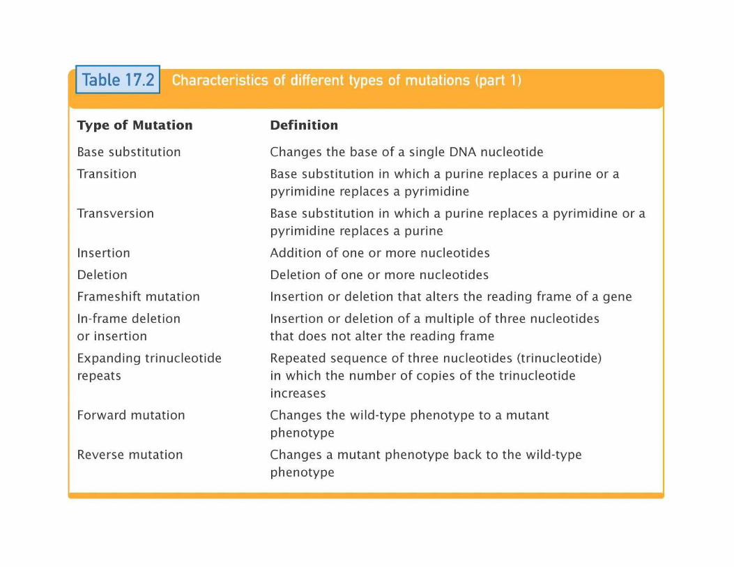
Mutation
Mutation types
• Alteração na sequência nucleotídica
• Várias classsificações
– Tipo de célula: somática ou linha germinal
– Tipo de alteração: molecular ou cromossómica
– Efeito fenotípico (na função)
– Origem:
ACERCA de MUTAÇÕES…
– Origem:
• espontâneas
• induzidas
– Agentes químicos (mutagéneos)
– Agentes físicos
• Sistemas de reparação
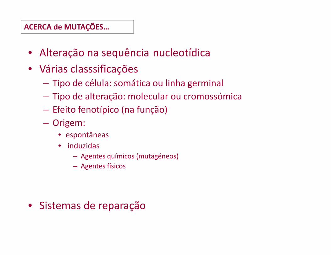
Mutation might also occur during DNA replication
Wilde-type
Mutant
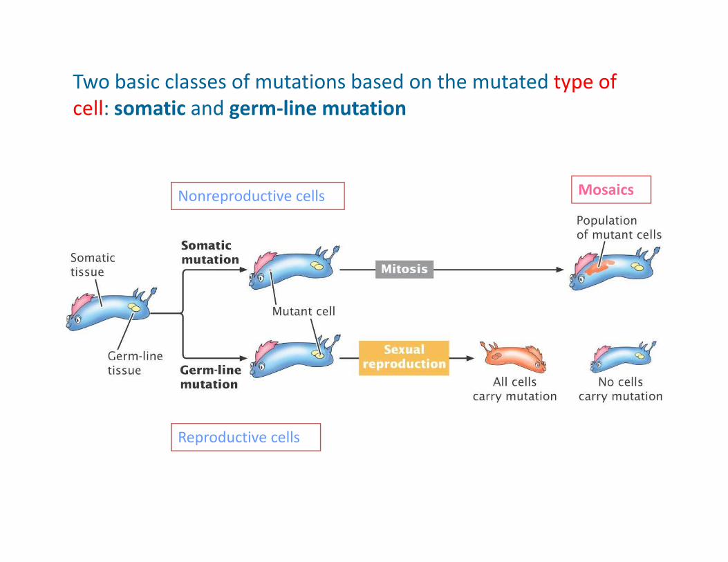
Two basic classes of mutations based on the mutated type of
cell: somatic and germ-line mutation
MosaicsNonreproductive cells
Reproductive cells
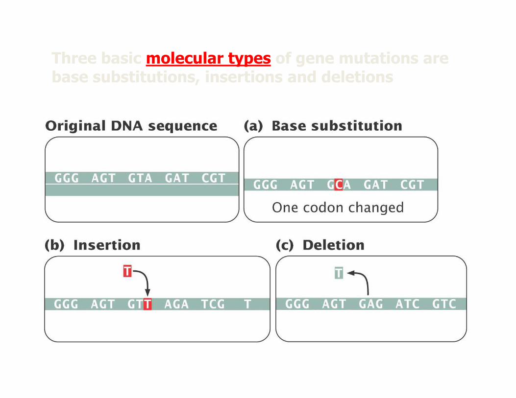
Three basic molecular types of gene mutations are base substitutions, insertions and deletions
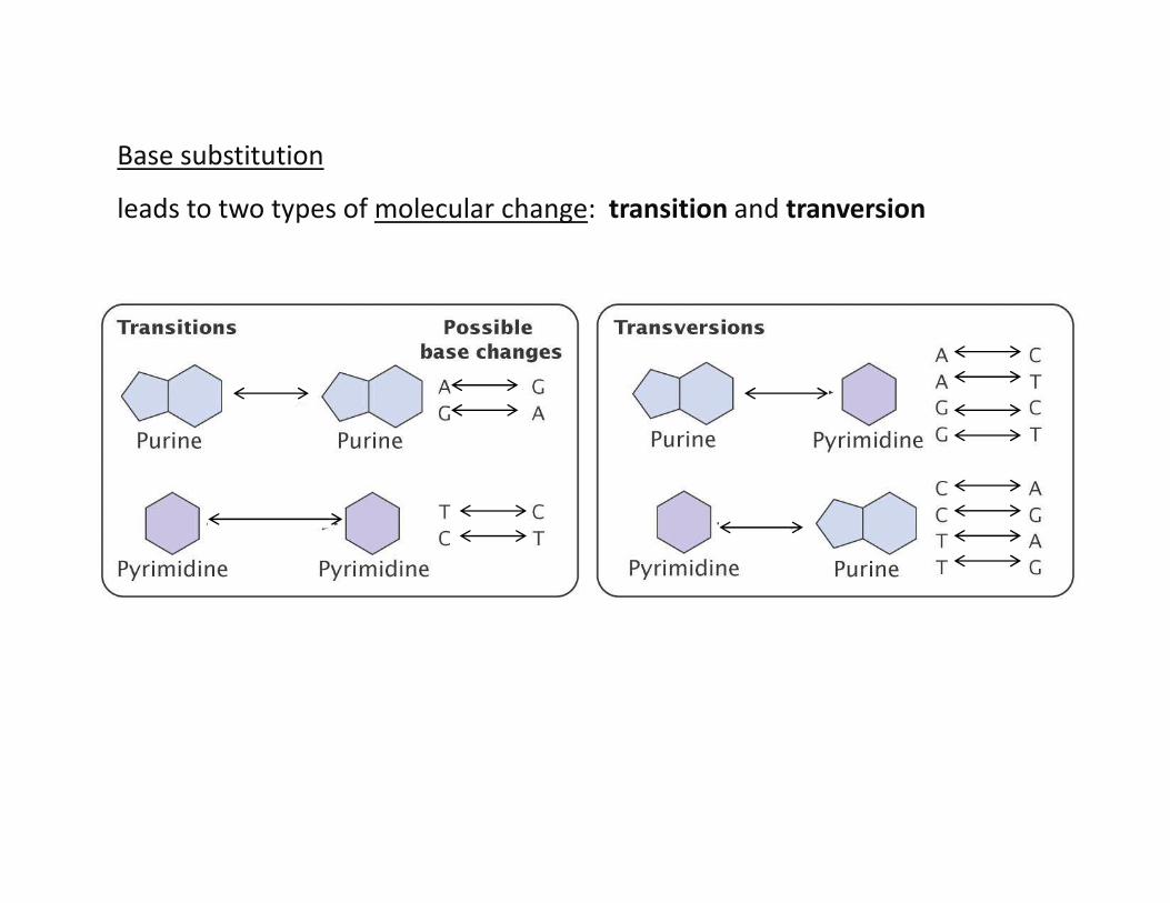
Base substitution
leads to two types of molecular change: transition and tranversion
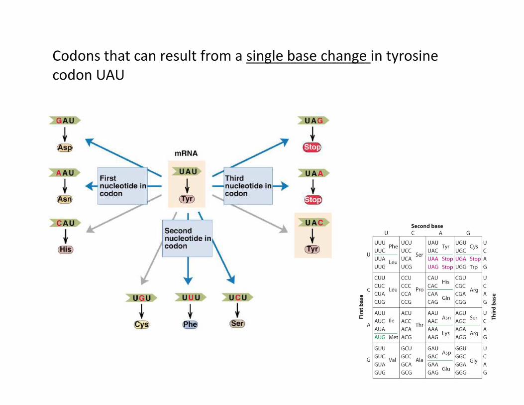
Codons that can result from a single base change in tyrosine
codon UAU
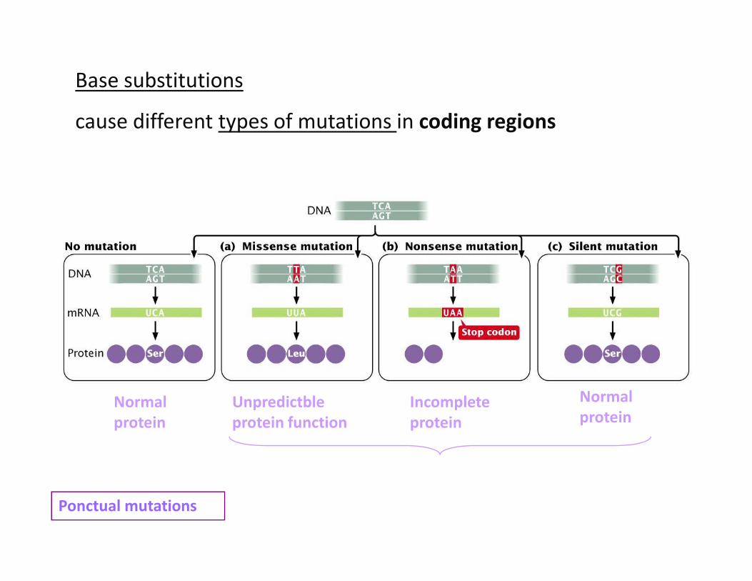
Base substitutions
cause different types of mutations in coding regions
Normalprotein
Normalprotein
Incompleteprotein
Unpredictbleprotein function
Ponctual mutations
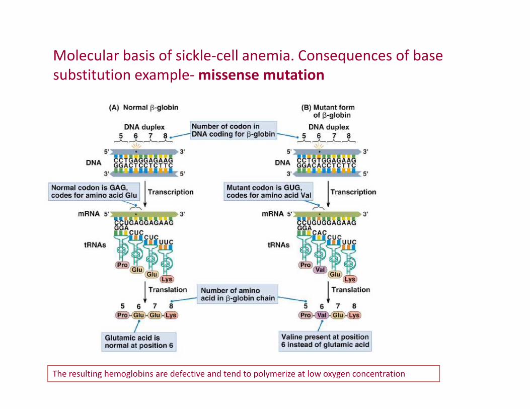
Molecular basis of sickle-cell anemia. Consequences of base
substitution example- missense mutation
The resulting hemoglobins are defective and tend to polymerize at low oxygen concentration
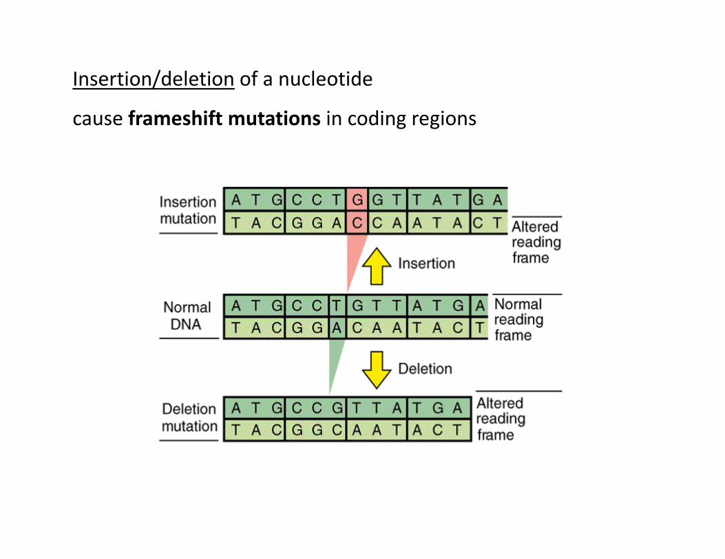
Insertion/deletion of a nucleotide
cause frameshift mutations in coding regions
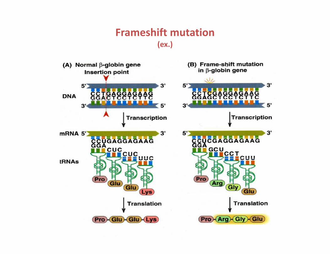
Frameshift mutation(ex.)

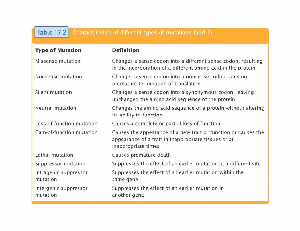
Classifications for phenotypic effects of mutations
• Loss-of-function (null or knockout) - eliminates normal function
• Gain-of-function- expressed at incorrect time, or in appropriate cell types (ectopic expression)
• Haploinsufficiency- a form of dominance in which na individual heterozygous for a wild-type allele and null allele shows na abnormal heterozygous for a wild-type allele and null allele shows na abnormal phenotype because the level of gene activity is not enough to produce a normal phenotype
• Hypomorphic (leaky)- reduces normal function, usually due to low level gene expression
• Hypermorphic- increases normal function, usually due to high level gene expression
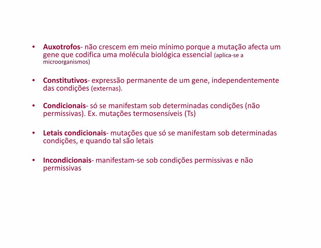
• Auxotrofos- não crescem em meio mínimo porque a mutação afecta um gene que codifica uma molécula biológica essencial (aplica-se a microorganismos)
• Constitutivos- expressão permanente de um gene, independentemente das condições (externas).
• Condicionais- só se manifestam sob determinadas condições (não permissivas). Ex. mutações termosensíveis (Ts)
• Letais condicionais- mutações que só se manifestam sob determinadas condições, e quando tal são letais
• Incondicionais- manifestam-se sob condições permissivas e não permissivas
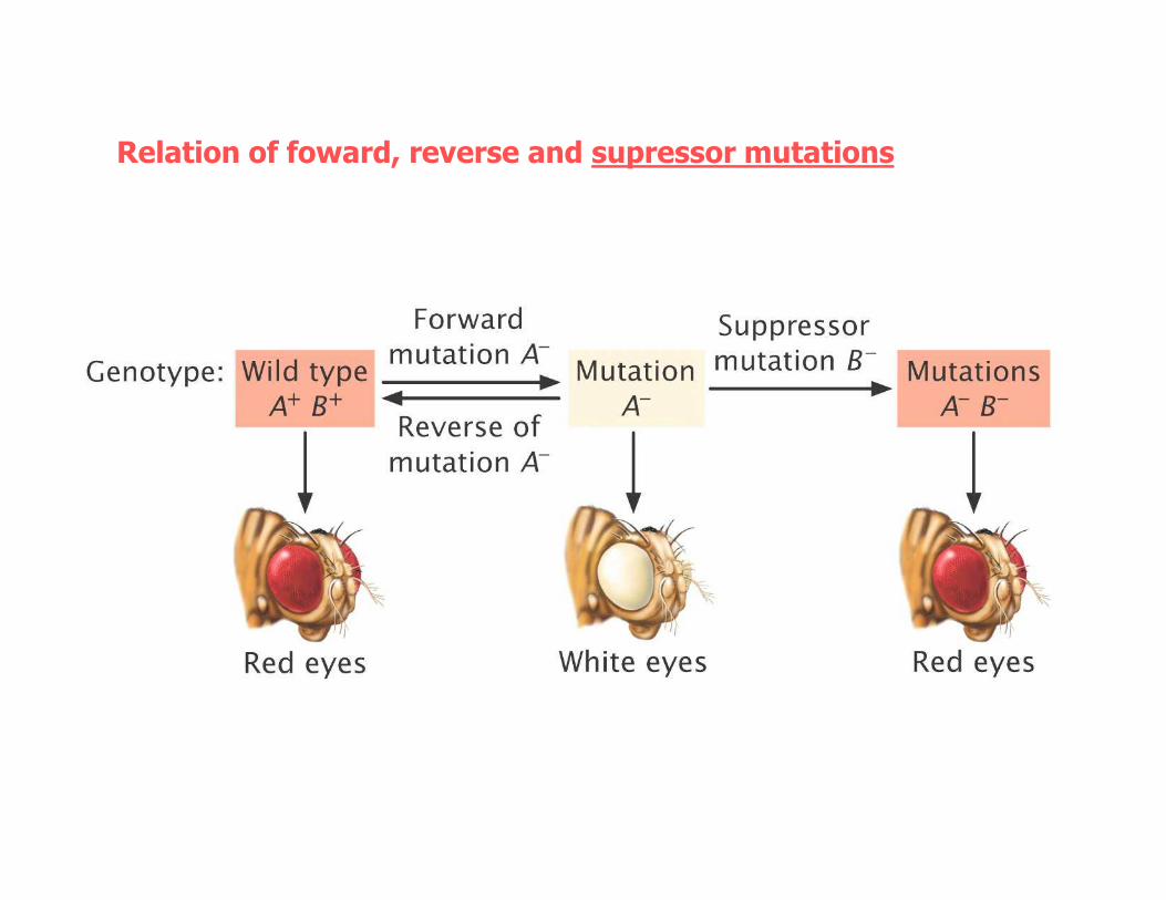
Relation of foward, reverse and supressor mutations
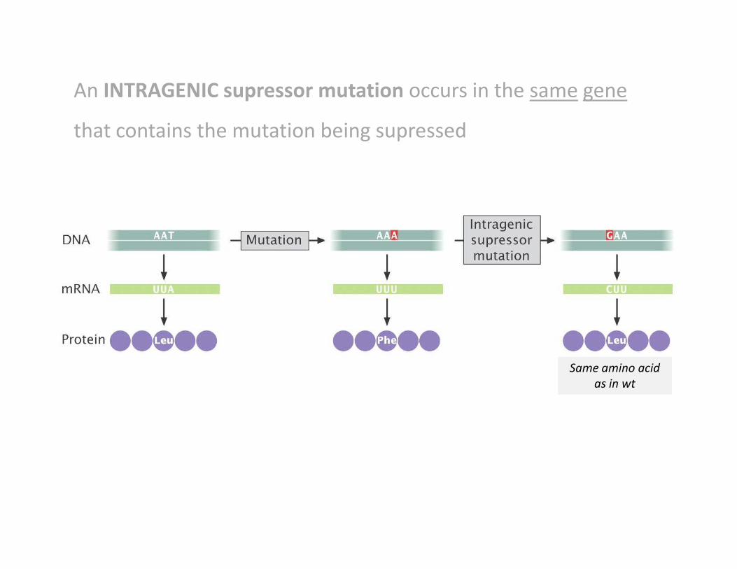
An INTRAGENIC supressor mutation occurs in the same gene
that contains the mutation being supressed
Same amino acid
as in wt
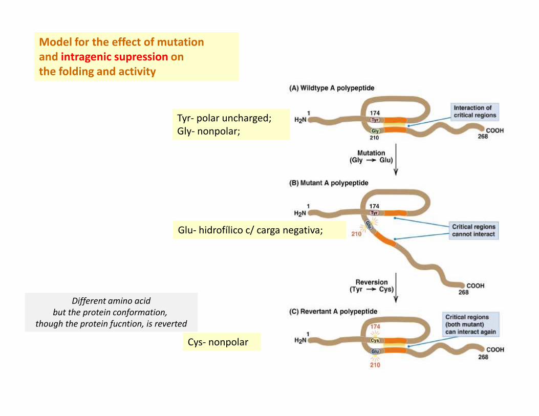
Model for the effect of mutation and intragenic supression onthe folding and activity
Tyr- polar uncharged;
Gly- nonpolar;
Glu- hidrofílico c/ carga negativa;
Cys- nonpolar
Different amino acid
but the protein conformation,
though the protein fucntion, is reverted
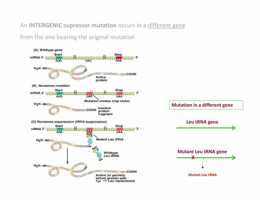
An INTERGENIC supressor mutation occurs in a different gene
from the one bearing the original mutation
Leu tRNA gene
Mutant Leu tRNA geneX
Mutant Leu tRNA
Mutation in a different gene
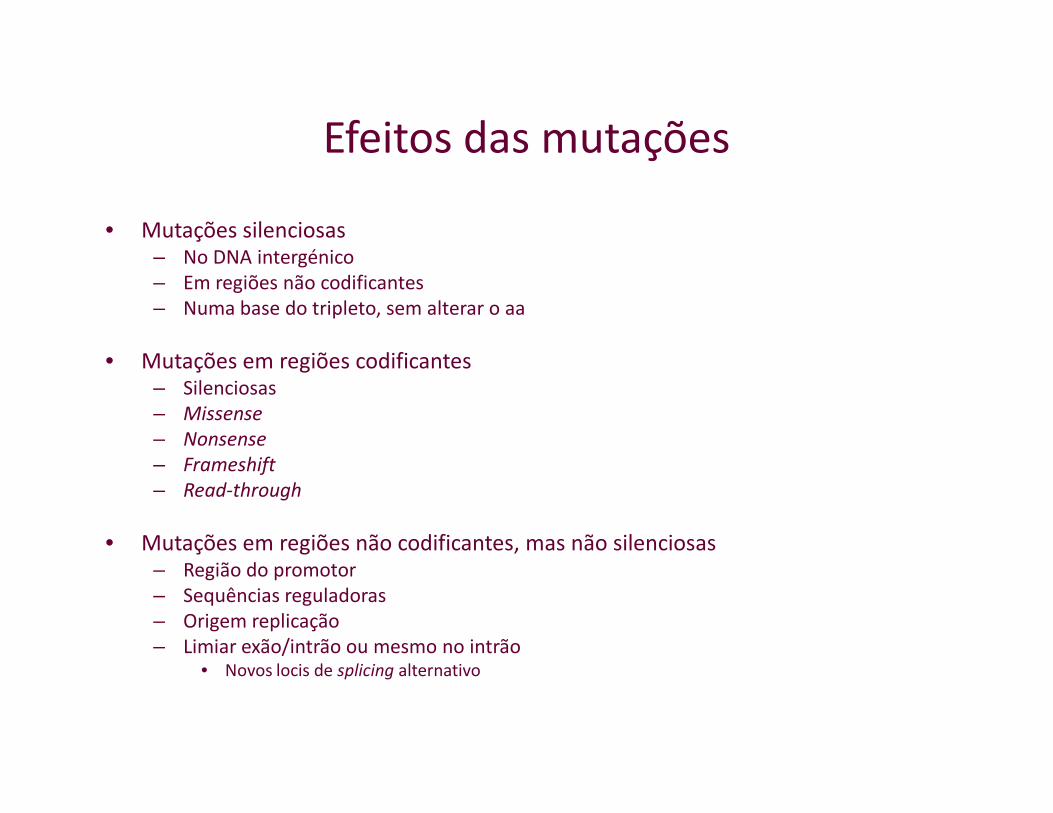
Efeitos das mutações
• Mutações silenciosas– No DNA intergénico
– Em regiões não codificantes
– Numa base do tripleto, sem alterar o aa
• Mutações em regiões codificantes– Silenciosas
– Missense– Missense
– Nonsense
– Frameshift
– Read-through
• Mutações em regiões não codificantes, mas não silenciosas– Região do promotor
– Sequências reguladoras
– Origem replicação
– Limiar exão/intrão ou mesmo no intrão• Novos locis de splicing alternativo
Spontaneous mutation(in absence of known mutagen)
vs vs
Induced mutation(in presence of known mutagen)
Spontaneous chemical changes
• Tautomerization
• Depurination
• Deamination (may also be induced by mutagenic chemicals)
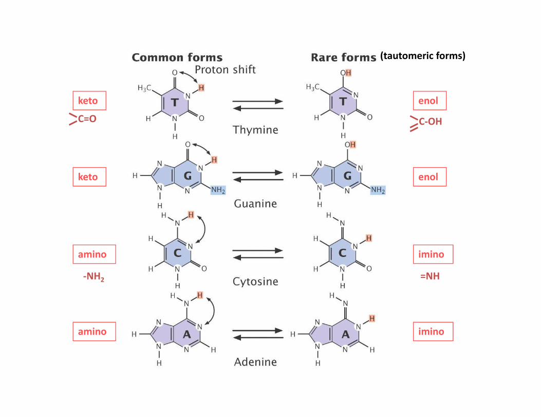
keto enol
(tautomeric forms)
keto enol
C-OHC=O
iminoamino
amino imino
-NH2 =NH
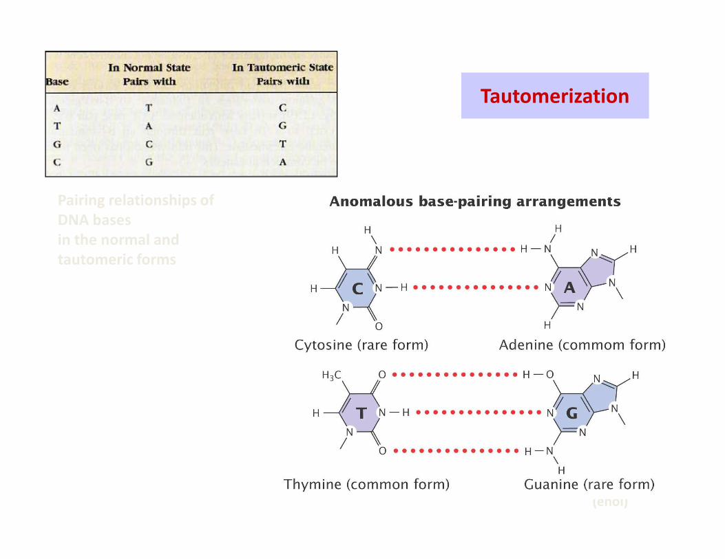
Pairing relationships of DNA basesin the normal andtautomeric forms
Tautomerization
(imino)
(enol)
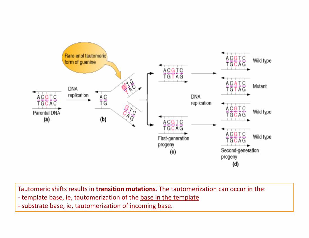
Tautomeric shifts results in transition mutations. The tautomerization can occur in the:
- template base, ie, tautomerization of the base in the template
- substrate base, ie, tautomerization of incoming base.
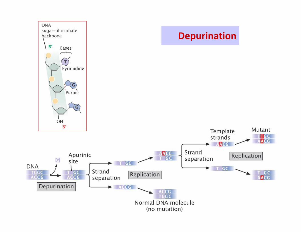
Depurination
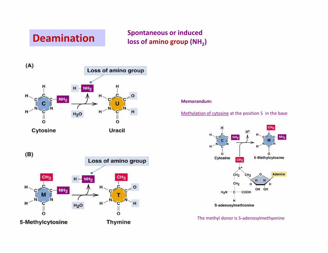
Spontaneous or inducedloss of amino group (NH2)Deamination
Memorandum:
Methylation of cytosine at the position 5 in the base
The methyl donor is S-adenosylmethyonine
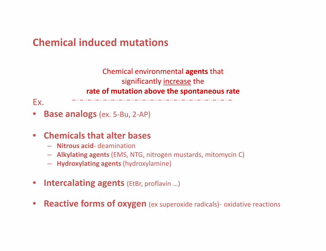
Chemical induced mutations
Chemical environmental agents that
significantly increase the
rate of mutation above the spontaneous rate
Ex.
• Base analogs (ex. 5-Bu, 2-AP)
• Chemicals that alter bases– Nitrous acid- deamination
– Alkylating agents (EMS, NTG, nitrogen mustards, mitomycin C)
– Hydroxylating agents (hydroxylamine)
• Intercalating agents (EtBr, proflavin …)
• Reactive forms of oxygen (ex superoxide radicals)- oxidative reactions
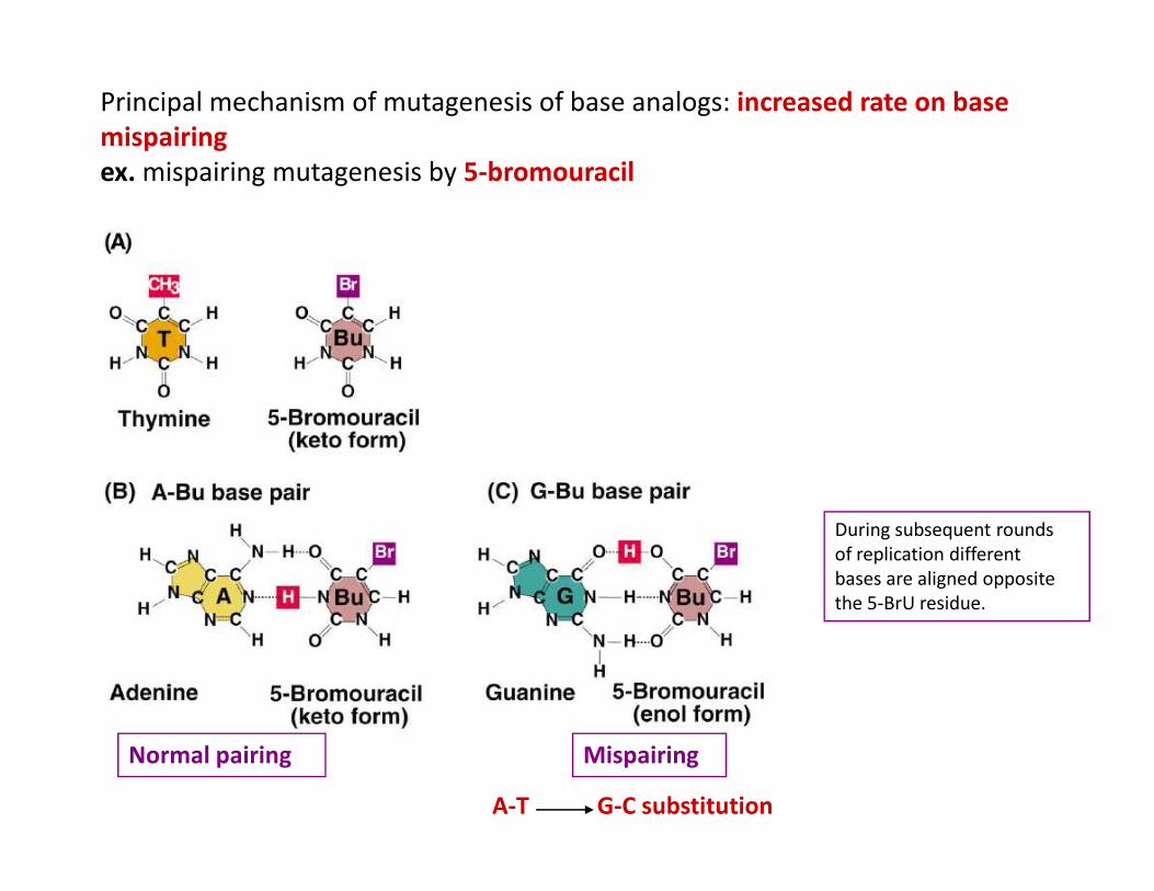
Principal mechanism of mutagenesis of base analogs: increased rate on base mispairingex. mispairing mutagenesis by 5-bromouracil
Normal pairing Mispairing
A-T G-C substitution
During subsequent rounds
of replication different
bases are aligned opposite
the 5-BrU residue.
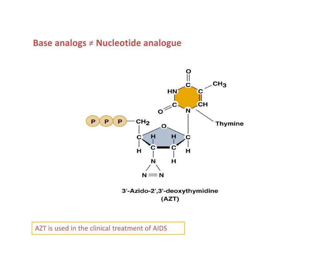
Base analogs ≠ Nucleotide analogue
AZT is used in the clinical treatment of AIDS
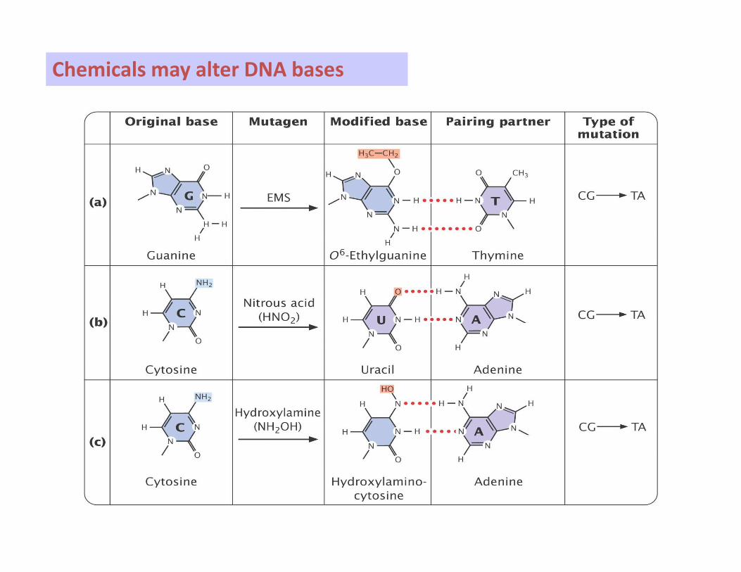
Chemicals may alter DNA bases
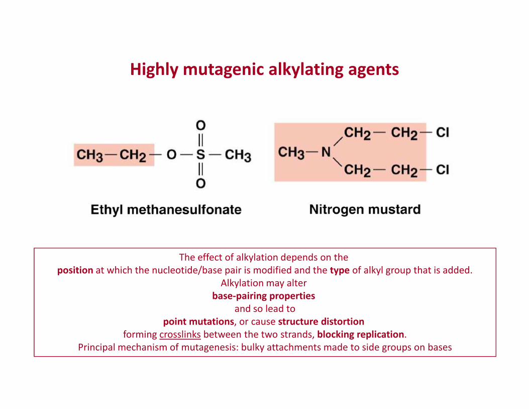
Highly mutagenic alkylating agents
The effect of alkylation depends on the
position at which the nucleotide/base pair is modified and the type of alkyl group that is added.
Alkylation may alter
base-pairing propertiesand so lead to
point mutations, or cause structure distortionforming crosslinks between the two strands, blocking replication.
Principal mechanism of mutagenesis: bulky attachments made to side groups on bases
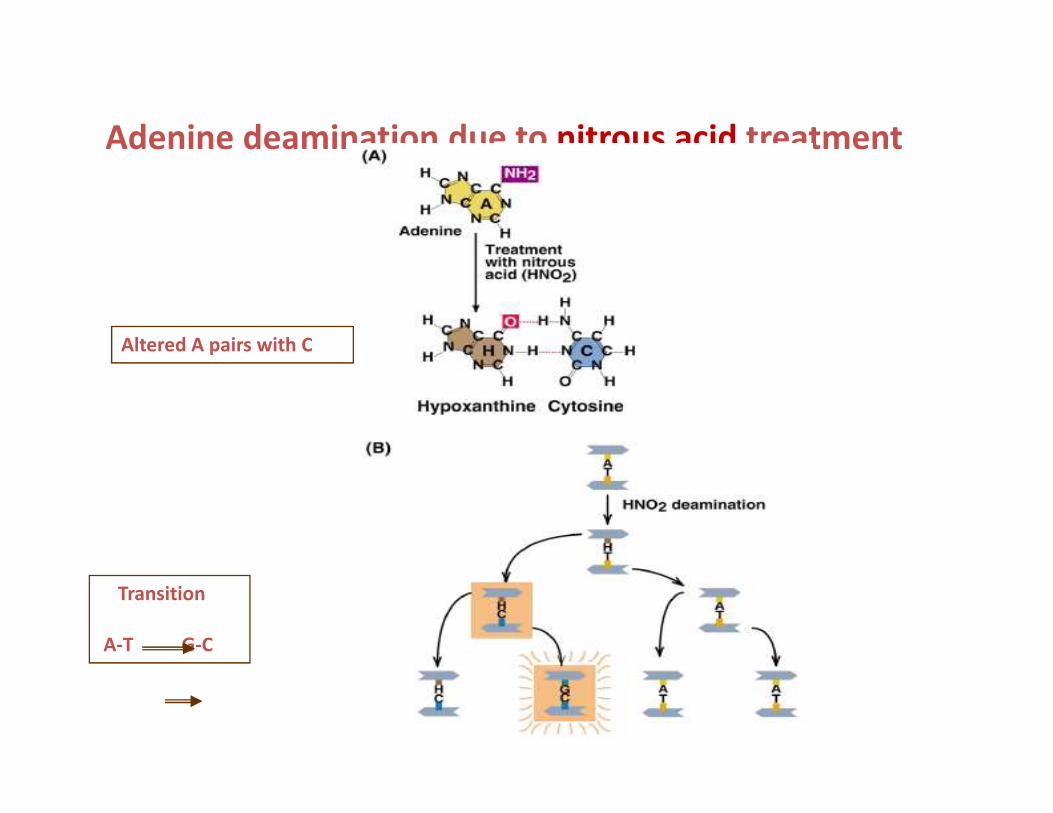
Adenine deamination due to nitrous acid treatment
Altered A pairs with C
Transition
A-T G-C
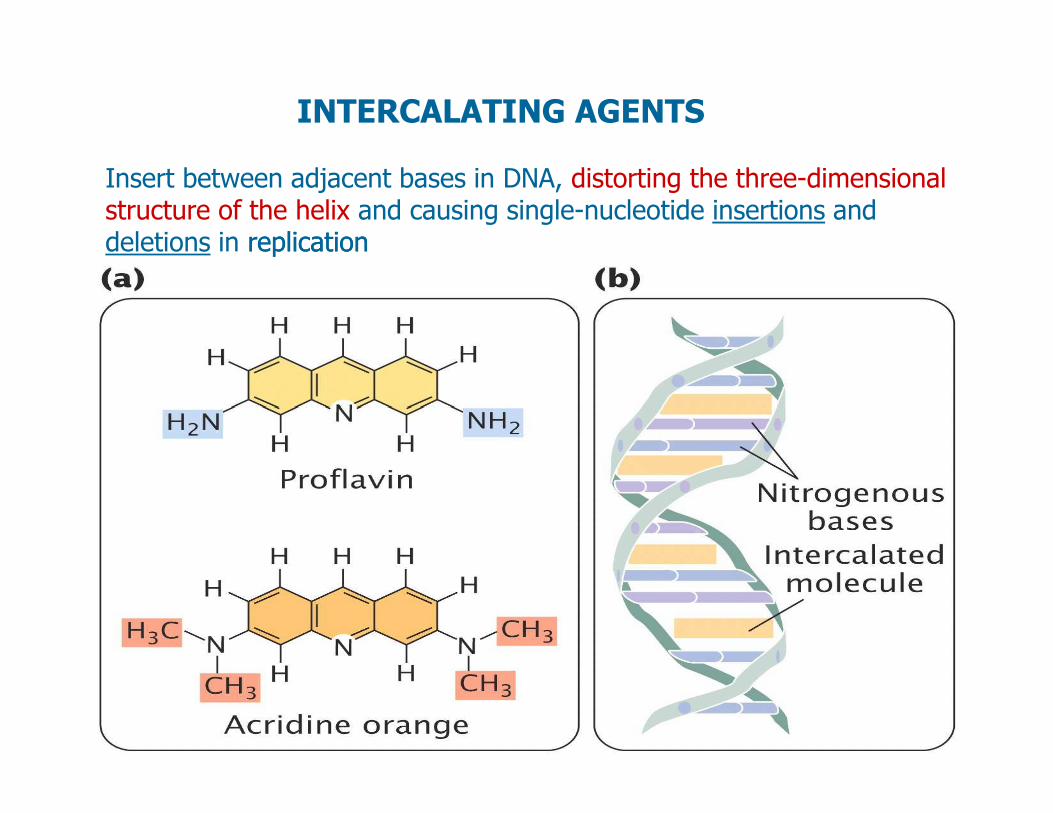
INTERCALATING AGENTS
Insert between adjacent bases in DNA, distorting the three-dimensional structure of the helix and causing single-nucleotide insertions and deletions in replicationreplication
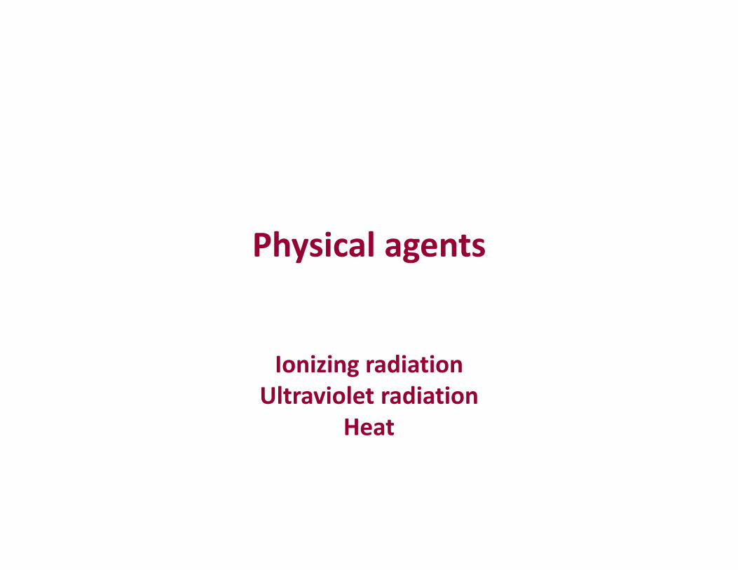
Physical agents
Ionizing radiationUltraviolet radiation
Heat
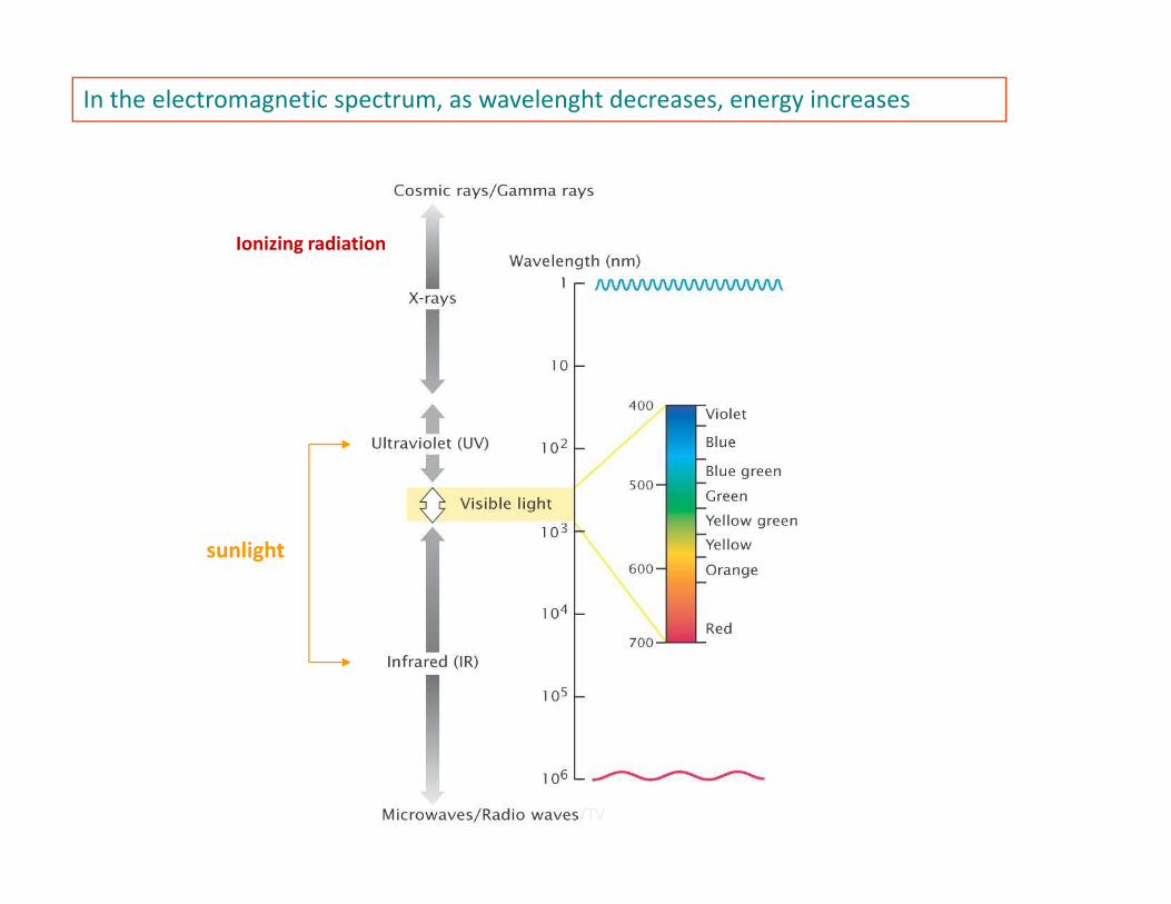
In the electromagnetic spectrum, as wavelenght decreases, energy increases
Ionizing radiation
sunlight
/TV
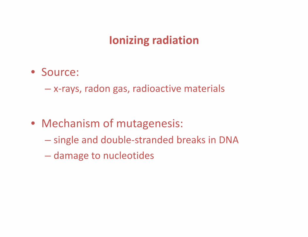
Ionizing radiation
• Source:
– x-rays, radon gas, radioactive materials
• Mechanism of mutagenesis: • Mechanism of mutagenesis:
– single and double-stranded breaks in DNA
– damage to nucleotides
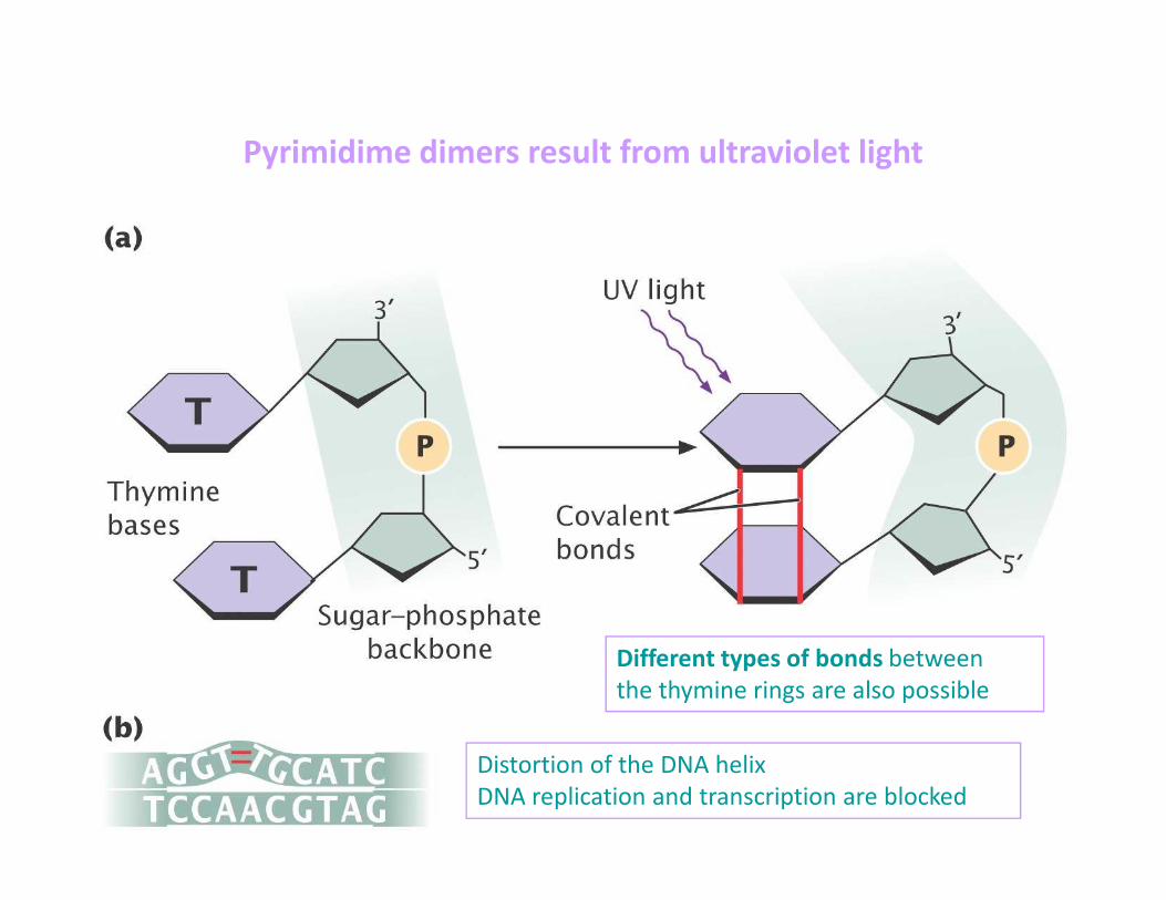
Pyrimidime dimers result from ultraviolet light
Distortion of the DNA helix
DNA replication and transcription are blocked
Different types of bonds between
the thymine rings are also possible
Técnicas de Mutagénese
Aleatória (random)
Dirigida
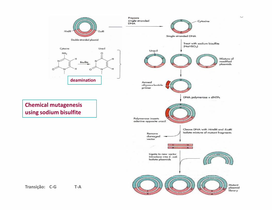
Chemical mutagenesis
deamination
Chemical mutagenesis using sodium bisulfite
Transição: C-G T-A
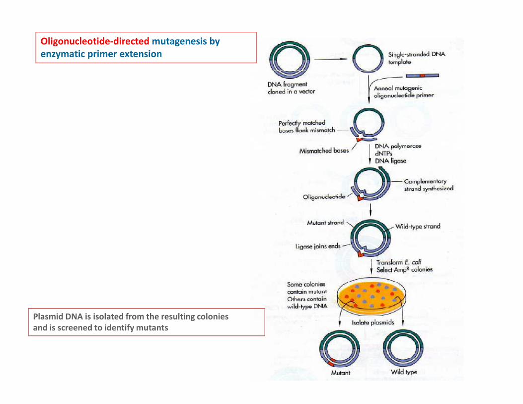
Oligonucleotide-directed mutagenesis by enzymatic primer extension
Plasmid DNA is isolated from the resulting coloniesand is screened to identify mutants

Quick-Change site directed mutagenesis
DpnI- is specific for methylated and
hemimethylated DNA
DNA isolated from most E. coli strains
is dam methylated
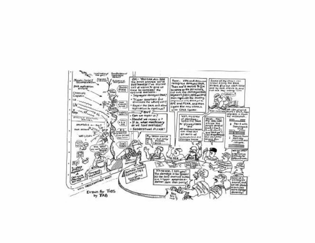
Mutation Repair
Sistemas de reparação
• Directos
– Recombinção homóloga em gaps ou cortes em cadeia dupla
– DNA ligase que actua sobre cortes em cadeia simples (nicks)
DNA polimerase I e DNA ligase (E. coli) que actuam em lacunas – DNA polimerase I e DNA ligase (E. coli) que actuam em lacunas (gaps)
– Fotoreparação enzimática. Ex. fotoliase de E. coli
– Remoção enzimática de grupos químicos que se ligam às bases dos nts e os alteram. Ex enzima ADA de E. coli que remove os grupos alquilo na posição 6 da guanina
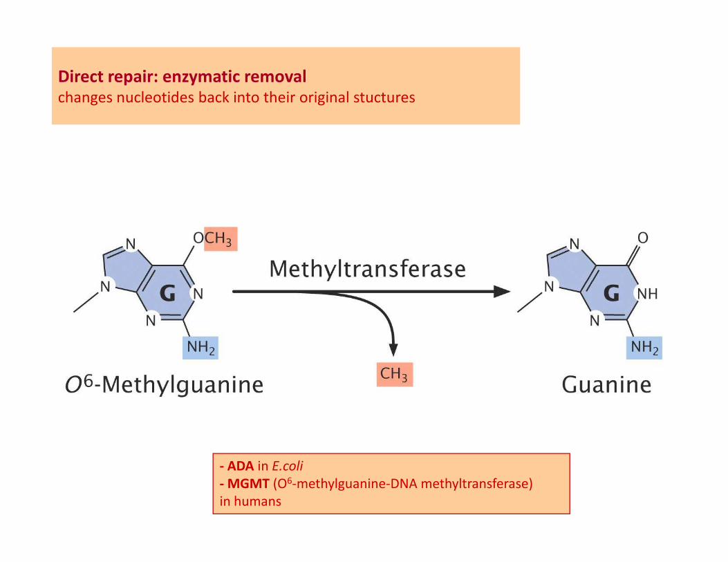
Direct repair: enzymatic removalchanges nucleotides back into their original stuctures
- ADA in E.coli
- MGMT (O6-methylguanine-DNA methyltransferase)
in humans
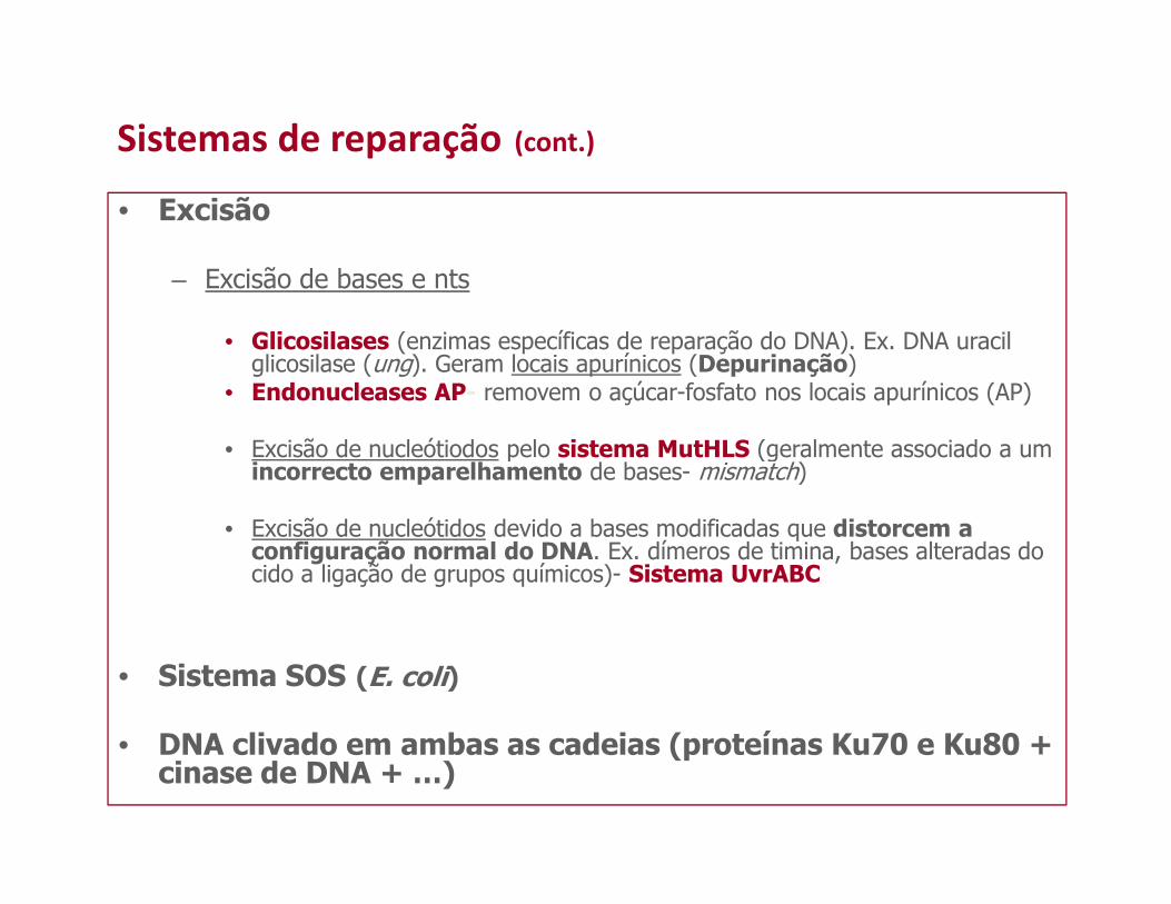
Sistemas de reparação (cont.)
• Excisão
– Excisão de bases e nts
• Glicosilases (enzimas específicas de reparação do DNA). Ex. DNA uracil glicosilase (ung). Geram locais apurínicos (Depurinação)
• Endonucleases AP- removem o açúcar-fosfato nos locais apurínicos (AP)
• Excisão de nucleótiodos pelo sistema MutHLS (geralmente associado a um • Excisão de nucleótiodos pelo sistema MutHLS (geralmente associado a um incorrecto emparelhamento de bases- mismatch)
• Excisão de nucleótidos devido a bases modificadas que distorcem a configuração normal do DNA. Ex. dímeros de timina, bases alteradas do cido a ligação de grupos químicos)- Sistema UvrABC
• Sistema SOS (E. coli)
• DNA clivado em ambas as cadeias (proteínas Ku70 e Ku80 + cinase de DNA + …)

Base and nucleotide excision repairExcises modified bases and then
replaces the entire nucleotide
Each DNA glycosylase enzymerecognizes and removes a specific
type of damaged base, producing
an apurinic or an apyrimidinic site
(AP site)
The endonuclease AP cleaves
the phosphodiester bond on
the 5’ side of the AP site and
Gap
the 5’ side of the AP site and
removes the deoxyribose sugar
Nick
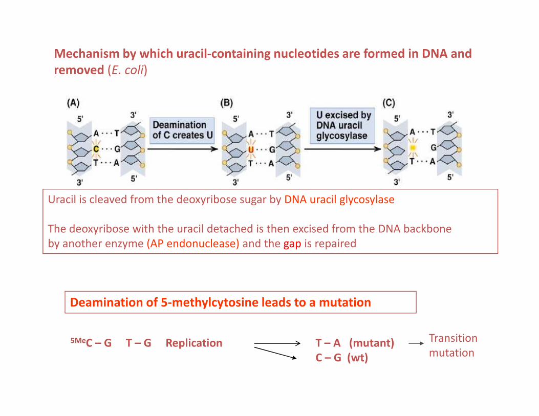
Mechanism by which uracil-containing nucleotides are formed in DNA and removed (E. coli)
Uracil is cleaved from the deoxyribose sugar by DNA uracil glycosylase
The deoxyribose with the uracil detached is then excised from the DNA backbone
by another enzyme (AP endonuclease) and the gap is repaired
Deamination of 5-methylcytosine leads to a mutation
5MeC – G T – G Replication T – A (mutant)C – G (wt)
Transition
mutation
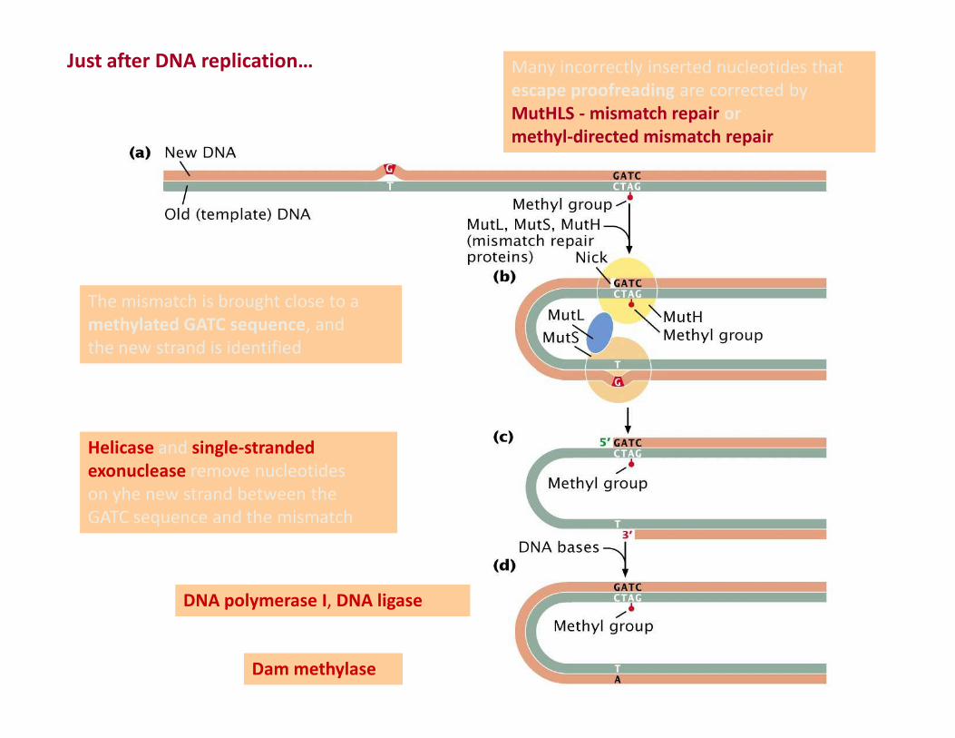
Many incorrectly inserted nucleotides that
escape proofreading are corrected by
MutHLS - mismatch repair or methyl-directed mismatch repair
Just after DNA replication…
The mismatch is brought close to a
methylated GATC sequence, and
the new strand is identified
Helicase and single-stranded exonuclease remove nucleotides
on yhe new strand between the
GATC sequence and the mismatch
DNA polymerase I, DNA ligase
Dam methylase
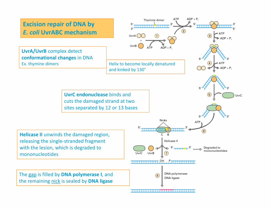
Excision repair of DNA by E. coli UvrABC mechanism
UvrA/UvrB complex detect
conformational changes in DNAEx. thymine dimers Helix to become locally denatured
and kinked by 130°
UvrC endonuclease binds and
cuts the damaged strand at two
sites separated by 12 or 13 basessites separated by 12 or 13 bases
Helicase II unwinds the damaged region,
releasing the single-stranded fragment
with the lesion, which is degraded to
mononucleotides
The gap is filled by DNA polymerase I, and
the remaining nick is sealed by DNA ligase