Embed Size (px)
Citation preview

Research Paper 177
Mutational analysis of active-site residues of the enterococcal D-Ala-o-Ala ciipeptidase VanX and comparison with Escherichja co/i D-Ala-D-Ala ligase and D-Ala-D-Ala carboxypeptidase VanY Ivan AD Lessard and Christopher T Walsh
Background: Vancomycin-resistant enterococci are pathogenic bacteria that
attenuate antibiotic sensitivity by producing peptidoglycan precursors that
terminate in D-Ala-D-lactate rather than D-Ala-D-Ala. A key enzyme in effecting
antibiotic resistance is the metallodipeptidase VanX, which reduces the cellular
pool of the D-Ala-D-Ala dipeptide.
Results: We constructed eleven mutants, using the recently determined VanX
structure as a basis, to investigate residue function. Mutating Asp1 42 or Serl 14
showed a large effect principally on KM, consistent with roles in recognition of
the D-Ala-D-Ala termini. The drastic reduction or absence of activity in the Arg71
mutants correlates with a role in the stabilization of an anionic tetrahedral
transition state. Three residues of the Escherichia co/i D-Ala-D-Ala ligase (Ddl),
Glul5, Ser 281 and Arg255, are similarly conserved and have equivalent
functions with respect to VanX, consistent with a convergent evolution of active
sites to bind D-Ala-D-Ala and lower energy barriers for formation of the
tetrahedral intermediate and transition states. In the N-acyl-D-Ala-D-Ala
carboxypeptidase VanY, all active-site residues are conserved (except for the
two responsible for recognition of the dipeptide amino terminus).
Address: Department of Biological Chemistry and Molecular Pharmacology, Harvard Medical School, Boston, MA 02115, USA.
Correspondence: Christopher T Walsh E-mail: [email protected]
Key words: D-Ala-D-Ala dipeptidase, DdlB ligase, mutagenesis, VanX, VanY
Received: 2 December 1998 Revisions requested: 5 January 1999 Revisions received: 13 January 1999 Accepted: 13 January 1999
Published: 22 February 1999
Chemistry & Biology March 1999, 6:177-l 87 http://biomednet.com/elecref/1074552100600177
% Elsevier Science Ltd ISSN 1074-5521
Conclusions: The mutagenesis results support structure-based functional
predictions and explain why the VanX dipeptidase and Ddl ligase show narrow
specificity for the D,D-dipeptide substrate. The results reveal that VanX and Ddl,
two enzymes that use the same substrate but proceed in opposite directions
driven by distinct cofactors (zinc versus ATP), evolved similar architectural
solutions to substrate recognition and catalysis acceleration. VanY sequence
analysis predicts an active site and mechanism of reaction similar to VanX.
Introduction The worldwide resurgence of infectious diseases, largely due to the appearance of antibiotic-resistant bacteria, is a serious medical problem [l-3]. Vancomycin-resistant enterococci (VRE) have become recognized as important opportunistic human pathogens over the past decade. Increased reliance on the glycopeptide antibiotic van- comycin to treat enterococcal infections and those caused by the dreaded methicillin-resistant Stap/zylococcus aurezlS (MRSA) [4] has not only caused a rise in clinically significant resistance and mortality from VRE, but also
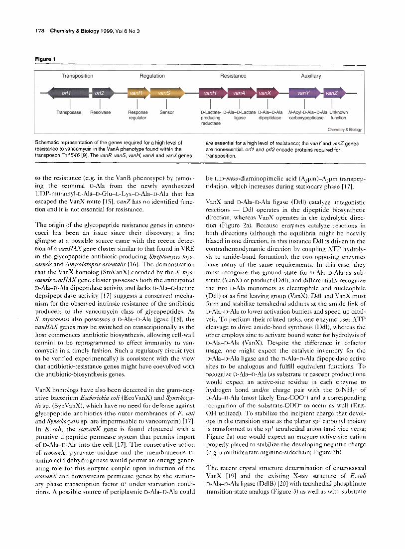
represents a looming infectious catastrophe for MRSA [d-8]. In the most prevalent clinical phenotypes of van- comycin resistance, VanA and VanB [8], bacteria have developed a strategy for reprogramming cell wall biosyn- thesis to new peptidoglycan precursor termini, which exhibit dramatically lower affinity for vancomycin. This paradigm of bacterial adaptation requires expression of the genes aanR, vans, aanH, canA and aatzX (using VanA phenotypic nomenclature, Figure 1) [9] to produce pepti- doglycan chain precursors terminating in D-alanyl-D-lactate
(D-Ala-D-lactate) in place of D-alanyl-D-alanine (D- Ala-D-Ala), resulting in a lOOO-fold decrease in van- comycin binding affinity and in unimpeded peptide strand cross-linking. The outcome is a mechanically strong cell wall whose synthesis is not inhibited by gly- copeptides [lo]. Vans and VanR act as a two-component regulatory system to mediate antibiotic-induced tran- scription of the genes vanH, nad and uanX, whose prod- ucts act sequentially (VanH, VanA) to synthesize the depsipeptide D-Ala-D-lactate in place of the normal D-
Ala-D-Ala dipeptide [ll,lZ]. VanX is a zinc-containing D,D-dipeptidase that hydrolyzes D-rZla-D-Ala but not D- Ala-D-lactate, allowing the D,D-depsipeptide to accumu- late and become incorporated in the growing peptidoglycan termini [13,14]. In the Van,4 VRE pheno- type, these genes are clustered on a transposon with two genes associated for transposition functions (o$ and o$‘) and two other oan genes, nanY and aanZ [9] (Figure 1). Although the VanE; D,D-carboxypeptidase is not essential for a high level of resistance in the VanA VRE phenotype, it is believed to contribute moderately
176 Chemistry & Biology 1999, Vol 6 No 3
Figure 1
Transposition Regulation Resistance Auxiliary
Transposase Aesolvase Response regulator
Sensor D-Lactate- D-Ala-D-Lactate D-Ala-D-Ala N-Acyl-D-Ala-D-Ala Unknown producing ligase dipeptidase carboxypeptidase function reductase
Chemistry & Elology
Schematic representation of the genes required for a high level of resistance to vancomycin in the VanA phenotype found within the transposon Tn 1546 [9]. The vanR, vanS, vanh’. vanA and vanX genes
are essential for a high level of resistance; the vanrand vanZ genes are nonessential. orfl and orf2 encode proteins required for transposition.
to the resistance (e.g. in the VanB phenotype) by remov- ing the terminal D-Ala from the newly synthesized UDP-muramyl-L-Ala-D-Glu-L-Lys-L>-Ala-II-Ala that has escaped the VanX route [ 151, snnZ has no identified func- tion and it is not essential for resistance.
The origin of the glycopeptide resistance genes in entero- cocci has been an issue since their discovery: a first glimpse at a possible source came with the recent detec- tion of a WZ~HAX gene cluster similar to that found in VRE in the glycopeptide antibiotic-producing Strepto7ByceJ toyo- caensis and Amycolatopsis orientalis [ 161. The demonstration that the VanX homolog (StoVanX) encoded by the S. toyo- caensis s?anHAX gene cluster possesses both the anticipated D-Ala-D-i\ia dipeptidase activity and lacks &Ala-r>-lactate depsipeptidase activity [ 171 suggests a conserved mecha- nism for the observed intrinsic resistance of the antibiotic producers to the vancomycin class of glycopeptides. As s. toyocaensis also possesses a D-Ala-D-Ala ligase [18], the uanHAX genes may be switched on transcriptionally as the host commences antibiotic biosynthesis, allowing cell-wall termini to be reprogrammed to effect immunity to van- comycin in a timely fashion. Such a regulatory circuit (yet to be verified experimentally) is consistent with the view that antibiotic-resistance genes might have coevolved with the antibiotic-biosynthesis genes.
VanX homologs have also been detected in the gram-neg- ative bacterium Eschetichia coli (EcoVanX) and Sy’nrchorys- tis sp. (SynVanX), which have no need for defense against glycopeptide antibiotics (the outer membranes of E. co/i and Syne&~~~tic sp. are impermeable to vancomycin) [17]. In E. cob, the ecooanX gene is found clustered with a putative dipeptide permease system that permits import of D-Ala-D-i\la into the cell [17]. The consecutive action of ecoaanx, pyruvate oxidase and the membraneous I)- amino acid dehydrogenase would permit an energy gener- ating role for this enzyme couple upon induction of the ecoaanX and downstream permease genes by the station- ary phase transcription factor CY under starvation condi- tions. A possible source of periplasmic D-Ala-D-Ala could
be r,,D-meso-diaminopimelic acid (AZpm)-A2pm transpep- tidation, which increases during stationary phase [17].
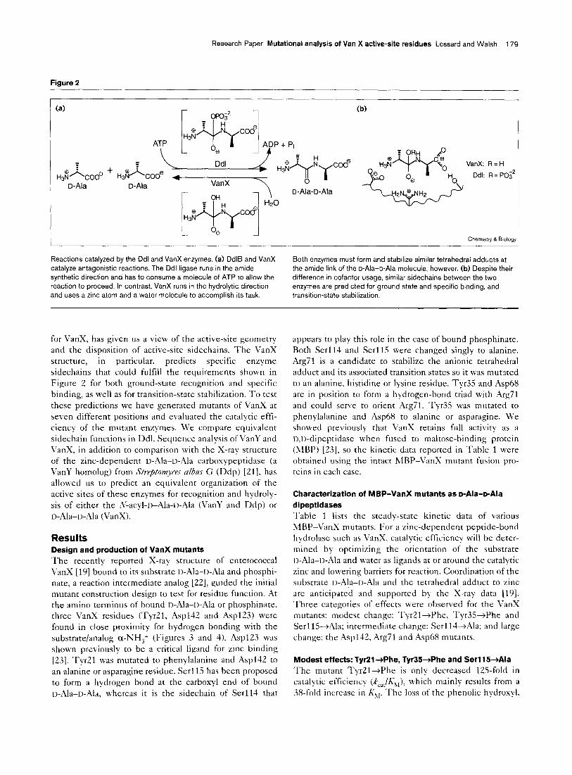
VanX and II-Ala-o-Ala ligase (Ddl) catalyze antagonistic reactions - Ddl operates in the dipeptide biosynthetic direction. whereas VanX operates in the hydrolytic direc- tion (Figure 2a). Because enzymes catalyze reactions in both directions (although the equilibria might be heavily biased in one direction. in this instance Ddl is driven in the contrathermodynamic direction by coupling ,4TP hydroly- sis to amide-bond formation), the two opposing enzymes have many of the same requirements. In this case, they must recognize the ground state for D-Ala-n-Ala as sub- strate (VanX) or product (Ddl), and differentially recognize the two 1,-,41a monomers as electrophile and nucleophile (Ddl) or as first leaving group (VanX). Ddl and VanX must form and stabilize tetrahedral adducts at the amide link of D-Ala-D-Ala to lower activation barriers and speed up catal- ysis. To perform their related tasks, one enzyme uses ,4TP cleavage to drive amide-bond synthesis (Ddl), tvhereas the other employs zinc to activate bound water for hydrolysis of I>-Ala-D-Ala (VanX). Despite the difference in cofactor usage, one might expect the catalytic inventory for the l&Ala-D-Ala ligase and the n-Ala--D-Ala dipeptidase active sites to be analogous and fulfill equivalent functions. To recognize D-Ala-D-Ala (as substrate or nascent product) one would expect an active-site residue in each enzyme to hydrogen bond and/or charge pair with the a-NH,+ of D-Ala-n-Ala (most likely Enz-COO-) and a corresponding recognition of the substrate-COO- to occur as well (Enz- OH utilized). To stabilize the incipient charge that devel- ops in the transition state as the planar sp2 carbonyl moiety is transformed to the sp” tetrahedral anion (and vice versa; Figure 2a) one would expect an enzyme active-site cation properly placed to stabilize the developing negative charge (e.g. a multidentate arginine-sidechain: Figure Zb).
The recent crystal structure determination of enterococcal VanX [19] and the existing X-ray structure of E. cob D-Ala-D-Ala ligase (DdlB) [20] with tetrahedral phosphinate transition-state analogs (Figure 3) as well as with substrate
Research Paper Mutational analysis of Van X active-site residues Lessard and Walsh 179
Figure 2
(a)
5
A H3N COOe D-Ala
ATP \
(W
VanX: R=H
Ddl: R = PO;’
Chem&y & Biology 1
Reactions catalyzed by the Ddl and VanX enzymes. (a) DdlB and VanX Both enzymes must form and stabilize similar tetrahedral adducts at catalyze antagonistic reactions. The Ddl ligase runs in the amide the amide link of the D-Ala-o-Ala molecule, however. (b) Despite their
synthetic direction and has to consume a molecule of ATP to allow the difference in cofactor usage, similar sidechains between the two reaction to proceed. In contrast, VanX runs in the hydrolytic direction enzymes are predicted for ground state and specific binding, and and uses a zinc atom and a water molecule to accomplish its task. transition-state stabilization.
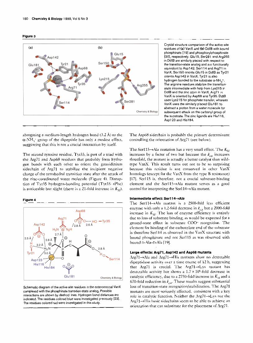
for VanX, has given us a view of the active-site geometry and the disposition of active-site sidechains. The VanX structure, in particular. predicts specific enzyme sidechains that could fulfill the requirements shown in Figure 2 for both ground-state recognition and specific binding, as well as for transition-state stabilization. To test these predictions we have generated mutants of VanX at seven different positions and evaluated the catalytic effi- ciency of the mutant enzymes. We compare equivalent sidechain functions in Ddl. Sequence analysis of VanY and
VanX, in addition to comparison with the X-ray structure of the zinc-dependent u-Ala-+-Ala carboxypeptidase (a VanY homolog) from Strepfomq’ces ahs G (Ddp) [Zl]. has allowed us to predict an equivalent organization of the active sites of these enzymes for recognition and hydroly- sis of either the ,V-acyl-D-Ala-[,-Ala (\?anY and Ddp) or I>-Ala-n-Ala (VanX).
Results Design and production of VanX mutants The recently reported X-ray structure of enterococcal VanX [19] bound to its substrate I,-Ala-[)-Ala and phosphi- nate, a reaction intermediate analog [ZZ], guided the initial mutant construction design to test for residue function. At the amino terminus of bound r)-Ala-n-Ala or phosphinate, three VanX residues (Tyr21, Asp142 and Asp123) were found in close proximity for hydrogen bonding with the substrate/analog cx-IKHj+ (Figures 3 and 4). Asp123 WiS
shown previously to be a critical ligand for zinc binding [23]. Tyr21 was mutated to phenylalanine and Asp142 to an alanine or asparagine residue. Serll.~ has been proposed to form a hydrogen bond at the carboxyl end of bound D-Ala-u-Ala, whereas it is the sidechain of Serl14 that
appears to play this role in the case of bound phosphinate. Both Serl14 and Serll5 were changed singly to alanine. Arg71 is a candidate to stabilize the anionic tetrahedral adduct and its associated transition states so it was mutated to an alanine, histidine or lysine residue. Tyr35 and Asp68 are in position to form a hydrogen-bond triad with Arg71 and could serve to orient ,4rg71. Tyr35 was mutated to phenylalanine and Asp68 to alanine or asparagine. We showed previously that VanX retains full activity as a I,,t)-dipeptidase when fused to maltose-binding protein (MBP) [23], so the kinetic data reported in Table 1 were obtained using the intact MBP-VanX mutant fusion pro- teins in each case.
Characterization of MBP-VanX mutants as D-Ala-D-Ala dipeptidases Table 1 lists the steady-state kinetic data of various MBP-VanX mutants. For a zinc-dependent peptide-bond hydrolase such as VanX, catalytic efficiency will be deter- mined by optimizing the orientation of the substrate t>-Ala-n-Ala and water as ligands at or around the catalytic zinc and lowering barriers for reaction. Coordination of the substrate I)-Ala-[)-Ala and the tetrahedral adducc to zinc are anticipated and supported by the X-ray data [19]. Three categories of effects were observed for the VanX mutants: modest change: TyQl+Phe, Tyr3S-+Phe and Serl lS-+Ala; intermediate change: Serl l++Ala: and large change: the AspllZ, Arg71 and ,4sp68 mutants.
Modest effects: TyRl+Phe, Tyr35+Phe and Serl15+Ala The mutant TyrZl-+Phe is only decreased 125fold in catalytic efficiency (k,,,/K,,). which mainly results from a 38-fold increase in K,,. The loss of the phenolic hydroxiyl.
180 Chemistry & Biology 1999, Vol 6 No 3
Figure 3
(a) (W Crystal structure comparison of the active-site
residues of (a) VanX and (b) Ddlf3 with bound
phosphinate [191 and phosphorylphosphinate [201, respectively. Glul5, Ser281 and Arg255 in DdlB are similarly placed with respect to
the transition-state analog and are functionally
equivalent to Asp1 42, Serl14 and Arg71 in VanX. Serl50 orients Glul5 in DdlB as TyrPl
orients Asp1 42 in VanX. TyrPl is also
hydrogen bonded to the substrate cx-NH,+. The arginine residues stabilize the transition-
state intermediate with help from Lys215 in
DdlB and the zinc atom in VanX. Arg71 in VanX is oriented by Asp68 and Tyr35. DdlB
uses Lys2 15 for phosphate transfer, whereas VanX uses the similarly placed Glu181 to
abstract a proton from a water molecule for
subsequent attack on the carbonyl group of the substrate. The zinc ligands are His1 16,
Asp1 23 and His1 84.
abrogating a medium-length hydrogen bond (3.2 A) to the cc-IKH,+ group of the dipeptide has only a modest effect, suggesting that this is not a crucial interaction by itself.
The second tyrosine residue, Tyr35, is part of a triad with the Arg71 and Asp68 residues that probably form hydro- gen bonds with each other to orient the guanidinium sidechain of Arg71 to stabilize the incipient negative charge of the tetrahedral transition state after the attack of the zinc-coordinated water molecule (Figure 4). Disrup- tion of Tyr35 hydrogen-bonding potential (Tyr35-+Phe) is noticeable but slight (there is a Zl-fold increase in K,,).
Figure 4
r Asp68
Arg7 1 A 0.6. O;6-A~o -Tyr35
:‘2.8 A
0
Serl 14
His1 16 ‘. ’ Hisi 0 .$o
Y Glti 81 Chemstry & Bmlog:
Schematic diagram of the active-site residues in the enterococcal VanX complexed with the phosphinate transition-state analog. Possible
interactions are shown by dashed lines. Hydrogen bond distances are indicated. The residues colored blue were investigated previously 1231.
The residues colored red were investigated in this study.
The Asp68 sidechain is probably the primary determinant controlling the orientation of Arg71 (see below).
The Serl lS--+Ala mutation has a very small effect. The Khl increases by a factor of two but because the k,,, increases threefold, the mutant is actually a better catalyst than wild- type VanX. This result turns out not to be so surprising because this residue is not conserved in other VanX homologs (except for the VanX from the type B resistance) [17]. Serl15 is, therefore. not a crucial substrate-binding element and the Serllj-+Ala mutant serves as a good control for interpreting the Serl14+Ala mutant.
intermediate effect: Serl14+Ala The Serll4-+Ala mutant is a 2500-fold less efficient enzyme with only a L.&fold decrease in k,,, but a 2000-fold increase in k;,. The loss of enzyme efficiency is entirely due to loss of substrate binding, as would be expected for a ground-state effect in substrate COO- recognition. The element for binding of the carboxylate end of the substrate is therefore Serll4 as obsen,ed in the VanX structure with bound phosphinate and not SerllS as was observed with bound II-Ala-n-Ala [ 191.
Large effects: Arg71, Asp142 and Asp66 mutants .4rg71-+Ala and Arg71-+His mutants show no detectable dipeptidase activity over a time course of 12 h, suggesting that Arg71 is crucial. The Arg71+Lys mutant has detectable activity but shows a 1.7 x 10h-fold decrease in catalytic efficiency, due to a 2750-fold increase in K,, and a 670-fold reduction in kcaf. These results suggest substantial loss of transition-state recognition/stabilization. ‘I’he Arg71 mutants are most seriously affected, consistent with a key role in catalytic function. Neither the Arg7l+Lys nor the Arg’ll+His basic sidechains seem to be able to achieve an orientation that can substitute for the placement of Arg71.
Research Paper Mutational analysis of Van X active-site residues Lessard and Walsh 181
Table 1
Kinetic parameters of purified MBP-VanX mutants fusion proteins using p-Ala-p-Ala as substrate.
Protein kc,, (s-‘I* Decrease (x-fold) KM hM) increase (x-fold) k,,lK, (s-ImM-I) Decrease (x-fold)
Wild type 26 TyrPl -+Phe 7.8
Asp1 42-+Ala 0.037 Asp1 42+Asn 0.30 Serll4-+Ala 22
Serll5+Ala 73 Arg71 +Ala nd
Arg71 +His nd Arg7 1 +Lys 0.041 Asp68-tAla 0.097
Asp68-+Asn 0.12 Tvr35-+Phe 10
3
703
87 1.2
0.36
38
81 1200
2000 2
125 58000
105000 2500
0.71 -
634
270 217
2.6
0.080
3.0
6.5 96
160
0.16
nd
nd 220
60
5.0 1.7
2750
750
63 21
330 2.6
0.0056 0.0031
0.13
460 nd
nd
0.00019 0.0016
0.024
5.8
1700000 200000
14000
56
MBP-VanX samples were prepared from cells grown in LB media cadmium-ninhydrin method (see the Materials and methods section).
supplemented with 200 PM ZnSO,. ‘Dipeptidase activity was assayed Protein concentrations were determined from the corrected absorbance by measuring the production of o-Ala using the modified at 280 nm in H,O [40]. nd, no detectable activity over 12 h.
The ,4rg142+Ala and Arg142-+,4sn mutants were con- structed because the X-ray structure of VanX shows that Asp142 is within 2.8 w of the D,D-substrate’s a-NH,+, con- sistent with participation in ground-state recognition (Figure 4). The Aspl4Z+Asn mutant is decreased only 87-fold in k,,, but is increased 1200-fold in Khl, suggesting that Asn142 might still be able to hydrogen bond with the D,D-substrate a-NH,+. The k,,, of the Asp142+Ala mutant is decreased 700-fold, an order of magnitude worse than the Asp142-+;2sn mutant. It is not clear why the K1, is only increased 81-fold, however. These effects in k,,, and KR, have not been subdivided into effects on micro- scopic rate constants, so we make no further interpretation about effects on elementary steps.
When the importance of Asp68 was examined, with the Asp68+Asn and Asp68--+Ala mutants, substitution of aspartate by asparagine preserves more function than the aspartate to alanine switch. Here, both mutants have a ZOO-300-fold decrease in k,,, but the Asp68jAsn mutant only has a 63-fold increase in K,,, whereas in the Asp68+Ala mutant, the K,,, was 750-fold worse. The VanX crystal structure shows that Asp68 is the central residue in the Arg71-Asp68-Tyr35 hydrogen-bonding triad. Presum- ably the asparagine sidechain in the Asp68+Asn mutant retains more contact with Arg71 than does the alanine sidechain in the ,4sp68+Ala mutant, assisting in produc- tive orientation of Arg71 for transition-state stabilization.
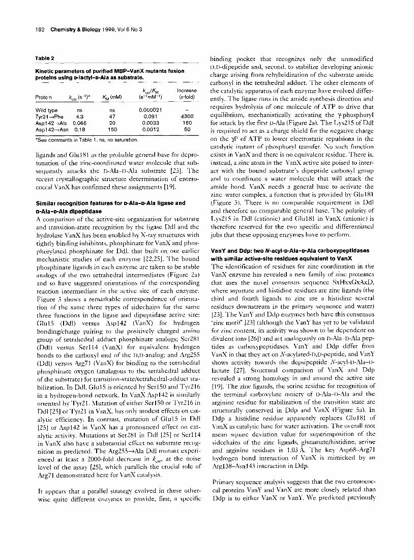
TyrZl+Phe, had an increased ability to hydrolyze I>-lactyl-D-Ala. The AsplilZ+Asn and Asp142+Ala mutants showed a 60- and 160-fold increase in peptidase activity, respectively, compared with the wild-type enzyme, which has a barely detectable k,,JK1, of 2 x lo-’ m,\l-‘s-l, a factor of 10’ worse than for I,-Ala-r>- Ala (Table 2). The Tyr2l-+Phe VanX mutant shows a much larger effect with an increase of 4300-fold in cat- alytic efficiency over the wild-type enzyme. For this mutant, II-Ala-u-Ala is only 30-fold better by k,,,/K:,, crite- rion than D-lactyl-D-Ala. This 30-fold difference is almost entirely due to K,,. The k,,, of TyrZl+Phe for o-lactyl- o-Ala cleavage is only sixfold lower than the wild-type enzyme for its natural substrate, D-Ala-D-Ala, suggesting a similar substrate orientation for peptide cleavage in each enzyme. In wild-type VanX Ty21, which hydrogen bonds to Aspl42, might exclude D-lact+II-.t\la by means of elec- trostatic repulsion between two oxygens. The Asp142 mutants have only a twofold increase in catalytic effi- ciency for n-Ala--u-Ala than I>-lactyl-D-Ala. Again removal of electron density close to the binding site for D-Ala-
n-Ala a-NH,+ allows better binding of the uncharged D-lactyl-D-Ala substrate. Interestingly, in DdlB? a D-lactyi-D-141a ligase activity was detected in the GlulS+Gln mutant [24]. Both Glu15 in DdlB and Asp142 in VanX are involved in the a-&H,+ recognition of the substrate n-Ala for DdlB and D-Ala-D-Ala for VanX.
Discussion Detection of novel activity in mutant proteins
We examined the mutant proteins for ‘gain of function’ novel activities by testing other potential enzyme sub- strates. In no case was ;V-acetyl-rj-Ala-D-Ala a substrate, even for the TyrZl-+Phe mutant, but the reverse regioiso- mer of D-Ala-D-lactate, the n-lactyl-D-Ala amide (which lacks an a-NH,+ group) showed an interesting substrate profile. Three mutants, Asp142-+Ala, Aspl4Z-+Asn and
Metalloproteases have been a successfully targeted for rationally designed therapeutic agents in other biological contexts (e.g. angiotensin converting enzyme inhibitors), suggesting that VanX is a prime candidate for design of inhibitors to combat VRE. In an earlier study of the zinc- dependent metallodipeptidase enterococcal VanX (VanA- type resistance). we identified, using mutagenesis, residues His1 16, Asp123 and His184 as probable zinc-coordination
18’2 Chemistry & Biology 1999, Vol 6 No 3
Table 2
Kinetic parameters of purified MBP-VanX mutants fusion proteins using D-lactyl-D-Ala as substrate.
Protein k (s-‘)* cat
Wild type TyrPl +Phe I”3 Asp1 42+Ala 0.066 Asp142-+Asn 0.18
K,,,, WI)
1; 20
150
katlKrv, (s’mM-‘)
0.000021 0.091
0.0033 0.0012
increase
(x-fold)
4300 160
60
*See comments in Table 1. ns, no saturation.
ligands and Glu181 as the probable general base for depro- tonation of the zinc-coordinated water molecule that sub- sequently attacks the I>-Ala-II-Ala substrate [23]. The recent crystallographic structure determination of entero- coccal VanX has confirmed these assignments [ 191.
Similar recognition features for D-Ala-D-Ala ligase and D-Ala-D-Ala dipeptidase A comparison of the active-site organization for substrate and transition-state recognition by the ligase Ddl and the hydrolase VanX has been enabled by X-ray structures with tightly binding inhibitors, phosphinate for VanX and phos- phorylated phosphinate for Ddl. that built on our earlier mechanistic studies of each enzyme [Z&25]. The bound phosphinate ligands in each enzyme are taken to be stable analogs of the two tetrahedral intermediates (Figure Za) and so have suggested orientations of the corresponding reaction intermediate in the active site of each enzyme. Figure 3 shows a remarkable correspondence of orienta- tion of the same three types of sidechains for the same three functions in the ligase and dipeptidase active site: Glu15 (Ddl) versus Asp142 (VanX) for hydrogen bonding/charge pairing to the positively charged amino group of tetrahedral adduct phosphinate analogs; Ser281 (Ddl) versus Serl1-l (VanX) for equivalent hydrogen bonds to the carboxyl end of the r),r,-analog; and ArgZSS (Ddl) versus Arg71 (VanX) for binding to the tetrahedral phosphinate oxygen (analagous to the tetrahedral adduct of the substrate) for transition-state/tetrahedral-adduct sta- bilization. In Ddl, Glu1.5 is oriented by SerlSO and Tyr216 in a hydrogen-bond network. In VanX Asp142 is similarly oriented by Tyr21. hlutation of either SerlSO or Tyr216 in Ddl [Zj] or Tyr21 in VanX, has only modest effects on cat- alytic efficiency. In contrast, mutation of Glu15 in Ddl [25] or Asp142 in VanX has a pronounced effect on cat- alytic activity. Mutations at Ser281 in Ddl [25] or Serll4 in VanX also have a substantial effect on substrate recog- nition as predicted. The Arg255+Ala Ddl mutant experi- enced at least a 2000-fold decrease in k,,,, at the noise level of the assay [25], which parallels the crucial role of Arg71 demonstrated here for VanX catalysis.
It appears that a parallel strategy evolved in these other-
wise quite different enzymes to provide, first, a specific
binding pocket that recognizes only the unmodified D,D-dipeptide and, second, to stabilize developing anionic charge arising from rehybridization of the substrate amide carbonyl in the tetrahedral adduct. The other elements of the catalytic apparatus of each enzyme have evolved differ- ently. The l&se runs in the amide synthesis direction and requires hydrolysis of one molecule of ATP to drive that equilibrium, mechanistically activating the y-phosphoryl for attack by the first D-Ala (Figure 2a). The Lys215 of Ddl is required to act as a charge shield for the negative charge on the p of ATP to lower electrostatic repulsions in the catalytic instant of phosphor);1 transfer. No such function exists in VanX and there is no equivalent residue. There is, instead, a zinc atom in the VanX active site poised to inter- act with the bound substrate’s dipeptide carbonyl group and to coordinate a water molecule that will attack the amide bond. VanX needs a general base to activate the zinc-water complex, a function that is provided by Glu181 (Figure 3). There is no comparable requirement in Ddl and therefore no comparable general base. The polarity of Lys215 in Ddl (cationic) and Glu181 in VanX (anionic) is therefore reserved for the two specific and differentiated jobs that these opposing enzymes have to perform.
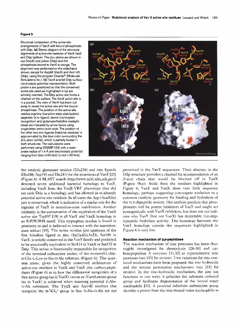
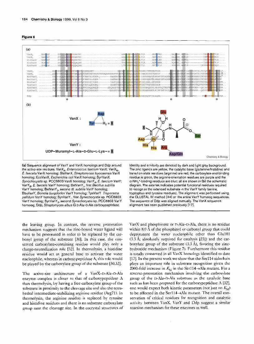
VanY and Ddp: two N-acyl-D-Ala-D-Ala carboxypeptidases with similar active-site residues equivalent to VanX The identification of residues for zinc coordination in the VanX enzyme has revealed a new family of zinc proteases that uses the novel consensus sequence SxHxxGxAxD, where aspartate and histidine residues are zinc ligands (the third and fourth ligands to zinc are a histidine several residues downstream in the primary sequence and water) [23]. The VanY and Ddp enzymes both have this consensus ‘zinc motif [23] (although the VanY has yet to be validated for zinc content, its activity was shown to be dependent on divalent ions [26]) and act analogously on D-.Ala--I>-!Lla pep- tides as carboxypeptidases. VanY and Ddp differ from VanX in that they act on ;V-acylated-D,t>-peptide, and VanY shows activity towards the depsipeptide N-acyl-n-Ala-r>- lactate [27]. Structural comparison of VanX and Ddp revealed a strong homology in and around the active site [lY]. The zinc ligands, the serine residue for recognition of the terminal carboxylate moiety of D-.Ala-D-L41a and the arginine residue for stabilization of the transition state are structurally conserved in Ddp and VanX (Figure 5,). In Ddp a histidine residue apparently replaces Glu181 of VanX as catalytic base for water activation. The overall root mean square deviation value for superimposition of the sidechains of the zinc ligands, glutamate/histidine, serine and arginine residues is 1.03 A. The key .4sp68-Arg71 hydrogen bond interaction of VanX is mimicked by an Arg138-A4sn143 interaction in Ddp.
Primary sequence analysis suggests that the two enterococ- cal proteins VanY and VanX are more closely related than Ddp is to either VanX or VanY. We predicted previously
Research Paper Mutational analysis of Van X active-site residues Lessard and Walsh 183
Figure 5
Structural comparison of the active-site arrangements of VanX with bound phosphinate
with Ddp. (a) Stereo diagram of the structural
alignments of active-site residues of VanX (red)
and Ddp (yellow). The zinc atoms are shown in red (VanX) and yellow (Ddp) and the
phosphinate bound to VanX is orange. The alignment was performed on the sidechains shown, except for Asp68 (VanX) and Asn143
(Ddp), using the program Cluanta@ (Molecular
Simulations Inc.). (b) VanX and (c) Ddp surface
electrostatic potential representation. Both proteins are positioned so that the conserved
active-site residues highlighted in (a) are similarly oriented. The Ddp active site forms a
channel on the surface. The VanX active site is
in a pocket. The view of VanX has been cut away to reveal the active site and the bound
phosphinate. The position of the active-site
residue arginine (transition-state stabilization), aspartate (zinc ligand), serine (carboxylate
recognition) and glutamatelhistidine (catalytic base) are indicated by arrow boxes using
single-letter amino-acid code. The position of the other two zinc ligands (histidine residues) is
approximated by the blue color surrounding the zinc atom (white), which is partially buried in
both structures. The calculations were
performed using GRASP (431 with a water
probe radius of 1.4 A and electrostatic potential ranging from blue (+50 kt/e) to red (-50 kt/e).
the catalytic glutamate residue (Glu’2.38) and zinc ligands (His188, Asp195 and His241) for the enterococcal VanY [23] (Figure 6). A BLAST search (http://www.ncbi.nlm.nih.gov/) detected seven additional bacterial homologs to VanY, including VanY from the VanB VRE phenotype (but did not rank Ddp as a homolog), and has allowed us to identify potential active-site residues. In all cases the Arg-(Asn/Gln) pair is conserved, which is indicative of a similar role for the arginine of VanY in transition-state stabilization. Another similarity is the conservation of the equivalent of the VanX active site Trp197 [19] in all VanY and VanX homologs in an E(P/W)WH motif. This tryptophan residue is found in proximity to and is believed to interact with the transition- state adduct [19]. The serine residue just upstream of the first histidine ligand to zinc (SxHxxGxAxD), Ser186 in VanY, is strictly conserved in the VanY family and predicted to be structurally equivalent to Serl14 in VanX or Ser152 in Ddp. This serine is functionally responsible for recognition of the terminal carboxylate moiety of the muramyl-L-iila- D-Glu-L-Lys-D-Ala-D-Ala substrate (Figure 6). The ques- tion arises, given the highly conserved architecture of active-site residues in VanX and VanY zinc carboxypepti- dases (Figure 6) as to how the differential recognition of a free amino group (as in VanX) versus an lv-acyl amino group (as in VanY) is achieved when scanning potential D-Ala- D-Ala substrates. The Tydl and Asp142 residues that recognize the a-NH,+ group in free D-Ala-D-Ala are not
preserved in the VanY sequences. Their absence in the Ddp structure provides a channel for accommodation of an .v-acyl chain that would be blocked off in VanX (Figure jb,c). Aside from the residues highlighted in Figure 6, VanY and VanX show very little sequence homology, perhaps suggesting convergent evolution to a common catalytic geometry for binding and hydrolysis of the D,D-dipeptide moiety. Our analysis predicts that phos- phinates will be potent inhibitors of VanY and might act synergistically with VanX inhibitors, but does not yet indi- cate why VanY (but not VanX) has detectable Il,D-dep- sipeptide hydrolase activity. The homology between the VanY homologs outside the sequences highlighted in Figure 6 is very low.
Reaction mechanism of D,D-peptidase
The reaction mechanism of zinc proteases has been thor- oughly investigated for thermolysin [28-301 and car- boxypeptidase A enzymes [31,32] as representative zinc proteases (see [33] for review). Two variations for zinc-cen- tered mechanisms have been proposed: the zinc hydroxide and the reverse protonation mechanisms (see [33] for review). In the zinc-hydroxide mechanism, the zinc ion functions in two ways; it polarizes the substrate carbonyl group and facilitates deprotonation of the bound water nucleophile [33]. A proximal sidechain carboxylate group shuttles a proton from the zinc-bound water nucleophile to
184 Chemistry & Biology 1999, Vol 6 No 3
Figure 6
(W
VanX :
VanY :
UDP-Muramyl-L-Ala-D-Glu-L-Lys-= 8
Chemistry&Biology
(a) Sequence alignment of VanY and VanX homologs and Ddp around Identity and similarity are denoted by dark and light gray background.
the active-site residues. VanX,, Enterococcus faecium VanX; VanX,, E. faecalis VanX homolog; StoVanX, Streptomyces toyocaensis VanX
homolog; EcoVanX, Escherichia co/i VanX homolog; SynVanX,
Synechocystis sp. PCC6803 VanX homolog; VanY,, E. faecium VanY; VanYa, E. faecalis VanY homolog; BstVanY,, first Bacillus subtilis
VanY homolog; BstVanY,, second B. subtilis VanY homolog; BbuVanY, Borrelia burgdorferiVanY homolog; TpaVanY. Treponema
pallidurn VanY homolog; SynVanY,, first Synechocystis sp. PCC6803 VanY homolog; SynVanY,, second Synechocystis sp. PCC6803 VanY
homolog; Ddp, Sfreptomyces a/bus G o-Ala-o-Ala carboxypeptidase.
The zinc ligands are yellow, the catalytic base (glutamine/histidine) and transition-state residues (arginine) are red, the carboxylate-end-binding
residue is green, the arginine-orientation residues are purple and the cc-NH,+-binding residues are blue; all are shown in (b) the schematic
diagram. The asterisk indicates potential functional residues required
to recognize the extended substrate in the VanY family (serine, tryptophan and tyrosine residues). The alignment was performed using
the CLUSTAL W method [441 on the entire VanY homolog sequences. The sequence of Ddp was aligned manually. The VanX sequence
alignment has been published previously [17].
the leaving group. In contrast, the reverse protonation mechanism suggests that the zinc-bound water ligand will have to be protonated in order to be replaced by the car- bony1 group of the substrate [30]. In this case, the con- served carboxylate-containing residue would play only a charge-neutralization role [32]. In thermolysin, a hiscidine residue would act as general base to activate the water nucleophile, whereas in carboxypeptidase A, this role would be played by the carboxylate group of the substrate [30,32].
The active-site architecture of a VanX-D-Ala-D-Ala enzyme complex is closer to that of carboxypeptidase A than thermolysin, by having a free carboxylate group of the substrate in proximity to the cleavage site and also the tetra- hedral intermediate-stabilizing arginine residue (Arg71). In thermolysin, the arginine residue is replaced by tyrosine and histidine residues and there is no substrate carboxylate group near the cleavage site. In the cocrystal structures of
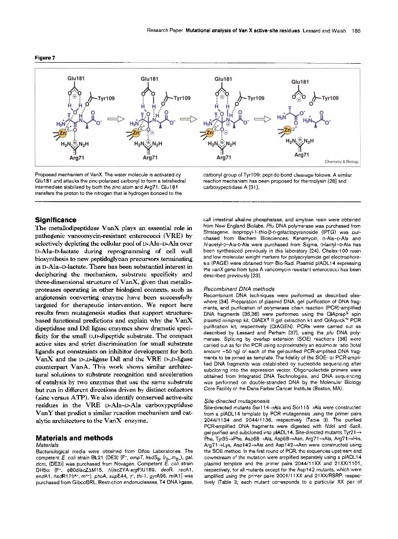
VanX and ghosphinate or n-Ala-D-Ala, there is no residue within 8.0 A of the phosphinyl or carbonyl group that could deprotonate the water nucleophile other than Glu181 (3.3 A; absolutely required for catalysis [23]) and the car- boxylate group of the substrate (3.3 A), favoring the zinc- hydroxide mechanism (Figure 7). Furthermore this residue is totally conserved in all VanX homologs identified to date [17]. In the present work we show that the Serl14 sidechain plays an important role in substrate recognition given the ZOOO-fold increase in K,, in the Serl14-+Ala mutant. For a reverse-protonation mechanism involving the carboxylate group of the D-Ala-D-Ala substrate as the catalytic base such as has been proposed for the carboxypeptidase A [32], one would expect both kinetic parameters (not just on K,,) to be affected in the Serl14-+Ala mutant. The overall con- servation of critical residues for recognition and catalytic activity between VanX, \‘anY and Ddp suggest a similar reaction mechanism for these enzymes as well.
Research Paper Mutational analysis of Van X active-site residues Lessard and Walsh 185
Figure 7
Arg71 Arg71
Chem&y & Biology
Proposed mechanism of VanX. The water molecule is activated by
Glul81 and attacks the zinc-polarized carbonyl to form a tetrahedral intermediate stabilized by both the zinc atom and Arg71. Glul81
transfers the proton to the nitrogen that is hydrogen bonded to the
carbonyl group of TyrlO9; peptide-bond cleavage follows. A similar reaction mechanism has been proposed for thermolysin [28] and
carboxypeptidase A [311.
Significance The metallodipeptidase VanX plays an essential role in pathogenic vancomycin-resistant enterococci (VRE) by selectively depleting the cellular pool of D-Ala-D-Ala over D-Ala-D-lactate during reprogramming of cell wall biosynthesis to new peptidoglycan precursors terminating in D-Ala-D-lactate. There has been substantial interest in deciphering the mechanism, substrate specificity and three-dimensional structure of VanX, given that metallo- proteases operating in other biological contexts, such as angiotensin converting enzyme have been successfully targeted for therapeutic intervention. We report here results from mutagenesis studies that support structure- based functional predictions and explain why the VanX dipeptidase and Ddl ligase enzymes show dramatic speci- ficity for the small D,D-dipeptide substrate. The compact active sites and strict discrimination for small substrate ligands put constraints on inhibitor development for both VanX and the D-,D-ligase Ddl and the VRE D-,D-ligase counterpart VanA. This work shows similar architec- tural solutions to substrate recognition and acceleration of catalysis by two enzymes that use the same substrate but run in different directions driven by distinct cofactors (zinc versus ATP). We also identify conserved active-site residues in the VRE D-Ala-D-Ala carboxypeptidase VanY that predict a similar reaction mechanism and cat- alytic architecture to the VanX enzyme.
Materials and methods Materials Bacteriological media were obtained from Difco Laboratories. The competent E. co/i strain BL21 (DE3) [F-, ompT, hsdS,, (r,..,m,-), gal,
dcm, (DE3)] was purchased from Novagen. Competent E. co/i strain DH5o [F-, $8Od/acZAM15, A(/acZYA-argF)U169, deoR, recA1,
endAl, hsdR17(+, mk+), phoA, supE44, y, thi-1, gyrA96, reelAl was purchased from GibcoBRL. Restriction endonucleases, T4 DNA ligase,
calf intestinal alkaline phosphatase, and amylose resin were obtained
from New England Biolabs. Pfu DNA polymerase was purchased from Stratagene. Isopropyl-l -thio-P-D-galactopyranoside (IF’TG) was pur-
chased from Bachem Biosciences. Kanamycin, D-Ala-o-Ala and
N-acetylbo-Ala-o-Ala were purchased from Sigma. o-lactyl-D-Ala has been synthesized previously in this laboratory [24]. Chelex-100 resin
and low molecular weight markers for polyacrylamide gel electrophore- sis (PAGE) were obtained from Bio-Rad. Plasmid plADLl4 expressing
the vanX gene from type A vancomycin resistant enterococci has been described previously [23].
Recombinant DNA methods Recombinant DNA techniques were performed as described else- where [341. Preparation of plasmid DNA, gel purification of DNA frag-
ments, and purification of polymerase chain reaction (PCR)-amplified
DNA fragments [35,361 were performed using the CIAprepe spin plasmid miniprep kit, QIAEXs II gel extraction kit and QIAquick”” PCR purification kit, respectively (QIAGEN). PCRs were carried out as
described by Lessard and Perham [371, using the pfu DNA poly-
merase. Splicing by overlap extension (SOE) reactions [38] were carried out as for the PCR using approximately an equimolar ratio (total
amount -50 ng) of each of the gel-purified PCR-amplified DNA frag-
ments to be joined as template. The fidelity of the SOE- or PCR-ampli- fied DNA fragments was established by nucleotide sequencing after
subcloning into the expression vector. Oligonucleotide primers were obtained from Integrated DNA Technologies, and DNA sequencing
was performed on double-stranded DNA by the Molecular Biology Core Facility of the Dana Farber Cancer Institute (Boston, MA).
Site-directed mutagenesis Site-directed mutants Serl14-+Ala and Serll5AAla were constructed from a plADLl4 template by PCR mutagenesis using the primer pairs
2044/l 134 and 2044/l 136, respectively (Table 3). The purified PCR-amplified DNA fragments were digested with Ndel and Sacll,
gel-purified and subcloned into plADLl4. Site-directed mutants Tyr21+
Phe, Tyr35+Phe, AspBB+Ala, Asp68+Asn, Arg71 -+Ala, Arg71jHi.s Arg7 1 -+Lys, Asp1 42-+Ala and Asp1 42-+Asn were constructed using
the SOE method. In the first round of PCR, the sequences upstream and downstream of the mutation were amplified separately using a plADLl4
plasmid template and the primer pairs 2044/l 1 XX and 21Xx/l 101,
respectively, for all mutants except for the Asp1 42 mutants, which were amplified using the primer pairs 2001/l 1Xx and 21XX/RSRP, respec- tively (Table 3; each mutant corresponds to a particular XX pair of
166 Chemistry & Biology 1999, Vol 6 No 3
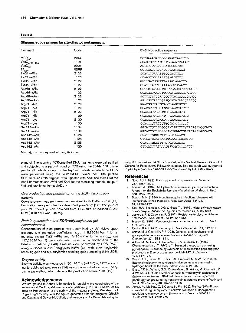
Table 3
Oligonucieotide primers for site-directed mutagenesis.
Comment Code 5’-3’ Nucleotide sequence
MB”,,,, VanXlntRD145A
VanXlntF T7,eml Tvr21 +Phe
Tir21 +Phe Tyr35+Phe
Tyr35+Phe Asp68+Ala
Asp68+Ala
Asp68-+Asn Asp68+Asn
Arg7 1 +Ala Arg71 +Ala
Arg71 +His
Arg7l+His
Arg71 -+Lys Arg71 -+Lys
Serll4-tAla
Serl 15-+Ala Asp1 42-+Ala
Asp1 42+Ala Asp1 42-+Asn
Asp1 42+Asn
2044 CCTGAAAGACGCGCAGACTAATTCG 1101 AGAGCGTTCAOCCATAAAATCWIATC 2001 ACTGTTTTATGCAATGGGCTGC
RSRP CGTGAACCATCACCCTAATCAAG 2126 GGACGCTZ'AATTTGCCACTTGG 1126 CCAAGTGGCAAATTTAGCGTCC 2127 GGTTGACGGTTZTGAAGTAAATCG 1127 CGATTTACTTCAA?+ACCGTCAACC 2122 GCTTCTATGGGCAGGTTACCGTCCTAAGC 1122 GGACGGTAACCTGCCCATAGAAGCAATCC 2123 GCTTCTATGGAACGGTTACCGTCCTAAGC 1123 GGACGGTAACCGTECATAGAAGCAATCC 2128 GGACGGTTACGCKCTAAGCGTGC 1128 GCACGCTTAGGAGCGTAACCGTCCC 2129 GGACGGTTACCATCCTAAGCGTG 1129 GCACGCTTAGGAZGGTAACCGTCCC 2130 GGACGGTTACMOCCTAAGCGTGCT 1130 GCACGCTTAGGCTlGTAACCGTCCC 1134 GGCACTGCCGCGGCTATGGCTTGC'I'TTTGAAGCCACG 1136 GGCACTGCCGCGGCTATGGGJCTGATTTTGUIGCCACG 2124 CGATTTGCCTTTATGGATGAACG 1124 GTTCATCCATX-bX!5ZAAATCGGCTCC 2125 CGATTTAACTTTATGGATGAACG 1125 GTTCATCCATAAAOTTAAATCGGCTCC
Mismatch mutations are bold and italicized.
primers). The resulting PCR-amplified DNA fragments were gel purified and subjected to a second round of PCR using the 2044/l 101 primer
pair for all mutants except for the Asp142 mutants, in which the PCRs were performed using the 200l/RSRP primer pair. The purified
SOE-amplified DNA fragment was digested with Sacll and HindIll for the
Asp1 42 mutants and Ndel and Sacll for the remaining mutants, gel puri-
fied and subcloned into a plADLl4.
Overproduction and purification of the MBP-VanX fusion
mutants Overexpression was performed as described in McCafferty et a/. [23].
Purification was performed as described previously [17]. The yield of pure MBP-VanX protein obtained from 1 I culture of induced f. co/i
BL21 (DE3) cells was -40 mg.
Protein quantitation and SDS-polyacrylamide gel electrophoresis Concentration of pure protein was determined by UV-visible spec- troscopy and extinction coefficients (Earn, 1 18,720 M-‘cm-’ for all
mutants, except TyrPl+Phe and Tyr35dPhe for which &280 was
1 17,230 M-‘cm-‘) were calculated based on a modification of the Edelhoch method [39,40]. Proteins were separated by SDS-PAGE
using a discontinuous Tris/giycine buffer [41] with loo/, acrylamide resolving gels and 5010 acrylamide stacking gels containing 0.1 O/o SDS.
Enzyme activity Enzyme activity was measured in 50 mM Tris (pH 8.0) at 37°C accord- ing to published procedures [14] using the modified cadmium-ninhy-
drin assay method, which detects the production of free o-Ala [421.
Acknowledgements We are grateful to Abbott Laboratories for providing the coordinates of the enterococcal VanX crystal structure and particularly to Dirk Bussiere for his input on interpretation of the activity of the mutants proteins. We also thank Roger Flugel for help with the figures generated with the programs GRASP and Quanta and Dewey McCafferty and members of the Walsh laboratoty for
insightful discussions. I.A.D.L. acknowledges the Medical Research Council of Canada for Postdoctoral Fellowshtp support. This research was supported in part by a grant from Abbott Laboratories and by NIH GM21 643.
References 1.
2.
Neu, H.C. (1992). The crisis in antibiotic resistance. Science 257, 1064-1073.
3.
4.
5.
6.
7. 6.
9.
10.
11
12
Tomasz, A. (1994). Multiple-antibiotic-resistant pathogenic bacteria. A report on the Rockefeller University Workshop. N. En.ql. J. Med. 330: 1247-1251. Swartz, M.N. (1994). Hospital-acquired infections: diseases with increasingly limited therapies. Proc. Nat/ Acad. SC;. USA 91, 2420-2427. Kirst, H.A., Thompson, D.G. & Nicas, T.I. (1998). Historical yearly usage of vancomycin. Antimicrob. Agents Chemother. 42, 1303-l 304. Leclercq, R. & Courvalin, P. (1997). Resistance to glycopeptides in enterococci. C/in. infect. Dis. 24, 545-554. Murray, E. (1997). Vancomycin-resistant enterococci. Am. J. Med. 102, 284-293. Cunha, B.A. (1995). Vancomycin. Med. C/in. N. Am. 19, 817-831. Arthur, M. & Courvalin, P. (1993). Genetics and mechamsms of glycopeptlde resistance in enterococci. Antimicroob. Agents Chemother. 37, 1563-l 571. Arthur, M., Molinas, C., Depardieu, F. & Courvalm. P. (1993). Characterization of Tn 7546, a Tn3-related transposon conferring glycopeptlde resistance by synthesis of depsipeptide peptidoglycan precursors in fnterococcus faecium BM4147. 1. Bacterial. 175, 117-l 27. Walsh, CT., Fisher, S.L., Park, I.-S., Prahalad, M. & Wu, Z. (1996). Bacterial resistance to vancomycin: five genes and one missing hydrogen bond tell the story. Chem. B/o/. 3, 21-28. Bugg, T.D.H., Wright, G.D., Dutka-Malen, S., Arthur, M., Courvalin, P. &Walsh, C.T. (1991). Molecular basis for vancomycin resistance in Entefococcus faecium BM4147: biosynthesis of a depsipeptide peptidoglycan precursor by vancomycin resistance protetns VanH and VanA. Biochemistry 30, 10408-l 0415. Arthur, M., Molinas, C. & Courvalin, P. (1992). The Vans-VanR two- component regulatory system controls synthesis of depsipeptide peptidoglycan precursors In Enterococcus faecium BM4147. J. Bacterial. 174, 2582-2591.
Research Paper Mutational analysis of Van X active-site residues Lessard and Walsh 187
13.
14.
15.
16.
17.
18.
19.
20.
21.
22.
23.
24.
25.
26.
27.
28.
29.
30.
31.
32.
33.
34.
Reynolds, P.E., Depardieu, F., Dutka-Malen, S., Arthur, M. & Courvalin, P. (1994). Glycopeptide resistance mediated by enterococcal transposon Tnl546 requires production of VanX for hydrolysis of D-alanyl-p-alanine. Mol. Microbial. 13, 1065-I 070. Wu, Z., Wright, G.D. &Walsh, C.T. (1995). Overexpression, purification, and characterization of VanX, a D;D-dipeptidase which is essential for vancomycin resistance in Enterococcus faecium BM4147. Biochemistry 34, 2455-2463. Reynolds, P.E. (1998). Control of peptidoglycan synthesis in vancomycin-resistant enterococci: D,D-peptidases and D,D-carboxypeptidases. Cell Mol. Life SC;. 54, 325-331. Marshall, C.G., Lessard, I.A.D., Park, I.-S. &Wright, G.D. (1998). Glycopeptide antibiotic genes in glycopeptide-producing organisms. Antimicrob. Agents Chemother. 42, 2215-2220. Lessard, I.A.D. et al., & Walsh, C.T. (1998). Homologs of the vancomycin resistance D-Ala-D-Ala dipeptidase VanX in Streptomyces toyocaensis, Escherichia co/i and Synechocystis: attributes of catalytic efficiency, stereoselectivity and regulation with implications for function. Chem. i3iol. 5,469.504. Marshall, C.G. &Wright, G.D. (1997). The glycopeptide antibiotic producer Streptomyces toyocaensis NRRL 15009 has both D-afanyf- D-afanine and D-alar@D-iaCtate ligases. EMS Micriobiol. Left. 157, 295-299. Bussiere, D.E., Pratt, S.D., Katz, L. Severin, J.M., Holzman, T. & Park, C. (1998). The structure of VanX reveals a novel amino- dipeptidase involved in mediating transposon-based vancomycin resistance. Mol. CeN 2, 75-84. Fan, C., Moews, P.C., Walsh, CT. & Knox, J.R. (1994). Vancomycin resistance: structure of D-afanine-D-aianine ligase at 2.3 A resolution. Science 266, 439-443. Dideberg, O., Charlier. P., Dive, G., Joris, B., Frere, J.M. & Ghuysen, J.M. (1982). Structure of a Zn2+- containing D-alanyl-D-alanine-CleaVing
carboxypeptidase at 2.5 A resolution. Nature 299, 469-470. Wu, Z. &Walsh, CT. (1995). Phosphinate analogs of D-,D- dipeptides: slow-binding inhibition and proteolysis protection of VanX, a D-,D-dipeptidase required for vancomycin resistance in Enterococcus faecium. Proc. Nat/ Acad. SC;. USA 92, 11603-l 1607. McCafferty, D.G., Lessard, I.A.D. &Walsh, CT. (1997). Mutational analysis of potential zinc-binding residues in the active site of the enterococcal D-Ala-o-Ala dipeptidase VanX. Biochemistry 36, 10498-l 0505. Park, I.-S., Lin, C.H. & Walsh, CT. (1996). Gain of D-alanyj-D-lactate or D-dactyl-D-ajanine synthetase activities in three active-site mutants of the Escherichia co/i D-alanyf-D-alanine ligase B. Biochemistry 35, 10464-l 0471. Shi, Y. & Walsh, CT. (1995). Active site mapping of Escherichia co/i o-Ala-D-Ala ligase by structured-based mutagenesis. Biochem/stry 34, 2786-2776. Arthur, M., Depardieu, F., Cabanie, L., Reynolds, P. & Courvalin, P. (1998). Requirement of the VanY and VanX D,D-peptidases for glycopeptide resistance in enterococci. Antimicrob. Agents Chemother. 36, 1514-1518. Wright, G.D., Molinas, C., Arthur, M., Courvalin, P., &Walsh, C.T. (1992). Characterization of VanY, a D,D-carboxypeptidase from vancomycin-resistant Enterococcus faecium BM4147. Antimicrob. Agents Chemother. 36, 1514-l 518. Holden, H.M., Tronrud, D.E., Monzingo, A.F., Weaver, L.H. & Matthews, B.W. (1987). Slow- and fast-binding inhibitors of thermolysin display different modes of binding: crystallographic analysis of extended phosphonamidate transition-state analogues. Biochemistry 26,8542-8553. Izquierdo-Martin, M. & Stein, R.L. (I 992). Mechanistic studies on the inhibition of thermolysin by a peptide hydroxamic acid. J. Am. Chem. Sot. 114, 325-331. Mock, W.L. & Stanford, D.J. (1996). Arazoformyl dipeptide substrates for thermolysm. Confirmation of a reverse protonation catalytic mechanism. Biochemistry 35, 7369-7377. Christianson, D.W. & Lipscomb, W.N. (1989). Carboxypeptidase A. Accounts Chem. Res. 22,62-69. Mock, W.L. & Zhang, J.Z. (1991). Mechanistically significant diastereoselection in the sulfoximine inhibition of carboxypeptidase A. 1. fliol. Chem. 266, 6393-6400. Lipscomb, W.N. & Strater, N. (1996). Recent advances in zinc enzymology. Chem. Rev. 96, 2375-2433. Sambrook, J. Fritsch, E.F. & Maniatis, T. (1969). Molecular Cloning: A Laboratory Manual, 2nd Ed. Cold Spring Harbor Laboratory, Cold Spring Harbor, NY.
35.
36.
37.
38.
39.
40.
41.
42.
43.
44.
Saiki, R.K., et al., & Arnheim, N. (1985). Enzymatic amplification of beta-globin genomic sequences and restriction site analysis for diagnosis of sickle cell anemia. Science 230, 1350-l 354. Saiki, R.K., et al., & Erlich, H.A. (1988). Primer-directed enzymatic amplification of DNA with a thermostable DNA polymerase. Science 239, 487-491. Lessard, I.A.D. & Perham, R.N. (1994). Expression in Eschenchia co/i of genes encoding the Ela and El 8 subunits of the pyruvate dehydrogenase complex of Bacillus stearothermophilus and assembly of a functional El component (a2 p2) in vitro. J. Biol. Chem. 269, 10378-l 0383. Ho, S.N., Hunt, H.D., Horton, R.M., Pullen, J.K. &Pease, L.R. (1989). Sate-directed mutagenesis by overlap extension using the polymerase chain reaction. Gene 77, 51-59. Edelhoch, H. (1967). Spectroscopic determination of tryptophan and tyrosine in proteins. Biochemistry 6, 1946-l 954. Pace, C.N., Vajdos, F. Fee, L., Grimsley, G. & Gray, T. (1995). How to measure and predict the molar absorption coefficient of a protein. Protein SC;. 4, 241 l-2423. Laemmli, U.K. (1970). Cleavage of structural proteins during the assembly of the head of bacteriophage T4. Nature 227, 680-665. Doi, E., Shibata, D. & Matoba, T. (1981). Modified calorimetric ninhydrin methods for peptidase assay. Anal. Biochem. 116, 173-I 84. Nicholls, A., Sharp, K.A. & Honig, B. (1991). Protein folding and association: insights from the interfacial and thermodynamic properties of hydrocarbons. Proteins 11, 57-68. Thompson, J.D., Higgins, D.G. & Gibson, T.J. (1994). CLUSTAL W: improving the sensibility of progressive multiple sequence alignment through sequence weighting, position-specific gap penalties and weight matrix choice. Nucleic Acids. Res. 22, 4673-4680.
Because Chemistry & Biology operates a ‘Continuous
Publication System’ for Research Papers, this paper has been
published via the internet before being printed. The paper can
be accessed from http://biomednet.wmkbiology/cmb - for
further information, see the explanation on the contents pages.


![Total Synthesis and Evaluation of [ψ[CH 2 NH]Tpg 4 ] Vancomycin Aglycon: Reengineering Vancomycin for Dual D -Ala- D - Ala and D -Ala- D -Lac Binding Brendan](https://img.pdfslide.net/doc/110x75/56649d1b5503460f949f12a1/total-synthesis-and-evaluation-of-ch-2-nhtpg-4-vancomycin-aglycon-reengineering.jpg)


![l*10B21agæ c D ífi D & a a a S AL Ala & 0000 0 00000 ... · yo.jp/ l*10B21agæ c D ífi D & a a a S AL Ala & 0000 0 00000 • :sunori ok tsunori.net/ [Twitter]@mit ñDßfiííŽ](https://img.pdfslide.net/doc/110x75/5d2a5dc688c993d8288d02dc/l10b21agae-c-d-ifi-d-a-a-a-s-al-ala-0000-0-00000-yojp-l10b21agae.jpg)