Embed Size (px)
Citation preview
HAL Id: hal-02087386https://hal.umontpellier.fr/hal-02087386
Submitted on 2 Apr 2019
HAL is a multi-disciplinary open accessarchive for the deposit and dissemination of sci-entific research documents, whether they are pub-lished or not. The documents may come fromteaching and research institutions in France orabroad, or from public or private research centers.
L’archive ouverte pluridisciplinaire HAL, estdestinée au dépôt et à la diffusion de documentsscientifiques de niveau recherche, publiés ou non,émanant des établissements d’enseignement et derecherche français ou étrangers, des laboratoirespublics ou privés.
Mycobacterium marinum MgtC Plays a Role inPhagocytosis but is Dispensable for Intracellular
MultiplicationClaudine Belon, Laïla Gannoun-Zaki, Georges Lutfalla, Laurent Kremer,
Anne-Béatrice Blanc-Potard
To cite this version:Claudine Belon, Laïla Gannoun-Zaki, Georges Lutfalla, Laurent Kremer, Anne-Béatrice Blanc-Potard.Mycobacterium marinum MgtC Plays a Role in Phagocytosis but is Dispensable for IntracellularMultiplication. PLoS ONE, Public Library of Science, 2014, 9 (12), pp.e116052. �10.1371/jour-nal.pone.0116052�. �hal-02087386�
RESEARCH ARTICLE
Mycobacterium marinum MgtC Plays aRole in Phagocytosis but is Dispensable forIntracellular MultiplicationClaudine Belon1,2, Laıla Gannoun-Zaki1,2, Georges Lutfalla1,2, Laurent Kremer1,2,3,Anne-Beatrice Blanc-Potard1,2*
1. Laboratoire de Dynamique des Interactions Membranaires Normales et Pathologiques, UniversitesMontpellier 2 et 1, Place Eugene Bataillon, 34095, Montpellier, Cedex 05, France, 2. Centre National de laRecherche Scientifique, UMR5235, Montpellier, France, 3. Institut national de la sante et de la recherchemedicale, Montpellier, France
Abstract
MgtC is a virulence factor involved in intramacrophage growth that has been
reported in several intracellular pathogens, including Mycobacterium tuberculosis
and Salmonella enterica serovar Typhimurium. MgtC participates also in adaptation
to Mg2+ deprivation. Herein, we have constructed a mgtC mutant in Mycobacterium
marinum to further investigate the role of MgtC in mycobacteria. We show that the
M. marinum mgtC gene (Mma mgtC) is strongly induced upon Mg2+ deprivation and
is required for optimal growth in Mg2+-deprived medium. The behaviour of the Mma
mgtC mutant has been investigated in the Danio rerio infection model using a
transgenic reporter zebrafish line that specifically labels neutrophils. Although the
mgtC mutant is not attenuated in the zebrafish embryo model based on survival
curves, our results indicate that phagocytosis by neutrophils is enhanced with the
mgtC mutant compared to the wild-type strain following subcutaneous injection.
Increased phagocytosis of the mutant strain is also observed ex vivo with the
murine J774 macrophage cell line. On the other hand, no difference was found
between the mgtC mutant and the wild-type strain in bacterial adhesion to
macrophages and in the internalization into epithelial cells. Unlike the role reported
for MgtC in other intracellular pathogens, Mma MgtC does not contribute
significantly to intramacrophage replication. Taken together, these results indicate
an unanticipated function of Mma MgtC at early step of infection within phagocytic
cells. Hence, our results indicate that although the MgtC function is conserved
among pathogens regarding adaptation to Mg2+ deprivation, its role towards
phagocytic cells can differ, possibly in relation with the specific pathogen’s
lifestyles.
OPEN ACCESS
Citation: Belon C, Gannoun-Zaki L, Lutfalla G,Kremer L, Blanc-Potard A-B (2014) Mycobacteriummarinum MgtC Plays a Role in Phagocytosis but isDispensable for Intracellular Multiplication. PLoSONE 9(12): e116052. doi:10.1371/journal.pone.0116052
Editor: Jerome Nigou, Centre National de laRecherche Scientifique - Universite de Toulouse,France
Received: September 18, 2014
Accepted: December 4, 2014
Published: December 29, 2014
Copyright: � 2014 Belon et al. This is an open-access article distributed under the terms of theCreative Commons Attribution License, whichpermits unrestricted use, distribution, and repro-duction in any medium, provided the original authorand source are credited.
Data Availability: The authors confirm that all dataunderlying the findings are fully available withoutrestriction. All relevant data are within the paperand its Supporting Information files.
Funding: French National Agency (ANRZebraFlam) and European Community’s SeventhFramework Programme (FP7-PEOPLE-2011-ITN)under Grant Agreement PITN-GA-2011- 289209 forthe Marie-Curie Initial Training NetworkFishForPharma. CB is supported by a MRTfellowship from the French Ministry of Researchand the Fondation for Medical Research (FRMFDT20140930905). The funders had no role instudy design, data collection and analysis, decisionto publish, or preparation of the manuscript.
Competing Interests: Laurent Kremer is a PLOSONE Editorial Board member. This does not alterthe authors’ adherence to PLOS ONE Editorialpolicies and criteria.
PLOS ONE | DOI:10.1371/journal.pone.0116052 December 29, 2014 1 / 23
Introduction
MgtC is a virulence factor common to several intracellular pathogens [1]. It was
first described in Salmonella enterica serovar Typhimurium (S. Typhimurium) as
required for intramacrophage multiplication and systemic infection in mice [2–4].
Later, it was described as a critical factor for the intramacrophage growth of
Mycobacterium tuberculosis, Brucella suis, Yersinia pestis, Burkholderia cenocepacia
and Salmonella enterica serovar Typhi [5–9]. Despite its importance in the
virulence of various bacterial pathogens, the mechanism by which MgtC promotes
intracellular growth remains unknown. Salmonella MgtC has been recently shown
to directly interact with bacterial F1Fo ATP synthase, thereby altering its ability to
translocate protons and to couple translocation to ATP synthesis [10]. Hence,
modulation of F-ATP synthase activity is proposed to drive the ability of MgtC to
promote intramacrophage replication.
MgtC has been associated to adaptation to low Mg2+ environments in broth
media in the various pathogens mentioned above, based on the observation that
growth of the corresponding mgtC mutants is impaired in Mg2+ deprived media
[1]. Moreover, expression of mgtC gene is highly induced by deprivation of
external Mg2+ concentration in S. Typhimurium and Y. pestis [11, 12], which are
two organisms where mgtC is cotranscribed with the Mg2+ transporter encoded by
mgtB. However, mgtC has been shown to be only slightly induced by Mg2+
limitation in M. tuberculosis [13, 14], suggesting that Mg2+ may regulate MgtC
only in bacterial species where it is cotranscribed with a Mg2+ transporter.
MgtC is a membrane-associated protein that harbors a conserved hydrophobic
N-terminal and a more divergent soluble C-terminal domain that exhibits
conservation among proteins from intracellular pathogens as S. Typhimurium
and M. tuberculosis [15]. MgtC has been shown to play a role in M. tuberculosis
virulence in macrophage and mice infection models [5] but the contribution of
mgtC in mycobacterial physiology and virulence needs further investigation.
Because MgtC is conserved between M. tuberculosis and Mycobacterium marinum,
we aimed to address its role in M. marinum virulence, as well as its regulation by
magnesium. M. marinum is closely related to M. tuberculosis not only in its
pathology but also genetically [16, 17], and has been increasingly used as a suitable
model for understanding the pathogenesis of tuberculosis. As a natural host for
this pathogen, zebrafish provides a powerful vertebrate model to study M.
marinum pathogenesis. Moreover, because of their genetic tractability and optical
transparency, zebrafish embryos have been successfully used to investigate host-
bacteria interactions and the role of innate immunity during M. marinum
infection [18–20].
The present results indicate that Mma mgtC transcription is strongly induced
by magnesium deprivation and, unexpectedly, that Mma MgtC appears
dispensable for intramacrophage replication but plays a role in phagocytosis, a
phenotype first uncovered in zebrafish embryos.
Role of M. marinum MgtC in Phagocytosis
PLOS ONE | DOI:10.1371/journal.pone.0116052 December 29, 2014 2 / 23

Materials and Methods
Bacterial strains and growth culture conditions
Mycobacterium marinum M and Mycobacterium smegmatis mc2155 strains were
grown at 30 C and 37 C, respectively, in Sauton’s medium containing 0.025% of
tyloxapol (Sigma) or on Middlebrook 7H10 agar plates supplemented with 10%
Oleic acid-Albumin-Dextrose-Catalase (OADC) enrichment, in the presence of
kanamycin (25 mg/ml), hygromycin (80 mg/ml) and zeocin (25 mg/ml), when
required. Low magnesium medium was obtained by replacing the magnesium
sulfate in the Sauton’s medium by a similar concentration of potassium sulfate.
Escherichia coli (DH5a) was used for cloning and was grown in LB medium with
zeocin (25 mg/ml) at 37 C.
RNA extraction and qRT-PCR
RNA was prepared from 5 ml of mid-logarithmic bacterial cultures (grown in
Sauton’s medium containing or not magnesium). Bacteria were harvested,
resuspended in 1 ml of RNA protect reagent (Qiagen) and incubated for 1 h at
room temperature. Bacteria were centrifuged and resuspended in 1 ml of RLT
buffer from RNeasy Mini kit (Qiagen), transferred in a Lysing matrix B tube (MP
Bio) and disrupted with a bead-beater apparatus (3 times, 45 sec, maximal speed).
RNA was purified with the RNeasy kit, according to manufacturer’s instructions.
DNA was further removed using DNAseI (Invitrogen). cDNA was produced using
Superscript III reverse transcriptase (Invitrogen). Controls without reverse
transcriptase were done on each RNA sample to rule out DNA contamination.
Quantitative real-time PCR was performed using an in-house SYBR Green mix
and a 480 light cycler instrument (Roche). PCR conditions were as follows: 3 min
denaturation at 98 C, 45 cycles of 98 C for 5 sec, 68 C for 10 sec and 72 C for
10 sec. The sigA gene (MMAR_2011) was used as internal control. The sequences
of primers used for qRT-PCR are listed in S1 Table.
Construction of M. marinum mgtC mutant and complemented
strains
A strain deleted for the mgtC gene was constructed in M. marinum M using
mycobacteriophage-mediated allelic exchange to replace mgtC by a hygromycin
cassette. The construction of the allelic exchange substrate (AES) phasmid, the
preparation of phage and the phage transduction was performed as described
[21]. One kb long sequences upstream and downstream of mgtC were cloned into
the pJSC347 on both sides flanking the hygromycin cassette. The mutant was
checked by PCR and southern blot using probes corresponding to the mgtC and
hygromycin genes. Complementation of the mgtC mutant was performed using a
chromosomal copy of the mgtC gene placed under the control of its own
promoter. A DNA fragment including the mgtC gene and 840 bp upstream
(which contains the intergenic region between MMAR_2685 and MMAR_2686 as
well as MMAR_2687 gene) was amplified by PCR using primers pMV306-XbaI-59
Role of M. marinum MgtC in Phagocytosis
PLOS ONE | DOI:10.1371/journal.pone.0116052 December 29, 2014 3 / 23

and pMV306-HindIII-39 and was cloned at the XbaI and HindIII sites of the
integrative vector pMV306 [22]. The resulting plasmid was electroporated in the
mgtC mutant to integrate the mgtC gene with its upstream sequences at the
chromosomal attB site with selection on 7H10 agar plates containing kanamycin.
As a control, the empty pMV306 vector was also electroporated into the wild-type
strain and the mgtC mutant strain.
Zebrafish embryos injections and microscopy
Experiments were performed using the golden zebrafish mutant [23] or the
Tg(mpx:EGFP) where GFP is specifically expressed in neutrophils [24]. Zebrafish
were raised and maintained according to standard procedures [25]. Embryos were
obtained from pairs of adult fish by natural spawning and raised at 28.5 C in tank
water. Ages are expressed as hours or days post fertilization (hpf or dpf).
Mycobacteria from mid-log phase cultures grown in Sauton’s medium were
washed twice in PBS, resuspended in PBS and homogenized with a 26G syringe
(five times) at an OD600 of 3. Zebrafish larvae of 30 hpf (caudal vein injection) or
3 dpf (subcutaneous injection) were anesthetized by immersion in buffered
tricaine (Sigma A-5040) and manually dechorionated, if needed. For subcuta-
neous infection, embryos were fed with tetrahymenas from 5 dpf to the end of the
experiment, 3 times per week. They were injected with 1 nl of bacterial suspension
using a Tritech Research digital microINJECTOR (MINJ-D). To determine
bacterial loads in infected embryos, groups of two infected embryos were collected
and dissociated using the BD mycoprep Kit. Suspensions were homogenized with
a 26G syringe (five times) and dilutions were plated on Middlebrook 7H10
supplemented with 25 mg/ml of kanamycin to determine the colony forming units
(CFU). All quantitative results are from triplicate experiments. Wide-field, bright-
field and fluorescence live microscopy of infected embryos were performed using
an Olympus MVX10 epifluorescence microscope. Images were acquired with a
digital color camera (Olympus XC50) and processed using CellSens (Olympus).
Fluorescence filters cubes (FITC-MVX10 and TRITC-MVX10) were used to detect
green and red light, respectively. Confocal fluorescence microscopy was
performed using a Leica DM2500CSQ upright microscope with a Leica TCS SPE
confocal scan head, differential interference contrast (DIC) optics and a
SuperZGalvo SPE z-step controller. For fixed-sample observations, embryos in
50% glycerol in PBS were mounted flat onto depression transparent slides with a
coverslip and observed with a 106 Leica Apo 0.3 NA, 406 Leica Apo oil 1.15 NA
or 636 Leica Apo oil 1.33 NA objectives. Overlays of fluorescent and DIC images
and 2D reconstructions of image stacks were processed and assembled using LAS-
AF software. Final image analysis and visualization were performed using GIMP
2.8 freeware to adjust levels and brightness and to remove out-of-focus
background fluorescence.
Role of M. marinum MgtC in Phagocytosis
PLOS ONE | DOI:10.1371/journal.pone.0116052 December 29, 2014 4 / 23
Ethics statement
All animal experiments described in the present study were conducted at the
University Montpellier 2 according to European Union guidelines for handling of
laboratory animals (http://ec.europa.eu/environment/chemicals/lab_animals/
home_en.htm) and were approved by the Direction Sanitaire et Veterinaire de
l’Herault and Comite d’Ethique pour l’Experimentation Animale under reference
CEEA-LR-13007. The breeding of adult fish adhered to the international
guidelines specified by the EU Animal Protection Directive 2010/63/EU and adult
zebrafish were not sacrificed for this study. For survival curves, cardiac rhythm
was used as a clinical criteria to fix the endpoint at which embryos are euthanized
using the anaesthetic Tricaine up to a lethal dose. Condition of the infected
animals was monitored three times a day. For CFU counts, embryos were
anaesthetized with Tricaine up to a lethal dose before lysis with triton. Embryos
that survive infection were anaesthetized with Tricaine up to a lethal dose before
bleach treatment. For live imaging analysis, embryos were anaesthetized with
Tricaine. For microscopic observation of fixed samples, embryos were fixed with
4% paraformaldehyde after being anaesthetized with Tricaine up to a lethal dose.
Macrophage infection assays
J774 cells were maintained at 37 C in 5% CO2 in Dulbecco’s modified Eagle
medium (DMEM) (Gibco) supplemented with 10% fetal bovine serum (FBS)
(Gibco). J774 cells were allowed to adhere in a 24-well plate at a density of 56104
cells/well for 24 h at 37 C in 5% CO2.
For infection, M. marinum cultures grown exponentially in Sauton’s medium
(OD600 around 0.8 to 1) were centrifuged, washed in phosphate buffer saline
(PBS) and bacterial clumps were disrupted by 8 successive passages through a 26G
needle. The remaining aggregates were then eliminated with a short spin (1 min at
1,100 rpm). For experiments including the wbbl2 mutant as control, mycobacteria
were grown on Sauton’s agar plates (to induce LOS production in the wild-type
strain) containing 10% OADC enrichment for 5–7 days as described [26].
Bacterial lawns were scraped off the plates, resuspended in 1 ml of PBS and
clumps were disrupted as indicated above followed by low speed centrifugation to
eliminate aggregates. In all experiments, macrophages were infected at a
multiplicity of infection (MOI) of approximately 2. The infection was allowed to
proceed for 3 h at 32 C in 5% CO2 prior to exposure to 200 mg/ml gentamicin for
60 min to kill the remaining extracellular bacteria. Infected cells were then washed
three times with PBS. Cells were then lysed with 0.1 ml of 1% Triton X-100 in PBS
and the number of intracellular mycobacteria counted by plating appropriate
dilutions onto Middlebrook 7H10 agar plates. To evaluate the numbers of
phagocytosed bacteria, the ratio between the count of internalized bacteria and the
count of bacteria in the inoculum was determined. For kinetic experiments, earlier
infection times (0.5, 1, and 2 h) were also assessed. To evaluate the multiplication
rate, after the three washes with PBS, infected cells were incubated for 5 days in
DMEM medium supplemented with 20 mg/ml gentamicin (a concentration that
Role of M. marinum MgtC in Phagocytosis
PLOS ONE | DOI:10.1371/journal.pone.0116052 December 29, 2014 5 / 23
prevents growth of M. marinum strains) at 32 C in 5% CO2 prior to cell lysis and
bacterial enumeration. The ratio between the number of internalized bacteria at
day 5 and the number of internalized bacteria after 3 h infection was calculated.
To evaluate bacterial adherence, the same procedure was applied, except that J774
cells were incubated 30 min with 10 mg/ml cytochalasin D prior to infection or
were incubated at 4 C during the phagocytic time. All results are derived from a
least three independent experiments where strains are tested in triplicate.
To visualize and count the number of bacterium-containing macrophages, a
similar protocol was used except that macrophages were allowed to adhere on
coverslips prior to infection with fluorescent bacteria carrying the
pMV261_mCherry (Table 1). Following exposure to gentamycin, infected cells
were washed three times in PBS and labeled with fluorescein-conjugated
phalloidin (Sigma), according to the manufacturer’s instructions to visualize the
shape of the cells. Stained cells were then observed using a Zeiss Axioimager and
quantification of mycobacterium-residing cells was determined by counting 20
fields per coverslip. Each strain was analyzed in triplicate.
HeLa cells infection assays
HeLa cells were maintained at 37 C in 5% CO2 in DMEM supplemented with
10% fetal bovine serum. HeLa cells were allowed to adhere in 24-well plates at a
density of 56104 cells per well for 24 h at 37 C in 5% CO2. For infection,
mycobacteria were prepared as described above from Sauton’s agar plates because
the wbbl2 mutant was also included in the experiment. The internalization
protocol was similar to the phagocytosis protocol.
Extraction and analysis of mycobacterial apolar lipids
M. marinum strains were grown to mid-log growth phase in Sauton’s medium
containing or not magnesium sulfate and labeled with 0.5 mCi/ml
[1-14C]propionate (specific activity of 54 mCi/mmol, American Radiolabeled
Chemicals, Inc) for 24 h. The non-polar fraction was separated from the polar
fraction as described [27]. The [1-14C]-propionate-labeled apolar lipids were
extracted by adding 2 ml of CH3OH/0.3% NaCl (100/10; v/v) and 2 ml of
petroleum ether to the cell pellet followed by stirring for 1 h. After centrifugation,
the upper petroleum ether layer was removed and 2 ml of petroleum ether was
added to the lower phase. The combined petroleum ether extracts were then
evaporated under a nitrogen stream to yield apolar lipids that were resuspended in
CH2Cl2 and analyzed by TLC using silica gel plates (5735 silica gel 60F254; Merck,
Darmstadt, Germany). Equivalent amounts of lipids (20 000 cpm) were spotted
on TLC plates that were run in various solvent systems: Petroleum ether/
diethylether 9:1 (v/v) for PDIM; chloroform/methanol 19:1 (v/v) for TDM and
PGL and chloroform/methanol 99:1 (v/v) for PAT. TLCs were exposed to a Kodak
Biomax MR film for 24 h.
Role of M. marinum MgtC in Phagocytosis
PLOS ONE | DOI:10.1371/journal.pone.0116052 December 29, 2014 6 / 23
Statistical analyses
For zebrafish embryos infections, statistical analyses were performed using
GraphPad Prism 4 (Graphpad Software). Survival curves were compared between
two groups by using the logrank test. For cells infection assays, averages were
compared by using an unpaired Student t-test. Only P-values ,0.05 were
considered as statistically significant.
Results
Identification of a MgtC-like protein in M. marinum and
construction of Mma mgtC mutant strain
An MgtC-like protein is encoded by the MMAR_2687 gene in M. marinum
genome (http://mycobrowser.epfl.ch/marinolist.html). The Mma MgtC protein
shares strong similarity to the Mtb MgtC protein (82% identity, 90% similarity)
(Fig. 1A), the major difference residing in the very C-terminal end of the protein.
The sequence of Mma MgtC is also well conserved with that of S. Typhimurium
MgtC in the N-terminal half of the protein (61% identity, 73% similarity), but
more divergent in the C-terminal half (102 last a.a. 20% identity, 37% similarity).
In addition, Mma MgtC harbors four conserved residues that have been shown to
be important for Salmonella MgtC function (Fig. 1A) [28]. The genomic
organization around mgtC is conserved between M. marinum and M. tuberculosis
(Fig. 1B). In both species, mgtC is located downstream of several genes coding for
PPE proteins and is adjacent to a gene coding for a protein of unknown function
(Rv1810 or MMAR_2686). This genomic organization differs from the one of S.
Typhimurium where mgtC is in operon with a gene that encodes a Mg2+
Table 1. Bacterial strains and plasmids.
Strains, plasmids, phages Description or phenotype Source/Ref.
Mycobacterium marinum strains
Mma M wild-type [16]
Mma DmgtC DmgtC This study
Mma DmgtC+mgtC DmgtC+attB::mgtC This study
Mma Dwbbl2 Dwbbl2 [26]
Mycobacterium smegmatis strains
mc2155 Electroporation-proficient ept mutant of M. smegmatis strain mc26 [49]
Plasmid
pJSC347 Vector for cloning allelic-exchange substrates to be used for specializedtransduction, HygR
[50]
pMV306 Single-copy-integrating vector, KanR [22]
pMV306-mgtC pMV306 carrying the mgtC gene and 840 bp upstream, KanR This study
pMV261-mCherry pMV261 carrying mCherry under the control of the hsp60 promoter, ZeoR [51]
Phage
phAE159 Conditionally replicating shuttle phasmid derived from the lytic mycobacter-iophage TM4
Kind gift from W.R. Jacobs
doi:10.1371/journal.pone.0116052.t001
Role of M. marinum MgtC in Phagocytosis
PLOS ONE | DOI:10.1371/journal.pone.0116052 December 29, 2014 7 / 23
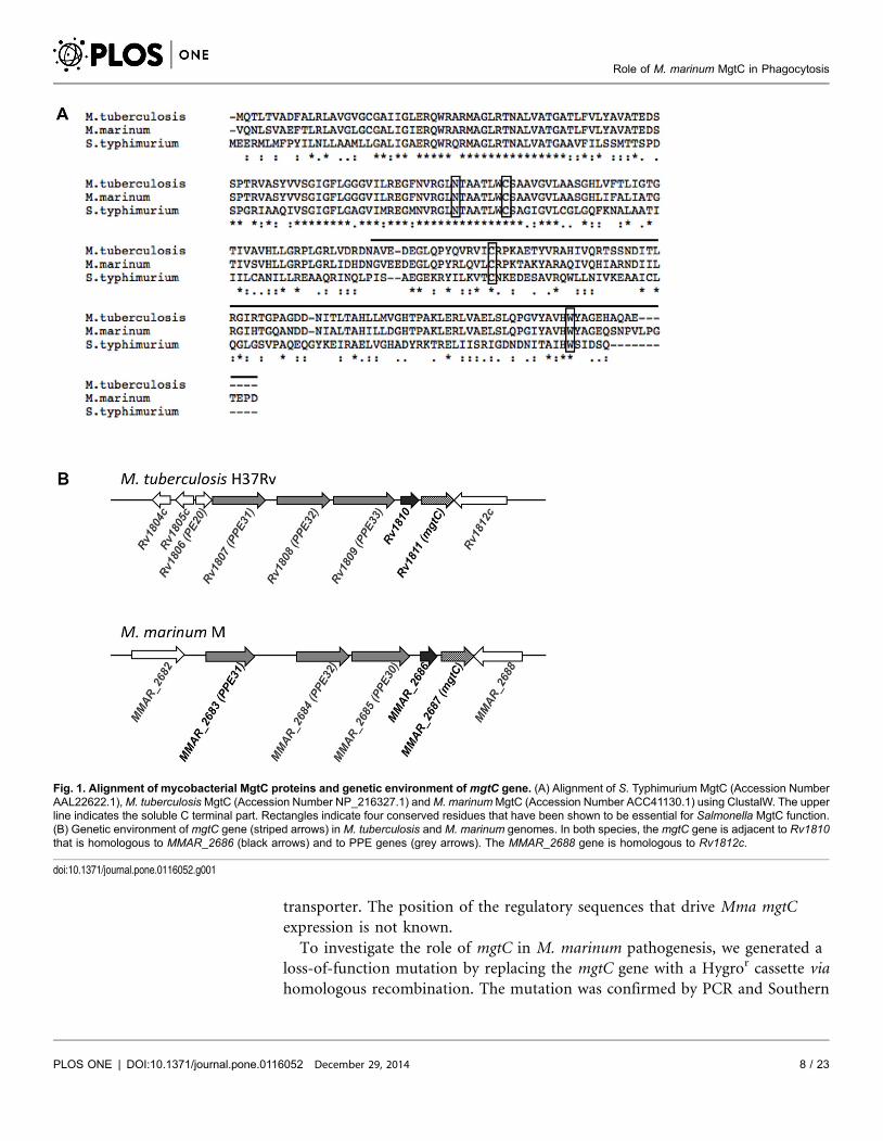
transporter. The position of the regulatory sequences that drive Mma mgtC
expression is not known.
To investigate the role of mgtC in M. marinum pathogenesis, we generated a
loss-of-function mutation by replacing the mgtC gene with a Hygror cassette via
homologous recombination. The mutation was confirmed by PCR and Southern
Fig. 1. Alignment of mycobacterial MgtC proteins and genetic environment of mgtC gene. (A) Alignment of S. Typhimurium MgtC (Accession NumberAAL22622.1),M. tuberculosisMgtC (Accession Number NP_216327.1) andM. marinumMgtC (Accession Number ACC41130.1) using ClustalW. The upperline indicates the soluble C terminal part. Rectangles indicate four conserved residues that have been shown to be essential for Salmonella MgtC function.(B) Genetic environment of mgtC gene (striped arrows) in M. tuberculosis and M. marinum genomes. In both species, the mgtC gene is adjacent to Rv1810that is homologous to MMAR_2686 (black arrows) and to PPE genes (grey arrows). The MMAR_2688 gene is homologous to Rv1812c.
doi:10.1371/journal.pone.0116052.g001
Role of M. marinum MgtC in Phagocytosis
PLOS ONE | DOI:10.1371/journal.pone.0116052 December 29, 2014 8 / 23
blotting (S1 Fig.). This mutation was complemented by introducing in the
chromosome the wild-type mgtC gene as well as upstream sequence at the
bacterial att site.
Regulation of Mma mgtC expression by Mg2+
and growth of mgtCmutant in Mg
2+deprived medium
MgtC is highly induced by low Mg2+ concentrations in S. Typhimurium [11]. In
M. tuberculosis, Mg2+ deprivation only slightly induced the mgtC gene (1.5 fold)
whereas genes upstream of mgtC (Rv1806 through Rv1809) are clearly induced
[13, 14]. M. marinum M strain was grown in Sauton’s medium with or without
Mg2+ and RNA was extracted to monitor the expression of mgtC along with two
upstream genes: MMAR_2686 that is located immediately upstream mgtC and
MMAR_2683 (PPE31), which is the first of the PPE genes. RT-PCR experiments
indicated that expression of all three genes is highly induced by Mg2+ deprivation
(Fig. 2A) whereas the control gene sigA is similarly transcribed in both conditions.
Quantitative RT-PCR using sigA gene as internal control indicated an induction
level by low Mg2+ of about 30 fold for PPE31 and mgtC (Fig. 2B). The induction
rate of MMAR_2686 is lower (about 5 fold), due to higher endogenous expression
in high Mg2+ medium.
RNA extraction was also performed from mgtC mutant and complemented
strain, to test the expression of mgtC from an ectopic location. As anticipated, the
mgtC gene is not expressed in the mgtC mutant (whereas PPE31 and MMAR_2686
are expressed and regulated similarly than in the wild-type context) (Fig. 2). The
mgtC gene is expressed and regulated by Mg2+ in the complemented strain to a
level similar to the one found in the wild-type strain. This result demonstrates that
mgtC is properly expressed and regulated at the attB locus in the complemented
strain. Thus, upstream sequences present in the complementation vector (i.e
included in the 840 bp upstream mgtC) are sufficient for Mg2+ regulation of
mgtC.
The growth rate of the mgtC mutant was evaluated in liquid cultures. The
mutant shows a slight growth defect at late exponential phase in Mg2+-deprived
broth medium (Fig. 3A), but not in medium supplemented with Mg2+ (Fig. 3B).
As expected, the complemented strain behaves similarly to the wild-type strain in
Mg2+-deprived medium, confirming the proper expression of the mgtC gene at the
attB locus.
Together, these data indicate that MgtC is induced by Mg2+ deprivation and
required for optimal growth in Mg2+-deprived medium in M. marinum. The
results allowed validating the complementation of mgtC mutant by an
extrachromosomal copy of the gene with its upstream DNA sequence.
Role of M. marinum MgtC in Phagocytosis
PLOS ONE | DOI:10.1371/journal.pone.0116052 December 29, 2014 9 / 23

Behaviour of mgtC mutant upon intravenous infection in zebrafish
embryos
Studies were undertaken using the zebrafish infection model to probe the
pathogenicity of the Mma mgtC mutant. MmaM, DmgtC mutant and
DmgtC+attB::mgtC strains were transformed with pMV261_mCherry (S1 Table)
and red fluorescent mycobacteria were injected intravenously (iv) in the Caudal
Haematopoietic Tissue (CHT) in 30 hpf embryos. In this biological system, iv-
injected mycobacteria are rapidly phagocytosed by circulating macrophages [29].
The infected embryos were monitored for survival and bacterial loads at different
time points. The survival curves indicated that the virulence of the mgtC mutant is
not significantly different from the parental Mma M or the complemented strains
Fig. 2. Expression of Mma mgtC and upstream genes in high Mg2+ and low Mg2+ conditions. (A) RT-PCR experiment on RNA isolated from M.marinum strains grown in high or low Mg2+ with primers specific for mgtC, MMAR_2686, MMAR_2683 (PPE31) and sigA. Experiment was carried out withwild-type strain, mgtC mutant strain and complemented strain. Controls where reverse transcriptase was omitted (indicated by RT -) are done to verify theabsence of genomic DNA contamination in the RNA sample. The sigA gene is used as control. (B) Quantification of mgtC, MMAR_2686 and MMAR_2683RNA by Q-RT-PCR experiment using RNA isolated from M. marinum strains grown in high or low Mg2+. The sigma factor sigA was used as an internalstandard. Results are expressed as means+standard deviations (SD) from a representative experiment performed in triplicate.
doi:10.1371/journal.pone.0116052.g002
Role of M. marinum MgtC in Phagocytosis
PLOS ONE | DOI:10.1371/journal.pone.0116052 December 29, 2014 10 / 23

(Fig. 4A). The bacterial loads after 3 dpi or 5 dpi were slightly lower with the
mgtC mutant, since less CFU were counted in embryos injected with the mutant
strain comparatively to the wild-type strain (Fig. 4B). The number of neutrophils
Fig. 3. Growth of Mma mgtC mutant in Mg2+ deprived liquid medium. (A) Growth curves of M. marinumwild-type, DmgtC and DmgtC+mgtC::attB strains grown in Sauton’s medium without magnesium. (B) or inregular Sauton’s medium supplemented with magnesium. OD600 is indicated over the growth period. Thecurves from two independent experiments are shown with SD.
doi:10.1371/journal.pone.0116052.g003
Role of M. marinum MgtC in Phagocytosis
PLOS ONE | DOI:10.1371/journal.pone.0116052 December 29, 2014 11 / 23
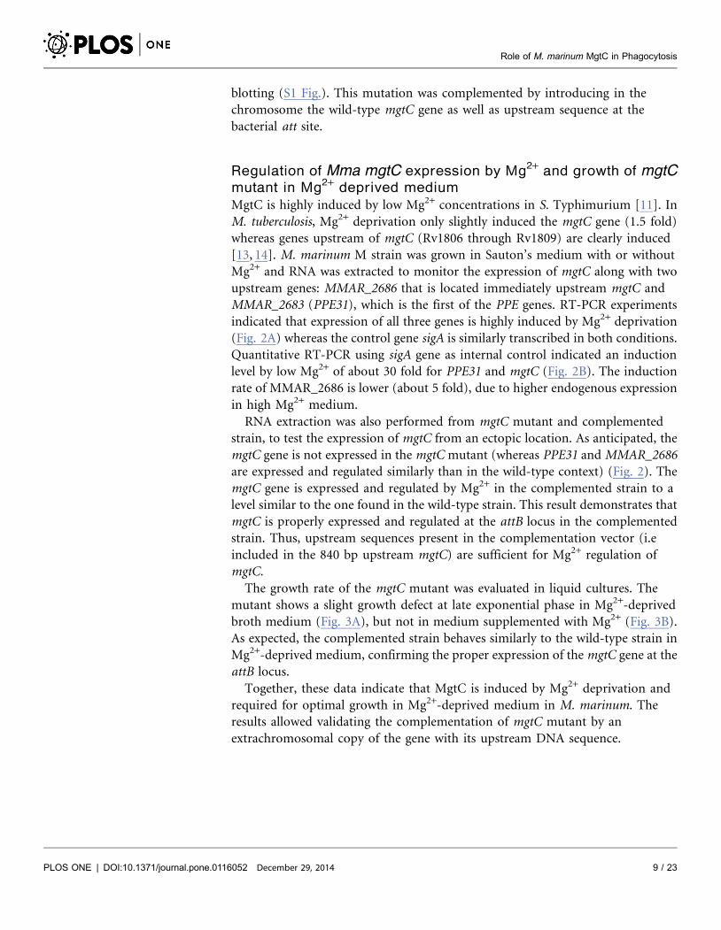
Fig. 4. Intravenous infection of zebrafish with the Mma mgtC mutant. (A) Survival of 30 hpf embryosintravenously infected with 150–200 CFU of wild-type M. marinum, DmgtC mutant or complemented straincompared to non-injected controls (n524). Results are from a representative experiment (infection with133 CFU for wild-type, 142 CFU for mgtC mutant and 205 CFU for complemented strain) out of threeindependent experiments. (B) Ratio of whole embryo bacterial counts between Mma M and mgtC mutantstrain-infected embryos at 0, 3 and 5 dpi. A ratio of 1 indicates equal CFU values. A ratio .1 indicates that WTCFU are higher than mgtC mutant CFU. Results are expressed as mean CFU per embryo+SD from fourindependent experiments (0 and 5 dpi) or two independent experiments (3 dpi). The mild difference betweenmutant and wild-type strains is not statistically significant (Student Test). (C) Visualization of neutrophils inmpx:GFP infected larvae at late stages of infection (one day before embryo’s death). Neutrophils fluoresce ingreen whilemcherry-expressing bacteria fluoresce in red. Neutropenia occurs in wild-type and complementedstrains but not in the mgtC mutant.
doi:10.1371/journal.pone.0116052.g004
Role of M. marinum MgtC in Phagocytosis
PLOS ONE | DOI:10.1371/journal.pone.0116052 December 29, 2014 12 / 23
has been shown to dramatically decrease in zebrafish larvae unable to control
bacterial proliferation upon injection with Staphylococcus or Shigella and
neutropenia has been proposed to correlate with bacterial overgrowth [30, 31].
We took advantage of the mpx:GFP transgenic line (harbouring green fluorescent
neutrophils) to follow the behaviour of neutrophils at late time of infection. By
infecting mpx:GFP embryos with M. marinum strains, we observed the day before
embryo’s death that the increased number of bacteria is associated with a drastic
decrease of green fluorescence in wild-type and complemented strains, indicative
of a neutropenia (Fig. 4C). Interestingly, neutropenia was not observed with the
mgtC mutant strain the day before embryo’s death.
Overall, these results suggest that the mgtC mutant may not replicate as
efficiently as the wild-type strain in zebrafish embryos, but that this effect is not
sufficient to influence the outcome of the infection since embryos died similarly
with both strains.
The Mma mgtC mutant is more efficiently phagocytosed than its
parental strain
To further explore the behaviour of Mma strains towards neutrophils at early
infection time, we carried out subcutaneous injections because it has been
reported that, with this injection route, bacteria are directly taken up by
neutrophils recruited at the infection site [32]. These previous subcutaneous
experiments were performed using non-pathogenic E. coli and we report here for
the first time subcutaneous injections of Mma. Comparing the death curves of
embryos failed to show differences between the wild-type and mutant strains
(Fig. 5A). Confocal microscopy was then used to study the recruitment of
neutrophils at the early stage of infection (4 hpi). In agreement with the previous
report on E. coli [32], we show here that neutrophils are recruited at the injection
site and that mycobacteria are taken up by neutrophils upon sub-cutaneous
injection (Fig. 5B). Interestingly, a higher proportion of red fluorescent bacteria
within the green fluorescent neutrophils was detected with the mutant than with
its parental or complemented strains, suggesting that the frequency at which the
mutant is phagocytosed by neutrophils is higher than the other strains (Fig. 5B).
The quantification of infected neutrophils confirmed a significant higher number
for mgtC mutant, which was approximately twice that of the two reference strains
(Fig. 5C).
To further investigate the behaviour of the mgtC mutant towards phagocytosis,
experiments were carried out using phagocytic and non-phagocytic cells.
Measurement of entry of bacteria into murine J774 macrophages indicated a two-
fold increased uptake with the mutant strain as compared to the wild-type or
complemented strains (Fig. 6A). The phagocytosis rate was next addressed by
visualization of fluorescent bacteria and numeration of infected macrophages,
leading to a similar pattern (Fig. 6B). An increased phagocytosis of similar
magnitude was also observed upon infection of primary bone-marrow derived
macrophages isolated from mice (BMDM) (data not shown). When the cells were
Role of M. marinum MgtC in Phagocytosis
PLOS ONE | DOI:10.1371/journal.pone.0116052 December 29, 2014 13 / 23

Role of M. marinum MgtC in Phagocytosis
PLOS ONE | DOI:10.1371/journal.pone.0116052 December 29, 2014 14 / 23
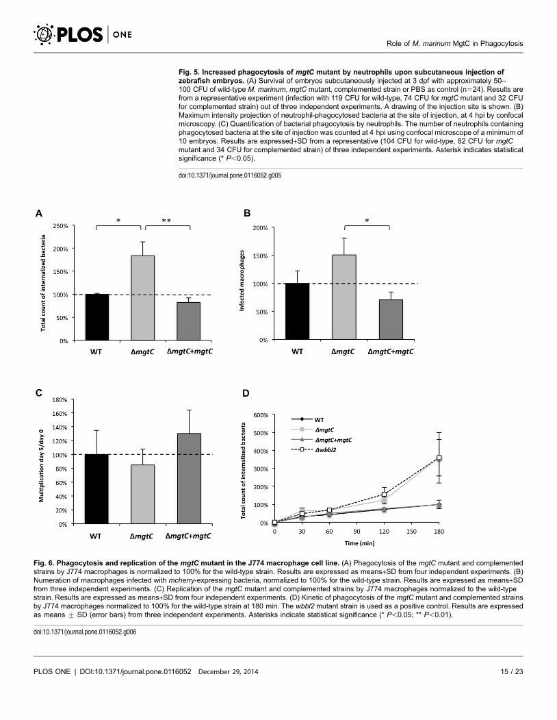
Fig. 5. Increased phagocytosis of mgtC mutant by neutrophils upon subcutaneous injection ofzebrafish embryos. (A) Survival of embryos subcutaneously injected at 3 dpf with approximately 50–100 CFU of wild-type M. marinum, mgtC mutant, complemented strain or PBS as control (n524). Results arefrom a representative experiment (infection with 119 CFU for wild-type, 74 CFU for mgtC mutant and 32 CFUfor complemented strain) out of three independent experiments. A drawing of the injection site is shown. (B)Maximum intensity projection of neutrophil-phagocytosed bacteria at the site of injection, at 4 hpi by confocalmicroscopy. (C) Quantification of bacterial phagocytosis by neutrophils. The number of neutrophils containingphagocytosed bacteria at the site of injection was counted at 4 hpi using confocal microscope of a minimum of10 embryos. Results are expressed+SD from a representative (104 CFU for wild-type, 82 CFU for mgtCmutant and 34 CFU for complemented strain) of three independent experiments. Asterisk indicates statisticalsignificance (* P,0.05).
doi:10.1371/journal.pone.0116052.g005
Fig. 6. Phagocytosis and replication of the mgtC mutant in the J774 macrophage cell line. (A) Phagocytosis of the mgtC mutant and complementedstrains by J774 macrophages is normalized to 100% for the wild-type strain. Results are expressed as means+SD from four independent experiments. (B)Numeration of macrophages infected with mcherry-expressing bacteria, normalized to 100% for the wild-type strain. Results are expressed as means+SDfrom three independent experiments. (C) Replication of the mgtC mutant and complemented strains by J774 macrophages normalized to the wild-typestrain. Results are expressed as means+SD from four independent experiments. (D) Kinetic of phagocytosis of the mgtC mutant and complemented strainsby J774 macrophages normalized to 100% for the wild-type strain at 180 min. The wbbl2 mutant strain is used as a positive control. Results are expressedas means ¡ SD (error bars) from three independent experiments. Asterisks indicate statistical significance (* P,0.05; ** P,0.01).
doi:10.1371/journal.pone.0116052.g006
Role of M. marinum MgtC in Phagocytosis
PLOS ONE | DOI:10.1371/journal.pone.0116052 December 29, 2014 15 / 23
lysed five days after infection to monitor the replication rate, the replication of the
mutant appeared slightly lower than the other strains but the difference was not
significant (Fig. 6C), which was also confirmed in BMDM (data not shown). We
next analyzed the kinetic of mgtC mutant phagocytosis by including a Mma
mutant defective in lipooligosaccharide (LOS) production (Dwbbl2 strain), which
had been shown to be more efficiently phagocytosed by macrophages [26]. Both
the mgtC and the wbbL2 mutant share a highly similar kinetic profile (Fig. 6D).
Differences with the parental strain appear more pronounced after 1 hr of
infection, suggesting that inactivation of mgtC does not alter very early step of
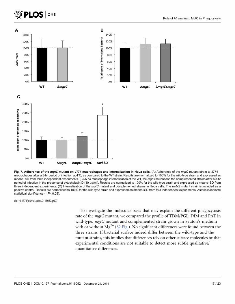
bacterial phagocytosis. Accordingly, when the experiment was carried out at 4 C
to prevent active phagocytosis, no difference between the mgtC mutant and the
parental strain was observed (Fig. 7A). A similar pattern was also observed
following addition of cytochalasin D that prevents actin-driven phagocytosis
(Fig. 7B). Hence, the increased phagocytosis of the mutant strain appears
mediated by an actin-dependent process. Collectively, these results suggest that
the higher phagocytic rate of mgtC mutant is not due to increased bacterial
adherence to macrophages but is very likely due to a higher uptake of bacteria.
Mycobacterium species have been shown to have the ability to invade non-
phagocytic cells, as epithelial cells [33, 34]. To investigate the behaviour of the
mgtC mutant towards non-phagocytic cells, internalization experiments were
performed using epithelial HeLa cells (Fig. 7C). Whereas the wbbL2 mutant
deficient for LOS synthesis shows higher internalization in HeLa cells, the mgtC
mutant, as well as its parental and complemented counterpart, were equally
internalized. From these results, it can be inferred that the phenotype of the mgtC
mutant is restricted to professional phagocytes.
Cell surface analysis
Whereas the function of MgtC in mycobacteria remains unknown, its recent
identification as a protein that modulates ATP-synthase in Salmonella implies that
MgtC may have pleiotropic effects. Previous studies in Salmonella indicated that
the level of some outer membrane proteins are modulated in a mgtC mutant
grown in low Mg2+ medium, which may be related with defect in bacterial
division and cell elongation [35]. Bacterial surface plays a role in phagocytosis. In
this respect, several mycobacterial (glycol)lipids are involved in many aspects of
host pathogenesis [36, 37], including the internalization of bacteria by phagocytic
and non-phagocytic cells. As mentioned above, LOS are bacterial surface
molecules capable to modulate Mma phagocytosis [26, 38]. That the wbbL2 and
mgtC mutants are different toward internalization within epithelial cells suggests
that mgtC mutant does not act by modulating expression of LOS. However, other
cell wall-associated molecules, such as diacyltrehaloses (DAT) and polyacyltre-
haloses (PAT), phtiocerol dimycocerosates (DIM) or phenolic glycolipids (PGL),
have been reported to participate in M. tuberculosis/M. leprae phagocytosis [39–
42].
Role of M. marinum MgtC in Phagocytosis
PLOS ONE | DOI:10.1371/journal.pone.0116052 December 29, 2014 16 / 23
To investigate the molecular basis that may explain the different phagocytosis
rate of the mgtC mutant, we compared the profile of TDM/PGL, DIM and PAT in
wild-type, mgtC mutant and complemented strain grown in Sauton’s medium
with or without Mg2+ (S2 Fig.). No significant differences were found between the
three strains. If bacterial surface indeed differ between the wild-type and the
mutant strains, this implies that differences rely on other surface molecules or that
experimental conditions are not suitable to detect more subtle qualitative/
quantitative differences.
Fig. 7. Adherence of the mgtC mutant on J774 macrophages and internalization in HeLa cells. (A) Adherence of the mgtC mutant strain to J774macrophages after a 3-hr period of infection at 4˚C, as compared to the WTstrain. Results are normalized to 100% for the wild-type strain and expressed asmeans+SD from three independent experiments. (B) J774 macrophage internalization of the WT, themgtCmutant and the complemented strains after a 3-hrperiod of infection in the presence of cytochalasin D (10 mg/ml). Results are normalized to 100% for the wild-type strain and expressed as means+SD fromthree independent experiments. (C) Internalization of the mgtC mutant and complemented strains in HeLa cells. The wbbl2 mutant strain is included as apositive control. Results are normalized to 100% for the wild-type strain and expressed as means+SD from four independent experiments. Asterisks indicatestatistical significance (* P,0.05).
doi:10.1371/journal.pone.0116052.g007
Role of M. marinum MgtC in Phagocytosis
PLOS ONE | DOI:10.1371/journal.pone.0116052 December 29, 2014 17 / 23
Discussion
MgtC appears as a unique virulence factor, shared by several intracellular bacterial
pathogens, which, at least in S. Typhimurium inhibits bacterial’s own F1Fo ATP
synthase [10]. To further investigate the role of MgtC in mycobacteria, we have
investigated its regulation and function in Mma virulence.
The transcription of the Mma mgtC gene and an upstream PPE gene (PPE31) is
highly induced by Mg2+ limitation (about 30 fold). Complementation experi-
ments demonstrated that the mgtC regulation is driven by a Mg2+-dependent
regulatory element present between the end of PPE genes and the mgtC gene and
do not rely on the PPE31 upstream sequences. The regulation of Mma mgtC is
similar to that of S. Typhimurium and Y. pestis but contrasts with that of M.
tuberculosis where the mgtC gene is only slightly induced (1.5 fold) by Mg2+
deprivation [13, 14]. Hence, a strong regulation by Mg2+ is not restricted to cases
where mgtC is co-transcribed with an Mg2+ transporter (as S. Typhimurium and
Y. pestis). The fact that magnesium dependent expression of MgtC is conserved in
phylogenetically distantly related bacteria is probably linked to the conserved
function of MgtC in adaptation to magnesium fluctuations as indicated by the
requirement of Mma MgtC for optimal growth in Mg2+-deprived broth medium.
Despite the conserved role of M. tuberculosis MgtC for optimal growth in low
Mg2+ media [5], the poor regulation of Mtb mgtC by Mg2+ suggests that mgtC
regulation has evolved differently in this species. This could be related to the fact
that M. tuberculosis is less exposed to environmental conditions, where
magnesium concentrations are fluctuating, than non-tuberculous pathogens like
M. marinum which have an external lifestyle and have to cope with various
environmental changes.
MgtC is regarded as an intramacrophage multiplication factor in several
intracellular bacterial pathogens that replicate in phagosomes, including M.
tuberculosis [1]. However, we failed to detect a significant multiplication defect of
the Mma mgtC mutant in either J774 macrophages or bone-marrow derived
macrophages, even though a slight defect was observed. The lack of strong
phenotype for the Mma mgtC mutant upon zebrafish embryos infection and
intramacrophage replication may be related to the Mma intracellular niche. Even
though Mma displays many similar virulence traits to M. tuberculosis, it exhibits
also notable differences such as the ability to promote actin tail formation in the
cytoplasm, probably to favor cell-to-cell spread [43]. Mma escapes from the
phagosome rapidly and with a frequent rate [44], which may explain the lack of
contribution of MgtC in intramacrophage replication. Another hypothesis to
explain the discrepancy between the intracellular phenotypes of M. tuberculosis
and Mma mgtC mutants may be related to the M. tuberculosis genetic background.
Whereas a mgtC mutant constructed in the Erdman background exhibited an
intramacrophage replication defect [5], an independent unpublished work
reported in a review [45] failed to observe an intracellular growth defect for an
mgtC mutant constructed in the H37Rv background, supporting the view that the
Role of M. marinum MgtC in Phagocytosis
PLOS ONE | DOI:10.1371/journal.pone.0116052 December 29, 2014 18 / 23

genetic requirements and/or macrophage cell type may account for these
differences.
The use of transgenic zebrafish embryos with fluorescent neutrophils allowed us
to follow neutrophil behaviour in vivo upon Mma infection. Earlier studies
demonstrated that neutrophils are very efficient to engulf E. coli on tissue surface
but are virtually unable to phagocytose microbes in fluid environments [32].
Consistently, we confirm here that Mma can be phagocytosed by neutrophils
shortly after infection upon subcutaneous injection, whereas neutrophils do not
phagocytose Mma at initial site of infection when injected in the circulation [46].
After injection in the circulation, neutrophils are recruited to the granulomas
where they phagocytize dying infected macrophages [46]. At later stages of
infection, we observed a neutrophil depletion associated with bacteremia
preceding the death of the larvae following infection with wild-type Mma.
Neutropenia has also been reported in zebrafish embryos unable to control
Staphylococcus or Shigella proliferation [30, 31]. This behaviour has been proposed
to be a critical correlate of bacterial overgrowth [30], supported by the fact that in
clinical infection, leukopenia is observed in overwhelming infections and is
regarded as a poor prognostic sign [47]. Interestingly, neutropenia is not seen in
embryos infected with the Mma mgtC mutant. The finding that bacterial loads in
embryos are restricted with the mutant strain supports the idea of a direct link
between neutropenia and the bacterial burden.
Mma MgtC is dispensable for intramacrophage replication, but we uncovered a
novel role for MgtC in the early phase of macrophage infection. Our results
indicate the Mma mgtC mutant is more efficiently phagocytosed than the wild-
type strain by neutrophils upon subcutaneous infection of zebrafish embryos. This
was subsequently confirmed in ex vivo experiments using various types of
macrophages. In addition, this phenotype appears specific to phagocytic cells
since no difference was found in epithelial cells, which contrasts with a LOS
defective mutant that clearly showed increased uptake by macrophages and
epithelial cells. In addition, kinetic experiments, as well as experiments carried out
at 4 C, indicate that MgtC does not play a role in the initial attachment events of
bacteria to macrophages but rather in later steps of the internalization process.
Moreover, experiments carried out in the presence of cytochalasine D confirmed
that this phenotype relies on an actin-based process. Cumulatively, our results
demonstrate that the presence of MgtC limits the phagocytic process. Despite of
its phagocytosis phenotype, the mgtC mutant is not attenuated in the zebrafish
larvae infection model. This finding is consistent with other studies in Mma or M.
tuberculosis mutants that also exhibited higher phagocytosis rate and were not
correlated with an increased virulence phenotype in animal models [26, 39].
Several surface/cell wall components, including LOS, DAT/PAT, DIM and PGL,
have been shown to modulate mycobacterial phagocytosis [36]. The glycan-rich
outer layer of M. tuberculosis cell wall can act as an antiphagocytic capsule but its
effect is mediated by limiting the association of the bacterium with macrophages
[48], which thus differs from MgtC effect. Given the distinct phenotypes
characterizing the mgtC and LOS mutants towards non-phagocytic cells, we
Role of M. marinum MgtC in Phagocytosis
PLOS ONE | DOI:10.1371/journal.pone.0116052 December 29, 2014 19 / 23
propose that the differences reside unlikely in these glycolipids. DAT/PAT
deficiency improved binding and entry of M. tuberculosis both in phagocytic and
non-phagocytic cells, thus also differing from the phenotype of Mma mgtC
mutant [39]. Interestingly, DIM deficiency reduced M. tuberculosis internalization
in macrophages in an actin-dependent process, without affecting the bacterial
binding to macrophages [40]. However, our TLC analysis failed to reveal major
differences in DIM between the strains in the conditions tested. Moreover, no
differences were found in the other lipids tested (DAT/PAT and PGL). Hence,
experimental conditions may not be optimized to detect quantitative differences
in those lipids or other surface molecules may be involved. Alternatively, the
uptake phenotype may be driven by a mechanism that triggers signaling pathways
of phagocytic receptors and/or early trafficking without noticeable bacterial
surface modification.
In conclusion, our results indicate that the Mg2+ regulation of MgtC and its role
for optimal growth in Mg2+-deprived media is conserved among bacteria that are
not phylogenetically linked as M. marinum and S. Typhimurium. The role of
MgtC in macrophages has been previously reported to be dissociated from its role
in low Mg2+ medium [28]. This view is further substantiated by the present study,
since Mma MgtC appears to have a role towards phagocytic cells linked to
phagocytosis rather than intracellular multiplication. Even though the precise role
of Mma MgtC during the infection process remains to be established, our results
suggest that the involvement of MgtC towards professional phagocytes has
evolved in bacterial pathogens, possibly to fit to the specific pathogen’s lifestyles.
Supporting Information
S1 Fig. Construction of mgtC mutant in M. marinum. A) A DNA substrate for
allelic replacement of the M. marinum mgtC gene was generated by cloning
979 bp upstream and downstream mgtC sequences to flank the hygR gene. The
locations of primers 1/19, 2/29 and 3/39 used to check the mgtC mutant by PCR are
indicated by arrows. Electrophoresis migration of PCR fragments 1 (primers
1/19), 2 (primers 2/29) and 3 (primers 3/39) amplified from cultures of wild-type
and mgtC mutant strain is shown. The upper lane indicates the 1569 bp DNA
fragment cloned in the integrative vector pMV306 to complement the Mma mgtC
mutant. B) Southern blot analysis of the mgtC mutant. The genomic structure of
gene replacement mutant was examined by Southern blot analysis. Chromosomal
DNA of wild-type and mgtC mutant strains were digested with XhoI and probed
with either a segment of DNA of mgtC or the hygR cassette. Hybridization signals
at the expected size are detected (1792 bp for the mgtC probe in the wild-type
strain and 3012 bp for hyg probe in the mutant strain).
doi:10.1371/journal.pone.0116052.s001 (TIFF)
S2 Fig. Lipid profiles. One-dimensional autoradiographic TLC of [1-14C]-
propionate-labeled apolar lipids from M. marinum wild-type, mgtC mutant and
complemented strains grown in Sauton’s liquid medium A) or in Sauton’s liquid
Role of M. marinum MgtC in Phagocytosis
PLOS ONE | DOI:10.1371/journal.pone.0116052 December 29, 2014 20 / 23

medium without magnesium B). Equal amount (20,000 cpm) of radiolabeled
lipids were spotted on TLC plates run in various solvents: chloroform/methanol
(19:1, v/v) for TDM/PGL, chloroform/methanol (99:1, v/v) for PAT and
petroleum ether/diethylether 9:1 (v/v) for PDIM.
doi:10.1371/journal.pone.0116052.s002 (TIFF)
S1 Table. Primers used in the study.
doi:10.1371/journal.pone.0116052.s003 (DOC)
Acknowledgments
We thank Steve Renshaw (University of Sheffield, UK) for providing the
mpx::GFP fish line and William Jacobs (Albert Einstein College of Medicine,
Bronx, NY) for the gift of phAE159 phage. We thank Catherine Gonzalez for fish
facility maintenance, Audrey Bernut for assistance with confocal microscopy and
the RIO Imaging platform at University Montpellier 2.
Author ContributionsConceived and designed the experiments: CB LGZ GL ABBP. Performed the
experiments: CB LGZ ABBP. Analyzed the data: CB LGZ GL LK ABBP.
Contributed reagents/materials/analysis tools: GL LK. Wrote the paper: CB ABBP.
References
1. Alix E, Blanc-Potard A-B (2007) MgtC: a key player in intramacrophage survival. Trends Microbiol 15:252–256.
2. Blanc-Potard A-B, Groisman EA (1997) The Salmonella selC locus contains a pathogenicity islandmediating intramacrophage survival. EMBO J 16: 5376–5385.
3. Lawley TD, Chan K, Thompson LJ, Kim CC, Govoni GR, et al. (2006) Genome-wide screen forSalmonella genes required for long-term systemic infection of the mouse. PLoS Pathog 2: e11.
4. Thompson JA, Liu M, Helaine S, Holden DW (2011) Contribution of the PhoP/Q regulon to survival andreplication of Salmonella enterica serovar Typhimurium in macrophages. Microbiology 157: 2084–2093.
5. Buchmeier N, Blanc-Potard A-B, Ehrt S, Piddington D, Riley L, et al. (2000) A parallelintraphagosomal survival strategy shared by Mycobacterium tuberculosis and Salmonella enterica.Mol Microbiol 35: 1375–1382.
6. Lavigne J-P, O’Callaghan D, Blanc-Potard A-B (2005) Requirement of MgtC for Brucella suisintramacrophage growth: a potential mechanism shared by Salmonella enterica and Mycobacteriumtuberculosis for adaptation to a low-Mg2+ environment. Infect Immun 73: 3160–3163.
7. Grabenstein JP, Fukuto HS, Palmer LE, Bliska JB (2006) Characterization of phagosome traffickingand identification of PhoP-regulated genes important for survival of Yersinia pestis in macrophages.Infect Immun 74: 3727–3741.
8. Maloney KE, Valvano MA (2006) The mgtC Gene of Burkholderia cenocepacia is required for growthunder magnesium limitation conditions and intracellular survival in macrophages. Infect Immun 74:5477–5486.
9. Retamal P, Castillo-Ruiz M, Mora GC (2009) Characterization of MgtC, a virulence factor of Salmonellaenterica serovar Typhi. PLoS One 4: e5551.
10. Lee E-J, Pontes MH, Groisman EA (2013) A bacterial virulence protein promotes pathogenicity byinhibiting the bacterium’s own F1Fo ATP synthase. Cell 154: 146–156.
Role of M. marinum MgtC in Phagocytosis
PLOS ONE | DOI:10.1371/journal.pone.0116052 December 29, 2014 21 / 23
11. Vescovi EG, Soncini FC, Groisman EA (1996) Mg2+ as an extracellular signal: environmentalregulation of Salmonella virulence. Cell 84: 165–174.
12. Zhou D, Han Y, Qin L, Chen Z, Qiu J, et al. (2005) Transcriptome analysis of the Mg2+-responsive PhoPregulator in Yersinia pestis. FEMS Microbiol Lett 250: 85–95.
13. Walters SB, Dubnau E, Kolesnikova I, Laval F, Daffe M, et al. (2006) The Mycobacterium tuberculosisPhoPR two-component system regulates genes essential for virulence and complex lipid biosynthesis.Mol Microbiol 60: 312–330.
14. Yang Y, Labesse G, Carrere-Kremer S, Esteves K, Kremer L, et al. (2012) The C-terminal domain ofthe virulence factor MgtC is a divergent ACT domain. J Bacteriol 194: 6255–6263.
15. Blanc-Potard A-B, Lafay B (2003) MgtC as a horizontally-acquired virulence factor of intracellularbacterial pathogens: evidence from molecular phylogeny and comparative genomics. J Mol Evol 57:479–486.
16. Stinear TP, Seemann T, Harrison PF, Jenkin GA, Davies JK, et al. (2008) Insights from the completegenome sequence ofMycobacterium marinum on the evolution ofMycobacterium tuberculosis. GenomeRes 18: 729–741.
17. Tobin DM, Ramakrishnan L (2008) Comparative pathogenesis of Mycobacterium marinum andMycobacterium tuberculosis. Cell Microbiol. 10: 1027–1039.
18. Ramakrishnan L (2013) Looking within the zebrafish to understand the tuberculous granuloma. In:Divangahi M, editor. The new paradigm of immunity to tuberculosis. Advances in experimental medicineand biology. Springer New York. pp. 251–266.
19. Meijer AH, van der Vaart M, Spaink HP (2014) Real-time imaging and genetic dissection of host–microbe interactions in zebrafish. Cell Microbiol 16: 39–49.
20. Torraca V, Masud S, Spaink HP, Meijer AH (2014) Macrophage-pathogen interactions in infectiousdiseases: new therapeutic insights from the zebrafish host model. Dis Model Mech 7: 785–797.
21. Larsen MH, Biermann K, Tandberg S, Hsu T, Jacobs William R (2007) Genetic manipulation ofMycobacterium tuberculosis. Current Protocols in Microbiology. John Wiley & Sons, Inc.
22. Stover CK, de la Cruz VF, Fuerst TR, Burlein JE, Benson LA, et al. (1991) New use of BCG forrecombinant vaccines. Nature 351: 456–460.
23. Lamason RL, Mohideen M-APK, Mest JR, Wong AC, Norton HL, et al. (2005) SLC24A5, a putativecation exchanger, affects pigmentation in zebrafish and humans. Science 310: 1782–1786.
24. Renshaw SA, Loynes CA, Trushell DMI, Elworthy S, Ingham PW, et al. (2006) A transgenic zebrafishmodel of neutrophilic inflammation. Blood 108: 3976–3978.
25. Westerfield M (2007) The zebrafish book: a guide for the laboratory use of zebrafish (Danio rerio), 5thed. Eugene: University of Oregon Press.
26. Alibaud L, Pawelczyk J, Gannoun-Zaki L, Singh VK, Rombouts Y, et al. (2014) Increasedphagocytosis of Mycobacterium marinum mutants defective in lipooligosaccharide production. J BiolChem 289: 215–228.
27. Dobson G, Minnikin DE, Minnikin SM, Parlett JH, Goodfellow M, et al. (1995) Systematic analysis ofcomplex mycobacterial lipids. Chemical Methods in Bacterial Systematics. Goodfellow M. and MinnikinD.E., (eds). pp. 237–265.
28. Rang C, Alix E, Felix C, Heitz A, Tasse L, et al. (2007) Dual role of the MgtC virulence factor in hostand non-host environments. Mol Microbiol 63: 605–622.
29. Clay H, Davis JM, Beery D, Huttenlocher A, Lyons SE, et al. (2007) Dichotomous role of themacrophage in early Mycobacterium marinum infection of the zebrafish. Cell Host Microbe 2: 29–39.
30. Mostowy S, Boucontet L, Mazon Moya MJ, Sirianni A, Boudinot P, et al. (2013) The zebrafish as anew model for the in vivo study of Shigella flexneri interaction with phagocytes and bacterial autophagy.PLoS Pathog 9: e1003588.
31. Prajsnar TK, Cunliffe VT, Foster SJ, Renshaw SA (2008) A novel vertebrate model of Staphylococcusaureus infection reveals phagocyte-dependent resistance of zebrafish to non-host specializedpathogens. Cell Microbiol 10: 2312–2325.
Role of M. marinum MgtC in Phagocytosis
PLOS ONE | DOI:10.1371/journal.pone.0116052 December 29, 2014 22 / 23
32. Colucci-Guyon E, Tinevez J-Y, Renshaw SA, Herbomel P (2011) Strategies of professionalphagocytes in vivo: unlike macrophages, neutrophils engulf only surface-associated microbes. J CellSci 124: 3053–3059.
33. Bermudez LE, Shelton K, Young LS (1995) Comparison of the ability of Mycobacterium avium, M.smegmatis and M. tuberculosis to invade and replicate within HEp-2 epithelial cells. Tuber Lung Dis 76:240–247.
34. Flesselles B, Anand NN, Remani J, Loosmore SM, Klein MH (1999) Disruption of the mycobacterialcell entry gene of Mycobacterium bovis BCG results in a mutant that exhibits a reduced invasiveness forepithelial cells. FEMS Microbiol Lett 177: 237–242.
35. Alix E, Miki T, Felix C, Rang C, Figueroa-Bossi N, et al. (2008) Interplay between MgtC and PagC inSalmonella enterica serovar Typhimurium. Microb Pathog 45: 236–240.
36. Astarie-Dequeker C, Nigou J, Passemar C, Guilhot C (2010) The role of mycobacterial lipids in hostpathogenesis. Drug Discov Today Dis Mech 7: e33–e41.
37. Tabouret G, Astarie-Dequeker C, Demangel C, Malaga W, Constant P, et al. (2010) Mycobacteriumleprae phenolglycolipid-1 expressed by engineered M. bovis BCG modulates early interaction withhuman phagocytes. PLoS Pathog. 6: e1001159.
38. Ren H, Dover LG, Islam ST, Alexander DC, Chen JM, et al. (2007) Identification of thelipooligosaccharide biosynthetic gene cluster from Mycobacterium marinum. Mol Microbiol 63: 1345–1359.
39. Rousseau C, Neyrolles O, Bordat Y, Giroux S, Sirakova TD, et al. (2003) Deficiency in mycolipenate-and mycosanoate-derived acyltrehaloses enhances early interactions of Mycobacterium tuberculosiswith host cells. Cell Microbiol 5: 405–415.
40. Astarie-Dequeker C, Le Guyader L, Malaga W, Seaphanh F-K, Chalut C, et al. (2009) Phthioceroldimycocerosates of M. tuberculosis participate in macrophage invasion by inducing changes in theorganization of plasma membrane lipids. PLoS Pathog 5: e1000289.
41. Schlesinger LS, Horwitz MA (1991) Phenolic glycolipid-1 of Mycobacterium leprae binds complementcomponent C3 in serum and mediates phagocytosis by human monocytes. J Exp Med 174: 1031–1038.
42. Vilcheze C, Molle V, Carrere-Kremer S, Leiba J, Mourey L, et al. (2014) Phosphorylation of KasBregulates virulence and acid-fastness in Mycobacterium tuberculosis. PLoS Pathog. 10: e1004115.
43. Stamm LM, Morisaki JH, Gao L-Y, Jeng RL, McDonald KL, et al. (2003) Mycobacterium marinumescapes from phagosomes and is propelled by actin-based motility. J Exp Med 198:1361–1368.
44. Collins CA, De Maziere A, van Dijk S, Carlsson F, Klumperman J, et al. (2009) Atg5-independentsequestration of ubiquitinated mycobacteria. PLoS Pathog 5: e1000430.
45. Smith I (2003) Mycobacterium tuberculosis pathogenesis and molecular determinants of virulence. ClinMicrobiol Rev 16: 463–496.
46. Yang C-T, Cambier CJ, Davis JM, Hall CJ, Crosier PS, et al. (2012) Neutrophils exert protection in theearly tuberculous granuloma by oxidative killing of mycobacteria phagocytosed from infectedmacrophages. Cell Host Microbe 12: 301–312.
47. Fine M, Smith M, Carson C, Mutha S, Sankey S, et al. (1996) Prognosis and outcomes of patients withcommunity-acquired pneumonia: A meta-analysis. JAMA 275: 134–141.
48. Stokes RW, Norris-Jones R, Brooks DE, Beveridge TJ, Doxsee D, et al. (2004) The glycan-rich outerlayer of the cell wall of Mycobacterium tuberculosis acts as an antiphagocytic capsule limiting theassociation of the bacterium with macrophages. Infect Immun 72: 5676–5686.
49. Snapper SB, Melton RE, Mustafa S, Kieser T, Jacobs WR (1990) Isolation and characterization ofefficient plasmid transformation mutants of Mycobacterium smegmatis. Mol Microbiol 4: 1911–1919.
50. Sambandamurthy VK, Wang X, Chen B, Russell RG, Derrick S, et al. (2002) A pantothenateauxotroph of Mycobacterium tuberculosis is highly attenuated and protects mice against tuberculosis.Nat Med 8: 1171–1174.
51. Alibaud L, Rombouts Y, Trivelli X, Burguiere A, Cirillo SLG, et al. (2011) A Mycobacterium marinumTesA mutant defective for major cell wall-associated lipids is highly attenuated in Dictyosteliumdiscoideum and zebrafish embryos. Mol Microbiol 80: 919–934.
Role of M. marinum MgtC in Phagocytosis
PLOS ONE | DOI:10.1371/journal.pone.0116052 December 29, 2014 23 / 23


![ReviewArticle Phagocytosis: A Fundamental Process in …downloads.hindawi.com/journals/bmri/2017/9042851.pdfresponses including phagocytosis [77]. Another molecule that negatively](https://img.pdfslide.net/doc/110x75/5f09f83a7e708231d429615f/reviewarticle-phagocytosis-a-fundamental-process-in-responses-including-phagocytosis.jpg)