Embed Size (px)
Citation preview
CASE REPORT Open Access
Mycobacterium tuberculosis as a cause ofmandibular osteomyelitis in a youngwoman: a case reportJorge Tellez-Rodriguez1, Rubi Lopez-Fernandez1, Rodolfo Rodriguez-Jurado2, Hayde Nallely Moreno-Sandoval3,Francisco Martinez-Perez4 and Juan Antonio Gonzalez-Barrios3*
Abstract
Background: Tuberculosis is considered an emerging disease worldwide; in the last 10 years, its incidence hasincreased to more than 9.6 million cases of active tuberculosis. In 2014, it resulted in 1.5 million patient deaths.However, oral presentation with bone involvement occurs in less than 3% of all reported cases and rarely arousesclinical suspicion on initial presentation.
Case presentation: A 15-year-old Mexican girl who had a previous diagnosis of neurofibromatosis presented toour hospital with pain and swelling in the region of the left mandibular body since November 2011. A clinicalexamination revealed pain in the mandibular region, a mass of soft consistency that seemed to involve bone, anda fistula with discharge of intraoral purulent material. Additionally, tachycardia and hyperthermia were observed.The left submental and submandibular regions had a 12-cm-diameter swelling, which was well-delineated andnonerythematous. The final diagnosis was established by real-time polymerase chain reaction.
Conclusions: The final diagnosis of rare cases of tuberculous osteomyelitis in the jaw can be established bydeoxyribonucleic acid (DNA) identification of Mycobacterium tuberculosis in the lesion. Simple and fastcomplementary diagnosis by real-time polymerase chain reaction is a fundamental approach to establishing earlyand effective pharmacological and surgical treatment.
Keywords: Tuberculosis, Osteomyelitis, Mycobacterium tuberculosis, Jaw, Case report
BackgroundTuberculosis is considered an emerging disease world-wide. The World Health Organization estimated thatthe incidence of tuberculosis increased in the last10 years to more than 9.6 million cases of active tuber-culosis, and that it caused 1.5 million patient deaths in2014. Approximately 1 million children were infectedby Mycobacterium tuberculosis and 140,000 childrendied as a result of tuberculosis. By the end of 2014, anadditional 480,000 cases of multidrug-resistant tubercu-losis were reported worldwide [1]. Tuberculosis is rarelyconfined to bone and develops frequently secondary to asystemic infection. However, oral presentation with bone
involvement occurs in less than 3% of all reportedcases; thus, it rarely arouses clinical suspicion at initialpresentation. Mandibular osteomyelitis caused by M.tuberculosis infection is a rare medical condition. Be-cause tuberculosis infection is rarely confined to bonetissue, such a manifestation may be considered as anextrapulmonary complication.Adults are more frequently affected, although cases in
children have also been described [2–5]. Tuberculous le-sions of the oral cavity are quite rare; despite the highincidence of the systemic disease, they can be explainedby M. tuberculosis inhibition by the salivary components[6]. The mechanism of propagation of tuberculosis infec-tion to the mandibular bone can be by direct inoculationthrough dental extraction, lesions of the mucosa duringteeth eruption, and spread from adjacent tissue or via ahematogenous route [7]. The clinical presentation and
* Correspondence: [email protected] de Medicina Genómica, Hospital Regional “1o. de Octubre”,Avenida Instituto Politécnico Nacional 1669, Mexico City 07760, MexicoFull list of author information is available at the end of the article
© The Author(s). 2016 Open Access This article is distributed under the terms of the Creative Commons Attribution 4.0International License (http://creativecommons.org/licenses/by/4.0/), which permits unrestricted use, distribution, andreproduction in any medium, provided you give appropriate credit to the original author(s) and the source, provide a link tothe Creative Commons license, and indicate if changes were made. The Creative Commons Public Domain Dedication waiver(http://creativecommons.org/publicdomain/zero/1.0/) applies to the data made available in this article, unless otherwise stated.
Tellez-Rodriguez et al. Journal of Medical Case Reports (2016) 10:366 DOI 10.1186/s13256-016-1118-x
radiological imaging of mandibular tuberculosis aresimilar to those of chronic secondary osteomyelitis andregular dentoalveolar abscess. However, cervical lymph-adenopathy produces discrete or diffuse masses, whichare often not sensitive to palpation. These can be dis-tinctive features in some patients [8]. This similarity toconventional cases of osteomyelitis emphasizes the im-portance of considering a differential diagnosis oftuberculous osteomyelitis of the jaw, especially if thepatient has a suspicious medical record for tuberculosis[9, 10]. The clinical lesions of oral tuberculosis presentas painful and irregular ulcers, especially in the poster-ior tongue area, pharynx, or palate, and they are fre-quently secondary to active pulmonary tuberculosis[11]. Bone involvement of the maxilla and mandiblecan produce tuberculous osteomyelitis by occult spreadof the microorganism [12], and regional lymphadenop-athy usually accompanies the oral lesions. For this rea-son, inclusion of scrofula in the differential diagnosis isobligatory [13]. Sometimes, bone invasion can lead totuberculosis-related osteomyelitis. The incidence is higherin poor and densely populated areas, areas of low socio-economic status, and in human immunodeficiency virus(HIV)-infected and immunocompromised individuals [11].The differential diagnosis of oral tuberculosis includes otherimportant diseases, such as squamous cell carcinoma; pri-mary syphilis and various oral lesions, such as actinomycosis;fungal lung diseases, such as histoplasmosis, coccidioido-mycosis, and blastomycosis; lymphoma; and submandibularsialadenitis [11]. It is important to remember that trauma isthe leading cause of oral ulcers and should be included inthe differential diagnosis, and all oral ulcers that raise suspi-cion of tuberculosis require biopsy to exclude cancer.Radiographic features of acute osteomyelitis are usu-
ally not immediately apparent. First, the exudate pro-gresses through the soft tissue component through thepreexisting medullary spaces. Until the trabecular bonecomponents have resorbed significantly, the magnitudeof the destruction will be evident on radiographs asmacular, mottled, or fuzzy in appearance with featherededges and accompanied by unresorbed fragments ofdead bone surrounded by large purulent areas. Thechronic osteomyelitis area has a mottled appearanceand is radio-opaque, which is designated as osteosclero-sis that can be limited to the surrounding tooth rootarea. In other cases, it can induce osteosclerosis oflarger areas or areas of edentulous bone [14]. M. tuber-culosis induces a specific response in infected tissuesthat are characterized by local areas of macrophagessurrounded by lymphocytes and fibroblasts, where themacrophages develop an abundant eosinophilic cyto-plasm similar to the epithelial cells that are known tobe like epithelioid cells. The fusion of macrophagesgives them the appearance of giant Langerhans cells in
which the nuclei are distributed in episomal form, character-istic of the histological diagnosis of bone tuberculosis [12].
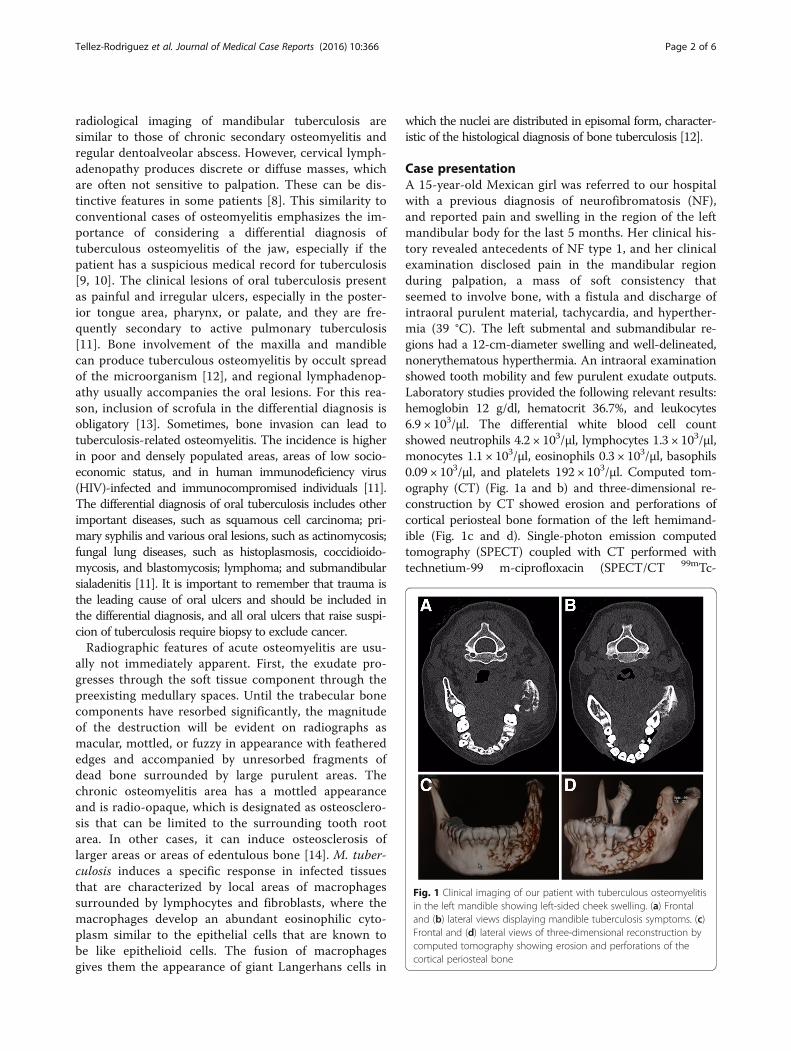
Case presentationA 15-year-old Mexican girl was referred to our hospitalwith a previous diagnosis of neurofibromatosis (NF),and reported pain and swelling in the region of the leftmandibular body for the last 5 months. Her clinical his-tory revealed antecedents of NF type 1, and her clinicalexamination disclosed pain in the mandibular regionduring palpation, a mass of soft consistency thatseemed to involve bone, with a fistula and discharge ofintraoral purulent material, tachycardia, and hyperther-mia (39 °C). The left submental and submandibular re-gions had a 12-cm-diameter swelling and well-delineated,nonerythematous hyperthermia. An intraoral examinationshowed tooth mobility and few purulent exudate outputs.Laboratory studies provided the following relevant results:hemoglobin 12 g/dl, hematocrit 36.7%, and leukocytes6.9 × 103/μl. The differential white blood cell countshowed neutrophils 4.2 × 10³/μl, lymphocytes 1.3 × 10³/μl,monocytes 1.1 × 10³/μl, eosinophils 0.3 × 10³/μl, basophils0.09 × 103/μl, and platelets 192 × 103/μl. Computed tom-ography (CT) (Fig. 1a and b) and three-dimensional re-construction by CT showed erosion and perforations ofcortical periosteal bone formation of the left hemimand-ible (Fig. 1c and d). Single-photon emission computedtomography (SPECT) coupled with CT performed withtechnetium-99 m-ciprofloxacin (SPECT/CT 99mTc-
Fig. 1 Clinical imaging of our patient with tuberculous osteomyelitisin the left mandible showing left-sided cheek swelling. (a) Frontaland (b) lateral views displaying mandible tuberculosis symptoms. (c)Frontal and (d) lateral views of three-dimensional reconstruction bycomputed tomography showing erosion and perforations of thecortical periosteal bone
Tellez-Rodriguez et al. Journal of Medical Case Reports (2016) 10:366 Page 2 of 6

ciprofloxacin) revealed an abnormally concentrated areaof radiolabeled antibiotic at the left mandible, right elbow,and soft tissues of the right hand, suggesting a dissemi-nated infectious process (Fig. 2a–c). An intraoral secretionbacteriological culture isolated Streptococcus parasangui-nis and high-level gentamicin- and vancomycin-resistant Enterococcus faecium. An antibiogram showsresistance to ampicillin, erythromycin, vancomycin,cefazolin, gentamicin, streptomycin, levofloxacin, oxacil-lin, and penicillin G, as well as sensitivity to daptomycin,doxycycline, linezolid, moxifloxacin, nitrofurantoin, qui-nupristin/dalfopristin, and rifampicin (RIF).The presumptive diagnosis was mandibular osteomyelitis.
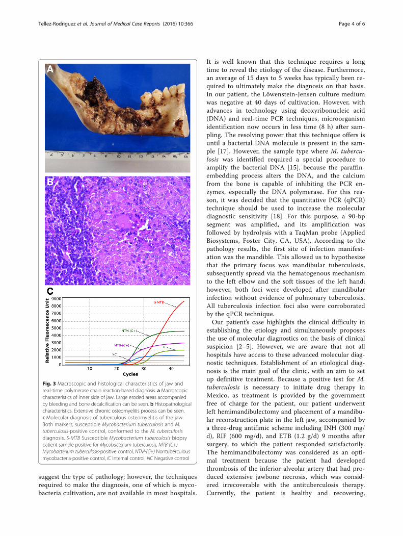
To corroborate the diagnosis, an incisional biopsy wasdone. The patient had received previous antibioticadministration with procaine benzylpenicillin (600,000 Uintravenous therapy) combined with benzylpenicillinsodium (200,000 U intravenous therapy) and metronidazole(500 mg oral therapy) every 8 h for 7 days. Macroscopicand histopathological study revealed an extensive chronicosteomyelitic process (Fig. 3a and b) due to infection thatdid not cease with antibiotic administration. This raisedthe possibility of tuberculous osteomyelitis. To identifymycobacteria as a causative agent, molecular diagnosticsfor tuberculosis was performed for the high diagnosticvalue [15]. M. tuberculosis molecular identificationand drug resistance to isoniazid (INH), RIF, fluoroquinolone(FQ), and aminoglycoside (AG) were done usingAnyplex plus mycobacterium tuberculosis/nontuberculousmycobacteria/drug-resistant tuberculosis (MTB/NTM/DR-TB) real-time test (Seegene, Seoul, Korea) in accordancewith the manufacturer’s specifications. The polymerase chainreaction (PCR) data confirmed the final molecular diagnosisof disseminated and chronic tuberculous osteomyelitis of themandible caused by M. tuberculosis (Fig. 3c), and noisoniazid (INH) or rifampicin (RIF) resistance was found bythe GenoType multidrug-resistant tuberculosis (MTBDR)test (Hain Lifescience, Nehren, Germany).
The patient received initial antituberculosis therapywith 300 mg INH, 600 mg RIF, 1.6 g pyrazinamide, and1 g ethambutol (EMB) for 2 months, followed by main-tenance treatment with 300 mg INH and 600 mg RIF. Atthe sixth month of tuberculosis therapy, the patient devel-oped thrombosis of the inferior alveolar artery that pro-duced extensive jawbone necrosis, which was consideredirrecoverable. Thus, an elective hemimandibulectomy wasperformed, and the surgical defect was reconstructed bytitanium plating with the condyle, and 1.2 g ETB wasadded to the antituberculosis maintenance therapy until15 months of antifimic drug therapy was complete.
DiscussionNF and tuberculosis rarely coincide in a patient; however,NF type 1 may be accompanied by a common variableimmunodeficiency that favors infection by M. tuberculosisand its systemic dissemination [16]. In this paper, we re-port a case of a young woman with NF and disseminatedtuberculosis affecting mainly the jaw that induced inferioralveolar artery thrombosis and irrecoverable mandibularnecrosis requiring an elective hemimandibulectomy andcompletion of antifimic drug therapy.Although bone tuberculosis, and especially the man-
dibular presentation [2–5], remains a rare disease ithas great significance because of the significant facialdeformity sequelae that occur. However, one of the di-lemmas faced by the doctor is the definitive etiologicdiagnosis (Table 1). In our patient’s case, differenttechniques used to support the clinical diagnosis in-cluded bacterial cultures, axial CT, and SPECT/CT99mTc-ciprofloxacin. All results obtained using thesediagnostic techniques were indicative of infectiousosteomyelitis. The incisional biopsy results were notconclusive for tuberculosis, because they showed onlychronic osteomyelitis with an exacerbation processwithout evidence of granulomatosis. It is well knownthat all these techniques only have the power to
Fig. 2 Technetium-99 m-ciprofloxacin accumulation in the mandible. Images show an abnormal concentrated area of radiolabeled antibiotic atthe level of the left mandible, suggesting an infectious disease process. (a) Frontal view. (b) Oblique view. (c) Lateral view. The arrows indicate thesite of 99mTc-Ciprofloxacine accumulation
Tellez-Rodriguez et al. Journal of Medical Case Reports (2016) 10:366 Page 3 of 6
suggest the type of pathology; however, the techniquesrequired to make the diagnosis, one of which is myco-bacteria cultivation, are not available in most hospitals.
It is well known that this technique requires a longtime to reveal the etiology of the disease. Furthermore,an average of 15 days to 5 weeks has typically been re-quired to ultimately make the diagnosis on that basis.In our patient, the Löwenstein-Jensen culture mediumwas negative at 40 days of cultivation. However, withadvances in technology using deoxyribonucleic acid(DNA) and real-time PCR techniques, microorganismidentification now occurs in less time (8 h) after sam-pling. The resolving power that this technique offers isuntil a bacterial DNA molecule is present in the sam-ple [17]. However, the sample type where M. tubercu-losis was identified required a special procedure toamplify the bacterial DNA [15], because the paraffin-embedding process alters the DNA, and the calciumfrom the bone is capable of inhibiting the PCR en-zymes, especially the DNA polymerase. For this rea-son, it was decided that the quantitative PCR (qPCR)technique should be used to increase the moleculardiagnostic sensitivity [18]. For this purpose, a 90-bpsegment was amplified, and its amplification wasfollowed by hydrolysis with a TaqMan probe (AppliedBiosystems, Foster City, CA, USA). According to thepathology results, the first site of infection manifest-ation was the mandible. This allowed us to hypothesizethat the primary focus was mandibular tuberculosis,subsequently spread via the hematogenous mechanismto the left elbow and the soft tissues of the left hand;however, both foci were developed after mandibularinfection without evidence of pulmonary tuberculosis.All tuberculosis infection foci also were corroboratedby the qPCR technique.Our patient’s case highlights the clinical difficulty in
establishing the etiology and simultaneously proposesthe use of molecular diagnostics on the basis of clinicalsuspicion [2–5]. However, we are aware that not allhospitals have access to these advanced molecular diag-nostic techniques. Establishment of an etiological diag-nosis is the main goal of the clinic, with an aim to setup definitive treatment. Because a positive test for M.tuberculosis is necessary to initiate drug therapy inMexico, as treatment is provided by the governmentfree of charge for the patient, our patient underwentleft hemimandibulectomy and placement of a mandibu-lar reconstruction plate in the left jaw, accompanied bya three-drug antifimic scheme including INH (300 mg/d), RIF (600 mg/d), and ETB (1.2 g/d) 9 months aftersurgery, to which the patient responded satisfactorily.The hemimandibulectomy was considered as an opti-mal treatment because the patient had developedthrombosis of the inferior alveolar artery that had pro-duced extensive jawbone necrosis, which was consid-ered irrecoverable with the antituberculosis therapy.Currently, the patient is healthy and recovering,
Fig. 3 Macroscopic and histological characteristics of jaw andreal-time polymerase chain reaction-based diagnosis. a Macroscopiccharacteristics of inner side of jaw. Large eroded areas accompaniedby bleeding and bone decalcification can be seen. b Histopathologicalcharacteristics. Extensive chronic osteomyelitis process can be seen.c Molecular diagnosis of tuberculous osteomyelitis of the jaw.Both markers, susceptible Mycobacterium tuberculosis and M.tuberculosis-positive control, conformed to the M. tuberculosisdiagnosis. S-MTB Susceptible Mycobacterium tuberculosis biopsypatient sample positive for Mycobacterium tuberculosis, MTB-(C+)Mycobacterium tuberculosis-positive control, NTM-(C+) Nontuberculousmycobacteria-positive control, IC Internal control, NC Negative control
Tellez-Rodriguez et al. Journal of Medical Case Reports (2016) 10:366 Page 4 of 6
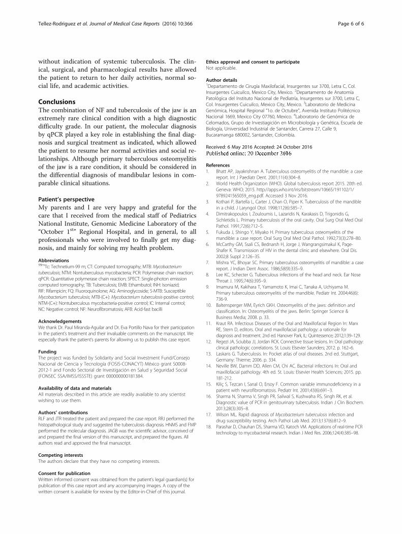
Table 1 Comparative characteristics of reported tuberculous osteomyelitis of the jaw during infancy
Sex, age [reference] Clinical findings Mandibular Region Histological diagnosis Methods for tuberculosis diagnosis Treatment (follow-up)
Male, 3 yearsold [2]
1 monthLeft submandibular massLymph enlargement
Angle Granulomatous osteomyelitisconsistent with tuberculosis
Primary.Ziehl-Neelsen staining and culturingnot performed
Antitubercular therapy(6 months)Without recurrence
Male, 4 yearsold [3]
1 monthProgressive swelling on cheek,right sideCaries in the right secondprimary molarLymph nodes
BodyAngle
Granulomatous osteomyelitis SecondaryGranulomatous areaGiant and epithelioid cells surroundedby lymphocytes and plasma cellsPositive Ziehl-Neelsen staining
Antitubercular chemotherapy(6 months)Complete resolution
Female, 10 yearsold [4]
2 monthsGradual swelling in left mandibularregionCaries in the lower left seconddeciduous molarNo evidence of lymphadenopathy
Body Caseous granuloma PrimaryFine-needle aspiration cytology ofswelling on left side of mandible
Antitubercular chemotherapyfor 8 monthsWithout recurrence
Female, 14 yearsold [5]
2 monthsProgressive swelling in the rightparotid regionTooth extraction 8 months ago
Angle Epithelioid cell granulomawith caseous necrosisGiant cell, Langerhans typePeripheral mantle oflymphocytes
PrimaryOpen biopsy
Antitubercular therapyComplete resolution
Female, 18 yearsold [6]
8 monthsEdema and trismus(preauricular region)of left side of faceUlcerative lesion (retromolar region)Lymph node enlargement
RamusCondyle
Caseous granuloma PrimaryFine-needle aspiration cytology
Not started
Female, 9 yearsold [7]
3 monthsProgressive mandibular edemaIntermittent feverLymph node
AngleRamus
Multiple epithelioid cellgranulomasMultinucleated giant cellsCaseation necrosis areasLymphocytic infiltrates
PrimaryPositive Ziehl-Neelsen stainingfor AFB
Antitubercular treatment(9 months)No recurrence
Female, 16 yearsold [8]
6 monthsNo important antecedentsNo tooth extractions or anyoral trauma
RamusCondyle
Granulation tissueNecrotic boneFocal epithelioid cellgranulomasLangerhans giant cellsChronic inflammatory cells
PrimaryNegative sputum for Ziehl-NeelsenstainingPositive tuberculin skin test
Antitubercular treatment(9 months)No recurrence
Female, 16 years old[present report]
7 monthsLeft mandibular inflammationUlcerative lesion over theretromolar region(impacted 37)Lymph node
BodyRamusCondyle
Chronic osteomyeliticprocess
PrimaryMolecular diagnosis (real-time PCR)
Antitubercular treatment(9 months)HemimandibulectomyMandibular reconstructionNo recurrence (15 months)
AFB Acid-fast bacilli, PCR Polymerase chain reaction
Tellez-Rodriguezet
al.JournalofMedicalCase
Reports (2016) 10:366
Page5of
6
without indication of systemic tuberculosis. The clin-ical, surgical, and pharmacological results have allowedthe patient to return to her daily activities, normal so-cial life, and academic activities.
ConclusionsThe combination of NF and tuberculosis of the jaw is anextremely rare clinical condition with a high diagnosticdifficulty grade. In our patient, the molecular diagnosisby qPCR played a key role in establishing the final diag-nosis and surgical treatment as indicated, which allowedthe patient to resume her normal activities and social re-lationships. Although primary tuberculous osteomyelitisof the jaw is a rare condition, it should be considered inthe differential diagnosis of mandibular lesions in com-parable clinical situations.
Patient’s perspectiveMy parents and I are very happy and grateful for thecare that I received from the medical staff of PediatricsNational Institute, Genomic Medicine Laboratory of the“October 1st” Regional Hospital, and in general, to allprofessionals who were involved to finally get my diag-nosis, and mainly for solving my health problem.
Abbreviations99mTc: Technetium-99 m; CT: Computed tomography; MTB: Mycobacteriumtuberculosis; NTM: Nontuberculous mycobacteria; PCR: Polymerase chain reaction;qPCR: Quantitative polymerase chain reaction; SPECT: Single-photon emissioncomputed tomography; TB: Tuberculosis; EMB: Ethambutol; INH: Isoniazid;RIF: Rifampicin; FQ: Fluoroquinolone; AG: Aminoglycoside; S-MTB: SusceptibleMycobacterium tuberculosis; MTB-(C+): Mycobacterium tuberculosis-positive control;NTM-(C+): Nontuberculous mycobacteria-positive control; IC: Internal control;NC: Negative control; NF: Neurofibromatosis; AFB: Acid-fast bacilli
AcknowledgementsWe thank Dr. Paul Miranda-Aguilar and Dr. Eva Portillo Nava for their participationin the patient’s treatment and their invaluable comments on the manuscript. Weespecially thank the patient’s parents for allowing us to publish this case report.
FundingThe project was funded by Solidarity and Social Investment Fund/ConsejoNacional de Ciencia y Tecnología (FOSIS-CONACYT) México grant S0008-2012-1 and Fondo Sectorial de Investigación en Salud y Seguridad Social(FONSEC SSA/IMSS/ISSSTE) grant 000000000181384.
Availability of data and materialsAll materials described in this article are readily available to any scientistwishing to use them.
Authors’ contributionsRLF and JTR treated the patient and prepared the case report. RRJ performed thehistopathological study and suggested the tuberculosis diagnosis. HNMS and FMPperformed the molecular diagnosis. JAGB was the scientific advisor, conceived ofand prepared the final version of this manuscript, and prepared the figures. Allauthors read and approved the final manuscript.
Competing interestsThe authors declare that they have no competing interests.
Consent for publicationWritten informed consent was obtained from the patient’s legal guardian(s) forpublication of this case report and any accompanying images. A copy of thewritten consent is available for review by the Editor-in-Chief of this journal.
Ethics approval and consent to participateNot applicable.
Author details1Departamento de Cirugía Maxilofacial, Insurgentes sur 3700, Letra C, Col.Insurgentes Cuicuilco, Mexico City, Mexico. 2Departamento de AnatomíaPatológica del Instituto Nacional de Pediatría, Insurgentes sur 3700, Letra C,Col. Insurgentes Cuicuilco, Mexico City, Mexico. 3Laboratorio de MedicinaGenómica, Hospital Regional “1o. de Octubre”, Avenida Instituto PolitécnicoNacional 1669, Mexico City 07760, Mexico. 4Laboratorio de Genómica deCelomados, Grupo de Investiagición en Microbiología y Genética, Escuela deBiología, Universidad Industrial de Santander, Carrera 27, Calle 9,Bucaramanga 680002, Santander, Colombia.
Received: 6 May 2016 Accepted: 24 October 2016
References1. Bhatt AP, Jayakrishnan A. Tuberculous osteomyelitis of the mandible: a case
report. Int J Paediatr Dent. 2001;11(4):304–8.2. World Health Organization (WHO). Global tuberculosis report 2015. 20th ed.
Geneva: WHO; 2015. http://apps.who.int/iris/bitstream/10665/191102/1/9789241565059_eng.pdf. Accessed 3 Nov 2016.
3. Kothari P, Bartella L, Carter J, Chan O, Piper K. Tuberculosis of the mandiblein a child. J Laryngol Otol. 1998;112(6):585–7.
4. Dimitrakopoulos I, Zouloumis L, Lazaridis N, Karakasis D, Trigonidis G,Sichletidis L. Primary tuberculosis of the oral cavity. Oral Surg Oral Med OralPathol. 1991;72(6):712–5.
5. Fukuda J, Shingo Y, Miyako H. Primary tuberculous osteomyelitis of themandible: a case report. Oral Surg Oral Med Oral Pathol. 1992;73(3):278–80.
6. McCarthy GM, Ssali CS, Bednarsh H, Jorge J, Wangrangsimakul K, Page-Shafer K. Transmission of HIV in the dental clinic and elsewhere. Oral Dis.2002;8 Suppl 2:126–35.
7. Mishra YC, Bhoyar SC. Primary tuberculous osteomyelitis of mandible: a casereport. J Indian Dent Assoc. 1986;58(9):335–9.
8. Lee KC, Schecter G. Tuberculous infections of the head and neck. Ear NoseThroat J. 1995;74(6):395–9.
9. Imamura M, Kakihara T, Yamamoto K, Imai C, Tanaka A, Uchiyama M.Primary tuberculous osteomyelitis of the mandible. Pediatr Int. 2004;46(6):736-9.
10. Baltensperger MM, Eyrich GKH. Osteomyelitis of the jaws: definition andclassification. In: Osteomyelitis of the jaws. Berlin: Springer Science &Business Media; 2008. p. 33.
11. Kraut RA. Infectious Diseases of the Oral and Maxillofacial Region In: MarxRE, Stern D, editors. Oral and maxillofacial pathology: a rationale fordiagnosis and treatment. 2nd ed. Hanover Park, IL: Quintessence; 2012;1:39–129.
12. Regezi JA, Sciubba JJ, Jordan RCK. Connective tissue lesions. In: Oral pathology:clinical pathologic correlations. St. Louis: Elsevier Saunders; 2012. p. 162–6.
13. Laskaris G. Tuberculosis. In: Pocket atlas of oral diseases. 2nd ed. Stuttgart,Germany: Thieme; 2006. p. 334.
14. Neville BW, Damm DD, Allen CM, Chi AC. Bacterial infections In: Oral andmaxillofacial pathology. 4th ed. St. Louis: Elsevier Health Sciences; 2015. pp.181-212.
15. Kiliç S, Tezcan I, Sanal O, Ersoy F. Common variable immunodeficiency in apatient with neurofibromatosis. Pediatr Int. 2001;43(6):691–3.
16. Sharma N, Sharma V, Singh PR, Sailwal S, Kushwaha RS, Singh RK, et al.Diagnostic value of PCR in genitourinary tuberculosis. Indian J Clin Biochem.2013;28(3):305–8.
17. Wilson ML. Rapid diagnosis of Mycobacterium tuberculosis infection anddrug susceptibility testing. Arch Pathol Lab Med. 2013;137(6):812–9.
18. Parashar D, Chauhan DS, Sharma VD, Katoch VM. Applications of real-time PCRtechnology to mycobacterial research. Indian J Med Res. 2006;124(4):385–98.
Tellez-Rodriguez et al. Journal of Medical Case Reports (2016) 10:366 Page 6 of 6