Embed Size (px)
Citation preview

MycologyClass 2
Fungal physiology, immune response,
antifungal

Claudio Cortes M. D.V.M., Ph.D
Medical Microbiology and ImmunologyThe University of Toledo3000 Arlington Avenue, mail stop 1021Toledo OH, 43614419.383.6386Email: [email protected]
Contact information

Characteristics of fungi relevant to medicine Immune response against fungal infectionMechanism of action of antifungal drugs
Treatment of mycotic diseases
OBJECTIVES

Activities of fungi - special relevance to medicine
1. Extracellular enzymes/components
2. Polyol production
3. Metabolites
4. Trace elements

Degrade polymers for cell nutrition Carbohydrates: mono- and disaccharides Proteins: di- and tri-peptides
Associated with wall and released from cell Virulence factors
e.g.; keratinase attack skin protein -> Dermatophytes. Collagenases: Zygomycetes (rhinocerebral zygomycosis).
Antigenic Modify the wall to allow branching: spore release Interact with defense in macrophages (evasive
mechanisms):
catalase: H2O2 H2O + O2
Extracellular enzymes/components

Polyol production: Nutrient sources Free radical scavengers may help
protect against phagocytic killing D-mannitol and D-arabinitol are
produced by several fungi. D-arabinitol D-Mannitol

Diagnostic value:1. D-arabinitol (yeast)
Produced during invasion by many Candida species: C. albicans
C. tropicalisC. parapsilosis
Cleared by kidneys in same manner as creatinine. Elevated arabinitol:creatinine ratio associated with invasive disease since arabinitol is higher than expected level
2. D-mannitol (filamentous fungi)
Aspergillosis
Cryptococcosis
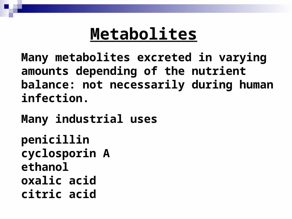
MetabolitesMany metabolites excreted in varying amounts depending of the nutrient balance: not necessarily during human infection.
Many industrial uses
penicillincyclosporin Aethanoloxalic acidcitric acid

Trace elementsIron (Fe) Battle for iron between host transferrin/ lactoferrin
and the fungus. Some fungi produce iron chelators that transport
Fe into cells. Ringworm species (dermatophytes) unable to
compete – may explain why restricted to dead cell layers of skin, and hair and nail.
Histoplasma capsulatum and Penicillium Marneffei-> intracellular pathogens. Glutathione-dependent extracellular ferric reductase
Zygomycosis and iron chelator (deferoxamine) to treat hemochromatosis.

Immune response against fungal infection

Ag. variation during development of the infection.
Distinct host humoral and cellular responses
Conidia spores (shedding, swelling and new antigens are expressed)
Hyphal filaments (more antigens exposed)
Aspergillus CandidaYeast, pseudohyphae, hyphae
Switching of surface antigens in vivo.
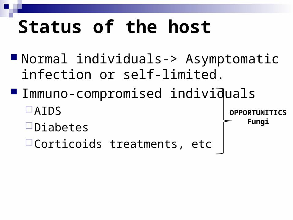
Status of the host
Normal individuals-> Asymptomatic infection or self-limited.
Immuno-compromised individualsAIDSDiabetesCorticoids treatments, etc
OPPORTUNITICSFungi

Non-immune factors in host defenses. Skin, mucous membrane, competition for
nutrients, mucocilliary clearance system of the respiratory tract. Burning, surgical wound Intravenous catheters Antibiotics treatment Iron chelators treatment (Deferoxamine)
Hemochromatosis: genetic or acquired - > Zygomycosis.

Host vs pathogen “Extreme immune response”
Mediastinal fibrosis -> Histoplasmosis Poor immune response
AIDS => High # Cryptococcosis (“no inflammatory responses)
Treatment with antiviral drugs-> life-threatening immune response inflammatory syndrome.
Immune-mediated hypersensitivity reactions. Asthma, allergic bronchopulmonary
aspergillosis.

Front Biosci (Elite Ed). 2009 Jun 1;1:1-12
Innate immune system- Complement system
Phagocytosis(opsonization)
-Chemotaxis(C5a release)
- CR1, CR3, CR4

Pentraxins (PTX3). Pattern recognition molecules-> impair phagocytosis (Aspergillosis)
Anticandidal peptides (β-defensin, Histatin) -> predisposition of patient with xerostomia (dry mouth) to oral candidiasis
Innate immune system-
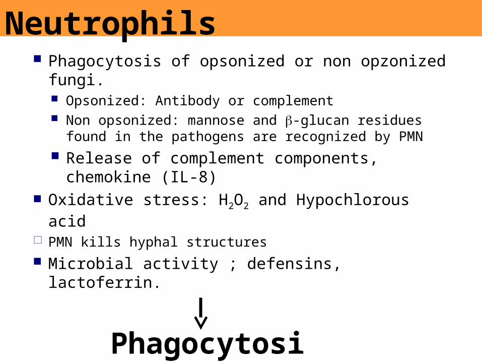
Phagocytosis of opsonized or non opzonized fungi. Opsonized: Antibody or complement Non opsonized: mannose and -glucan residues
found in the pathogens are recognized by PMN
Release of complement components, chemokine (IL-8)
Oxidative stress: H2O2 and Hypochlorous acid PMN kills hyphal structures
Microbial activity ; defensins, lactoferrin.
Neutrophils
Phagocytosis

Candida spp (disseminated disease) Aspergillus spp (52% neutropenia) Zygomycosis (20%) Fusarium spp Trichosporon spp
Chronic granulomatous disease (CGD); failure of NADPH oxidase-> susceptible to: Catalase (+) bacteria (S. aureus) Aspergillus spp Candida spp
Neutropenia
Mononuclear cells
Macrophage. Kills efficiently conidia but not hyphal structures. H capsulatum can survive inside macrophages.

B-deficiency (animal model)-> no effect on infection with Candida, H. capsulatum.
Humoral immunity -> disseminated candidiasis Specific immune response against certain Ags. Cryptoccosis in patients with
Hypogammaglobulinemia Pathogenesis of allergic responses to inhaled
fungi. IgE and IgG specific to fungal Ags ->pathogenesis of chronic rhinosinusitis.
Remain controversial
Acquired immune system- Antibody

CD4; CD8 CD4 T helpers
TH1 and TH17
Conidia and yeast->TH1. Candida spp, A. fumigatus (conidia), dimorphic fungi
Candida spp, Aspergillus -> TH17
TH2 -> Hyphal forms
Disease C. neoformans, P.jiroveci, and dimorphic infection. Candida infection (mucosal infection)-> HIV
Acquired immune system- T cells

DC (TGF-, IL-6) (Dectin 1)
Candida (-glucan)
IL-23
TH17; CD4 T cells (IL-17A and 17B (IL-25))
endothelial, epithelial, fibroblast, keratinocytes
Recruit neutrophils, Augment neutrophils and
Macrophages production in BM Increase differentiation of local
monocytes into macrophages.
IL-6, IL-8 (CXCL8), G-CSF, GM-
CSF
Link: CD4 and neutrophil and macrophage

Predisposing factor Etiologic agents(s) Traumatized skin and mucosa surfaces Candida spp
Neutropenia Candida spp (disseminated diseae) Aspergillus spp Zygomycosis agents Fusarium spp Trichosporon spp
Impaired T cell-mediated immunity Candida spp (mucocutaneous disease) Cryptococcus neoformans Histoplama capsulatum Coccidiodes immitis Pneumocystis jiroveci Paracocciodoiodes brasiliensis Penicillium marneffei
Chronic granulomatous disease Aspergillus spp Candida albicans (disseminated disease)
Diabetes mellitus/Ketoacidosis Agents of zygomycosis
Deferoxamine therapy Agents of zygomycosis
Graft-versus-host disease Aspergillus spp

Antifungal agents

Cell membrane and cell wall
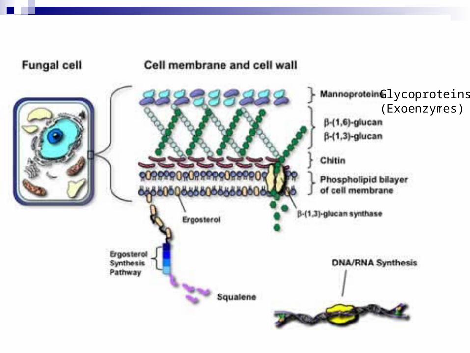
Glycoproteins(Exoenzymes)

Best target is only in fungi not in humans. Fungitoxic drugs:
Cause fungal death
Fungistatic drugs: Prevent further growth (gives immune system
time to catch up)

PRIMARY ANTI-FUNGAL AGENTS1. 5-fluorocytosine (5-FC)2. Polyenes3. Azoles4. Griseofulvin5. Allylamines6. Echinocandins7. Whitfield ointment8. Trimethoprim –sulfamethoxazole (*).
*Used mainly in infections caused by bacteria, but also for fungal infection caused by Pneumocystis jiroveci (HIV + individuals)

permease
5-FC 5-FC
Inside fungal cell
5-FU
deaminase
RNA translation
DNAsynthesisinhibition
5-fluorocytosine (5-FC) Fungicidal (Candida and Cryptococcus)
Enters via cytosine permease Deaminated to 5-fluorouracil (5-FU) (cytosine deaminase absent in human cells)
Bone marrow suppression and alopecia Resistance often seen when not used in combination with other
antifungal drugs (Candida) Permease reduction Deaminase reduction

Polyenes (Fungal sterols) Bind to ergosterol and form ion channels in fungal membrane The permeability of the fungal cell wall is altered and the intracellular
contents leak (potassium leak) Ergosterol: C28 sterols (Humans cells have C27 sterols i.e.;
cholesterol) First fungitoxic drugs (candida)
Nystatin Mucosal infection (topical) and oral to decrease intestinal levels of Candida (not absorbed)
Amphotericin B deoxycholate: (Broad spectrum; amphotericin B lipid complex (ABLC), amphotericin B colloidal dispersion (ABCD), and liposomal amphotericin B (L-amphotericin B)
intravenously or intraperitoneally (systemic candidiasis)
fever, chills, and myalgia
Nephrotoxic: azotemina, decreased glomerular filtration, loss of urinary
concentrating ability, renal loss of sodium and potassium, and renal tubular acidosis
(reduced nephrotoxicity if given as lipid complex or in liposomes
Resistance is uncommon but when present usually is associated with reduced sterol in the cell membranes

Fungal sterols
Ergosterol: C28 sterols (Humans cells have C27 sterols i.e.; cholesterol)
Target for polyene antifungals (nystatin, amphotericin B)

Effect Mechanism Antifungals Involved
Suggested clinical management
Increased accumulation of renally-cleared drugs and/or drug vehicles Flucytosine, Fluconazole, Beta-lactams and many others...
Decrease in glomerular filtration
Amphotericin B Consult package insert. Most drug dosages can be adjusted based on estimates of glomerular filtration (i.e. creatinine clearance). Use of a lipid amphotericin B formulation may help stabilize or slow declines in renal function.
Enhanced nephrotoxicity AminoglycosidesCyclosporineIntravenous Contrast DyeFoscarnet and others...
Enhanced glomerular and tubular toxicity in the kidney
Amphotericin B Minimize co-administration of nephrotoxic agents whenever possible. Consider first-line use of lipid amphotericin B formulation (L-amphotericin B).

AMPHOTERICIN BDisadvantages Intravenous administration Thrombophlebitis Nephrotoxic (Blood Urea Nitrogen (BUN), Creatinine)
Fever Chills Anemia Long term administration

Acetyl CoA
Squalene
Squalene epoxide
Lanosterol
Ergosterol
Ergosterol synthesisPolyenes
(Nystatin and amphotericin B)

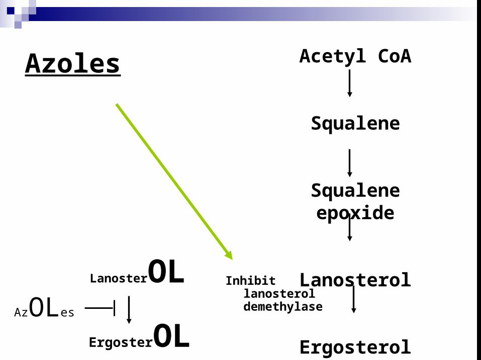
Azoles (Candida spp, Cryptococcus neoformans) Inhibit lanosterol demethylase
Inhibit Ergosterol synthesis Fungistatic (Wide and Broad; Dermatophytes, Systemic fungi,
Candidiosis) Miconazole, Ketoconazole, Fluconazole, Itraconazole*, Voriconazole*,
Posaconazole* ((*) Aspergillus, blastomycosis and histoplasmosis) Oral absorption and solubility is optimal at acidic gastric pH (Ketoconazole) Azoles, specially Ketoconazole, may have antiandrogenic effect, adrenal
supression and liver dysfunction. Teratogenic drugs (avoid pregnant women).
Azole resistance in Candida albicans Several different types of resistance
Mutation in lanosterol demethylase Upregulation of pumps exporting drug
ABC transporter MDR (multiple drug resistance) type In 1996, 10% cases of oral candidiasis in San Francisco were untreatable with
fluconazole. Mutations in C. albicans over 2 years: AIDS. fluconazole resistance
Different yeast species that are inherently resistant to azoles are appearing as important pathogens
Acetyl CoA
Squalene
Squalene epoxide
Lanosterol
Ergosterol
Azoles
Inhibit lanosterol demethylase
AzOLes
LanosterOL
ErgosterOL

Effect Mechanism Antifungals Involved
Suggested clinical management
Decreased serum concentration of azoleAntacids H2 Receptor
antagonism Proton Pump Inhibitors Sulcrafate Didanosine (oral)
Decreased dissolution/absorption of solid dosage form
Ketoconazole, itraconazole (capsules),
Use solution formulation of itraconazole or other azole if indicated (i.e. voriconazole)
Avoid taking antacids within 2 hours of oral azole therapy
Increased metabolism of azole Isoniazid, Rifampin, Phenytoin CarbamazepinePhenobarbitalRitonavir (voriconazole)
Induction of mammalian cytochrome-P450 mediated metabolism of azole
Ketoconazole,itraconazole,fluconazole,voriconazole,posaconazole
Avoid concomitant use of these agents if possible. May require switch to amphotericin B formulation or echinocandin
Consideration of therapy

Increased serum concentration of co-administered drug or metabolite Oral hypoglycemics, S-warfarinR-Wafarin Cyclosporin, Tacrolimus Phenytoin, Protease inhibitors(saquinavir, ritonavir)Busulfan DiltiazemLovastatinIsoniazid, Rifampin, Rifabutin, Quinidine , etc.
Inhibition of cytochrome P450, P-gp, or both
Ketoconazole, itraconazole, voriconazole > fluconazole (usual doses)
Avoid concomitant use if possible. Severity of possible interaction is drug-dependent. Consult prescribing information of each drug to address interaction severity
Effect Mechanism Antifungals Involved
Suggested clinical management

Allylamines
Fungistatic/toxicTerbinafine = Lamisil®, naftidine Inhibit squalene epoxidase
Accumulate in stratum corneum. High activity for ringworm infections

Acetyl CoA
Squalene
Squalene epoxide
Lanosterol
Ergosterol
Allylamines
squalene epoxidaseAllylamines
Allylamines
Squalene
Squalene epoxide
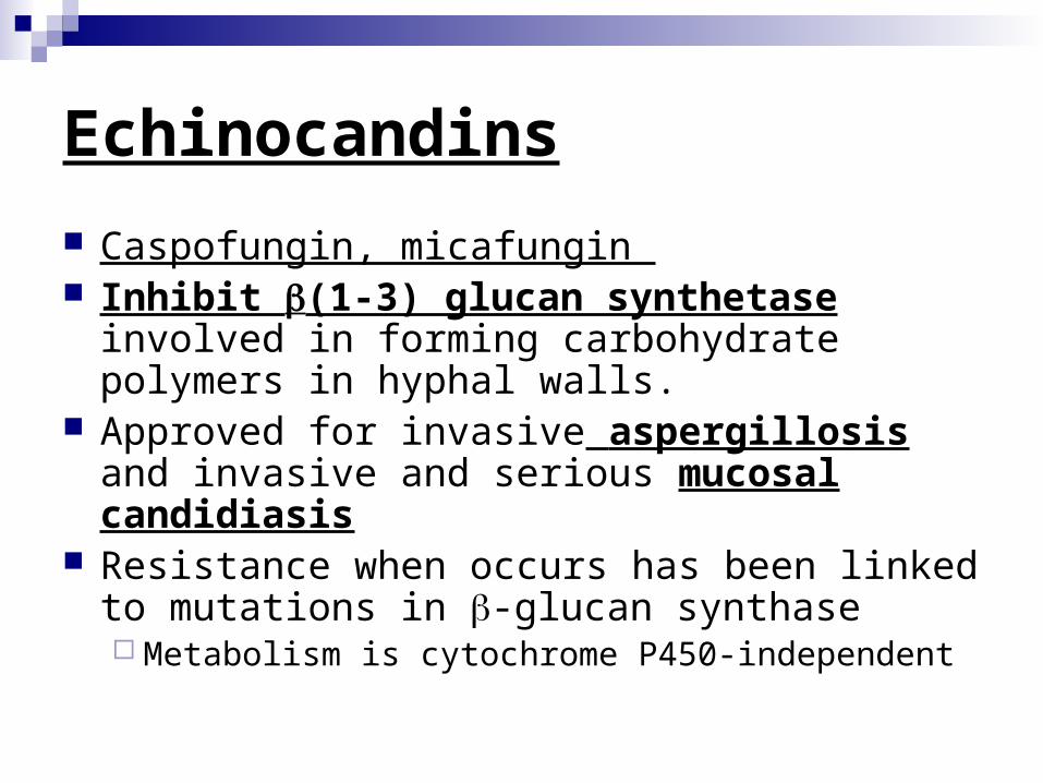
Echinocandins
Caspofungin, micafungin Inhibit (1-3) glucan synthetase involved in
forming carbohydrate polymers in hyphal walls. Approved for invasive aspergillosis and
invasive and serious mucosal candidiasis Resistance when occurs has been linked to
mutations in -glucan synthase Metabolism is cytochrome P450-independent
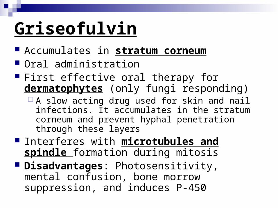
Griseofulvin Accumulates in stratum corneum Oral administration First effective oral therapy for dermatophytes
(only fungi responding) A slow acting drug used for skin and nail infections. It
accumulates in the stratum corneum and prevent hyphal penetration through these layers
Interferes with microtubules and spindle formation during mitosis
Disadvantages: Photosensitivity, mental confusion, bone morrow suppression, and induces P-450

Whitfield ointment May be caustic and impossible to use as
systemic therapy E.g. Salicylic acid, benzoic acid (weak acids, not
ionized at lower pH)
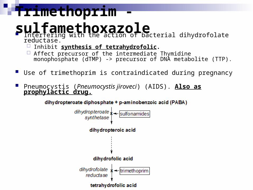
Trimethoprim - sulfamethoxazole Interfering with the action of bacterial dihydrofolate reductase.
Inhibit synthesis of tetrahydrofolic. Affect precursor of the intermediate Thymidine monophosphate (dTMP)
-> precursor of DNA metabolite (TTP).
Use of trimethoprim is contraindicated during pregnancy
Pneumocystis (Pneumocystis jiroveci) (AIDS). Also as prophylactic drug.

Acetyl CoA
Squalene
Squalene epoxide
Lanosterol
Ergosterol
Allylamines(Terbinafine)
squalene epoxidase
Inhibit lanosterol demethylase
Azoles
Polyenes(Nystatin and amphotericin B)
Summary

5-fluorocytosine
Echinocandins(Caspofungin, micafungin)
Griseofulvin
Inhibit (1-3) glucan synthetase; inhibit carbohydrate polymers in hyphal walls.
Inhibit RNA translation and DNA synthesis
Interferes with microtubules and spindle formation during mitosis
Summary

Drug Mechanism of action Treatment Fluorocytosine (5-FC) Inhibit RNA and DNA synthesis Candida and Cryptococcus
Polyenes Binds polyenes; alter membrane permeability
- Nystatin Mucosal (oral/intestinal) infection: candida - Amphotericin B Broad spectrum: systemic infection; penicilliosis
Azoles Inhibit lanosterol demethylase Broad spectrum: systemic infection
- Fluconazole Dermatophytes, Candida (exception: C. krusei, glabrata, guiiermondii); C neoformans, C. immitis.
- Itraconazole Dermatophytes, Candida and C. neoformans (IV no accessible in US; no for systemic candidiasis); dimorphic fungi ; Aspergillus; Phaeohyphomycetes
- Voriconazole Dermatophytes, Candida and C. neoformans; dimorphic fungi; aspergillus; phaeohyphomycetes
- Posaconazole Dermatophytes, Candida and C. neoformans; dimorphic fungi; aspergillus; phaeohyphomycetes
Griseofulvin Interferes with microtubules and spindle
Dermatophytes
Allylamines Inhibit squalene epoxidase - Terbinafine Dermatophytes
Echinocandins Inhibit b(1-3) glucan synthetase Candida and Aspergillus
(Caspo-, mica-, anidula- fungin
Trimethoprim –sulfamethoxazole Inhibit synthesis of tetrahydrofolic
Pneumocystis jiroveci; also prophylactic

End

Supplemental informationClasses 1 and 2

Overview of medically important fungiGroup (criteria
based on Morphology and
disease Disease Etiologic agentDiagnostic in vivo form
Diagnostic in vitro form Natural habitat
1. Molds Chromoblastomycosis Cladosporium Chestnut brown Branching chains of Woody plantBlack fungi carrionii thick-walled single-celled conidia material
Unifomr cells 10 um in diameter
Fonsecaea Same Series of single- Samepedrosoi celled conidia
giving rise to seriesof secondary conidia
Phaeohyphomycosis Exophiala Hyaline to brown Single-celled conidia Woody plant materialjeanselmei yeast-like cells, in balls at the apices
filaments, septate of annellideshyphae, in variouscombinations
Wangiella Same Single-celled conidia Soil and similar ambientsdermatitidis in balls a thte apices
of phialidesand annellides
Xylohypha Brown septate Sparsely branched, bantiana Hypahe long chains of
single-celled conidia

Overview of medically important fungi (cont.)Group (criteria
based on Morphology and
disease Disease Etiologic agentDiagnostic in vivo
formDiagnostic in
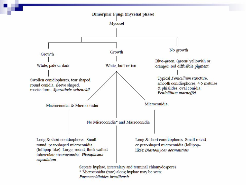
vitro form Natural habitat3. Dimorphic Blastomycosis Blastomyces Round to oval Small, round, smooth Woody plants
dermatiditis yeast s 8-15 um in diameter single-celled conidia materialhaving broad-based budded daugther cells
Coccidioidomycosis Coccidiodes Spherules 30-60 um Alternating barrel Soilimmitis containing single-celled shaped arthroconidia
endospores 2-5 um in diameter2.5 - 4 by 3-6 um
Histoplasmosis Histoplasma Oval, intracellular yeasts Tuberculate macro- Soil enriched by bat, starling capsulatum 2-5 - 3.5 um in diameter microconidiaconidia and smooth- or chicken droppings
walled
Paracoccidioidomycosis Paracoccidioides Multiples budding yeast, roundTypically sterile Probably woody plantsbrasiliensis budded cells
2-10 um attached to mature cells30-60 um in diameter
Sporotrichosis Sporothrix Round to oval yeast 3-5 Conidia develpoing Woody plant materialschenckii um in diameter from sympodial
conidiophores and fromthe hyphae
4 Oportunistic Aspergillosis Aspergillus Septate, dichotomously branchingChain of conidia Sameinfections favus hypahe 2.5 - 3.5 um in from phialides
diameter Same
Aspergillus Samefumigatus Usually sterile, some
isolate from phialidesMycetoma Madurella granules in tissue and draining
mycetomatis sinuses
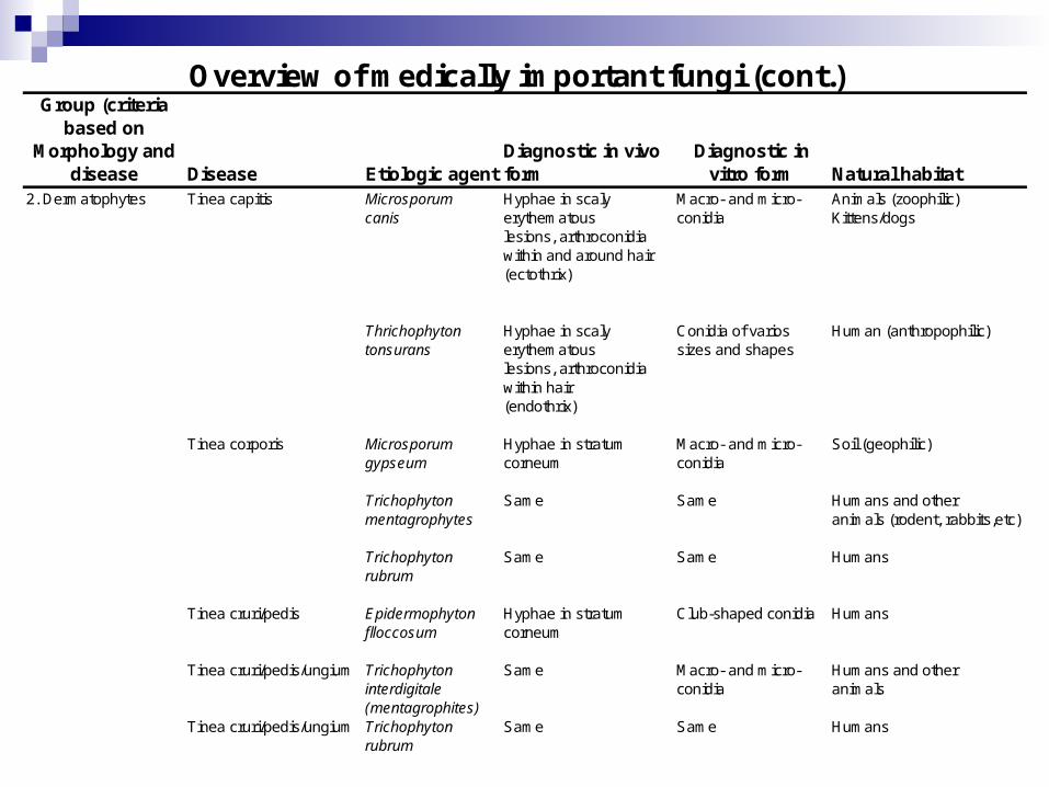
Overview of medically important fungi (cont.)Group (criteria
based on Morphology and
disease Disease Etiologic agentDiagnostic in vivo form
Diagnostic in vitro form Natural habitat
2. Dermatophytes Tinea capitis Microsporum Hyphae in scaly Macro- and micro- Animals (zoophilic)canis erythematous conidia Kittens/dogs
lesions, arthroconidia within and around hair(ectothrix)
Thrichophyton Hyphae in scaly Conidia of varios Human (anthropophilic)tonsurans erythematous sizes and shapes
lesions, arthroconidia within hair(endothrix)
Tinea corporis Microsporum Hyphae in stratum Macro- and micro- Soil (geophilic)gypseum corneum conidia
Trichophyton Same Same Humans and othermentagrophytes animals (rodent, rabbits,etc)
Trichophyton Same Same Humansrubrum
Tinea cruri/pedis Epidermophyton Hyphae in stratum Club-shaped conidia Humansflloccosum corneum
Tinea cruri/pedis/ungium Trichophyton Same Macro- and micro- Humans and otherinterdigitale conidia animals(mentagrophites)
Tinea cruri/pedis/ungium Trichophyton Same Same Humansrubrum

Cell wall composition and taxonomic classification of representative medically important fungi
Principal cell wall polymer Taxonomic Group Examples
Chitin-chitosan Zygomycetes Rhizopus arrhizus
Chitin-glucan Ascomycetes (mycelial) Pseudallescheria boydii
Basidiomycetes (mycelial) Schizophyllum commune
Glucanmannan Ascomycetes (yeast) Sacchamomyces cerivisea
Chitin-mannan Fungi imperfecti Candida albicans
Basidiomycetes (yeast) Filobasidiella neoformans

Hair Perforation Test
Urease Test
Growth at 37°C
Macro-conidia Micro-conidia Distinguishing Characteristics
Trichophyton rubrum
Negative Negative Positive Pencil shaped/cigar shaped
Club shaped to pyriform, along the sides of the hyphae
Red reverse pigment
Hair perf. test neg.
Club shaped microconidia
Trichophyton mentagrophytes
Positive Positive Positive Club shaped when present
Numerous
Unicellular to round in grape like clusters
Round microconidia in grape like clusters Spiral
hyphae
Trichophyton tonsurans
Usually (-)
Occasionally +Positive Positive Cylindrical to cigar
shaped and sinuous, if present
Numerous, varying in shape and size, club shaped to balloon
shaped
Microconidia varying in shape and size
Growth enhanced by thiamine
Trichophyton verrucosum
Negative Negative Positive “Rat-tailed” if present
Rare or Absent
Chlamydospores in chains typically seen
Chlamydospores in chains
Growth better on media with thiamine and inositol
Trichophyton terrestre
Positive Positive Negative 2-8 celled borne at right angles to
hyphae
Club shaped with squared-off base on
pedicels
Microconidia with squared-off base on short pedicels
Epidermophyton floccosum
Negative Positive Positive Club shaped, often in clusters
Absent Khaki colored colony with brown reverse
Microconidia absent
Microsporum
canis
Positive Positive NA Fusoid, thick, rough walled with
recurved apex
Typically absent
Club shaped if present
Fusoid, rough walled macroconidia with
recurved apex
Microsporum gypseum
Positive Positive NA Ellipsoidal to fusiform, thin, Rough walled
Moderately abundant Club shaped
Thin walled macroconidia
Tawny-buff granular colony
Microsporum nanum
Positive Positive NA Typically 2 celled Pear or egg shaped
Rough walled
Clavate when present 2 celled pear shaped macroconidia

Pathogen Fungal ligand(s) Phagocytic receptor(s)
Aspergillus fumigatus Mannans, B-glucans DC-SIGN, dectin-1
Blastomyces dermatitidis BAD1 CR3, CD14
Candida albicans Mannans, B-glucans DC-SIGN, dectin-1
Coccidiodes posadasii Mannans, B-glucans DC-SIGN, dectin-1
Cryptococcus neoformans GlucoronoxylomannanTLR2, TLR4, CD14, CD18, FcyRII
Histoplasma capsulatum HSP60 CD18, VLA-5
Pneumocystis jiroveci Mannans, B-glucans DC-SIGN, dectin-1
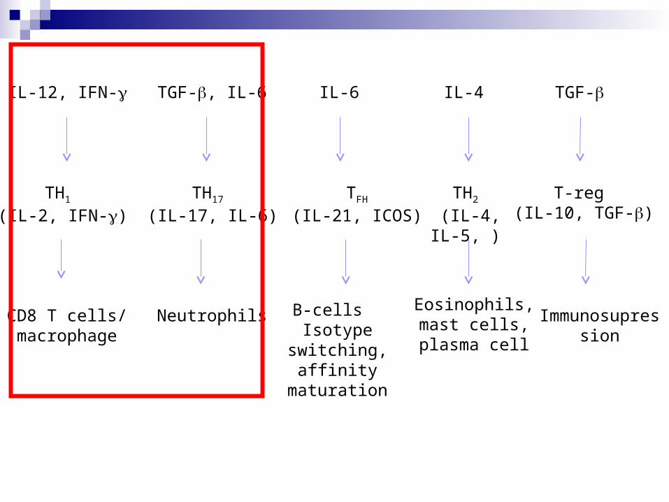
TFH
(IL-21, ICOS)TH1
(IL-2, IFN-)
TGF-TGF-, IL-6 IL-6IL-12, IFN- IL-4
T-reg (IL-10, TGF-)
TH17 (IL-17, IL-6)
TH2 (IL-4, IL-5, )
CD8 T cells/ macrophage
Eosinophils, mast cells, plasma cell
B-cells Isotype switching, affinity maturation
Immunosupression
Neutrophils

C: cream L: lotion NL: nail lacquer O: ointmentOS: oral suspension P: powderS: solution/spray VO: vaginal ointmentVS: vaginal suppository T: troche VT: vaginal tabletD: dermatophytosis CC: cutaneous candidiasisOC: oropharyngeal candidiasis VC: vulvovaginal candidiasis
Chemical ClassGeneric Name
Formulations Indications
Polyenes
Amphotericin B C, L, O CC
Nystatin C, O, OS, P, VT, T CC, OC, VC
Azoles (Imidazoles)
Butoconazole C VC
Clotrimazole C, L, S, T, VT D, CC, OC, VC
Econazole C D, CC
Ketoconazole C, S D, CC
Miconazole C, L, S, P, VS D, CC, VC
Oxiconazole C, L D, CC
Sulconazole C, S D, CC
Terconazole C, VS VC
Tioconazole C, VO VC

Chemical ClassGeneric Name
Formulations Indications
Allylamines and other non-azole ergosterol synthesis inhibitors
Amorolfine NL O
Butenafine HCl C D
Naftifine C, O, P D
Terbinafine C, S D
Other agents
Ciclopirox olamine C, L D, CC
NL O
Haloprogin C D, CC
Tolnaftate C, S, P D
Undecylenate C, P, O, S D
C: cream L: lotion NL: nail lacquer O: ointmentOS: oral suspension P: powderS: solution/spray VO: vaginal ointmentVS: vaginal suppository T: troche VT: vaginal tabletD: dermatophytosis CC: cutaneous candidiasisOC: oropharyngeal candidiasis VC: vulvovaginal candidiasis

Definitions and Nomenclature http://labmed.ucsf.edu/education/residency/fung_morph/fungal_site/
page1.01.html#cleistothecia
Anamorph
Asexual or "imperfect" form of a fungus; for example, Scedosporium apiospermum is the anamorphic form of the teleomorph Pseudallescheria boydii
Arthroconidia
Conidia arising from pre-existing cells in the mycelium; adjacent cells collapse to release the mature form; see, for example, Geotrichum and Coccidioidomycosis
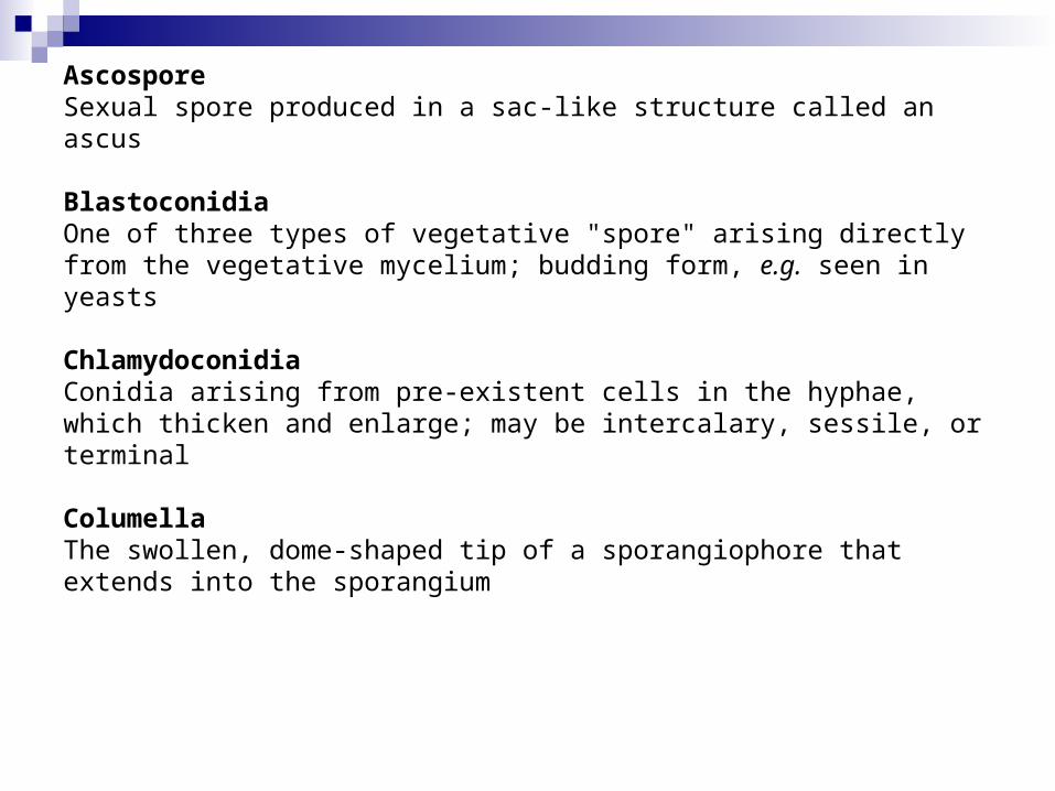
AscosporeSexual spore produced in a sac-like structure called an ascus
BlastoconidiaOne of three types of vegetative "spore" arising directly from the vegetative mycelium; budding form, e.g. seen in yeasts
ChlamydoconidiaConidia arising from pre-existent cells in the hyphae, which thicken and enlarge; may be intercalary, sessile, or terminal
ColumellaThe swollen, dome-shaped tip of a sporangiophore that extends into the sporangium

Conidia(singular conidium) Asexual "spores" of fungus
ConidiophoreSpecialized hyphal element bearing conidia
HolomorphTaxonomic name including teleomorphic and anamorphic forms of a fungus; the name of the teleomorph also serves as the name of the holomorph
Hyphae(singular hypha) The fundamental, threadlike structure of molds
Metula(plural metulae) Structure below the phialide in some Penicillium and Aspergillus species; see for example Aspergillus terreus

Mycelium(plural mycelia) The mass of filaments that constitutes the body of a mold; may be vegetative or aerial (reproductive)
Phialide
A conidiogenous cell that produces conidia from within its apex, which does not increase in width or length during conidiogenesis
Rhizoid
Root-like, branched hyphae which usually extend into growth medium; found especially in Zygomycetes. See, for example, Rhizopus
Sporangia
A fruiting body which forms a closed sac; see, for example, Absidia , Rhizopus

SporangiophoreA specialized hyphal element that bears the sporangium
StolonHorizontal hyphae growing along the surface of growth medium; runner
TeleomorphSexual or "perfect" form of a fungus; see Anamorph and Holomorph, above

1. Antifungal Agents. Encyclopedia of Microbiology, 2009, Pages 205-222. A. Espinel-Ingroff2. Recent Advances in Antifungal Prevention and Treatment. Seminars in Hematology, Volume 46, Issue 3, July
2009, Pages 212-229. Andreas H. Groll, 3. Triazole-resistant candidaemia following posaconazole exposure. International Journal of Antimicrobial Agents,
Volume 33, Issue 5, May 2009, Pages 494-495. Stefan Weiler, Cornelia Lass-Flörl, Jutta Auberger, Rosa Bellmann-Weiler, Markus Stein, Michael Joannidis, Romuald Bellmann
4. Fungal Infections in Hematopoietic Stem Cell Transplantation and Solid-Organ Transplantation—Focus on Aspergillosis. Clinics in Chest Medicine, Volume 30, Issue 2, June 2009, Pages 295-306. Marcio Nucci, Elias Anaissie
5. Reduced fluconazole susceptibility of Candida albicans isolates in women with recurrent vulvovaginal candidiasis: effects of long-term fluconazole therapy. Diagnostic Microbiology and Infectious Disease, Volume 64, Issue 3, July 2009, Pages 354-356. Zainab Shahid, Jack D. Sobel
6. A comparison of the fungicidal activity of amphotericin B and posaconazole against Zygomycetes in vitro Diagnostic Microbiology and Infectious Disease, Volume 63, Issue 4, April 2009, Pages 361-364. Suganthini Krishnan-Natesan, Elias K. Manavathu, George J. Alangaden, Pranatharthi H. Chandrasekar
7. Candida in the ICU. Clinics in Chest Medicine, Volume 30, Issue 2, June 2009, Pages 287-293. Rabih O. Darouiche
8. In vitro synergistic effects of antituberculous drugs plus antifungals against Coccidioides posadasii. International Journal of Antimicrobial Agents, Volume 34, Issue 3, September 2009, Pages 278-280. Rossana de Aguiar Cordeiro, Raimunda Sâmia Nogueira Brilhante, Marcos Fábio Gadelha Rocha, Delia Jessica Astete Medrano, André Jalles Monteiro, Juliane Lira Tavares, Rita Amanda Chaves de Lima, Zoilo Pires de Camargo, José Júlio Costa Sidrim
9. Articular aspergillosis: case report and review of the literature. International Journal of Infectious Diseases, In Press, Corrected Proof, Available online 4 August 2009Oriana Hoi Yun Yu, Annick Wong Wong Keet, Donald C. Sheppard, Timothy Brewer
10. Fungal Infections in the ICU.Infectious Disease Clinics of North America , Volume 23, Issue 3, September 2009, Pages 625-642. Marya D. Zilberberg, Andrew F. Shorr
11. Two Triazole Anti-fungal Drugs Modify IgE-dependent Cytokine Release from Human Lung Tissue Explants. Journal of Allergy and Clinical Immunology, Volume 123, Issue 2, Supplement 1, February 2009, Page S255. J.A. Warner, S.L. Williams
12. Aspergillus osteomyelitis: review of 12 cases identified by the Prospective Antifungal Therapy Alliance registry. Diagnostic Microbiology and Infectious Disease, Volume 63, Issue 4, April 2009, Pages 384-387. David Horn, Sutthichai Sae-Tia, Dionissios Neofytos
13. Noninvasive Pulmonary Aspergillus Infections. Clinics in Chest Medicine, Volume 30, Issue 2, June 2009, Pages 315-335. Brent P. Riscili, Karen L. Wood
14. Coccidioidomycosis: A Review of Recent Advances. Clinics in Chest Medicine, Volume 30, Issue 2, June 2009, Pages 241-251. Neil M. Ampel
Additional information (www.pubmed.org)

3. Pictures of dermatomycosishttp://www.mycology.adelaide.edu.au/Mycoses/Cutaneous/Dermatophytosis
4. An Introduction to MycologyBy R. S. Mehrotra, K. R. Aneja An Introduction to Mycologyhttp://books.google.com/books?id=UUorj_O2dcsC&pg=PR11&lpg=PR11&dq=mycology+class&source=bl&ots=r0woQyTj3P&sig=kEn9blHfksUnxGVpr1ekdS8TfxE&hl=en&ei=eU5hSoW5HMKktgfEuMyyAg&sa=X&oi=book_result&ct=result&resnum=2#v=onepage&q=mycology%20class&f=false
2. Hickey, P.C. & Read, N.D. (2003). The biology of the living fungi. British Mycological Society: Wokingham, UK. Available. Sponsored and published by The British Mycological Society http://www.fungalcell.org/cdrom/http://www.britmycolsoc.org.uk
1. Doctor Fungushttp://www.doctorfungus.org/
5. Medical Microbiology 6Th edition. Murray Patrick R. 2009.
Additional information (online)
6. Merck manual online medical library http://www.merck.com/mmhe/sec17/ch197/ch197e.html7. emedicine http://emedicine.medscape.com/public/about
8. Conidiospores formationhttp://www.mycolog.com/CHAP4a.htm