Embed Size (px)
Citation preview

RESEARCH Open Access
MyD88 is pivotal for immune recognition ofCitrobacter koseri and astrocyte activation duringCNS infection†
Shuliang Liu1,3 and Tammy Kielian2*
Abstract
Citrobacter koseri (C. koseri) is a Gram-negative bacterium that can cause a highly aggressive form of neonatalmeningitis, which often progresses to establish multi-focal brain abscesses. The roles of Toll-like receptor 4 (TLR4)and its signaling adaptor MyD88 during CNS C. koseri infection have not yet been examined, which is importantsince recent evidence indicates that innate immune responses are tailored towards specific pathogen classes. HereTLR4 WT (C3H/FeJ) and TLR4 mutant (C3H/HeJ) mice as well as MyD88 KO animals were infected intracerebrallywith live C. koseri, resulting in meningitis and ventriculitis with accompanying brain abscess formation. MyD88 KOmice were exquisitely sensitive to C. koseri, demonstrating enhanced mortality rates and significantly elevatedbacterial burdens compared to WT animals. Interestingly, although early proinflammatory mediator release (i.e.12 h) was MyD88-dependent, a role for MyD88-independent signaling was evident at 24 h, revealing acompensatory response to CNS C. koseri infection. In contrast, TLR4 did not significantly impact bacterial burdensor proinflammatory mediator production in response to C. koseri. Similar findings were obtained with primaryastrocytes, where MyD88-dependent pathways were essential for chemokine release in response to intact C. koseri,whereas TLR4 was dispensable; implicating the involvement of alternative TLRs since highly enriched astrocytes didnot produce IL-1 upon bacterial exposure, which also signals via MyD88. Collectively, these findings demonstratethe importance of MyD88-dependent mechanisms in eliciting maximal proinflammatory responses, astrocyteactivation, and bacterial containment during CNS C. koseri infection, as well as a late-phase MyD88-independentsignaling pathway for cytokine/chemokine production.
Keywords: MyD88 Toll-like receptor 4 (TLR4), C. koseri, meningitis, brain abscess, astrocyte
IntroductionCitrobacter koseri (formerly known as C. diversus) is aGram-negative bacillus with a predilection for causingmeningitis and multi-focal brain abscesses in humanneonates [1,2]. In fact, almost one-third of infants andyoung children infected with C. koseri succumb to thedisease, and approximately half of those who surviveinfection experience long-term neurological deficits dueto focal or diffuse brain damage [1,2]. Increasing evi-dence has accumulated demonstrating that innateimmune responses are tailored towards specific patho-gen classes [3-5]. Specifically, the types of responses
elicited by Gram-positive bacteria can differ significantlyfrom those triggered by Gram-negative pathogens. Inaddition, the majority of studies examining bacterialpathogenesis in the CNS have utilized Gram-positiveorganisms [6-8], which eliminates the involvement ofkey Toll-like receptors (TLRs) that may trigger distinctpathways during Gram-negative infections (i.e. TLR4,TLR5). Therefore, it is important to investigate the CNSresponse to divergent pathogens to identify unique aswell as conserved responses, which may facilitate thedevelopment of novel treatment strategies that wouldcross multiple bacterial species.The innate immune system recognizes multiple patho-
gen classes via highly conserved molecular motifs,termed pathogen-associated molecular patterns(PAMPs), through a limited set of germ-line encoded
* Correspondence: [email protected] of Pathology and Microbiology, University of Nebraska MedicalCenter, Omaha, NE 68198 USAFull list of author information is available at the end of the article
Liu and Kielian Journal of Neuroinflammation 2011, 8:35http://www.jneuroinflammation.com/content/8/1/35
JOURNAL OF NEUROINFLAMMATION
© 2011 Liu and Kielian; licensee BioMed Central Ltd. This is an Open Access article distributed under the terms of the CreativeCommons Attribution License (http://creativecommons.org/licenses/by/2.0), which permits unrestricted use, distribution, andreproduction in any medium, provided the original work is properly cited.

receptors known as pattern recognition receptors (PRRs)[9,10]. Toll-like receptors (TLRs) are a family of PRRsresponsible for sensing numerous PAMPs of bacterial,viral, and fungal species [9]. For example, lipoproteinsand LPS contained in the outer cell wall of Gram-nega-tive bacteria are agonists for TLR2 and TLR4, respec-tively; flagellin, which is the main component ofbacterial flagella, engages TLR5; and bacterial DNA con-taining unmethylated CpG motifs binds to TLR9. SinceCitrobacter are Gram-negative bacilli and possess anouter cell wall rich in LPS, it was anticipated that TLR4-mediated signaling would be important in eliciting hostinflammatory responses. However, LPS is not the onlyCitrobacter-derived PAMP that could be recognized bythe host. Other candidates include, but are not limitedto, lipoproteins, flagellin, and bacterial DNA that arerecognized by TLR2, TLR5, and TLR9, respectively.Therefore, all of these TLRs could conceivably contri-bute to activation of the host inflammatory responseduring CNS C. koseri infection and since they all utilizethe common adaptor molecule myeloid differentiationfactor 88 (MyD88) [11,12], we also examined responsesto bacterial challenge in MyD88 KO mice.Although their primary function is to provide support
for maintaining CNS homeostasis, astrocytes can alsoparticipate in innate immune responses and serve as amajor source of chemokines [13,14]. Astrocytes expressTLRs and can clearly contribute to innate immune pro-cesses in the CNS [13,15]. Although astrocytes are stra-tegically positioned at the blood-brain barrier, a sitewhere C. koseri must traverse to colonize the CNS par-enchyma during infection, to date no studies have exam-ined whether astrocytes are capable of recognizing thispathogen and the downstream consequences elicited. Inaddition, since astrocytes are the most numerous celltype in the CNS parenchyma these cells likely play amajor role in dictating the course of C. koseri ventriculi-tis, meningitis, and brain abscess formation.Following TLR engagement, a number of diverse sig-
naling pathways can be triggered, the nature of whichdepends on the specific PAMP [9,16]. Most TLR-mediated signaling pathways converge to utilize thecommon intracellular adaptor molecule MyD88, withthe exception of TLR3. However, in addition to utilizingMyD88, TLR4-mediated signaling can also occurthrough alternative adapter molecules, namely TIR-domain-containing adaptor protein-inducing IFN-b(TRIF) and TRIF-related adaptor molecule (TRAM)[9,16]. The MyD88-dependent pathway is responsiblefor early-phase NF-�B and MAPK activation, whichinduces proinflammatory cytokine/chemokine expres-sion. In contrast, the MyD88-independent, TRIF-depen-dent pathway activates interferon regulatory factor 3(IRF3), which is required for the expression of IFN-
inducible genes. Furthermore, this pathway mediateslate-phase NF-�B as well as MAPK activation, also con-tributing to inflammatory responses [17]. Since C. koseriis a Gram-negative pathogen with abundant LPS in itsouter cell wall, it was important to consider the possiblecontribution of MyD88-independent pathways for elicit-ing inflammatory gene expression during CNS infection.To assess the functional importance of TLR4 and
MyD88 in C. koseri-induced parenchymal infection,TLR4 WT (C3H/FeJ) and TLR4 mutant (C3H/HeJ)mice as well as MyD88 KO animals were infected intra-cerebrally with live C. koseri. MyD88 KO mice wereexquisitely sensitive to C. koseri and succumbed toinfection within 24-36 h following bacterial exposure.The enhanced mortality rate of MyD88 KO mice wastypified by significantly elevated bacterial burdens andattenuated cytokine/chemokine expression and immunecell influx early after infection (i.e. 12 h). Interestingly,MyD88-independent pathways were triggered with adelayed kinetics (i.e. 24 h), likely representing a compen-satory mechanism in an attempt to contain infection. Incontrast to MyD88, TLR4 had minimal impact on bac-terial burdens or proinflammatory mediator production.Similar findings were obtained with astrocytes, whereMyD88-dependent signaling was essential for chemokineproduction by C. koseri treated cells, whereas TLR4 wasdispensable. Collectively, these findings demonstrate theimportance of MyD88-dependent mechanisms in elicit-ing maximal proinflammatory responses and bacterialcontainment during CNS C. koseri infection, as well as alate-phase MyD88-independent signaling pathway forcytokine/chemokine production.
Materials and methodsMouse strainsMyD88 KO mice (kindly provided by Dr. Shizuo Akira,Osaka University, Osaka, Japan) have been backcrossedwith C57BL/6 mice for over 10 generations and age-and sex-matched C57BL/6 mice were purchased fromthe National Cancer Institute (NCI-Frederick, Frederick,MD) as WT controls. C3H/FeJ and C3H/HeJ mice werepurchased from the Jackson Laboratory (Bar Harbor,ME). The C3H/HeJ strain carries a spontaneous muta-tion in the cytoplasmic domain of TLR4 making itunable to transduce a signal in response to LPS (here-after referred to as TLR4 mutant) [18,19] and C3H/FeJmice were used as WT controls.
Ethical approvalThe animal use protocols were approved by the Institu-tional Animal Care and Use Committees at the Univer-sity of Nebraska Medical Center and University ofArkansas for Medical Sciences, and are in accord withNIH guidelines for the use of rodents.
Liu and Kielian Journal of Neuroinflammation 2011, 8:35http://www.jneuroinflammation.com/content/8/1/35
Page 2 of 14

Citrobacter koseri isolateC. koseri strain 4036 was originally isolated from thecerebrospinal fluid of an infant with meningitis andmultiple brain abscesses [20] and was kindly providedby Dr. J. G. Vallejo (Baylor College of Medicine, Hous-ton, TX). Based on antibiotic sensitivity profiles (per-formed by the Clinical Microbiology Laboratory atArkansas Children’s Hospital, Little Rock, AR), thestrain was propagated in the presence of ampicillin (20μg/ml) in brain-heart infusion broth with constant agita-tion (250 rpm) and recovered at mid-log-phase (12-18 hincubation) by washing twice with ice-cold PBS. To pre-pare live C. koseri stocks, freshly recovered bacteriawere resuspended in PBS containing 10% DMSO and5% bovine serum albumin (Sigma-Aldrich, St. Louis,MO) and aliquots stored at -80°C. Immediately beforeinfection, aliquots were washed twice with ice-cold PBSto remove residual DMSO, serially diluted 10-fold inPBS, and plated on blood agar plates to determine infec-tious titers.
Generation of C. koseri-induced meningitis and brainabscessAge- and gender-matched TLR4 WT and TLR4 mutant,or C57BL/6 WT and MyD88 KO mice (4-5 wks old)were used for in vivo studies to investigate C. koseri-induced meningitis and brain abscess pathogenesis.Briefly, live bacteria were directly inoculated into thebrain parenchyma by stereotactic injection as previouslydescribed [21]. C. koseri was resuspended in PBS ratherthan encapsulated in agarose beads, the latter of whichrepresents the standard method for inducing experimen-tal brain abscesses [22]. The rationale for this approachis that the meninges are punctured during intracerebralinjections and the introduction of bacteria in an aqueoussuspension allows for pathogen reflux into the subarach-noid space via the needle tract, leading to meningitisformation. This procedure recapitulates clinical diseasesince many infants infected with C. koseri developmeningitis and ventriculitis that is often complicated byabscess formation [2]. Since pilot studies indicated thatMyD88 KO animals were extremely sensitive toC. koseri, succumbing to infection within the first 24-36h following bacterial exposure, MyD88 KO and WTmice were euthanized at either 12 or 24 h post-infectionto eliminate potential bias from only examining animalsthat had survived the infection (i.e. “survival bias”).
Preparation of brain homogenates for quantitation ofbacterial titers and proinflammatory mediator productionMice were sacrificed with an overdose of inhaled isoflur-ane and perfused transcardially with ice cold PBS. Boththe infected and contralateral hemispheres were col-lected, weighed, and homogenized in 500 μl of
homogenization buffer [1X PBS supplemented with aprotease inhibitor cocktail tablet (Roche, Indianapolis,IN) and RNase inhibitor (Promega, Madison, WI)] onice. To quantitate the numbers of viable bacteria, serial10-fold dilutions of brain tissue homogenates were pla-ted onto blood agar. Titers were calculated by enumer-ating colony growth and are expressed as colonyforming units (CFU) per gram wet tissue weight. Super-natants were collected from brain homogenates follow-ing centrifugation at 14,000 rpm for 20 min at 4°C fordownstream analyses of cytokine and chemokine expres-sion by Milliplex multi-analyte bead arrays. Protein con-centrations of brain homogenates were determinedusing a colorimetric Bio-Rad DC Protein Assay kit asspecified by the manufacturer (Bio-Rad, Hercules, CA).
Milliplex multi-analyte bead arrayTo compare proinflammatory mediator expression profilesbetween the various mutant and WT mice, a custom-designed mouse cytokine/chemokine microbead array wasutilized according to the manufacturer’s instructions(Milliplex; Millipore, Billerica, MA). This microbead arrayallows for the simultaneous detection of 19 individualinflammatory molecules in a single 75 μl brain homoge-nate including IL-1a, IL-1b, TNF-a, IFN-g, IL-6, IL-9, IL-10, IL-12p70, IL-12p40, IL-15, IL-17, CXCL1/keratinocytechemoattractant (KC), CXCL2/macrophage inflammatoryprotein-2 (MIP-2), CXCL9/monokine induced by IFN-g(MIG), CXCL10/IFN-g-induced protein 10 (IP-10), CCL2/monocyte chemoattractant protein-1 (MCP-1), CCL3/MIP-1a, CCL4/MIP-1b, and CCL5/regulated upon activa-tion T cell expressed and secreted (RANTES). Resultswere analyzed using a Bio-Plex workstation (Bio-Rad) andadjusted based on the amount of total protein extractedfrom brain tissue homogenates for normalization.
Quantitation of cellular infiltrates in C. koseri infectedbrain tissues by FACSTo determine whether MyD88 loss affected neutrophilor macrophage influx and/or microglial percentages fol-lowing C. koseri infection, cells were quantitated byFACS analysis as previously described [6,23]. Briefly,mice were perfused with PBS to eliminate leukocytesfrom the vasculature and both hemispheres were col-lected to recover infection-associated cells. Brain tissueswere minced in HBSS supplemented with 10% FBS andfiltered through a 70 μm nylon mesh strainer. Theresulting tissue suspensions were digested for 30 min at37°C in HBSS supplemented with collagenase type I(Sigma-Aldrich) and DNase I (Invitrogen, San Diego,CA; 2 mg/ml and 28 U/ml final concentrations, respec-tively) to obtain a single-cell suspension. Followingenzyme neutralization, cells were layered onto a discon-tinuous Percoll gradient (1.03-1.088 g/ml), whereupon
Liu and Kielian Journal of Neuroinflammation 2011, 8:35http://www.jneuroinflammation.com/content/8/1/35
Page 3 of 14

myelin debris was carefully aspirated and the cell inter-face collected. Following extensive washes and incuba-tion in Fc Block™ (BD Biosciences, San Diego, CA) tominimize non-specific antibody binding to Fc receptors,cells were stained with directly conjugated Abs (Ly-6G-PE, F4/80-Alexa 488, and CD45-APC, all from BD Bios-ciences) to identify neutrophils (Ly-6G+, F4/80-, andCD45high), macrophages (Ly-6G-, F4/80+, and CD45high),and microglia (Ly-6G-, F4/80+, and CD45low-intermediate).The vital stain 7-AAD (eBiosciences, San Diego, CA)was included to discriminate viable from dead cells.Cells were analyzed using a BD FACSAria cytometer(BD Biosciences) with compensation set based on thestaining of each individual fluorochrome alone and cor-rection for autofluorescence with unstained cells. Con-trols included cells stained with directly conjugatedisotype control Abs (BD Biosciences) to assess thedegree of nonspecific staining. Results are presented asthe percentage of viable neutrophils, macrophages, andmicroglia recovered from MyD88 KO animals comparedto WT mice (set to 100%).After sorting, viable neutrophils, macrophages, and
microglia recovered from C. koseri infected MyD88 WTand KO mice were enumerated and added to 96-wellplates without further stimulation for 24 h, whereuponconditioned medium was collected to quantitate proin-flammatory mediator expression using Milli-Plex multi-analyte bead arrays. Results are expressed as the concen-tration of each inflammatory mediator per 103 cells fornormalization.
Processing of brain tissues for histological andimmunofluorescence analysisBrain tissues were collected from C. koseri infectedMyD88 KO and WT animals at 24 h post-injection toevaluate neuropathological changes. To prepare tissuesfor immunostaining, mice were perfused with 4% par-aformaldehyde (PFA) in 1X PBS using a peristaltic per-fusion pump at a flow rate of 10 ml/min. Brain tissueswere post-fixed in cold 4% PFA for 1 h and cryopro-tected in 30% sucrose prior to freezing and embeddingin OCT (Optimal Cutting Temperature compound,Sakura Finetek USA Inc., Torrance, CA) for cryostatsectioning. Serial sections of brain tissues (10 μm)were prepared and processed for immunofluorescencestaining with Iba-1 (Biocare Medical, Concord, CA) toassess microglial/macrophage reactivity and imagedusing a Zeiss LSM 510 META confocal microscope(Zeiss, New York, NY). In addition, infected brain tis-sues were processed for Gram staining to visualizeextracellular C. koseri and the extent of bacterialdissemination.
Purification of primary astrocytes by FACSPrimary astrocytes were derived from mixed glial cul-tures of TLR4 WT, TLR4 mutant, C57BL/6 WT, andMyD88 KO mice (1-4 days of age) as previouslydescribed [24]. One concern when working with primaryastrocytes relates to the issue of cell purity, principallydue to low levels of residual microglia [25]. Therefore,after a minimum of three passages while cultured in thepresence of 0.1 mM L-LME, a microglial cytotoxic agent[25,26], the resulting cells were stained with CD11b, acell surface marker expressed on microglia, but notastrocytes, and collected by negative selection to obtainhighly enriched astrocytes. Briefly, cells were incubatedwith Fc Block™ followed by a CD11b-PerCP-Cy5.5 anti-body (both from BD Biosciences). Cells were sorted on aFACSAria (BD Biosciences) at a low flow rate through alarge nozzle to minimize astrocyte damage during thesorting procedure. Post-sort analysis revealed less than1% contamination with CD11b+ microglia. An additionalassessment of astrocyte purity after sorting was obtainedby culturing astrocytes on glass cover slips followed byimmunofluorescence staining with GFAP and CD11b(Additional File 1, Figure S2).
Enzyme linked immunosorbent assay (ELISA)Quantitation of CCL2 and TNF-a (mouse OptEIA, BDBiosciences) and CXCL2 and IL-1b (mouse DuoSet; R &D Systems, Minneapolis, MN) levels in conditionedsupernatants from astrocytes treated with C. koseri wasperformed using standard sandwich ELISA kits accord-ing to the manufacturer’s instructions.
Nitrite assayLevels of nitrite, a stable end product of nitric oxide(NO) following its reaction with O2, were quantitatedin conditioned supernatants of astrocytes treated withC. koseri using the Griess reagent (0.1% naphtyletylene-diamine dihydrochloride, 1% sulfanilamide, and 2.5%phosphoric acid, all from Sigma). The absorbance at550 nm was measured on a plate reader (Spectra Max190; Molecular Devices, Sunnyvale, CA, USA), andnitrite concentrations determined using a standardcurve with sodium nitrite (NaNO2; Sigma; level of sen-sitivity 0.4 μM).
Statistical analysisSignificant differences were determined either by a Stu-dent’s t-test or one-way analysis of variance (ANOVA)followed by the Holm-Sidak method for multiple pair-wise comparisons as indicated (SPSS Science, Chicago,IL). For all analyses, a p-value of less than 0.05 was con-sidered to be statistically significant.
Liu and Kielian Journal of Neuroinflammation 2011, 8:35http://www.jneuroinflammation.com/content/8/1/35
Page 4 of 14

ResultsMyD88-dependent signals are critical for bacterialcontainment and inflammatory mediator productionduring C. koseri brain infectionTo evaluate the functional importance of MyD88-depen-dent pathways for CNS immune responses to C. koseri,we employed a mouse model of meningitis and brainabscess formation using a C. koseri clinical isolate [20].After subjecting C57BL/6 mice to a series of escalatinginoculums, a dose of approximately 3 × 10⁴ cfu resultedin a 30% mortality rate concomitant with meningitis andventriculitis, and brain abscess formation in a subset ofanimals (Additional File 2, Figure S1). Therefore, thisdose was used for all subsequent experiments.MyD88 KO mice were exquisitely sensitive to intracer-
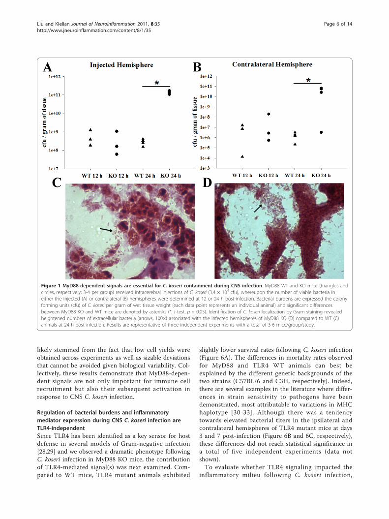
ebral C. koseri, with the majority of animals succumbingto infection within 24-36 h following bacterial exposure(data not shown). Because MyD88 was essential for sur-vival, this dictated acute sampling intervals of 12 and 24h following bacterial exposure to avoid potential artifactsdue to “survival bias”. Bacterial burdens were similarbetween MyD88 KO and WT mice at 12 h post-infec-tion (Figure 1A and 1B). In contrast, C. koseri titerswere significantly elevated in both the injected and con-tralateral hemispheres of MyD88 KO mice at 24 h fol-lowing bacterial exposure compared to WT animals(Figure 1A and 1B), revealing a pivotal role for MyD88in controlling C. koseri infection. This finding was cor-roborated by Gram-staining, where MyD88 KO miceexhibited greater extracellular C. koseri compared toWT animals (Figure 1D and 1C, respectively). Collec-tively, these findings reveal the critical role that MyD88-dependent signals play in bacterial containment duringCNS C. koseri infection.The dramatic sensitivity of MyD88 KO mice to
intracerebral C. koseri suggested defects in mounting aprotective antibacterial inflammatory response in theCNS parenchyma. To examine this possibility, proin-flammatory mediator expression was evaluated inMyD88 KO and WT animals using a multi-analytebead array. Indeed, the expression of numerous cyto-kines (i.e. IL-1b, TNF-a, and IL-6) and chemokines (i.e. CXCL1, CXCL2, CXCL10, and CCL2) were signifi-cantly attenuated in infected MyD88 KO mice at 12 hfollowing bacterial exposure, reflecting impairedimmune activation (Figure 2). Surprisingly, the levelsof these same mediators were significantly increasedin MyD88 KO animals at 24 h post-infection, suggest-ing a late induction of MyD88-independent signalingto elicit proinflammatory mediator release (Figure 2).It is possible that this MyD88-independent responseresulted from heightened bacterial burdens observedin MyD88 KO animals at this interval post-infection(Figure 1).
Neutrophil and macrophage infiltration and activationinto C. koseri-infected brain parenchyma is attenuated inMyD88 KO miceThe differential impact of MyD88 signaling on chemo-kine expression at 12 versus 24 h post-infection led usto examine whether these changes correlated withalterations in peripheral immune cell influx. To quanti-tate the potential differences in neutrophils, macro-phages, and microglia between MyD88 KO and WTmice, three-color FACS analysis was utilized. A dramaticreduction in neutrophil and macrophage infiltrates wasobserved in MyD88 KO mice at 12 h following C. koseriexposure (data not shown), which correlated with thediminished chemokine expression observed at this timepoint (Figure 2). Interestingly, neutrophil and macro-phage influx remained significantly lower in MyD88 KOcompared to WT animals at 24 h post-infection (Figure3), which contrasted with the heightened chemokineexpression observed at this interval (Figure 2). The per-centage of microglia recovered following C. koseri infec-tion was also significantly attenuated in MyD88 KOmice, which may be a consequence of the extensivenecrosis that accompanied infection in these animals. Inaddition, Iba-1 staining revealed that macrophage/microglial reactivity was attenuated in MyD88 KOanimals (Figure 4), which corroborates the decreasedpercentages of each as revealed by FACS.To determine whether viable abscess-associated neu-
trophils, macrophages, or microglia recovered from theinfected CNS of MyD88 KO or WT mice displayed anydifferences in inflammatory mediator expression, weperformed microbead array analysis on conditionedsupernatants from each cell type following a 24 h incu-bation period in vitro without bacterial re-stimulation,in an attempt to capture cellular activation states thatwere ongoing in vivo. MyD88 was important for elicitingmicroglial proinflammatory mediator release duringCNS C. koseri infection as demonstrated by decreasedproduction of CXCL1, CXCL2, CCL3, CCL4, and IL-6(Figure 5A). This finding agrees with our previousreport where MyD88-dependent signaling was shown tobe important for microglial activation in response to C.koseri in vitro [27]. Similarly, macrophage activation inresponse to CNS C. koseri infection was also influencedby MyD88 as revealed by attenuated CXCL1, CXCL2,and IL-6 production (Figure 5B). Infiltrating neutrophilsassociated with C. koseri infected brain also requiredMyD88-dependent activation signals for proinflamma-tory mediator release (Figure 5C). None of these differ-ences in cytokine/chemokine expression between celltypes recovered from MyD88 KO versus WT micereached statistical significance, although trends towardsreduced mediator levels were observed in all threepopulations. The failure to detect significant changes
Liu and Kielian Journal of Neuroinflammation 2011, 8:35http://www.jneuroinflammation.com/content/8/1/35
Page 5 of 14
likely stemmed from the fact that low cell yields wereobtained across experiments as well as sizable deviationsthat cannot be avoided given biological variability. Col-lectively, these results demonstrate that MyD88-depen-dent signals are not only important for immune cellrecruitment but also their subsequent activation inresponse to CNS C. koseri infection.
Regulation of bacterial burdens and inflammatorymediator expression during CNS C. koseri infection areTLR4-independentSince TLR4 has been identified as a key sensor for hostdefense in several models of Gram-negative infection[28,29] and we observed a dramatic phenotype followingC. koseri infection in MyD88 KO mice, the contributionof TLR4-mediated signal(s) was next examined. Com-pared to WT mice, TLR4 mutant animals exhibited
slightly lower survival rates following C. koseri infection(Figure 6A). The differences in mortality rates observedfor MyD88 and TLR4 WT animals can best beexplained by the different genetic backgrounds of thetwo strains (C57BL/6 and C3H, respectively). Indeed,there are several examples in the literature where differ-ences in strain sensitivity to pathogens have beendemonstrated, most attributable to variations in MHChaplotype [30-33]. Although there was a tendencytowards elevated bacterial titers in the ipsilateral andcontralateral hemispheres of TLR4 mutant mice at days3 and 7 post-infection (Figure 6B and 6C, respectively),these differences did not reach statistical significance ina total of five independent experiments (data notshown).To evaluate whether TLR4 signaling impacted the
inflammatory milieu following C. koseri infection,
Figure 1 MyD88-dependent signals are essential for C. koseri containment during CNS infection. MyD88 WT and KO mice (triangles andcircles, respectively; 3-4 per group) received intracerebral injections of C. koseri (3.4 × 10⁴ cfu), whereupon the number of viable bacteria ineither the injected (A) or contralateral (B) hemispheres were determined at 12 or 24 h post-infection. Bacterial burdens are expressed the colonyforming units (cfu) of C. koseri per gram of wet tissue weight (each data point represents an individual animal) and significant differencesbetween MyD88 KO and WT mice are denoted by asterisks (*, t-test, p < 0.05). Identification of C. koseri localization by Gram staining revealedheightened numbers of extracellular bacteria (arrows, 100×) associated with the infected hemispheres of MyD88 KO (D) compared to WT (C)animals at 24 h post-infection. Results are representative of three independent experiments with a total of 3-6 mice/group/study.
Liu and Kielian Journal of Neuroinflammation 2011, 8:35http://www.jneuroinflammation.com/content/8/1/35
Page 6 of 14

proinflammatory mediator expression was quantifiedusing a microbead array. In general, chemokine expres-sion was significantly higher at day 3 compared to day 7post-infection, regardless of mouse strain (Figure 7Aand 7B). Only CXCL10 expression was significantly atte-nuated in TLR4 mutant mice at day 3 post-infection
Figure 2 Proinflammatory mediator production following CNS C. koseri infection is influenced by both MyD88-dependent and-independent signals. MyD88 WT and KO mice (4-6 per group) received intracerebral injections of C. koseri (3.4 × 10⁴ cfu), whereuponsupernatants were prepared from infected hemispheres and cytokine/chemokine expression evaluated using multiplex microbead arrays for IL-1b(A), TNF-a (B), IL-6 (C), CXCL1 (D), CXCL2 (E), CXCL10 (F), CCL2 (G), and CCL5 (H). Cytokine/chemokine expression was normalized based on theamount of total protein obtained for each sample to correct for differences in tissue sampling size. Significant differences in mediator expressionbetween MyD88 WT and KO mice at 12 h post-infection are denoted by asterisks (*, p < 0.05, **, p < 0.01, t-test), whereas significant differencesin mediator levels between MyD88 WT and KO animals at 24 h post-infection are denoted by hatched signs (#, p < 0.05, ##, p < 0.01, t-test).Results are representative of two independent experiments with a total of 3-6 mice/group/study.
Figure 3 Neutrophil and macrophage influx into C. koseri-infected brain is dictated by MyD88-dependent signaling.MyD88 KO and WT mice (5-8 per group) received intracerebralinjections of C. koseri and were sacrificed at 24 h following bacterialexposure, whereupon the percentages of viable neutrophils (Ly-6G+,F4/80-, and CD45high), macrophages (Ly-6G-, F4/80+, and CD45high),and microglia (Ly-6G-, F4/80+, and CD45low-intermediate) werequantitated by FACS analysis with the vital stain 7-AAD. Results areexpressed as the percentage of cells normalized to WT values (setto 100%) and represent the mean ± SEM from three independentexperiments. Significant differences between MyD88 KO versus WTmice are denoted by asterisks (*, p < 0.05, **, p < 0.01, t-test).
Figure 4 Microglial/macrophage activation in response to C.koseri infection is impaired in MyD88 KO mice. MyD88 KO andWT mice (6 per group) were sacrificed at 24 h following C. koseriexposure, whereupon brain tissues were fixed by paraformaldehydeperfusion and processed for cryostat sectioning. Ten μm thick brainsections were subjected to immunofluorescence staining using Iba-1 (green) and imaged by confocal microscopy (magnification, 10×).Results are representative of six independent animals per group.
Liu and Kielian Journal of Neuroinflammation 2011, 8:35http://www.jneuroinflammation.com/content/8/1/35
Page 7 of 14

(Figure 7D), whereas the majority of mediators, includ-ing CCL2, CXCL1, and IL-1b were similar betweenTLR4 mutant and WT mice across all time pointsexamined (Figure 7). The similarity in proinflammatorymediator expression between WT and TLR4 mutantmice are in agreement with the nearly equivalent bacter-ial burdens (Figure 6B and 6C). Collectively, these find-ings indicate that TLR4-mediated signaling does notplay a major role in controlling C. koseri infection andthe resultant inflammatory response, at least under theconditions examined in the present study.
MyD88, but not TLR4, is essential for astrocytechemokine production in response to C. koseriAstrocytes are capable of bacterial recognition and serveas a major source of chemokines in response to CNSinfection/injury [13,14,24]. To date, no studies havedemonstrated whether astrocytes are responsive to C.koseri, which is relevant since astrocytes are the mostnumerous cell type in the CNS and would directlyencounter bacteria during CNS colonization at theblood-brain barrier as well as within the parenchyma.Only a few reports have examined the role of MyD88-dependent signaling in astrocytes; however, this was inresponse to purified PAMPs or LCMV [34-36] and notan intact bacterium such as C. koseri, which presentscells with a complex milieu of PAMPs. Both NO andCXCL2 production were significantly inhibited inMyD88 KO astrocytes compared to WT cells (Figure 8),indicating that MyD88-dependent pathway(s) are criticalfor transducing signals required for the production ofthese inflammatory mediators by astrocytes.To assess the functional importance of TLR4 in indu-
cing astrocytic inflammatory mediator production inresponse to C. koseri, we compared responses of WTand TLR4 mutant astrocytes enriched by FACS analysisto remove residual contaminating microglia because of
the controversy regarding TLR4 expression in astrocytes[37-39]. Both CCL2 and CXCL2 production was equiva-lent between WT and TLR4 mutant astrocytes inresponse to heat-inactivated C. koseri (Figure 9A and9B, respectively), suggesting C. koseri recognition byastrocytes is TLR4-independent. Importantly, IL-1b andTNF-a were below the limit of detection by ELISA(data not shown) indicating that C. koseri is not stronginducer of cytokine expression in astrocytes.As virulence factors produced by viable C. koseri may
contribute to astrocyte activation via pathways distinctfrom those elicited by heat-inactivated bacteria, we nextstudied astrocyte responses to live C. koseri. Similarly,astrocyte activation by live C. koseri was also TLR4-independent (Figure 9D and 9E). Neither heat-inacti-vated nor live C. koseri led to significant changes inastrocyte viability as revealed by MTT assays (Figure 9Cand 9F, respectively). Taken together, these data suggestthat enriched astrocytes recognize C. koseri and respondwith robust production of chemokines in a MyD88-dependent, TLR4-independent manner.
DiscussionC. koseri is a Gram-negative pathogen that can causemeningitis and ventriculitis, which frequently invadesthe surrounding brain parenchyma resulting in multipleabscesses [1,2]. Although a few reports have examinedvarious aspects of C. koseri meningitis/brain abscess inrodent models [40-42], little is known regarding themechanism(s) responsible for bacterial recognition andinflammatory responses within the CNS. In addition, theimpact of TLR signaling in CNS Gram-negative infec-tions has received less attention compared to the num-ber of studies focusing on Gram-positive pathogens[7,8,21]. This is an important point since recent evi-dence indicates that cytokine responses are tailored forspecific pathogen classes and are not relegated to a “one
Figure 5 MyD88 signaling regulates inflammatory mediator production by microglia and infiltrating immune cells associated withCNS C. koseri infection. Viable microglia (A), macrophages (B), and neutrophils (C) were recovered from lesions of MyD88 KO and WT mice at24 h following C. koseri infection by FACS based on exclusion of the vital dye 7-AAD. Cells were cultured overnight without additionalstimulation and conditioned supernatants were analyzed for mediator expression by multi-analyte microbead arrays. The amount of eachcytokine/chemokine was normalized based on total cell numbers and represents the mean ± SEM from three independent experiments.
Liu and Kielian Journal of Neuroinflammation 2011, 8:35http://www.jneuroinflammation.com/content/8/1/35
Page 8 of 14

size fits all” response [3-5]. Indeed, this point is high-lighted by comparing the current study with an earlierreport from our laboratory where CNS infection with S.aureus was examined. Both utilized MyD88 KO mice;however, here we demonstrate that C. koseri was capableof eliciting cytokine/chemokine production via MyD88-independent pathways, whereas S. aureus recognitionwas strictly MyD88-dependent [6]. Another distinctionwas that MyD88 was essential for C. koseri containment,whereas this was not the case with S. aureus where bac-terial titers were similar between MyD88 KO and WTmice [6]. Similar differences between MyD88 involve-ment in CNS C. koseri versus S. aureus infection wereobserved in a follow up study from our group. Namely,MyD88-dependent signaling was critical in CNS intrin-sic cells to generate maximal innate immune responsesduring CNS S. aureus infection, whereas little impact ofMyD88-independent pathways was observed [43]. How-ever, we did find that astrocytic chemokine inductionfollowing C. koseri stimulation was MyD88-dependent,suggesting some degree of conservation between patho-gen sensing mechanisms. Collectively, these findingshighlight the ability of CNS immune mechanisms to dis-criminate between Gram-positive versus -negative spe-cies. The level of sophistication for pathogenrecognition (i.e. whether distinctions can be madewithin similar pathogen classes) remains to bedetermined.Our results revealed an intriguing dichotomy for
MyD88-dependent signals in regulating host immunityduring CNS C. koseri infection. Specifically, MyD88 waspivotal for inducing proinflammatory mediator releaseimmediately following bacterial exposure (i.e. 12 h);however, within 24 h a transition occurred to elicitthese same mediators via a MyD88-independentmechanism, with cytokine/chemokine levels in MyD88KO mice exceeding those detected in infected WT ani-mals. This deferred induction of MyD88-independenteffects is in agreement with the delayed kinetics of thissignaling pathway as reported by others [44,45]. How-ever, it was apparent that the elevated MyD88-indepen-dent proinflammatory response elicited in KO mice at24 h was unable to impact C. koseri clearance since bac-terial burdens were significantly elevated in MyD88 KOcompared to WT mice, reaching approximately 3-loghigher levels in the former. However, another interpreta-tion is that MyD88-mediated responses increase anti-inflammatory mediator expression that would normallyrepress potentially damaging inflammation while pro-moting the production of anti-microbial substances. Inthis case, MyD88 loss would be expected to attenuateanti-inflammatory mediator release, allowing theMyD88-independent production of damaging moleculesto remain unchecked. However, we did not observe any
Figure 6 TLR4 has minimal impact on the course of CNS C.koseri infection. TLR4 WT and TLR4 mutant mice (triangles andcircles, respectively; 4-5 per group) received intracerebral injectionsof C. koseri (2.8 × 10⁴ cfu) to induce meningitis and brain abscessformation. Survival rates of WT and TLR4 mutant mice were 100%and 84%, respectively (A). TLR4 WT or TLR4 mutant (Mut) mice wereeuthanized at days 3 or 7 post injection (4-5 mice per group),whereupon the number of viable bacteria in either the injected (B)or contralateral (C) hemispheres were determined. Bacterial burdensare expressed as colony forming units (cfu) of C. koseri per gram ofwet tissue weight (each data point represents an individual animal).Results are representative of three independent experiments with atotal of 4-6 mice/group/study.
Liu and Kielian Journal of Neuroinflammation 2011, 8:35http://www.jneuroinflammation.com/content/8/1/35
Page 9 of 14

significant differences in IL-10 expression in MyD88 KOor WT mice at either time point following C. koseriinfection (data not shown). Nonetheless, we cannotexclude the possibility that alternative immune suppres-sive mediators not examined here (i.e. TGF-b, suppres-sor of cytokine signaling (SOCS) proteins) are driven byMyD88-dependent signaling, and the absence of thesemolecules exacerbates late stage C. koseri inflammatoryresponses. With regard to the mechanism(s) responsiblefor eliciting MyD88-independent inflammation duringC. koseri infection, one possibility is signaling via thealternative adaptor molecule TRIF. Indeed, activation of
TRIF-dependent signaling leads to the expression ofIFN-inducible genes and late phase NF-�B activation[17,46]. However, the only TLRs that utilize TRIF areTLR3 and TLR4 and the former is likely not involved inC. koseri recognition since its ligand, dsRNA, is typicalof viral infections [47]. Likewise, our results demonstratea minor role for TLR4 in terms of eliciting proinflam-matory mediator release and bacterial containment.Therefore, the contribution of TRIF towards theMyD88-independent proinflammatory response duringC. koseri CNS infection remains uncertain. Other poten-tial candidates include the cytoplasmic PRRs NOD1 and
Figure 7 Proinflammatory mediator production in response to CNS C. koseri infection is primarily TLR4-independent. TLR4 WT and TLR4mutant mice (4-5 per group) received intracerebral injections of C. koseri (2.8 × 10⁴ cfu), whereupon supernatants were prepared from infectedhemispheres and cytokine/chemokine expression evaluated using multiplex microbead arrays for CCL2 (A), CXCL1 (B), IL-1b (C), and CXCL10 (D).Cytokine/chemokine expression was normalized based on the amount of total protein obtained for each sample to correct for differences intissue sampling size. Significant differences in mediator expression between days 3 and 7 post-infection are denoted by asterisks (*, p < 0.05,One-Way ANOVA), whereas the difference in CXCL10 levels between TLR4 WT mice and TLR4 mutant mice at day 3 post-infection is denoted bya hatched sign (#, p < 0.05, One-Way ANOVA). Results are representative of three independent experiments with a total of 4-6 mice/group/study.
Liu and Kielian Journal of Neuroinflammation 2011, 8:35http://www.jneuroinflammation.com/content/8/1/35
Page 10 of 14
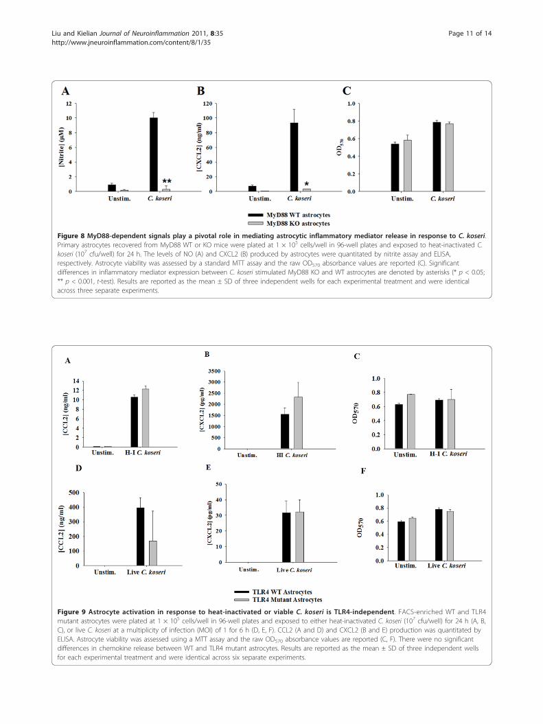
Figure 8 MyD88-dependent signals play a pivotal role in mediating astrocytic inflammatory mediator release in response to C. koseri.Primary astrocytes recovered from MyD88 WT or KO mice were plated at 1 × 10⁵ cells/well in 96-well plates and exposed to heat-inactivated C.koseri (107 cfu/well) for 24 h. The levels of NO (A) and CXCL2 (B) produced by astrocytes were quantitated by nitrite assay and ELISA,respectively. Astrocyte viability was assessed by a standard MTT assay and the raw OD570 absorbance values are reported (C). Significantdifferences in inflammatory mediator expression between C. koseri stimulated MyD88 KO and WT astrocytes are denoted by asterisks (* p < 0.05;** p < 0.001, t-test). Results are reported as the mean ± SD of three independent wells for each experimental treatment and were identicalacross three separate experiments.
Figure 9 Astrocyte activation in response to heat-inactivated or viable C. koseri is TLR4-independent. FACS-enriched WT and TLR4mutant astrocytes were plated at 1 × 10⁵ cells/well in 96-well plates and exposed to either heat-inactivated C. koseri (107 cfu/well) for 24 h (A, B,C), or live C. koseri at a multiplicity of infection (MOI) of 1 for 6 h (D, E, F). CCL2 (A and D) and CXCL2 (B and E) production was quantitated byELISA. Astrocyte viability was assessed using a MTT assay and the raw OD570 absorbance values are reported (C, F). There were no significantdifferences in chemokine release between WT and TLR4 mutant astrocytes. Results are reported as the mean ± SD of three independent wellsfor each experimental treatment and were identical across six separate experiments.
Liu and Kielian Journal of Neuroinflammation 2011, 8:35http://www.jneuroinflammation.com/content/8/1/35
Page 11 of 14

2 that sense PGN moieties as well as NLRs that havebeen implicated in the recognition of flagellated bacteriasuch as Ipaf and Naip [48-50]. However, the involve-ment of these alternative PRRs in regulating proinflam-matory mediator release during CNS C. koseri infectionwaits testing in future studies.One unexpected finding was that elevated chemokine
expression in MyD88 KO animals at 24 h post-infectiondid not translate into enhanced immune cell recruit-ment. Rather, neutrophil and macrophage infiltratesremained significantly attenuated in MyD88 KO micecompared to WT animals. The reason for this finding isunclear; however, since multiple events are required forimmune cell extravasation across the blood-brain bar-rier, it is possible that another phase is aberrant inMyD88 KO mice. For example, early IL-1b and TNF-aproduction is important for inducing adhesion moleculeexpression on BBB endothelial cells [51-53]. Since theproduction of both cytokines is significantly bluntedearly in MyD88 KO animals, sufficient time would berequired to induce adhesion molecule expression toovercome the migration block. In this case, by the timethat IL-1b and TNF-a were upregulated in MyD88 KOanimals, this may prove too late to impact immune cellmigration into the CNS. Yet, even those neutrophils andmacrophages that were recruited to the site of infectionin MyD88 KO mice were less activated as revealed byreduced levels of cytokine/chemokine production. It ispossible that the residual recruitment of these leukocytesubsets in MyD88 KO animals resulted from alternativechemotactic factors such as microbial-derived peptides(i.e. formyl peptides) or complement split products (i.e.C3a or C5a). However, once these cells enter the CNSparenchyma they are unable to effectively respond to C.koseri due to the lack of MyD88 signaling. Since heigh-tened inflammatory mediator release was evident inbrain abscess homogenates but not purified immune cellpopulations at 24 h post-infection, this suggests MyD88-independent contributions from alternative cell types,the identity of which remains unknown at the presenttime.The host possesses multiple redundant mechanisms to
ensure rapid immune responses to pathogens and infec-tion resolution [9,10,48,54]. When taken together, ourresults strongly suggest a collaborative role for numer-ous recognition pathways in generating a potent anti-bacterial immune response to CNS C. koseri infection.First, since the majority of TLRs utilize MyD88 as anadaptor, MyD88 deficiency will globally abolish all TLR-mediated signals in addition to signaling via the IL-1Rand IL-18R. This is particularly relevant since C. koseriharbors numerous ligands that can stimulate MyD88-dependent TLR signaling including lipoproteins (TLR2),LPS (TLR4), flagella (TLR5), and CpG DNA (TLR9) as
well as the ability of C. koseri to elicit IL-1b release dur-ing CNS infection (Figure 2). In addition to TLRs, cyto-plasmic PRRs such as NLRTC4 and NAIP that areactivated by flagellin [48] could also contribute to proin-flammatory mediator production since C. koseri is a fla-gellated bacterium; however, this possibility remainsspeculative.The concept of receptor redundancy for eliciting max-
imal responses to C. koseri was also evident when exam-ining primary astrocytes isolated from MyD88 KO orTLR4 mutant mice. Similar to our in vivo observations,chemokine production was negligible in MyD88 KOastrocytes in response to intact bacteria, whereas TLR4mutant cells responded identically to their WT counter-parts. This indicates that MyD88-dependent receptorsare critical for astrocyte activation and potential candi-dates include TLR2, TLR5, and TLR9 that recognizelipoproteins, flagellin, and CpG DNA, respectively.Although it is clear that TLR4 is not involved in astro-cyte responses to intact C. koseri, the extent of TLR4expression in astrocytes and whether it plays a role inLPS sensing remains controversial [37-39]. This issuehas been hampered by the poor quality of commerciallyavailable TLR4 antibodies in the mouse as well as con-cerns regarding the purity of primary astrocyte cultures[25]. However, as this topic was not the focus of thecurrent report, future studies using highly purified astro-cytes obtained by FACS analysis or alternative methodssuch as positive selection via magnetic beads are neededto address these issues related to LPS responsiveness.In summary, MyD88 is crucial for eliciting a protec-
tive host immune response during C. koseri brain infec-tion. Despite a late-phase induction of MyD88-independent signaling to trigger proinflammatoryrelease, this response was not sufficient to negateuncontrolled bacterial replication and lethality inMyD88 KO animals. The fact that TLR4 had littleimpact on the course of infection strongly suggests theinvolvement of alternative TLRs as well as the IL-1Rand IL-18R, which also utilize MyD88 as an adaptor.The identity of these alternative MyD88-dependentreceptors remains the topic for future studies.
Additional material
Additional file 1: Astrocyte enrichment by sub-culturing and FACS.Primary astrocytes were sub-cultured by shaking and passage three timesbefore collection. Astrocytes were harvested by trypsinization and stainedwith a CD11b antibody conjugated to PerCP-Cy5.5. The majority of cellsrecovered from astrocyte flasks were CD11b-negative, with an average of3-10% contaminating CD11b-positive microglia (A). Residual CD11b-positive cells were depleted from astrocytes by sorting, as indicated bypost-sort analysis (B). Purified microglia were included as a positivecontrol for CD11b staining (C). Subsequently, sorted astrocytes wereplated on cover slips at a density of 1 × 10⁴ cells/ml. After 24 h, cellswere stained for GFAP (D) and Iba-1 (E) to visualize astrocytes and
Liu and Kielian Journal of Neuroinflammation 2011, 8:35http://www.jneuroinflammation.com/content/8/1/35
Page 12 of 14

residual microglia, respectively. Purified microglia were stained with Iba-1as positive control (F).
Additional file 2: Establishment of CNS C. koseri infection and brainabscess formation. (A) C57BL/6 mice (4-5 per group) were used tooptimize C. koseri infectious doses and survival rates out to day 7 post-infection are presented. (B) A brain abscess induced by C. koseri at day 7post-infection is shown (arrows; magnification, 12.5×).
List of Abbreviations7-AAD: 7-Aminoactinomycin D; CFU: colony forming unit; DMSO: dimethylsulfoxide; FACS: fluorescent activated cell sorting; HBSS: Hank’s Balanced SaltSolution; IFN: interferon; IRF3: interferon regulatory factor 3; KO: knockout;LPS: lipopolysaccharide; MyD88: myeloid differentiation factor 88; NLR: NOD-like receptor; NOD1: nucleotide-binding oligomerization domain containing1; PAMP: pathogen-associated molecular pattern; PRR: pattern recognitionreceptor; TLR: Toll-like receptor; TRAM: TRIF-related adaptor molecule; TRIF:TIR-domain-containing adaptor protein-inducing IFN-β; WT: wild type.
AcknowledgementsThe authors thank Dr. Charles Kuszynski, Megan Michalak, and Victoria Smithin the UNMC Cell Analysis Facility for assistance with FACS analysis, TeresaFritz for performing immunostaining techniques, Amanda Angle for mousecolony maintenance, Debbie Vidlak for performing the Milliplex microbeadarrays, and Ms. Kari Nelson for grammatical review of the manuscript. Thiswork was supported by the NIH National Institute of Neurological Disordersand Stroke (NINDS) [R01NS055385] to T.K. Shuliang Liu was the recipient of aGSRF award from the UAMS Graduate School.
Author details1Department of Neurobiology and Developmental Sciences, University ofArkansas for Medical Sciences, Little Rock, AR 72205 USA. 2Department ofPathology and Microbiology, University of Nebraska Medical Center, Omaha,NE 68198 USA. 3Division of Neurotoxicology, National Center forToxicological Research, FDA, Jefferson, AR 72079 USA.
Authors’ contributionsSL performed the experiments, participated in study design, and helped todraft the manuscript. TK conceived the study, participated in study design,and wrote the manuscript. Both authors have read and approved the finalversion of the manuscript.
Competing interestsThe authors declare that they have no competing interests.
Received: 5 January 2011 Accepted: 16 April 2011Published: 16 April 2011
References1. Doran TI: The role of Citrobacter in clinical disease of children: review.
Clin Infect Dis 1999, 28:384-394.2. Agrawal D, Mahapatra AK: Vertically acquired neonatal citrobacter brain
abscess - case report and review of the literature. J Clin Neurosci 2005,12:188-190.
3. Jack CS, Arbour N, Manusow J, Montgrain V, Blain M, McCrea E, Shapiro A,Antel JP: TLR signaling tailors innate immune responses in humanmicroglia and astrocytes. J Immunol 2005, 175:4320-4330.
4. Santos-Sierra S, Golenbock DT, Henneke P: Toll-like receptor-dependentdiscrimination of streptococci. J Endotoxin Res 2006, 12:307-312.
5. Vance RE, Isberg RR, Portnoy DA: Patterns of pathogenesis: discriminationof pathogenic and nonpathogenic microbes by the innate immunesystem. Cell Host Microbe 2009, 6:10-21.
6. Kielian T, Phulwani NK, Esen N, Syed MM, Haney AC, McCastlain K,Johnson J: MyD88-dependent signals are essential for the host immuneresponse in experimental brain abscess. J Immunol 2007, 178:4528-4537.
7. Echchannaoui H, Frei K, Schnell C, Leib SL, Zimmerli W, Landmann R: Toll-like receptor 2-deficient mice are highly susceptible to Streptococcuspneumoniae meningitis because of reduced bacterial clearing andenhanced inflammation. J Infect Dis 2002, 186:798-806.
8. Koedel U, Rupprecht T, Angele B, Heesemann J, Wagner H, Pfister HW,Kirschning CJ: MyD88 is required for mounting a robust host immuneresponse to Streptococcus pneumoniae in the CNS. Brain 2004,127:1437-1445.
9. Takeuchi O, Akira S: Pattern recognition receptors and inflammation. Cell2010, 140:805-820.
10. Mogensen TH: Pathogen recognition and inflammatory signaling ininnate immune defenses. Clin Microbiol Rev 2009, 22:240-273, Table ofContents.
11. Adachi O, Kawai T, Takeda K, Matsumoto M, Tsutsui H, Sakagami M,Nakanishi K, Akira S: Targeted disruption of the MyD88 gene results inloss of IL-1- and IL-18-mediated function. Immunity 1998, 9:143-150.
12. Burns K, Martinon F, Esslinger C, Pahl H, Schneider P, Bodmer JL, DiMarco F, French L, Tschopp J: MyD88, an adapter protein involved ininterleukin-1 signaling. J Biol Chem 1998, 273:12203-12209.
13. Farina C, Aloisi F, Meinl E: Astrocytes are active players in cerebral innateimmunity. Trends Immunol 2007, 28:138-145.
14. Dong Y, Benveniste EN: Immune function of astrocytes. Glia 2001,36:180-190.
15. Kielian T: Toll-like receptors in central nervous system glial inflammationand homeostasis. J Neurosci Res 2006, 83:711-730.
16. O’Neill LA, Bowie AG: The family of five: TIR-domain-containing adaptorsin Toll-like receptor signalling. Nat Rev Immunol 2007, 7:353-364.
17. Kagan JC, Su T, Horng T, Chow A, Akira S, Medzhitov R: TRAM couplesendocytosis of Toll-like receptor 4 to the induction of interferon-beta.Nat Immunol 2008, 9:361-368.
18. Poltorak A, He X, Smirnova I, Liu MY, Van Huffel C, Du X, Birdwell D,Alejos E, Silva M, Galanos C, et al: Defective LPS signaling in C3H/HeJ andC57BL/10ScCr mice: mutations in Tlr4 gene. Science 1998, 282:2085-2088.
19. Qureshi ST, Lariviere L, Leveque G, Clermont S, Moore KJ, Gros P, Malo D:Endotoxin-tolerant mice have mutations in Toll-like receptor 4 (Tlr4).J Exp Med 1999, 189:615-625.
20. Kline MW, Mason EO Jr, Kaplan SL: Characterization of Citrobacterdiversus strains causing neonatal meningitis. J Infect Dis 1988,157:101-105.
21. Kielian T, Haney A, Mayes PM, Garg S, Esen N: Toll-like receptor 2modulates the proinflammatory milieu in Staphylococcus aureus-induced brain abscess. Infect Immun 2005, 73:7428-7435.
22. Kielian T, Barry B, Hickey WF: CXC chemokine receptor-2 ligands arerequired for neutrophil-mediated host defense in experimental brainabscesses. J Immunol 2001, 166:4634-4643.
23. Carson MJ, Reilly CR, Sutcliffe JG, Lo D: Mature microglia resembleimmature antigen-presenting cells. Glia 1998, 22:72-85.
24. Esen N, Tanga FY, DeLeo JA, Kielian T: Toll-like receptor 2 (TLR2) mediatesastrocyte activation in response to the Gram-positive bacteriumStaphylococcus aureus. J Neurochem 2004, 88:746-758.
25. Saura J: Microglial cells in astroglial cultures: a cautionary note.J Neuroinflammation 2007, 4:26.
26. Hamby ME, Uliasz TF, Hewett SJ, Hewett JA: Characterization of animproved procedure for the removal of microglia from confluentmonolayers of primary astrocytes. J Neurosci Methods 2006, 150:128-137.
27. Liu S, Kielian T: Microglial activation by Citrobacter koseri is mediated byTLR4- and MyD88-dependent pathways. J Immunol 2009, 183:5537-5547.
28. Abel B, Thieblemont N, Quesniaux VJ, Brown N, Mpagi J, Miyake K, Bihl F,Ryffel B: Toll-like receptor 4 expression is required to control chronicMycobacterium tuberculosis infection in mice. J Immunol 2002,169:3155-3162.
29. Branger J, Knapp S, Weijer S, Leemans JC, Pater JM, Speelman P, Florquin S,van der Poll T: Role of Toll-like receptor 4 in gram-positive and gram-negative pneumonia in mice. Infect Immun 2004, 72:788-794.
30. Lipton HL, Melvold R: Genetic analysis of susceptibility to Theiler’s virus-induced demyelinating disease in mice. J Immunol 1984, 132:1821-1825.
31. Jin YH, Kang HS, Mohindru M, Kim BS: Preferential induction of protectiveT cell responses to Theiler’s virus in resistant (C57BL/6 × SJL)F1 mice.J Virol 2011, 85:3033-3040.
32. Stevenson MM, Tam MF: Differential induction of helper T cell subsetsduring blood-stage Plasmodium chabaudi AS infection in resistant andsusceptible mice. Clin Exp Immunol 1993, 92:77-83.
33. Sayles PC, Wassom DL: Immunoregulation in murine malaria.Susceptibility of inbred mice to infection with Plasmodium yoelii
Liu and Kielian Journal of Neuroinflammation 2011, 8:35http://www.jneuroinflammation.com/content/8/1/35
Page 13 of 14
depends on the dynamic interplay of host and parasite genes.J Immunol 1988, 141:241-248.
34. Stevens NT, Sadovskaya I, Jabbouri S, Sattar T, O’Gara JP, Humphreys H,Greene CM: Staphylococcus epidermidis polysaccharide intercellularadhesin induces IL-8 expression in human astrocytes via a mechanisminvolving TLR2. Cell Microbiol 2009, 11:421-432.
35. Zhou S, Halle A, Kurt-Jones EA, Cerny AM, Porpiglia E, Rogers M,Golenbock DT, Finberg RW: Lymphocytic choriomeningitis virus (LCMV)infection of CNS glial cells results in TLR2-MyD88/Mal-dependentinflammatory responses. J Neuroimmunol 2008, 194:70-82.
36. Gorina R, Font-Nieves M, Marquez-Kisinousky L, Santalucia T, Planas AM:Astrocyte TLR4 activation induces a proinflammatory environmentthrough the interplay between MyD88-dependent NFkappaB signaling,MAPK, and Jak1/Stat1 pathways. Glia 2011, 59:242-255.
37. Lehnardt S, Lachance C, Patrizi S, Lefebvre S, Follett PL, Jensen FE,Rosenberg PA, Volpe JJ, Vartanian T: The toll-like receptor TLR4 isnecessary for lipopolysaccharide-induced oligodendrocyte injury in theCNS. J Neurosci 2002, 22:2478-2486.
38. Lehnardt S, Massillon L, Follett P, Jensen FE, Ratan R, Rosenberg PA,Volpe JJ, Vartanian T: Activation of innate immunity in the CNS triggersneurodegeneration through a Toll-like receptor 4-dependent pathway.Proc Natl Acad Sci USA 2003, 100:8514-8519.
39. Farina C, Krumbholz M, Giese T, Hartmann G, Aloisi F, Meinl E: Preferentialexpression and function of Toll-like receptor 3 in human astrocytes.J Neuroimmunol 2005, 159:12-19.
40. Soriano AL, Russell RG, Johnson D, Lagos R, Sechter I, Morris JG Jr:Pathophysiology of Citrobacter diversus neonatal meningitis:comparative studies in an infant mouse model. Infect Immun 1991,59:1352-1358.
41. Townsend SM, Gonzalez-Gomez I, Badger JL: fliP influences Citrobacterkoseri macrophage uptake, cytokine expression and brain abscessformation in the neonatal rat. J Med Microbiol 2006, 55:1631-1640.
42. Townsend SM, Pollack HA, Gonzalez-Gomez I, Shimada H, Badger JL:Citrobacter koseri brain abscess in the neonatal rat: survival andreplication within human and rat macrophages. Infect Immun 2003,71:5871-5880.
43. Garg S, Nichols JR, Esen N, Liu S, Phulwani NK, Syed MM, Wood WH,Zhang Y, Becker KG, Aldrich A, Kielian T: MyD88 expression by CNS-resident cells is pivotal for eliciting protective immunity in brainabscesses. ASN Neuro 2009, 1.
44. Hoebe K, Du X, Georgel P, Janssen E, Tabeta K, Kim SO, Goode J, Lin P,Mann N, Mudd S, et al: Identification of Lps2 as a key transducer ofMyD88-independent TIR signalling. Nature 2003, 424:743-748.
45. Yamamoto M, Sato S, Hemmi H, Hoshino K, Kaisho T, Sanjo H, Takeuchi O,Sugiyama M, Okabe M, Takeda K, Akira S: Role of adaptor TRIF in theMyD88-independent toll-like receptor signaling pathway. Science 2003,301:640-643.
46. Palsson-McDermott EM, O’Neill LA: Signal transduction by thelipopolysaccharide receptor, Toll-like receptor-4. Immunology 2004,113:153-162.
47. Alexopoulou L, Holt AC, Medzhitov R, Flavell RA: Recognition of double-stranded RNA and activation of NF-kappaB by Toll-like receptor 3. Nature2001, 413:732-738.
48. Wilmanski JM, Petnicki-Ocwieja T, Kobayashi KS: NLR proteins: integralmembers of innate immunity and mediators of inflammatory diseases.J Leukoc Biol 2008, 83:13-30.
49. Liu X, Chauhan VS, Marriott I: NOD2 contributes to the inflammatoryresponses of primary murine microglia and astrocytes to Staphylococcusaureus. Neurosci Lett 2010, 474:93-98.
50. Chauhan VS, Sterka DG Jr, Furr SR, Young AB, Marriott I: NOD2 plays animportant role in the inflammatory responses of microglia andastrocytes to bacterial CNS pathogens. Glia 2009, 57:414-423.
51. Blamire AM, Anthony DC, Rajagopalan B, Sibson NR, Perry VH, Styles P:Interleukin-1beta-induced changes in blood-brain barrier permeability,apparent diffusion coefficient, and cerebral blood volume in the ratbrain: a magnetic resonance study. J Neurosci 2000, 20:8153-8159.
52. Didier N, Romero IA, Creminon C, Wijkhuisen A, Grassi J, Mabondzo A:Secretion of interleukin-1beta by astrocytes mediates endothelin-1 andtumour necrosis factor-alpha effects on human brain microvascularendothelial cell permeability. J Neurochem 2003, 86:246-254.
53. Quagliarello VJ, Wispelwey B, Long WJ Jr, Scheld WM: Recombinant humaninterleukin-1 induces meningitis and blood-brain barrier injury in the rat.Characterization and comparison with tumor necrosis factor. J Clin Invest1991, 87:1360-1366.
54. Franchi L, Eigenbrod T, Munoz-Planillo R, Nunez G: The inflammasome: acaspase-1-activation platform that regulates immune responses anddisease pathogenesis. Nat Immunol 2009, 10:241-247.
doi:10.1186/1742-2094-8-35Cite this article as: Liu and Kielian: MyD88 is pivotal for immunerecognition of Citrobacter koseri and astrocyte activation during CNSinfection†. Journal of Neuroinflammation 2011 8:35.
Submit your next manuscript to BioMed Centraland take full advantage of:
• Convenient online submission
• Thorough peer review
• No space constraints or color figure charges
• Immediate publication on acceptance
• Inclusion in PubMed, CAS, Scopus and Google Scholar
• Research which is freely available for redistribution
Submit your manuscript at www.biomedcentral.com/submit
Liu and Kielian Journal of Neuroinflammation 2011, 8:35http://www.jneuroinflammation.com/content/8/1/35
Page 14 of 14