Embed Size (px)
Citation preview
1
Robert P Hasserjian, MDAssociate ProfessorMassachusetts General Hospital and Harvard Medical School
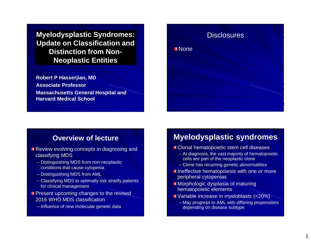
Myelodysplastic Syndromes:Update on Classification and
Distinction from Non-Neoplastic Entities
Disclosures
None
Overview of lectureReview evolving concepts in diagnosing and classifying MDS– Distinguishing MDS from non-neoplastic
conditions that cause cytopenia– Distinguishing MDS from AML– Classifying MDS to optimally risk stratify patients
for clinical management
Present upcoming changes to the revised 2016 WHO MDS classification– Influence of new molecular genetic data
Myelodysplastic syndromesClonal hematopoietic stem cell diseases– At diagnosis, the vast majority of hematopoietic
cells are part of the neoplastic clone – Clone has recurring genetic abnormalities
Ineffective hematopoiesis with one or more peripheral cytopeniasMorphologic dysplasia of maturing hematopoietic elementsVariable increase in myeloblasts (<20%)– May progress to AML with differing propensities
depending on disease subtype
2
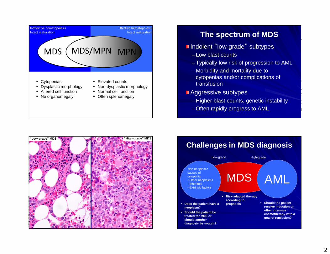
Ineffective hematopoiesisIntact maturation
Effective hematopoiesisIntact maturation
MDS/MPNMDS MPN
� Cytopenias� Dysplastic morphology� Altered cell function� No organomegaly
� Elevated counts� Non-dysplastic morphology� Normal cell function� Often splenomegaly
The spectrum of MDS
Indolent “low-grade” subtypes– Low blast counts
– Typically low risk of progression to AML
– Morbidity and mortality due to cytopenias and/or complications of transfusion
Aggressive subtypes– Higher blast counts, genetic instability– Often rapidly progress to AML
“Low-grade” MDS “High-grade” MDS
Challenges in MDS diagnosis
MDSNon-neoplasticcauses of cytopenia--Other neoplasms--Inherited--Extrinsic factors
AML
Low-grade High-grade
� Does the patient have a neoplasm?
� Should the patient be treated for MDS or should another diagnosis be sought?
� Should the patient receive induction or other intensive chemotherapy with a goal of remission?
� Risk-adapted therapy according to prognosis
3
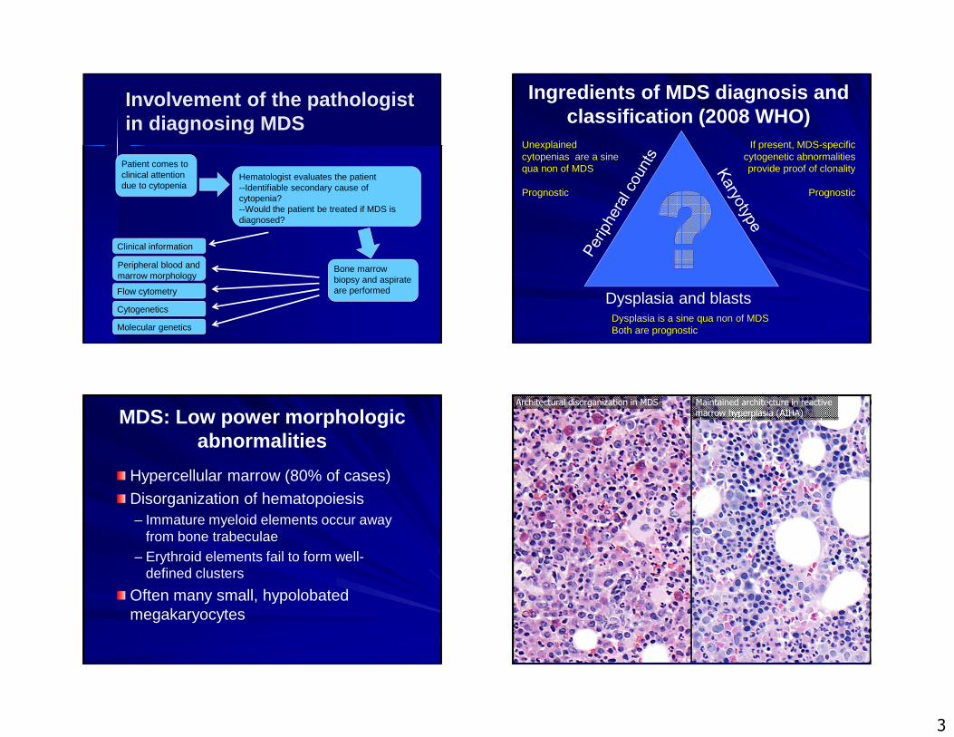
Involvement of the pathologist in diagnosing MDS
Patient comes to clinical attention due to cytopenia
Hematologist evaluates the patient--Identifiable secondary cause of cytopenia?--Would the patient be treated if MDS is diagnosed?
Bone marrow biopsy and aspirate are performed
Peripheral blood and marrow morphology
Flow cytometry
Cytogenetics
Molecular genetics
Clinical information
Ingredients of MDS diagnosis and classification (2008 WHO)
Dysplasia and blasts
Unexplained cytopenias are a sine qua non of MDS
Prognostic
If present, MDS-specific cytogenetic abnormalities provide proof of clonality
Prognostic
Dysplasia is a sine qua non of MDSBoth are prognostic
MDS: Low power morphologic abnormalities
Hypercellular marrow (80% of cases)
Disorganization of hematopoiesis– Immature myeloid elements occur away
from bone trabeculae– Erythroid elements fail to form well-
defined clusters
Often many small, hypolobatedmegakaryocytes
Architectural disorganization in MDS Maintained architecture in reactive marrow hyperplasia (AIHA)
4
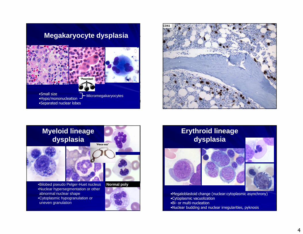
Megakaryocyte dysplasia
•Small size•Hypo/mononucleation•Separated nuclear lobes
Micromegakaryocytes
‘‘‘‘Pawnball’’’’
CD61
Myeloid lineage dysplasia
•Bilobed pseudo Pelger-Huet nucleus •Nuclear hypersegmentation or other abnormal nuclear shape
•Cytoplasmic hypogranulation or uneven granulation
Normal poly
‘‘‘‘Pince-nez’’’’
Erythroid lineage dysplasia
•Megaloblastoid change (nuclear:cytoplasmic asynchrony)•Cytoplasmic vacuolization•Bi- or multi-nucleation•Nuclear budding and nuclear irregularities, pyknosis
5
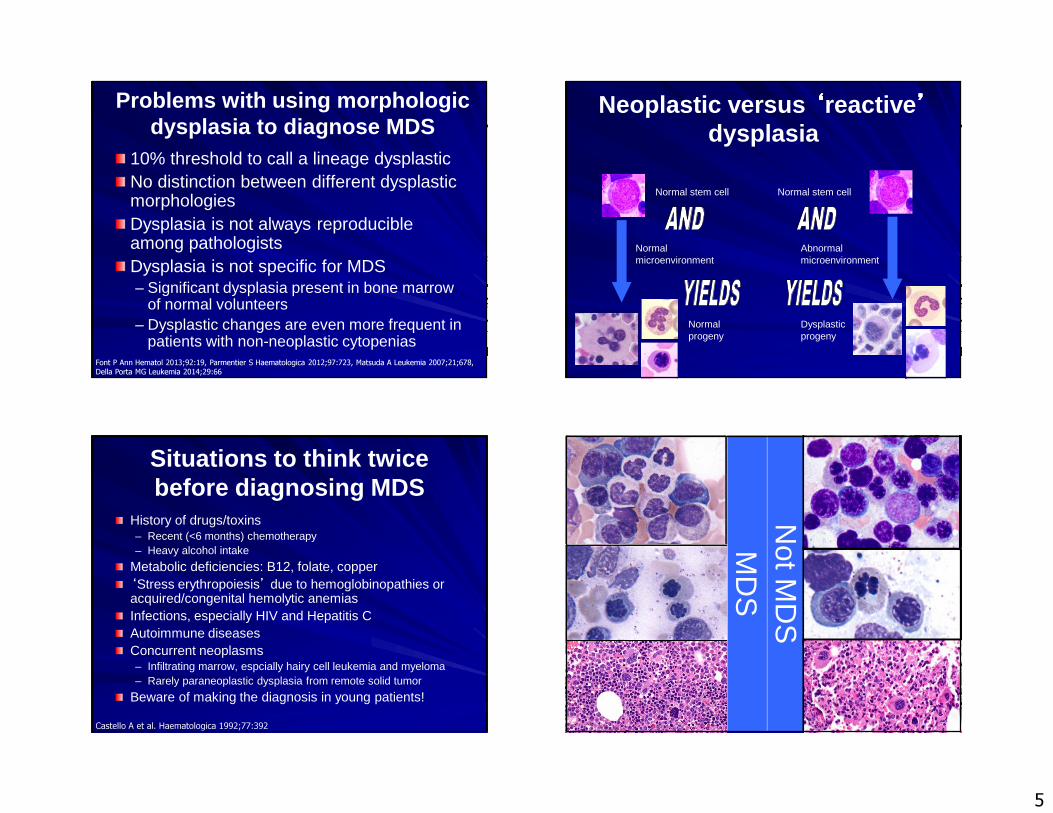
Problems with using morphologic dysplasia to diagnose MDS
10% threshold to call a lineage dysplasticNo distinction between different dysplastic morphologiesDysplasia is not always reproducible among pathologistsDysplasia is not specific for MDS– Significant dysplasia present in bone marrow
of normal volunteers– Dysplastic changes are even more frequent in
patients with non-neoplastic cytopeniasFont P Ann Hematol 2013;92:19, Parmentier S Haematologica 2012;97:723, Matsuda A Leukemia 2007;21;678, Della Porta MG Leukemia 2014;29:66
Neoplastic versus ‘‘‘‘reactive ’’’’
dysplasia
Abnormal stem cell
Normal microenvironment
Normalprogeny
Normal stem cell
Dysplasticprogeny
Abnormalmicroenvironment
Normal stem cell
Situations to think twice before diagnosing MDS
History of drugs/toxins– Recent (<6 months) chemotherapy– Heavy alcohol intake
Metabolic deficiencies: B12, folate, copper‘Stress erythropoiesis’ due to hemoglobinopathies or acquired/congenital hemolytic anemiasInfections, especially HIV and Hepatitis CAutoimmune diseasesConcurrent neoplasms– Infiltrating marrow, espcially hairy cell leukemia and myeloma– Rarely paraneoplastic dysplasia from remote solid tumor
Beware of making the diagnosis in young patients!
Castello A et al. Haematologica 1992;77:392
Not M
DS
MD
S
6
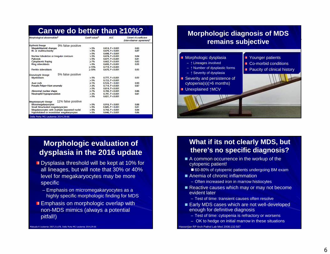
Can we do better than ≥10%?
Della Porta MG Leukemia 2014;29:66
9% false positive
5% false positive
11% false positive
Morphologic diagnosis of MDS remains subjective
Younger patientsCo-morbid conditionsPaucity of clinical history
Morphologic dysplasia– ↑ Lineages involved– ↑ Number of dysplastic forms– ↑ Severity of dysplasia
Severity and persistence of cytopenia(s)(>6 months)Unexplained ↑MCV
Morphologic evaluation of dysplasia in the 2016 updateDysplasia threshold will be kept at 10% for all lineages, but will note that 30% or 40% level for megakaryocytes may be more specific– Emphasis on micromegakaryocytes as a
highly specific morphologic finding for MDS
Emphasis on morphologic overlap with non-MDS mimics (always a potential pitfall!)
Matsuda A Leukemia 2007;21;678, Della Porta MG Leukemia 2014;29:66
What if its not clearly MDS, but there’s no specific diagnosis?� A common occurrence in the workup of the
cytopenic patient!� 60-80% of cytopenic patients undergoing BM exam
� Anemia of chronic inflammation– Often increased iron in marrow histiocytes
� Reactive causes which may or may not become evident later– Test of time: transient causes often resolve
� Early MDS cases which are not well-developed enough for definitive diagnosis– Test of time: cytopenia is refractory or worsens– OK to hedge on initial marrow in these situations
Hasserjian RP Arch Pathol Lab Med 2008;132:587
7
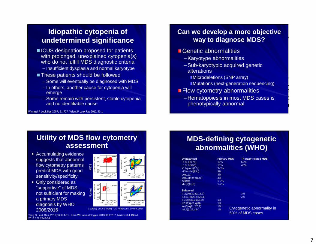
Idiopathic cytopenia of undetermined significance � ICUS designation proposed for patients
with prolonged, unexplained cytopenia(s) who do not fulfill MDS diagnostic criteria– Insufficient dysplasia and normal karyotype
�These patients should be followed– Some will eventually be diagnosed with MDS– In others, another cause for cytopenia will
emerge– Some remain with persistent, stable cytopenia
and no identifiable cause
Wimazal F Leuk Res 2007; 31:727, Valent P Leuk Res 2012;36:1
Can we develop a more objective way to diagnose MDS?
Genetic abnormalities– Karyotype abnormalities– Sub-karyotypic acquired genetic
alterationsMicrodeletions (SNP array)Mutations (next-generation sequencing)
Flow cytometry abnormalities– Hematopoiesis in most MDS cases is
phenotypically abnormal
Utility of MDS flow cytometry assessment
� Accumulating evidence suggests that abnormal flow cytometry patterns predict MDS with good sensitivity/specificity
� Only considered as “supportive” of MDS, not sufficient for making a primary MDS diagnosis by WHO 2008/2016
Tang G Leuk Res. 2012;36:974-81, Kern W Haematologica 2013;98:201-7, Malcovati L Blood 2013;122:2943-64
CD16 FITC-A
CD
13 A
PC
-A
-102
103
104
105
-102
102
103
104
105
CD16 FITC-A
CD
13 A
PC
-A
-102
103
104
105
-102
102
103
104
105
CD7 FITC-A
CD
34 P
erC
P-C
y5-5
-A
-102
103
104
105
-102
102
103
104
105 84.8%
CD7 FITC-A
CD
34 P
erC
P-C
y5-5
-A
-102
103
104
105
-102
102
103
104
105
5.39%
MD
SN
orm
al
Courtesy of Dr S Wang, MD Anderson Cancer Center
MDS-defining cytogenetic abnormalities (WHO)
Unbalanced Primary MDS Therapy-related MDS-7 or del(7q) 10% 50%-5 or del(5q) 10% 40%i(17q) or t(17p) 3-5%-13 or del(13q) 3%del(11q) 3%del(12p) or t(12p) 3%del(9q) 1-2%idic(X)(q13) 1-2%
Balancedt(11;16)(q23;p13.3) 3%t(3;21)(q26.2;q22.1) 2%t(1;3)(p36.3;q21.2) 1%t(2;11)(p21;q23) 1%inv(3)(q21q26.2) 1%t(6;9)(p23;q34) 1% Cytogenetic abnormality in
50% of MDS cases
8
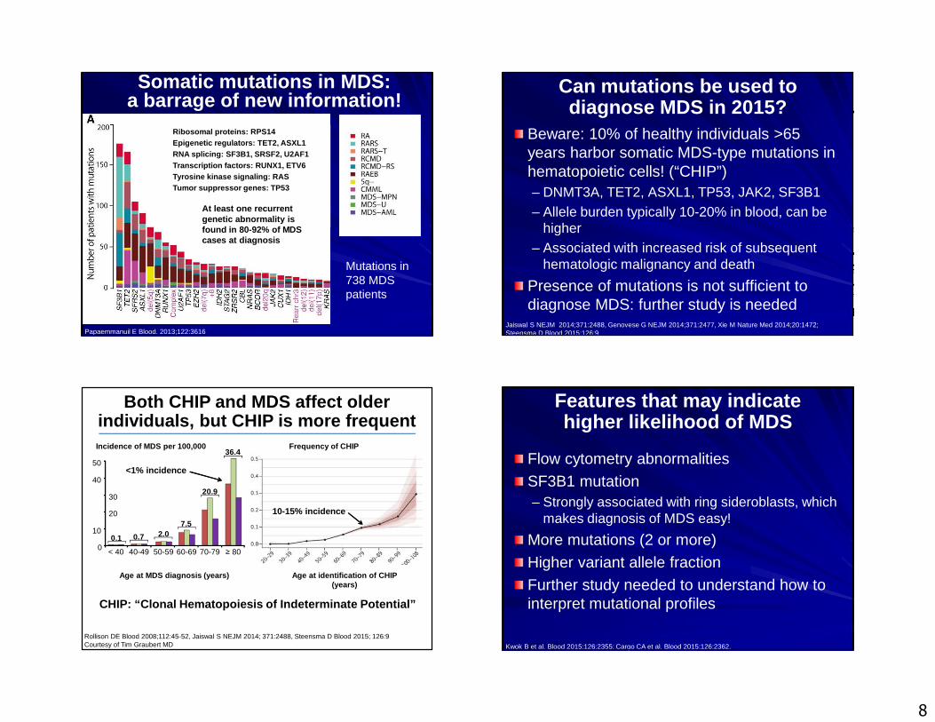
Somatic mutations in MDS: a barrage of new information!
Papaemmanuil E Blood. 2013;122:3616
Ribosomal proteins: RPS14
Epigenetic regulators: TET2, ASXL1RNA splicing: SF3B1, SRSF2, U2AF1Transcription factors: RUNX1, ETV6Tyrosine kinase signaling: RAS
Tumor suppressor genes: TP53
Ribosomal proteins: RPS14Epigenetic regulators: TET2, ASXL1RNA splicing: SF3B1, SRSF2, U2AF1Transcription factors: RUNX1, ETV6Tyrosine kinase signaling: RASTumor suppressor genes: TP53
At least one recurrent genetic abnormality is found in 80-92% of MDS cases at diagnosis
Mutations in 738 MDS patients
Can mutations be used to diagnose MDS in 2015?
Beware: 10% of healthy individuals >65 years harbor somatic MDS-type mutations in hematopoietic cells! (“CHIP”)– DNMT3A, TET2, ASXL1, TP53, JAK2, SF3B1– Allele burden typically 10-20% in blood, can be
higher– Associated with increased risk of subsequent
hematologic malignancy and death
Presence of mutations is not sufficient to diagnose MDS: further study is needed
Jaiswal S NEJM 2014;371:2488, Genovese G NEJM 2014;371:2477, Xie M Nature Med 2014;20:1472; Steensma D Blood 2015;126:9
Age at MDS diagnosis (years)
0
10
20
30
40
50
< 40 40-49 50-59 60-69 70-79 ≥ 80
0.1 0.7 2.07.5
20.9
36.4
Age at identification of CHIP (years)
Rollison DE Blood 2008;112:45-52, Jaiswal S NEJM 2014; 371:2488, Steensma D Blood 2015; 126:9 Courtesy of Tim Graubert MD
Incidence of MDS per 100,000 Frequency of CHIP
Both CHIP and MDS affect older individuals, but CHIP is more frequent
CHIP: “Clonal Hematopoiesis of Indeterminate Potent ial”
<1% incidence
10-15% incidence
Features that may indicate higher likelihood of MDS
Flow cytometry abnormalities
SF3B1 mutation– Strongly associated with ring sideroblasts, which
makes diagnosis of MDS easy!
More mutations (2 or more)
Higher variant allele fraction
Further study needed to understand how to interpret mutational profiles
Kwok B et al. Blood 2015;126:2355; Cargo CA et al. Blood 2015;126:2362.
9
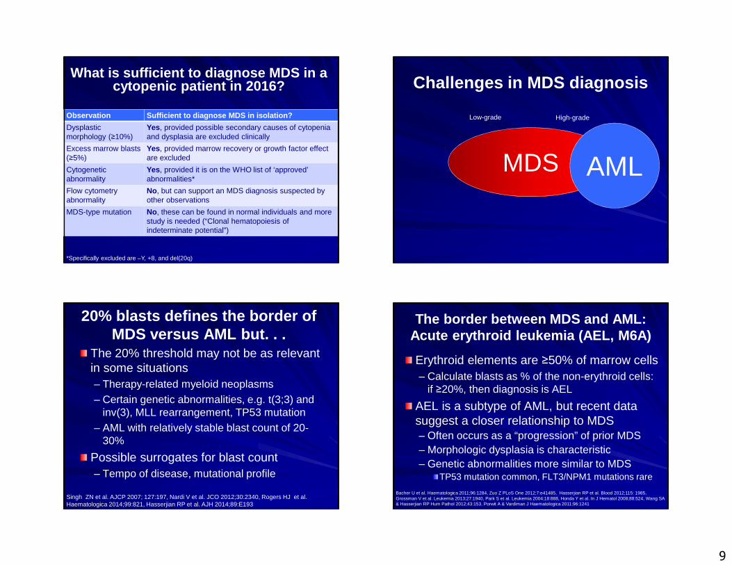
What is sufficient to diagnose MDS in a cytopenic patient in 2016?
Observation Sufficient to diagnose MDS in isolation?
Dysplastic morphology (≥10%)
Yes, provided possible secondary causes of cytopeniaand dysplasia are excluded clinically
Excess marrow blasts (≥5%)
Yes, provided marrow recovery or growth factor effect are excluded
Cytogenetic abnormality
Yes, provided it is on the WHO list of ‘approved’ abnormalities*
Flow cytometryabnormality
No, but can support an MDS diagnosis suspected by other observations
MDS-type mutation No, these can be found in normal individuals and more study is needed (“Clonal hematopoiesis of indeterminate potential”)
*Specifically excluded are –Y, +8, and del(20q)
Challenges in MDS diagnosis
MDS AML
Low-grade High-grade
20% blasts defines the border of MDS versus AML but. . .
The 20% threshold may not be as relevant in some situations– Therapy-related myeloid neoplasms– Certain genetic abnormalities, e.g. t(3;3) and
inv(3), MLL rearrangement, TP53 mutation– AML with relatively stable blast count of 20-
30%
Possible surrogates for blast count– Tempo of disease, mutational profile
Singh ZN et al. AJCP 2007; 127:197, Nardi V et al. JCO 2012;30:2340, Rogers HJ et al. Haematologica 2014;99:821, Hasserjian RP et al. AJH 2014;89:E193
Erythroid elements are ≥50% of marrow cells– Calculate blasts as % of the non-erythroid cells:
if ≥20%, then diagnosis is AEL
AEL is a subtype of AML, but recent data suggest a closer relationship to MDS– Often occurs as a “progression” of prior MDS– Morphologic dysplasia is characteristic– Genetic abnormalities more similar to MDS
TP53 mutation common, FLT3/NPM1 mutations rare
The border between MDS and AML: Acute erythroid leukemia (AEL, M6A)
Bacher U et al. Haematologica 2011;96:1284, Zuo Z PLoS One 2012;7:e41485, Hasserjian RP et al. Blood 2012;115: 1985, Grossman V et al. Leukemia 2013;27:1940, Park S et al. Leukemia 2004;18:888, Honda Y et al. In J Hematol 2008;88:524, Wang SA & Hasserjian RP Hum Pathol 2012;43:153, Porwit A & Vardiman J Haematologica 2011;96:1241
10
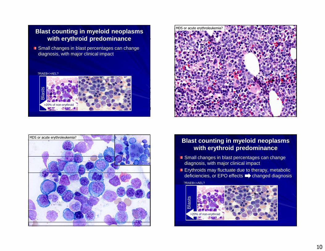
Blast counting in myeloid neoplasms with erythroid predominance
Small changes in blast percentages can change diagnosis, with major clinical impact
Bla
sts
?RAEB<>AEL?
>20% of non-erythroid
MDS or acute erythroleukemia?
MDS or acute erythroleukemia?
Small changes in blast percentages can change diagnosis, with major clinical impactErythroids may fluctuate due to therapy, metabolic deficiencies, or EPO effects changed diagnosis
Bla
sts
>20% of non-erythroid
?RAEB<>AEL?
Blast counting in myeloid neoplasmswith erythroid predominance
11
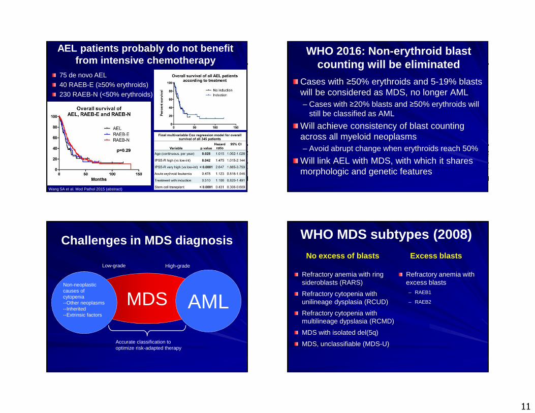
AEL patients probably do not benefit from intensive chemotherapy
Wang SA et al. Mod Pathol 2015 (abstract)
75 de novo AEL40 RAEB-E (≥50% erythroids)230 RAEB-N (<50% erythroids)
WHO 2016: Non-erythroid blast counting will be eliminated
Cases with ≥50% erythroids and 5-19% blasts will be considered as MDS, no longer AML– Cases with ≥20% blasts and ≥50% erythroids will
still be classified as AML
Will achieve consistency of blast counting across all myeloid neoplasms– Avoid abrupt change when erythroids reach 50%
Will link AEL with MDS, with which it shares morphologic and genetic features
Challenges in MDS diagnosis
MDSNon-neoplasticcauses of cytopenia--Other neoplasms--Inherited--Extrinsic factors
AML
Low-grade High-grade
Accurate classification to optimize risk-adapted therapy
WHO MDS subtypes (2008)
Refractory anemia with ring sideroblasts (RARS)
Refractory cytopenia with unilineage dysplasia (RCUD)
Refractory cytopenia with multilineage dypslasia (RCMD)
MDS with isolated del(5q)
MDS, unclassifiable (MDS-U)
Refractory anemia with excess blasts– RAEB1
– RAEB2
No excess of blasts Excess blasts
12
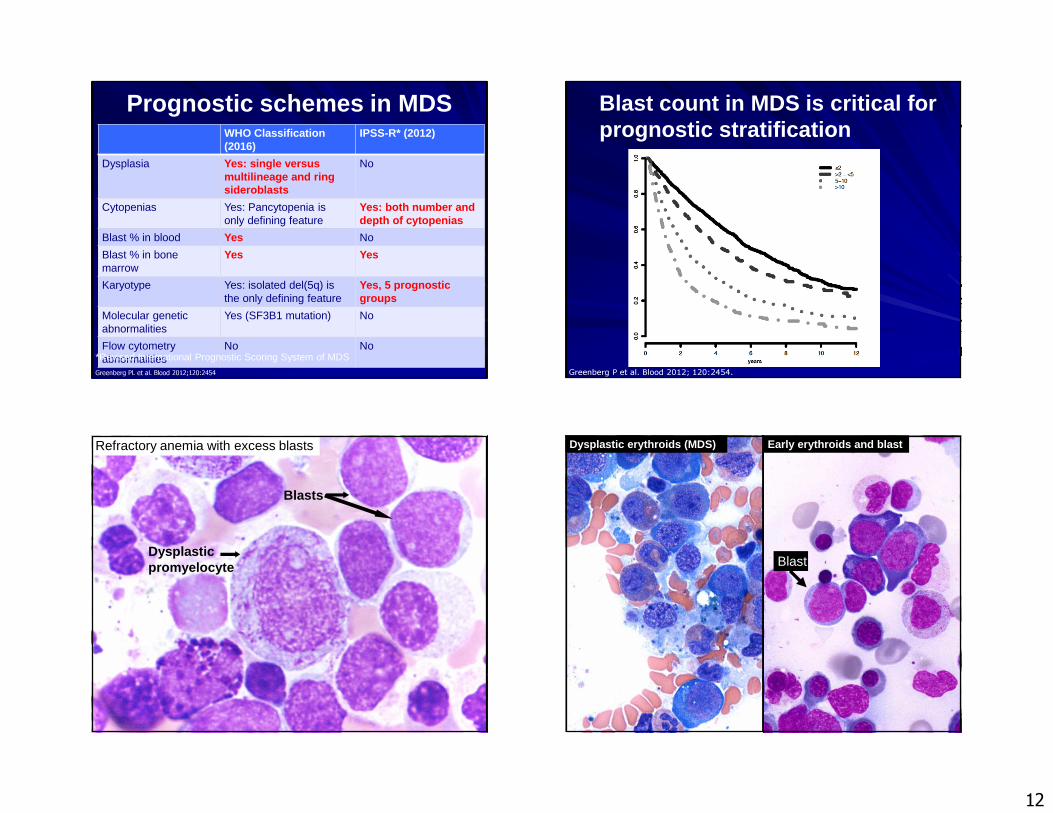
Prognostic schemes in MDSWHO Classification (2016)
IPSS-R* (2012)
Dysplasia Yes: single versus multilineage and ring sideroblasts
No
Cytopenias Yes: Pancytopenia is only defining feature
Yes: both number and depth of cytopenias
Blast % in blood Yes No
Blast % in bone marrow
Yes Yes
Karyotype Yes: isolated del(5q) is the only defining feature
Yes, 5 prognostic groups
Molecular genetic abnormalities
Yes (SF3B1 mutation) No
Flow cytometry abnormalities
No No
Greenberg PL et al. Blood 2012;120:2454 *Revised International Prognostic Scoring System of MDS
Greenberg P et al. Blood 2012; 120:2454.
Blast count in MDS is critical for prognostic stratification
Refractory anemia with excess blasts
Blasts
Dysplasticpromyelocyte
Dysplastic erythroids (MDS) Early erythroids and blast
Blast
13
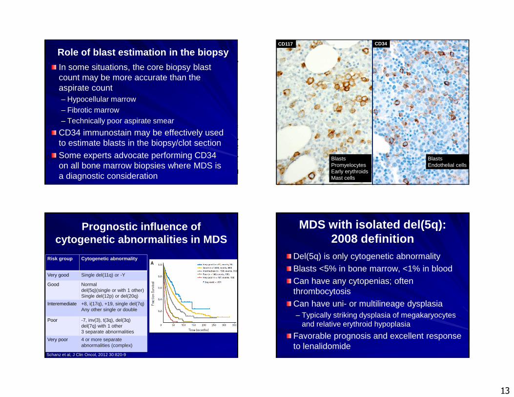
Role of blast estimation in the biopsy
In some situations, the core biopsy blast count may be more accurate than the aspirate count– Hypocellular marrow– Fibrotic marrow– Technically poor aspirate smear
CD34 immunostain may be effectively used to estimate blasts in the biopsy/clot section
Some experts advocate performing CD34 on all bone marrow biopsies where MDS is a diagnostic consideration
CD117 CD34
BlastsPromyelocytesEarly erythroidsMast cells
BlastsEndothelial cells
Prognostic influence of cytogenetic abnormalities in MDS
Risk group Cytogenetic abnormality
Very good Single del(11q) or -Y
Good Normaldel(5q)(single or with 1 other)Single del(12p) or del(20q)
Interemediate +8, i(17q), +19, single del(7q)Any other single or double
Poor -7, inv(3), t(3q), del(3q)del(7q) with 1 other3 separate abnormalities
Very poor 4 or more separate abnormalities (complex)
Schanz et al, J Clin Oncol, 2012 30:820-9
MDS with isolated del(5q): 2008 definition
Del(5q) is only cytogenetic abnormality
Blasts <5% in bone marrow, <1% in blood
Can have any cytopenias; often thrombocytosis
Can have uni- or multilineage dysplasia– Typically striking dysplasia of megakaryocytes
and relative erythroid hypoplasia
Favorable prognosis and excellent response to lenalidomide
14
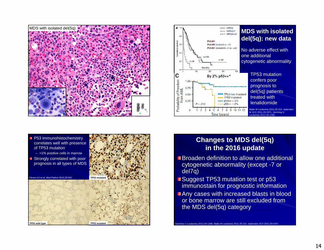
MDS with isolated del(5q)
No adverse effect with one additional cytogenetic abnormality
TP53 mutation confers poor prognosis to del(5q) patients treated with lenalidomide
Mallo M Leukemia 2011;25:110, JaderstenM JCO 2011;29:1971, Germing U Leukemia 2012;26:1286
Months
MDS with isolated del(5q): new data
P53 immunohistochemistry correlates well with presence of TP53 mutation– >1% positive cells in marrow
Strongly correlated with poor prognosis in all types of MDS
Cleven AJ et al. Mod Pathol 2015;28:552
TP53 wild type TP53 mutated
TP53 mutated
Changes to MDS del(5q) in the 2016 update
Broaden definition to allow one additional cytogenetic abnormality (except -7 or del7q)Suggest TP53 mutation test or p53 immunostain for prognostic informationAny cases with increased blasts in blood or bone marrow are still excluded from the MDS del(5q) category
Germing U Leukemia 2012;26:1286, Mallo M Leukemia 2011;25:110, Jadersten JCO 2011;29:1971
15
Specific mutations also carry prognostic impact
Bejar R NEJM 2011;364:2496
TP53, EZH2, ETV6, RUNX1, or ASXL1 mutations confer adverse prognosis
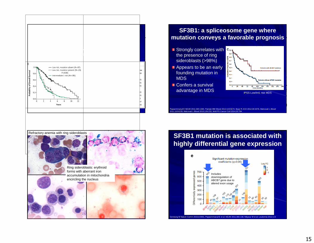
SF3B1: a spliceosome gene where mutation conveys a favorable prognosis
Strongly correlates with the presence of ring sideroblasts (>98%)Appears to be an early founding mutation in MDSConfers a survival advantage in MDS
IPSS Low/Int1 risk MDS
Papaemmanuil E NEJM 2011;365:1384, Patniak MM Blood 2012;119:5674, Bejar R JCO 2012;30:3376, Malcovati L Blood 2011;118:6239, Malcovati L Blood 2015;126:233, Woll PS Cancer Cell 2014;25:794
Ring sideroblasts: erythroidforms with aberrant iron accumulation in mitochondria encircling the nucleus
Refractory anemia with ring sideroblastsSF3B1 mutation is associated with highly differential gene expression
Gerstung M Nature Comm 2015;6:5901, Papaemmanuil E et al. NEJM 2011;365:138, Nikpour M et al. Leukemia 2012;1-8
Includes downregulation of ABCB7 gene due to altered exon usage
16
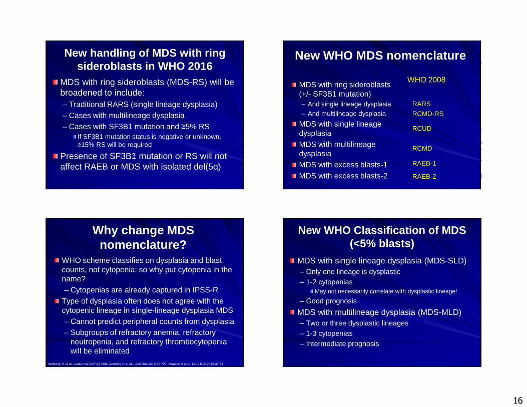
New handling of MDS with ring sideroblasts in WHO 2016
MDS with ring sideroblasts (MDS-RS) will be broadened to include:– Traditional RARS (single lineage dysplasia)– Cases with multilineage dysplasia – Cases with SF3B1 mutation and ≥5% RS
If SF3B1 mutation status is negative or unknown, ≥15% RS will be required
Presence of SF3B1 mutation or RS will not affect RAEB or MDS with isolated del(5q)
New WHO MDS nomenclature
MDS with ring sideroblasts(+/- SF3B1 mutation)– And single lineage dysplasia– And multilineage dysplasia
MDS with single lineage dysplasiaMDS with multilineagedysplasiaMDS with excess blasts-1MDS with excess blasts-2
RARS
RCMD-RS
RCUD
RCMD
RAEB-1
RAEB-2
WHO 2008
Why change MDS nomenclature?
WHO scheme classifies on dysplasia and blast counts, not cytopenia: so why put cytopenia in the name? – Cytopenias are already captured in IPSS-R
Type of dysplasia often does not agree with the cytopenic lineage in single-lineage dysplasia MDS– Cannot predict peripheral counts from dysplasia– Subgroups of refractory anemia, refractory
neutropenia, and refractory thrombocytopenia will be eliminated
Verburgh E et al. Leukemia 2007;21:668, Germing U et al. Leuk Res 2012;36:727, Maasen A et al. Leuk Res 2013;37:64
New WHO Classification of MDS (<5% blasts)
MDS with single lineage dysplasia (MDS-SLD)– Only one lineage is dysplastic– 1-2 cytopenias
May not necessarily correlate with dysplastic lineage!
– Good prognosis
MDS with multilineage dysplasia (MDS-MLD)– Two or three dysplastic lineages– 1-3 cytopenias– Intermediate prognosis
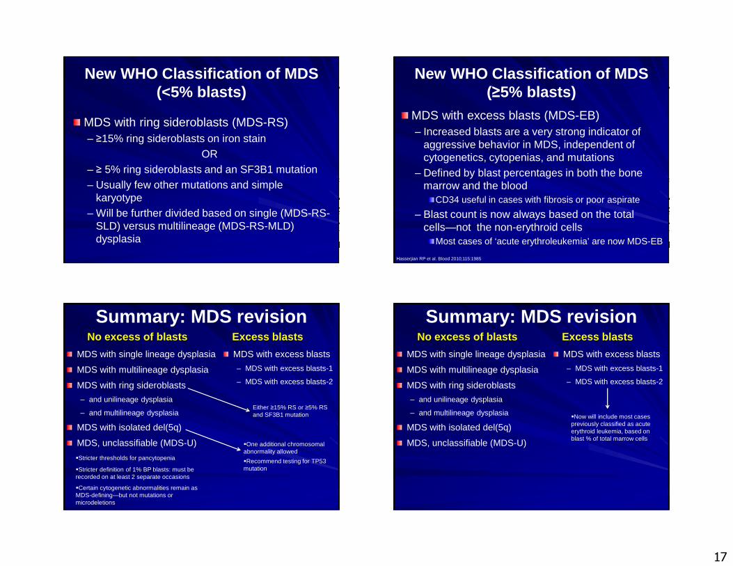
17
New WHO Classification of MDS (<5% blasts)
MDS with ring sideroblasts (MDS-RS)– ≥15% ring sideroblasts on iron stain
OR– ≥ 5% ring sideroblasts and an SF3B1 mutation– Usually few other mutations and simple
karyotype– Will be further divided based on single (MDS-RS-
SLD) versus multilineage (MDS-RS-MLD) dysplasia
New WHO Classification of MDS (≥5% blasts)
MDS with excess blasts (MDS-EB)– Increased blasts are a very strong indicator of
aggressive behavior in MDS, independent of cytogenetics, cytopenias, and mutations
– Defined by blast percentages in both the bone marrow and the blood
CD34 useful in cases with fibrosis or poor aspirate
– Blast count is now always based on the total cells—not the non-erythroid cells
Most cases of ‘acute erythroleukemia’ are now MDS-EB
Hasserjian RP et al. Blood 2010;115:1985
Summary: MDS revision
MDS with single lineage dysplasia
MDS with multilineage dysplasia
MDS with ring sideroblasts
– and unilineage dysplasia
– and multilineage dysplasia
MDS with isolated del(5q)
MDS, unclassifiable (MDS-U)
No excess of blasts
MDS with excess blasts
– MDS with excess blasts-1
– MDS with excess blasts-2
Excess blasts
Either ≥15% RS or ≥5% RS and SF3B1 mutation
�One additional chromosomal abnormality allowed
�Recommend testing for TP53 mutation
�Stricter thresholds for pancytopenia
�Stricter definition of 1% BP blasts: must be recorded on at least 2 separate occasions
�Certain cytogenetic abnormalities remain as MDS-defining—but not mutations or microdeletions
Summary: MDS revision
MDS with single lineage dysplasia
MDS with multilineage dysplasia
MDS with ring sideroblasts
– and unilineage dysplasia
– and multilineage dysplasia
MDS with isolated del(5q)
MDS, unclassifiable (MDS-U)
No excess of blasts
MDS with excess blasts
– MDS with excess blasts-1
– MDS with excess blasts-2
Excess blasts
�Now will include most cases previously classified as acute erythroid leukemia, based on blast % of total marrow cells
18
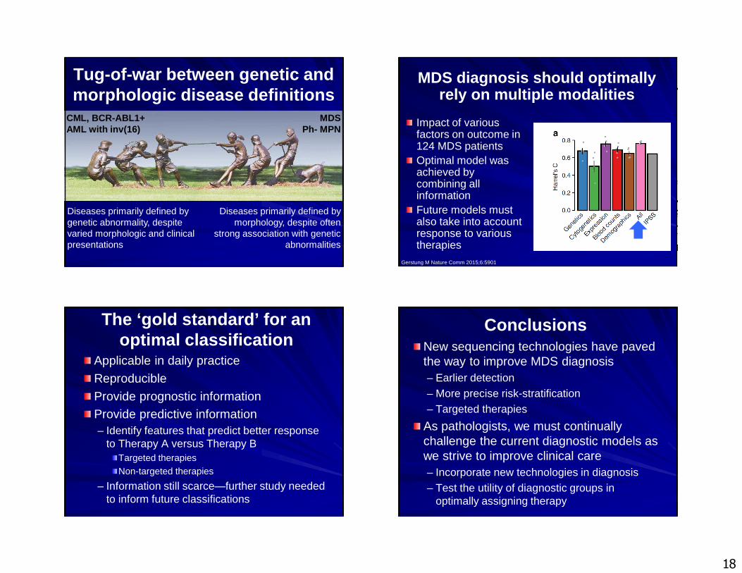
Tug-of-war between genetic and morphologic disease definitions
CML, BCR-ABL1+AML with inv(16)
MDSPh- MPN
Diseases primarily defined by genetic abnormality, despite varied morphologic and clinical presentations
Diseases primarily defined by morphology, despite often
strong association with genetic abnormalities
MDS diagnosis should optimallyrely on multiple modalities
Impact of various factors on outcome in 124 MDS patientsOptimal model was achieved by combining all informationFuture models must also take into accountresponse to various therapies
Gerstung M Nature Comm 2015;6:5901
The ‘gold standard’ for an optimal classification
Applicable in daily practice
Reproducible
Provide prognostic informationProvide predictive information– Identify features that predict better response
to Therapy A versus Therapy BTargeted therapiesNon-targeted therapies
– Information still scarce—further study needed to inform future classifications
ConclusionsNew sequencing technologies have paved the way to improve MDS diagnosis– Earlier detection– More precise risk-stratification– Targeted therapies
As pathologists, we must continually challenge the current diagnostic models as we strive to improve clinical care– Incorporate new technologies in diagnosis– Test the utility of diagnostic groups in
optimally assigning therapy