Embed Size (px)
Citation preview
George N. Sfakianakis MDProfessor of Radiology and Pediatrics
Director Division of Nuclear Medicine , University of Miami, Florida
MYOCARDIAL PERFUSION SCINTIGRAPHY
October 2009
EVALUATION OF THE FUNCTION AND DISEASES OF THE HEART
WITH NUCLEAR MEDICINE PROCEDURES (CARDIAC SCINTIGRAPHY)
The real function of the heart
The real trouble of the heart

PREVENTION
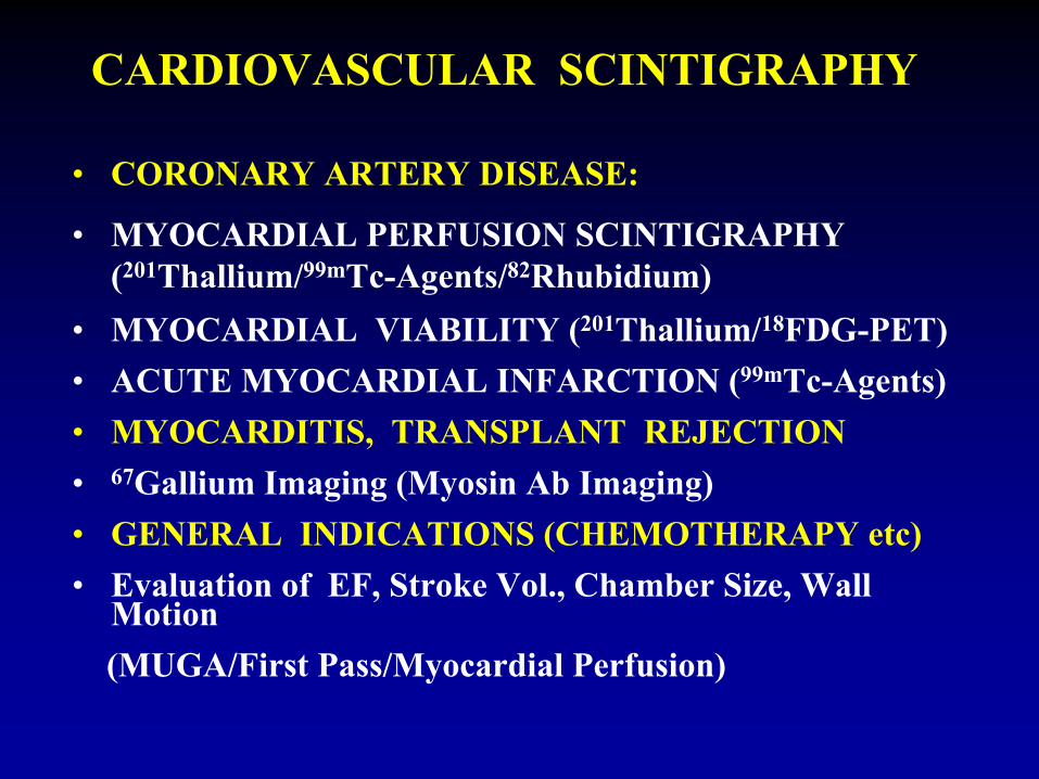
CARDIOVASCULAR SCINTIGRAPHY
• CORONARY ARTERY DISEASE:
• MYOCARDIAL PERFUSION SCINTIGRAPHY (201Thallium/99mTc-Agents/82Rhubidium)
• MYOCARDIAL VIABILITY (201Thallium/18FDG-PET)• ACUTE MYOCARDIAL INFARCTION (99mTc-Agents)• MYOCARDITIS, TRANSPLANT REJECTION• 67Gallium Imaging (Myosin Ab
Imaging)
• GENERAL INDICATIONS (CHEMOTHERAPY etc)• Evaluation of EF, Stroke Vol., Chamber Size, Wall
Motion(MUGA/First Pass/Myocardial Perfusion)
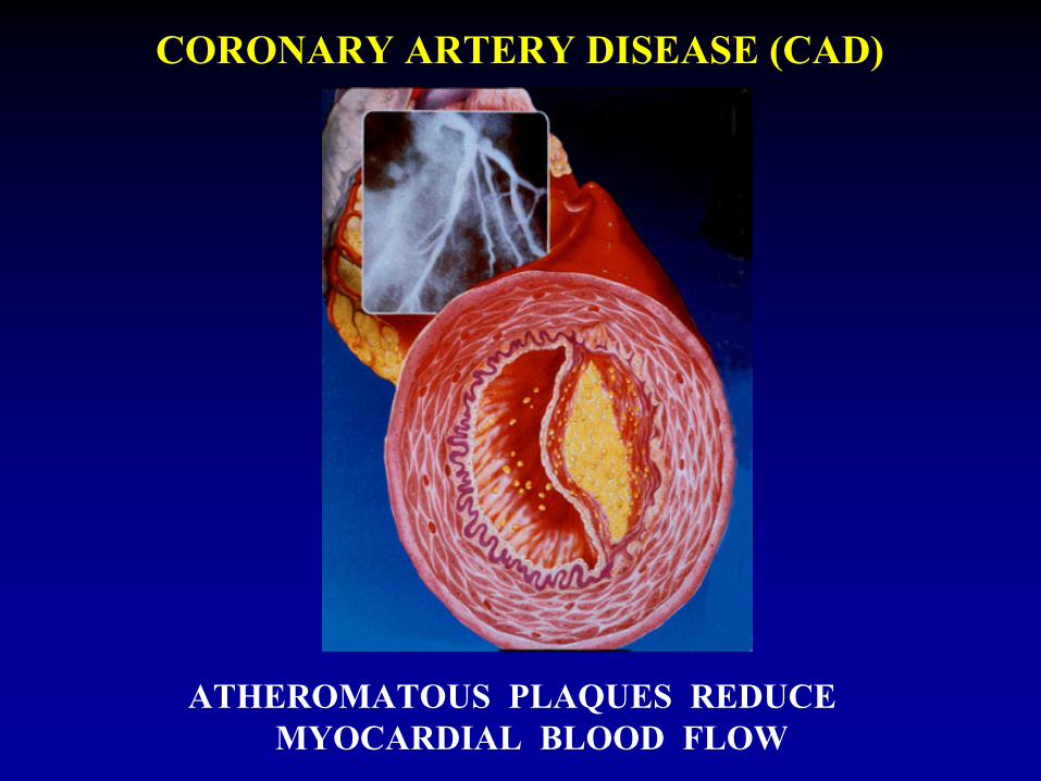
ATHEROMATOUS PLAQUES REDUCE MYOCARDIAL BLOOD FLOW
CORONARY ARTERY DISEASE (CAD)
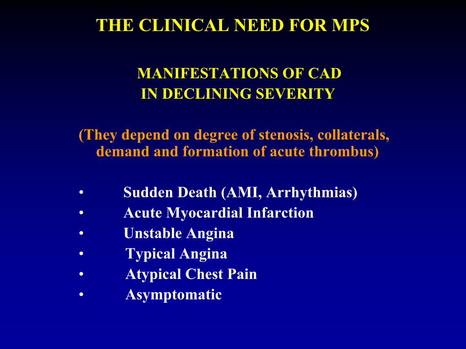
THE CLINICAL NEED FOR MPS
MANIFESTATIONS OF CADIN DECLINING SEVERITY
(They depend on degree of stenosis, collaterals, demand and formation of acute thrombus)
• Sudden Death (AMI, Arrhythmias)• Acute Myocardial Infarction• Unstable Angina• Typical Angina• Atypical Chest Pain• Asymptomatic
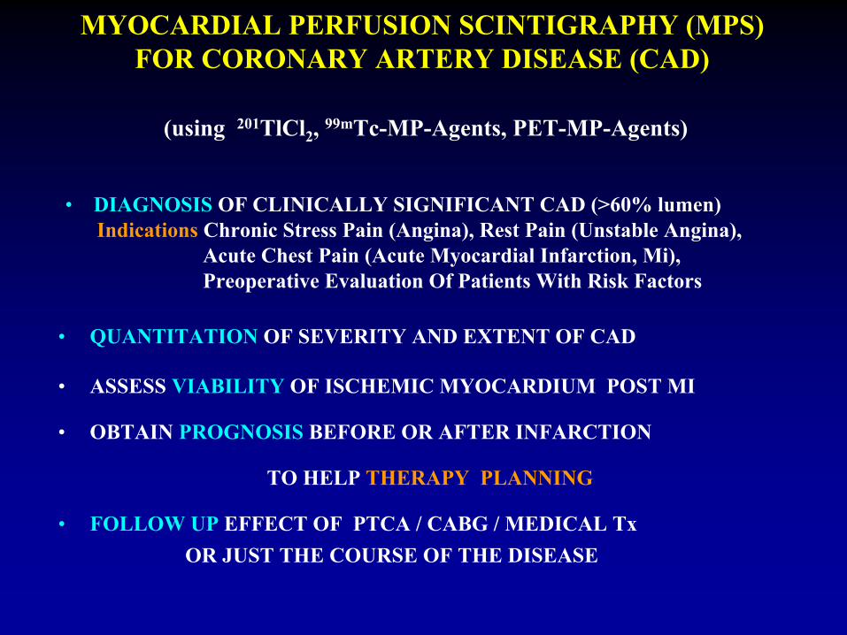
MYOCARDIAL PERFUSION SCINTIGRAPHY (MPS) FOR CORONARY ARTERY DISEASE (CAD)
(using 201TlCl2
, 99mTc-MP-Agents, PET-MP-Agents)
• QUANTITATION
OF SEVERITY AND EXTENT OF CAD
• ASSESS
VIABILITY
OF ISCHEMIC MYOCARDIUM POST MI
• OBTAIN
PROGNOSIS
BEFORE OR AFTER INFARCTION
TO HELP THERAPY PLANNING
• FOLLOW UP
EFFECT OF PTCA / CABG / MEDICAL TxOR JUST THE COURSE OF THE DISEASE
• DIAGNOSIS
OF CLINICALLY SIGNIFICANT CAD (>60% lumen)Indications Chronic Stress Pain (Angina), Rest Pain (Unstable Angina),
Acute Chest Pain (Acute Myocardial Infarction, Mi), Preoperative Evaluation Of Patients With Risk Factors
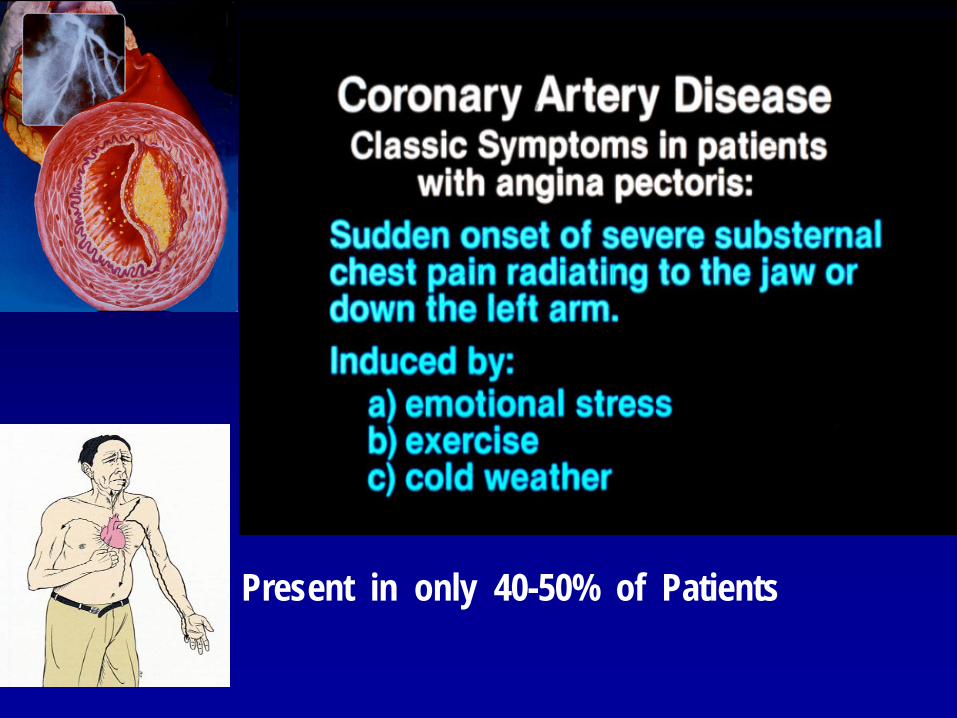
Present in only 40-50% of Patients
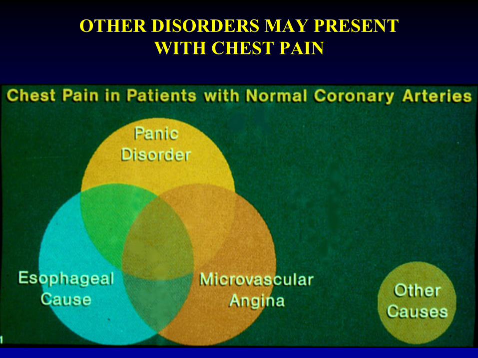
OTHER DISORDERS MAY PRESENT WITH CHEST PAIN
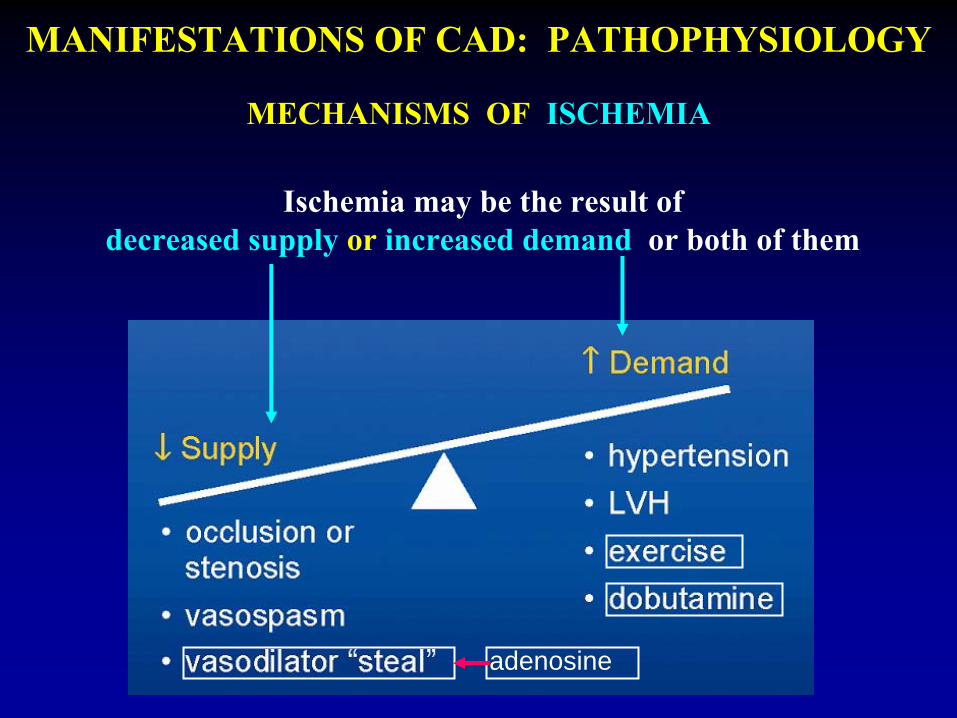
MANIFESTATIONS OF CAD: PATHOPHYSIOLOGY
MECHANISMS OF ISCHEMIA
adenosine
Ischemia may be the result of decreased supply
or increased demand
or both of them
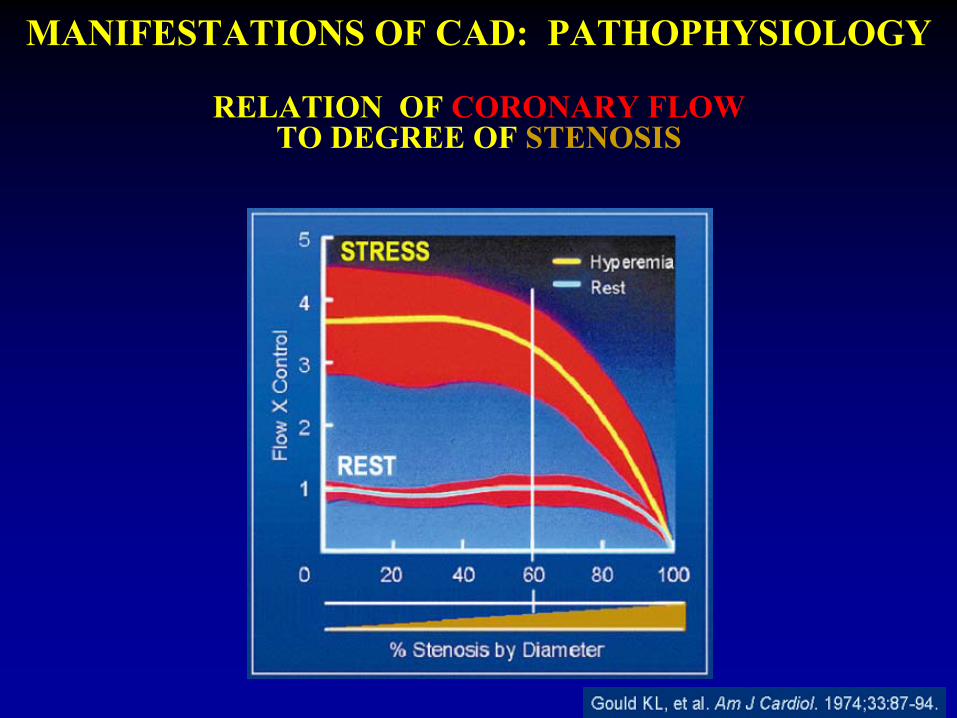
MANIFESTATIONS OF CAD: PATHOPHYSIOLOGY
RELATION OF CORONARY FLOW TO DEGREE OF STENOSIS
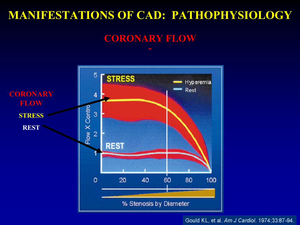
MANIFESTATIONS OF CAD: PATHOPHYSIOLOGY
CORONARY FLOW -
CORONARY FLOWSTRESS
REST
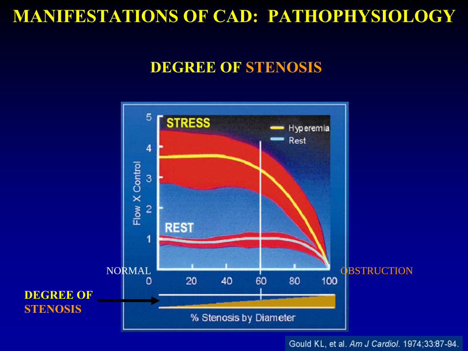
MANIFESTATIONS OF CAD: PATHOPHYSIOLOGY
DEGREE OF STENOSIS
DEGREE OF STENOSIS
NORMAL OBSTRUCTION
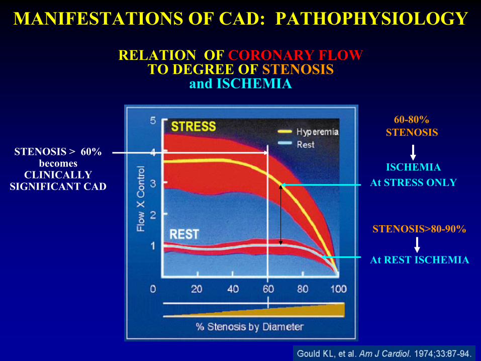
STENOSIS > 60%becomes
CLINICALLYSIGNIFICANT CAD
ISCHEMIA At STRESS ONLY
STENOSIS>80-90%
At REST ISCHEMIA
60-80% STENOSIS
MANIFESTATIONS OF CAD: PATHOPHYSIOLOGY
RELATION OF CORONARY FLOW TO DEGREE OF STENOSIS
and
ISCHEMIA
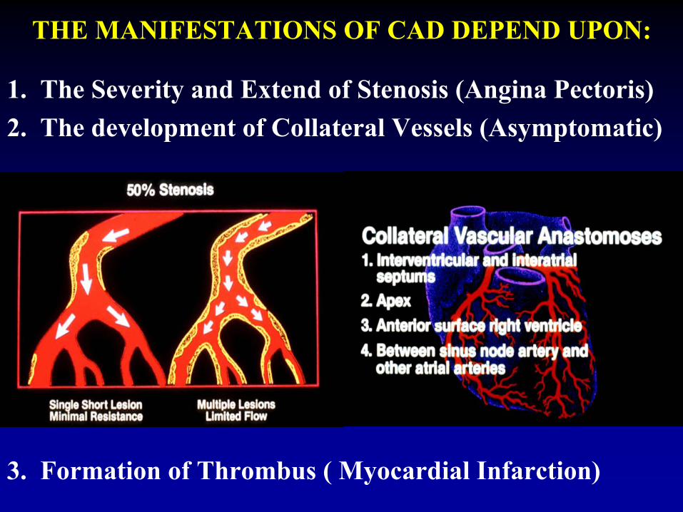
1. The Severity and Extend of Stenosis (Angina Pectoris) 2. The development of Collateral Vessels (Asymptomatic)
THE MANIFESTATIONS OF CAD DEPEND UPON:
3. Formation of Thrombus ( Myocardial Infarction)
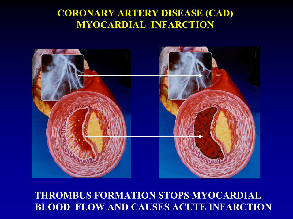
THROMBUS FORMATION STOPS MYOCARDIAL BLOOD FLOW AND CAUSES ACUTE INFARCTION
CORONARY ARTERY DISEASE (CAD) MYOCARDIAL INFARCTION

THE CLINICAL NEED FOR MPS
MANIFESTATIONS OF CADIN DECLINING SEVERITY
(They depend on degree of stenosis, collaterals, demand and formation of acute thrombus)
• Sudden Death (AMI, Arrhythmias)• Acute Myocardial Infarction• Unstable Angina• Typical Angina• Atypical Chest Pain• Asymptomatic
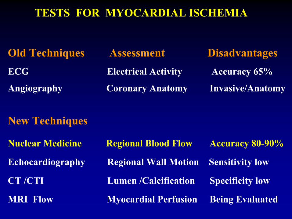
TESTS FOR MYOCARDIAL ISCHEMIA
Old Techniques Assessment Disadvantages ECG Electrical Activity Accuracy 65%
Angiography Coronary Anatomy Invasive/Anatomy
New Techniques
Nuclear Medicine Regional Blood Flow Accuracy 80-90%
Echocardiography Regional Wall Motion Sensitivity low
CT /CTI Lumen /Calcification Specificity low
MRI Flow Myocardial Perfusion Being Evaluated
THE CLINICAL NEED FOR MPS
Clinical Examination and EKG at Restand Special Tests are Suboptimal or Invasive
TREADMILL ECG TEST
• Informative, noninvasive, low risk
• Accuracy 65%
• Both FP and FN results
CARDIAC CATHETERIZATION
• Excellent anatomic
definition of CAD
• Insufficient physiologic information
• Limited definition of viability
• Invasive, expensive, subjective
• Evaluation and Therapy (Angioplasty)
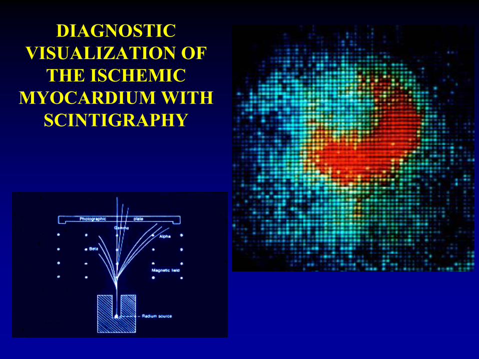
DIAGNOSTIC VISUALIZATION OF
THE ISCHEMIC MYOCARDIUM WITH
SCINTIGRAPHY
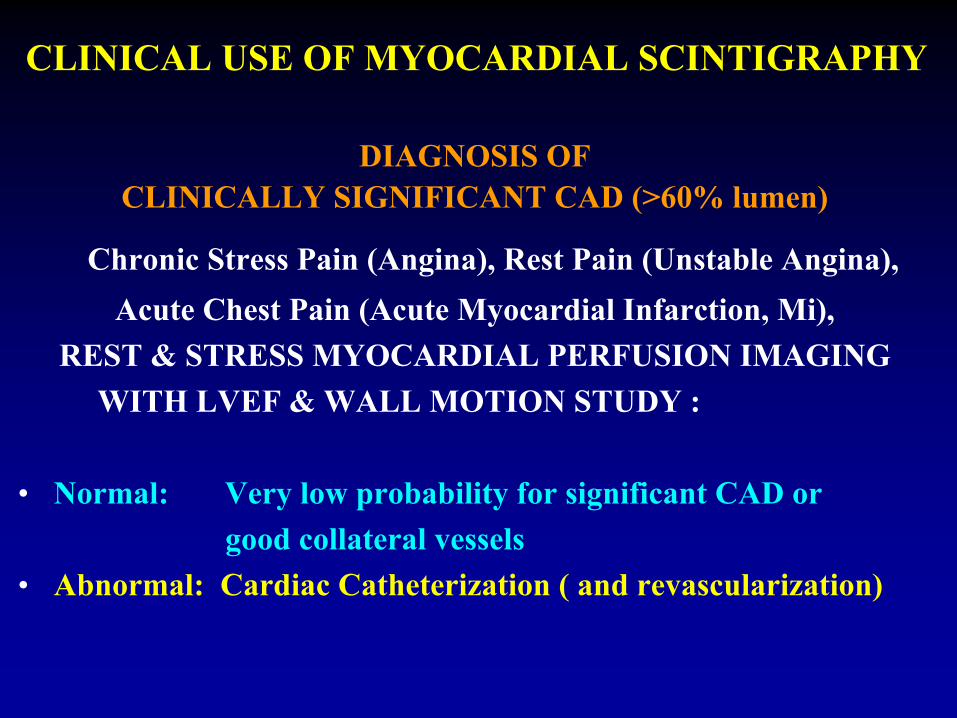
DIAGNOSIS OF CLINICALLY SIGNIFICANT CAD (>60% lumen)
Chronic Stress Pain (Angina), Rest Pain (Unstable Angina), Acute Chest Pain (Acute Myocardial Infarction, Mi),
REST & STRESS MYOCARDIAL PERFUSION IMAGINGWITH LVEF & WALL MOTION STUDY :
• Normal: Very low probability for significant CAD orgood collateral vessels
• Abnormal: Cardiac Catheterization ( and revascularization)
CLINICAL USE OF MYOCARDIAL SCINTIGRAPHY
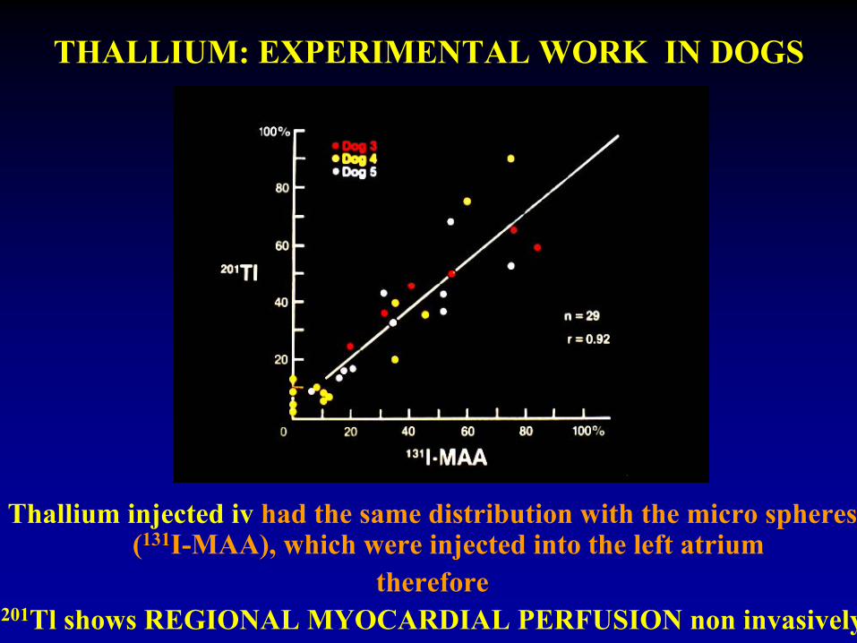
Thallium injected iv had the same distribution with the micro spheres (131I-MAA), which were injected into the left atrium
therefore201Tl shows REGIONAL MYOCARDIAL PERFUSION non invasively
THALLIUM: EXPERIMENTAL WORK IN DOGS
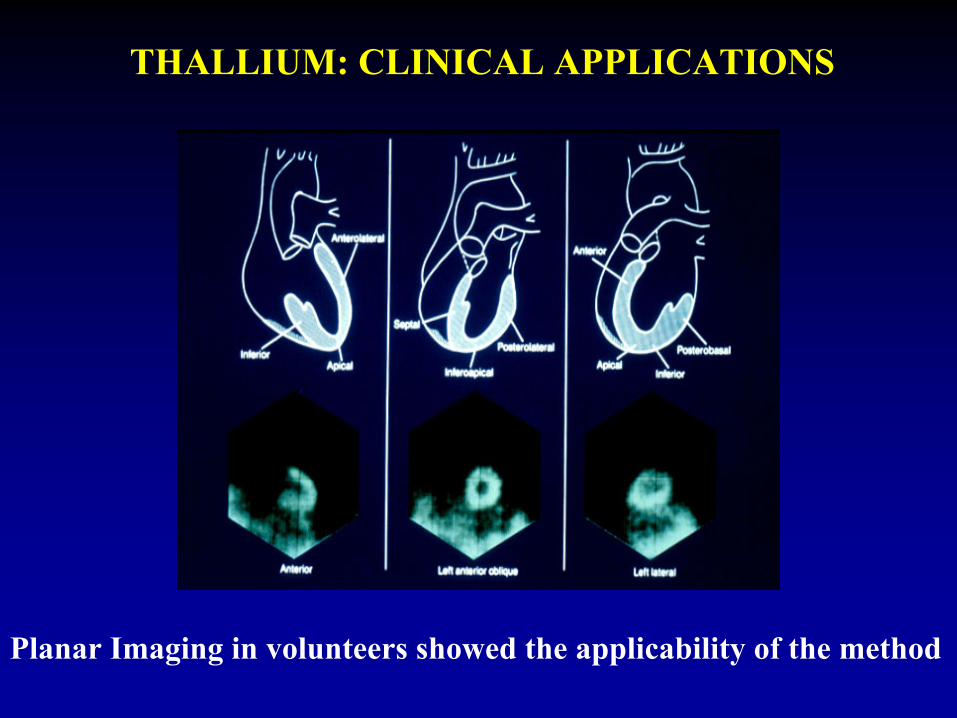
Planar Imaging in volunteers showed the applicability of the method
THALLIUM: CLINICAL APPLICATIONS
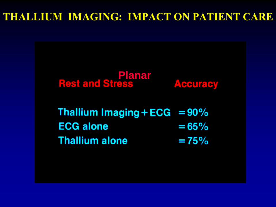
THALLIUM IMAGING: IMPACT ON PATIENT CARE
Planar

WHY WE NEED MYOCARDIAL PERFUSION SCINTIGRAPHY (MPS)
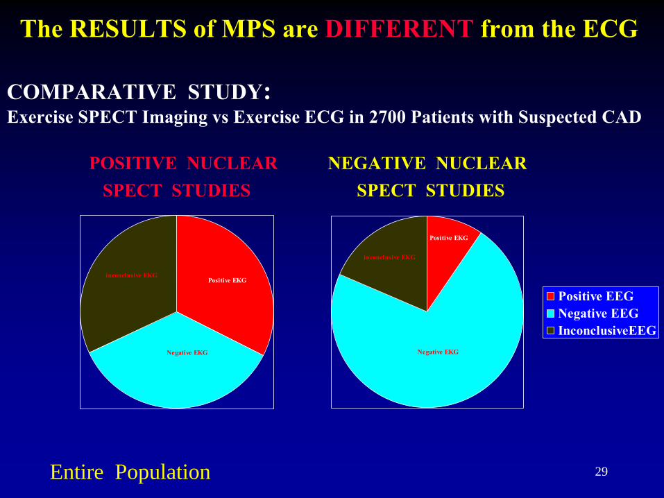
The RESULTS of MPS are DIFFERENT
from the ECG
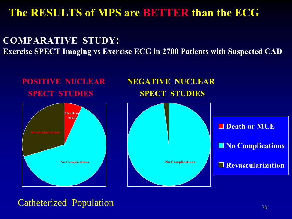
The RESULTS of MPS are BETTER
than the ECG
MPS is NON-INVASIVE as compared to Angiography
29
POSITIVE NUCLEAR
NEGATIVE NUCLEARSPECT STUDIES
SPECT STUDIES
The RESULTS of MPS are DIFFERENT
from the ECG
COMPARATIVE STUDY: Exercise SPECT Imaging vs
Exercise ECG in 2700 Patients with Suspected CAD
Positive EEGNegative EEGInconclusiveEEG
inconclusive EKG
Negative EKG
Positive EKG
inconclusive EKG
Negative EKG
Positive EKG
Entire Population
30
POSITIVE NUCLEAR
NEGATIVE NUCLEARSPECT STUDIES
SPECT STUDIES
The RESULTS of MPS are BETTER
than the ECG
COMPARATIVE STUDY: Exercise SPECT Imaging vs
Exercise ECG in 2700 Patients with Suspected CAD
Catheterized Population
Revascularization
No Complications
Death orMCE
Death or MCE
No Complications
RevascularizationNo Complications
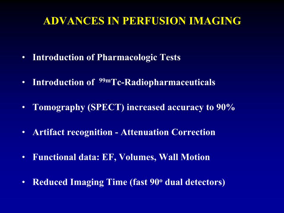
ADVANCES IN PERFUSION IMAGING
• Introduction of Pharmacologic Tests
• Introduction of 99mTc-Radiopharmaceuticals
• Tomography (SPECT) increased accuracy to 90%
• Artifact recognition -
Attenuation Correction
• Functional data: EF, Volumes, Wall Motion
• Reduced Imaging Time (fast 90o
dual detectors)
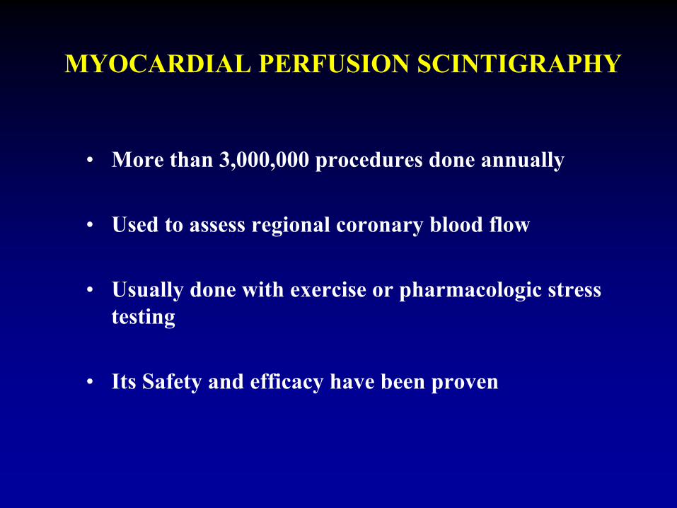
MYOCARDIAL PERFUSION SCINTIGRAPHY
• More than 3,000,000 procedures done annually
• Used to assess regional coronary blood flow
• Usually done with exercise or pharmacologic stress testing
• Its Safety and efficacy have been proven
MYOCARDIAL PERFUSION SCINTIGRAPHY (SPECT)
METHODS AND APPLICATIONS
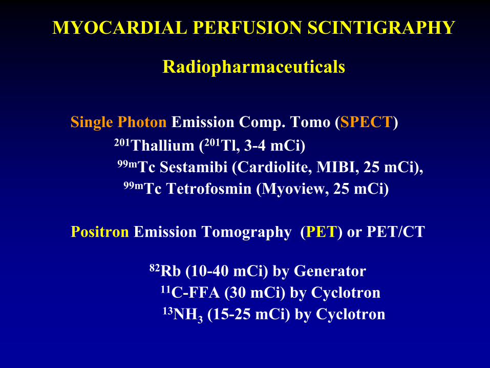
MYOCARDIAL PERFUSION SCINTIGRAPHY
Radiopharmaceuticals
Single Photon
Emission Comp. Tomo
(SPECT)201Thallium (201Tl, 3-4 mCi)99mTc Sestamibi (Cardiolite, MIBI, 25 mCi),
99mTc Tetrofosmin (Myoview, 25 mCi)
Positron
Emission Tomography (PET) or PET/CT
82Rb (10-40 mCi) by Generator11C-FFA (30 mCi) by Cyclotron13NH3
(15-25 mCi) by Cyclotron
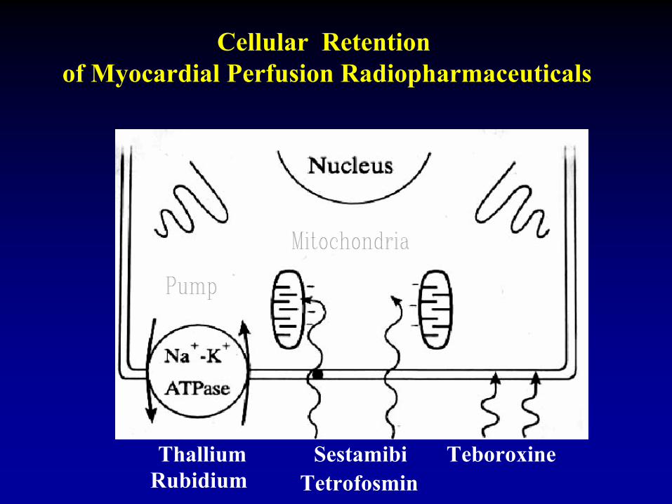
Cellular Retention of Myocardial Perfusion Radiopharmaceuticals
Thallium Sestamibi TeboroxineTetrofosmin Rubidium
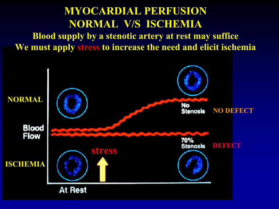
MYOCARDIAL PERFUSIONNORMAL V/S ISCHEMIA
Blood supply by a stenotic artery at rest may sufficeWe must apply stress
to increase the need and elicit ischemia
DEFECT
NO DEFECTNORMAL
ISCHEMIA
stress
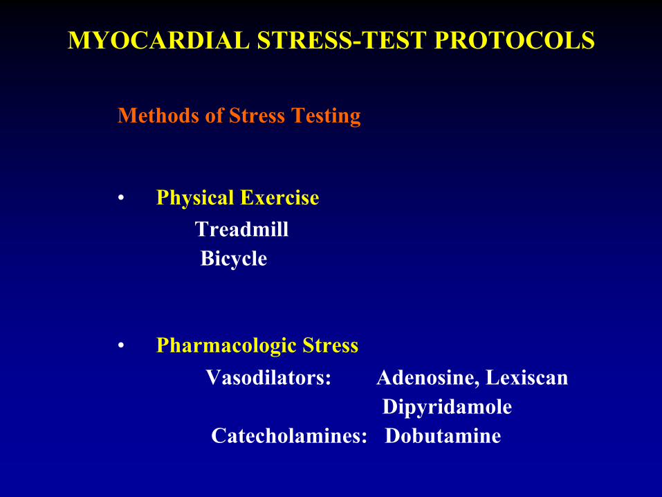
MYOCARDIAL STRESS-TEST PROTOCOLS
Methods of Stress Testing
• Physical ExerciseTreadmillBicycle
• Pharmacologic StressVasodilators: Adenosine, Lexiscan
DipyridamoleCatecholamines: Dobutamine
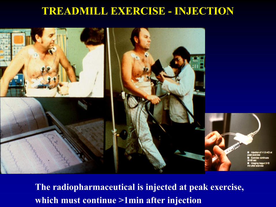
The radiopharmaceutical is injected at peak exercise,which must continue >1min after injection
TREADMILL EXERCISE -
INJECTION
MPS DIAGNOSIS and PROGNOSIS
Coronary Artery Disease (CAD) may be treatable
if diagnosed in time and before myocardial infarction or death occurs.
For the diagnosis of CAD Myocardial Perfusion Scintigraphy (MPS)
is a Sensitive and Specific method
MPS is proven effective for the early Diagnosis and Quantification of CAD,
helps in Prognosis, in decision making about mode of Therapy,
and can be used for follow up the course of the disease and results of treatment.
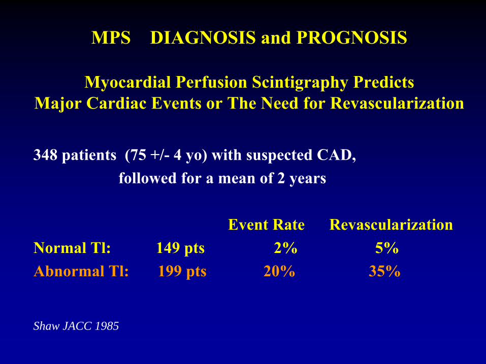
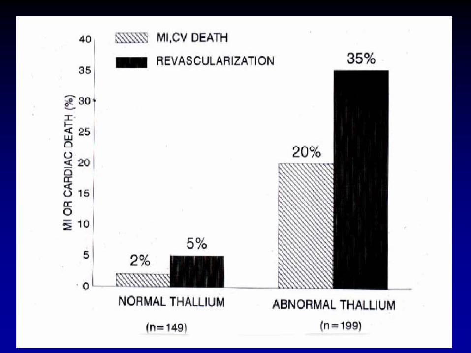
MPS DIAGNOSIS and PROGNOSIS
Myocardial Perfusion Scintigraphy Predicts Major Cardiac Events or The Need for Revascularization
348 patients (75 +/-
4 yo) with suspected CAD, followed for a mean of 2 years
Event Rate
RevascularizationNormal Tl: 149 pts
2% 5%
Abnormal Tl: 199 pts 20% 35%
Shaw JACC 1985
MPS DIAGNOSIS and PROGNOSIS
• There is only a requirement:
Adequate stress
is essential to uncover ischemia
during MPS.
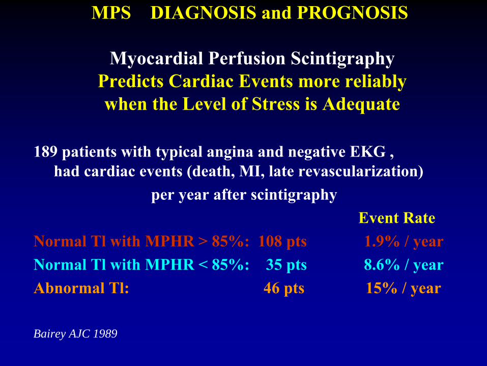
MPS DIAGNOSIS and PROGNOSIS
Myocardial Perfusion Scintigraphy Predicts Cardiac Events more reliably
when the Level of Stress is Adequate
189 patients with typical angina and negative EKG , had cardiac events (death, MI, late revascularization)
per year after scintigraphyEvent Rate
Normal Tl
with MPHR > 85%: 108 pts 1.9% / yearNormal Tl
with MPHR < 85%: 35 pts 8.6% / year
Abnormal Tl: 46 pts 15% / year
Bairey AJC 1989
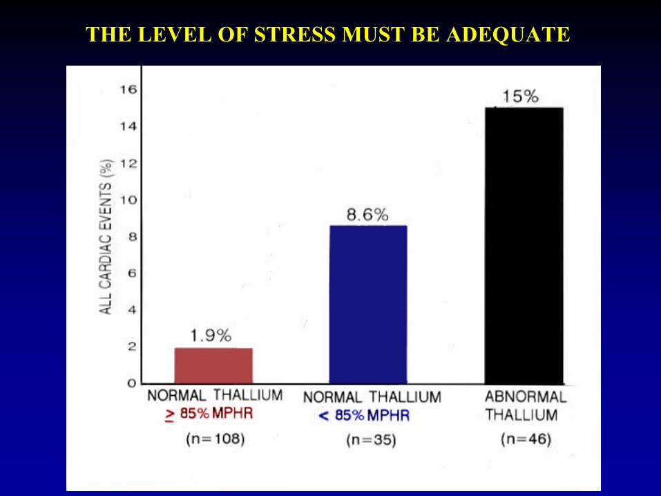
THE LEVEL OF STRESS MUST BE ADEQUATE
• There is indeed a requirement for a successful MPS: Adequate stress is essential
to uncover ischemia
• However, most of our patients, especially female, are unable to exercise to a level the test becomes diagnostic and fully useful. As a result many examinations are either useless or even misleading.
• Pharmacologic stress must be used when fitness for physical exercise is anticipated to be low.
LIMITATIONS OF THE PHYSICAL STRESS
CONTRAINDICATIONS TO PHYSICAL STRESS
• Aortic Stenosis
• Abdominal Aortic Aneurysm
• Left Bundle Branch Block
• Ventricular Paced Rhythm
• Inability to Exercise CNS or Orthopedic
• Limited Capacity for Exercise
• Peripheral Vascular Disease, COPD,
• Medications (eg. β
blockers),
• Poor Motivation,
Heart Failure,
• Previous Submaximal
Exercise Test
MOST FREQUENT INDICATIONS FOR PHARMACOLOGIC STRESS
• Inability to Exercise
• Contraindication to Exercise
• Limited capacity to Exercise
• Left Bundle Branch Block: ( use Adenosine, Dipyridamole )
• Asthma, Obstructive Lung Disease:( use Dobutamine )
PHARMACOLOGIC STRESS TESTING MEDICATIONS
VASODILATORS
ADENOSINE or
LEXISCAN:
Direct receptor Effect, Rapid Onset/Offset
DIPYRIDAMOLE:
Indirect Effect, Prolonged Action
SYMPATHETICOMIMETIC
DOBUTAMINE:
Indirect Effect (Enhances Myocardial Contraction)
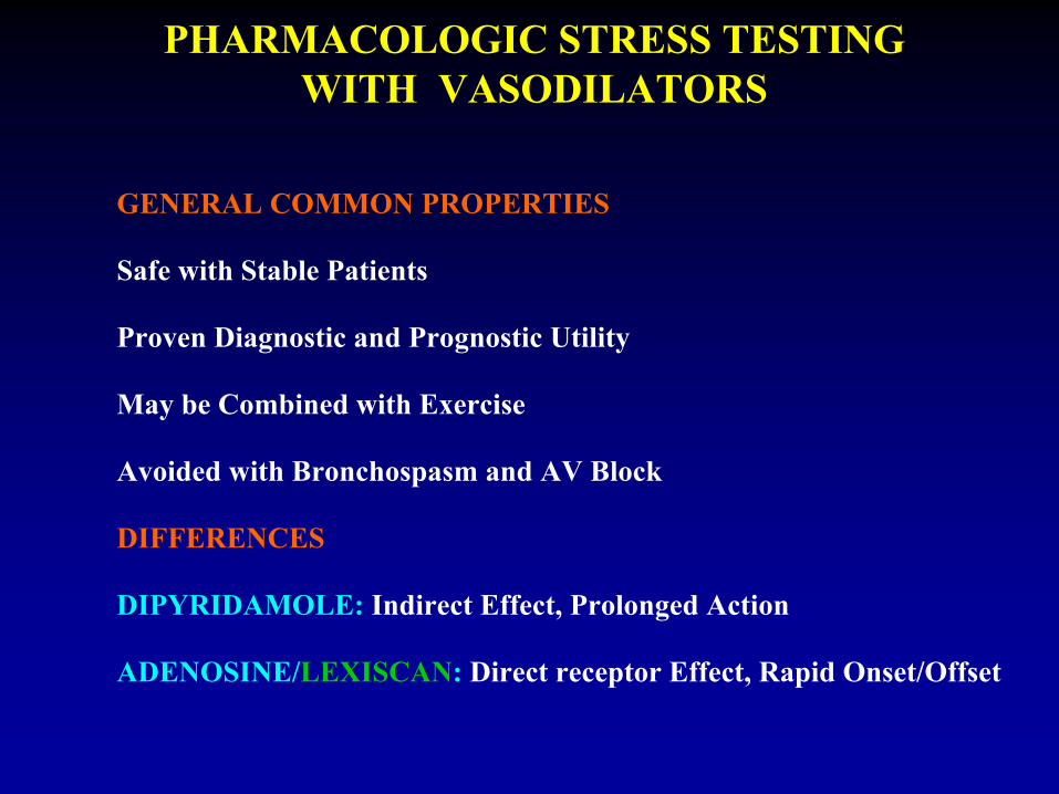
PHARMACOLOGIC STRESS TESTING WITH VASODILATORS
GENERAL COMMON PROPERTIES
Safe with Stable Patients
Proven Diagnostic and Prognostic Utility
May be Combined with Exercise
Avoided with Bronchospasm
and AV Block
DIFFERENCES
DIPYRIDAMOLE: Indirect Effect, Prolonged Action
ADENOSINE/LEXISCAN: Direct receptor Effect, Rapid Onset/Offset
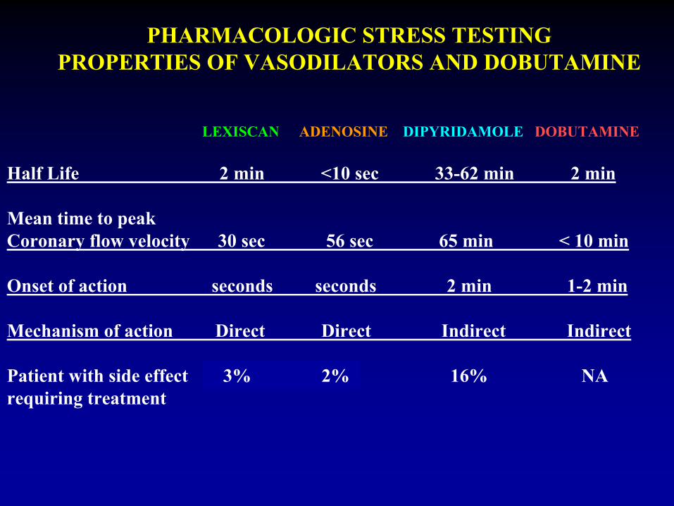
PHARMACOLOGIC STRESS TESTINGPROPERTIES OF VASODILATORS AND DOBUTAMINE
ADENOSINE DIPYRIDAMOLE DOBUTAMINELEXISCAN
Half Life 2 min <10 sec 33-62 min 2 min
Mean time to peakCoronary flow velocity 30 sec 56 sec 65 min < 10 min
Onset of action seconds seconds
2 min 1-2 min
Mechanism of action Direct Direct
Indirect Indirect
Patient with side effect 0.6% 16% NArequiring treatment
3% 2%
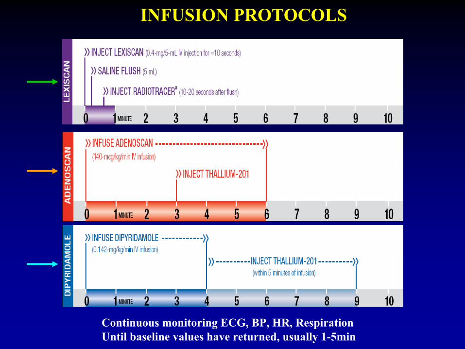
Continuous monitoring ECG, BP, HR, RespirationUntil baseline values have returned, usually 1-5min
INFUSION PROTOCOLS
At least 24 hr no products containingCaffeine, theophylline and dipyridamole
PREPARATION FOR ADENOSINE STRESS
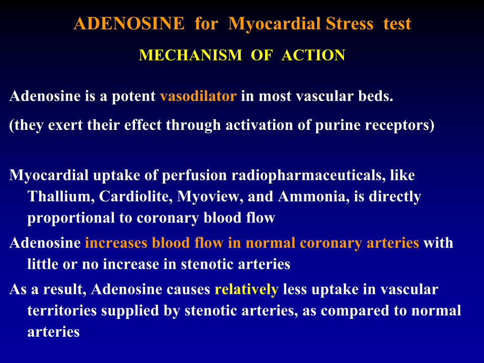
MECHANISM OF ACTION
Adenosine is a potent vasodilator
in most vascular beds.
(they exert their effect through activation of purine
receptors)
Myocardial uptake of perfusion radiopharmaceuticals, like Thallium, Cardiolite, Myoview, and Ammonia, is directly proportional to coronary blood flow
Adenosine increases blood flow in normal coronary arteries
with little or no increase in stenotic arteries
As a result, Adenosine causes relatively
less uptake in vascular territories supplied by stenotic arteries, as compared to normal
arteries
ADENOSINE for Myocardial Stress test
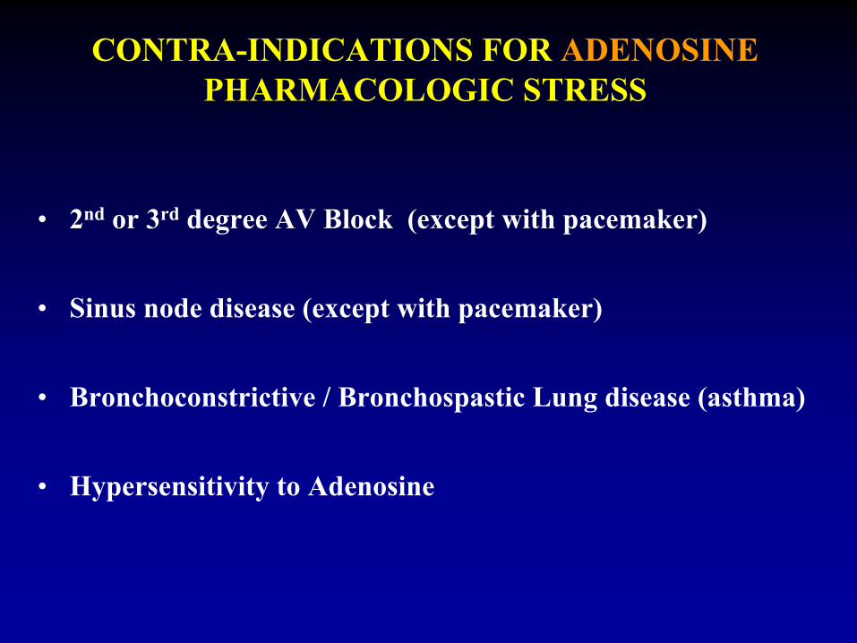
CONTRA-INDICATIONS FOR ADENOSINE PHARMACOLOGIC STRESS
• 2nd
or 3rd
degree AV Block (except with pacemaker)
• Sinus node disease (except with pacemaker)
• Bronchoconstrictive
/ Bronchospastic
Lung disease (asthma)
• Hypersensitivity to Adenosine
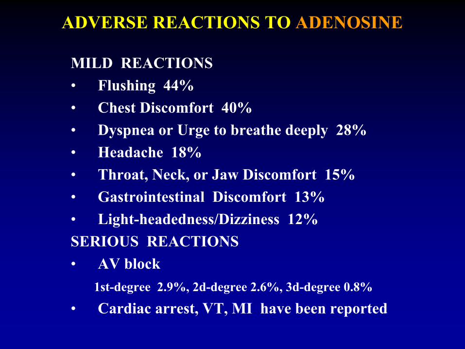
MILD REACTIONS• Flushing 44%• Chest Discomfort 40%• Dyspnea or Urge to breathe deeply 28%• Headache 18%• Throat, Neck, or Jaw Discomfort 15%• Gastrointestinal Discomfort 13%• Light-headedness/Dizziness 12%SERIOUS REACTIONS• AV block
1st-degree 2.9%, 2d-degree 2.6%, 3d-degree 0.8%
• Cardiac arrest, VT, MI have been reported
ADVERSE REACTIONS TO ADENOSINE
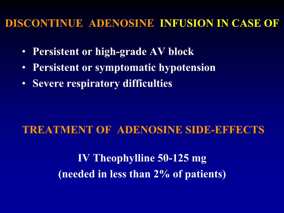
• Persistent or high-grade AV block• Persistent or symptomatic hypotension• Severe respiratory difficulties
DISCONTINUE ADENOSINE
INFUSION IN CASE OF
TREATMENT OF ADENOSINE SIDE-EFFECTS
IV Theophylline 50-125 mg(needed in less than 2% of patients)
LEXISCAN FOR MYOCARDIAL STRESS TEST
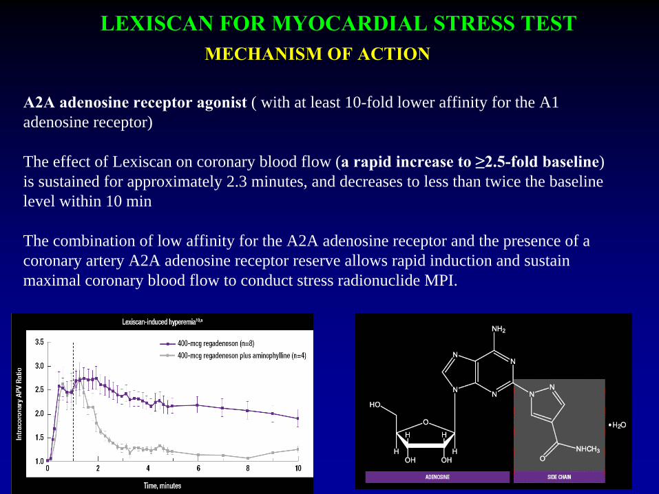
A2A adenosine receptor agonist
( with at least 10-fold lower affinity for the A1 adenosine receptor)
The effect of Lexiscan on coronary blood flow (a rapid increase to ≥2.5-fold baseline) is sustained for approximately 2.3 minutes, and decreases to less than twice the baseline level within 10 min
The combination of low affinity for the A2A adenosine receptor and the presence of a coronary artery A2A adenosine receptor reserve allows rapid induction and sustain maximal coronary blood flow to conduct stress radionuclide MPI.
MECHANISM OF ACTION
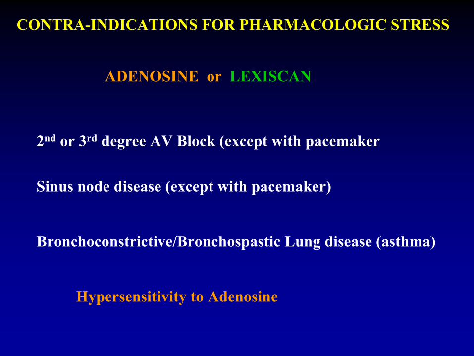
CONTRA-INDICATIONS FOR PHARMACOLOGIC STRESS
ADENOSINE or LEXISCAN
2nd
or 3rd
degree AV Block (except with pacemaker
Sinus node disease (except with pacemaker)
Bronchoconstrictive/Bronchospastic
Lung disease (asthma)
Hypersensitivity to Adenosine
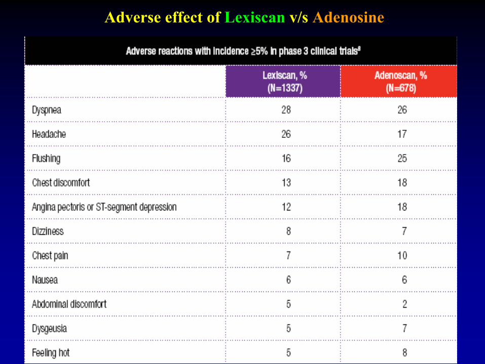
Adverse effect of Lexiscan
v/s
Adenosine
TREATMENT OF LEXISCAN SIDE-EFFECTS
IV Theophylline 50-100 mg
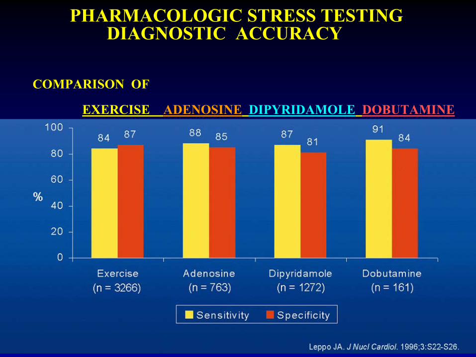
PHARMACOLOGIC STRESS TESTING DIAGNOSTIC ACCURACY
COMPARISON OF EXERCISE ADENOSINE
DIPYRIDAMOLE
DOBUTAMINE
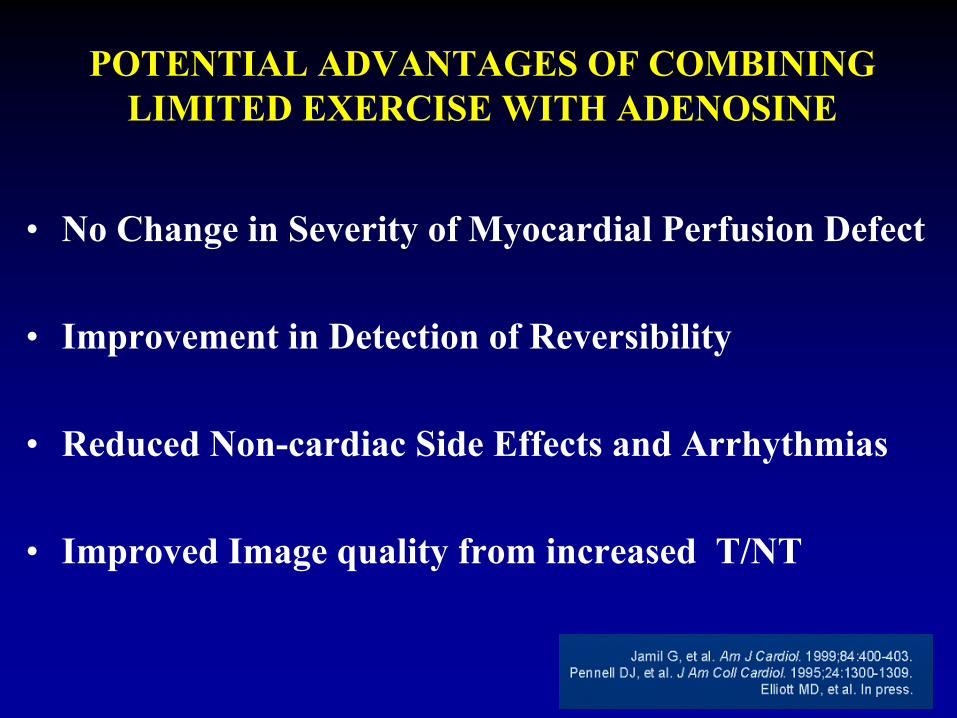
• No Change in Severity of Myocardial Perfusion Defect
• Improvement in Detection of Reversibility
• Reduced Non-cardiac Side Effects and Arrhythmias
• Improved Image quality from increased T/NT
POTENTIAL ADVANTAGES OF COMBINING LIMITED EXERCISE WITH ADENOSINE
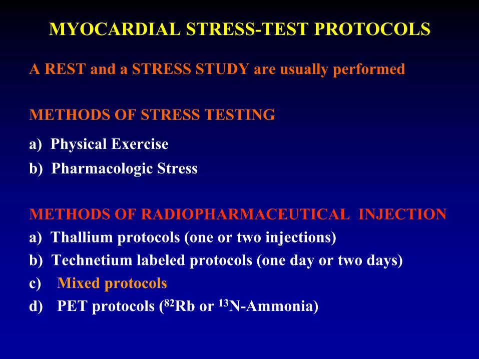
MYOCARDIAL STRESS-TEST PROTOCOLS
A REST and a STRESS STUDY are usually performed
METHODS OF STRESS TESTING
a) Physical Exerciseb) Pharmacologic Stress
METHODS OF RADIOPHARMACEUTICAL INJECTIONa) Thallium protocols (one or two injections)b) Technetium labeled protocols (one day or two days)c)
Mixed protocols
d)
PET protocols (82Rb or 13N-Ammonia)
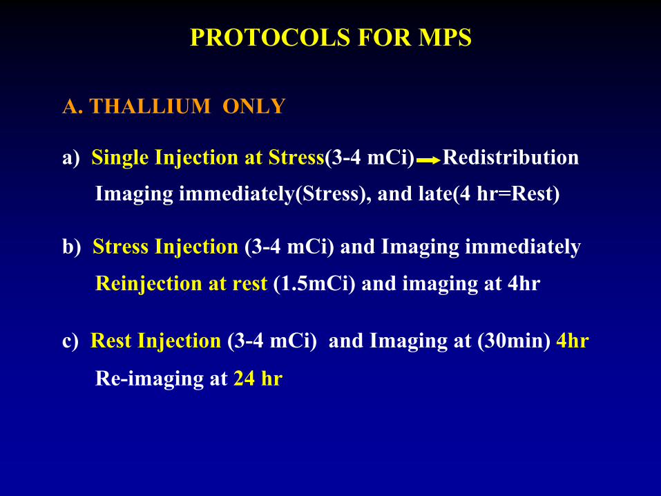
PROTOCOLS FOR MPS
A. THALLIUM ONLY
a) Single Injection
at Stress(3-4 mCi) Redistribution
Imaging immediately(Stress), and late(4 hr=Rest)
b) Stress Injection
(3-4 mCi) and Imaging immediately
Reinjection at rest
(1.5mCi) and imaging at 4hr
c) Rest Injection
(3-4 mCi) and Imaging at (30min) 4hr
Re-imaging at 24 hr
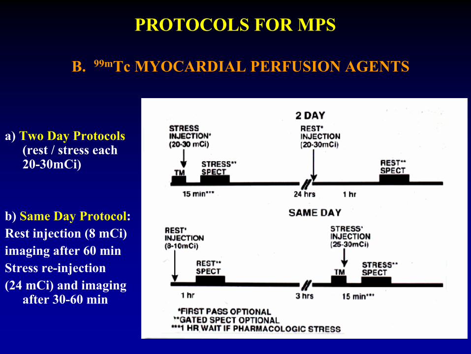
PROTOCOLS FOR MPS
a) Two Day Protocols (rest / stress each
20-30mCi)
b) Same Day Protocol:Rest injection (8 mCi)imaging after 60 minStress re-injection(24 mCi) and imaging
after 30-60 min
B. 99mTc MYOCARDIAL PERFUSION AGENTS
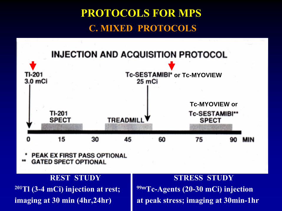
REST STUDY201Tl (3-4 mCi) injection at rest;imaging at 30 min (4hr,24hr)
PROTOCOLS FOR MPSC. MIXED PROTOCOLS
STRESS STUDY99mTc-Agents (20-30 mCi) injection at peak stress; imaging at 30min-1hr
or Tc-MYOVIEW
Tc-MYOVIEW or
MIXED PROTOCOLS FOR MPS
ADVANTAGES
• IMPROVED QUALITY 201Tl more sensitive for viability than 99mTc-Agents 99mTc-Agents had better photons and short half life, the higher dose increases sensitivity for ischemia, allows functional evaluations (EF, Volumes,WM, )
• BETTER LOGISTICS
Facilitates imaging, utilizes resources better, shortens time the patient stays in the lab
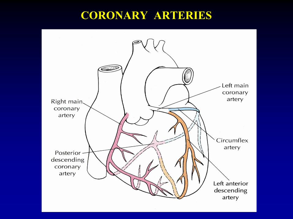
CORONARY ARTERIES
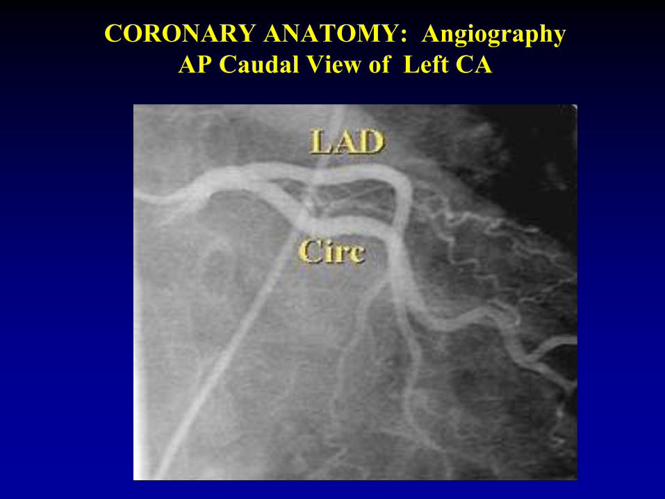
CORONARY ANATOMY: Angiography AP Caudal View of Left CA
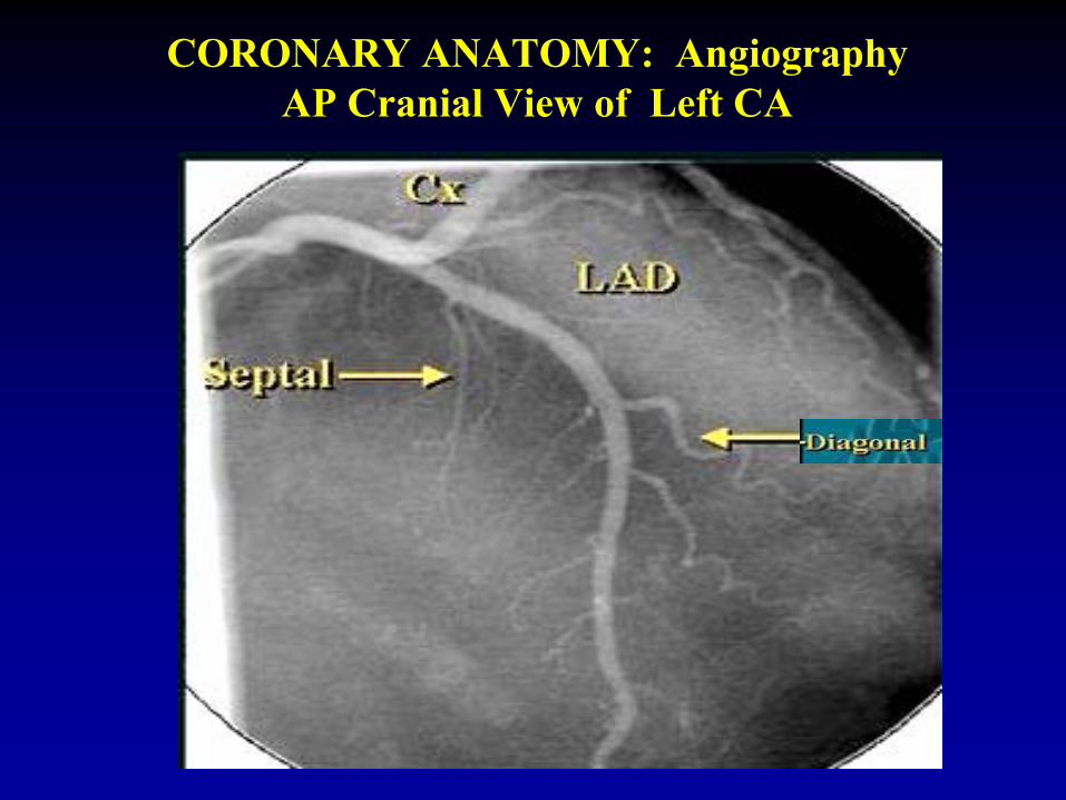
CORONARY ANATOMY: Angiography AP Cranial View of Left CA
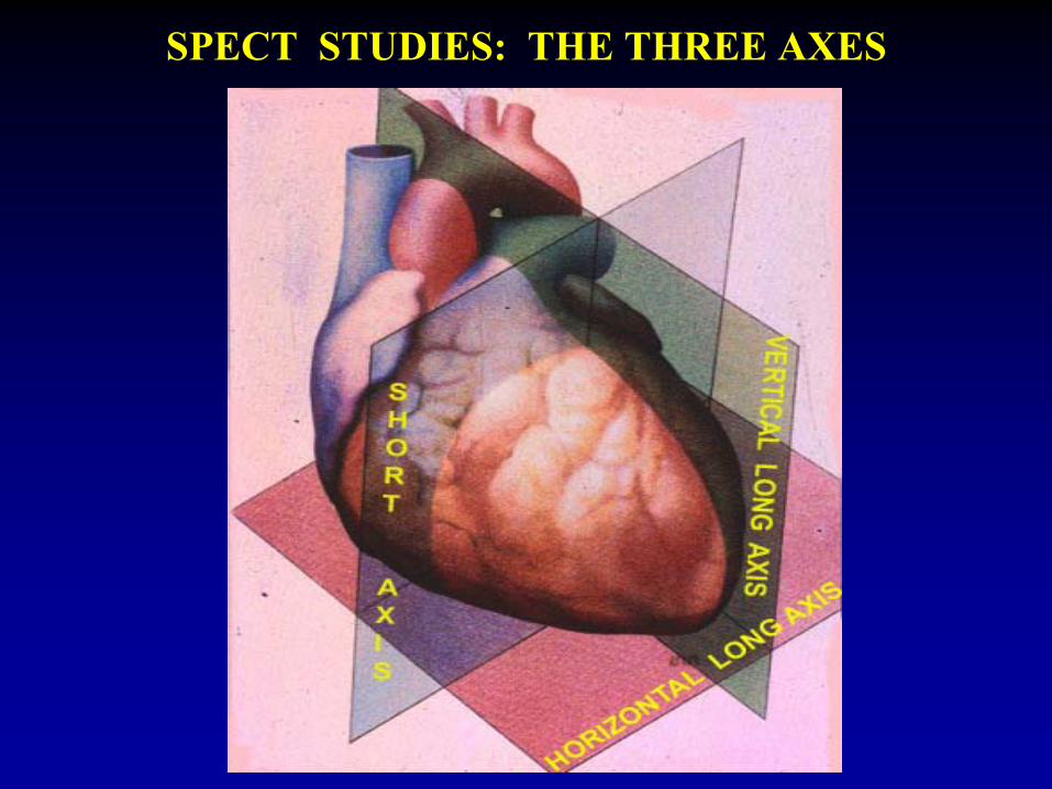
SPECT STUDIES: THE THREE AXES

A 55 yo man with typical angina pectoris has a stress and rest MPS
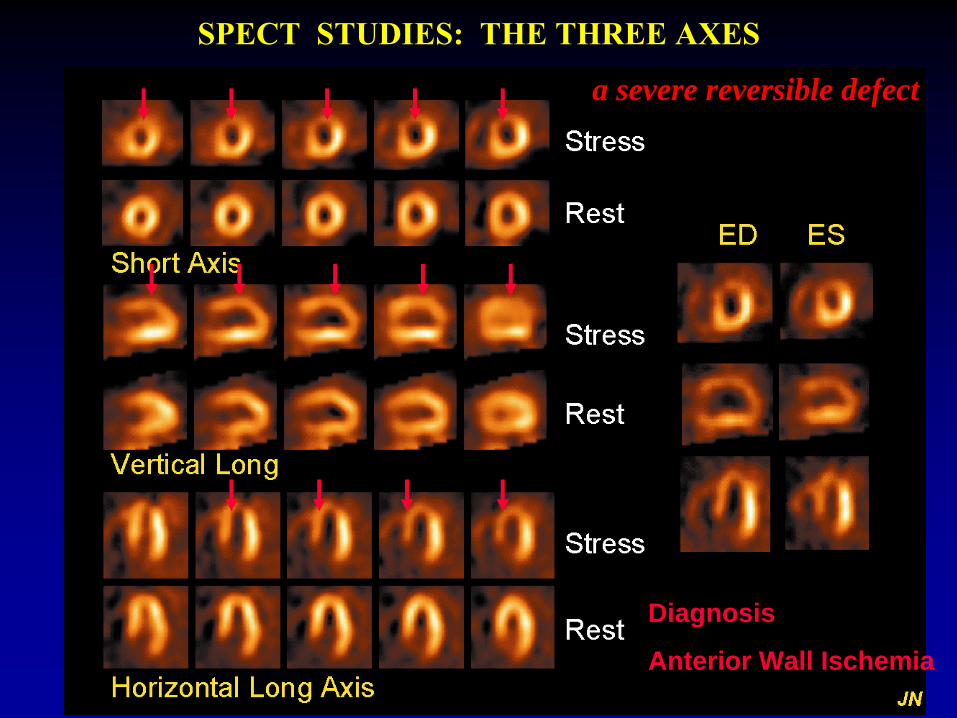
SPECT STUDIES: THE THREE AXES
Diagnosis
Anterior Wall Ischemia
a severe reversible defect
ASSESSMENT OF VENTRICULAR FUNCTION IN CONJUNCTION WITH PERFUSION IMAGING
GATED SPECT AT REST
• Global and regional thickening and wall motion
• LV Ejection Fraction, global and regional
• LV Volumes: Stroke Volume, ESV, EDV
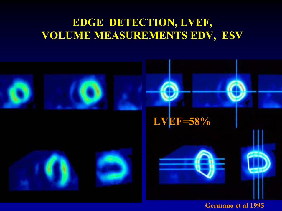
EDGE DETECTION, LVEF, VOLUME MEASUREMENTS EDV, ESV
LVEF=58%
Germano
et al 1995
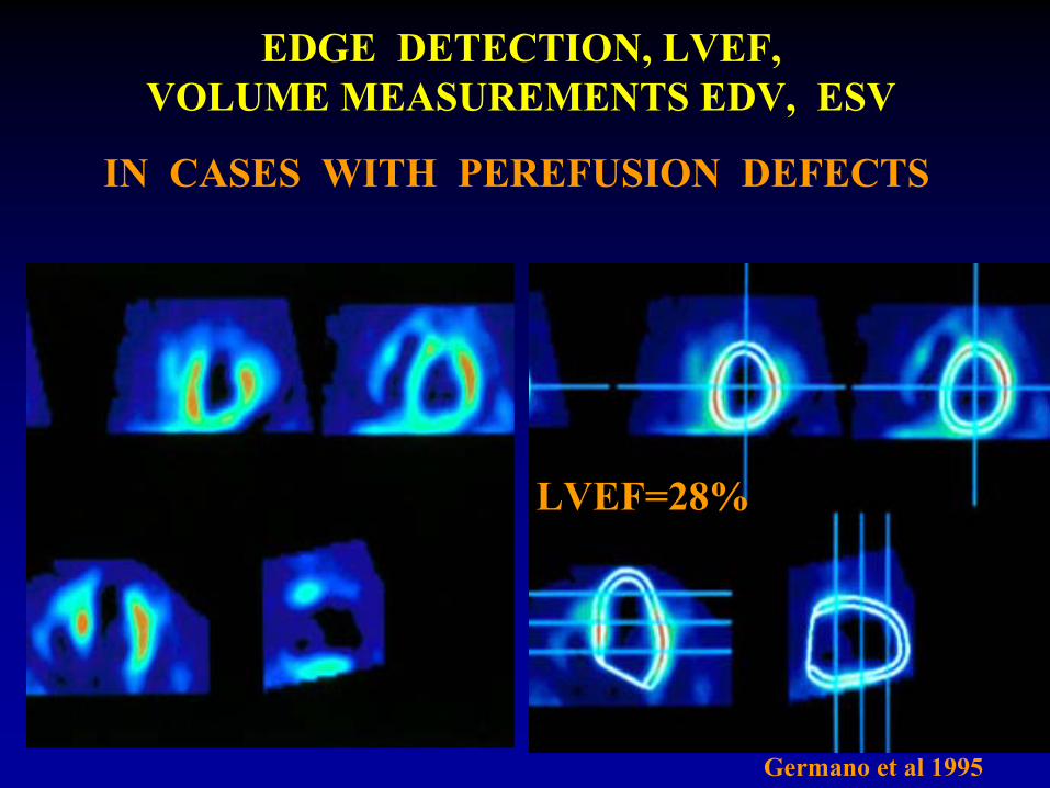
IN CASES WITH PEREFUSION DEFECTS
EDGE DETECTION, LVEF, VOLUME MEASUREMENTS EDV, ESV
LVEF=28%
Germano
et al 1995
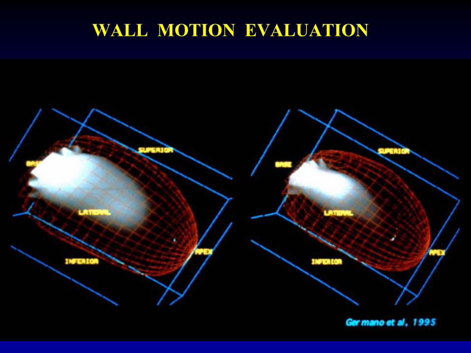
WALL MOTION EVALUATION
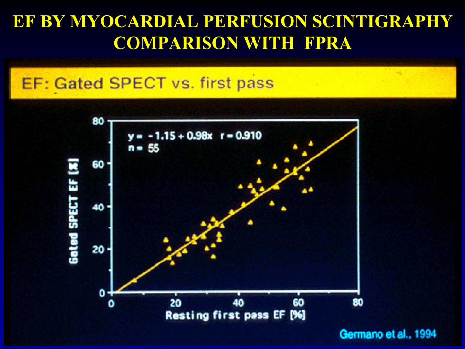
EF BY MYOCARDIAL PERFUSION SCINTIGRAPHY COMPARISON WITH FPRA
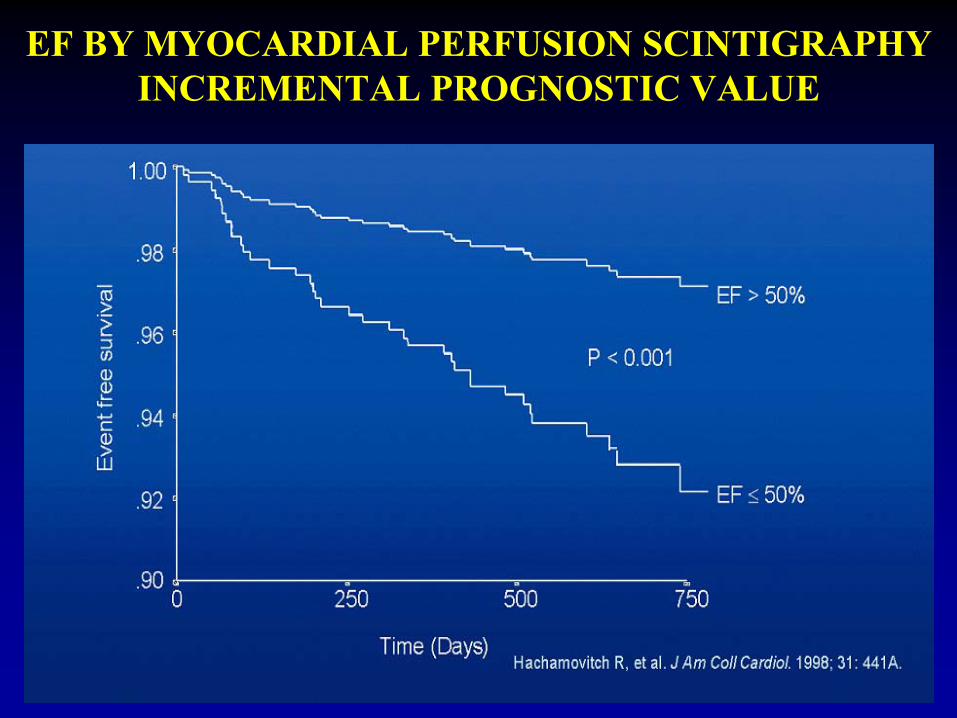
EF BY MYOCARDIAL PERFUSION SCINTIGRAPHY INCREMENTAL PROGNOSTIC VALUE
CLINICAL USE OF MYOCARDIAL SCINTIGRAPHY
QUANTITATION OF EXTENT AND SEVERITY OF CAD
(Before or after cardiac catheterization )
REST & STRESS MYOCARDIAL PERFUSION IMAGING
WITH LVEF & WALL MOTION STUDY IS PERFORMED
a) Severity of ischemia: mild, moderate, severe
b) Extend of ischemia: percent of total wall found ischemic
c) Vessel(s) involved: Single, Two, Multiple, LAD/LCx/RCA
d) Function:
Wall motion, Volumes, EF ( global and regional)
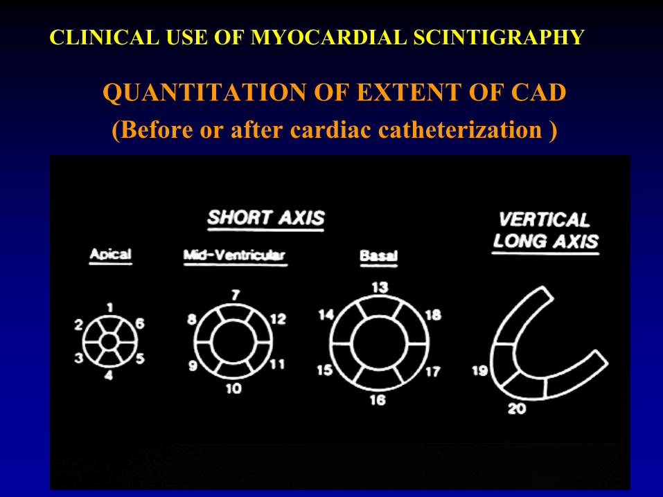
CLINICAL USE OF MYOCARDIAL SCINTIGRAPHY
QUANTITATION OF EXTENT OF CAD (Before or after cardiac catheterization )
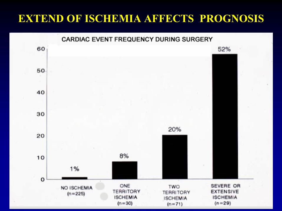
EXTEND OF ISCHEMIA AFFECTS PROGNOSIS
A 61 yo lady is evaluated for chest pain (not typical)
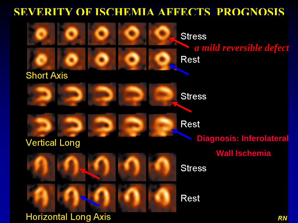
SEVERITY OF ISCHEMIA AFFECTS PROGNOSIS
a mild reversible defect
Diagnosis: Inferolateral
Wall Ischemia

A 55 yo man with typical angina pectoris has a stress and rest MPS
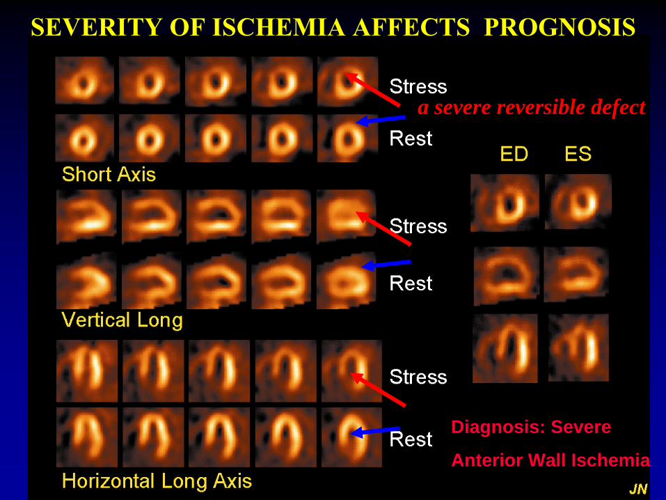
a severe reversible defect
SEVERITY OF ISCHEMIA AFFECTS PROGNOSIS
Diagnosis: Severe
Anterior Wall Ischemia
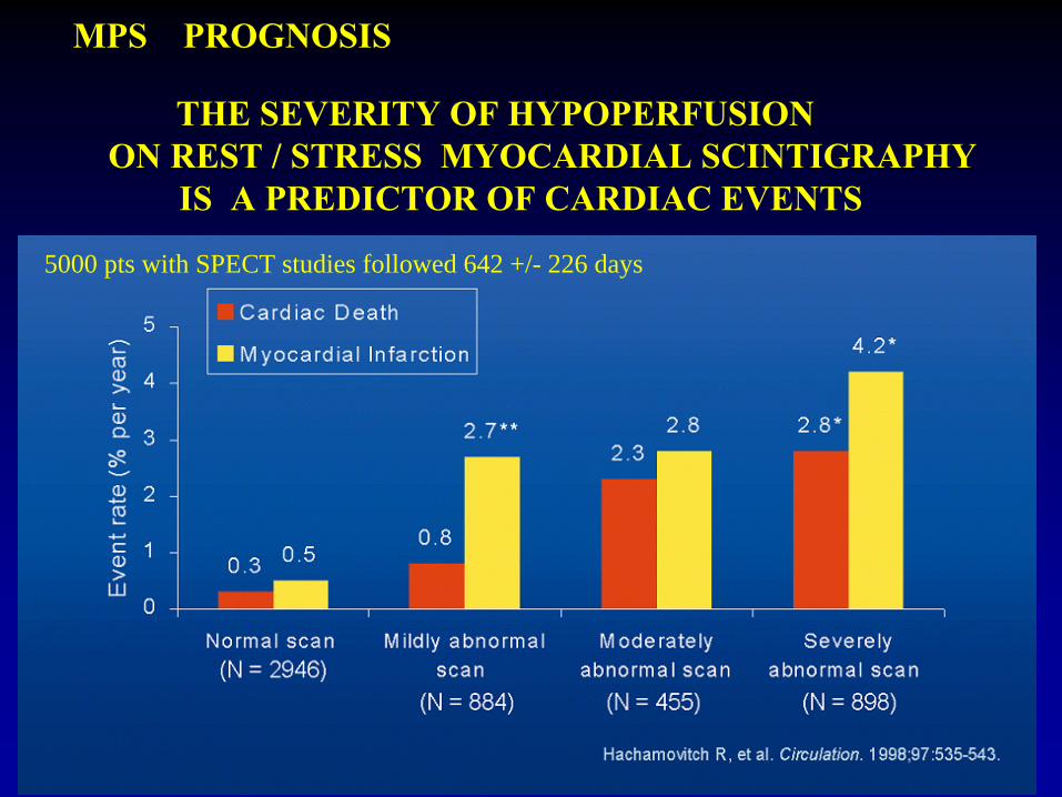
MPS PROGNOSIS
THE SEVERITY OF HYPOPERFUSION ON REST / STRESS MYOCARDIAL SCINTIGRAPHY
IS A PREDICTOR OF CARDIAC EVENTS
5000 pts with SPECT studies followed 642 +/- 226 days
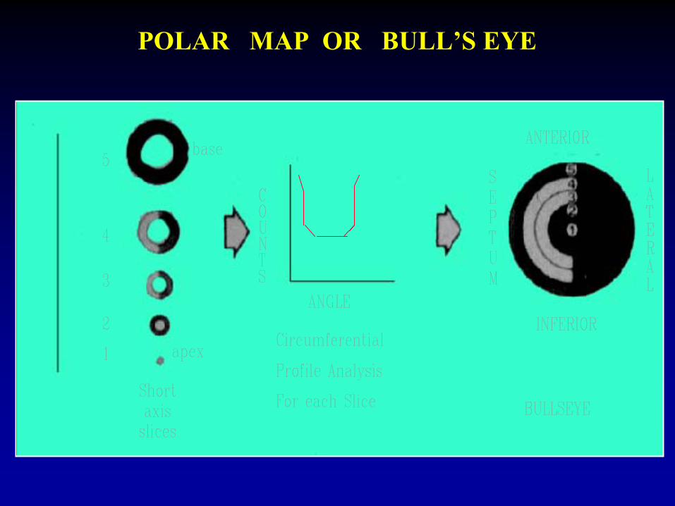
POLAR MAP OR BULL’S EYE
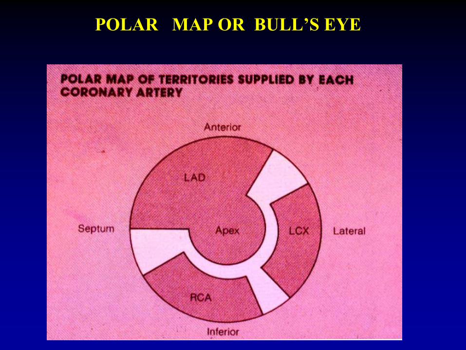
POLAR MAP OR BULL’S EYE
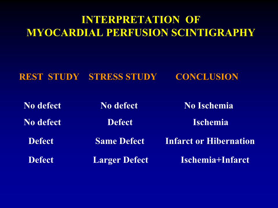
INTERPRETATION OF MYOCARDIAL PERFUSION SCINTIGRAPHY
REST STUDY STRESS STUDY CONCLUSION
No defect No defect No Ischemia
No defect Defect
Ischemia
Defect Same Defect Infarct or Hibernation
Defect Larger Defect Ischemia+Infarct
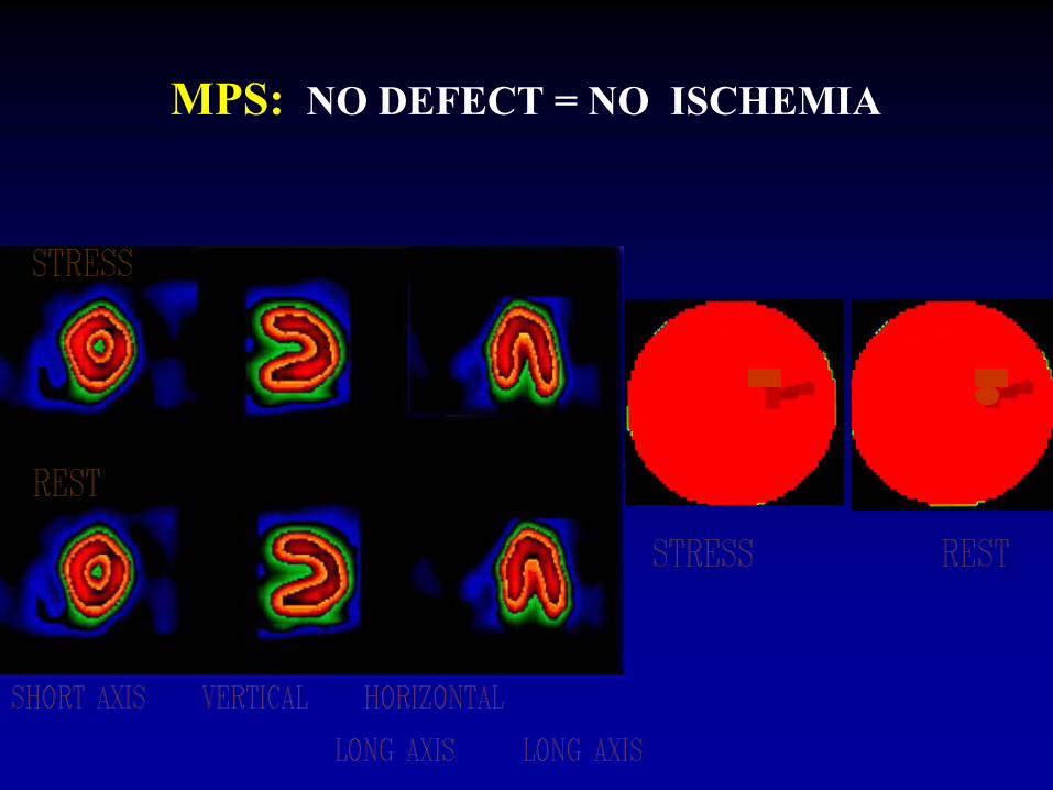
MPS: NO DEFECT =
NO ISCHEMIA
A 45 yo patient presents with typical angina pectorisHas an exercise and rest MPS
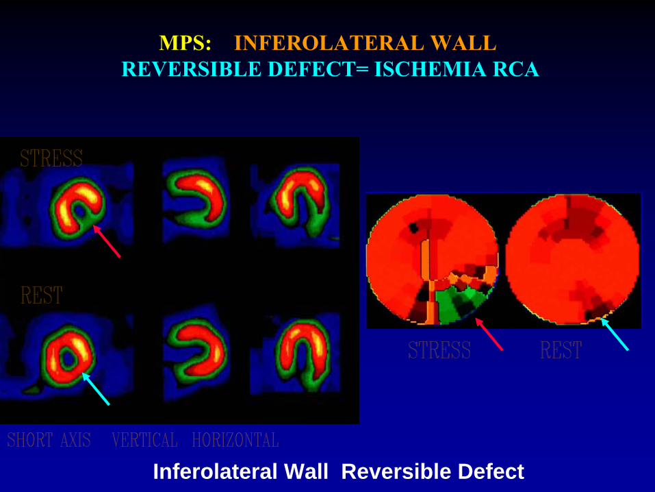
Inferolateral Wall Reversible Defect
MPS: INFEROLATERAL WALL REVERSIBLE DEFECT= ISCHEMIA RCA

A 79 yo man with history af
CAD and MI has an Adenosine MPS
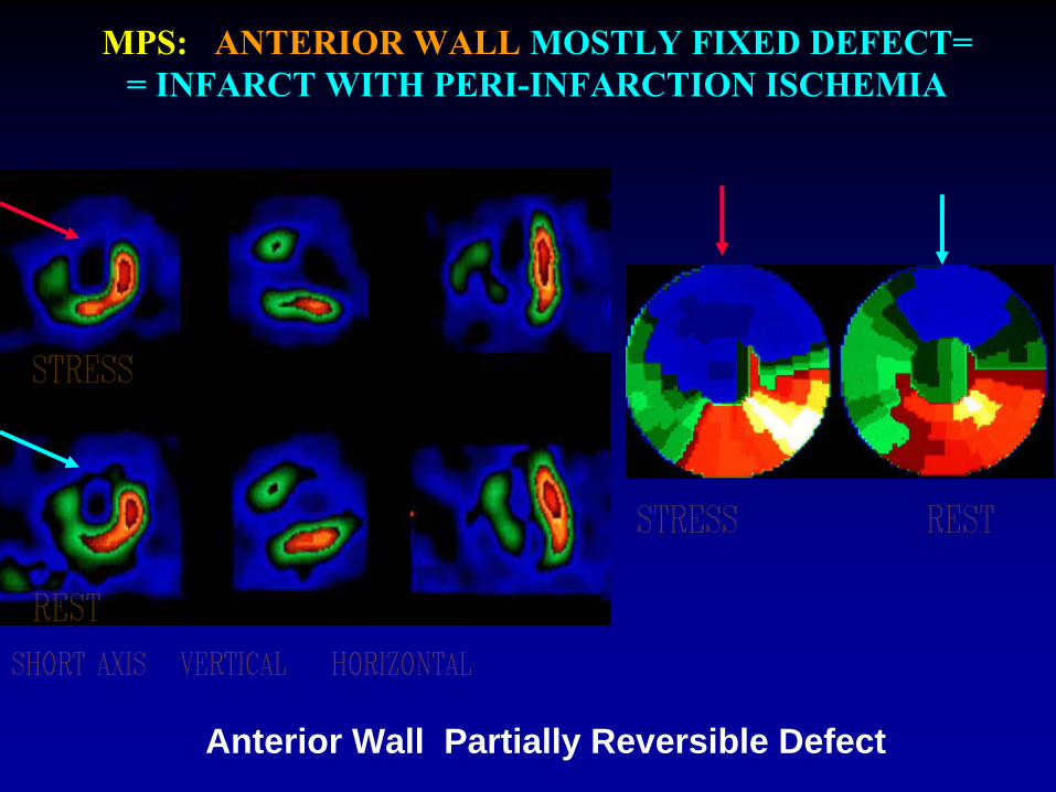
MPS: ANTERIOR WALL MOSTLY FIXED DEFECT= =
INFARCT WITH PERI-INFARCTION ISCHEMIA
Anterior Wall Partially Reversible Defect
A 45 yo lady with atypical chest pain has an exercise and rest MPS
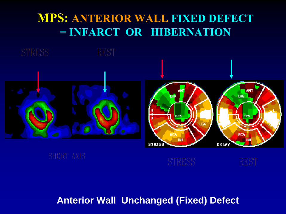
MPS: ANTERIOR WALL FIXED DEFECT =
INFARCT OR HIBERNATION
Anterior Wall Unchanged (Fixed) Defect
Based on tens of thousands of publications
The Tests is ConsideredSafe
AccurateCost Effective
THREE DECADES OF EXPERIENCE IN MYOCARDIAL PERFUSION
SCINTIGRAPHY
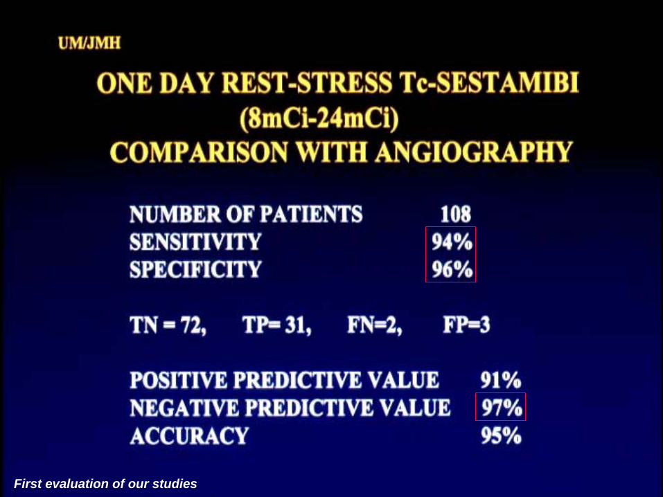
First evaluation of our studies
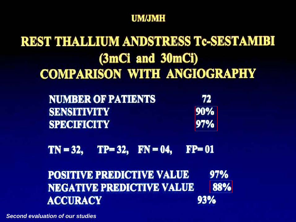
Second evaluation of our studies
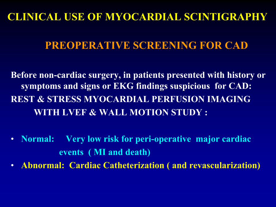
CLINICAL USE OF MYOCARDIAL SCINTIGRAPHY
PREOPERATIVE SCREENING FOR CAD
Before non-cardiac surgery, in patients presented with history or symptoms and signs or EKG findings suspicious for CAD:
REST & STRESS MYOCARDIAL PERFUSION IMAGINGWITH LVEF & WALL MOTION STUDY :
• Normal: Very low risk for peri-operative major cardiacevents ( MI and death)
• Abnormal: Cardiac Catheterization ( and revascularization)
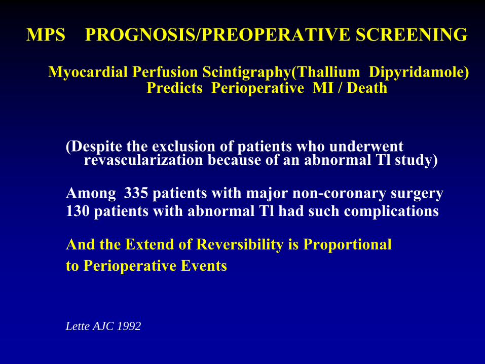
MPS PROGNOSIS/PREOPERATIVE SCREENING
Myocardial Perfusion Scintigraphy(Thallium
Dipyridamole) Predicts Perioperative
MI / Death
(Despite the exclusion of patients who underwent revascularization because of an abnormal Tl
study)
Among 335 patients with major non-coronary surgery130 patients with abnormal Tl had such complications
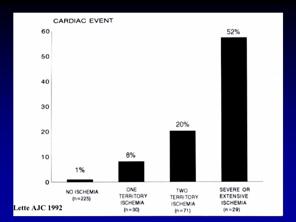
And the Extend of Reversibility is Proportionalto Perioperative
Events
Lette AJC 1992
Lette
AJC 1992
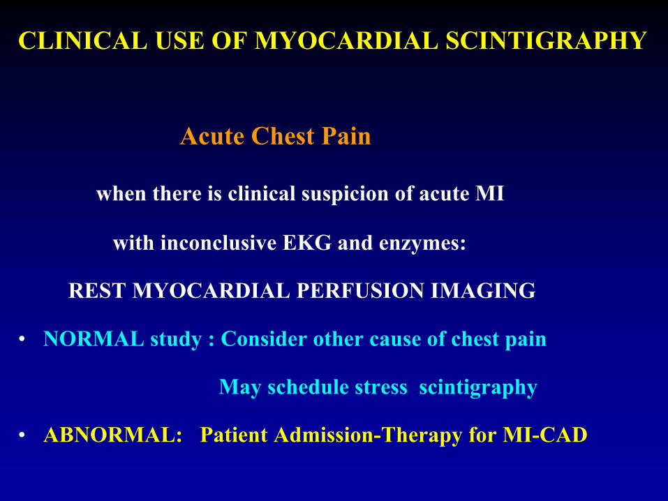
Acute Chest Pain
when there is clinical suspicion of acute MI
with inconclusive EKG and enzymes:
REST MYOCARDIAL PERFUSION IMAGING
• NORMAL study : Consider other cause of chest pain
May schedule stress scintigraphy
• ABNORMAL: Patient Admission-Therapy for MI-CAD
CLINICAL USE OF MYOCARDIAL SCINTIGRAPHY
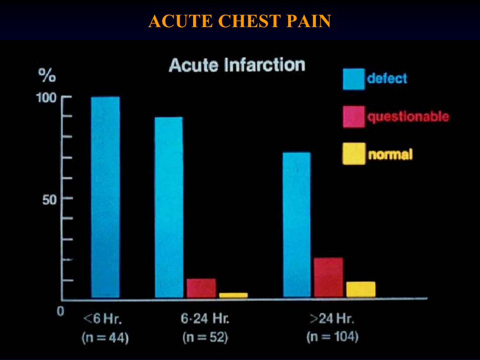
ACUTE CHEST PAIN
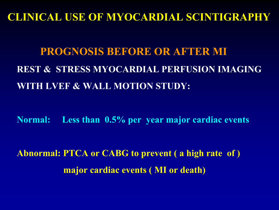
PROGNOSIS BEFORE OR AFTER MIREST & STRESS MYOCARDIAL PERFUSION IMAGING
WITH LVEF & WALL MOTION STUDY:
Normal: Less than 0.5% per year major cardiac events
Abnormal: PTCA or CABG to prevent ( a high rate of )
major cardiac events ( MI or death)
CLINICAL USE OF MYOCARDIAL SCINTIGRAPHY
MPS PROGNOSIS (RISK STRATIFICATION)
MYOCARDIAL PERFUSION SCINTIGRAPHY (THALLIUM EXERCISE)
PREDICTS MAJOR CARDIAC EVENTS
In 404 patients with a mean age of 65 yearswho were followed for a mean of 2 years
Incidence of MI or cardiac death was0.5%in normal, 5% in one vessel, 13% in =>2 vessel disease
Iskandrian AJC 1988
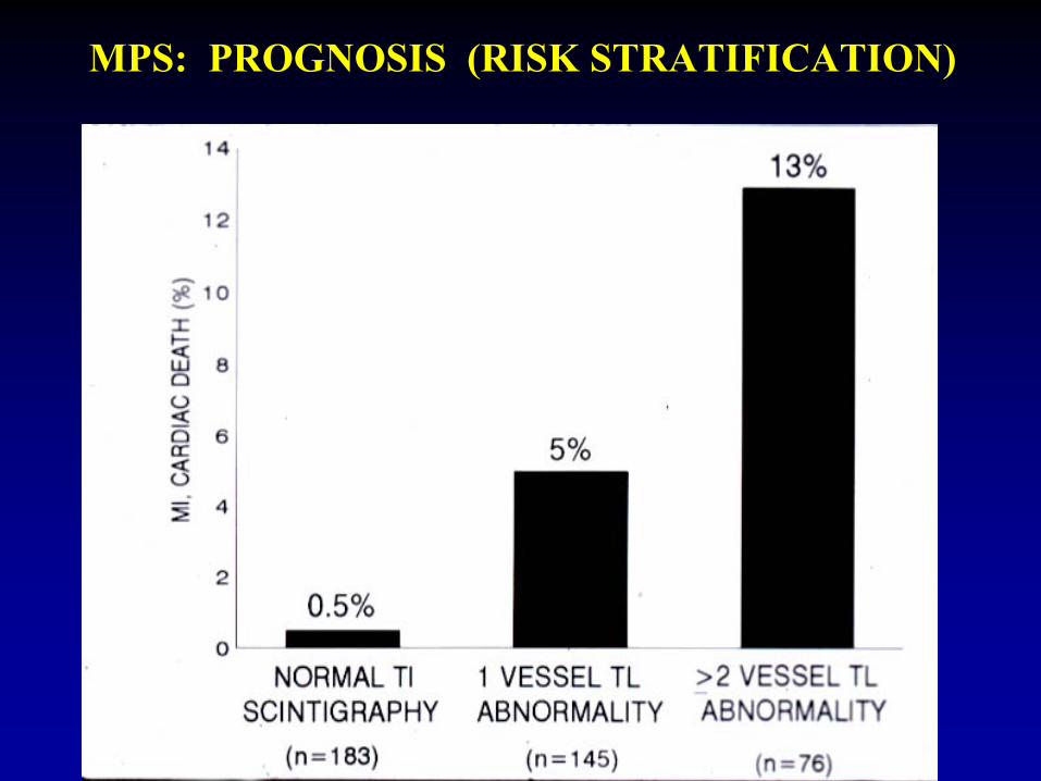
MPS: PROGNOSIS (RISK STRATIFICATION)
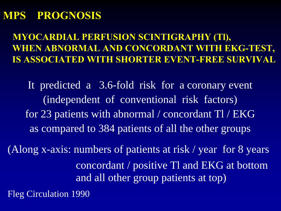
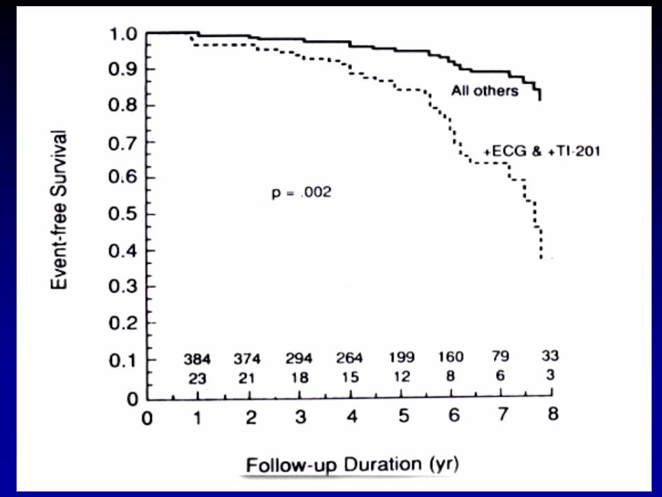
MPS PROGNOSIS
MYOCARDIAL PERFUSION SCINTIGRAPHY (Tl), WHEN ABNORMAL AND CONCORDANT WITH EKG-TEST,
IS ASSOCIATED WITH SHORTER EVENT-FREE SURVIVAL
It predicted a 3.6-fold risk for a coronary event(independent of conventional risk factors)
for 23 patients with abnormal / concordant Tl / EKGas compared to 384 patients of all the other groups
(Along x-axis: numbers of patients at risk / year for 8 yearsconcordant / positive Tl and EKG at bottom and all other group patients at top)
Fleg Circulation 1990
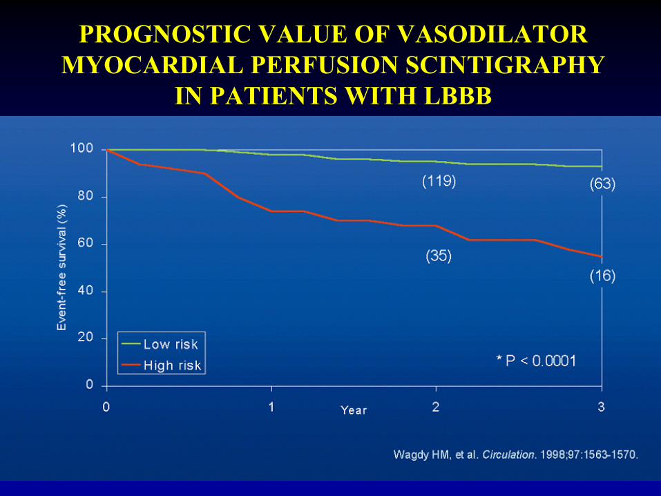
PROGNOSTIC VALUE OF VASODILATOR MYOCARDIAL PERFUSION SCINTIGRAPHY
IN PATIENTS WITH LBBB
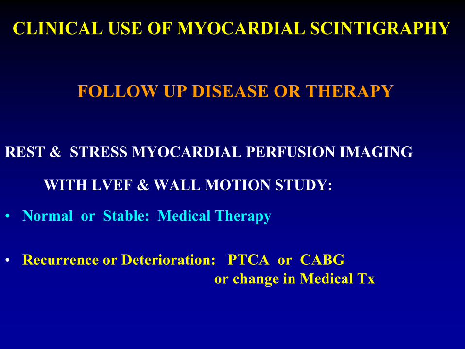
FOLLOW UP DISEASE OR THERAPY
REST & STRESS MYOCARDIAL PERFUSION IMAGING
WITH LVEF & WALL MOTION STUDY:
• Normal or Stable: Medical Therapy
• Recurrence or Deterioration: PTCA or CABG or change in Medical Tx
CLINICAL USE OF MYOCARDIAL SCINTIGRAPHY

A 57 yo man underwent angioplasty because of LAD disease He had MPS before and after Angioplasty
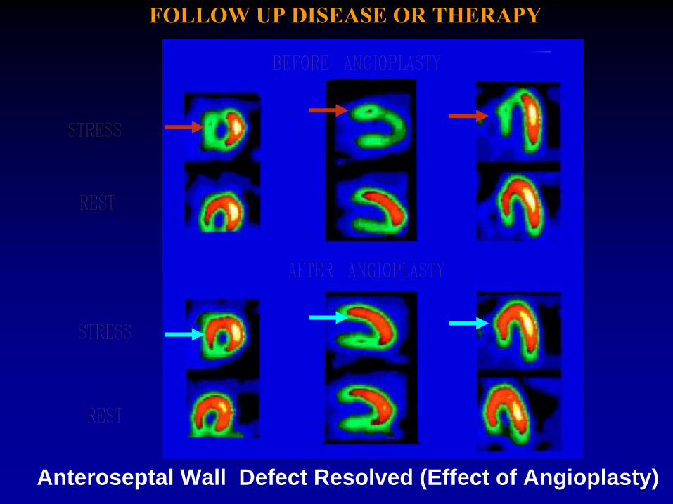
FOLLOW UP DISEASE OR THERAPY
Anteroseptal Wall Defect Resolved (Effect of Angioplasty)
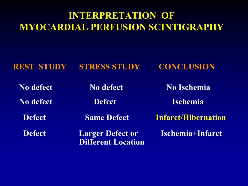
INTERPRETATION OF MYOCARDIAL PERFUSION SCINTIGRAPHY
REST STUDY STRESS STUDY CONCLUSION
No defect No defect No Ischemia
No defect Defect
Ischemia
Defect Same Defect Infarct/Hibernation
Defect Larger Defect or Ischemia+InfarctDifferent Location
A 45 yo lady with atypical chest pain has an exercise and rest MPS
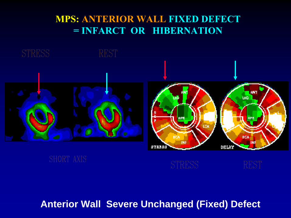
MPS: ANTERIOR WALL FIXED DEFECT =
INFARCT OR HIBERNATION
Anterior Wall Severe Unchanged (Fixed) Defect
A 55 yo man with typical chest pain has an exercise and rest MPS
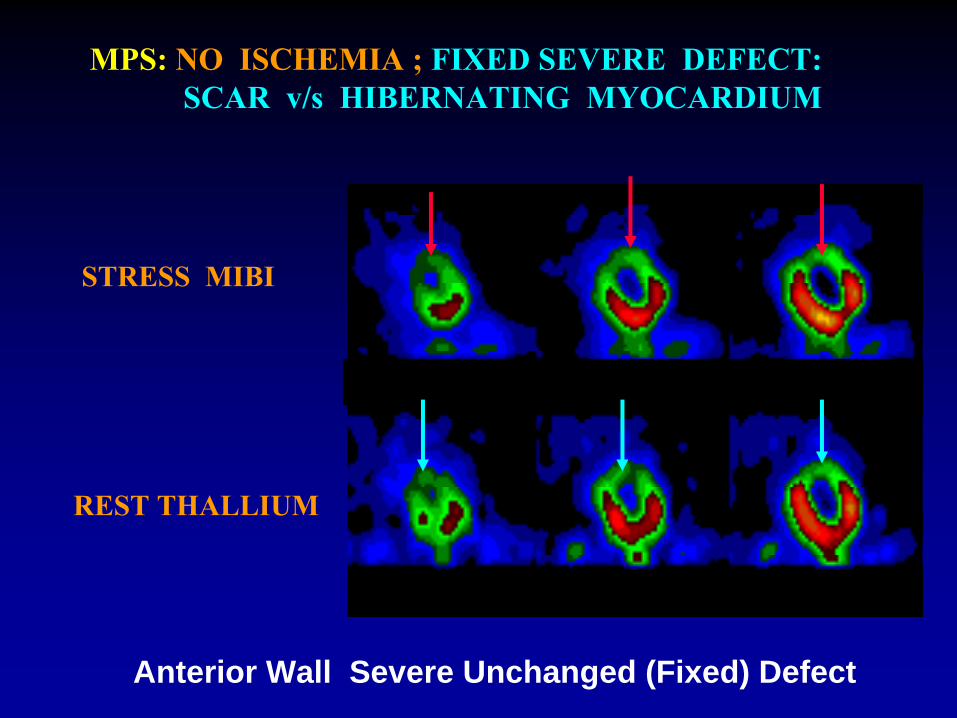
STRESS MIBI
REST THALLIUM
MPS: NO ISCHEMIA ; FIXED SEVERE DEFECT:SCAR v/s
HIBERNATING MYOCARDIUM
Anterior Wall Severe Unchanged (Fixed) Defect
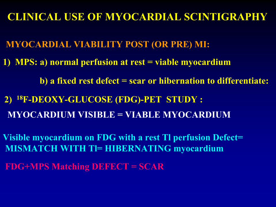
MYOCARDIAL VIABILITY POST (OR PRE) MI:
1) MPS: a) normal perfusion at rest = viable myocardium
b) a fixed rest defect = scar or hibernation to differentiate:
2) 18F-DEOXY-GLUCOSE (FDG)-PET STUDY :
MYOCARDIUM
VISIBLE = VIABLE
MYOCARDIUM
Visible myocardium on FDG with a rest Tl
perfusion Defect=MISMATCH WITH Tl= HIBERNATING myocardium
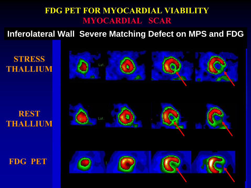
FDG+MPS Matching DEFECT = SCAR
CLINICAL USE OF MYOCARDIAL SCINTIGRAPHY
A 63 yo patient with a history of heart attack and an inconclusive EKG had MPS and FDG/PET study
REST THALLIUM
FDG PET
FDG PET FOR MYOCARDIAL VIABILITY MYOCARDIAL SCAR
STRESS THALLIUM
Inferolateral Wall Severe Matching Defect on MPS and FDG

Compare Perfusion with Viability
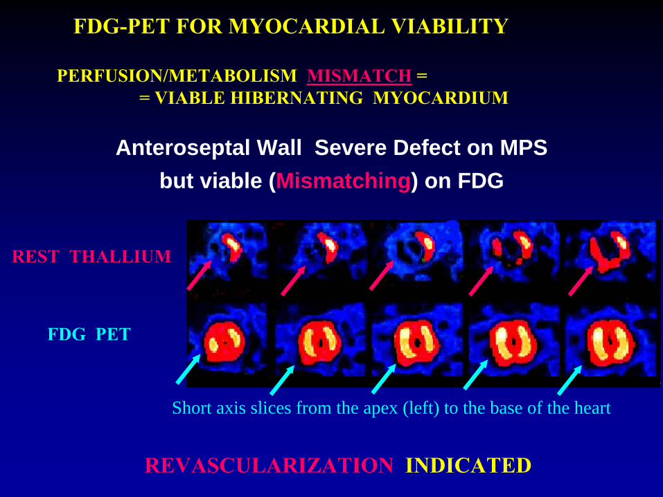
FDG-PET FOR MYOCARDIAL VIABILITY
FDG PET
Short axis slices from the apex (left) to the base of the heart
REST THALLIUM
REVASCULARIZATION
INDICATED
Anteroseptal Wall Severe Defect on MPS but viable (Mismatching) on FDG
PERFUSION/METABOLISM MISMATCH
= = VIABLE HIBERNATING MYOCARDIUM
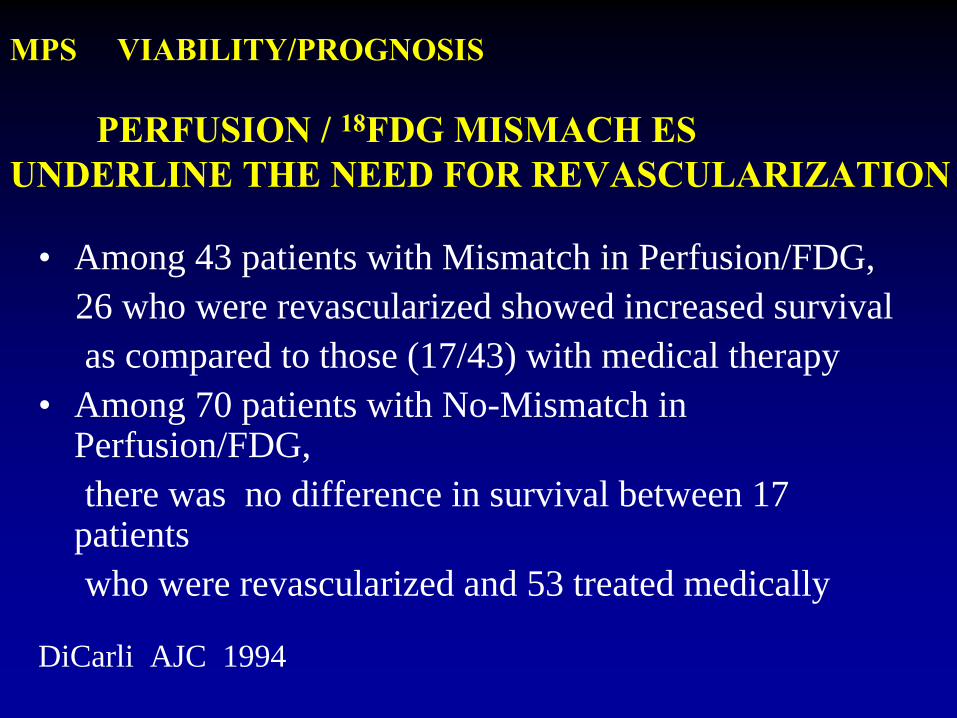
MPS VIABILITY/PROGNOSIS
PERFUSION / 18FDG MISMACH ES UNDERLINE THE NEED FOR REVASCULARIZATION
• Among 43 patients with Mismatch in Perfusion/FDG, 26 who were revascularized showed increased survival as compared to those (17/43) with medical therapy
• Among 70 patients with No-Mismatch in Perfusion/FDG, there was no difference in survival between 17
patients who were revascularized and 53 treated medically
DiCarli AJC 1994

Please explain to us what we see in the next picture and what lessons we learn from it
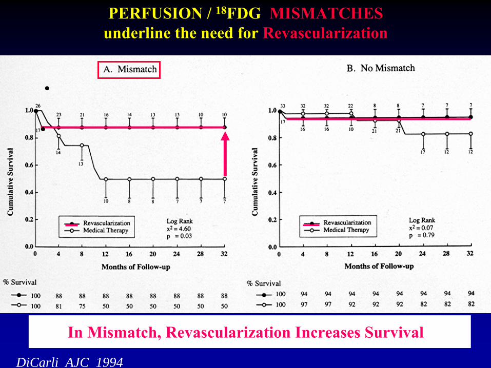
PERFUSION / 18FDG MISMATCHES underline the need for Revascularization
In Mismatch, Revascularization Increases Survival
DiCarli AJC 1994
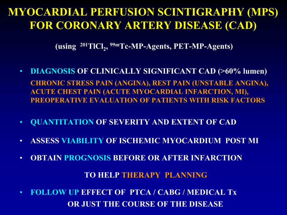
MYOCARDIAL PERFUSION SCINTIGRAPHY (MPS) FOR CORONARY ARTERY DISEASE (CAD)
(using 201TlCl2
, 99mTc-MP-Agents, PET-MP-Agents)
• DIAGNOSIS
OF CLINICALLY SIGNIFICANT CAD (>60% lumen)CHRONIC STRESS PAIN (ANGINA), REST PAIN (UNSTABLE ANGINA),
ACUTE CHEST PAIN (ACUTE MYOCARDIAL INFARCTION, MI), PREOPERATIVE EVALUATION OF PATIENTS WITH RISK FACTORS
• QUANTITATION
OF SEVERITY AND EXTENT OF CAD
• ASSESS
VIABILITY
OF ISCHEMIC MYOCARDIUM POST MI
• OBTAIN
PROGNOSIS
BEFORE OR AFTER INFARCTION
TO HELP THERAPY PLANNING
• FOLLOW UP
EFFECT OF PTCA / CABG / MEDICAL TxOR JUST THE COURSE OF THE DISEASE
COMPARISON OF ECHO WITH SCINTIGRAPHY
Advantages of ScintigraphyIschemia not required for regional abnormalityQuantification of perfusion abnormality -
extent and severityGreater reproducibility through quantificationDetection of peri-infarction ischemiaHigh success rateNot as dependent on physician’s technical expertiseExtensive literature on clinical value
COMPARISON OF ECHO WITH SCINTIGRAPHY
Equal ValueDetection of disease in patients with normal
function at restLeft ventricular function
Advantages of EchocardiographyEase and rapidity of study“On-line” visualization of the heartEvaluation of pericardium, valves, myocardium
MYOCARDIAL PERFUSION SCINTIGRAPHY
George N. Sfakianakis MDProfessor of Radiology and Pediatrics
Director, Division of Nuclear MedicineUM/JMMCMiami FL