Embed Size (px)
Citation preview

Myocardial Viability In Ischemic Syndromes
Cover: Two-dimensional echocardiographic enddiastolic and endsystolic still
frames of a patient before and after coronary artery bypass graft surgery,
demonstrating functional recovery.
CIP-DATA KONINKLIJKE BIBLIOTHEEK, DEN HAAG
Cornel, Jan Hein
Myocardial viability in ischemic syndromes / Jan Hein Cornel - Rotterdam
Erasmus University, Department of Cardiology.
Thesis Erasmus University Rotterdam. - With ref. - With summary in Dutch.
ISBN 90-9009694-9
NUGI 742
Subject headings: dobutamine stress echocardiography, FDG, myocardial viability,
SPECT.
W Printed by Ridderprint bv, Ridderkerk.
© 1996 J.H. Cornel, Rotterdam, the Netherlands. All rights reserved.
No part of this publication may be reproduced, stored in a retrieval system, in any
form or by any means without prior permission from the author.
Myocardial Viability in Ischemic Syndromes
evaluatie van diagnostische methoden naar myocardiale
vitaliteit
Proefschrift
Ter verkrijging van de graad van doctor
aan de Erasmus Universiteit Rotterdam
op gezag van de Rector Magnificus
Prof. Dr. P.W.c. Akkermans M.A.
en volgens Besluit van het College voor Promoties.
De openbare verdediging zal plaatsvinden op
20 november 1996 om 13.45 uur
door
Jan Hein Cornel
geboren te Harlingen
Promotiecommissie
Promotor
Co-promotor
Overige leden
Prof. Dr. I.R.T.C. Roelandt
Dr. P.M. Fioretti
Prof. Dr. E.P. Krenning
Prof. Dr. M.L. Simoons
Prof. Dr C.A. Visser
Financial support by the Netherlands Heart Foundation for the publication of this
thesis is gratefully aclrnowledged.

Voor allen die rnij dierbaar zijn
Contents
Chapter 1
Introduction and outline of the thesis 9
Part I Methodology
Chapter 2 27
Assessment of myocardial viability before revascularization: can sestamibi
accurately predict functional recovery?
In: Imaging and Intervention in Cardiology. C.A. Nienaber and U. Sechtem
(Eds). Kluwer Academic Publish~rs, the Netherlands, 1996:249-258
Chapter 3 41
Dobutamine stress-redistribution-reinjection versus rest-redistribution
201Thallium SPECT in the assessment of myocardial viability.
International Journal of Cardiac Imaging, in press
Chapter 4 57
Safety and feasibility of dobutamine-atropine stress echocardiography in
patients with ischemic left ventricular dysfunction.
Journal of the American Society of Echocardiography 1996;9:27-32
Part II Recovery of contractile function
Chapter 5 73
Prediction of improvement of ventricular function after first acute
myocardial infarction using low-dose dobutamine stress echocardiography.
American Journal of Cardiology 1994;74:853-856
Chapter 6 85
T-wave normalization during dobutamine echocardiography for diagnosis of
viable myocardium.
American Journal of Cardiology 1995,'75:505-507
Chapter 7 95
Prediction of improvement of regional left ventricular function after surgical
revascularization: a comparison of low-dose dobutamine echocardiography
with 201TI single-photon emission computed tomography.
Circulation 1995;91:2748-2752
Addendum
Correspondence
Circulation 1996,'93:396-397
Chapter 8
111
115
18P-Pluorodeoxyglucose SPECT for the prediction of reversibility of
ventricular dysfunction after revascularization. A comparison with rest
redistribution 201Thallium SPECT.
Submitted for publication
Chapter 9 139
Prediction of improvement of ventricular function after revascularization:
quantitative 18P-fluorodeoxyglucose single-photon emission computed
tomography versus low-dose dobutamine echocardiography.
Submitted for publication
Chapter 10 159
Biphasic response to dobutamine predicts recovery of global ventricular
function after surgical revascularization. Implications of time course of
recovery on diagnostic accuracy.
Submitted for publication
Chapter 11 185
Summary and future perspectives
Samenvatting 193
Dankwoord 199
Curriculum vitae 203
List of publications 205

Chapter 1
Introduction and Outline of the Thesis
Chapter 1 10
Introduction
Background
Currently cardiologists face a substantial growth in the number of patients
with congestive heart failure, a clinical syndrome with a poor prognosis. In the
United States, more than 3 million people suffer from heart failure and more than
100,000 die from end-stage congestive heart failure annually. I In the Netherlands
the prevalence of heart failure is currently 4 % and rises firmly in the elderly.
Although heart failure can be caused by various pathological processes, the single
most frequent cause is left ventricular (LV) ~ystolic dysfunction resulting from
myocardial ischemic injury due to coronary artery disease. Myocardial dysfunction
is the major determinant of the severity of heart failure and prognosis. 2-5 The risk
of developing heart failure in ischemic heart disease is much lower and the
prognosis better when LV systolic function is preserved. Therefore preservation
of LV function by limiting infarct size or improvement of chronic ischemic LV
dysfunction must be considered as major therapeutic goals for the prevention of
ischemic heart failure. The introduction of aggressive reperfusion strategies in the
treatment of acute myocardial infarction, such as thrombolytic therapy and direct
PTCA, has dramatically improved prognosis of patients with myocardial
infarctions. 6-8 Due to the progress in management of acute ischemic events, there
are increasing numbers of patients with (asymptomatic ) LV dysfunction. However,
also in the thrombolytic era, mortality rises steeply in patients with a LV ejection
fraction (L YEP) of < 35 % .9 Below this break point even a small gain in function
may imply substantially improved prognosis.
Apart from medical therapy, available treatment options in the management
of patients with coronary artery disease and advanced LV dysfunction are heart
transplantation, myocardial revascularization (PTCA or CABG) and other forms
of reparative surgery (i.e. mitral valve surgery, dynamic cardiomyoplasty, LV
aneurysmectomy). Reparative surgery will not be discussed in this thesis.
Heart transplantation has been successfully performed in patients with end
stage heart failure. The Thoraxcenter Rotterdam has reported a five year survival
rate of 84 % . 10 However, in the first 9 years of the heart transplant program only
200 patients could be treated. Shortage of donor hearts and the significant morbitity
of immunosuppressive therapy has led to a search for alternative treatment choices.
Introduction 11
It is well recognized that CABG may improve LV performance and functional
status in patients with chronic advanced LV dysfunction,u Several studies have
shown that myocardial revascularization may improve regional and global LV
function. l1-13 Additionally, long-term survival may improve after revascula
rization. 14 In patients with a LVEF :0;;35% Pigott and coworkers14 reported a 7-
year survival of 63 % after CABG compared with 34 % when on medical therapy.
However the CABG procedure is associated with high mortality in patients with
poor LV function. 15,16 Therefore careful preoperative selection of patients with poor
LV function who will benefit from a revascularization procedure is warranted.
Appropriate decision making prevents patients, in whom revascularization would
not result in functional benefit from being exposed to high risk surgery. Distinction
between dyssynergic regions containing viable and non-viable myocardium may
identify patients with high and low probability to improve LV performance and
functional status after revascularization, This may help the physician in clinical
decision making.
Pathophysiology of myocardial dyssynergy
Several different conditions may lead to myocardial contractile dysfunction.
LV dyssynergy may be due to myocardial necrosis, (repetitive) stunning or
myocardial hibernation.
In myocardial necrosis scar formation involves either the entire myocardium,
is limited to the subendocardium or is scattered throughout the myocardium. In all
these conditions it is not expected that revascularization results in improvement of
contractile function.
Myocardial stunning is defined as transient prolonged postischemic
dysfunction that may occur after the restoration of normal flOW. 17.18 Despite the
absence of irreversible damage, mechanical dysfunction may persist after coronary
reperfusion in different clinical situations following, for example coronary
angioplasty,19,20 coronary artery bypass surgery, unstable angina,19-21 exercise
induced ischemia22 or acute myocardial infarction with early reperfusion. 23-3o
Spontaneous recovery may occur within weeks after the event and is dependent on
the "area at risk", the duration of coronary occlusion, the amount and location of
myocardial necrosis and the presence and extent of collateral vessels. 31 Repeated
Chapter 1 12
episodes of myocardial ischemia may however also lead to a chronic reduction in
contractility without a concomitant reduction in myocardial perfusion. 32,33 This
phenomenon has recently been described as repetitive stunning. The impairment
of contractile function can be reversed after adequate revascularization,
The concept of hibernation has been introduced by Rahimtoola. He
hypothesizes that chronic reduction in myocardial blood flow may lead to a
matched downregulation of the contractile cellular function which is reversible after
revascularization. 34,35 It has been postulated that reduction of contractile function
may be a protective response of myocytes to counteract the reduced supply of
oxygen and substrates. The restoration of the delicate balance between perfusion
and contraction may prevent cell death. 34-36
In individual patients, all types of reversible and irreversible contractile
dysfunction may coexist. Both experimental and clinical studies have conclusively
demonstrated that the simple assessment of wall motion does not adequately
distinguish non-viable from viable (either stunned or hibernating) but dyssynergic segments. 37-39
Positron emission tomography
Among the available techniques, positron emission tomography (PET) of , myocardial perfusion and metabolism (using 18F_ fluorodeoxyglucose (FDG» is
considered the gold standard for the identification of viable myocardium. 40-45 Under
normal physiologic conditions myocardial perfusion is closely matched with the
energy requirements of the myocardium. Under ischemic conditions exogeneous
glucose becomes the preferred substrate for myocytes. 46,47 The rationale for the use
of FDG is based on the fact that after uptake FDG is intracellularly trapped. The
hallmark of jeopardized and viable myocardium is either increased FDG uptake in
areas of myocardial hypoperfusion (mismatch) or, normal or increased FDG uptake
in areas of normal perfusion (repetitive stunning). With these characteristics
present there is a high likelihood of functional recovery after revascularization. On
the other hand, a mild reduction in both perfusion and FDG uptake has also been
considered to represent viable myocardium (viable match) which is however
unlikely to improve in contractile performance after revascularization. 41 Compari
son with myocardial biopsies have provided histological evidence for the presence
Introduction 13
of viable tissue in dyssynergic segments with preserved FDG uptake. 32.42.48 By
differentiating between perfusion-metabolism match or mismatch PET can
adequately predict recovery of regional and global LV function after revasculariza
tion. 40,49-56 Furthermore, retrospective follow-up studies suggest that PET carries
prognostic information. 57-61 Patients with hibernating myocardium have an increased
risk to suffer cardiac events in comparison to patients without myocardium in
jeopardy. The presence of a mismatch pattern in medically treated patients is
associated with a higher morbidity and mortality. In these patients revascularization
substantially improves symptoms related to heart failure and reduces the number
of cardiac events. However high costs and limited availibility restrict the use of
PET.
Alternative diagnostic methods
To respond to the increasing demand for viability studies, other techniques
have been proposed including imaging myocardial FDG uptake (using special 511
keY collimators) with single photon emission computed tomography (SPECT),62-66
201-thallium eOITI) SPECT,50,67-73 technetium-99m sestamibi SPECT74 and dobuta
mine stress echocardiography. 74-79 These techniques are attractive from a clinical
point of view, because they are widely available and may be more cost-effective.
However they reflect different cellular mechanisms of viability.
During the last decade 20lTI SPECT has received much attention and is now
considered to be a reliable technique for the identification of viable myocardium. 80
Assessment of myocardial viability using 20lTI SPECT is based on the principle that
initial tracer uptake is proportional to regional flow, whereas delayed imaging
reflects cell membrane integrity by demonstrating redistribution. 81 In direct
comparison with PET, several different 20lTI protocols (especially stress
redistribution-reinj ection and rest-redistribution) have demons trated good agreement
with the detection of viable myocardium. 5o,82,83 In practical terms, the most
important aspect of viability is recovery of mechanical function either spontaneous
ly or after intervention. Whether 20lTI SPECT can accurately predict recovery of
LV function is still unclear and will be addressed in this thesis.
Low-dose dobutamine echocardiography has been proposed as another alter
native method for the assessment of residual myocardial viability both in patients
Chapter 1 14
soon after acute myocardial infarction7S-77 and in patients with stable chronic
ischemic heart disease. 74 ,78,79 The rationale is based upon the fact that low-dose
dobutamine increases myocardial contractility with a minimal increase in heart rate.
The inotropic effect of dobutamine is already nearly maximal at doses of 5 to 15
Ilg/kg/min and is dependent on infarct size, the severity of stenosis of the infarct
related coronary artery and the duration of infusion. The echocardiographic
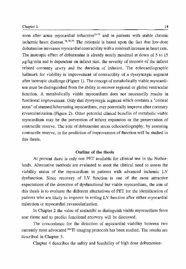
hallmark for viability is improvement of contractility of a dyssynergic segment
after inotropic challenge (Figure 1). The concept of metabolically viable myocardi
um must be distinguished from the ability to recover regional or global ventricular
function. A metabolically viable myocardium does not necessarily results in
functional improvement. Only that dyssynergic segment which contains a "critical
mass" of stunned/hibernating myocardium, may potentially improve after coronary
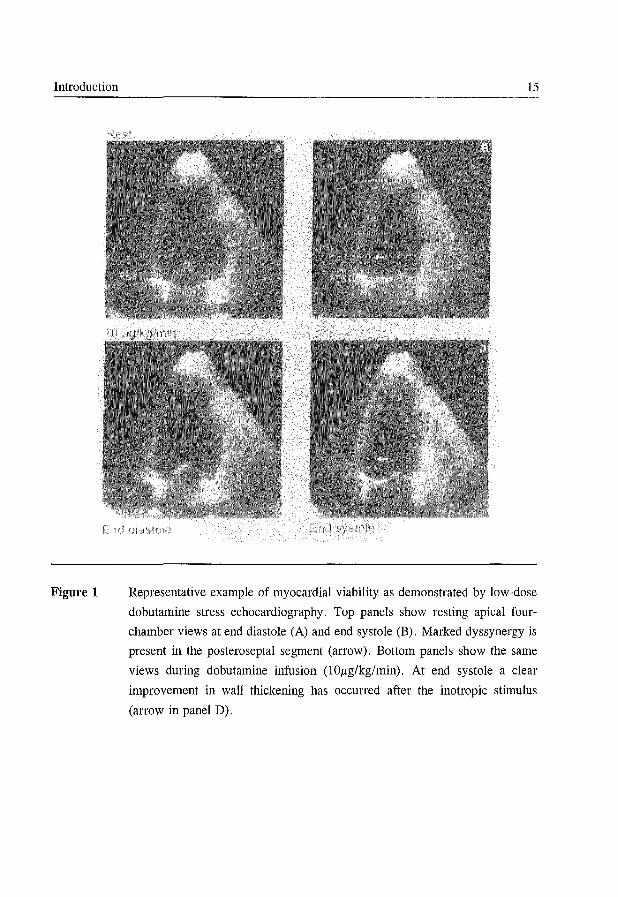
revascularization (Figure 2). Other potential clinical benefits of metabolic viable
myocardium may be the prevention of infarct expansion or the preservation of
contractile reserve. The role of dobutamine stress echocardiography, by assessing
contractile reserve, in the prediction of improvement of function will be studied in
this thesis.
Outline of the thesis
At present there is only one PET available for clinical use in the Nether
lands. Alternative methods are evaluated to meet the clinical need to assess the
viability status of the myocardium in patients with advanced ischemic LV
dysfunction. Since recovery of LV function is one of the most attractive
expectations of the detection of dysfunctional but viable myocardium, the aim of
this thesis is to evaluate the different alternatives of PET for the identification of
patients who are likely to improve in resting LV function after either myocardial
infarction or myocardial revascularization.
In Chapter 2 the value of sestamibi to distinguish viable myocardium from
scar tissue and to predict functional recovery will be discussed.
The concordance for the detection of myocardial viability between two
currently most advocated 201TI imaging protocols has been studied. The results are
Clescribed in Chapter 3.
Chapter 4 describes the safety and feasibility of high dose dobutamine-
Introduction 15
Figure 1 Representative example of myocardial viability as demonstrated by low-dose
dobutamine stress echocardiography. Top panels show resting apical four
chamber views at end diastole (A) and end systole (B). Marked dyssynergy is
present in the posteroseptal segment (arrow). Bottom panels show the same
views during dobutamine infusion (l0/lg/kg/min). At end systole a clear
improvement in wall thickening has occurred after the inotropic stimulus
(arrow in panel D).
Chapter 1
Figure 2
16
Example of post-revascularization improvement of posterior wall thickening
as predicted by low-dose dobutamine stress echocardiography. M-mode
tracings of the parasternal long axis view demonstrate at rest severe
hypokinesia of the posterior wall (panel A). At low-dose dobutamine infusion
wall thickening improved (panel B), after successful revascularization wall
thickening at rest recovered to the level as predicted by low-dose dobutamine
infusion.
Introduction 17
atropine stress echocardiography in patients with ischemic LV dysfunction.
The second part of this thesis is dedicated to the value of the different
techniques to predict recovery of LV function at rest, either spontaneously after
acute MI or after revascularization in chronic coronary artery disease. The studies
involved close co-operation with the Department of Cardiothoracic Surgery (Head:
Prof. Dr . E.Bos) and the referring 'Rijnmond' cardiologists.
Chapters 5 and 6 deal with the value of low-dose dobutamine echocardio
graphy and T-wave normalization for the prediction of late spontaneous recovery
of regional LV function after acute myocardial infarction.
A direct comparison between low-dose dobutamine echocardiography and
post-stress reiI\iection 201Tl SPECT for the prediction of recovery of mechanical
dysfunction after CABG in patients with chronic coronary artery disease is
discussed in Chapter 7.
Chapters 8 and 9 describe co-operative studies with the Department of
Cardiology of the Free University Hospital, Amsterdam.
The aim in Chapter 8 was twofold; 1) to evaluate the use of FDG SPECT
in the prediction of functional improvement of regional and global LV function
after revascularization, and 2) to compare FDG SPECT with rest-redistribution
201Tl SPECT in a head-to-head fashion.
Chapter 9 describes a study designed to determine the agreement between
FDG SPECT and low-dose dobutamine echocardiography to identify viable
myocardium and to predict improvement of regional LV function after revasculari
zation.
To overcome the limitations of low-dose instead of high-dose dobutamine
infusion and to circumvent the methodological problems caused by the lack of an
independent technique to detect changes in LV performance over time, we then
designed a high-dose dobutamine/atropine echo cardiographic study using
radionuc1ide ventriculography as independent method to determine improvement
of LV function (Chapter 10). In the studies described in Chapters 5 to 9 functional
recovery was measured by resting echocardiography at 3 months. We realised
however, that recovery in some patients may be delayed for more than 3 months,
given the severity of structural changes observed in hibernating myocardium. 42 In
Chapter 1 18
the study described in Chapter 10, the time course of recovery of LV function is
studied in more detail by extending the follow-up period to 1 year after CABG.
References
1. Miller LW. Candidate selection for heart transplantation. Cardiol Clin 1995;13:93-
100.
2. Alderman EL, Fisher LD, Litwin P, et al. Results of coronary artery surgery in
patients with poor left ventricular function (CASS). Circulation 1983;68:785-795.
3. Mock MB, Ringquist I, Fisher LD, et al. Survival of medically treated patients in the
Coronary Artery Surgery Study (CASS) registry. Circulation 1992;66:562-568.
4. The Multicenter Postinfarction Research Group. Risk stratification and survival after
myocardial infarction. N Eng J Med 1983;309:331-336.
5. White RD, Norris RM, Brown MA, Brandt PWT, Whitlock RML, Wild CJ. Left
ventricular end-systolic volume as the major determinant of survival after recovery
from myocardial infarction. Circulation 1987;76:44-51.
6. Gruppo Italiano per 10 Studio della Streptochinasi nell'Infarto Miocardico (GISSI).
Effectiveness of intravenous thrombolytic treatment in acute myocardial infarction.
Lancet 1986;i;397-401.
7. ISIS-2 (Second International Study of Infarct Survival) collaborative group.
Randomised trial of intravenous streptokinase, oral aspirin, both, or neither among
17,187 cases of suspected acute myocardial infarction: ISIS-2. Lancet 1988; ii: 349-360.
8. Zijlstra F, de Boer MJ, Roorntje JCA, Reiffers S, Reiber JRC, Suryapranata R. A
comparison of immediate coronary angioplasty with intravenous streptokinase in acute
myocardial infarction. N Engl J Med 1993;328:680-684.
9. Volpi A, De Vita C, Franzosi MG, et al. Determinants of 6-month mortality in
survivors of myocardial infarction after thrombolysis. Results of the GISSI-2 data
base. Circulation 1993;88:416-429.
10. Balk ARMM. Clinical aspects of heart transplantation. Thesis, Rotterdam 1993.
11. Elefteriades JA, Tolis G, Levi E, Mills LK, Zaret BL. Coronary artery bypass
grafting in severe left ventricular dysfunction: excellent survival with improved
ejection fraction and functional state. J Am ColI Cardiol 1993;22:1411-1417.
12. Rees G, Bristow JD, Kremkau EL, et al. Influence of aortocoronary bypass surgery
on left ventricular performance. N Engl J Med 1971;284:1116-1120.
13. Brundage BR, Massie BM, Botvinick EH. Improved regional ventricular function
after successful surgical revascularization. J Am ColI Cardiol 1984;3 :902-908.
14. Pigott JD, Kouchoukos NT, Oberman A, Cutter GR. Late results of surgical and
Introduction 19
medical therapy for patients with coronary artery disease and depressed left ventricu
lar function. J Am ColI Cardiol 1985;5: 1036-1045.
15. Kron IL, Flanagan TL, Blackbourne LH, Schroeder RA, Nolan SP. Coronary
revascularization rather than cardiac transplantation for chronic ischemic cardiomyo
pathy. Ann Surg 1989;210:348-352.
16. Louie HW, Laks H, Milgalter E, et al. Ischemic cardiomyopathy: criteria for
coronary revascularization and cardiac transplantation. Circulation 1991;84 (suppl
III):290-295.
17. Bolli R. Myocardial "stunning" in man. Circulation 1992;86:1671-1691.
18. Braunwald E, Kloner R. The stunned myocardium: prolonged, postischemic
ventricular dysfunction. Circulation 1982; 66: 1146-1149.
19. Marzullo P, Parodi 0, Sambuceti G, et al. Does the myocardium become 'stunned'
after episodes of angina at rest, angina on effort, and coronary angioplasty? Am J
CardioI1993;71:1045-1051.
20. De Fey tel' PJ, Suryapranata H, Serruys PW, Beatt K, van den Brand M, Hugenholtz
PG. Effects of successful percutaneous transluminal coronary angioplasty on global
and regional left ventricular function in unstable angina pectoris. Am J Cardiol
1987;60:993-997.
21. Nixon JV, Brown CN, Smitherman TC. Identification of transient and persistent
segmental wall motion abnormalities in patients with unstable angina by two
dimensional echocardiography. Circulation 1982;65: 1497-1503.
22. Kloner RA, Allen J, Cox TA, Zheng Y, Ruiz CEo Stunned left ventricular
myocardium after exercise treadmill testing in coronary artery disease. Am J Cardiol
1991 ;68:329-334.
23. Patel B, Kloner RA, Przyklenk K, Braunwald E. Postischemic myocardial "stunning":
a clinically relevant phenomenon. Ann Intern Med 1988;108:626-628.
24. Bourdillon PDV, Broderick TM, Williams ES, et al. Early recovery of regional left
ventricular function after reperfusion in acute myocardial infarction assessed by serial
two-dimensional echocardiography. Am J Cardiol 1989; 63: 641-646.
25. Serruys PW, Simoons ML, Suryapranata H, et al. Preservation of global and regional
left ventricular function after early thrombolysis in acute myocardial infarction. J Am
ColI CardioI1986;7:729-742.
26. Charuzi Y, Beeder C, Marshall LA, et al. Improvement in regional and global left
ventricular function after intracoronary thrombolysis: assessment with two
dimensional echocardiography. Am J Cardiol 1984;53:662-665.
27. Widimsky P, Cervenka V, Gregor P, et al. First month course of left ventricular
asynergy after intracoronary thrombolysis in acute myocardial infarction. A
Chapter 1 20
longitudinal echocardiographic study. Eur Heart J 1985;6:759-765.
28. Touchstone DA, Beller GA, Nygaard TW, Tedesco C, Kaul S. Effects of succesful
intravenous reperfusion therapy on myocardial function and geometry in humans: a
tomographic assessment using two-dimensional echocardiography. J Am Coil Cardiol
1989; 13: 1506-1513.
29. Penco M, Romano S, Agati L, et al. Influence of reperfusion induced by thrombolytic
treatment on natural history of left ventricular regional wall motion abnormality in
acute myocardial infarction. Am J Cardiol 1993;71: 1015-1020.
30. Marino P, Zanolla L, Zardini P, on behalf of the GISSI study. Effect of streptokinase
on left ventricular modeling and function after myocardial infarction: the GISSI trial.
J Am Coil Cardiol 1989; 14: 1149-1158.
31. Sabia P, Powers ER, Ragosta M, Sarenbock II, Burwell LR, Kaul S. An association
between collateral blood flow and myocardial viability in patients with recent
myocardial infarction. N Eng J Med 1992;327:1825-1831.
32. Vanoverschelde J-LJ, Wijns W, Depre C, et al. Mechanisms of chronic regional
postischemic dysfunction in humans: New insights from the study of noninfarcted
collateral-dependent myocardium. Circulation 1993 ;87: 1513-1523.
33. Marinho NVS, Keogh BE, Costa DC, Lammertsma AA, Ell PJ, Camici PG.
Pathophysiology of chronic left ventricular dysfunction. New insights from the
measurement of absolute myocardial blood flow and glucose utilization. Circulation
1996;93 :737-744.
34. Rahimtoola SR A perspective on the three large multicenter randomized clinical
trails of coronary bypass surgery for chronic stable angina. Circulation 1985;72
(suppl V): 123-135.
35. Braunwald E, Rutherford JD. Reversible ischemic left ventricular dysfunction:
evidence for the hibernating myocardium. J Am Coil CardioI1986;8:1467-1470.
36. Ross Jr J. Myocardial perfusion-contraction matching. Implications for coronary heart
disease and hibernation. Circulation 1991;83:1076-1083.
37. Heyndrickx GR, Baig H, Nelkins P, Leusen K, Fishbein MC, Vatner SF. Depression
of regional blood flow and wall thicknening after brief coronary occlusions. Am J
Physiol 1978;234:H653-H659.
38. Matsuzaki M, Gallagher KP, Kemper WS, White F, Ross Jr J. Sustained regional
dysfunction produced by prolonged coronary stenosis. Gradual recovery after
reperfusion. Circulation 1983; 68: 170-182.
39. Perrone-Filardi P, Bacharach SL, Dilsizian V, et al. Metabolic evidence of viable
myocardium in regions with reduced wall thickness and absent wall thickening in
patients with chronic ischemic left ventricular dysfunction. J Am Coil Cardiol
Introduction 21
1992;20: 161-168.
40. Tillisch J, Brunken R, Marshall R, et al. Reversibility of cardiac wall-motion
abnormalities predicted by positron tomography. N Engl J Med 1986;314:884-888.
41. vom Dahl J, Eitzman DT, AI-Aouar ZR, et al. Relation of regional function,
perfusion and metabolism in patients with advanced coronary artery disease under
going surgical revascularization. Circulation 1994;90:2356-2366.
42. Maes A, Flameng W, Nuyts J, et al. Histological alterations in chronically hypoperfu
sed myocardium. Correlation with PET findings. Circulation 1994; 90: 735-7 45.
43. Knuuti MJ, Nuutila P, Ruotsalainen U, et al. The value of quantitative analysis of
glucose utilization in detection of myocardial viability by PET. J Nucl Med
1993;34:2068-2075.
44. Eitzman D, AI-Aouar ZR, Kanter HL, et al. Clinical outcome of patients with advan
ced coronary artery disease after viability studies with positron emission tomography.
J Am ColI Cardiol 1992;20:559-565.
45. Di Carli M, Davidson M, Little R, et al. Value of metabolic imaging with positron
emission tomography for evaluating prognosis in patients with coronary artery disease
and left ventricular dysfunction. Am J Cardiol 1994;73:527-533.
46. Liedtke AJ. Alterations of carbohydrate and lipid metabolism in the acutely ischemic
heart. Prog Cardiovasc Dis 1981;23:321-336.
47. Camici P, Ferrannini E, Opie LH. Myocardial metabolism in ischemic heart disease:
Basic principles and application to imaging by positron emission tomography. Prog
Cardiovasc Dis 1989;32:217-238.
48. Depre C, Vanoverschelde JLJ, Melin JA, et al. Structural and metabolic correlates
of the reversibility of chronic left ventricular ischemic dysfunction in humans. Am
J Physiol 1995;268:HI265-HI275.
49. Tamaki N, Yonekura Y, Yamashita K, et al. PET using fluorine-18-deoxyglucose in
evaluation of coronary artery bypass grafting. Am J Cardiol 1989;64:860-865.
50. Tamaki N, Ohtani H, Yamashita K, et al. Metabolic activity in the areas of new fill
in after thallium-201 reinjection: Comparison with positron emission tomography
using fluorine-18-deoxyglucose. J Nucl Med 1991;32:673-678.
51. Lucignani G, Paolini G, Landoni C, et al. Presurgical identification of hibernating
myocardium by combined use of technetium-99m hexakis 2-methoxyisobutylisonitrile
SPECT and fluorine-18 fluoro-2-deoxy-D-glucose positron emission tomography in
patients with coronary artery disease. Eur J Nucl Med 1992; 19: 874-881.
52. Carrel T, Jenni R, Haubold-Reuter S, Von Schulthess G, Pasic M, Turina M.
Improvement of severely reduced left ventricular function after surgical revasculariza
tion in patients with preoperative myocardial infarction. Eur J Cardiothorac Surg
Chapter 1 22
1992;6:479-484.
53. Marwick TH, MacIntyre WJ, Lafont A, Nemec n, Salcedo EE. Metabolic responses
of hibernating and infarcted myocardium to revascularization. Circulation 1992;85:
1347-1353.
54. Gropler RJ, GeHman EM, Sampathkumaran K, et al. Comparison of carbon-ll
acetate with fluorine-18-fluorodeoxyglucose for delineating viable myocardium by
positron emission tomography. J Am CoIl CardioI1993;22:1587-1597.
55. Gropler RJ, GeHman EM, Sampathkumaran K, et al. Functional recovery after
coronary revascularization for chronic coronary artery disease is dependent on
maintainance of oxidative metabolism. J Am CoIl Cardiol 1992;20:569-577.
56. Tamaki N, Kawamoto M, Tadamura E, et al. Prediction of reversible ischemia after
revascularization. Perfusion and metabolic studies with positron emission tomography.
Circulation 1995;91: 1697-1705.
57. Eitzman D, AI-Azouar ZR, Kanter HL, et al. Clinical outcome of patients with
advanced coronary artery disease after viability studies with positron emission
tomography. J Am ColI Cardiol 1992;20:559-565.
58. Tamaki N, Kawamoto M, Takahashi N, et al. Prognostic value of an increase in
fluorine-18 deoxyglucose uptake in patients with myocardial infarction: Comparison
with stress thallium imaging. J Am ColI CardioI1993;22:1621-1627.
59. Di Carli MF, Davidson M, Little R, et al. Value of metabolic imaging with positron
emission tomography for evaluating prognosis in patients with coronary artery disease
and left ventricular dysfunction. Am J Cardiol 1994;73:527-533.
60. Lee KS, Marwick TH, Cook SA, et al. Prognosis of patients with left ventricular
dysfunction, with and without viable myocardium after myocardial infarction. Relative
efficacy of medical therapy and revascularization. Circulation 1994;90:2687-2694.
61. Di Carli MF, Asgarzadie F, Schelbert HR, Laks H, Phelps ME, Maddahi J.
Quantitative relation between myocardial viability and improvement in heart failure
symptoms after revascularization in patients with ischemic cardiomyopathy.
Circulation 1995;92:3436-3444.
62. Bax n, Visser FC, Blanksma PK, et al. Comparison of myocardial uptake of 18F_
fluorodeoxyglucose imaged with positron emission tomography and single photon
emission computed tomography in dyssynergic myocardium. J Nucl Med 1996; in
press.
63. Bax n, Visser FC, van Lingen A, et al. Relation between myocardial uptake of
thallium-201 chloride and fluorine-18 fluorodeoxyglucose imaged with single-photon
emission tomography in normal individuals. Eur J Nucl Med 1995;22:56-60.
64. Burt RW, Perkins OW, Oppenheim BE, et al. Direct comparison offluorine-18-FDG
Introduction 23
SPECT, fluorine-18-FDG PET and rest thallium-201 SPECT for detection of
myocardial viability. J Nucl Med 1995;36:176-179.
65. Delbeke D, Videlefsky S, Patton JA, et al. Rest myocardial perfusion/metabolism
imaging using simultaneous dual-isotope acquisition SPECT with technetium-99m
MIBIlfluorine-18-FDG. J Nucl Med 1995;36:2110-2119.
66. Stoll HP, Helwig N, Alexander C, Ozbek C, Schieffer H, Oberhausen E. Myocardial
metabolic imaging by means of fluorine-18 deoxyglucose/technetium-99m sestamibi
dual isotope single-photon emission tomography. Eur J Nucl Med 1994;21: 1085-
1093.
67. Ragosta M, Beller GA, Watson DD, Kaul S, Gimple LW. Quantitative planar rest
redistribution 20lTl imaging in detection of myocardial viability and prediction of
improvement in left ventricular function after coronary bypass surgery in patients with
severely depressed left ventricular function. Circulation 1993; 87: 1630-1641.
68. Berger BC, Watson DD, Burwell LR, et al. Redistribution of thallium at rest in
patients with stable and unstable angina and the effect of coronary artery bypass
surgery. Circulation 1979;60: 1114-1125.
69. Iskandrian AS, Hakki A, Kane SA, et al. Rest and redistribution thallium-201 myo
cardial scintigraphy to predict improvement in left ventricular function after coronary
arterial bypass grafting. Am J CardioI1983;51:1312-1316.
70. Mori T, Minamiji K, Kurogane H, Ogawa K, Yoshida Y. Rest-injected thallium-201
imaging for assessing viability of severe asynergic regions. J Nuc1 Med 1991 ;32:
1718-1724.
71. Alfieri 0, La Canna G, Giubbini R, Pardini A, Zogno M, Fucci C. Recovery of
myocardial function. The ultimate target of coronary revascularization. Eur J Cardio
thorac Surg 1993;7:325-330.
72. Dilsizian V, Rocco TP, Freedman NM, Leon MB, Bonow RO. Enhanced detection
of ischemic but viable myocardium by the reinjection of thallium after stress
redistribution imaging. N Eng J Med 1990;323:141-146.
73. Ohtani H, Tamaki N, Yonekura Y, et al. Value of thallium-201 reinjection after
delayed SPECT imaging for predicting reversible ischemia ischemia after coronary
artery bypass grafting. Am J Cardiol 1990;66:394-399.
74. Marzullo P, Parodi 0, Reisenhofer B, et al. Value of rest Thallium-201lTechnetium-
99m sestamibi scans and dobutamine echocardiography for detecting myocardial
viability. Am J Cardiol 1993;71:166-172.
75. Pierard LA, De Landsheere CM, Berthe C, Rigo P, Kulbertus HE. Identification of
viable myocardium by echo cardiography during dobutamine infusion in patients with
myocardial infarction after thrombolytic therapy: comparison with positron emission
Chapter 1 24
tomography. J Am ColI CardioI1990;15:1021-1031.
76. Barilla F, Gheorghiade M, Alam M, Khaja F, Goldstein S. Low-dose dobutamine in
patients with acute myocardial infarction identifies viable but not contractile
myocardium and predicts the magnitude of improvement in wall motion abnormalities
in response to coronary revascularization. Am Heart J 1991;122:1522-153l.
77. Smart SC, Sawada S, Ryan T, et al. Low-dose dobutamine echocardiography detects
reversible dysfunction after thrombolytic therapy of acute myocardial infarction.
Circulation 1993;88:405-415.
78. Cigarroa CG, de Filippi CR, Brickner ME, Alvarez LG, Wait MA, Grayburn PA.
Dobutamine stress echocardiography identifies hibernating myocardium and predicts
recovery of left ventricular function after coronary revascularization. Circulation
1993;88:430-436.
79. La Canna G, Alfieri 0, Giubbini R, Gargano M, Ferrari R, Visioli 0. Echocardio
graphy during infusion of dobutamine for identification of reversible dysfunction in
patients with chronic coronary artery disease. J Am ColI CardioI1994;23:617-626.
80. Dilsizian V, Bonow RO. Current diagnostic techniques of assessing viability in
patients with hibernating and stunned myocardium. Circulation 1993 ;87: 1-20.
81. Zimmermann R, Mall G, Rauch B, et al. Residual 201TI activity in irreversible defects
as a marker of myocardial viability. Clinicopathological study. Circulation 1995; 91 :
1016-1021.
82. Bonow RO, Dilsizian V, Cuocolo A, Bacharach SL. Identification of viable myocar
dium in patients with chronic coronary artery disease and left ventricular dysfunction:
Comparison of thallium scintigraphy with reinjection and PET imaging with 18F_
fluorodeoxyglucose. Circulation 1991; 83 :26-37.
83. Dilsizian V, Perrone-Filardi P, Arrighi JA, et al. Concordance and discordance
between stress-redistribution-reinjection and rest-redistribution thallium imaging for
assessing viable myocardium. Comparison with metabolic activity by positron
emission tomography. Circulation 1993; 88: 941-952.

Part I
METHODOLOGY

Chapter 2
Assessment of Myocardial Viability Before Revascularization: Can
Sestamibi Accurately Predict Functional Recovery?
JH Cornel, AEM Reijs*, J Postma-Tjoa*, PM Fioretti.
From the Thoraxcenter and the Department of Nuclear Medicine * ,
University Hospital Rotterdam-Dijkzigt, Rotterdam, the Netherlands.
In: Imaging and intervention in cardiology.
CA Nienaber and U Sechtem (Eds). Kluwer Academic Publishers,
the Netherlands, 1996:249-258.
Chapter 2 28
Introduction
The assessment of myocardial viability is an issue of considerable clinical
relevance in the current era of thrombolytic therapy and coronary revasculari
zation. 1,2 The awareness of the potential of even severe regional and global
dyssynergic myocardium to improve its functional state, has resulted in a search
for the optimal diagnostic approach for its noninvasive assessment. The
identification of myocardial regions with high and low probability of functional
improvement after revascularization is of vital importance since this can be crucial
for the decision of performing revascularization procedures in individual patients
with mUltiple severe wall motion abnormalities.
Viability, defined by reversible myocardial dysfunction, may be caused by
stunning or hibernation. Myocardial stunning is transient prolonged postischemic
dysfunction that may occur after the restoration of normal flow. 1 Despite the
absence of irreversible damage, mechanical dysfunction may persist after coronary
reperfusion in different clinical scenarios such as after percutaneous trans luminal
coronary angioplasty, coronary artery bypass surgery or acute myocardial
infarction with early reperfusion. 3,4 Spontaneous recovery may occur within weeks
and is dependent on the "area at risk", the duration of coronary occlusion and the
presence and extent of collateral vessels. 5 In hibernating myocardium, chronic
reduction in myocardial blood flow is thought to be matched by downregulation of
the contractile cellular function. 6•7 Successful coronary revascularization may lead
to functional recovery of this chronic process. In contrast, myocardial necrosis and
scar tissue formation do not lead to reversibility of contractile dysfunction. In
individual patients, all types of reversible and irreversible contractile dysfunction
may coexist with areas of normal contractile myocardium.
Recovery of function
Although normal contractile myocardium is obviously viable, a mixture of
normal myocardium with scar or hibernating myocardium can both be present in
a hypokinetic myocardial region, but only that segment which hibernates may
potentially improve after coronary revascularization. Thus accurate noninvasive
methods are needed to discriminate between the different pathophysiologic
mechanisms of hypo- or akinesis. Randomized trials in patients with coronary
Sestamibi and myocardial viability 29
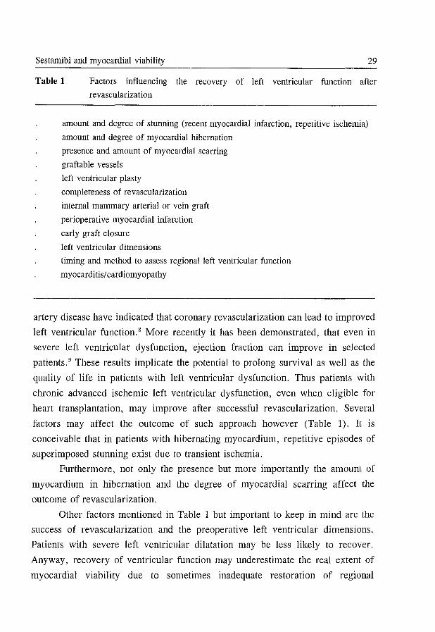
Table 1 Factors influencing the recovery of left ventricular function after
revascularization
amount and degree of stunning (recent myocardial infarction, repetitive ischemia)
amount and degree of myocardial hibernation
presence and amount of myocardial scarring
graftable vessels
left ventricular plasty
completeness of revascularization
internal mammary arterial or vein graft
perioperative myocardial infarction
early graft closure
left ventricular dimensions
timing and method to assess regional left ventricular function
myocarditis/ cardiomyopathy
artery disease have indicated that coronary revascularization can lead to improved
left ventricular function. 8 More recently it has been demonstrated, that even in
severe left ventricular dysfunction, ejection fraction can improve in selected
patients. 9 These results implicate the potential to prolong survival as well as the
quality of life in patients with left ventricular dysfunction. Thus patients with
chronic advanced ischemic left ventricular dysfunction, even when eligible for
heart transplantation, may improve after successful revascularization. Several
factors may affect the outcome of such approach however (Table 1). It is
conceivable that in patients with hibernating myocardium, repetitive episodes of
superimposed stunning exist due to transient ischemia.
Furthermore, not only the presence but more importantly the amount of
myocardium in hibernation and the degree of myocardial scarring affect the
outcome of revascularization.
Other factors mentioned in Table 1 but important to keep in mind are the
success of revascularization and the preoperative left ventricular dimensions.
Patients with severe left ventricular dilatation may be less likely to recover.
Anyway, recovery of ventricular function may underestimate the real extent of
myocardial viability due to sometimes inadequate restoration of regional
Chapter 2 30
myocardial blood flow. Various nuclear methods have received attention for the
assessment of myocardial viability. This chapter focusses on the approach with
technetium-99m (Tc-99m) labeled sestamibi as a perfusion agent to (1) distinguish
hibernation (with or without superimposed stunning) from non-viable myocardium
and (2) to predict functional recovery after coronary revascularization.
Properties of Tc-99m sestamibi
Sestamibi is a Tc-99m labeled myocardial perfusion agent, providing similar
information as thallium-201 eOITI) for the detection of coronary artery disease.lO
In comparison with 20ITI, sestamibi has the advantage of better imaging properties,
particularly when single photon emission computed tomography (SPECT) is
considered. 11 The gamma emission of Tc-99m sestamibi is higher (141 keV versus
68 to 80 keV) and its physical half life is shorter (6 versus 73 hours). Another
difference between 20lTI and sestamibi is the lower first pass extraction for
sestamibi (40% versus 80%).11.12 Although minimal myocardial redistribution
occurs « 25 % in 4 hours), the slow myocardial clearance of sestamibi
compensates for its low first pass extraction. 13 The tissue uptake of sestamibi is
parallel to coronary blood flow, with the exception of high flow conditions. Even
under conditions of low coronary blood flow and in stunned myocardium, the
myocardial uptake of sestamibi is comparable with that of 20lTl. 14 Since the uptake
of sestamibi is dependent on cell membrane integrity and mitochondrial function
(membrane potential), it may from a cellular point of view also reflect myocardial
viability. 15
While the use of sestamibi for myocardial perfusion is well accepted, its role
for the assessment of myocardial viability is still controversia1.2.11 ,16 On the basis
of these experiments, one may expect that sestamibi is comparable to 20lTI for the
detection of viable, stunned myocardium, when myocardial perfusion has been
restored after an ischemic episode. In contrast, 20lTI seems more suitable than
sestamibi in the setting of hibernating myocardium, due to its properties to
redistribute in a chronic low flow state. 16 In this condition, a sestamibi scan at rest
is expected to show a perfusion defect, most likely underestimating the presence
of viable myocardium. However, in the clinical setting stunned and hibernating
myocardium often coexist and constitute a dynamic condition. Therefore, a
Sestamibi and myocardial viability 31
distinction between the 2 syndromes is more theoretical than real in our daily
clinical practice.
Sestamibi in chronic left ventricular dysfunction
Several recent publications describe the merit of sestamibi in the setting of
chronic ischemic left ventricular dysfunction in order to distinguish viable
myocardium from scar. There are two kinds of data available: first, comparative
studies between sestamibi and other viability tracers like 201Tp7-24 or 18F-FDG using
positron emission tomography25.26 and second, studies using the improvement of
left ventricular wall motion after succesful revascularization as a standard for
myocardial viability. 27-33
Comparison of sestamibi with 201TI
All studies comparing sestamibi and 20lTl have reached similar conclusions,
suggesting that myocardial regions with severely reduced sestamibi uptake at rest
may contain viable tissue. Post-stress reinjection 2°'TI imaging has been compared
with sestamibi imaging at rest by different authors. 17-19 Cuocolo et aP7 compared
exercise-redistribution-reinjection with exercise-rest sestamibi (two day protocol)
planar imaging in 20 patients with coronary artery disease and chronic left
ventricular dysfunction (ejection fraction 30 ± 8 % ). Qualitative segmental analysis
showed 122 myocardial segments (41 %) with irreversible 20lTl uptake defects at
redistribution. After 20lTI reinjection, in 57/122 of these segments (47 %) tracer fill
in was noted. In contrast, 100/122 segments appeared as fixed defects (without
reversibility) on the sestamibi images at rest. Furthermore, the resting sestamibi
mean uptake score in the segments with perfusion defects was significantly worse
compared to the reinjection 2°'Tl mean uptake score (5 point grading system).
Since quantitative analysis of SPECT images may improve diagnostic
accuracy of comparative data, 26 patients with advanced chronic left ventricular
dysfunction (mean ejection fraction 32 ± 6 %) due to coronary artery disease, were
studied with post-stress reinjection 20lTl as well as sestamibi SPECT at rest within
7 days.'9 The images were acquired 20 minutes after reinjection of 40 MBq of 2°'TI
and 2 hours after intravenous administration of 370 MBq of sestamibi. All images
were acquired using a single head rota gamma camera with a low-energy, all
Chapter 2
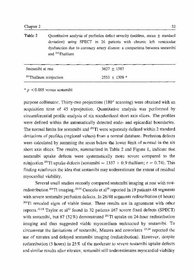
Table 2
32
Quantitative analysis of perfusion defect severity (unitless, mean ± standard
deviation) using SPECT in 26 patients with chronic left ventricular
dysfunction due to coronary artery disease: a comparison between sestamibi
and 201Thallium
Sestamibi at rest 3627 ± 1587
2553 ± 1309 * 201Thallium reinjection
* p < O. 005 versus sestamibi
purpose collimator. Thirty-two projections (180° scanning) were obtained with an
acquisition time of 45 s/projection. Quantitative analysis was performed by
circumferential profile analysis of six standardized short axis slices. The profiles
were defined within the automatically detected endo- and epicardial boundaries.
The normal limits for sestamibi and 201TI were separately defined within 2 standard
deviations of profiles (regional values) from a normal database. Perfusion defects
were calculated by summing the areas below the lower limit of normal in the six
short axis slices. The results, summarized in Table 2 and Figure 1, indicate that
sestamibi uptake defects were systematically more severe compared to the
reinjection 201TI uptake defects (sestamibi = 1337 + 0.9 thallium; r = 0.74). This
finding reinforces the idea that sestamibi may underestimate the extent of residual
myocardial viability.
Several small studies recently compared sestamibi imaging at rest with rest
redistribution 201TI imaging. 20-24 Cuocolo et apo reported in 19 patients 48 segments
with severe sestamibi perfusion defects. In 26/48 segments redistribution (4 hours)
201TI revealed signs of viable tissue. These results are in agreement with other
reports. 21 -24 Taylor et al22 found in 32 patients 167 severe fixed defects (SPECT)
with sestamibi, but 87 (52 %) demonstrated 201TI uptake on 24-hour redistribution
imaging and thus suggested viable myocardium undetected by sestamibi. To
circumvent the limitations of sestamibi, Maurea and coworkers 23,24 reported the
use of nitrates and delayed sestamibi imaging (redistribution). However, despite
redistribution (5 hours) in 25 % of the moderate to severe sestamibi uptake defects
and similar results after nitrates, sestamibi still underestimates myocardial viability
Sestamibi and myocardial viability 33
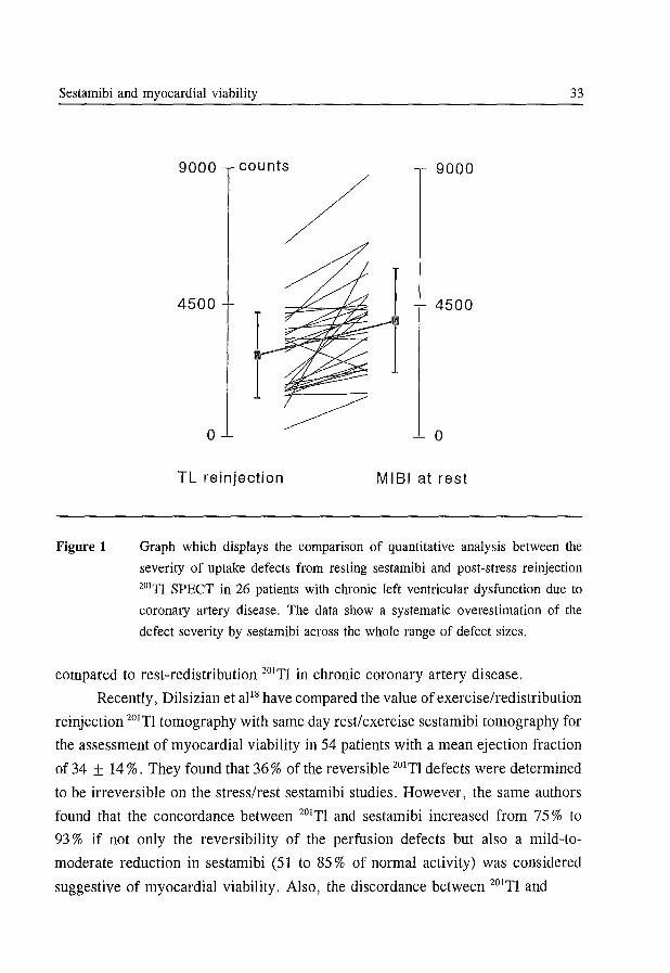
Figure 1
9000 9000
4500 4500
- a
TL reinjection MIBI at rest
Graph which displays the comparison of quantitative analysis between the
severity of uptake defects from resting sestamibi and post-stress reirUection
201TI SPECT in 26 patients with chronic left ventricular dysfunction due to
coronary artery disease. The data show a systematic overestimation of the
defect severity by sestamibi across the whole range of defect sizes.
compared to rest-redistribution 20lTI in chronic coronary artery disease.
Recently, Dilsizian et al18 have compared the value of exercise/redistribution
reinjection 20lTI tomography with same day rest/exercise sestamibi tomography for
the assessment of myocardial viability in 54 patients with a mean ejection fraction
of34 ± 14%. They found that 36% of the reversible 20lTI defects were determined
to be irreversible on the stress/rest sestamibi studies. However, the same authors
found that the concordance between 20lTI and sestamibi increased from 75 % to
93 % if not only the reversibility of the perfusion defects but also a mild-to
moderate reduction in sestamibi (51 to 85 % of normal activity) was considered
suggestive of myocardial viability. Also, the discordance between 20lTI and
Chapter 2 34
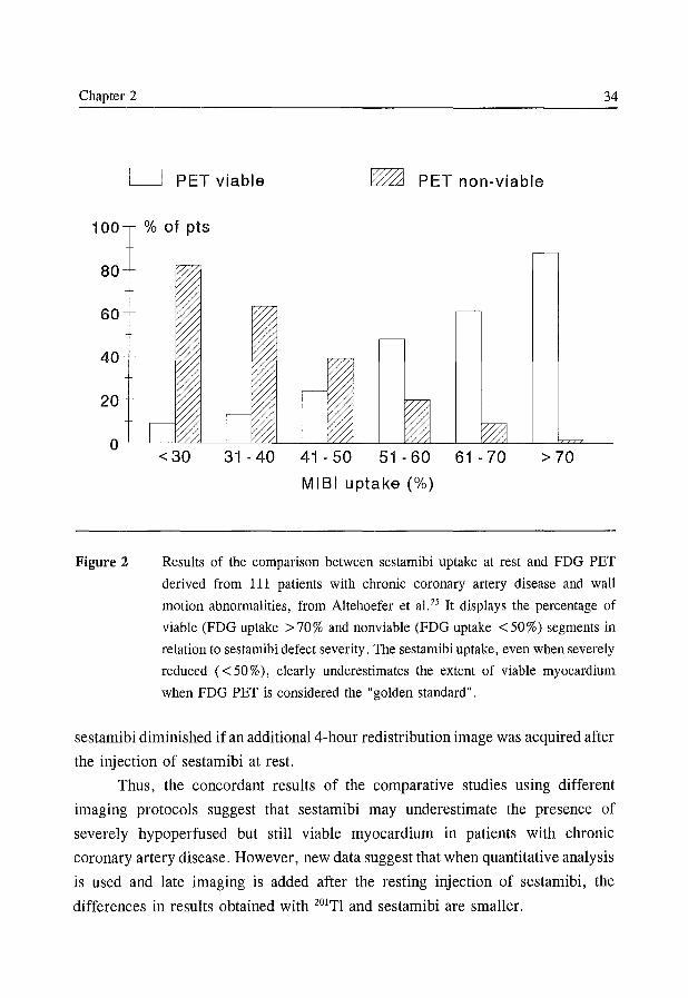
o PET viable ~ PET non-viable
100 % of pts
80
60
40
20
o
Figure 2
<30 31 -40 41 - 50 51 - 60 61 - 70 > 70
MISI uptake (%)
Results of the comparison between sestamibi uptake at rest and FDG PET
derived from 111 patients with chronic coronary artery disease and wall
motion abnormalities, from Altehoefer et al. 25 It displays the percentage of
viable (FDG uptake > 70 % and nonviable (FDG uptake < 50 %) segments in
relation to sestamibi defect severity. The sestamibi uptake, even when severely
reduced « 50 %), clearly underestimates the extent of viable myocardium
when FDG PET is considered the "golden standard" .
sestamibi diminished if an additional4-hour redistribution image was acquired after
the injection of sestamibi at rest.
Thus, the concordant results of the comparative studies using different
imaging protocols suggest that sestamibi may underestimate the presence of
severely hypoperfused but still viable myocardium in patients with chronic
coronary artery disease. However, new data suggest that when quantitative analysis
is used and late imaging is added after the resting injection of sestamibi, the
differences in results obtained with 201TI and sestamibi are smaller.
Sestamibi and myocardial viability 35
Comparison with F-18 FDG PET
Recently, Altehoefer et al25 evaluated the relationship between sestamibi
uptake at rest and glucose metabolism by FDG PET. They found, in a group of
111 patients with coronary artery disease and wall motion abnormalities, preserved
glucose metabolism (FDG uptake > 70 %) in a substantial number of patients with
reduced sestamibi uptake at rest (SPECT). Furthermore, of the myocardial
segments with 31-70 % of peak sestamibi uptake (moderate to severe defects) 13-
61 % were viable by PET criteria (Figure 2). On the other hand, sestamibi defects
with 5 30 % of peak activity are highly predictive for scar tissue (82 %).
Thus, the severity of a given sestamibi perfusion defect may yield an
indirect estimate of the likelihood of myocardial viability. Nevertheless, 30% of
the patients had at least 1 viable segment with severely reduced uptake of sestamibi
( < 50 % of peak activity). Similarly, Sawada and coworkers26 showed in 20 patients
(7 after recent myocardial infarction) that 50 % of the moderate or severe sestamibi
defects had preserved glucose metabolism (> 60 % FDG uptake). They found no
lower limit of sestamibi activity that excluded significant FDG uptake. Thus,
sestamibi uptake clearly underestimates myocardial viability in comparison with
FDG PET in patients with chronic coronary artery disease. These data are in
agreement with the comparative data with various 20lTI imaging protocols as
previously discussed. However, regardless of the lower sensitivity of sestamibi for
detecting viable myocardium, tracer uptake may still allow for correct prediction
of functional recovery after successful revascularization. The extent of
underestimation is of key importance since failure to detect limited amounts of
viable myocardium may not have an impact on the outcome of revascularization.
Dilsizian et al18 have recently found in 25 patients that the agreement
between same day rest/exercise sestamibi and PET imaging (FDG/blood flow
matching) is greatly enhanced if an additional late redistribution imaging is
acquired after the resting injection of sestamibi or if the severity of the sestamibi
activity within the irreversible defects is taken into account. However, larger
comparative studies are needed to answer the question to what myocardial extent
'Sestamibi underestimates myocardial viability relative to the amount of myocardial
scar.
Chapter 2 36
Can sestamibi reliably predict functional recovery after revascularization?
The number of studies dealing with this topic is limited. 27-33 The results
indicate, although small numbers of patients were studied, that sestamibi is not
always a good indicator of functional recovery after revascularization. In
particular, Maublant et aF8 observed, in a group of 18 patients, that wall motion
improved in 8 of 9 segments with a initial severe sestamibi defect 3 months after
revascularization. Marzullo and coworkers29 studied 14 patients with chronic
coronary artery disease before and 3 months after revascularization. Sestamibi
uptake at rest and planar rest-redistribution (16 hour) 20ITl-scans were acquired
before revascularization. Compared to delayed redistribution 20lTl imaging,
sestamibi uptake had a lower sensitivity (75 vs 86 %) and specificity (84 vs 92 %)
to predict recovery of wall motion. Sciagra et aPI reported their initial results with
the use of an infusion of nitrates during tracer injection. They studied in 22
patients the recovery of function after revascularization with first-pass radionuclide
ventriculography. They showed a predictive accuracy for sestamibi SPECT of
82 %. This approach is of interest and confirms previous work by Galli and
coworkers32 and Maurea et aIl3 also using sestamibi and Medrano et aP4 utilizing
20lTl reinjection. Zafrir and collegues35 demonstrated in 18 patients with severe left
ventricular dysfunction the additional value of simultaneous assessment of function
and perfusion by sestamibi for the prediction of functional recovery. A recent study
from Udelson et al33 performed a head-to-head comparison between rest
redistribution 20lTl tomography and rest injection of sestamibi (with imaging 1 hour
post-injection), to predict the recovery of regional left ventricular dysfunction after
successful revascularization. Thirty-one patients were studied with echocardio
graphy before and on average 20 days after revascularization. 20lThallium and
sestamibi had similar positive (75 % for 20lTl and 80 % for sestamibi) and negative
(92 % and 96 %, respectively) predictive values for recovery of regional left
ventricular dysfunction after revascularization. For both tracers, the "best" cut-off
for the discrimination of viable and not viable myocardium was 60 % of peak
activity. These excellent results are somewhat surprising because sestamibi
scintigraphy was acquired with no pre-medication of nitrates nor was late imaging
performed. 18 These results, although very promising, should be viewed with some
caution because of 1) the very short follow-up of 20 days after revascularization,
Sestamibi and myocardial viability 37
and 2) the lack of information on the changes of global left ventricular function
after revascularization.
Conclusion
Myocardial perfusion imaging may reflect viability, since tracer uptake
requires adequate perfusion, cellular integrity and metabolic function. 99m
Technetium is an excellent myocardial flow tracer, but from the data so far
available it seems less suitable than 20lTI for the assessment of myocardial viability
in chronic coronary artery disease. It tends to overestimate the extent of
myocardial necrosis or scar.
However, since sestamibi possesses superior imaging properties, the
underestimation of myocardial viability has lead to the development of alternative
protocols. For the detection of viable myocardium, imaging 1 hour after sestamibi
injection may not be optimal. The tracer is known to redistribute to a small degree,
thus delayed imaging may enhance the detection of viable myocardium in severe
perfusion defects. 13.18 Furthermore, nitroglycerin administration before rest imaging
seems usefuF3,32 and also with the addition of functional data (first pass and/or
gated imaging), revealing a complementary data set on function and perfusion, the
proper identification of viability may be improved. 27 ,35 Quantification of regional
tracer uptake may also enhance the discrimination between viable and non-viable
myocardium. 18,25,27,33 However, even with quantitation, a significant amount of
severe defects still represents viable tissue. 25,29 Finally, new methods for attenuation
correction may reduce the number of artifacts, in particular false positive perfusion
defects, in order to enhance diagnostic specificity.
Thus, although it seems that sestamibi SPECT with 1) quantification of the
activity, 2) late imaging and 3) pre-medication with nitrates provides information
on myocardial viability close to that of 20lTI and PET, further studies are needed
to substantiate the usefullness of sestamibi for addressing the issue of viability. In
particular we feel that the role of sestamibi for the prediction of functional
recovery after revascularization has still to be defined in larger and possibly
multicenter studies.
Chapter 2 38
References
1. Bolli R. Myocardial 'stunning' in man. Circulation 1992;86: 1671-1691.
2. Dilsizian V, Bonow RO. Current diagnostic techniques of assessing myocardial
viability in patients with hibernating and stunned myocardium. Circulation 1993;87: 1-
20.
3. Patel B, Kloner RA, Przyklenk K, Braunwald E. Postischemic myocardial "stunning":
a clinically relevant phenomenon. Ann Intern Med 1988;108:626-628.
4. Bourdillon PDV, Broderick TM, Williams ES, et al. Early recovery of regional left
ventricular function after reperfusion in acute myocardial infarction assessed by serial
two-dimensional echocardiography. Am J Cardiol 1989; 63: 641-646.
5. Sabia P, Powers ER, Ragosta M, Sarenbock IJ Burwell LR, Kaul S. An association
between collateral blood flow and myocardial viability in patients with recent
myocardial infarction. N Engl J Med 1992;327:1825-1831.
6. Rahimtoola SR. A perspective on the three large multicenter randomized clinical
trails of coronary bypass surgery for chronic stable angina. Circulation 1985;72
(suppl V):123-135.
7. Braunwald E, Rutherford JD. Reversible ischemic left ventricular dysfunction:
evidence for the hibernating myocardium. J Am CoIl Cardiol1986;8:1467-1470.
8. Alderman EL, Bourassa MG, Cohen LS, et al. Ten-year follow-up of survival and
myocardial infarction in the randomized coronary artery surgery study. Circulation
1990;82: 1629-1646.
9. Elefteriades JA, Tolis G, Levi E, Mills LK, Zaret BL. Coronary artery bypass
grafting in severe left ventricular dysfunction: excellent survival with improved
ejection fraction and functional state. J Am CoIl Cardiol1993;22:1411-1417.
10. Kahn JK, McGhie I, Akers MS, et al. Quantitative rotational tomography with Tl-201
and Tc-99m 2-methoxy-isobutyl-isonitrile: a direct comparison in normal individuals
and patients with coronary artery disease. Circulation 1989;79:1282-1293.
11. Liu P. New technetium 99m imaging agents: promising windows for myocardial
perfusion and viability. Am J Card Imaging 1992;6:28-41.
12. Okada RD, Glover D, Gaffney T, Williams S. Myocardial kinetics of technetium-
99m-hexakis-2-methoxy-2-methylpropyl-isonitrile. Circulation 1988;77:491-498.
13. Taillefer R, Primeau M, Costi P, Lambert R, Leveille J, Latour Y. Technetium-99m
sestamibi myocardial perfusion imaging in detection coronary artery disease:
comparison between initial (I-hour) and delayed (3-hour) postexercise images. J Nucl
Med 1991;32:1961-1965.
14. Sinusas AJ, Watson DD, Cannon Jr JM, Beller GA. Effect of ischemia and
postischemic dysfunction on myocardial uptake of technetium-99m-labeled
Sestamibi and myocardial viability 39
methoxyisobutyl isonitrile and thallium-201. J Am CoIl CardioI1989;14:1785-1793.
15. Beanlands RSB, Dawood F, Wen WH, et al. Are the kinetics of technetium 99m
methoxy isobutyl isonitrile affected by cell metabolism and viability? Circulation
1990; 82: 1802-1814.
16. Bonow RO, Dilsizian V. Thallium-201 and technetium-99m sestamibi for assessing
viable myocardium. J Nucl Med 1992;33:815-818.
17. Cuocolo A, Pace L, Ricciardelli B, Chiariello M, Trimarco B, Salvatore M.
Identification of viable myocardium in patients with chronic coronary artery disease:
comparison of thallium-201 scintigraphy with reinjection and technetium-99m
methoxyisobutyl isonitrile. J Nucl Med 1992;33:505-511.
18. Dilsizian V, Arrighi JA, Diodati JG, et al. Myocardial viability in patients with
chronic coronary artery disease: comparison of 99mTc-sestamibi with thallium
reinjection and ['BF]fluorodeoxyglucose. Circulation 1994;89: 578-587.
19. Cornel JH, Arnes.e M, Forster T, Postma-Tjoa J, Reijs AEM, Fioretti PM. Potential
and limitations of Tc-99m sestamibi scintigraphy for the diagnosis of myocardial
viability. Herz 1994;19:19-27.
20. Cuocolo A, Maurea S, Pace L, et al. Resting technetium-99m methoxyisobutyl
isonitrile cardiac imaging in chronic coronary artery disease: comparison with rest
redistribution thallium-201 scintigraphy. Eur J Nucl Med 1993;20:1186-1192.
21. Coleman PS, Metherall JA, Oao Q, et al. Comparison of rest-redistribution thallium-
201 uptake with resting sestamibi uptake in coronary artery disease (abstract). J Nucl
Med 1992;33:905.
22. Taylor AM, Merhige ME. Detection of myocardial viability: sestamibi overestimates
necrosis compared with thallium (abstract). J Am Coll Cardiol 1993;21 :283A.
23. Maurea S, Cuocolo A, Soricelli A, et al. Resting technetium-99m mibi redistribution
in patients with chronic coronary artery disease (abstract). J Nucl Med 1994;35: 114P.
24. Maurea S, Cuocolo A, Soricelli A, et al. Nitrates improve the identification of viable
myocardium by technetium-99m mibi spet imaging in chronic coronary artery disease
(abstract). J Nucl Med 1994;35:115P.
25. Altehoefer C, vom Dahl J, Biedermann M, et al. Significance of defect severity in
technetium-99m-MIBI SPECT at rest to assess myocardial viability: comparison with
fluorine-18-FDG PET. J Nucl Med 1994;35:569-574.
26. Sawada SG, Allman KC, Muzik 0, et al. Positron emission tomography detects
evidence of viability in rest technetium-99m sestamibi defects. J Am ColI Cardiol
1994;23:92-98.
27. Rocco TP, Dilsizian V, Strauss HW, Boucher CA. Technetium-99m isonitrile
myocardial uptake at rest: II. Relation to clinical markers of potential viability. J Am
Chapter 2 40
CoIl Cardiol 1989; 14: 1678-1684.
28. Maublant JC, Citron B, Lipiecki J, et al. Predictive value of Tc-99m-sestamibi
tomographic imaging as test for myocardial viability in hibernating myocardium
(abstract). J Am CoIl Cardiol 1993;21 :282A.
29. Marzullo P, Parodi 0, Reisenhofer B, et al. Value of rest thallium-201ltechnetium-
99m sestamibi scans and dobutamine echocardiography for detecting myocardial
viability. Am J Cardiol 1993; 71: 166-172.
30. Marzullo P, Sambuceti G, Parodi 0. The role of sestamibi scintigraphy in the
radioisotopic assessment of myocardial viability. J Nuc1 Med 1992; 33: 1925-1930.
31. Sciagra R, Bisi G, Santoro GM, Rossi V, Fazzini PF. Tc-99m-sestamibi nitrate
imaging: comparison of functional and perfusion changes in asynergic territories for
the prediction of post-rev as cuI ariz at ion recovery (abstract). J Nuc1 Med 1994;35:49P.
32. Galli M, Marcassa C, Silva P, Zoccarato 0, Campini R. Improvement of resting
99mTc-sestamibi myocardial uptake by acute nitroglycerine administration (abstract).
J Am CoIl Cardiol 1993;21:221A.
33. Udelson JE, Coleman PS, Metherall JHA, et al. Predicting recovery of severe
regional ventricular dysfunction. Comparison of resting scintigraphy with 201thallium
and 99mTc-sestamibi. Circulation 1994;89:2552-2561.
34. Medrano R, Mahmarian JJ, Ashmore RF, et al. The enhanced detection of myocardial
viability with thallium-201 reinjection after nitroglycerine: a randomized, double
blind, parallel, placebo controlled trail using quantitative tomography (abstract).
Circulation 1992;86(suppl i):I-109.
35. Zafrir N, Vidne B, Bassevitch R, Lubin E. Extent of function-perfusion mismatch -
a predictor for efficacy of coronary artery bypass grafting in patients with severe left
ventricular dysfunction (abstract). J Nuc1 Med 1994;35: 126P.
Chapter 3
Dobutamine Stress-Redistribution-Reinjection Versus Rest
Redistribution 201Thallium SPECT in the Assessment of Myocardial
Viability
JH Cornel, JJ Bax, A Elhendy, AEM Reijs*, PM Fioretti.
From the Thoraxcenter, Division of Cardiology, and Department of Nuclear
Medicine * , University Hospital Rotterdam-Dijkzigt, Rotterdam, the
Netherlands.
International Journal of Cardiac Imaging, in press
Chapter 3 42
Abstract
The aim of this study was to evaluate the value of thallium-201 chloride
eOITI) reinjection imaging following dobutamine stress (DRi) to identify viable
myocardium in comparison with a rest-redistribution 20lTI protocol (RR).
The identification of viable myocardium bears important consequences for adequate
selection of patients with poor left ventricular function, often unable to exercise,
who are considered for revascularization.
Twenty-six patients with chronic coronary artery disease and depressed left
ventricular function (ejection fraction 36 ± 10 %) were studied by both DRi and
RR single photon emission computed tomography (SPECT). Semi-quantitative
analysis of regional 20lTI activity (5-point score) and wall motion by echocardio
graphy using a 16-segment model was performed. Regions were classified as viable
(normal/reversible/fixed moderate defects) or nonviable (fixed severe defects) and
related to regional wall motion. Target heart rate was reached in 25 patients.
Myocardial viability was demonstrated in 353/416 (85%) by DRi SPECT and in
346/416 (83%) by RR SPECT. The agreement between the 2 protocols was 98%
with a Kvalue of 0.94; similar results were obtained when the analysis was limited
to dyscontractile segments.
In conclusion, this study confirms the feasibility and diagnostic value of DRi
SPECT to identify viable myocardium.
Introduction
In patients with coronary artery disease, dyscontractile myocardium may
represent either viable or necrotic myocardium. I Recovery of regional contractile
function after revascularization may occur when residual viability is present,
whereas necrotic myocardium will not improve in function. I If a substantial amount
of dyscontractile but viable myocardium is present, this may translate in
improvement of global ventricular function. 2-4 Thus, the identification of viable
myocardium bears important consequences for adequate selection of patients
considered for revascularization. This is even more important in patients with
severely depressed ventricular function, since surgical intervention in this subset
of patients is associated with high mortality5, and also improves long-term survival
as compared to conservative treatment. 6
Thallium SPECT and myocardial viability 43
At present, many laboratories use the potassium analogue thallium-201 chloride
eO'TI) to assess cellular integrity, hence indicating viability. Both 2°'TI rest
redistribution4,7-'o and stress-redistribution-reinjection"-13 are frequently used
protocols to assess myocardial viability. Stress-redistribution-reinjection provides
information on both stress-induced ischemia and viability, whereas rest-redistributi
on provides only information on viability.
Previous studies employed physical exercise to provoke myocardial ischemia
using the stress-redistribution-reinjection protocol. 11-13 Since patients with advanced
left ventricular dysfunction are often unable to exercise, pharmacological stress
with adenosine, 14 dipyridamole'5 or dobutamine'6 has been be used as an alternative
to induce myocardial ischemia. The value of a 2°'TI reinjection protocol using
pharmacological stress to assess myocardial viability is unclear, although a major
difference with maximal exercise testing is unlikely. No comparative studies
between such protocol and positron emission tomography or 2°'TI rest-redistribution
single photon emission computed tomography (SPECT) have been described.
Therefore, the aim of the present study was to evaluate the value of 2°'TI
reinjection imaging following dobutamine infusion to identify viable myocardium
using rest-redistribution 20lTl imaging as a reference method.
Methods
Study population
We studied 26 patients with chronic coronary artery disease and depressed
LV function (mean left ventricular ejection fraction 36 ± 10 %). All patients
underwent two-dimensional echocardiography, cardiac catheterization, dobutamine
stress-redistribution-reinjection and rest-redistribution SPECT.
The patients had a mean age 60 ± 7 years; there were 20 men and 6 women.
Twenty-three of the study patients (88 %) suffered a previous myocardial infarction,
but not within 6 months before the study (mean 62 ± 64 months, range 6-228).
Twenty-one patients had a Q wave on the ECG (10 anterior, 11 inferior). All
patients were symptomatic. Fifteen patients were in New York Heart Association
functional class II and 11 in class III.
In each patient coronary angiography showed significant narrowing (;:::: 50 %
reduction in luminal diameter) of at least one major coronary artery. Three patients
Chapter 3 44
had single vessel disease, 5 had two-vessel disease and 18 had 3-vessel disease.
Informed consent was obtained in all patients. The study protocol was approved
by the ethical committee of the University Hospital Rotterdam.
Two-dimensional echo cardiography
Echocardiography was performed with a commercially available scanner
(Vingmed CFM 800). Standard views of the left ventricle (parasternal long- and
short-axis, apical 2- and 4-chamber views) were recorded. To evaluate regional
wall motion, the left ventricle was divided into 16 segments, as previously descri
bed.!7 The interpretation was done by two experienced observers who were blinded
to the nuclear data of the individual patients. Each segment was scored semiquanti
tatively as: 1 = normal (normal endocardial excursion and systolic wall thicke
ning), 2 = hypokinesia (moderate to severe reduction in excursion and wall thic
kening), 3 = akinesia (absence of excursion and wall thickening) or 4 = dyski
nesia (paradoxic outward movement in systole). We previously reported the good
inter- (84 %) and intraobserver (87 %) concordances of resting wall motion analysis
of our laboratory. 18
Dobutamine stress-redistribution-reinjection 201TI SPECT
Increasing doses of dobutamine were infused through an antecubital vein
with steps of 10 ug/kg/min every 3 min to a maximum of 40 ug/kg/min. Atropine
(up to 1 mg) was given if there were no signs of ischemia and if the 85 % of the
age predicted maximal heart rate was not reached. The test was interrupted
prematurely if severe chest pain, > 2mm ST-segment deviation, significant tachy
arrhythmias, severe hypotension or other severe unsuspected side effects occurred.
Approximately 1 min before termination of the test an intravenous dose of 2 mCi
of 2°!TI was administered.
The acquisition of the stress 201TI SPECT imaging was started within 10 min
after completing the dobutamine-atropine stress test. SPECT imaging was
performed on a Siemens Gammasonics single-head Rota Camera (Orbiter; Siemens
Corp.,Iselin,N.J.). For each study, 32 projections were obtained in a 180 0 orbit
beginning from the 40 0 left posterior oblique to the right anterior oblique
projection with an acquisition time of 60 seconds per projection. The images were
Thallium SPECT and myocardial viability 45
obtained using a low energy, all purpose collimator. A energy window of 20 % was
centered on the 68-80 keY peak. Images were stored on a 64x64, 16-bit matrix.
A Gamma 11 computer system was used to process the tomographic data. Long
and short-axis tomograms were constructed from the 3-dimensional voxel matrix.
Four hours after stress imaging, a redistribution image was obtained, followed by
reinjection of 1 mCi of 201Tl. A third acquisition was started 20 min after
reinjection. In 8 patients the acquisition of a redistribution image was omitted.
Rest-redistribution 201TI SPECT
A rest-redistribution study was performed within 10 days of the stress
reil\iection protocol. A single dosis of 3 mCi 201TI was administered at rest.
SPECT data were acquired within 10 min after tracer injection and 4 hours later.
The same camera system and acquisition protocol as described above were used.
SPECT data analysis
As previously described,18,19 the interpretation of the images was based on
six short-axis slices, three longitudinal long-axis slices and three transverse long
axis slices (for each study). The analysis was performed visually with the
assistance of quantitative measurement (circumferential profiles). The same 16-
segment model used for the interpretation of the echocardiograms was applied for
the interpretation of the SPECT images. 18,19 20lTl uptake in each segment was
scored with the consensus of 2 experienced observers using a 5-point scoring
system: 0 = normal uptake, 1 = mildly reduced, 2 = moderately reduced, 3 =
severely reduced, 4 = absent uptake.
In the stress-protocol, a segment with reduced 20lTI uptake at stress was
considered reversibly ischemic (viable) if the defect showed fill-in on the
redistribution or reinjection image. A defect was considered fixed in the absence
of fill-in. These fixed defects were divided into mild-moderate and severe defects.
A defect was classified as severe if the 20lTI uptake of a segment was :::;; 50 %
compared to maximal activity on the quantitative circumferential profile analysis20
and if it was consistent with a severe visually assessed defect. Severe fixed defects
were considered nonviable, whereas mild-moderate fixed defects were considered
viable. 20 Similar criteria for the identification of viable myocardium were used in
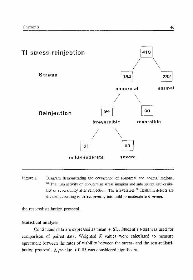
Chapter 3 46
TI stress-reinjection [~~~~-I
/ \
Figure 1
Stress
abnormal normal
/ \ Reinjection
irreversible reversible
/ EJ
mild-moderate
\
severe
Diagram demonstrating the occurrence of abnormal and normal regional
201Thallium activity on dobutamine stress imaging and subsequent irreversibi
lity or reversibility after reinjection. The irreversible 201Thallium defects are
divided according to defect severity into mild to moderate and severe.
the rest-redistribution protocol.
Statistical analysis
Continuous data are expressed as mean ± SD. Student's (-test was used for
comparison of paired data. Weighted K values were calculated to measure
agreement between the rates of viability between the stress- and the rest-redistri
bution protocol. A p-value < 0.05 was considered significant.
Thallium SPECT and myocardial viability 47
Results
Dobutamine stress test
Beta-blockade was continued in 10 of 26 patients. Seventeen patients
experienced angina during the test. Target heart rate was reached in 25 patients
(dobutamine infusion was discontinued prematurely in 1 patient due to symptomatic
hypotension). Atropine was administered in 11 patients in order to reach the target
heart rate. The mean heart rate increased from 73 ± 13 to 139 ± 15 beats/min (p
<0.001). Systolic blood pressure did not change significantly (130 ± 21 to 125
± 20 mm Hg). The rate pressure product increased from 9638 ± 2953 to 17268
± 3022 (p <0.001).
Stress-redistribution-reinjection 201TI SPECT
The initial 18 patients underwent the complete protocol, whereas in the last
8 patients the redistribution image was omitted.
In 18 patients, 288 segments were evaluated, of which 154 (53 %) showed no
perfusion abnormalities, 42 (15 %) were reversible and 92 (32 %) showed
irreversible defects at the conventional redistribution images. At reinjection, 29
(32 %) of the 92 fixed defects demonstrated significant fill-in. Of the 63 fixed
defects at reinjection, 22 were classified as mild-moderate (> 50% 201Tl uptake)
and 41 as severe (~50% 201Tl uptake). Thus, 247 (86%) of the segments were
classified as viable and 41 (14 %) as nonviable. Omission of the conventional
redistribution image did not change the identification of viable segments.
In the entire group of 26 patients, viability was then assessed using the stress and
reinjection images only (Figure 1). In a total of 416 analyzed regions, 184 had a
perfusion defect at stress, with 90 (49 %) being reversible and 94 (51 %)
irreversible. Of the irreversible defects at reinjection, 31 (33 %) were mild
moderate and 63 (67 %) were severe. Hence, in the entire patient group (n = 26)
15 % (63/416) of the segments were identified as nonviable.
Rest-redistribution 201TI SPECT
With the rest-redistribution protocol, 252 (61 %) of the 416 segments had
normal 201Tl uptake and I"64 (39%) showed a perfusion defect at rest (Figure 2).
Of these perfusion defects 28 (17 % ) demonstrated reversibility at redistribution and
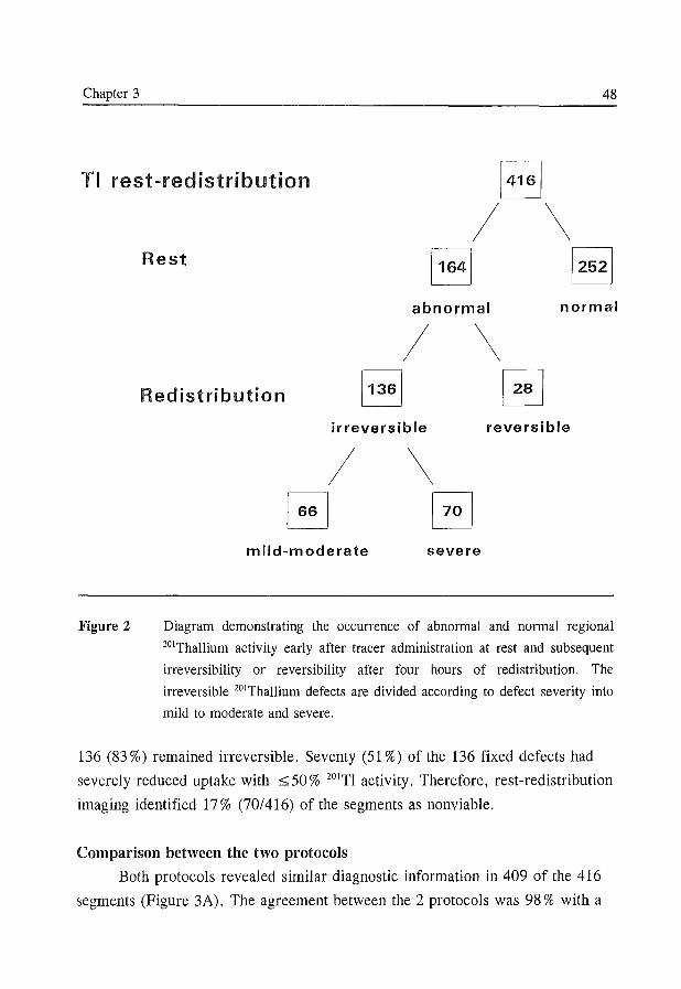
Chapter 3 48
TI rest-redistribution 1~~~1
/ \
Figure 2
Rest
abnormal normal
/ \ Redistribution
irreversible reversible
/ B
mild-moderate
\ G severe
Diagram demonstrating the occurrence of abnormal and normal regional
201Thallium activity early after tracer administration at rest and subsequent
irreversibility or reversibility after four hours of redistribution. The
irreversible 201Thallium defects are divided according to defect severity into
mild to moderate and severe.
136 (83%) remained irreversible. Seventy (51 %) of the 136 fixed defects had
severely reduced uptake with :::;; 50 % 201Tl activity. Therefore, rest-redistribution
imaging identified 17 % (70/416) of the segments as nonviable.
Comparison between the two protocols
Both protocols revealed similar diagnostic information in 409 of the 416
segments (Figure 3A). The agreement between the 2 protocols was 98 % with a
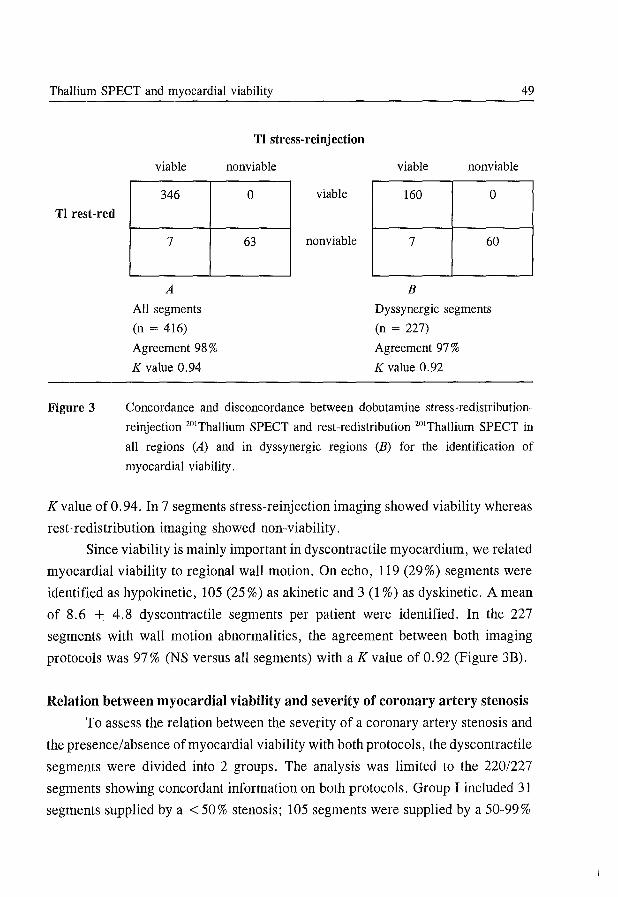
Thallium SPECT and myocardial viability 49
TI rest-red
Figure 3
TI stress-reinjection
viable nonviable viable nonviable
346 0 viable 160 0
7 63 nonviable 7 60
A B
All segments Dyssynergic segments
(n = 416) (n = 227)
Agreement 98 % Agreement 97 %
K value 0.94 K value 0.92
Concordance and disconcordance between dobutamine stress-redistribution
reinjection 201Thallium SPECT and rest-redistribution 201Thallium SPECT in
all regions (A) and in dyssynergic regions (B) for the identification of
myocardial viability.
Kvalue of 0.94. In 7 segments stress-reinjection imaging showed viability whereas
rest-redistribution imaging showed non-viability.
Since viability is mainly important in dyscontractile myocardium, we related
myocardial viability to regional wall motion. On echo, 119 (29%) segments were
identified as hypokinetic, 105 (25 %) as akinetic and 3 (1 %) as dyskinetic. A mean
of 8.6 ± 4.8 dyscontractile segments per patient were identified. In the 227
segments with wall motion abnormalities, the agreement between both imaging
protocols was 97% (NS versus all segments) with a K value of 0.92 (Figure 3B).
Relation between myocardial viability and severity of coronary artery stenosis
To assess the relation between the severity of a coronary artery stenosis and
the presence/absence of myocardial viability with both protocols, the dyscontractile
segments were divided into 2 groups. The analysis was limited to the 220/227
segments showing concordant information on both protocols. Group I included 31
segments supplied by a < 50% stenosis; 105 segments were supplied by a 50-99%
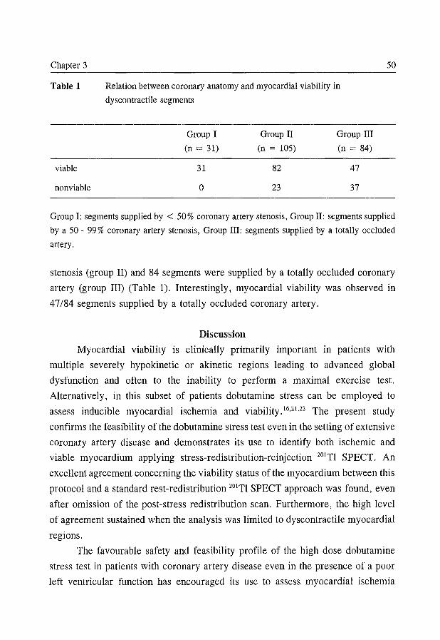
Chapter 3 50
Table 1 Relation between coronary anatomy and myocardial viability in
dyscontractile segments
Group I Group II Group III
(n = 31) (n = 105) (n = 84)
viable 31 82 47
nonviable 0 23 37
Group I: segments supplied by < 50% coronary artery stenosis, Group II: segments supplied
by a 50 - 99% coronary artery stenosis, Group III: segments supplied by a totally occluded
artery.
stenosis (group II) and 84 segments were supplied by a totally occluded coronary
artery (group III) (Table 1). Interestingly, myocardial viability was observed in
47/84 segments supplied by a totally occluded coronary artery.
Discussion
Myocardial viability is clinically primarily important in patients with
multiple severely hypokinetic or akinetic regions leading to advanced global
dysfunction and often to the inability to perform a maximal exercise test.
Alternatively, in this subset of patients dobutamine stress can be employed to
assess inducible myocardial ischemia and viability.16.21.22 The present study
confirms the feasibility of the dobutamine stress test even in the setting of extensive
coronary artery disease and demonstrates its use to identify both ischemic and
viable myocardium applying stress-redistribution-reinjection 201Tl SPECT. An
excellent agreement concerning the viability status of the myocardium between this
protocol and a standard rest-redistribution 201Tl SPECT approach was found, even
after omission of the post-stress redistribution scan. Furthermore, the high level
of agreement sustained when the analysis was limited to dyscontractile myocardial
regions.
The favourable safety and feasibility profile of the high dose dobutamine
stress test in patients with coronary artery disease even in the presence of a poor
left ventricular function has encouraged its use to assess myocardial ischemia
Thallium SPECT and myocardial viability 51
and/or viability using echocardiography or SPECT as imaging modality.16-18,21-23
Dobutamine is a selective 61-adrenoceptor agonist with relatively weak a and 62-
adrenoceptor activity. Its inotropic and chronotropic effects at high-dose infusion
act as an exercise simulator, 24 In comparative studies dobutamine induces reversible
perfusion defects very similarily in direct comparison to maximal exercise, 22,25,26
Therefore this test is increasingly used to assess myocardial ischemia, 16,19,21
Two studies have already demonstrated an excellent agreement between
exercise-redistribution-reinjection imaging and rest-redistribution imaging for the
assessment of myocardial viability27,28 in patients with advanced coronary artery
disease. The results of the present study are in line with these reports. It shows that
also semi-quantitative dobutamine-redistribution-reinjection 20lTI SPECT provides
concordant information regarding myocardial viability when compared with a
conventional rest-redistribution 20lTI protocol. The use of semi-quantitative analysis
of the SPECT data is crucial to reach such high concordance. 20,27,28
However the induction of myocardial ischemia/dysfunction may limit the proper
assessment of the amount of potential reversible dyscontractile myocardium.
Therefore, the in the literature reported diagnostic accuracy of rest-redistribution
20lTI SPECT for predicting recovery of LV function after revascularization is
somewhat higher and less variable in comparison with stress-reinjection 20lTI
SPECT. 29
Our results demonstrate that omission of the post stress redistribution image
did not substantially change the level of concordance. Dilsizian and Bonow30
decribed the phenomenon of "differential uptake", indicating the magnitude of
change in 20lTI activity after reinjection in segments identified as abnormal at
stress. The authors showed that apparent washout occurred after reinjection in a
small number of segments (8 %) identified as abnormal at stress. Due to differential
uptake, regions with fill-in at redistribution may appear fixed at reinjection when
deleting the conventional redistribution image. Such segments however had a mean
20lTI activity of 58 % at reinjection. 30 In another study, Dilsizian et aFo indicated
that segments with 20lTI activity> 50%, although fixed, were viable as compared
with PET imaging. In our study fixed defects with a 20lTI activity> 50% were
defined as viable, thereby circumventing the problem of underestimation of
viability due to differential uptake. Viable myocardium was demonstrated in 56%
Chapter 3 52
of the dyscontractile segments supplied by a totally occluded coronary artery. This
finding is not surprising since the metabolic state of the myocardium is not fully
dependent on the status of the epicardial coronary arteries. The presence of
collateral blood flow may also play an important role in the maintenance of cellular
integrity.31 In our study we did not have the opportunity to compare the results of
both 20lTl protocols with positron emision tomography, nor with functional
recovery after successful revascularization.
In conclusion, an excellent agreement between semi-quantitative dobutamine
redistribution-reinjection 20lTl SPECT and conventional rest-redistribution 201Tl
SPECT to distinguish viable from necrotic myocardium was found. The high level
of agreement sustained when the analysis was limited to dyscontractile myocardial
regions.
References
1. Dilsizian V, Bonow RO. Current diagnostic techniques of assessing myocardial
viability in hibernating and stunned myocardium. Circulation 1993 ;87: 1-20.
2. Tillisch J, Brunken R, Marshall R, et al. Reversibility of cardiac wall motion
abnormalities predicted by positron tomography. N Eng J Med 1986;314:884-888.
3. Vom Dahl J, Eitzman DT, AI-Aouar ZR, et al. Relation of regional function,
perfusion and metabolism in patients with advanced coronary artery disease under
going surgical revascularization. Circulation 1994;90:2356-2366.
4. Ragosta M, Beller GA, Watson DD, Kaul S, Gimple LW. Quantitative planar rest
redistribution 20lTl imaging in detection of myocardial viability and prediction of
improvement in left ventricular function after coronary bypass surgery in patients with
severely depressed left ventricular function. Circulation 1993; 87: 1630-1641.
5. Kennedy JW, Kaiser GC, Fisher LD, et al. Clinical and angiographic predictors of
operative mortality from the collaborative study in coronary artery surgery (CASS).
Circulation 1981; 63: 793-802.
6. Pigott JD, Kouchoukos NT, Oberman A, Cutter GR. Late results of surgical and
medical therapy for patients with coronary artery disease and depressed left ventricu
lar function. J Am ColI Cardiol 1985; 5: 1036-1045.
7. Berger BC, Watson DD, Burwell LR, et al. Redistribution of thallium at rest in
patients with stable and unstable angina and the effect of coronary artery bypass
surgery. Circulation 1979;60: 1114-1125.
Thallium SPECT and myocardial viability 53
8. Iskandrian AS, Hakki A, Kane SA, et al. Rest and redistribution thallium-201 myo
cardial scintigraphy to predict improvement in left ventricular function after coronary
arterial bypass grafting. Am J CardioI1983;51:1312-1316.
9. Mori T, Minamiji K, Kurogane H, Ogawa K, Yoshida Y. Rest-injected thallium-201
imaging for assessing viability of severe asynergic regions. J Nuc1 Med 1991; 32:
1718-1724.
10. Alfieri 0, La Canna G, Giubbini R, Pardini A, Zogno M, Fucci C. Recovery of
myocardial function. The ultimate target of coronary revascularization. Eur J Cardio
thorac Surg 1993;7:325-330.
11. Dilsizian V, Rocco TP, Freedman NM, Leon MB, Bonow RO. Enhanced detection
of ischemic but viable myocardium by the reinjection of thallium after stress-redistri
bution imaging. N Eng J Med 1990;323:141-146.
12. Ohtani H, Tamaki N, Yonekura Y, et al. Value of thallium-201 reinjection after
delayed SPECT imaging for predicting reversible ischemia ischemia after coronary
artery bypass grafting. Am J Cardiol 1990;66:394-399.
13. Tamaki N, Ohtani H, Yamashita K, et al. Metabolic activity in the areas of new fill
in after thallium-201 reinjection: Comparison with positron emission tomography
using fluorine-18-deoxyglucose. J Nuc1 Med 1991;32:673-678.
14. Verani MS, Mahmarian II. Myocardial perfusion scintigraphy during maximal
coronary artery vasodilation with adenosine. Am J Cardiol 1991;67:12D-17D.
15. Ranhosky A, Kempthorne-Rawson J, and the Intravenous Dipyridamole Thallium
Imaging Study Group. The safety of intravenous dipyridamole thallium myocardial
perfusion imaging. Circulation 1990;81:1205-1209.
16. Pennell DJ, Underwood SR, Swanton RH, Walker JM, Ell PI. Dobutamine thallium
myocardial perfusion tomography. J Am Coll Cardiol 1991; 18: 1471-1479.
17. Cornel JH, Balk AHMM, Boersma E, et al. Safety and feasibility of dobutamine
atropine stress echocardiography in patients with ischemic left ventricular dysfunction.
J Am Soc Echocardiogr 1996;9:27-32.
18. Arnese M, Cornel JH, Salustri A, et al. Prediction of improvement of regional left
ventricular function after surgical revascularization. A comparison of low-dose
dobutamine echocardiography with 20lTl single-photon emission computed tomograp
hy. Circulation 1995;91:2748-2752.
19. Forster T, McNeill AJ, Salustri A, et al. Simultaneous dobutamine stress echocardio
graphy and technetium-99 isonitrile single-photon emission computed tomography in
patients with suspected coronary artery disease. J Am Coli Cardiol 1993;21:1591-
1596.
20. Dilsizian V, Freedman NMT, Bacharach SL, Perrone-Filardi P, Bonow RO. Regional
Chapter 3 54
thallium uptake in irreversible defects. Magnitude of change in thallium activity after
reinjection distinguishes viable from nonviable myocardium. Circulation 1992; 85: 627-
634.
21. Hays JT, Mahmarian JJ, Cochran AJ, Verani MS. Dobutamine thallium-201
tomography for evaluating patients with suspected coronary artery disease unable to
undergo exercise or vasodilator pharmacologic stress testing. J Am Coll Cardiol
1993 ;21: 1583-1590.
22. Wallbridge DR, Tweddel AC, Martin W, Hutton 1. A comparison of dobutamine and
maximal exercise as stress for thallium scintigraphy. Eur J Nucl Med 1993;20:319-
323.
23. Mertes H, Sawada SG, Ryan T, et al. Symptoms, adverse effects, and complications
associated with dobutamine stress echocardiography. Exercise in 1118 patients.
Circulation 1993;88:15-19.
24. Meyer SL, Curry GC, Donsky MS, Twieg DB, Parkey RW, WillersonJT. Influence
of dobutamine on hemodynamics and coronary blood flow in patients with and
without coronary artery disease. Am J CardioI1976;38:103-108.
25. Herman SD, LaBresh KA, Santos-Ocampo CD, et al. Comparison of dobutamine and
exercise using technetium-99m sestamibi imaging for the evaluation of coronary
artery disease. Am J CardioI1994;73:164-169.
26. Marwick TH, D'Hondt AM, Mairesse GH, et al. Comparative ability of dobutamine
and exercise stress in inducing myocardial ischaemia in active patients. Br Heart J
1994;72:31-38.
27. Galassi AR, Centamore G, Fiscella A, et al. Comparison of rest-redistribution
thallium-201 imaging and reinjection after stress-redistribution for the assessment of
myocardial viability in patients with left ventricular dysfunction secondary to coronary
artery disease. Am J Oirdiol 1995;75:436-442.
28. Dilsizian V, Perrone-Filardi P, Arrighi JA, et al. Concordance and discordance
between stress-redistribution-reinjection and rest-redistribution thallium imaging for
assessing viable myocardium. Comparison with metabolic activity by positron
emission tomography. Circulation 1993; 88: 941-952.
29. Maddahi J, Schelbert H, Brunken R, Di Carli M. Role of Thallium-201 and PET
imaging in evaluation of myocardial viability and management of patients with
coronary artery disease and left ventricular dysfunction. J Nucl Med 1994;35:707-
715.
30. Dilsizian V, Bonow RO. Differential uptake and apparent 201Tl washout after thallium
reinjection. Options regarding early redistribution imaging before reinjection or late
redistribution imaging after reinjection. Circulation 1992; 85: 1032-1038.

Thallium SPECT and myocardial viability 55
31. Vanoverschelde J-LJ, Wijns W, Depre C, et al. Mechanisms of chronic regional
postischemic dysfunction in humans. New insights from the study of noninfarcted
collateral-dependent myocardium. Circulation 1993 ;87: 1513-1523.

Chapter 4
Safety and Feasibility of Dobutamine-Atropine Stress
Echocardiography in Patients With Ischemic Left Ventricular
Dysfunction
JH Cornel, AHMM Balk, E Boersma, APWM Maat*, A Elhendy, M Arne&c,
A Salustri, JRTC Roelandt, PM Fioretti.
From the Thoraxcenter, Departments of Cardiology and Cardiac Surgery*,
University Hospital Rotterdam-Dijkzigt, Erasmus University, Rotterdam, the
Netherlands.
Journal of the American Society of Echocardiography 1996;9:27-32.
Chapter 4 58
Abstract
The aim of this study was to analyze whether left ventricular dysfunction
affects the safety and feasibility of high-dose dobutamine-atropine stress echocardi
ography. We examined the results of the test in 318 consecutive patients who were
referred for high-dose dobutamine-atropine stress echocardiography and also
underwent diagnostic cardiac catheterization. Forty-four patients had a left
ventricular ejection fraction of 25% or less (mean, 21 %; range, 15% to 25%).
In the entire group of 318 patients, no serious complications (death,
myocardial infarction, or ventricular fibrillation) occurred. The overall feasibility
of completing the test was excellent (97 %). Atrial fibrillation occurred in four
patients, nonsustained ventricular tachycardia in 12, and sustained ventricular
tachycardia in one. A.decrease in systolic blood pressure of greater than 40 mm
Hg or a peak systolic pressure of less than 80 mm Hg was present in eight cases.
In the group with an ejection fraction of 25 % or less, there was a higher rate of
significant tachyarrhythmias (14 % versus 5 % ; p = 0.03), whereas the feasibility
of the test was slightly lower (89 % ; p < 0.01), but no difference for hypotension
was found. By multivariate analysis, a history of tachyarrhythmias was the only
predictor of stress-induced arrhythmias.
Advanced left ventricular dysfunction does not represent a contra-indication
for dobutamine-atropine stress testing.
Introduction
High dose dobutamine-atropine stress echocardiography (DASE) has been
shown to be highly feasible and safe and to provide useful diagnostic and
prognostic information in patients with suspected or proven coronary artery
disease. 1-10 Therefore it has entered the clinical arena for the assessment of stress
induced ischemia.
Because there is increasing evidence that coronary revascularization may
prolong survival and improve the quality of life in selected patients with left
~entricular (LV) dysfunctionll-13 by reducing stress-induced ischemia and
improving LV function, the issue of a proper assessment of residual ischemia in
these patients is of great clinical relevance. 14
Pharmacologic stress testing is particularly attractive in these patients, who
Safety and feasibility of DASE 59
often have a reduced exercise capacity. However, few data are available on the
feasibility and safety of high-dose dobutamine-atropine stress tests in patients with
advanced LV dysfunction.
Accordingly, this study was undertaken to describe the safety profile,
hemodynamic response, incidence of stress-induced ischemia, and feasibility of the
test in a consecutive group of patients referred for stress testing who also
underwent diagnostic cardiac catheterization. The specific objective was to assess
how strongly the severity of LV dysfunction affects the safety and feasibility of the
test.
Methods
Patients
Between November 1992 and December 1994, 318 consecutive patients with
stable proven or suspected coronary artery disease referred for DASE also
underwent diagnostic coronary arteriography and LV angiography within 3 months.
These 318 patients were included in this study. The patients were derived from 876
patients who underwent DASE at our institution during that period. DASE was
performed for evaluation of chest pain, after recent myocardial infarction, or as
part of research protocols on myocardial viability. They were included in the study
if no unstable angina, active congestive heart failure, significant valvular disease,
or protruding thrombus in the left ventricle was present and after informed consent
was obtained. There were 244 men and 74 women, aged 31 to 81 years (mean, 58
years). Previous myocardial infarction was present in 239 patients, 84 had a
history of heart failure, and 221 had angina. Of 249 patients with no previous
coronary bypass grafting, 206 had coronary artery disease: 136 had multivessel
disease and 70 had one-vessel disease. A history of ventricular or atrial tachyarrhy
thmias was present in 24 and 26 cases, respectively. Antianginal medication was
continued before the study, including B-blockers in 127 patients.
Dobutamine-atropine stress echo cardiography
A two-dimensional precordial echocardiogram was recorded at baseline, with
a commercially available wide-angle phased-array system (Esaote Biomedica SIM
7000 CFM [Esaote, Biomedica, Florence, Italy] or Vingmed CFM 800 [Vingmed
Chapter 4 60
Sound, Horton, Norway]). Standard apical and parasternal views were obtained.
DASE was performed as described previously.4 Briefly, dobutamine was infused
through an antecubital cannula starting at a dose of 5 ~g/kg/min for 3 minutes,
increased with 5 ~g/kg for the next 3 minutes, and continued with increases of 10
~g/kg every 3 minutes to a maximum of 40 ~g/kg/min. In patients not achieving
85 % of their age-predicted maximal heart rate (in men [220 - age] x 85 %; in
women [200 - age] x 85 %) and without signs or symptoms of myocardial ischemia
or major adverse effects, atropine (starting with 0.25 mg increasing to a maximum
of 1 mg) was added intravenously and dobutamine was continued. Thus the
maximum dose of pharmacologic agents used in this study was similar to the
generally applied protocol at our institution.
Throughout the test the electrocardiogram (three leads) was monitored. A
12-lead electrocardiogram was recorded every minute, whereas blood pressure was
measured by sphygmomanometer every 3 minutes. A two-dimensional echocardio
gram monitored LV wall motion continuously, obtaining the three standard apical
views sometimes together with the short-axis view, and the images were recorded
on videotape during the last minute of each stage of the test and continuously after
atropine administration. The images were also digitized on-line and subsequently
displayed side by side on a quad-screen format, for comparison of baseline and
stress images.
The following interruption criteria for the dobutamine-atropine stress test
were used: severe chest pain or dyspnea, cardiac arrhythmias with hemodynamic
or subjective deterioration, ST segment elevation or horizontal / downsloping ST
segment depression greater than 2 mm 80 millisecond after the J point (in the
absence of significant ST segment deviation on baseline electrocardiogram),
reduction in systolic blood pressure greater than 40 mm Hg compared with
baseline, systolic blood pressure less than 80 mm Hg, or any other serious adverse
effect during dobutamine infusion. New wall motion abnormalities were considered
a r.eason for interruption in the absence of serious adverse effects only if it was
severe and extensive, leading to LV dilation.
To analyze wall motion, the LV wall was divided into 16 segments and each
segment was scored on a 4-point scale: 1 = normal wall motion and thickening,
2 = hypokinesis, 3 = akinesis, and 4 = dyskinesis (absence of systolic wall
Safety and feasibility of DASE 61
motion with thinning). The analysis of the echo cardiographic images was
performed by two observers, blinded to the clinical data of the individual patient
but with knowledge of the doses of dobutamine and atropine used. A wall motion
score index (total score divided by the number of segments visualized) at both
baseline and peak stress was calculated for each patient. DASE was considered
positive for myocardial ischemia when a new wall motion abnormality or a
worsening of hypokinesis occurred in one or more segments during stress. 15
DASE was defined as feasible when symptoms (severe chest pain) or signs
(electrocardiographic or echo cardiographic ) of myocardial ischemia were detected
or, in the absence of ischemia, at least the 85 % of age-predicted maximal heart
rate was achieved.
Cardiac catheterization
The arteriograms were analyzed visually to assess the degree of stenosis of
the coronary arterial segments of the three major vessels. Significant stenosis was
defined as a diameter stenosis of greater than 50 %. The number of vessels with
significant lesions was assessed per patient. LV ejection fraction (L VEF) was
calculated from volume measurements by single-plane cineangiocardiography
according to the area-length method.
Data management
Patients characteristics such as demographic features and medical history,
stress test results (adverse effects, hemodynamic response, reasons to stop, and
feasibility), and echocardiographic and angiographic findings were loaded into a
database at the time of the stress test. Death, myocardial infarction, severe
hypotension defined as a drop in systolic pressure of greater than 40 mmHg or a
peak systolic blood pressure of less than 80 mmHg, and significant tachy
arrhythmias (supraventricular or ventricular tachycardias, nonsustained or
sustained) were defined as end points concerning the safety of the stress test. For
further analysis, the patients were also stratified into three groups with LVEF used
as a measure of LV function (525%, >25% but 540%, or >40%).
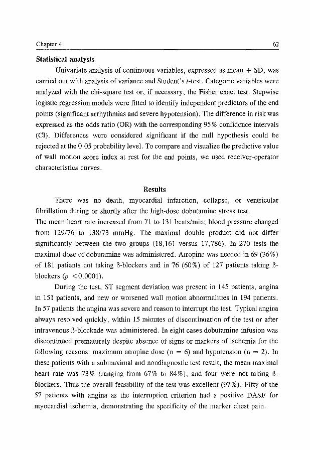
Chapter 4 62
Statistical analysis
Univariate analysis of continuous variables, expressed as mean ± SD, was
carried out with analysis of variance and Student's t-test. Categoric variables were
analyzed with the chi-square test or, if necessary, the Fisher exact test. Stepwise
logistic regression models were fitted to identify independent predictors of the end
points (significant arrhythmias and severe hypotension). The difference in risk was
expressed as the odds ratio (OR) with the corresponding 95 % confidence intervals
(CI). Differences were considered significant if the null hypothesis could be
rejected at the 0.05 probability level. To compare and visualize the predictive value
of wall motion score index at rest for the end points, we used receiver-operator
characteristics curves.
Results
There was no death, myocardial infarction, collapse, or ventricular
fibrillation during or shortly after the high-dose dobutamine stress test.
The mean heart rate increased from 71 to 131 beats/min; blood pressure changed
from 129/76 to 138/73 mmHg. The maximal double product did not differ
significantly between the two groups (18,161 versus 17,786). In 270 tests the
maximal dose of dobutamine was administered. Atropine was needed in 69 (36 %)
of 181 patients not taking 13-blockers and in 76 (60%) of 127 patients taking 13-
blockers (p <0.0001).
During the test, ST segment deviation was present in 145 patients, angina
in 151 patients, and new or worsened wall motion abnormalities in 194 patients.
In 57 patients the angina was severe and reason to interrupt the test. Typical angina
always resolved quickly, within 15 minutes of discontinuation of the test or after
intravenous 13-blockade was administered. In eight cases dobutamine infusion was
discontinued prematurely despite absence of signs or markers of ischemia for the
following reasons: maximum atropine dose (n = 6) and hypotension (n = 2). In
these patients with a submaximal and nondiagnostic test result, the mean maximal
heart rate was 73 % (ranging from 67 % to 84 % ), and four were not taking 13-
blockers. Thus the overall feasibility of the test was excellent (97 %). Fifty of the
57 patients with angina as the interruption criterion had a positive DASE for
myocardial ischemia, demonstrating the specificity of the marker chest pain.
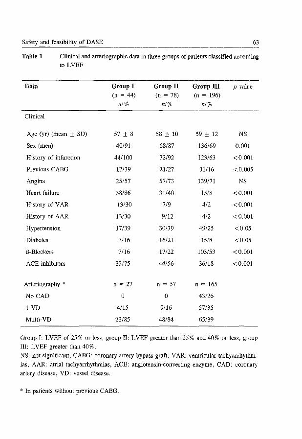
Safety and feasibility of DASE 63
Table 1 Clinical and arteriographic data in three groups of patients classified according to LVEF
Data Group I Group II Group III p value (n = 44) (n = 78) (n = 196)
n/% n/% n/%
Clinical
Age (yr) (mean ± SD) 57 ± 8 58 ± 10 59 ± 12 NS
Sex (men) 40/91 68/87 136/69 0.001
History of infarction 44/100 72/92 123/63 <0.001
Previous CABG 17/39 21127 31116 <0.005
Angina 25/57 57/73 139/71 NS
Heart failure 38/86 31140 15/8 <0.001
History of V AR 13/30 7/9 4/2 <0.001
History of AAR 13/30 9/12 4/2 <0.001
Hypertension 17/39 30/39 49/25 <0.05
Diabetes 7/16 16/21 15/8 <0.05
B-Blockers 7/16 17/22 103/53 <0.001
ACE inhibitors 33/75 44/56 36/18 <0.001
Arteriography * n = 27 n = 57 n = 165
No CAD 0 0 43/26
1 VD 4/15 9/16 57/35
Multi-VD 23/85 48/84 65/39
Group I: L VEF of 25 % or less, group II: L VEF greater than 25 % and 40 % or less, group
III: L VEF greater than 40 % . NS: not significant, CABG: coronary artery bypass graft, VAR: ventricular tachyarrhythm-
ias, AAR: atrial tachyarrhythmias, ACE: angiotensin-converting enzyme, CAD: coronary
artery disease, VD: vessel disease.
* In patients without previous CABG.
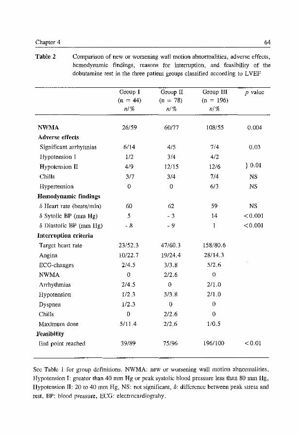
Chapter 4 64
Table 2 Comparison of new or worsening wall motion abnormalities, adverse effects, hemodynamic findings, reasons for interruption, and feasibility of the dobutamine test in the three patient groups classified according to L VEF
Group I Group II Group III p value (n = 44) (n = 78) (n = 196)
n/% n/% n/%
NWMA 26/59 60/77 108/55 0.004
Adverse effects
Significant arrhytmias 6/14 4/5 7/4 0.03
Hypotension I 112 3/4 4/2
Hypotension II 4/9 12/15 12/6 } 0.01
Chills 3/7 3/4 7/4 NS
Hypertension 0 0 6/3 NS
Hemodynamic findings
o Heart rate (beats/min) 60 62 59 NS
o Sytolic BP (rom Hg) 5 - 3 14 <0.001
o Diastolic BP (rom Hg) - .8 - 9 1 <0.001
Interruption criteria
Target heart rate 23/52.3 47/60.3 158/80.6
Angina 10122.7 19/24.4 28/14.3
ECG-changes 2/4.5 3/3.8 512.6
NWMA 0 212.6 0
Arrhythmias 2/4.5 0 2/1.0
Hypotension 112.3 3/3.8 2/1.0
Dyspnea 112.3 0 0
Chills 0 212.6 0
Maximum dose 5/11.4 212.6 110.5
Feasibility
End point reached 39/89 75/96 196/100 <0.01
See Table 1 for group definitions. NWMA: new or worsening wall motion abnormalities,
Hypotension I: greater than 40 mm Hg or peak systolic blood pressure less than 80 rom Hg, Hypotension II: 20 to 40 mm Hg, NS: not significant, 0: difference between peak stress and
rest, BP: blood pressure, ECG: electrocardiograhy.
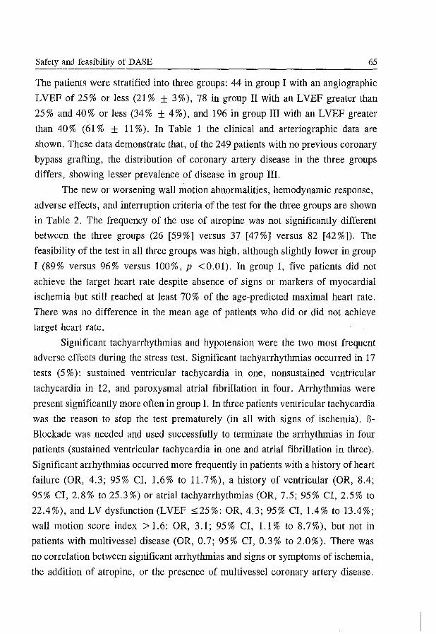
Safety and feasibility of DASE 65
The patients were stratified into three groups: 44 in group I with an angiographic
LVEF of 25% or less (21 % ± 3%), 78 in group II with an LVEF greater than
25 % and 40 % or less (34 % ± 4 % ), and 196 in group III with an L VEF greater
than 40 % (61 % ± 11 %). In Table 1 the clinical and arteriographic data are
shown. These data demonstrate that, of the 249 patients with no previous coronary
bypass grafting, the distribution of coronary artery disease in the three groups
differs, showing lesser prevalence of disease in group III.
The new or worsening wall motion abnormalities, hemodynamic response,
adverse effects, and interruption criteria of the test for the three groups are shown
in Table 2. The frequency of the use of atropine was not significantly different
between the three groups (26 [59%] versus 37 [47%] versus 82 [42%]). The
feasibility of the test in all three groups was high, although slightly lower in group
I (89% versus 96% versus 100%, p <0.01). In group I, five patients did not
achieve the target heart rate despite absence of signs or markers of myocardial
ischemia but still reached at least 70% of the age-predicted maximal heart rate.
There was no difference in the mean age of patients who did or did not achieve
target heart rate.
Significant tachyarrhythmias and hypotension were the two most frequent
adverse effects during the stress test. Significant tachyarrhythmias occurred in 17
tests (5 %): sustained ventricular tachycardia in one, nonsustained ventricular
tachycardia in 12, and paroxysmal atrial fibrillation in four. Arrhythmias were
present significantly more often in group I. In three patients ventricular tachycardia
was the reason to stop the test prematurely (in all with signs of ischemia). 13-
Blockade was needed and used successfully to terminate the arrhythmias in four
patients (sustained ventricular tachycardia in one and atrial fibrillation in three).
Significant arrhythmias occurred more frequently in patients with a history of heart
failure (OR, 4.3; 95% CI, 1.6% to 11.7%), a history of ventricular (OR, 8.4;
95% CI, 2.8% to 25.3%) or atrial tachyarrhythmias (OR, 7.5; 95% CI, 2.5% to
22.4%), and LV dysfunction (LVEF :::;25%: OR, 4.3; 95% CI, 1.4% to 13.4%;
wall motion score index> 1.6: OR, 3.1; 95% CI, 1.1 % to 8.7%), but not in
patients with multivessel disease (OR, 0.7; 95% CI, 0.3% to 2.0%). There was
no correlation between significant arrhythmias and signs or symptoms of ischemia,
the addition of atropine, or the presence of multivessel coronary artery disease.
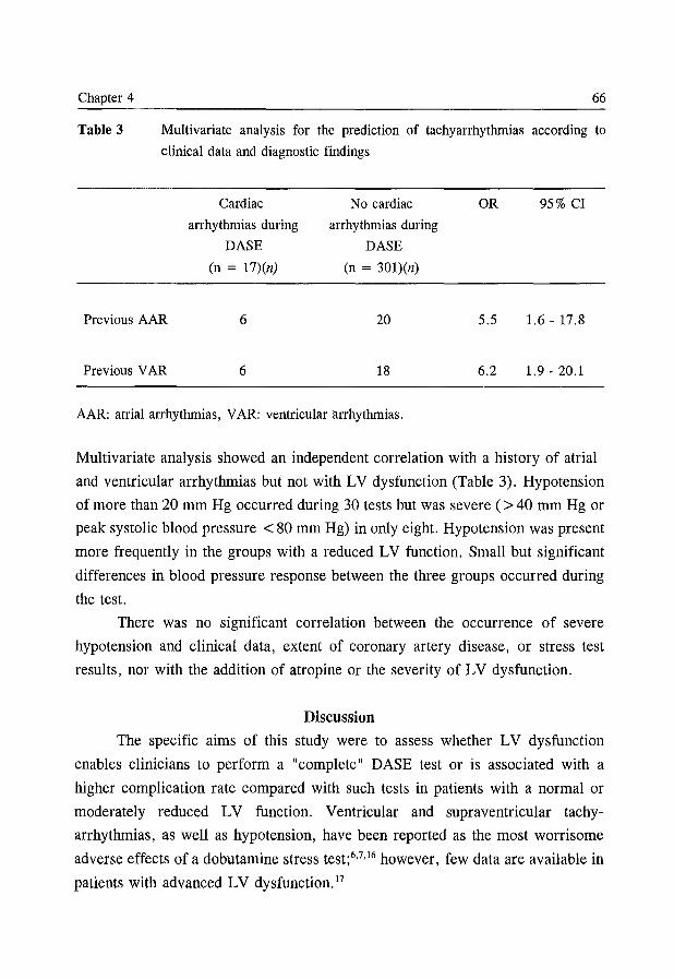
Chapter 4 66
Table 3 Multivariate analysis for the prediction of tachyarrhythmias according to
clinical data and diagnostic findings
Previous AAR
Previous V AR
Cardiac
arrhythmias during
DASE
(n = 17)(n)
6
6
No cardiac
arrhythmias during
DASE
(n = 301)(n)
20
18
AAR: atrial arrhythmias, VAR: ventricular arrhythmias.
OR 95% CI
5.5 1.6 - 17.8
6.2 1.9 - 20.1
Multivariate analysis showed an independent correlation with a history of atrial
and ventricular arrhythmias but not with LV dysfunction (Table 3). Hypotension
of more than 20 mm Hg occurred during 30 tests but was severe (> 40 mm Hg or
peak systolic blood pressure < 80 mm Hg) in only eight. Hypotension was present
more frequently in the groups with a reduced LV function. Small but significant
differences in blood pressure response between the three groups occurred during
the test.
There was no significant correlation between the occurrence of severe
hypotension and clinical data, extent of coronary artery disease, or stress test
results, nor with the addition of atropine or the severity of LV dysfunction.
Discussion
The specific aims of this study were to assess whether LV dysfunction
enables clinicians to perform a "complete" DASE test or is associated with a
higher complication rate compared with such tests in patients with a normal or
moderately reduced LV function. Ventricular and supraventricular tachy
arrhythmias, as well as hypotension, have been reported as the most worrisome
adverse effects of a dobutamine stress test;6.7,16 however, few data are available in
patients with advanced LV dysfunction. 17
Safety and feasibility of DASE 67
Safety of DASE
The initial hypothesis was that there could be an increased risk to induce
ischemic changes with high-dose dobutamine and atropine in patients with severely
reduced L VEF and that the test would be potentially dangerous in these patients.
Despite our aggressive approach, with atropine used in addition to dobutamine, our
results reject the initial hypothesis. Although frequently myocardial ischemia was
induced, there was no major complication during DASE such as death, myocardial
infarction, or ventricular fibrillation.
A decrease in systolic blood pressure decrease of 20 mm Hg or greater but
less than 40 mm Hg during the test has not been considered by us to be a serious
adverse effect, in contrast to others.7.16,18 Significant tachyarrhythmias were
infrequent (5%), similar to that found by others;6,7,16 only in four patients were
intravenous B-blockers needed to restore sinus rhythm. Our study shows that a
history of atrial or ventricular tachyarrhythmias but not LV (dys )function is
independently correlated with the occurrence of significant tachyarrhythmias. In
a previous study we reported that LV dysfunction is a risk factor for paroxysmal
tachyarrhythmias.6 However, the number of patients with advanced LV dys
function was small. This study includes a new study population with a broad range
of LV dysfunction, all with measurement of angiographic L VEF. This strengthens
our conclusion that LV function does not influence the occurrence of cardiac
arrhythmias, not even in a patient popUlation with extensive LV dysfunction after
an aggressive dobutamine-atropine protocol. Furthermore, we confirm that,
although atropine by withdrawal of parasympathetic tone may increase the risk of
significant cardiac arrhythmias, the addition of atropine does not actually increase
the occurrence of arrhythmias. 5-7 The strong correlation between dobutamine stress
induced cardiac arrhythmias and a history of arrhythmias in itself is not surprising
and confirms previous reports. 6
Hypotension has been considered a reason for termination of the test only
if symptoms or severe (> 40 mm Hg) hypotension occurred. This happened in
eight cases, in two accompanied by sinus bradycardia. The occurrence rate of
severe hypotension was infrequent and similar in the groups with different LVEF.
It was not related to the occurrence of new wall motion abnormalities or other
markers of ischemia. These data are consistent with those we found previously in
Chapter 4 68
a larger group of patients with less advanced LV dysfunction. 6 Vasodepressor
reflex mechanisms induced by vigorous myocardial contractions, dynamic
intraventricular outflow obstruction, or the vasodilator effects of dobutamine are
possible mechanisms of dobutamine-induced hypotension. 19,20 Other adverse effects,
such as chills, hypertension, or dyspnea, were minor and present infrequently.
They seldom were the cause of termination of the test.
Feasibility of DASE
The criteria for interruption of the test were similar in the different groups.
We rarely considered the appearance of new wall motion abnormalities in the
absence of symptoms or hemodynamic deterioration as an interruption criterion.
Despite the more frequent use of B-blockers, the test could be completed (reaching
the end points of ischemia or target heart rate) in a high proportion of patients with
normal and mildly reduced LV function (Table 2). The frequency of the use of
atropine in addition to dobutamine was similar in the three groups with different
severities of LV dysfunction. The slightly lower feasibility of the test in the group
with advanced LV dysfunction c,an be explained by a lower maximal (but at least
70 % was reached) heart rate reached in the absence of signs or symptoms of
myocardial ischemia.
Our definition of a submaximal nondiagnostic test is rather arbitrary. One
could argue that because nearly all patients in group I reached 75 % of the target
heart rate, the feasibility in this group with predominance of multivessel disease
was excellent. A possible explanation for the lower increase in heart rate in group
I is the down-regulation of the cardiac B-receptors that has been described in
patients with advanced LV dysfunction. 21
In conclusion, in advanced ischemic LV dysfunction, high-dose DASE is
feasible and diagnostic in a high proportion of patients and does not represent a
risk factor for serious complications or adverse effects. A history of significant
arrhythmias represents an increased risk for the occurrence of arrhythmias during
the test.
We acknowledge Peter Klootwijk for his critical support.
Safety and feasibility of DASE 69
References
1. Berthe C, Pierard LA, Hiernaux M, et al. Predicting the extent and location of
coronary artery disease in acute myocardial infarction by echocardiography during
dobutamine infusion. Am J Cardiol 1986;58: 1167-1172.
2. Sawada SG, Segar DS, Ryan T, et al. Echocardiographic detection of coronary artery
disease during dobutamine infusion. Circulation 1991; 83: 1605-1614.
3. Forster T, McNeill AJ, Salustri A, et al. Simultaneous dobutamine stress echocar
diography and technetium-99m isonitrile single-photon emission computed tomography
in patients with suspected coronary artery disease. J Am ColI Cardiol 1993;21: 1591-
1596.
4. McNeill AJ, Fioretti PM, EI-Said EM, Salustri A, Forster T, Roelandt JRTC.
Enhanced sensitivity for the detection of coronary artery disease by addition of
atropine to dobutamine stress echocardiography. Am J Cardiol 1992;70:41-46.
5. Akosah KO, Porter TR, Simon R, Funia JT, Minisi AJ, Mohanty PK. Ischemia
induced regional wall motion abnormality is improved after coronary angioplasty:
demonstration by dobutamine stress echocardiography. J Am CoIl Cardiol 1993 ;21:
584-589.
6. Poldermans D, Fioretti PM, Boersma E, et al. Safety of dobutamine-atropine stress
echocardiography in patients with suspected or proven coronary artery disease. Am
J CardioI1994,73:456-459.
7. Mertes H, Sawada SG, Ryan T, et al. Symptoms, adverse effects, and complications
associated with dobutamine stress echocardiography: experience in 1118 patients.
Circulation 1993;88:15-19.
8. Picano E, Mathias Jr W, Pingitore A, Bigi R, Previtali M for the Echo Dobutamine
International Cooperative Study Group. Safety and tolerability of dobutamine-atropine
stress echocardiography: a prospective, multicentre study. Lancet 1994;344: 1190-
1192.
9. Poldermans D, Fioretti PM, Forster T, et al. Dobutamine stress echocardiography for
assessment of perioperative cardiac risk in patients undergoing major vascular
surgery. Circulation 1993; 87: 1506-1512.
10. Mazeika PK, Nadazdin A, Oakley CM. Prognostic value of dobutamine echocardio
graphy in patients with high pretest likelihood of coronary artery disease. Am J
CardioI1993;71:33-39.
11. The Veterans Administration Coronary Artery Bypass Surgery Cooperative Study
Group. Eleven-year survival in the Veterans Administration Randomized Trial of
Coronary Bypass Surgery for Stable Angina. N Engl J Med 1984;311:1333-1339.
12. Alderman EL, Bourassa MG, Cohen LS, et al for the Coronary Artery Surgery Study
Chapter 4 70
investigators. Ten-year follow-up of survival and myocardial infarction in the
randomized coronary artery surgery study. Circulation 1990;82:1629-1646.
13. Elefteriades JA, Tolis G, Levi E, Mills LK, Zaret BL. Coronary artery bypass
grafting in severe left ventricular dysfunction: excellent survival with improved
ejection fraction and functional state. J Am CoIl Cardiol 1993 ;22: 1411-1417.
14. Cigarroa CG, deFilippi CR, Brickner E, Alvarez LG, Wait MA, Grayburn PA.
Dobutamine stress echocardiography identifies hibernating myocardium and predicts
recovery of left ventricular function after coronary revascularization. Circulation
1993;88:430-436.
15. Arnese M, Fioretti PM, Cornel JH, Postma-Tjoa J, Reijs AEM, Roelandt JRTC.
Akinesis becoming dyskinesis during high-dose dobutamine stress echocardiography:
a marker of myocardial ischemia or a mechanical phenomenon? Am J Cardiol
1994;73:896-899.
16. Marwick T, D'Hondt A, Baudhuin T, et al. Optimal use of dobutamine stress for the
detection and evaluation of coronary artery disease: combination with echocardio
graphy or scintigraphy, or both? :r Am CoIl Cardiol1993;22:159-167.
17. Hepner AM, Bach DS, Deeb GM, et al. Safety of dobutamine stress echocardio
graphy in patients with chronic ischemic left ventricular dysfunction. [Abstract]
Circulation 1993;88:1-404.
18. Mazeika PK, Nadazdin A, Oakley CM. Dobutamine stress echocardiography for
detection and assessment of coronary artery disease. J Am CoIl Cardiol 1992; 19:
1203-1211.
19. Marcovitz PA, Bach DS, Mathias W, Shayna V, Armstrong WF. Paradoxic
hypotension during dobutamine stress echocardiography: clinical and diagnostic
implications. J Am CoIl Cardiol 1993 ;21: 1080-1086.
20. Pellikka PA, Oh JK, Bailey KR, Nichols BA, Monahan KH, Tajik AI. Dynamic
intraventricular obstruction during dobutamine stress echocardiography; a new
observation. Circulation 1992;86: 1429-1432.
21. Bristow MR, Ginsburg R, Umans V, et al. 81- and 82-adrenergic-receptor
subpopulations in nonfailing and failing human ventricular myocardium: coupling of
both receptor subtypes to muscle contraction and selective 81-receptor down
regulation in heart failure. Circ Res 1986;59:297-309.

Part II
RECOVERY OF CONTRACTILE FUNCTION

Chapter 5
Prediction of Improvement of Ventricular Function After First Acute
Myocardial Infarction Using Low-Dose Dobutamine Stress
Echocardiography
A Salustri, A Elhendy, P Garyfallydis*, M Ciavatti*, JH Cornel, FJ ten Cate,
E Boersma, A Gemelli*, JRTC Roelandt, PM Fioretti.
From the Thoraxcenter, University Hospital Rotterdam-Dijkzigt and Erasmus
University, Rotterdam, the Netherlands, and the Division of Cardiology*,
Sandro Pertini Hospital Rome, Italy.
American Journal of Cardiology 1994;74:853-856
Chapter 5 74
Abstract
This study was performed to assess the prevalence of spontaneous
improvement of regional left ventricular dysfunction in patients after acute
myocardial infarction, and to evaluate the role of low-dose dobutamine stress
echocardiography for its prediction.
In 57 patients with a first acute myocardial infarction (thrombolysis, n = 27;
Q-wave, n = 49), regional wall motion was evaluated with two-dimensional
echocardiography at rest, during a low-dose dobutamine stress test performed
within 1 week after hospital admission, and at 3-month follow-up.
Myocardial viability was considered if there was an improvement of 2::: 1
grade in dyssynergic segments from rest to low-dose dobutamine infusion; recovery
of regional function was defined as an improvement of 2::: 1 grade between rest and
follow-up echocardiograms. Wall motion score index decreased from rest to low
dose dobutamine echocardiography (1.46 ± 0.29 to 1.39 ± 0.30, P <0.0001),
and this change persisted at follow-up study (1.37 ± 0.30). No differences were
found between patients who did and did not undergo thrombolysis, or between
those who had Q-wave and non-Q-wave infarction. At baseline echocardiography,
189 of 627 segments were dyssynergic (85 hypokinetic, 104 akinetic). Viability at
low-dose dobutamine stress echocardiography was more frequent in hypo kinetic
than in akinetic segments (30 of 85 versus 12 of 104, odds ratio 4.18, 95 %
confidence interval [CI] 1.87 to 9.48). Spontaneous recovery was more frequent
in hypokinetic than in akinetic segments (30 of 85 versus 20 of 104, odds ratio
2.29, CI 1.13 to 4.68). Sensitivity, specificity, and positive and negative predictive
values oflow-dose dobutamine stress echocardiography for predicting late recovery
of regional function were 66 %, 94 %, 79 %, and 88 %, respectively. Sensitivity was
lower in akinetic segments than in hypokinetic segments (35 %, CI 0.14 to 0.56,
versus 87%, CI 0.75 to 0.99). An improvement during low-dose dobutamine stress
echocardiography was a strong predictor of reversible postischemic dysfunction
(odds ratio 17.1, CI 3.5 to 97.1).
In conclusion, in patients after a first, relatively uncomplicated acute
myocardial infarction, late spontaneous recovery occurs in 26 % of the dyssynergic
segments. Low-dose dobutamine stress echocardiography provides very specific
information for predicting lack of improvement and has a high sensitivity for
Viability after acute myocardial infarction 75
predicting improvement in hypokinetic segments, but is not useful in identifying
akinetic segments that will spontaneously improve.
Introduction
Myocardial stunning is defined as transient prolonged postischemic
dysfunction that may occur after the restoration of normal coronary flow. 1 It has
been observed in several clinical conditions, including in patients after acute
myocardial infarction treated with thrombolysis. 2-5 The natural history in patients
with acute myocardial infarction not treated with thrombolysis includes potential
improvement in left ventricular function at follow-up. 6 If this is true, the evaluation
of viable myocardium should be desirable in all patients early after an acute
myocardial infarction, and could influence the choice between medical treatment
or coronary revascularization in selected patients. With the combined analysis of
flow and metabolism, positron emission tomography is the reference noninvasive
method for assessing the presence of viable myocardium. 7
Recently, myocardial perfusion scintigraphy with different isotopes and with
different protocols has been proposed for the same purpose. 8 The administration
of dobutamine in conjunction with echo cardiographic wall motion analysis has been
proposed as a simpler alternative to the more sophisticated and expensive nuclear
techniques. 9-12 Despite interest in these findings, little is known about the
spontaneous recovery of segmental left ventricular function and the potential role
of low-dose dobutamine stress echocardiography for its prediction.
Accordingly, the aims of this study were to assess the incidence of late (3
months) spontaneous improvement of regional left ventricular function in an
unselected series of patients after a first acute myocardial infarction, and to
evaluate the potential role of low-dose dobutamine stress echocardiography
(performed within 1 week after myocardial infarction) for its prediction.
Methods
Patient group
Fifty-seven consecutive patients (48 men and 9 women, mean age 58 ± 10
years, range 25 to 76) were prospectively enrolled in this study. Criteria for
recruitment were: 1) admission at our institutions with a diagnosis of first acute
Chapter 5 76
myocardial infarction (prolonged chest pain, 2:: Imm ST-segment deviation in 2::
2 leads on the initial electrocardiogram, and typical creatine kinase-MB isoenzyme
pattern); and 2) wall motion abnormalities on a resting echocardiogram performed
within 7 days of admission. Patients with valvular heart disease (n = 2), prior
myocardial infarction (n = 4), previous coronary artery bypass graft or
percutaneous transluminal coronary angioplasty (n = 3), postinfarction angina
requiring revascularization procedures (n = 3), or infarction complicated by severe
hemodynamic instability (n = 2) were excluded. No patient was excluded because
of inadequate echocardiogram. Twenty-seven patients were treated with thromboly
tic agents within 6 hours from the beginning of symptoms, and 30 had contra
indications or late presentation to the hospital. Eight patients had a non-Q-wave
myocardial infarction, and 2 of them underwent thrombolytic therapy. The site of
myocardial infarction was anterior in 30 patients and inferior and/or lateral in 27.
Predischarge dobutamine stress echo cardiography and follow-up study
After giving verbal informed consent, all patients underwent dobutamine
stress echo cardiogram within 7 days after hospital admission. Antianginal drugs,
digitalis and other drugs that might alter myocardial contractility were withdrawn
24 to 48 hours before the test. Dobutamine was infused by a volumetric pump at
incremental doses according to a protocol based on 2 stages of 5 and 10 {tg/kg/min
(5 min/dose) and 3-minute stages of 20, 30, and 40 {tg/kg/min, plus the addition
of atropine (0.25 to 1 mg) in patients not achieving 85 % of their age-predicted
maximal heart rate who had no symptoms or signs of ischemia. The first 2 stages
were considered a "low-dose stress test" and were evaluated for the presence of
viable myocardium. Throughout the dobutamine infusion, an electrocardiogram
lead was continuously monitored on the echocardiographic monitor. Twelve-lead
electrocardiograms and blood pressures were recorded at rest and at the end of
each stage. A two-dimensional echocardiogram was monitored throughout the test
and recorded on videotape at rest and during the last minute of each stage. The test
was stopped in case of severe new wall motion abnormalities, typical angina,
significant cardiac arrhythmias, or any other limiting side effects.
At 3-month follow-up, two-dimensional echocardiograms were obtained at
rest in all patients after adequate pharmacologic washout. Echocardiograms were
Viability after acute myocardial infarction 77
recorded on videotape for subsequent analysis and comparison with the previous
examination. All echo cardiograms were reviewed on the videotapes and a
consensus was achieved by 2 observers unaware of the clinical data. For the
purposes of this study, the left ventricular wall was divided into 11 segments Y Both inward wall motion and wall thickening were evaluated, and each segment
was graded as 1 = normal or hyperkinetic, 2 = hypokinetic, 3 = akinetic, and
4 = dyskinetic. Baseline images before dobutamine infusion were used as
references and compared with the corresponding images during low-dose
dobutamine infusion. An improvement of 2: 1 grade in the dyssynergic segments
during dobutamine infusion was considered as a marker of viable myocardium.
Thus, hypokinetic segments returning to normal, and systolic myocardial
thickening becoming apparent in a segment that was either akinetic or dyskinetic
were considered as a positive test for the presence of viable myocardium. Follow
up echocardiograms were compared with the corresponding rest images before
dobutamine infusion for comparative segmental analysis. For each segment, a
recovery of function was defined as an improvement of 2:: 1 grade. For each study,
a wall motion score index was calculated, dividing the sum of the scores by the
number of the segments. Because hyperdynamic left ventricular segments during
low-dose dobutamine stress echo cardiography were scored as normal (1), wall
motion score index was affected only by abnormally contracting segments.
Statistical analysis
Continuous data are expressed as mean ± SD. Continuous variables were
analyzed using analysis of variance for repeated measurements and paired t test
with the Bonferroni correction. An unpaired t test was used when appropriate.
Sensitivity, specificity, and positive and negative predictive values rely on the
standard definition and are reported with the corresponding 95 % confidence
interval (CI). The difference in late recovery was expressed as the odds ratio with
the corresponding CI. Differences were considered significant if the null hypothesis
could be rejected at the 0.05 probability level.
Results
The median interval from myocardial infarction to dobutamine test was 4
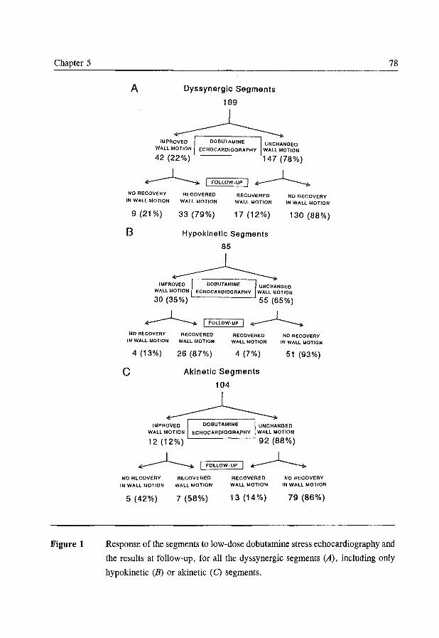
Chapter 5
Figure 1
A Dyssynergic Segments
189
IMPROVED DOBUTAMINE UNCHANGED WALL MOTION ECHOCARDIOGRAPHY WALL MOTION
42 (22%) 147 (78%)
~ I FOLLOW·UP I ~ NO RECOVERY RECOVERED RECOVERED
WALL MOTION NO RECOVERY
IN WALL MOTION IN WALL MOTION WALL MOTION
9 (21 %) 33 (79%) 17 (12%) 130 (88%)
B Hypokinetic Segments
85
~ IMPROVED DOBUTAIdINE UNCHANGED
WALL MOTION ECHOCARDIOGRAPHY WALL MOTION
30 (35%) 55 (65%)
~ I FOLLOW·UP I ~ NO RECOVERY RECOVERED RECOVERED
WALL MOTION NO RECOVERY
IN WALL IADTION IN WALL MOTION WALL MOTION
c 4 (13%) 26 (87%) 4 (7%)
Akinetic Segments
104
~
51 (93%)
IMPROVED DOBUTAMINE UNCHANGED WALL MOTION ECHOCARDIOGRAPHY WALL MOTION
12 (12%) 92 (88%)
~ I FOLLOW·UP I ~ NO RECOVERY RECOVERED
IN WALL MOTION WALL MOTION
5 (42%) 7 (58%)
RECOVERED WALL MOTION
13 (14%)
NO RECOVERY IN WALL MOTION
79 (86%)
78
Response of the segments to low-dose dobutamine stress echocardiography and
the results at follow-up, for all the dyssynergic segments (A), including only
hypokinetic (B) or akinetic (C) segments.
Viability after acute myocardial infarction 79
days (range 3 to 7). Atropine was added to dobutamine in 35 patients. No
complications occurred during the dobutamine stress test. Heart rate and systolic
blood pressure were 80 ± 19 beats/min and 143 ± 38 mm Hg at rest and 82 ± 20 beats/min and 143 ± 38 mm Hg after low-dose of dobutamine infusion.
Nineteen patients (33 %) had a positive low-dose dobutamine stress
echocardiogram, and 16 of these (84 %) had recovery of at least 1 segment at
follow-up, whereas 29 of the 38 patients (76%) with a negative low-dose
dobutamine stress echocardiogram had no improvement (n = 28) or even a
worsening (n = 1) at follow-up.
There was a significant reduction in wall motion score index between rest
and both low-dose dobutamine infusion and follow-up echocardiograms (1.46 ± 0.29 versus 1.39 ± 0.30 versus 1.37 ± 0.30, P <0.0001). Patients did not differ
according to the type of treatment (thrombolysis versus no thrombolysis) or the
type of infarction (Q-wave versus non-Q wave).
Analysis of segments
At baseline, 189 of 627 segments were dyssynergic (30%). Eighty-five of
them were hypokinetic and 104 akinetic. Low-dose dobutamine stress echocardio
graphy revealed the presence of viable myocardium in 42 of 189 segments (22 %).
At baseline, 30 of these were hypokinetic and 12 were akinetic (7 became
hypokinetic, 5 normal). Thus, viability was detected more frequently in hypokine
tic than in akinetic segments (35 % versus 12 %, odds ratio 4.18, CI 1.87 to 9.48).
At follow-up, 50 of 189 segments (26%) recovered: 10 from akinetic to normal,
10 from akinetic to hypokinetic, and 30 from hypokinetic to normal. Recovery
occurred in 33 of 42 segments (79 %) that improved with low-dose dobutamine
stress echocardiography, and in only 17 of 147 (12 %) that did not improve.
Recovery was more frequent in hypokinetic than in akinetic segments (35 % versus
19%, odds ratio 2.29, CI 1.13 to 4.68) (Figure 1).
When considering only the "viable" segments at low-dose dobutamine stress
echocardiography, there was a trend toward more frequent late recovery in the
hypokinetic than in the akinetic segments (87% versus 58%, odds ratio 4.64, CI
0.78 to 29.55). Sensitivities, specificities, and predictive values of low-dose
dobutamine stress echocardiography for the prediction of spontaneous recovery at
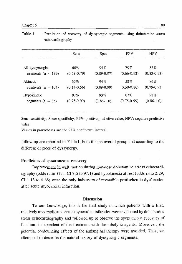
Chapter 5 80
Table 1 Prediction of recovery of dyssynergic segments using dobutamine stress
echocardiography
Sens Spec PPV NPV
All dyssynergic 66% 94% 79% 88%
segments (n = 189) (0.53-0.79) (0.89-0.97) (0.66-0.92) (0.83-0.93)
Akinetic 35% 94% 58% 86%
segments (n = 104) (0.14-0.56) (0.89-0.99) (0.30-0.86) (0.79-0.93)
Hypokinetic 87% 93% 87% 93%
segments (n = 85) (0.75-0.99) (0.86-1.0) (0.75-0.99) (0.86-1.0)
Sens: sensitivity, Spec: specificity, PPV: positive predictive value, NPV: negative predictive
value.
Values in parentheses are the 95 % confidence interval.
follow-up are reported in Table I, both for the overall group and according to the
different degrees of dyssynergy.
Predictors of spontaneous recovery
Improvement in wall motion during low-dose dobutamine stress echocardi
ography (odds ratio 17.1, CI 3.5 to 97.1) and hypokinesia at rest (odds ratio 2.29,
CI 1.13 to 4.68) were the only indicators of reversible postischemic dysfunction
after acute myocardial infarction.
Discussion
To our knowledge, this is the first study in which patients with a first,
relatively uncomplicated acute myocardial infarction were evaluated by dobutamine
stress echocardiography and followed up to observe the spontaneous recovery of
function, independent of the treatment with thrombolytic agents. Moreover, the
potential confounding effects of the anti anginal therapy were avoided. Thus, we
attempted to describe the natural history of dyssynergic segments.
Viability after acute myocardial infarction 81
The main findings of the present study can be summarized as follows:
1) Approximately one fourth of the dyssynergic segments after acute myocardial
infarction show a spontaneous recovery at 3-month follow-up.
2) The incidence of the recovery is higher in hypokinetic than in akinetic segments.
3) Low-dose dobutamine stress echocardiography is very specific for predicting the
lack of improvement in segmental left ventricular function (94 %).
4) The sensitivity is high (87 %) in hypokinetic segments, and lowest (35 %) in
akinetic segments.
Among the affected left ventricular segments in our population, the
prevalence of akinesia and hypokinesia was similar (55 % versus 45 %, respective
ly). However, improvement during the low-dose dobutamine stress test and late
spontaneous recovery occurred more frequently in the hypokinetic segments. This
is not surprising, since it seems logical to hypothesize that hypokinetic segments
contain a mixture of scar, normal, and viable myocardium, whereas in the akinetic
segments the amount of scar is predominant. The time course of recovery of the
hypokinetic segments demonstrates that in one third of the cases, the dysfunction
was probably based on the presence of myocardial stunning. This phenomenon is
less frequent when akinetic segments are considered. Among the segments that
were viable during low-dose dobutamine stress echocardiography, there was a clear
trend toward less frequent spontaneous recovery in akinetic than in hypokinetic
segments (58 % versus 87 %). It is conceivable that some of these akinetic segments
sustain incomplete reperfusion, resulting in a combination of myocardial stunning
and hibernation, with less chance of spontaneous recovery.
The comparative role of low-dose dobutamine stress echocardiography and
positron emission tomography for detecting viable myocardium was first assessed
by Pierard et al. 10 Viability was found in 10 patients with acute anterior myocardial
infarction, and recovery occurred in 6 of them; patients with no viable myocardi
um detected by echocardiography had no functional recovery at follow-up. Patients
in whom echocardiography revealed viable myocardium but who did not have late
functional recovery were characterized by an abnormally high glucose-to-perfusion
ratio, suggesting jeopardized myocardium and persistent ischemia. This pattern
may explain our finding of absence of recovery in some segments that showed
"viability" with low-dose dobutamine stress echocardiography.
Chapter 5 82
The role of low-dose dobutamine stress echocardiography for identifying viable
myocardium and predicting improvement after coronary revascularization (either
with percutaneous transluminal coronary angioplasty or coronary artery bypass
grafting) was assessed by Barilla et a1. 12 In this study, 21 patients with anterior
non-Q-wave myocardial infarction or post-thrombolytic therapy, or both, were
evaluated. Wall motion improved during dobutamine stress echocardiography in
all but 1 patient, as indicated by a reduction in wall motion score index. At follow
up, all patients had an improvement in contractility, although the magnitude was
greater in the 13 patients who underwent revascularization. Recently, Smart et al"
investigated the role of different indicators of reversible postischemic dysfunction
(wall motion at different doses of dobutamine, non-Q-wave myocardial infarction,
peak creatine kinase) in 51 patients after thrombolytic therapy. Low-dose
dobutamine stress echocardiography had a sensitivity of 86 % and a specificity of
90% for reversible dysfunction, and was sensitive in all infarct locations. Of the
other variables not related to stress echocardiography, non-Q-wave myocardial
infarction was sensitive only in anterior infarction. However, in that study, 22
patients underwent revascularization before hospital discharge on the basis of
angiographic findings alone, and this can affect the real assessment of myocardial
stunning. Although these data indicate that low-dose dobutamine-induced wall
motion improvement may be sensitive for reversible postischemic dysfunction, all
these studies focused on patients after thrombolytic therapy, often limited to
anterior myocardial infarction; furthermore, the role of rev as cui ariz at ion is difficult
to evaluate, and the relative importance of the pattern of dyssynergy at rest has not
been considered.
Study limitations
Several limitations of the present study deserve further consideration.
Coronary arteriography was not performed in our patients on a routine basis and
neither positron emission tomographic scanning for metabolic activity nor thallium
scintigraphy was performed. However, we believe that the reference method for
stunned myocardium should be spontaneous recovery of wall motion as we have
evaluated.
Echocardiograms were evaluated qualitatively and digital cineloop systems
Viability after acute myocardial infarction 83
were not used. In a previous study using dobutamine stress echocardiography,
digital techniques did not offer advantages over analysis on videotapes in terms of
diagnosis of myocardial ischemia. 14
Changes in regional wall motion, and particularly hypokinetic segments
becoming normal, are subtle and difficult to evaluate. Dobutamine stress
echocardiography was performed within 7 days hospital admission. Thus, some
segments could have already recovered, lowering the prevalence of a positive low
dose dobutamine stress echocardiogram.
Finally, the results of this study cannot be extrapolated to patients with
complicated acute myocardial infarction, who represent the ideal target population
and for whom proper identification of myocardial viability is clinically most
relevant.
Acknowledgements
We are indebted to all nurses working at our institutions for assistance in
performing the echocardiographic studies.
References
1. Bolli R. Mechanism of myocardial "stunning". Circulation 1990;82:723-738.
2. Bolli R. Myocardial "stunning" in man. Circulation 1992;86:1671-1691.
3. Bourdillon PDV, Broderick TM, Williams ES, et al. Early recovery of regional left
ventricular function after reperfusion in acute myocardial infarction assessed by serial
two-dimensional echocardiography. Am J Cardiol 1989; 63: 641-646.
4. Serruys PW, Simoons ML, Suryapranata H, et al. Preservation of global and regional
left ventricular function after early thrombolysis in acute myocardial infarction. J Am
ColI Cardiol 1986;7:729-742.
5. Patel B, Kloner RA, Przyklenk K, Braunwald E. Postischemic myocardial "stunning":
a clinically relevant phenomenon. Ann Intern Med 1988; 108: 626-628.
6. Picard MH, Wilkins GT, Ray PA, Weyman AE. Natural history of left ventricular
size and function after acute myocardial infarction. Assessment and prediction by
echo cardiographic endocardial surface mapping. Circulation 1990;82:484-494.
7. Gould KL, Yoshida K, Hess MJ, Haynie M, Mullani N, Smalling RW. Myocardial
metabolism of fluorodeoxyglucose compared to cell membrane integrity for the
potassium analogue rubidium-82 for assessing infarct size in man by PET. J Nucl
Chapter 5 84
Med 1991;32:1-9.
8. Dilsizian V, Bonow RO. Current diagnostic techniques of assessing viability in
patients with hibernating and stunned myocardium. Circulation 1993;87:1-20.
9. Marzullo P, Parodi 0, Reisenhofer B, et al. Value of rest thallium-201ltechnetium-
99m sestamibi scans and dobutamine echocardiography for detecting myocardial
viability. Am J CardiolI993;71:166-172.
10. Pierard LA, De Landsheere CM, Berthe C, Rigo P, Kulbertus HA. Identification of
viable myocardium by echocardiography during dobutamine infusion in patients with
myocardial infarction after thrombolytic therapy: comparison with positron emission
tomography. JAm Coll CardiolI990;15:1021-1031.
11. Smart SC, Sawada S, Ryan T, et al. Low-dose dobutamine echocardiography detects
reversible dysfunction after thrombolytic therapy of acute myocardial infarction.
Circulation 1993;88:405-415.
12. Barilla F, Gheorghiade M, Alam M, Khaja F, Goldstein S. Low-dose dobutamine in
patients with acute myocardial infarction identifies viable but not contractile
myocardium and predicts the magnitude of improvement in wall motion abnormalities
in response to coronary revascularization. Am Heart J 1991;122:1522-1531.
13. Picano E, Landi P, Bolognese L, et al. for the EPIC Study Group. Prognostic value
of dipyridamole echocardiography early after uncomplicated myocardial infarction:
a large-scale, multicenter trial. Am J Med 1993;95:608-618.
14. Salustri A, Fioretti PM, Pozzoli MMA, McNeill AJ, Roelandt JRTC. Dobutamine
stress echocardiography: its role in the diagnosis of coronary artery disease. Eur
Heart J 1992; 13 :70-77.
Chapter 6
T -wave Normalization During Dobutamine Echocardiography for
Diagnosis of Viable Myocardium
A Salustri, P Garyfallidis*, A Elhendy, M Ciavatti*, JH Cornel, A Gemelli * ,
FJ ten Cate, JRTC Roelandt, PM Fioretti.
From the Thoraxcenter, Division of Cardiology, University Hospital Rotterdam
Dijkzigt, Rotterdam, the Netherlands and the Division of Cardiology*,
Sandro Pertini Hospital, Rome, Italy.
American Journal of Cardiology 1995; 75:505-507
Chapter 6 86
Introduction
Low-dose dobutamine stress echocardiography has been proposed as a useful
tool for assessing reversible dysfunction after myocardial infarction. \,2 However,
subtle regional wall motion improvement during low-dose dobutamine infusion can
be difficult to evaluate, especially in akinetic segments. 3 In our experience with
dobutamine stress testing in postinfarction patients, we frequently observed the
occurrence of T-wave normalization with low-dose dobutamine without signs of
myocardial ischemia. Because we were puzzled by this electrocardiographic (EeG)
finding, we wondered whether this pattern could reflect the presence of reversible
mechanical dysfunction. In particular, we wanted to test the hypothesis that
normalization of inverted T wave during low-dose dobutamine infusion represents
a sign of dysfunctioning but still viable myocardium in patients after recent
myocardial infarction.
Methods
With this aim in mind, we enrolled in a prospective study 90 consecutive
patients admitted to our institutions who fulfilled the following admission criteria:
1) first Q-wave myocardial infarction (as documented by history, typical
isoenzymes curves, and EeG changes), 2) negative T waves in ~2 infarct-related
EeG leads, and 3) segmental wall motion abnormalities on the resting echocardio
gram. Patients with overt heart failure and early postinfarction angina requiring
revascularization were excluded. Fifty patients were treated with thrombolytic
agents. The EeG location of the infarction was anterior in 39 and inferior and/or
lateral in 51 patients.
Low-dose dobutamine stress echocardiography (5 and 10 ltg/kg/min, each
stage lasting 5 minutes) was performed within 7 days after hospital admission, with
adequate withdrawal of all antianginal and other cardioactive drugs. Regional wall I
motion was evaluated on a II-segment 4-grade scale, where 1 = normal, 2 =
hypokinesia, 3 = akinesia, and 4 = dyskinesia. Any improvement in wall motion
during low-dose dobutamine infusion in ~ 1 segment already dyssynergic at rest
was considered positive for the presence of viable myocardium. A wall motion
score index was derived by dividing the sum of the individual scores by the
number of the segments. A 12-lead electrocardiogram was recorded at rest and
Viability and T-wave nonnalization during LDDE 87
every minute during the test. T-wave normalization was defined as negative T
waves becoming upright in ~2 infarct-related BeG leads during dobutamine
infusion. Both echocardiograms and electrocardiograms were reviewed and a
consensus reached by 2 observers unaware of the results of the other test. In 59
patients a rest echocardiogram was obtained after 3 months. During this period,
no cardiac events (unstable angina, myocardial infarction, revascularization
procedure) occurred. Late spontaneous recovery of function was defined as any
improvement of wall motion in the infarct area from predobutamine to follow-up
echocardiograms.
Statistical analysis
The agreement between echocardiography and electrocardiography was
defined as the percentage of concordant diagnosis, and it was also assessed by
calculating the kappa (K) value and its standard error; K values between 0.50 and
0.75 were considered indicative of good agreement. Sensitivity, specificity,
accuracy, and positive and negative predictive values of both echocardiography and
electrocardiography for predicting late spontaneous recovery of function relied on
standard definitions, and their values were expressed as percentage with 95 %
confidence intervals.
Results
All patients completed low-dose dobutamine stress testing without adverse
effects. Both heart rate and systolic blood pressure were similar at rest and at low
dose dobutamine infusion (76 ± 18 versus 78 ± 18 beats/min; 140 ± 36 versus
140 ± 36 mmHg, respectively). Improvement in wall motion in the infarct area
during dobutamine infusion occurred in 29 patients (32 %), while the T wave
normalized in 23 (26 %). An example of T -wave normalization during dobutamine
infusion is shown in Figure 1. The overall agreement between echocardiography
and electrocardiography was 82 % (K = 0.57) (Figure 2).
Patients with improvement in wall motion and T-wave normalization (group
1, n = 18) were compared with those with improvement in wall motion but no
change in the negative T waves (group 2, n = 11). In these 2 subgroups, both the
number of dyssynergic segments at rest and the changes in wall motion score index
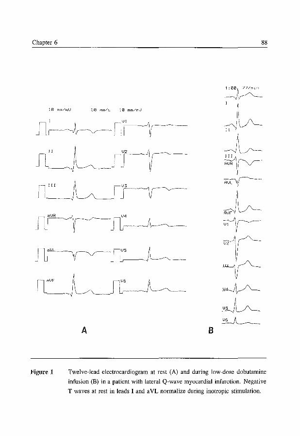
Chapter 6
10 mm/mV 50 mm/s 10 mm/mV
II
J~~
~J~
J~
A B
88
1 : 00 i~ 77 /m 1 n
~VJ-~ I
;\
I~ ~)
J~ ~rr·
Figure 1 Twelve-lead electrocardiogram at rest (A) and during low-dose dobutamine
infusion (B) in a patient with lateral Q-wave myocardial infarction. Negative
T waves at rest in leads I and a VL normalize during inotropic stimulation.
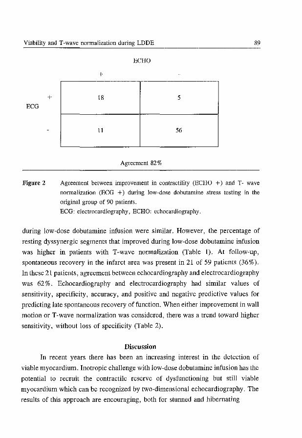
Viability and T-wave nonnalization during LDDE 89
+ ECG
Figure 2
ECHO
+
18 5
11 56
Agreement 82 %
Agreement between improvement in contractility (ECHO +) and T - wave
nonnalization (ECG +) during low-dose dobutamine stress testing in the
original group of 90 patients.
ECG: electrocardiography, ECHO: echocardiography.
during low-dose dobutamine infusion were similar. However, the percentage of
resting dyssynergic segments that improved during low-dose dobutamine infusion
was higher in patients with T-wave normalization (Table 1). At follow-up,
spontaneous recovery in the infarct area was present in 21 of 59 patients (36%).
In these 21 patients, agreement between echo cardiography and electrocardiography
was 62 %. Echocardiography and electrocardiography had similar values of
sensitivity, specificity, accuracy, and positive and negative predictive values for
predicting late spontaneous recovery of function. When either improvement in wall
motion or T-wave normalization was considered, there was a trend toward higher
sensitivity, without loss of specificity (Table 2).
Discussion
In recent years there has been an increasing interest in the detection of
viable myocardium. Inotropic challenge with low-dose dobutamine infusion has the
potential to recruit the contractile reserve of dysfunctioning but still viable
myocardium which can be recognized by two-dimensional echocardiography. The
results of this approach are encouraging, both for stunned and hibernating
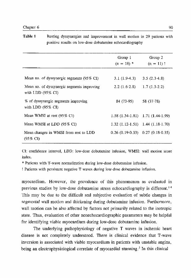
Chapter 6 90
Table 1 Resting dyssynergies and improvement in wall motion in 29 patients with
positive results on low-dose dobutamine echocardiography
Mean no. of dyssynergic segments (95% CI)
Mean no. of dyssynergic segments improving
with LDD (95 % CI)
% of dyssynergic segments improving
with LDD (95 % CI)
Mean WMSI at rest (95 % CI)
Mean WMSI at LDD (95 % CI)
Mean changes in WMSI from rest to LDD
(95% CI)
Group 1
(n = 18) *
3.1 (1.9-4.3)
2.2 (1.6-2.8)
84 (73-95)
1.58 (1.34-1.81)
1.32 (1.12-1.51)
0.26 (0.19-0.33)
Group 2
(n = 11) !
3.5 (2.3-4.8)
1.7 (1.3-2.2)
58 (37-78)
1. 71 (1.44-1. 99)
1.44 (1.18-1.70)
0.27 (0.18-0.35)
CI: confidence interval, LDD: low-dose dobutamine infusion, WMSI: wall motion score
index.
* Patients with T-wave normalization during low-dose dobutamine infusion.
! Patients with persistent negative T waves during low-dose dobutamine infusion.
myocardium. However, the prevalence of this phenomenon as evaluated in
previous studies by low-dose dobutamine stress echocardiography is different. 1-4
This may be due to the difficult and SUbjective evaluation of subtle changes in
segmental wall motion and thickening during dobutamine infusion. Furthermore,
wall motion can be also affected by factors not primarily related to the inotropic
state. Thus, evaluation of other nonechocardiographic parameters may be helpful
for identifying viable myocardium during low-dose dobutamine infusion.
The underlying pathophysiology of negative T waves in ischemic heart
disease is not completely understood. There is clinical evidence that T-wave
inversion is associated with viable myocardium in patients with unstable angina,
being an electrophysiological correlate of myocardial stunning. 5 In this clinical
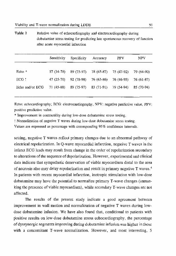
Viability and T-wave normalization during LDDE 91
Table 2 Relative value of echocardiography and electrocardiography during
dobutamine stress testing for predicting late spontaneous recovery of function
after acute myocardial infarction
Sensitivity Specificity Accuracy PPV NPV
Echo * 57 (34-78) 89 (75-97) 78 (65-87) 75 (47-92) 79 (64-90)
EeG! 47 (25-70) 92 (78-98) 76 (63-86) 76 (46-95) 76 (61-87)
Echo and/or EeG 71 (45-88) 89 (75-97) 83 (71-91) 79 (54-94) 85 (70-94)
Echo: echocardiography; EeG: electrocardiography, NPV: negative predictive value, PPV:
positive predictive value.
* Improvement in contractility during low-dose dobutamine stress testing.
! Normalization of negative T waves during low-dose dobutamine stress testing.
Values are expressed as percentage with corresponding 95 % confidence intervals.
setting, negative T waves reflect primary changes due to an abnormal pathway of
electrical repolarization. In Q-wave myocardial infarction, negative T waves in the
infarct BeG leads may result from change in the order of repolarization secondary
to alterations of the sequence of depolarization. However, experimental and clinical
data indicate that sympathetic denervation of viable myocardium distal to the area
of necrosis also may delay repolarization and result in primary negative T waves. 6
In patients with recent myocardial infarction, inotropic stimulation with low-dose
dobutamine may have the potential to normalize primary T-wave changes (unmas
king the presence of viable myocardium), while secondary T-wave changes are not
affected.
The results of the present study indicate a good agreement between
improvement in wall motion and normalization of negative T waves during low
dose dobutamine infusion. We have also found that, conditional to patients with
positive results on low-dose dobutamine stress echocardiography, the percentage
of dyssynergic segments improving during dobutamine infusion was higher in those
with a concomitant T-wave normalization. However, and most interesting, 5
Chapter 6 92
patients with negative results on low-dose dobutamine echocardiography had T
wave normalization as well, with late spontaneous improvement in wall motion in
all 3 who were evaluated at follow-up. Thus, the main finding of the present study
is the additional value of this EeG pattern for predicting late spontaneous recovery
of segmental wall motion. The higher sensitivity of electrocardiography added to
echocardiography compared with that of echocardiography alone was not
corroborated by a clear separation of the corresponding 95 % confidence intervals.
However, from this figure we estimated that ::::: 1,000 patients should be needed
to reach a statistical significance. The results of the present study may also explain
the low specificity for myocardial ischemia of normalization of negative T waves
during exercise reported in previous studies. 7 This finding may represent viable
myocardium not at risk. Finally, the relatively low sensitivity for predicting
recovery with low-dose dobutamine echocardiography in our study compared with
available datal can be explained by several factors: the different patients selected,
the different echocardiographic left ventricular model and definition of improved
wall motion and reversible dysfunction, and the prevalence of akinetic segments
at rest, which has been shown to lower sensitivity of the test. 3
In conclusion, these preliminary data indicate that T -wave normalization
during low-dose dobutamine stress testing is an ancillary sign of viable myocardi
um after acute myocardial infarction and increases the sensitivity of echocardio
graphy for predicting late spontaneous recovery of function.
Acknowledgement
We thank all nurses working at our institutions for their assistance and
expertise in performing the stress echo cardiographic studies.
References
1. Smart SC, Sawada S, Ryan T, et al. Low-dose dobutamine echocardiography detects
reversible dysfunction after thrombolytic therapy of acute myocardial infarction.
Circulation 1993;88:405-415.
2. Barilla F, Gheorghiade M, Alam M, Khaja F, Goldstein S. Low-dose dobutamine in
patients with acute myocardial infarction identifies viable but not contractile
myocardium and predicts the magnitude of improvement in wall motion abnormalities
Viability and T-wave normalization during LDDE 93
in response to coronary revascularization. Am Heart J 1991;122:1522-1531.
3. Salustri A, Elhendy A, Garyfallidis P, et al. Prediction of recovery of ventricular
dysfunction after first acute myocardial infarction using low-dose dobutamine stress
echocardiography. Am J Cardiol 1994;74:853-856.
4. Cigarroa CG, de Filippi CR, Brickner ME, Alvarez LG, Wait MA, Grayburn PA.
Dobutamine stress echocardiography identifies hibernating myocardium and predicts
recovery of left ventricular function after coronary revascularization. Circulation
1993; 88 :430-436.
5. Renkin J, Wijns W, Ladha Z, Col 1. Reversal of segmental hypokinesis by coronary
angioplasty in patients with unstable angina, persistent T wave inversion, and left
anterior descending coronary artery stenosis: additional evidence for myocardial
stunning in humans. Circulation 1990;82:913-921.
6. Matetzky S, Barabash GI, Shahar A, et al. Early T wave inversion after thrombolytic
therapy predicts better coronary perfusion: clinical and angiographic study. J Am Coll
Cardiol 1994;24:378-383.
7. Wagoner LE, Movahed A, Reeves WC, Jolly SR. Clinical significance of electrocar
diographic T-wave normalization with exercise. Am J Noninvas Cardiol 1993;7:27-
32.

Chapter 7
Prediction of Improvement of Regional Left Ventricular Function
After Surgical Revascularization
A Comparison of Low-Dose Dobutamine Echocardiography With 201TI
Single-Photon Emission Computed Tomography
M Arnese, JH Cornel, A Salustri, APWM Maat*, A Elhendy, AEM Reijs**,
FJ ten Cate, D Keane, AHMM Balk, JRTC Roelandt, PM Fioretti.
From the Thoraxcenter, Departments of Cardiology and Cardiac Surgery * , and
the Department of Nuclear Medicine * * , University Hospital Rotterdam-Dijkzigt
and Erasmus University, Rotterdam, the Netherlands.
Circulation 1995;91 :2748-2752
Chapter 7 96
Abstract
Background
Although both 201Thallium e01TI) scintigraphy and low-dose dobutamine
echocardiography (LDDE) have been proposed as effective methods of assessing
myocardial viability, their relative efficacies are unknown. The aim of the present
study was to compare the two imaging techniques in the prediction of improvement
of regional left ventricular (LV) function after surgical revascularization.
Methods and Results
Thirty-eight patients with severe chronic LV dysfunction (ejection fraction
:::; 40 %, one or more akinetic (Ak) or severely hypokinetic (SH) segments on
resting echocardiogram) who underwent uncomplicated coronary artery bypass
graft surgery were studied with simultaneous dobutamine stress echocardiography
and poststress reinjection 201Tl single-photon emission computed tomography
(SPECT) before surgery. The Ak or SH segments were considered viable by
LDDE when wall thickening improved during the infusion of 10 j-tg.kg -I. min-I
dobutamine. Scintigraphic definition of viability was the presence of normal 210TI
uptake, totally reversible defect, partially reversible defect, or moderately severe
fixed defect. The postoperative improvement of dyssynergic segments was
determined with a rest echocardiogram 3 months after surgery.
Of 608 LV segments, 169 were classified as Ak and 51 as SH on resting
preoperative echocardiography. Of these, 170 were successfully revascularized.
Wall motion during LDDE improved in 33 severely dyssynergic segments and was
more frequent in SH than in Ak segments (19 of 44 versus 14 of 126, P < 0.0001).
Viability was detected by 2°'TI SPECT criteria in 103 SH or Ak segments. Thirty
two of the 33 segments from LDDE responders were judged viable on 201TI
SPECT, whereas 201TI viability was also detected in 71 of 137 segments from
LDDE nonresponders.
The sensitivity and the specificity for the prediction of postoperative
improvement of segmental wall motion were 74 % (95 % confidence interval [CI],
67% to 81 %) and 96% (95% CI, 93% to 99%) by LDDE, and 89% (95% CI,
84% to 94%) and 48% (95% CI, 40% to 56%) by 2°'TI SPECT, respectively.
Positive predictive value of LDDE was higher than that of 2°'TI SPECT (85 %,
201Thallium SPECT versus LDDE for prediction of functional recovery 97
[95% CI, 80% to 90%] versus 33% [95% CI, 26% to 40%]).
Thirty-six patknts had angina before and only 1 had angina 3 months after
revascularization. High-dose dobutamine echocardiography demonstrated significant
reduction in stress-induced ischemia (new or worsening of preexisting wall motion
abnormalities) after surgery (from 163 to 23 LV segments).
Conclusions
In patients with severe chronic LV dysfunction, LDDE is a good predictor
of the improvement of dyssynergic segments after revascularization. Because 20lTI
SPECT overestimates the probability of postoperative improvement of dyssynergic
segments, LDDE should be the preferred imaging technique for preoperative
assessment of these patients.
Introduction
Coronary artery bypass graft surgery can improve regional and global
ventricular performance and the functional status of patients with chronic left
ventricular (LV) dysfunction. 1 The concepts of stunned and hibernating myocar
dium have been advocated to explain such improvement. 2,3 Reliable preoperative
prediction of patients in whom regional and/or global LV dysfunction will improve
after revascularization would present several clinical advantages, including the
appropriate referral for cardiac surgery of patients who are currently considered
to be unsuitable for revascularization, the referral of patients for revascularization
who would at present be considered only for cardiac transplantation, and the
avoidance of cardiac surgery in patients in whom revascularization would result in
no functional benefit (but would carry significant risk of peri operative morbidity
and mortality).
Both 201TI single-photon emission computed tomography (SPECT)2-5 and
low-dose dobutamine echocardiography (LDDE) have been proposed as effective
techniques for the evaluation of myocardial viability.6-11 Although LDDE is more
widely available, it is unknown whether its efficacy and reliability equal those of
20lTI SPECT, which is more established in this role. 2-5
To determine the relative merits of the two imaging techniques, we
compared 20lTl SPECT and LDDE in the prediction of functional recovery in 38
Chapter 7 98
patients with LV dysfunction who were undergoing coronary artery bypass graft
surgery. Postoperative resting echocardiography at 3 months was used to determine
LV improvement.
Methods
Study population
Forty-three patients with stable LV dysfunction who were to undergo
coronary artery bypass graft surgery fulfilled the study inclusion criteria: ejection
fraction of ::;; 40 % on contrast ventriculography, history of previous (> 3 months
old) myocardial infarction, one or more akinetic (Ak) or severely hypokinetic (SH)
segments on preoperative resting echocardiography (16-segment left ventricular
model), absence of recent episodes of unstable angina, absence of significant
( > 50 %) left main stem stenosis, and absence of (hemodynamically) significant
valvular disease.
Five patients were withdrawn from the study because of perioperative
myocardial infarction (3 patients), resection of all dyssynergic segments (1 patient),
or inability to graft any of the Ak of SH segments (1 patient). Thirty-eight patients
constituted the final study population. Mean patient age was 59 years (range, 36
to 73) and 26 were men. All patients were symptomatic - 36 had angina pectoris
and 20 had dyspnea on effort. Mean angiographic LV ejection fraction was 31 %
(range, 18 to 40%). One-vessel disease, defined as diameter stenosis of a major
coronary artery > 50%, was present in 3 patients; 16 patients had two-vessel
disease; and 19 patients had three-vessel disease. Four patients had undergone
previous coronary artery bypass graft surgery. Four patients were on B-blockers
during the preoperative diagnostic work-up.
Dobutamine stress echo cardiography
The dobutamine stress test was performed as follows. A two-dimensional
transthoracic echo cardiogram in standard views and a 12-lead EeG were recorded
with the patient at rest. Dobutamine was infused through an antecubital vein at
doses of 5 and 10 J,tg.kg-I.min -I, for 3 minutes at each dose. Subsequently, three
other steps from 20 to 40 J,tg.kg-I . min-I (3 minutes each) were added. Finally,
atropine (up to 1 mg) was injected when 85 % of the maximal heart rate had not
201Thallium SPECT versus LDDE for prediction of functional recovery 99
been reached.!2 The echo cardiogram was monitored during the test, and the last
minute of each stage was recorded on videotape. The echocardiographic images
were also digitized (on optical disk [Vingmed CFM 800] or on floppy disk [Esaote
Biomedica SIM 7000]) and displayed side-by-side in quad screen format to facilitate
the comparison of images at rest and after dobutamine with subsequent postoperati
ve images. A 3-lead ECG was monitored continuously, and a 12-lead ECG was
recorded every minute. Blood pressure was measured by sphygmomanometer at
each 3-minute stage.
Post-operative echo cardiography
To assess the functional outcome of the dyssynergic segments, we obtained
resting two-dimensional echocardiograms in all patients 3 months after cardiac
surgery. In addition, high-dose dobutamine/atropine stress echocardiography was
obtained in 32 patients.
Analysis of preoperative and postoperative echocardiograms
The interpretation of echo cardiograms was performed by two experienced
observers who were blinded to the clinical, angiographic and previous echocardio
graphic results of the individual patients. In a subset of 11 patients (176 segments),
the interobserver and intraobserver variabilities of the classification of resting wall
motion and the response to LDDE were also assessed. The assessment was based
on both the digitized images displayed in a quadscreen format and a review of the
images recorded on the videotape. The assessment was semiquantitative, and a 16-
segment model!3 was used. The wall motion, including wall thickening, of every
segment was scored with a 5-point scoring system, where 1 is normal wall motion
and thickening, 2 is moderately hypokinetic, 3 is SH, 4 is Ak, and 5 is dyskinetic.
We defined a segment as SH in the presence of minimal wall thickening with a
limited inward motion of < 2 mm; as Ak in the absence of systolic wall motion
and thickening and, whenever possible, confirmed by M-mode tracing; and as
dyskinetic in the presence of systolic outward wall motion with thinning.
Wall thickening was primarily used for the classification of wall motion,
preventing the problem of postoperative paradoxical septal motion. Also, to reduce
the confounding effect of tethering from adjacent segments, segmental wall
Chapter 7 100
thickening was analyzed frame-by-frame during the first half of systole.
Myocardial viability was judged to be present in a dyssynergic (either Ak or SH)
segment when wall motion improved during the infusion of low-dose dobutamine
by at least one point of the scoring system. Thus, a severe hypokinesis becoming
moderately hypokinetic or systolic myocardial thickening becoming apparent in a
previously Ak segment was considered a marker of viability. Myocardial ischemia
was judged to be present when there was worsening by ~ 1 ofthe segmental score.
Ak and dyskinetic segments were not evaluated for this purpose.
Follow-up echocardiograms were compared with the corresponding
preoperative resting images. For each segment, improvement of function was
defined as a decrease of one or more grades. Moreover, we used the preoperative
and postoperative wall motion score indexes (WMSls) to evaluate the effect of
revascularization on global LV function. WMSI was defined as the sum of the
degrees of each segment divided by the total number of segments analyzed.
201Thallium SPECT imaging
Briefly, as previously described, 14-16 20lTl (2 mCi) was injected intravenously
1 minute before the termination of the infusion of high-dose dobutamine (up to 40
j.tg.kg-I .min- I, with the addition of atropine if there were no signs of ischemia and
if 85 % of the maximal heart rate had not been reached). The acquisition of the
posts tress SPECT imaging was started within 10 minutes after the interruption of
the dobutamine infusion. All images were acquired with a Siemens Gammasonics
single-head Rota Camera (Orbiter) and a low-energy, all-purpose collimator.
Thirty-two projections were obtained, from left posterior oblique to right anterior
oblique, with an acquisition time of 45 seconds for each projection. A Gamma 11
computer system was used to process the tomographic data. Four hours after the
stress imaging, a second acquisition was performed 20 minutes following the
reinjection of 1 mCi of 20lTl.
As previously described,15.16 the interpretation of the images was based on
six short-axis slices, three longitudinal slices, and three transverse long-axis slices
(both stress and after reinjection). The analysis was performed visually with the
assistance of quantitative measurement (circumferential profiles). The same 16-
segment model used for interpretation of the echocardiograms was applied for the
201Thallium SPECT versus LDDE for prediction of functional recovery 101
interpretation of the SPECT images. Scintigraphic images from the short-axis and
the long-axis views wtfre matched with the echocardiographic images. Each defect
was classified as fixed, partially reversible, or totally reversible. A myocardial
segment was considered nonviable in the presence of a severe irreversible defect.
A defect was classified as severe if the 201TI uptake of a segment was < 50 % of
the uptake of the "normal" segments on the quantitative circumferential profile
analysis and if it was consistent with a severe visually assessed defect. Scintigra
phic definition of viability was based on the presence of normal 201TI uptake,
totally reversible defect, partially reversible defect, or moderately severe fixed
defect.
Statistical analysis
Continuous data are expressed as mean ± SD. Univariate analysis for
categorical variables was performed using the chi-square test with Yate's
correction. Differences were considered significant if the null hypothesis could be
rejected at the .05 probability level. Sensitivity, specificity and positive and
negative predictive values were based on their standard definitions and are reported
with the corresponding 95 % confidence interval (CI). The interobserver and
intraobserver variabilities of regional wall motion pattern were assessed as percent
agreement and K value.
Results
Pre-operative data
Of a total of 608 LV segments, 169 were classified as Ak and 51 as SR.
Forty-three Ak segments were excluded from the postoperative evaluation because
of aneurysmectomy (n = 41), or because they were not grafted (n = 2). Of the
51 SR segments, 7 were excluded from the postoperative evaluation because of
aneurysmectomy (n = 3) or because they were not graftable (n = 4).
Low-dose dobutamine echo cardiography
Only 14 of the 126 Ak segments that could be successfully revascularized
showed the presence of wall thickening during low-dose dobutamine on preoperati
ve echocardiogram. These 14 Ak segments were detected in 8 patients. Wall
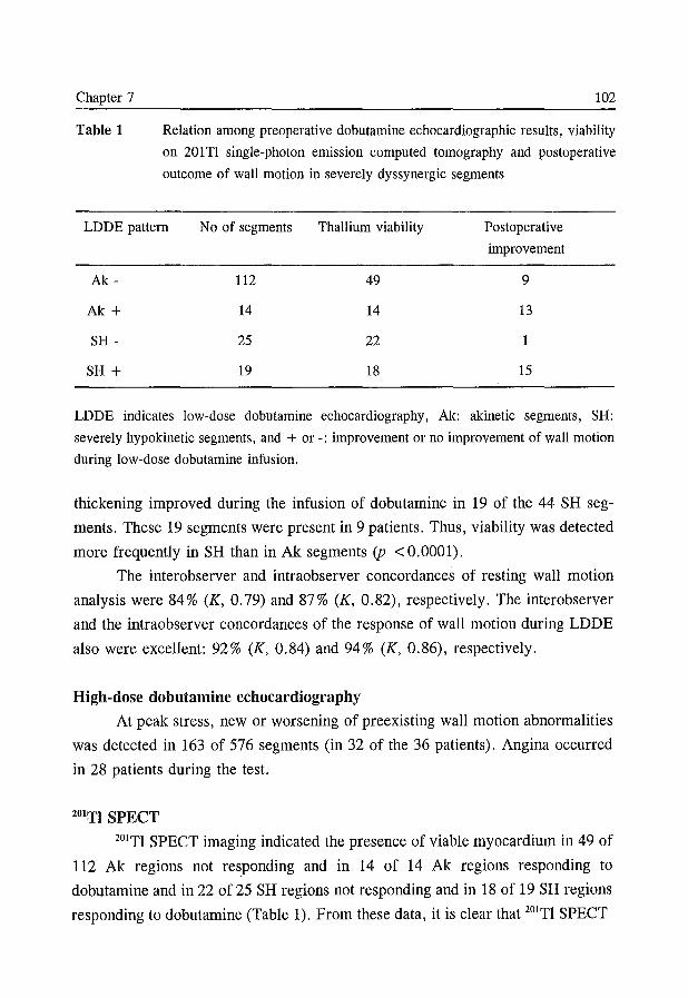
Chapter 7
Table 1
102
Relation among preoperative dobutamine echocardiographic results, viability
on 201 Tl single-photon emission computed tomography and postoperative
outcome of wall motion in severely dyssynergic segments
LDDE pattern No of segments Thallium viability Postoperative
improvement
Ak - 112 49 9
Ak + 14 14 13
SH - 25 22 1
SH + 19 18 15
LDDE indicates low-dose dobutamine echocardiography, Ak: akinetic segments, SH:
severely hypokinetic segments, and + or -: improvement or no improvement of wall motion
during low-dose dobutamine infusion.
thickening improved during the infusion of dobutamine in 19 of the 44 SH seg
ments. These 19 segments were present in 9 patients. Thus, viability was detected
more frequently in SH than in Ak segments (p <0.0001).
The interobserver and intraobserver concordances of resting wall motion
analysis were 84% (K, 0.79) and 87% (K, 0.82), respectively. The interobserver
and the intraobserver concordances of the response of wall motion during LDDE
also were excellent: 92% (K, 0.84) and 94% (K, 0.86), respectively.
High-dose dobutamine echo cardiography
At peak stress, new or worsening of preexisting wall motion abnormalities
was detected in 163 of 576 segments (in 32 of the 36 patients). Angina occurred
in 28 patients during the test.
201TI SPECT
201TI SPECT imaging indicated the presence of viable myocardium in 49 of
112 Ak regions not responding and in 14 of 14 Ak regions responding to
dobutamine and in 22 of 25 SH regions not responding and in 18 of 19 SH regions
responding to dobutamine (Table 1). From these data, it is clear that 201TI SPECT
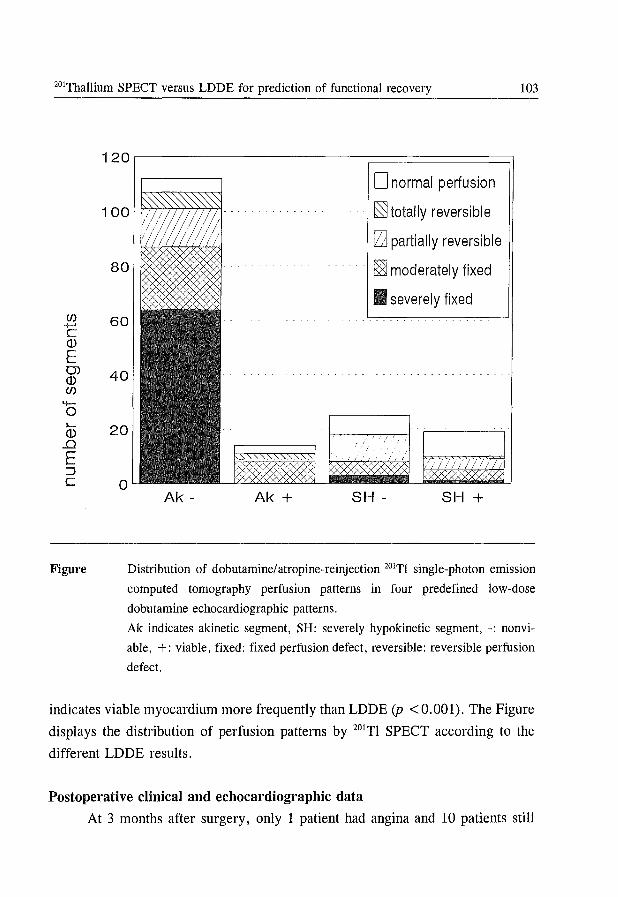
20lrhallium SPEer versus LDDE for prediction of functional recovery 103
(f) +-' C (])
E 0) (]) (f) -0 "-(])
..0 E ::i C
Figure
120
o normal perfusion
~ totally reversible
rza partially reversible
~ moderately fixed
• severely fixed 60
40
20
0 Ak - Ak + SH - SH +
Distribution of dobutamineiatropine-reinjection 201T! single-photon emission
computed tomography perfusion patterns in four predefined low-dose
dobutamine echocardiographic patterns.
Ak indicates akinetic segment, SH: severely hypokinetic segment, -: nonvi
able, +: viable, fixed: fixed perfusion defect, reversible: reversible perfusion
defect.
indicates viable myocardium more frequently than LDDE (p < 0.001). The Figure
displays the distribution of perfusion patterns by 20lTI SPECT according to the
different LDDE results.
Postoperative clinical and echo cardiographic data
At 3 months after surgery, only 1 patient had angina and 10 patients still
Chapter 7 104
complained of dyspnea on effort.
Improvement of regional wall motion
Resting echocardiograms at 3-months postoperative follow-up revealed an
improvement of wall motion in 38 (22 %) of the 170 dyssynergic segments. The
improvement was found in 22 (17%) of the 126 Ak segments (change to SH in 6
segments, to moderate hypokinesis in 13 segments and to normal in 3 segments)
and in 16 (36 %) of the 44 SH segments (change to moderate hypokinesis in 8
segments and to normal in 8 segments) (p = .02).
Reduction of dobutamine induced myocardial ischemia
Of 32 patients who underwent a high-dose dobutamine stress test during
follow-up, 3 had angina at peak stress. New or worsened wall motion abnormali
ties were detected in 10 patients and in 23 of 512 LV segments.
Prediction of regional improvement
Postoperative improvement occurred in 28 of the 33 segments that improved
during LDDE and in only 10 of 137 segments that did not improve (Table 1). Of
the segments judged to be viable by LDDE, postoperative improvement occurred
in 79% of SH segments and in 93% of Ak segments.
201TI SPECT detected the presence of viable myocardium in 32 of the 33 matched
segments considered viable by LDDE. Despite the frequent indication of viability
by 201TI SPECT in the Ak segments unresponsive to dobutamine (44%),
improvement after surgery was found in only 8% of these segments. Similarly,
wall thickening improved after revascularization in only 1 of the 25 SH regions
unresponsive to dobutamine, despite signs of viability by 201TI SPECT in 22 of
these 25 segments. Table 2 shows the predictive accury with 95 % CI of the two
methods for the postoperative improvement of SH and of AK segments.
Global left ventricular function
The WMSI revealed that there were no significant differences before and
after coronary artery bypass graft surgery in either the subset in whom myocardial
viability was predicted by LDDE (13 patients) (2.6 ± 0.5 versus 2.4 ± 0.4) or in
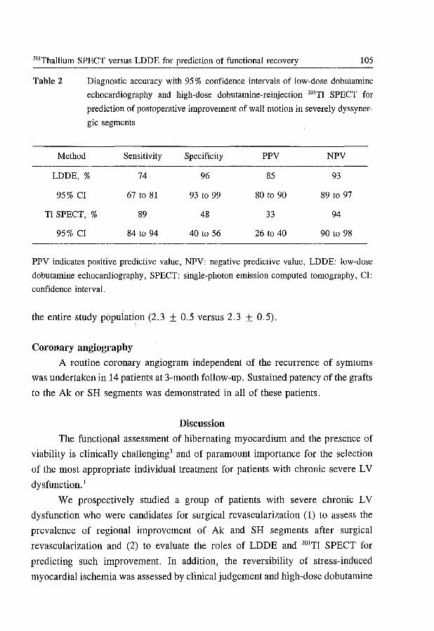
201Thallium SPECT versus LDDE for prediction of functional recovery 105
Table 2 Diagnostic accuracy with 95 % confidence intervals of low-dose dobutamine
echocardiography and high-dose dobutamine-reinjection 201TI SPECT for
prediction of postoperative improvement of wall motion in severely dyssyner-
gic segments
Method Sensitivity Specificity PPV NPV
LDDE, % 74 96 85 93
95% CI 67 to 81 93 to 99 80 to 90 89 to 97
Tl SPECT, % 89 48 33 94
95% CI 84 to 94 40 to 56 26 to 40 90 to 98
PPV indicates positive predictive value, NPV: negative predictive value, LDDE: low-dose
dobutamine echocardiography, SPECT: single-photon emission computed tomography, CI:
confidence interval.
the entire study population (2.3 ± 0.5 versus 2.3 ± 0.5).
Coronary angiography
A routine coronary angiogram independent of the recurrence of symtoms
was undertaken in 14 patients at 3-month follow-up. Sustained patency of the grafts
to the Ak or SH segments was demonstrated in all of these patients.
Discussion
The functional assessment of hibernating myocardium and the presence of
viability is clinically challenging3 and of paramount importance for the selection
of the most appropriate individual treatment for patients with chronic severe LV
dysfunction. 1
We prospectively studied a group of patients with severe chronic LV
dysfunction who were candidates for surgical revascularization (1) to assess the
prevalence of regional improvement of Ak and SH segments after surgical
revascularization and (2) to evaluate the roles of LDDE and 201TI SPECT for
predicting such improvement. In addition, the reversibility of stress-induced
myocardial ischemia was assessed by clinical judgement and high-dose dobutamine
Chapter 7 106
stress test.
In this series of patients, improvement of regional function after revasculari
zation was found in 22 % of Ak and SH segments. This percentage is lower than
that of other series?·9.10 and might be related to the selection of patients with
severely impaired LV function in a tertiary referral center with an ongoing heart
transplantation program.
We have demonstrated that preoperative LDDE is both a sensitive (28 of 38
segments) and a specific (127 of 132 segments) predictor of postoperative outcome
of regional myocardial function. The pattern of improvement of wall thickening in
severely dyssynergic regions during the infusion of low-dose dobutamine was
found to be a reliable predictor of functional recovery of wall motion after
successful and uncomplicated surgical revascularization, with a positive predictive
value of 85% (95% CI, 80% to 90%), whereas the pattern of Ak or SH
unresponsive to low-dose dobutamine is indicative of nonviable tissue and offers
a negative predictive value of 93% (95% CI, 89% to 97%).
2°!TI SPECT (matched for echocardiographic segments) indicated the
presence of viable myocardium more frequently than LDDE (103 of 170 versus 33
of 170). However, this imaging technique appears to be less suitable than LDDE
to predict the postoperative improvement of regional wall motion in patients with
severe LV dysfunction. In particular, the high prevalence of viability detected
before surgery by 2°!TI SPECT and the low prevalence of postoperative functional
improvement result in a low specificity and in a low positive predictive value
(Table 2).
Overestimation of myocardial viability by perfusion scintigraphy may relate
to several factors. First, scintigraphy may detect islands of jeopardized vital
myocardial cells of inadequate size to revert LV dysfunction despite successful
revascularization. Second, tethering by scar tissue may restrict the improvement
in wall motion of adjacent viable segments. Third, functional recovery may not be
complete by 3 months ("embalmed myocardium").!? Finally, since subendocardial
layers playa major role in wall motion, a necrosis limited to the subendocardium
may result in severe dyssynergy despite the presence of viable myocardium in the
subepicardiallayers.!8
In our study group, there was no significant postoperative improvement of
201rhallium SPECr versus LDDE for prediction of functional recovery 107
global LV function; however, bypass surgery alleviated myocardial ischemia, since
the number of patients with angina and the extent of stress induced ischemia were
greatly reduced. This is consistent with the high postoperative patency rate of
bypass coronary grafts and confirmed the usefulness of dobutamine stress
echocardiography in the assessment of stress-induced myocardial ischemia after
coronary revascularization. 12
Previous studies Although several studies6-8.11.19 have addressed the role of dobutamine
echocardiography for the assessment of LV functional recovery in patients with
recent myocardial infarction, few data are available regarding its predictive value
for postrevascularization functional improvement.9.10 In two previous studies on
postrevascularization recovery, Marzullo et al 9 and Cigarroa et al lO reported a
higher incidence of wall thickening during low-dose dobutamine in Ak regions
(47% and 39%, respectively, compared with 11 % in the present study). This
discrepancy may relate to different methodologies (absence of subclassification for
SH segments in their studies) and patient selection (inclusion in the present study
of patients with more severe and more longstanding ventricular dysfunction, where
stunned myocardium is less likely to be present). Considering the value of LDDE
in predicting postoperative functional outcome, our findings are in agreement with
those of the two previous reports.
Study limitations
First, the number of viable Ak and SH segments identified in the present
study was limited despite our analysis of 608 segments both before and after
coronary bypass graft surgery. This, however, reflects our stringent inclusion
criteria and our strict method of analysis (panel review with simultaneous, quad
screen format).
Second, we arbitrarely timed the outcome of dyssynergic segments 3 months
after surgical revascularization. However, it cannot be excluded that functional
improvement can also occur later.
Finally, we focused on the postoperative phase of regional wall motion. We are
aware that the improvement in a limited area of myocardium can be clinically not
Chapter 7 108
relevant to global LV function. However, this was not the primary aim of the
study.
Conclusions
Our observations in the setting of severe chronic LV dysfunction indicate
that 1) wall thickening during low-dose dobutamine in Ak segments is infrequent,
2) responsiveness of Ak and SH segments to low-dose dobutamine is both a
specific and a sensitive predictor of postrevascularization functional improvement,
and 3) compared with LDDE, 201Tl SPECT has an equivalent sensitivity for the
prediction of postoperative myocardial functional improvement but a lower
specificity. Thus, LDDE should be the preferred imaging technique for predicting
the functional outcome of patients with severe LV dysfunction who are under
consideration for coronary artery bypass graft surgery.
Acknowledgements
We are deeply grateful to Eric Boersma, MSc for the statistical assistance.
Also, we appreciate the co-operation of the referring Rijnmond cardiologists.
References
1. Elefteriades JA, Tolis G, Levi E, Mills LK, Zaret BL. Coronary artery bypass
grafting in severe left ventricular dysfunction: excellent survival with improved
ejection fraction and functional state. I Am ColI CardioI1993;22:1411-1417.
2. Bonow RO, Dilsizian V. Thallium-201 and technetium-99-m-sestamibi for assessing
viable myocardium. I Nucl Med 1992;33:815-818.
3. Dilsizian V, Bonow RO. Current diagnostic techniques of assessing viability in
patients with hibernating and stunned myocardium. Circulation 1993 ;87: 1-20.
4. Zaret BL, Wackers FI. Nuclear Cardiology (First of Two Parts). New Engl I Med
1993;329:775-783.
5. Zaret BL, Wackers FI. Nuclear Cardiology (Second of Two Parts). New Engl I Med
1993;329:855-863.
6. Pierard LA, De Landsheere CM, Berthe C, Rigo P, Kulbertus HE. Identification of
viable myocardium by echocardiography during dobutamine infusion in patients with
myocardial infarction after thrombolytic therapy: comparison with positron emission
tomography. I Am ColI CardioI1990;15:1021-1031.
7. Barilla F, Gheorghiade M, Alam M, Khaja F, Goldstein S. Low-dose dobutamine in
201Thallium SPECT versus LDDE for prediction of functional recovery 109
patients with acute myocardial infarction identifies viable but not contractile myocar
dium and predicts the magnitude of improvement in wall motion abnormalities in
response to coronary revascularization. Am Heart J 1991;122:1522-1531.
8. Smart SC, Sawada S, Ryan T, et al. Low-dose dobutamine echocardiography detects
reversible dysfunction after thrombolytic therapy of acute myocardial infarction.
Circulation 1993;88:405-415.
9. Marzullo P, Parodi 0, Reisenhofer B, et al. Value of rest thallium-201ltechnetium-
99m sestamibi scans and dobutamine echocardiography for detecting myocardial
viability. Am J Cardiol 1993;71:166-172.
10. Cigarroa CG, de Filippi CR, Brickner ME, Alvarez LG, Wait MA, Grayburn PA.
Dobutamine stress echocardiography identifies hibernating myocardium and predicts
recovery of left ventricular function after coronary revascularization. Circulation
1993;88:430-436.
11. Salustri A, Elhendy A, Garyfallydis P, et al. Prediction of improvement of ventricular
function after first acute myocardial infarction using low-dose dobutamine stress
echocardiography. Am J Cardiol 1994;74:853-856.
12. McNeill AJ, Fioretti PM, EI-Said EM, Salustri A, de Fey tel' PJ, Roelandt JRTC.
Dobutamine stress echocardiography before and after angioplasty. Am J Cardiol
1992;69:740-745.
13. Sawada SG, Segar DS, Ryan T, et al. Echocardiographic detection of coronary artery
disease during dobutamine infusion. Circulation 1991; 83: 1605-1614.
14. Cornel JH, Arnese M, Forster T, Postma-Tjoa J, Reijs AEM, Fioretti PM. Potential
and limitations of Tc-99m Sestamibi scintigraphy for the diagnosis of myocardial
viability. Herz 1994; 19: 19-27.
15. Pozzoli MMA, Fioretti PM, Salustri A, Reijs AEM, Roelandt JRTC. Exercise
echocardiography and technetium-99m MIBI single-photon emission computed
tomography in the detection of coronary artery disease. Am J Cardiol 1991;67:350-
355.
16. Forster T, McNeill AJ, Salustri A, et al. Simultaneous dobutamine stress echocardio
graphy and technetium-99m isonitrile single-photon emission tomography in patients
with suspected coronary artery disease. J Am Coil CardioI1993;21:1591-1596.
17. Bashour TT, Mason DT. Myocardial hibernation and "embalment". Am Heart 1990;
119:706-708.
18. Sklenar J, Ismail S, Villanueva FS, Goodman NC, Glasheen WP, Kaul S.
Dobutamine echocardiography for determining the extent of myocardial salvage after
reperfusion: an experimental evaluation. Circulation 1994; 90: 1502-1512.
19. Picano E, Marzullo P, Gigli G, et al. Identification of viable myocardium by

Chapter 7 110
dipyridamole-induced improvement in regional left ventricular function assessed by
echocardiography in myocardial infarction and comparison with thallium scintigraphy
at rest. Am J Cardiol 1992;70:703-710.
Addendum 111
Addendum
Correspondence
Prediction of improvement of regional left ventricular function after surgical
revascularization.
Circulation 1996;93:396-397
To the editor:
Arnese et all in the article titled: "Prediction of improvement of regional left
ventricular function after surgical revascularization" concluded that compared with
low-dose dobutamine echocardiography (LDDE), 20lThallium single photon
emission computed tomography (SPECT) imaging has an equivalent sensitivity for
the prediction of postoperative myocardial functional improvement but a lower
specificity. Thus, LDDE should be the preferred imaging technique for predicting
the functional outcome of patients with severe left ventricular dysfunction who are
under consideration for coronary artery bypass surgery. In this article, the
specificity and positive predictive value of dobutamine 20lThallium imaging with
reinjection were significantly lower (48 % and 33 %, respectively) compared with
LDDE.
The lower specificity and positive predictive value compared with other
studies2-4 may be attributable to the fact that moderate fixed defects on 20lThallium
imaging were considered viable.
As shown in the Figure, the largest percentage of "false-positive"
20lThallium results were noted in the segments with moderate fixed defects.
Although it is true that viabilitiy has been shown in fixed defects of mild or
moderate severity using positron emission tomography imaging with I 8FDG4 ,
resting wall motion may not improve post-revascularization in these segments if
they are composed of an admixture of scar tissue and viable tissue (normally
perfused at rest). However, other benefits such as symptomatic improvement and
reduction of ventricular remodeling may be derived from revascularizing these
segments. The specificity and positive predictive value of viability assessment by
Chapter 7 112
SPECT 201Thallium imaging may be significantly better if other clinical outcomes
are considered.
The authors demonstrate a difference in the ability to predict the functional
outcome of myocardial segments between dobutamine echocardiography and
201Thallium imaging. Consequently, it is inappropriate to conclude that LDDE
should be the preferred imging technique for predicting the functional outcome of
patients with severe left ventricular dysfunction who are under consideration for
coronary artery bypass surgery.
Olakunle O. Akinboboye, MD
Columbia University College of Physicians and Surgeons
New York, U.S.A
References
1. Arnese M, Cornel JH, Salustri A, et al. Prediction of improvement of regional left
ventricular function after surgical revascularization: a comparison of low-dose
dobutamine echocardiography with 201Tl Single-photon emission computed tomograp
hy. Circulation 1995;91:2748-2752.
2. Dilsizian V, Rocco PT, Freedman NM, Leon MB, Bonow RO. Enhanced detection
of ischemic but viable myocardium by the reinjection of thallium after stress
redistribution imaging. N Engl J Med 1990;323:141-146.
3. Tamaki N, Ohtani H, Yamashita K, et al. Metabolic activity in the areas of new fill
in after thallium-20l reinjection: comparison with positron emission tomography using
fluorine- 18-deoxyglucose. J Nuel Med 1991;32:673-678.
4. Bonow RO, Dilsizian V, Cuocolo A, Bacharach SL. Identification of viable
myocardium in patients with chronic coronary artery disease and left ventricular
dysfunction: comparison of thallium scintigraphy with reinjection and PET imaging
with 18F-fluorodeoxyglucose. Circulation 1991 ;83 :26-37.
Response:
We appreciate the interest of Dr Akinboboye in our work. In response to his
comments, we want to recall the aim of our study. 1 This study was undertaken to
compare 201Tl stress-reinjection single photon emission computed tomography with
Addendum 113
low-dose dobutamine echocardiography (LDDE) in the prediction of improvement
of regional left ventricular function after surgical revascularization. The study
group comprised mostly patients with severe regional and global chronic left
ventricular dysfunction. To our knowledge only three other studies,2-4 including
Ohtani et aP but not Bonow et aV have used 201TI stress-reinjection protocols for
the prediction of improvement of regional left ventricular dysfunction after
revascularization. In these, a limited number of patients (n = 55) was studied and
the majority of patients had well or relatively well preserved left ventricular
function. A highly variable specifity from 40%4 to 80%2 has been reported, and
in the largest series of patients, studied by Ohtani et al,3 25 % of the preoperatively
hypoperfused segments had normal wall motion but were still included in the
analysis. Furthermore, in these studies no distinction was made between
moderately and severely fixed perfusion defects, possibly explaining the variability
of the specificity of the test.
Indeed as shown in the Figure in our article, the reported specificity and
positive predictive value may be influenced by the fact that moderately fixed
perfusion defects, although in comparative "state-of- the-art" studies with FDG
positive emission tomography (PET) mostly viable, were considered viable. For
predicting reversibility of wall motion abnormalities this may not be the case.
Therefore, we re-analyzed our data with the assumption of nonviability of
moderately fixed defects. Of the 40 moderately fixed defects, 13 recovered
postoperatively, resulting in an increase in specificity from 48 % to 68 % but with
a much lower sensitivity of 55 % .
It is highly speculative to suggest that revascularization of moderately fixed
defects may lead to symptomatic improvement or reduction of ventricular
remodeling. There are no studies to support this hypothesis. On the contrary, PET
data show that only patients with jeopardized myocardium (mismatch pattern)
benefit from revascularisation, in terms of symptomatic improvement or
prognosis. 6
About the last comment, we agree that our statement in the conclusions
("low dose dobutamine echocardiography should be the preferred imaging
technique for predicting the functional outcome of patients") is inappropriate based
on the reported results. However, preliminary results from our institution suggest
Chapter 7 114
that an improvement of global left ventricular function is highly likely to occur
when at least 25 % of the myocardium is akinetic and is responsive to dobutamine. 7
Jan Rein Cornel, MD
Paolo M Fioretti, MD
Department of Cardiology
University Hospital Dijkzigt
Rotterdam, the Netherlands
References
1. Arnese M, Cornel JH, Salustri A, et al. Prediction of improvement of regional left
ventricular function after surgical revascularization: a comparison of low-dose
dobutamine echocardiography with 201TI Single-photon emission computed tomograp
hy. Circulation 1995;91:2748-2752.
2. Dilsizian V, Rocco PT, Freedman NM, Leon MB, Bonow RO. Enhanced detection
of ischemic but viable myocardium by the reirtiection of thallium after stress
redistribution imaging. N Engl J Med 1990;323:141-146.
3. Ohtani H, Tamaki N, Yonekura Y, et al. Value of thallium-201 reinjection after
delayed SPECT imaging for predicting reversible ischemia after coronary artery
bypass grafting. Am J Cardiol 1990;66:394-399.
4. Tamaki N, Ohtani H, Yamashita K, et al. Metabolic activity in the areas of new fill
in after thallium-201 reinjection: comparison with positron emission tomography using
fluorine- 18-deoxyglucose. J Nucl Med 1991;32:673-678.
5. Bonow RO, Dilsizian V, Cuocolo A, Bacharach SL. Identification of viable
myocardium in patients with chronic coronary artery disease and left ventricular
dysfunction: comparison of thallium scintigraphy with reinjection and PET imaging
with 18F-fluorodeoxyglucose. Circulation 1991 ;83 :26-37.
6. Di Carli MF, Davidson M, Little R, et al. Value of metabolic imaging with positron
emission tomography for evaluating prognosis in patients with coronary artery disease
and left ventricular dysfunction. Am J Cardiol 1994;73:527-533.
7. Cornel JH, Bax JJ, Fioretti PM, Visser FC. Prediction of improvement of regional
left ventricular functi()n after revascularization: FDG/TI SPECT versus dobutamine
echocardiography. J Nucl Med 1995;36:35P. Abstract.
Chapter 8
18F -Fluorodeoxyglucose SPECT for the Prediction of Reversibility of
Ventricular Dysfunction After Revascularization
A Comparison with Rest-Redistribution 201Thallium SPECT
JH Cornel, JJ Bax*, FC Visser*, PM Fioretti, JM Huitink*, A van Lingen**,
o Kamp*, GJJ Teule**, JRTC Roelandt, CA Visser*.
From the Department of Cardiology, University Hospital Rotterdam-Dijkzigt,
Rotterdam, and the Departments of Cardiology* and Nuclear Medicine**, Free
University Hospital Amsterdam, the Netherlands.
Submitted for publication
Chapter 8 116
Abstract
Background
Improvement of left ventricular dysfunction after coronary revascularization
can be predicted by positron emission tomography using '8F-fluorodeoxyglucose
(FDG) as a metabolic marker. We evaluated the use of FDG in combination with
single photon emission computed tomography (SPECT) and compared this
technique with rest-redistribution 2°'Thallium eO'Tl) SPECT to predict functional
recovery after revascularization.
Methods
Fifty-five patients with regional wall motion abnormalities were studied with
FDG SPECT during hyperinsulinemic glucose clamping. Subsequently a resting
2°'Tl SPECT was performed to assess myocardial perfusion, 24 patients also
underwent redistribution 2°'Tl SPECT. Regional contractile function was evaluated
with echocardiography, using a 13-segment model and a 4-grade scoring system,
before and 3 months after revascularization. Left ventricular ejection fraction
(L VEF) was also determined, either by echo (n = 37) or by radionuclide
ventriculography (n = 18). The SPECT data were analyzed quantitatively and the
optimal diagnostic accuracy for functional recovery was determined using receiver
operating characteristic analysis.
Results
One third of dysfunctional segments showed improvement of wall thickening
at follow-up. The FDG approach reached a sensitivity of 85 % (95 % confidence
interval [CI], 78% to 92%) and a specificity of 75% (95% CI, 69% to 81 %) to
predict regional functional recovery. Similar results were obtained in the subgroup
of patients with a L VEF of :::; 30 % (n = 22). L VEF improved from 28 ± 8 % to
35 ± 9 % (p < 0.05) in patients with 23 viable segments on FDG SPECT. In
patients with :::;2 viable segments LVEF remained unchanged. In direct compari
son FDG SPECT showed a better specificity (77 % (95 % CI, 67 % to 87 %) versus
57% (95% CI, 45% to 69%» as compared to rest-redistribution 2°'Tl SPECT.
Stepwise logistic regression analysis of the FDG and 2°'Tl rest-redistribution data
showed that increased FDG uptake in perfusion defects was the best predictor of
FDG SPECr for prediction of functional recovery 117
functional recovery. All other parameters did not contribute independently.
Conclusions
This study shows that FDG SPECT is superior to rest-redistribution 20lTl
SPECT in the identification of patients in whom regional and global LV function
can improve after revascularization. The SPECT approach may contribute to a
more routine use of FDG for management of patients with chronic ischemic LV
dysfunction.
Introduction
Heart failure is an increasing problem in cardiology due to its rising
incidence over the last decade. In coronary artery disease impaired left ventricular
(LV) function is not necessarily an irreversible process as recovery of LV function
after revascularization has been demonstrated. The concepts of hibernation and
repetitive stunning have been introduced to explain such improvement. 1,2 Nesto et
al have described improvement in left ventricular ejection fraction (L VEF) and
favourable prognosis after revascularization of viable myocardium. 3 Thus, the
differentiation between viable and scarred myocardium may have important clinical
implications.
Identification of viable myocardium is possible with positron emission
tomography (PET) using F8F]fluorodeoxyglucose (FDG). 4,5 Reversibility of
regional wall motion abnormalities after revascularization has been demonstrated
in regions with normal perfusion and in hypoperfused regions with relatively
preserved FDG uptake (FDG-perfusion mismatch). In contrast, regions with
hypoperfusion and concomitantly decreased FDG uptake (FDG-perfusion match)
did not show recovery of function. 6-12 Improvement of global LV function has also
been observed in patients with viable myocardium on FDG PET. 6,1l,12
Relatively few PET devices are avialable for clinical use. Therefore, several
laboratories have evaluated the feasibility of imaging myocardial FDG uptake with
SPECT using 511 keY collimatorsY-18 No data however are available on the
diagnostic value of FDG SPECT to predict functional recovery after revas
cularization. Therefore, the aim of the present studywas to evaluate whether FDG
SPECT can predict improvement of resting wall motion abnormalities after
Chapter 8 118
revascularization. For comparison of regional FDG uptake with regional perfusion
early resting 201Tl SPECT was performed. 19 In addition the results were compared
with rest-redistribution 20lTl SPECT, which is currently one of the most widely
used techniques to assess myocardial viability. 20-24
Methods
Study population
We prospectively studied 55 consecutive patients referred for coronary
revascularization: all had chronic regional wall motion abnormalities at rest obser
ved on LV angiography and on two-dimensional echocardiography. Forty-five
patients (82%) underwent coronary artery bypass graft surgery (CABG), 10
patients (18%) a percutaneous transluminal coronary angioplasty (PTCA). The
time-interval of the SPECT study to revascularization was 2.0 ± 2.7 months. The
SPECT studies were performed in patients who were already scheduled for
revascularization. The results did not influence patient management.
Study protocol
All patients underwent an early resting 20lTl SPECT, followed by FDG
SPECT during a hyperinsulinemic euglycemic clamp. In 24 of these patients rest
redistribution 20lTl SPECT was performed. Improvement of regional wall motion
was determined from serial resting echocardiograms before and 3.4 ± 2.5 months
after revascularization. To assess improvement of L VEF 18 patients had
radionuclide ventriculography before and after revascularization, whereas in the
remaining 37 patients LVEF was calculated from echocardiograms. The SPECT
studies were performed preferably on the same day, with a maximum interval of
1 week. Medication was continued during the study. Each patient gave informed
consent to the study protocol that was approved by the ethical committees of the
participating hospitals.
201TI SPECT
To delineate regional myocardial perfusion a resting 20lTl SPECT was
performed as described previously. 18,19 After an overnight fast, a single dose of 111
MBq (3 mCi) 20lTl chloride was administered intravenously at rest. Imaging was
FDG SPECT for prediction of functional recovery 119
started 10 to 15 minutes after injection. In 24 patients a redistribution image was
acquired, 4 hours after tracer injection. A large-field-of-view rotating dual head
gamma camera (ADAC Laboratories, Milpitas CA) was used, equipped with a
low-energy high-resolution (LEHR) collimator. The energy was centered on the
89 keY photon peak with a 20% window and on the 167 keY photon peak with a
20 % window. The dual head gamma camera system was rotated over 360 0, collec
ting 64 views for 30 seconds each, resulting in a total data collection time of 16
minutes. Data were stored in a 64 x 64, 16-bit matrix. From the raw scintigraphic
data 6 mm-thick (1 pixel) transaxial slices were reconstructed by filtered back
projection using a Hanning filter (fc = 0.63 cycle/cm). Slices were not corrected
for attenuation. Further reconstruction yielded long- and short-axis projections per
pendicular to the heart-axis.
FDG SPECT
FDG (tlh of 18F = 110 min) was produced and synthesized at the cyclotron
facility of the Free University Amsterdam according to previously described
methods. 25 The radiochemical purity of the product was 98 %. After an overnight
fast, subjects underwent a hyperinsulinemic glucose clamp26 to standardize
metabolic conditions throughout FDG SPECT. FDG (185 MBq, 5 mCi) was
injected after 60 min of clamping, another 45 min was allowed for myocardial
FDG uptake. 27 ,28 Data acquisition was performed with the same camera system as
described for 20lTl. Especially designed collimators were used for the detection of
511 keY photons (van Mullekom, Nuclear Fields, Boxmeer, The Netherlands).29
Reconstruction of data was identical to that of 201TI SPECT.
Hyperinsulinemic euglycemic clamping
For this purpose, 2 catheters were placed in the left and right antecubital
veins. One catheter was used for separate infusions of glucose and insulin. The
contralateral catheter was used for the administration of FDG and to draw plasma
samples to monitor glucose levels. Insulin (Human Velosulin, Novo Nordisk, 100
IE/ml) in 0.65% sodium chloride was used. To prevent adhesion of insulin to the
infusion system 3.8 ml human albumin (20 % Human Albumin, CLB) was added.
The insulin infusion rate was 100 mU/kg/hr and was not changed throughout
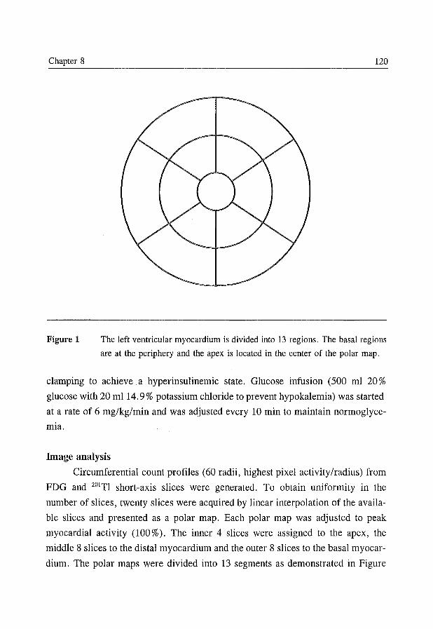
Chapter 8 120
Figure 1 The left ventricular myocardium is divided into 13 regions. The basal regions
are at the periphery and the apex is located in the center of the polar map.
clamping to achieve a hyperinsulinemic state. Glucose infusion (500 ml 20%
glucose with 20 mI14.9% potassium chloride to prevent hypokalemia) was started
at a rate of 6 mg/kg/min and was adjusted every 10 min to maintain normoglyce
mia.
Image analysis
Circumferential count profiles (60 radii, highest pixel activity/radius) from
FDG and 201TI short-axis slices were generated. To obtain uniformity in the
number of slices, twenty slices were acquired by linear interpolation of the availa
ble slices and presented as a polar map. Each polar map was adjusted to peak
myocardial activity (100 % ). The inner 4 slices were assigned to the apex, the
middle 8 slices to the distal myocardium and the outer 8 slices to the basal myocar
dium. The polar maps were divided into 13 segments as demonstrated in Figure
FDG SPECT for prediction of functional recovery 121
1. A region of normal perfusion was drawn on the early 201TI polar map. This
region was defined as the area with the highest 201TI uptake associated with normal
wall motion on the echocardiogram. The activity of this area was normalized to the
mean activity of the same area of a data base containing data of normal indivi
duals14 . All other polar map data were adjusted subsequently. The region of normal
perfusion was projected on the FDG polar map and the same normalization
procedure was followed. For this purpose another database containing data on FDG
activities in normal individuals was used. 14 Pixels with a 201TI activity < 2 SD
below the normal reference value were considered abnormal. The extent of the
perfusion defects within each of the 13 segments was expressed as the percentage
of abnormal pixels relative to the total number of pixels/segment. A perfusion
defect was considered present only if the extent exceeded 15 % of the myocardial
segment. The 201TI and FDG activities were expressed as percentage of the
corresponding normal reference values.
Definition of myocardial viability
For the FDG SPECT approach a segment was considered viable if perfusion
was normal or if FDG uptake was relatively increased in a perfusion defect. 4.5 The
cut-off level of increased FDG uptake was defined by receiver-operating charac
teristic (ROC) analysis.
For the rest-redistribution 201TI SPECT approach a segment was considered
viable if perfusion was normal, if significant redistribution occurred in a perfusion
defect or if segmental 201TI activity on the late image exceeded a threshold. 20-24 The
optimal cut-off levels for 201TI redistribution and 201TI activity were defined by
ROC analysis.
Assessment of improvement of regional dyssynergy
Regional wall motion was assessed by resting echocardiography before and
after revascularization. Echocardiographic images were recorded on videotape.
Four standard views of the left ventricle were obtained: parasternal long- and
short-axis views and apical two- and four-chamber views. The images were
reviewed off-line and consensus was achieved by 2 observers unaware of the
SPECT data. For comparison with the SPECT data the left ventricle was divided
Chapter 8 122
into 13 comparable segments. 30 Both inward wall motion and wall thickening were
analyzed. Each segment was assigned a wall motion score (WMS) of 0 to 3: nor
mal = 0, hypo kinetic = 1 (decreased endocardial excursion and systolic wall
thickening), akinetic = 2 (absence of endocardial excursion and systolic wall
thickening), and dyskinetic = 3 (paradoxic outward movement in systole). The
pre- and post-interventional images were compared on a segmental basis. Post
interventional improvement of regional wall motion was considered if systolic
thickening (hypo- or normokinesia) was detected in a prior a- or dyskinetic
segment, or if normal wall motion was detected in hypokinetic segments. Only
segments that were succesfully revascularized were analyzed. Wall thickening was
primarily utilized for the classification of septal wall motion, thereby preventing
the problem of paradoxical septal motion after CABGY We previously reported
a low level of inter- and intra-observer variability for the classification of resting
wall motion (agreement 84 % and 87 % ).32
Assessment of improvement of global ventricular function
Serial LVEF measurements were obtained using radionuclide ventriculo
graphy in 18 patients. This was performed at rest with the patient in supine
position after intravenous administration of 740 MBq of 99mtechnetium. Images
were acquired with a small-field-of-view gamma camera (Orbiter, Siemens Corp,
Iselin, NJ, USA), oriented in the 45 0 left anterior oblique position with a 5-10 0
caudal tilt. L VEF was calculated from the 45 0 left anterior oblique view by an
automated technique. In 37 patients LVEF was calculated by cross-sectional
echocardiography utilizing the apical biplanar Simpson's technique. 33
Improvement of global function after revascularization was defined as an
increase of L VEF 25%, exceeding our reproducibility limits.
Statistical analysis
All results were expressed as mean ± SD. Patient data were compared using
the Student's t-test for paired and unpaired data when appropriate. Comparison of
proportions was performed using chi-square analysis. A p-value of <0.05 was
considered significant. ROC analysis was performed to determine the optimal cut
off point for increased FDG uptake, 201Tl redistribution and 201Tl activity for the
FDG SPECT for prediction of functional recovery 123
prediction of functional recovery.
The best cut-off point of increased FDG uptake and 201Tl redistribution had
to exceed our reproducibility limits (> 4 % ). The optimal cut -off was chosen as the
maximal sum of sensitivity and specificity. Sensitivity, specificity, positive and
negative predictive values rely on the standard definition and are reported with
95 % confidence intervals (CI). Stepwise logistic regression analysis was performed
to determine which parameters (such as early and late 201TI activities, 201TI redistri
bution, FDG activity, increased FDG uptake in perfusion defects, extent of
perfusion defect, severity of resting wall motion abnormalities before revascu
larization) independently predict absence or presence of functional recovery.
Results
Patient characteristics
The clinical characteristics of the study population are presented in Table 1.
Fifty-one patients (93 %) had a previous infarction. The time-interval of infarction
to the SPECT study was more than 1 month in 93 % of the patients. Eight patients
had diabetes mellitus type II, which was well-regulated on oral medication. All
patients had significant coronary artery disease on angiography. They had a mean
L VEF of 39 %; 22 patients had an L VEF :::; 30 % .
Baseline characteristics
Of a total of 715 segments analyzed by echocardiography, 305 (43 %)
showed abnormal wall motion. The mean number of abnormal segments per patient
was 5.5 ± 3.1. Twenty-four segments were excluded from analysis due to
inadequate revascularization. Therefore, 281 dyssynergic segments were selected
for serial analysis. Hypokinesis was observed in 128 segments (46%), whereas 153
were akinetic/dyskinetic (54%). Forty-nine segments (17%) showed normal
perfusion on 201TI SPECT.
Prediction of regional functional recovery by FDG SPECT
Using recovery of wall motion abnormalities as "golden standard", 281
segments with abnormal wall motion at baseline were divided into 2 groups: seg
ments with improvement after revascularization (group I, n = 94) and segments
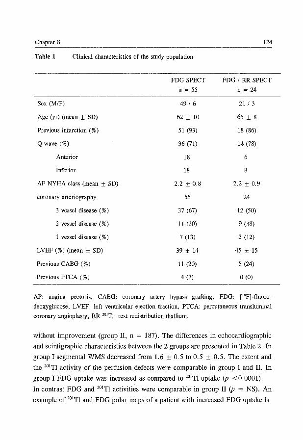
Chapter 8 124
Table 1 Clinical characteristics of the study population
FDG SPECT FDG / RR SPECT
n = 55 n = 24
Sex (M/F) 49/6 21 /3
Age (yr) (mean ± SD) 62 ± 10 65 ± 8
Previous infarction (%) 51 (93) 18 (86)
Q wave (%) 36 (71) 14 (78)
Anterior 18 6
Inferior 18 8
AP NYHA class (mean ± SD) 2.2 ± 0.8 2.2 ± 0.9
coronary arteriography 55 24
3 vessel disease (%) 37 (67) 12 (50)
2 vessel disease (%) 11 (20) 9 (38)
1 vessel disease (%) 7 (13) 3 (12)
LVEF (%) (mean ± SD) 39 ± 14 45 ± 15
Previous CABG (%) 11 (20) 5 (24)
Previous PTCA (%) 4(7) o (0)
AP: angina pectoris, CABG: coronary artery bypass grafting, FDG: [18F]-fluoro
deoxyglucose, LVEF: left ventricular ejection fraction, PTCA: percutaneous transluminal
coronary angioplasty, RR 201Tl: rest redistribution thallium.
without improvement (group II, n = 187). The differences in echocardiographic
and scintigraphic characteristics between the 2 groups are presented in Table 2. In
group I segmental WMS decreased from 1.6 ± 0.5 to 0.5 ± 0.5. The extent and
the 201TI activity of the perfusion defects were comparable in group I and II. In
group I FDG uptake was increased as compared to 201TI uptake (p <0.0001).
In contrast FDG and 201TI activities were comparable in group II (p = NS). An
example of 201TI and FDG polar maps of a patient with increased FDG uptake is
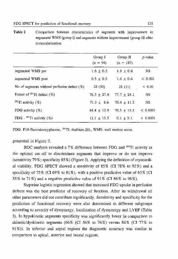
FDG SPECr for prediction of functional recovery 125
Table 2 Comparison between characteristics of segments with improvement in
segmental WMS (group I) and segments without improvement (group II) after
revascularization
Group I Group II p-value
(n = 94) (n = 187)
segmental WMS pre 1.6 ± 0.5 1.6 ± 0.6 NS
segmental WMS post 0.5 ± 0.5 1.6 ± 0.6 < 0.001
No of segments without perfusion defect (%) 28 (30) 21 (11) < 0.01
Extent of 20Iri defect (%) 76.3 ± 27.8 77.7 ± 24.1 NS
20ITl activity (%) 71.3 ± 8.6 70.4 ± 11.2 NS
FDG activity (%) 84.4 ± 13.0 70.5 ± 13.3 < 0.0001
FDG - 20Iri activity (%) 13.1 ± 13.5 0.1 ± 9.1 < 0.0001
FDG: F18-fluorodeoxyglucose, 20Irl: thallium-201, WMS: wall motion score.
presented in Figure 2.
ROC analysis revealed a 7 % difference between FDG and 20ITl activity as
the optimal cut-off to discriminate segments that improve or do not improve
(sensitivity 79 %; specificity 85 %) (Figure 3). Applying the definition of myocardi
al viability, FDG SPECT showed a sensitivity of 85 % (CI 78 % to 92 %) and a
specificity of 75 % (CI 69% to 81 %), with a positive predictive value of 63 % (CI
55% to 71 %) and a negative predictive value of 91 % (CI 86% to 96%).
Stepwise logistic regression showed that increased FDG uptake in perfusion
defects was the best predictor of recovery of function. After its withdrawal all
other parameters did not contribute significantly. Sensitivity and specificity for the
prediction of functional recovery were also determined in different subgroups
according to severity of dyssynergy, localization of dyssynergy and L VEF (Table
3). In hypokinetic segments specificity was significantly lower in comparison to
akinetic/ dyskinetic segments (66 % (CI 56 % to 76 %) versus 84 % (CI 77 % to
91 %». In inferior and septal regions the diagnostic accuracy was similar in
comparison to apical, anterior and lateral regions.
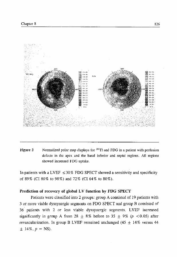
Chapter 8 126
AI'" ANT
90~% 90-96
B4<10 114-90
76-B4. FDG 7e-liI4
7'J.7f!, 72-78
6I:>-I':t 66-72
60-66
5~~G(I 5'~60
U-:H 413-54
42-U
36-U
)(l.~jG 30-31'1
24-)0 24-30
If!~24. ;U!~24
12-1Q 12-.1B ~-12
'-6 0-6
INF INF
Figure 2 Normalized polar map displays for 20lTl and FDG in a patient with perfusion
defects in the apex and the basal inferior and septal regions. All regions
showed increased FDG uptake.
In patients with a L VEF ~ 30 % FDG SPECr showed a sensitivity and specificity
of 89% (CI 80% to 98%) and 72% (CI 64% to 80%).
Prediction of recovery of global LV function by FDG SPECT
Patients were classified into 2 groups: group A consisted of 19 patients with
3 or more viable dyssynergic segments on FDG SPECr and group B consisted of
36 patients with 2 or less viable dyssynergic segments. LVEF increased
significantly in group A from 28 ± 8% before to 35 ± 9% (p <0.05) after
revascularization. In group B LVEF remained unchanged (45 ± 14% versus 44
± 14%, P = NS).
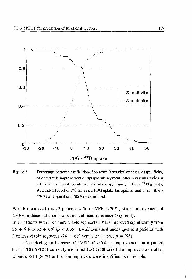
FDG SPECY for prediction of functional recovery 127
11'======~----------------~'~'~"~"~"~'~"~'------~
0.8 ~
0.6 ~
0.4
0.2 ~
- Sensitivity
.... Specificity
o~~~~----~----~----~----~----~----~----~
-30
Figure 3
-20 -10 o 10 20 30 40 50
FDG - 20lTI uptake
Percentage correct classification of presence (sensivity) or absence (specificity)
of contractile improvement of dyssynergic segments after revascularization as
a function of cut-off points over the whole spectrum of FDG - 201YI activity.
At a cut-off level of 7% increased FDG uptake the optimal sum of sensitivity
(79 %) and specificity (85 %) was reached.
We also analyzed the 22 patients with a L VEF ::::; 30 %, since improvement of
L VEF in these patients is of utmost clinical relevance (Figure 4).
In 14 patients with 3 or more viable segments L VEF improved significantly from
25 ± 6% to 32 ± 6% (p <0.05). LVEF remained unchanged in 8 patients with
2 or less viable segments (24 ± 6 % versus 25 ± 6 %, p = NS).
Considering an increase of L VEF of ~ 5 % as improvement on a patient
basis, FDG SPECT correctly identified 12112 (100%) of the improvers as viable,
whereas 8/10 (80%) of the non-improvers were identified as nonviable.
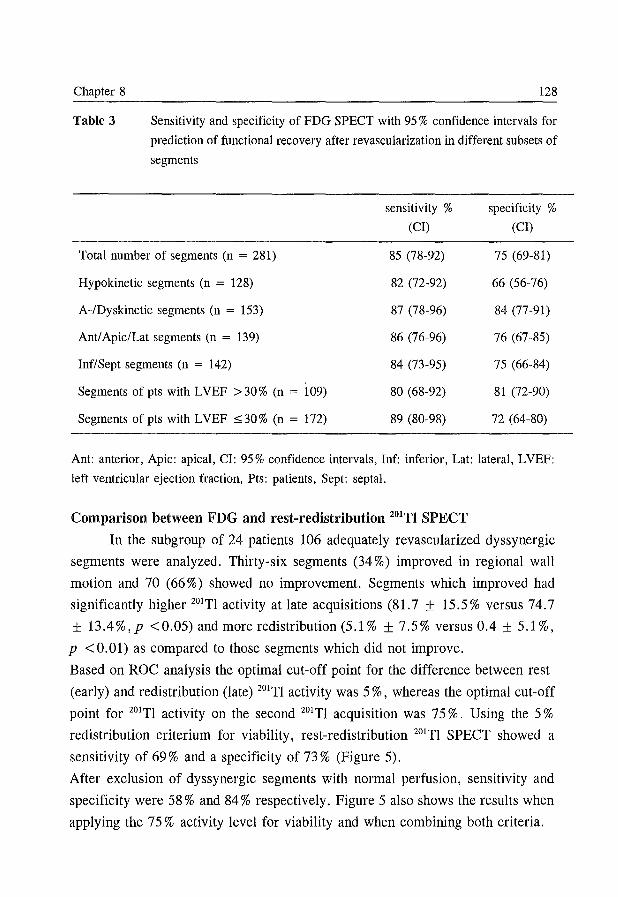
Chapter 8 128
Table 3 Sensitivity and specificity of FDG SPECT with 95 % confidence intervals for
prediction of functional recovery after revascularization in different subsets of
segments
sensitivity % specificity % (CI) (CI)
Total number of segments (n = 281) 85 (78-92) 75 (69-81)
Hypokinetic segments (n = 128) 82 (72-92) 66 (56-76)
A-lDyskinetic segments (n = 153) 87 (78-96) 84 (77-91)
Anti Apic/Lat segments (n = 139) 86 (76-96) 76 (67-85)
Inf/Sept segments (n = 142) 84 (73-95) 75 (66-84)
Segments of pts with L VEF > 30 % (n = 109) 80 (68-92) 81 (72-90)
Segments of pts with L VEF :::; 30 % (n = 172) 89 (80-98) 72 (64-80)
Ant: anterior, Apic: apical, CI: 95% confidence intervals, Inf: inferior, Lat: lateral, LVEF:
left ventricular ejection fraction, Pts: patients, Sept: septal.
Comparison between FDG and rest-redistribution 201Tl SPECT
In the subgroup of 24 patients 106 adequately revascularized dyssynergic
segments were analyzed. Thirty-six segments (34 %) improved in regional wall
motion and 70 (66 %) showed no improvement. Segments which improved had
significantly higher 201TI activity at late acquisitions (81.7 ± 15.5% versus 74.7
± 13.4 % ,p < 0.05) and more redistribution (5.1 % ± 7.5 % versus 0.4 ± 5.1 % ,
P <0.01) as compared to those segments which did not improve.
Based on ROC analysis the optimal cut-off point for the difference between rest
(early) and redistribution (late) 201TI activity was 5 %, whereas the optimal cut-off
point for 201TI activity on the second 201TI acquisition was 75%. Using the 5 %
redistribution criterium for viability, rest-redistribution 201TI SPECT showed a
sensitivity of 69 % and a specificity of 73 % (Figure 5).
After exclusion of dyssynergic segments with normal perfusion, sensitivity and
specificity were 58 % and 84 % respectively. Figure 5 also shows the results when
applying the 75 % activity level for viability and when combining both criteria.
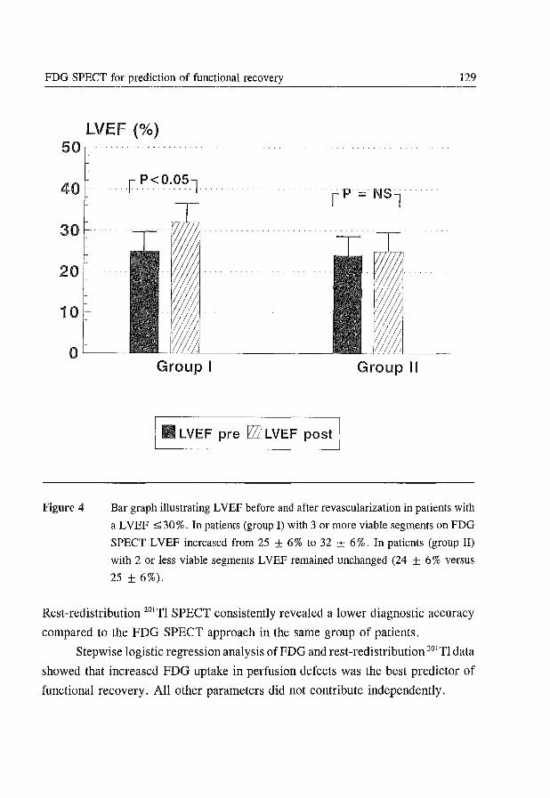
FDG SPECT for prediction of functional recovery 129
LVEF (%) 50
30
20
10
0'----
Figure 4
Group I Group II
I_ LVEF pre r2Zl LVEF post I
Bar graph illustrating L VEF before and after revascularization in patients with
a L VEF :::; 30 %. In patients (group I) with 3 or more viable segments on FDG
SPECT L VEF increased from 25 ± 6 % to 32 ± 6 %. In patients (group II)
with 2 or less viable segments L VEF remained unchanged (24 ± 6 % versus
25 ± 6%).
Rest-redistribution 201Tl SPECT consistently revealed a lower diagnostic accuracy
compared to the FOG SPECT approach in the same group of patients.
Stepwise logistic regression analysis of FOG and rest-redistribution 201Tl data
showed that increased FOG uptake in perfusion defects was the best predictor of
functional recovery. All other parameters did not contribute independently.
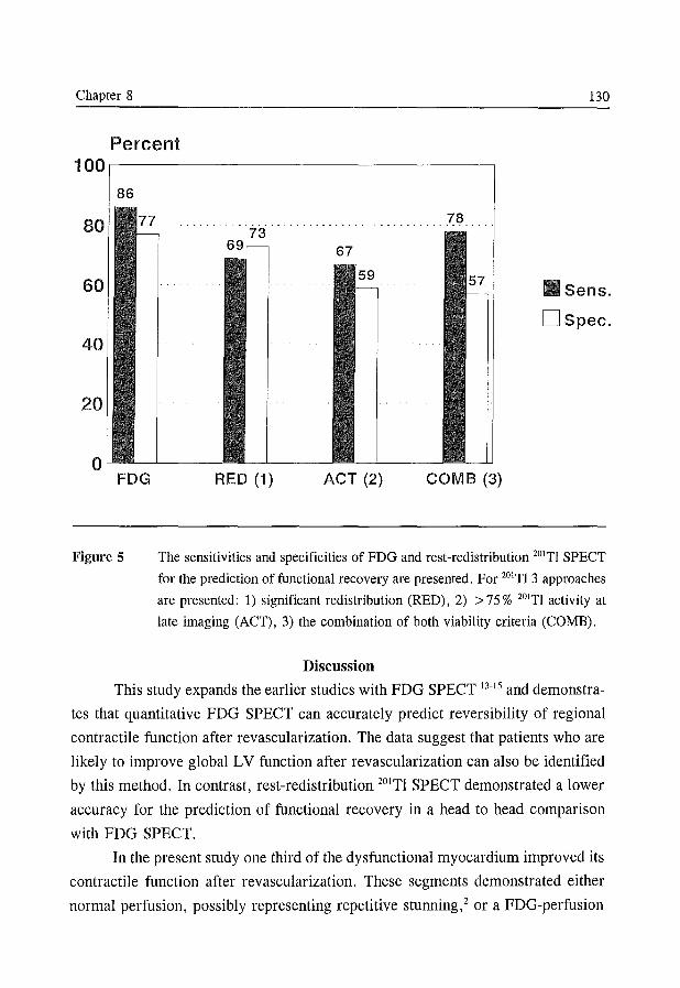
Chapter 8 130
Percent 100,-----------------------------~
86
FDG
Figure 5
78
67
Sens.
DSpec.
RED (1) ACT (2) COMB (3)
The sensitivities and specificities of FDG and rest-redistribution 201TI SPECT
for the prediction of functional recovery are presented. For 201TI 3 approaches
are presented: 1) significant redistribution (RED), 2) > 75 % 201Tl activity at
late imaging (ACT), 3) the combination of both viability criteria (COMB).
Discussion
This study expands the earlier studies with FDG SPECT 13-15 and demonstra
tes that quantitative FDG SPECT can accurately predict reversibility of regional
contractile function after revascularization. The data suggest that patients who are
likely to improve global LV function after revascularization can also be identified
by this method. In contrast, rest-redistribution 20lTl SPECT demonstrated a lower
accuracy for the prediction of functional recovery in a head to head comparison
with FDG SPECT.
In the present study one third of the dysfunctional myocardium improved its
contractile function after revascularization. These segments demonstrated either
normal perfusion, possibly representing repetitive stunning,2 or a FDG-perfusion
FDG SPECT for prediction of functional recovery 131
mismatch likely to represent hibernation. 1 The majority of segments without functi
onal improvement showed a FDG-perfusion match suggesting scar tissue. 4•5 The
results obtained with FDG SPECT .are in line with previous results obtained with
FDG PET. In FDG PET studies sensitivity ranged from 71 % to 100 % (mean 88 %)
in predicting functional recovery, whereas specificity ranged from 38 % to 86 %
(mean 74 %).34
Sensitivity and specificity of FDG SPECT are not different in the subgroup
of patients with severely depressed LV function (L VEF $ 30 %), in whom
prediction of functional recovery has the most clinical relevance. Patients with 3
or more dyssynergic but viable segments on FDG SPECT are likely to improve
global LV function. These results are also valid for the subgroup with L VEF
$ 30 %. This finding corroborates previous data6.'2.22 and emphasizes that a
substantial amount of viable myocardium is conditional for establishing
improvement of global LV function after revascularization.
Although absence of viable myocardium was highly predictive for absence
of recovery after revascularization, the presence of viable myocardium was less
predictive (positive predictive value of 63 %) for recovery of regional contractility.
Recently vom Dahl et al described comparable results using FDG PET. 12 Several
factors may have accounted for the lack of recovery of segments viable on FDG
SPECT. It has been demonstrated that morphological degeneration occurs in viable
myocytes before actual cell death takes place. 35.36 Marwick et al suggested that,
despite increased FDG uptake, some segments may contain myocytes that are too
severely injured to recover. 9 In addition an area with a mixture of viable myocytes
and necrotic myocardium may result in a FDG-perfusion mismatch pattern.
However, if the amount of viable cells in the area is relatively low, the segment
may not improve in function after revascularization. Furthermore attempted
revascularization may not be completely successful. Post-interventional reocclusion
may have lead to incomplete recovery in contractile function in segments that were
viable on FDG SPECT. Finally deterioration of viable myocytes in necrosis may
have occurred silently preventing recovery in function even after adequate revas
cularization.
The lower specificity of FDG SPECT in hypokinetic segments is in
agreement with PET literature. '2 The predominance of normal myocytes with
Chapter 8 132
normal 2°'TI and FDG uptake in these hypokinetic regions may obscure uptake
defects of small islands of nonviable cells, providing a false positive SPECT result.
Since in clinical practice the issue of myocardial viability is mainly relevant in
severe contraction abnormalities, this is not an important limitation of the
technique.
2°'TI stress-redistribution-reinjection and 2°'TI rest-redistribution have been
used extensively to detect viable myocardium. 21 ,22 Several studies have addressed
the value of 2°'TI rest-redistribution for the prediction of improvement of regional
myocardial dysfunction after revascularization.20.22-24 The mean sensitivity and
specificity for the prediction of functional recovery in these studies were 88 %
(range 44 % to 95 %) and 51 % (range 31 % to 88 %) respectively. In the present
study sensitivity varied from 67 % to 78 % dependent on which criterium was used
eO'TI redistribution, 2D1Tl activity or the combination). Specificity ranged from
57 % to 73 %. However, FDG SPECT consistently showed a higher diagnostic
accuracy than rest-redistribution 2°'TI SPECT.
The criteria for viability on 2°'TI rest-redistribution imaging included normal
perfusion, substantial redistribution and 20lTl activity on the late image. Previous
studies generally use a cut-off level of 50% of 2°'TI activity.2' We determined an
optimal cut-off level of 75 %. The difference between these levels may have
resulted from the fact that in all previous studies 180 0 imaging was performed,
whereas we used 360 0 imaging. Previously it has been reported that the 360 0
approach results in a lesser reduction of activity in 2°'TI defects as compared to
180 0 imaging. 37 In addition normalization to normal reference values leads to less
severe defects in comparison with normalization to maximal 2°'TI activity. 20,22
The use of a 180 0 versus a 360 0 orbit remains an issue of controversy, 38 In a
comparative study, Go et al have demonstrated that 180 0 data sampling results in
more false-positive segmental perfusion abnormalities as compared to 360 0 data
sampling. 39 In addition, geometric distortion is reduced using 360 0 data sampling. 40
With the newer generation of multi-headed SPECT systems 360 0 data sampling
does not result in longer acquisition time and is therefore recommended. 41
The reduction of 2°'TI uptake on the early image was not significantly different in
segments with or without improvement. It demonstrates that perfusion alone is
unable to discriminate between viable and nonviable segments. Segments with a
FDG SPECT for prediction of functional recovery 133
perfusion defect d~monstrating improved contractility after revascularization
showed significant increased FDG uptake and redistribution of 20lTl. These seg
ments may represent hibernating myocardium, defined as myocardium with
chronically decreased perfusion at rest. I The level of 20lTl activity on the late
image was significantly higher in segments that improved, which is in line with
other studies. 20,22 Although myocardial viability can be demonstrated by both 20lTl
redistribution and increased FDG uptake, FDG activity was much more increased
in the present study. This difference is probably due to different mechanisms of
uptake of the two tracers. Since FDG uptake is able to exceed FDG uptake of
normal myocardium (absolutely increased FDG uptake), 20lTI uptake at redistri
bution may only reach the level of normal myocardium. Thus, the FDG "signal"
may be more discriminative as compared to the 20lTI redistribution "signal".
Methodological considerations
Early resting 20lTI SPECT was used to measure regional perfusion since
regional FDG uptake needs to be compared with regional perfusion. Melin and
coworkers l9 demonstrated that initial myocardial uptake of 20lTl was proportional
to regional perfusion (determined with microspheres) even under ischemic
conditions. The FDG SPECT study was performed during hyperinsulinemic
euglycemic clamping. The clamping technique results in superior image quality,42
particularly in patients with diabetes mellitus. 43,44 In addition, the clamping
technique minimizes regional inhomogeneity in myocardial FDG uptake. 45
Hariharan et al46 recently emphasized that only under steady-state conditions uptake
and retention of FDG in the myocardium is linearly related to glucose utilization.
For the FDG SPECT approach the criteria for viability were either normal
perfusion or increased FDG uptake in 20lTI perfusion defects. 20lTI has lower
photon energy than FDG. This may lead to differences in attenuation especially in
the inferoseptal region of the myocardium. However we did not use attenuation
correction. In normal volunteers we previously described no differences between
tracer activities in various regions of the myocardium. 14 The normalization to
normal reference values of 20lTI and FDG reduces the effects of attenuation. In the
present study we demonstrated that sensitivity and specificity for the detection of
recovery were comparable in anterior, lateral and apical regions versus inferior and
Chapter 8 134
septal regions.
The spatial resolution of SPECT is inferior to PET. Potentially, SPECT may
not be able to detect small areas with increased FDG uptake. Its clinical relevance
however is uncertain. PET studies have already demonstrated that a substantial
amount of viable myocardium is a prerequisite for improvement of global LV function. 6,12.22
Limitations
In this study improvement of regional myocardial function after revasculari
zation was used as the gold standard for myocardial viability. Although
revascularization reports were carefully reviewed it is possible that inadequate
revascularization has occurred. Graft or vessel patency was not evaluated by
repeated coronary angiography. Furthermore functional recovery may have been
incomplete at the time of follow-up echocardiography. However these potential
limitations may have affected both techniques equally.
Quantitative measurement of systolic wall thickening by MRI may allow
more precise comparison between contractile function and quantitative SPECT
data. 47 However we visually analyzed the echocardiograms.
Clinical implications
The results of the present study validate the use of FDG SPECT in the
identification of potentially reversible wall motion abnormalities. Since FDG
SPECT appears capable of determining the likelihood of functional recovery, it
may lead to a more widespread use of this technique. It may support the clinician
in selecting the appropriate treatment for patients with advanced LV dysfunction
due to coronary artery disease.
Although FDG PET has been succesfully used in this clinical setting, PET
is not widely available for routine clinical use. FDG SPECT therefore may
contribute to a more routine use of FDG imaging for viability studies, if 511 keY
collimators are available and distribution of FDG can be optimalized.
Conclusions
This study demonstrates the ability of FDG SPECT to predict functional
FDG SPECT for prediction of functional recovery 135
outcome of revascularization procedures on LV dysfunction. In contrast a direct
comparison between FDG and rest-redistribution 201TI SPECT revealed a lower
predictive value ofthe latter technique. Since gamma cameras are widely available,
FDG SPECT may contribute to a more routine use of FDG imaging for studying
myocardial viability.
References
1. Rahimtoola SH. The hibernating myocardium. Am Heart J 1989;117:211-221.
2. Vanoverschelde JLJ, Wijns W, Depre C, et al. Mechanisms of chronic regional
postischemic dysfunction in humans. New insights from the study of noninfarcted
collateral-dependent myocardium. Circulation 1993 ;87: 1513-1523.
3. Nesto RW, Cohn LH, Collins JH, Wynne J, Holman L, Cohn PF. Inotropic
contractile reserve: a useful predictor of increased 5 year survival and improved
postoperative left ventricular function in patients with coronary aretery disease and
reduced ejection fraction. Am J Cardiol 1982;50:39-44.
4. Schwaiger M, Hicks R. The clinical role of metabolic imaging of the heart by
positron emission tomography. J Nuc1 Med 1991;32:565-578.
5. Schelbert HR. Positron Emission Tomography for the assessment of myocardial
viability. Circulation 1991;84 (suppl 1):1-122-1-131.
6. Tillisch J, Brunken R, Marshall R, et al. Reversibility of cardiac wall motion
abnormalities predicted by positron tomography. N Engl J Med 1986;314:884-888.
7. Tamaki N, Y onekura Y, Yamashita K, et al. Positron emission tomography using
fluorine-18 deoxyglucose in evaluation of coronary artery bypass grafting. Am J
Cardiol 1989;64:860-865.
8. Nienaber CA, Brunken RC, Shennan CT, et al. Metabolic and functional recovery
of ischemic human myocardium after coronary angioplasty. J Am ColI Cardiol
1991; 18:966-978.
9. Marwick TH, Macintyre WJ, Lafont A, Nemec JJ, Salcedo EE. Metabolic responses
of hibernating and infarcted myocardium to revascularization. Circulation
1992;85:1347-1353.
10. Knuuti MJ, Nuutila P, Ruotsalainen U, et al. The value of quantitative analysis of
glucose utilization in detection of myocardial viability by PET. JNuc1 Med 1993;
34:2068-2075.
11. Maes A, Flameng W, Nuyts J, et al. Histological alterations in chronically hypoperfu
sed myocardium. Correlation with PET findings. Circulation 1994;90:735-745.
Chapter 8 136
12. Vom Dahl J, Eitzman DT, AI-Aouar ZR, et al. Relation of regional function,
perfusion and metabolism in patients with advanced coronary artery disease
undergoing surgical revascularization. Circulation 1994;90:2356-2366.
13. Bax JJ, Visser FC, van Lingen A, et al. Feasibility of assessing regional myocardial
uptake of 18F-fluorodeoxyglucose using single photon emission computed tomo
graphy. Eur Heart J 1993; 14:1675-1682.
14. Bax JJ, Visser FC, van Lingen A, et al. Relation between myocardial uptake of
thallium-201 chloride and F18-fluorodeoxyglucose imaged with SPECT in normal
volunteers. Eur J Nucl Med 1995;22:56-60.
15. Bax JJ, Visser FC, van Lingen A, Huitink JM, Visser CA, Teule GJJ. Myocardial
F18-Fluorodeoxyglucose imaging by Single Photon Emission Computed Tomography.
Clin Nucl Med 1996; in press.
16. Martin WH, Delbeke D, Patton JA, et al. FDG-SPECT: correlation with FDG-PET.
J Nucl Med 1995;36:988-995.
17. Burt RW, Perkins OW, Oppenheim BE, et al. Direct comparison of fluorine-18-FDG
SPECT, fluorine-18-FDG PET and rest thallium-201 SPECT for the detection of
myocardial viability. J Nucl Med 1995;36:176-179.
18. Kelly MJ, Kalff V. Fluorine 18-labeled fluorodeoxyglucose myocardial scintigraphy
with Anger gamma cameras for assessing myocardial viability. J Nucl Cardiol
1995;2:360-365.
19. Melin JA, Becker LC. Quantitative relationship between global left ventricular
thallium uptake and blood flow: effects of propranolol, ouabain, dipyridamole and
coronary artery occlusion. J Nucl Med 1986;27:641-652.
20. Mori T, Minamiji K, Kurogane H, Ogawa K, Yoshida Y. Rest-injected thallium-201
imaging for assessing viability of severe asynergic regions. J Nucl Med 1991;
32: 1718-1724.
21. Dilsizian V, Perrone-Filardi P, Arrighi JA, et al. Concordance and discordance be
tween stress-redistribution-reinjection and rest-redistribution thallium imaging for
assessing viable myocardium. Comparison with metabolic activity by positron
emission tomography. Circulation 1993; 88: 941-952.
22. Ragosta M, Beller GA, Watson DD, Kaul S, Gimple LW. Quantitative planar rest
redistribution 201TI imaging in detection of myocardial viability and prediction of
improvement in left ventricular function after coronary bypass surgery in patients with
severely depressed left ventricular function. Circulation 1993; 87: 1630-1641 .
23. Alfieri 0, La Canna G, Giubbini R, Pardini A, Zogno M, Fucci C. Recovery of
myocardial function. Eur J Cardio-Thorac Surg 1993;7:325-330.
24. Charney R, Schwinger ME, Chun J, et al. Dobutamine echocardiography and resting-
FDG SPECT for prediction of functional recovery 137
redistribution thallium-201 scintigraphy predicts recovery of hibernating myocardium
after coronary revascularization. Am Heart J 1994;128:864-869.
25. Hamacher K, Coenen HH, Stocklin G. Efficient Stereospecific synthesis of no-carrier
added 2-[18F] -fluoro-2-deoxy -D-glucose using aminopolyether supported nucleophilic
substitution. J Nucl Med 1986;27:235-238.
26. DeFronzo RA, Tobin JD, Andres R. Glucose clamp technique: a method for
quantifying insulin secretion and resistance. Am J Physiol 1979;237:E214-E223.
27. Phelps ME, Hoffman EJ, Selin C, et al. Investigation of [18F]2-fluoro-2-
deoxyglucose for the measure of myocardial glucose metabolism. J Nucl Med
1978; 19: 1311-1319.
28. Gallagher BM, Fowler JS, Gutterson NI, MacGregor RR, Wan C, Wolf AP.
Metabolic trapping as a principle of radiopharmaceutical design: some factors
responsible for the biodistribution of [18F] 2-deoxy-2-fluoro-D-glucose. J Nucl Med
1978; 19: 1154-1161.
29. Van Lingen A, Huijgens PC, Visser FC, et al. Performance characteristics of a 511-
keV collimator for imaging positron emitters with a standard gamma-camera. Eur J
Nucl Med 1992;19:315-321.
30. Jaarsma W, Visser CA, Eenige van MJ, et al. Prognostic implications of regional
hyperkinesia and remote asynergy of noninfarcted myocardium. Am J Cardiol
1986;58:394-398.
31. Righetti A, Crawford MH, O'Rourke RA, Schelbert H, Daily PD, Ross J Jr.
Interventricular septal motion and left ventricular function after coronary bypass
surgery: evaluation with echocardiography and radionuclide ventriculography. Am J
CardioI1977;39:372-377.
32. Arnese M, Cornel JH; Salustri A, et al. Prediction of improvement of regional left
ventricular function after surgical revascularization: A comparison of low-dose
dobutamine echocardiography with 201-TL SPECT. Circulation 1995; 91: 2748-2752.
33. Folland ED, Parisi AF, Moynihann PF, Jones DR, Feldman CL, Tow DE.
Assessment of left ventricular ejection fraction and volumes by real-time, two
dimensional echocardiography: A comparison of cineangiographic and radionuclide
techniques. Circulation 1979;60:760-766.
34. Schelbert HR. Metabolic imaging to assess myocardial viability. J Nucl Med
1994;35(Suppl): 8S-14S.
35. Flameng W, Vanhaeke J, Van Belle H, Borgers M, De Beer L, Minten J. Relation
between coronary artery stenosis and myocardial purine metabolism, histology and
regional function in humans. J Am ColI CardioI1987;9:1235-1242.
36. Flameng W, Suy R, Schwartz F, Borgers M. Ultrastructural correlates of left
Chapter 8 138
ventricular contraction abnonnalities in patients with chronic ischemic heart disease:
detenninants of reversible segmental asynergy post revascularization surgery. Am
Heart J 1981;102:846-857.
37. Tamaki N, Mukai T, Ishii Y, et al. Comparative study of thallium emission
myocardial tomography with 180 0 and 360 0 data collection. J Nucl Med 1982;661-
666.
38. Faber TL. Multiheaded rotating gamma cameras in cardiac single-photon emission
computed tomographic imaging. J Nucl Cardiol 1994;1:292-303.
39. Go RT, MacIntyre WJ, Houser TS, et al. Clinical evaluation of 360 0 and 180 0 data
sampling techniques for transaxial SPECT thallium-201 myocardial perfusion
imaging. J Nucl Med 1985;26:695-706.
40. Knesaurek K, King MA, Glick SJ, Penney BC, Investigation of causes of geometric
distortion in 1800 and 360 0 angular sampling in SPECT. J Nucl Med 1989;30: 1666-
1675.
41. Cullom SJ. Principles of cardiac SPECT. In: DePuey EG, Bennan DS, Garcia EV,
eds. Cardiac SPECT imaging. New York: Raven Press;1995:1-19.
42. Knuuti J, Nuutila P, Ruotsalainen D, et al. Euglycemic hyperinsulinemic clamp and
oral glucose load in stimulating myocardial glucose utilization during positron
emission tomography. J Nucl Med 1992;33:1255-1262.
43. Bax JJ, Visser FC, van Lingen A, Raijmakers PGHM, Teule GJJ, Visser CA. Image
quality of Fl8-fluorodeoxyglucose SPECT studies in patients with coronary artery
disease and diabetes mellitus type II. Eur J Nucl Med 1994;21:824 [abstract].
44. Vom Dahl J, Hennan WH, Hicks RJ, et al. Myocardial glucose uptake in patients
with insulin-dependent diabetes mellitus assessed quantitatively by dynamic positron
emission tomography. Circulation 1993;88:395-404.
45. Hicks RJ, Hennan WH, Kalff V, et al. Quantitative evaluation of regional substrate
metabolism in the human heart by positron emission tomography. J Am ColI Cardiol
1991; 18: 101-111.
46. Hariharan R, Bray M, Ganim R, Doenst T, Goodwin GW, Taegtmeyer H.
Fundamental limitations of PSF]2-deoxy-2-fluoro-D-glucose for assessing myocardial
glucose uptake. Circulation 1995;91:2435-2444.
47 van Rugge FP, Van der Wall EE, Spanjersberg S, et al. Magnetic resonance imaging
during dobutamine stress for detection and localization of coronary artery disease.
Quantitative wall motion analysis using a modification of the centerline method.
Circulation 1994;90: 127-138.
Chapter 9
Prediction of Improvement of Ventricular Function After
Revascularization: Quantitative 18F-Fluorodeoxyglucose Single-Photon
Emission Computed Tomography Versus Low-Dose Dobutamine
Echocardiography
JH Cornel, JJ Bax*, PM Fioretti, FC Visser*, APWM Maat, E Boersma,
A van Lingen, A Elhendy, CA Visser*, JRTC Roelandt.
From the Department of Cardiology, University Hospital Rotterdam-Dijkzigt,
Rotterdam, and the Department of Cardiology*, Free University Hospital,
Amsterdam, the Netherlands.
Submitted for publication
Chapter 9 140
Abstract
Aims
To compare 18F-fluorodeoxyglucose (FDG) imaged with single-photon emission
computed tomography (SPECT) and low-dose dobutamine echocardiography
(LDDE) in predicting improvement of regional and global left ventricular (LV)
function after coronary artery bypass graft surgery (CABG).
Methods and results
Thirty patients with regional wall motion abnormalities (mean ejection fraction 32
± 19%) who underwent uncomplicated CABG were studied with LDDE (5 and 10
ltg/kg/min) and 201Thallium e01TI)/FDG SPECT prior to surgery. For comparative
analysis, a 13-segment model was used. Reversibility of wall motion abnormalities
during dobutamine infusion on echo as well as normal perfusion or a relatively
increased FDG uptake in perfusion defects (mismatch) in dyssynergic segments on
SPECT were considered predictive for post-operative improvement. After CABG
the LV function was reassessed with a rest echocardiogram at 3 months follow-up.
Regional wall motion improved in 62 / 168 (37 % ) revascularized segments. LD D E
showed a sensitivity of 89% and specificity of 82 % in predicting post-operative
functional outcome whereas 201TlIFDG SPECT had a sensitivity and specificity of
84% and 86%. In patients with 22 viable segments on either technique, the wall
motion score index, a derivative of global LV function, improved significantly
after follow-up.
Conclusion
Both 201TlIFDG SPECT and LDDE seem valuable in predicting improvement
of LV function after CABG.
Introduction
Assessment of myocardial viability is relevant for the optimal treatment of
the rising number of patients with left ventricular (LV) dysfunction due to signifi
cant coronary artery disease. It has been demonstrated that in some patients,
coronary artery bypass grafting (CABG) can improve LV function, functional state
and survival even in the presence of severe LV dysfunction. I-3 The presence of
FDG SPECT versus LDDE 141
residual viability in dyscontractile myocardial tissue has been used to explain
improvement of LV function after revascularization. However it is likely that a
"critical mass" of viable tissue is necessary for functional recovery to occur. The
identification of myocardial regions with high and low probability of functional
improvement after revascularization is important for the decision to perform revas
cularization procedures in individual patients with severe wall motion abnormalities
and reduced global LV function, as an alternative to heart transplantation. 4
Among the available techniques, positron emission tomography (PET) of
myocardial perfusion and metabolism (using 18P-fluorodeoxyglucose (PDG) is
considered the most accurate method for the identification of viable myocardium. s
IS This technique is able, by detecting normal perfusion, perfusion-metabolism mis
matches or matches, to predict reversibility of regional as well as global LV
function. 5-7 However high costs and limited availability restrict the use of this
technique for clinical routine.
To respond to the increasing demand for viability studies, other techniques
have been proposed, including imaging myocardial PDG uptake with single-photon
emission computed tomography (SPECT) using special 511 keV collimators,16-20
and low-dose dobutamine echo cardiography (LDDE).21-25 These techniques are
attractive from a clinical point of view, but they elucidate different cellular mecha
nisms of viability. The present study was designed to compare 2lOThallium
e01TI)/PDG SPECT and LDDE in predicting improvement of regional and global
LV function after uncomplicated CABG. Post-operative improvement of LV
function was determined with a rest echocardiogram 3 months after surgery.
Methods
Patients and study protocol
Thirty patients with stable LV dysfunction (ejection fraction ranging from
13 % to 50 %; 4 patients with an ejection fraction> 45 %) were prospectively enrol
led in the present study prior to scheduled CABG. They fulfilled the following cri
teria: 1) history of myocardial infarction before the study, 2) regional dyssynergy
on resting echocardiogram, 3) no recent episodes of unstable angina, 4) no signi
ficant valvular disease. All had undergone coronary arteriography and contrast
ventriculography prior to the study. The decision to revascularize was based on
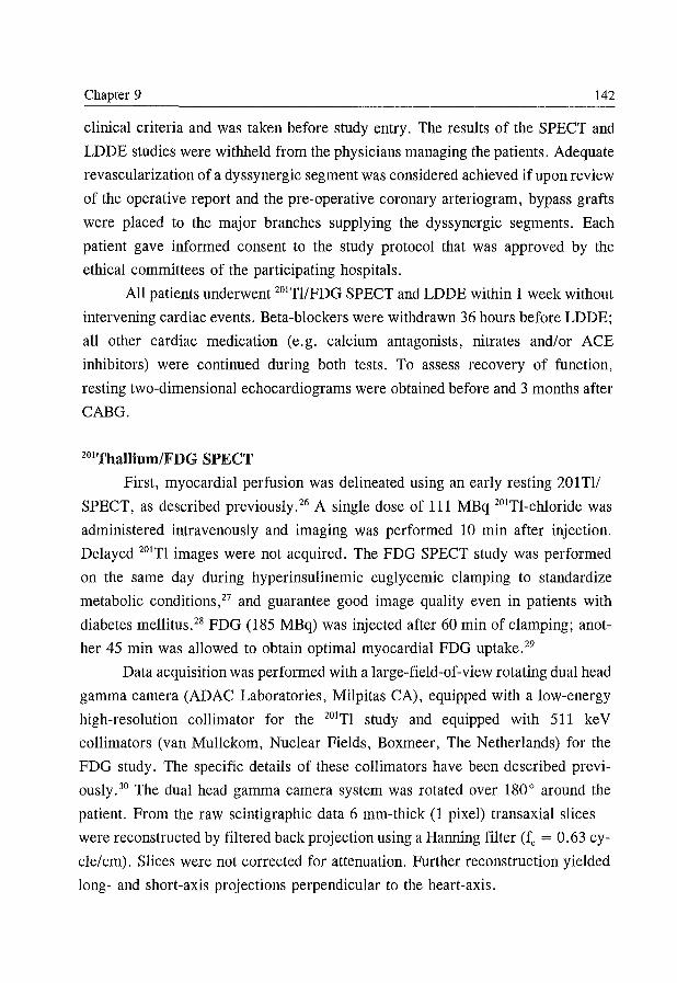
Chapter 9 142
clinical criteria and was taken before study entry. The results of the SPECT and
LDDE studies were withheld from the physicians managing the patients. Adequate
revascularization of a dyssynergic segment was considered achieved if upon review
of the operative report and the pre-operative coronary arteriogram, bypass grafts
were placed to the major branches supplying the dyssynergic segments. Each
patient gave informed consent to the study protocol that was approved by the
ethical committees of the participating hospitals.
All patients underwent 20lTlIFDG SPECT and LDDE within 1 week without
intervening cardiac events. Beta-blockers were withdrawn 36 hours before LDDE;
all other cardiac medication (e.g. calcium antagonists, nitrates and/or ACE
inhibitors) were continued during both tests. To assess recovery of function,
resting two-dimensional echocardiograms were obtained before and 3 months after
CABG.
201Thallium/FDG SPECT
First, myocardial perfusion was delineated using an early resting 201 TI/
SPECT, as described previously.26 A single dose of 111 MBq 20ITI-chloride was
administered intravenously and imaging was performed 10 min after injection.
Delayed 20lTl images were not acquired. The FDG SPECT study was performed
on the same day during hyperinsulinemic euglycemic clamping to standardize
metabolic conditions,27 and guarantee good image quality even in patients with
diabetes mellitus. 28 FDG (185 MBq) was injected after 60 min of clamping; anot
her 45 min was allowed to obtain optimal myocardial FDG uptake. 29
Data acquisition was performed with a large-field-of-view rotating dual head
gamma camera (ADAC Laboratories, Milpitas CA), equipped with a low-energy
high-resolution collimator for the 20lTI study and equipped with 511 keY
collimators (van Mullekom, Nuclear Fields, Boxmeer, The Netherlands) for the
FDG study. The specific details of these collimators have been described previ
ously.30 The dual head gamma camera system was rotated over 180 0 around the
patient. From the raw scintigraphic data 6 mm-thick (1 pixel) transaxial slices
were reconstructed by filtered back projection using a Hanning filter (fe = 0.63 cy
cle/cm). Slices were not corrected for attenuation. Further reconstruction yielded
long- and short-axis projections perpendicular to the heart-axis.
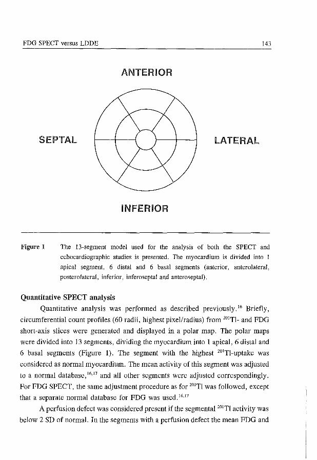
FDG SPECT versus LDDE 143
ANTERIOR
SEPTAL LATERAL
Figure 1
INFERIOR
The 13-segment model used for the analysis of both the SPECT and
echocardiographic studies is presented. The myocardium is divided into 1
apical segment, 6 distal and 6 basal segments (anterior, anterolateral,
posterolateral, inferior, inferoseptal and anteroseptal).
Quantitative SPEeT analysis
Quantitative analysis was performed as described previously. 16 Briefly,
circumferential count profiles (60 radii, highest pixel/radius) from 20ITl_ and FDG
short-axis slices were generated and displayed in a polar map. The polar maps
were divided into 13 segments, dividing the myocardium into 1 apical, 6 distal and
6 basal segments (Figure 1). The segment with the highest 20ITl-uptake was
considered as normal myocardium. The mean activity of this segment was adjusted
to a normal database,16.17 and all other segments were adjusted correspondingly.
For FDG SPECT, the same adjustment procedure as for 20lTl was followed, except
that a separate normal database for FDG was used. 16.17
A perfusion defect was considered present if the segmental 20lTl activity was
below 2 SD of normal. In the segments with a perfusion defect the mean FDG and
Chapter 9 144
201Tl activities were compared. Dyssynergic segments with either normal perfusion
or a 7 % increased FDG uptake in perfusion defects (perfusion-metabolism
mismatch) were considered myocardial regions with a high probability of
functional improvement after revascularization. In contrast, dyssynergic segments
with a 201Tl perfusion defect without increased FDG uptake (perfusion-metabolism
match) were classified as having a low probability of functional recovery. The 7 %
cutoff value was defined previously using ROC analysis in a different group of
patients undergoing revascularization. 31
For analysis on a patient basis, patients were considered to have the ability
to recover functionally ("recoverable") if 22 adjacent dyssynergic segments
showed normal perfusion or a mismatch pattern on 201TlIFDG SPECT.
Low-dose dobutamine echo cardiography
A two-dimensional transthoracic echocardiogram in standard apical and para
sternal views and a 12-lead ECG were recorded at rest. Dobutamine was infused
by a volumetric pump through an antecubital vein at doses of 5 and 10 /-tg/kg/min,
for 5 min at each dose. Continuous monitoring of the echocardiogram was obtained
during the test, and recorded on video tape at the last minute of both stages.
The echocardiographic images were also digitized (Prevue-III, Nova-Micro
sonics or Vingmed CFM 800) and displayed in quad-screen format to facilitate the
comparison of rest and dobutamine images. A 3-lead ECG was continuously moni
tored and a 12-lead ECG was recorded every minute. Blood pressure was measu
red by sphygmomanometer at each stage.
Analysis of echocardiograms
The interpretation of echocardiograms was performed by two experienced
observers, blinded to the clinical data and SPECT results. In case of disagreement,
a third observer reviewed the study and a majority decision was achieved. The
stress- and post-operative resting echocardiograms were interpreted within 1 week
after acquisition. Post-operative resting echocardiograms were analyzed without
knowledge of the LDDE results. For analysis of echocardiograms we used a 13-
segment model to allow comparison with the 13 segments of the SPECT polar
maps. 16
FDG SPECT versus LDDE 145
Wall motion of every segment, including wall thickening, was scored with a 4-
point scoring system (0 = normal, 1 = mildly hypokinetic, 2 = severely hypoki
netic, 3 = akinetic or dyskinetic). We defined a segment as severely hypokinetic
in the presence of minimal wall thickening ( < 2mm) with a limited inward motion;
as akinetic in the absence of systolic wall motion and thickening, and as dyskinetic
in the presence of systolic outward motion with thinning. Wall thickening was
primarily utilized for the classification of wall motion, pre-empting the problem of
post-operative paradoxical septal motion. Additionally, in order to reduce the
confounding effect of tethering, segmental wall thickening was analyzed frame by
frame during the first half of systole.
The LDDE studies were analyzed using the digitized rest and dobutamine
images, displayed in a quad-screen format and also by reviewing the images recor
ded on video tape. Dyscontractile regions were considered to have a high
probability of functional improvement after revascularization when wall motion
improved by at least one point of the scoring system during the infusion of low
dose dobutamine. A lack of improvement or direct worsening in wall motion were
considered a low probability of functional recovery.
For analysis on a patient basis, patients were considered to have the ability
to recover functionally ("recoverable") if 22 adjacent dyssynergic segments
showed improved wall motion during dobutamine infusion. Follow-up echocardio
grams were compared with the corresponding pre-operative resting images, without
knowledge of the dobutamine studies. For each segment, recovery of function was
defined as an improvement of one or more grades.
We previously reported a low level of inter- and intra-observer variability
for the classification of resting wall motion (agreement 84 % and 87 %) and the
response to low-dose dobutamine (agreement 92 % and 94 %) in a different but
comparable patient group.22 For individual patients, functional recovery was
defined as an improvement of segmental wall motion score after revascularization
in 22 adjacent segments. Finally, a wall motion score index (WMSI) was
calculated from the pre-operative and post-operative resting echo cardiograms to
evaluate change in global LV function. WMSI was defined as the sum of the scores
of each segment divided by the total number of segments analyzed.
Chapter 9 146
Statistical analysis
Continuous data are expressed as means ± standard deviation (SD). A
paired or unpaired Student's t-test was used when appropriate. Univariate analysis
for categorical variables was performed using the chi-square test with Yates'
correction. Sensitivity, specificity, positive and negative predictive values were
based upon their standard definitions. McNemar's test was used to compare the
sensitivity and specificity of 201TlIFDG SPECT and LDDE in the prediction of
functional recovery. Differences were considered significant if the null hypothesis
could be rejected at the 0.05 probability level.
Results
Characteristics of the study group
The study group comprised 30 patients (25 men, 5 women) with stable
coronary artery disease and regional LV dysfunction who underwent an uncom
plicated CABG procedure. The demographic, clinical and angiographic data are
summarized in Table 1.
Of the 390 segments, 388 were visualized by resting echocardiography. Of
these segments 212 (55%) had normal wall motion and 176 (45%) showed
abnormal wall motion at rest. The mean number of abnormal segments per patient
was 5.6 ± 3.5. Eight dyssynergic segments were excluded from post-operative
evaluation due to inadequate revascularization. Thus, 168 segments were available
for serial analysis. Sixty-six (39 %) dyssynergic segments were classified as mildly
hypokinetic and 102 (61 %) as severely dyssynergic (33 severely hypokinetic and
69 a- or dyskinetic).
Post-operative results
At 3 months follow-up 62 (37%) of the 168 dyssynergic segments showed
an improvement of wall thickening and motion. The improvement was observed
in 35 (53%) of the 66 mildly hypokinetic segments and in 27 (26%) of the 102
severely dyssynergic segments (in 16 severely hypokinetic and 11 a- or dyskinetic
segments; p < 0.01 severely dyssynergic versus mildly hypokinetic segments). The
mean WMSI decreased significantly from 0.90 ± 0.48 to 0.72 ± 0.44 (p <0.05).
Patient-by-patient analysis revealed improvement of wall motion in ;::;: 2 adjacent
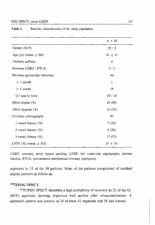
FDG SPECT versus LDDE 147
Table 1 Baseline characteristics of the study population
n = 30
Gender (M/F) 25 15
Age (yr) (mean ± SD) 61 ± 11
Diabetes mellitus 4
Previous CABG I PTCA 5 I 1
Previous myocardial infarction 30
< 1 month 1
> 1 month 19
Q I non-Q wave 20 I 10
Effort angina (%) 24 (80)
Effort dyspnea (%) 16 (53)
Coronary arteriography 30
1 vessel disease (%) 7 (23)
2 vessel disease (%) 6 (20)
3 vessel disease (%) 17 (57)
LVEF (%) (mean ± SD) 35 ± 10
CABG: coronary artery bypass grafting, LVEF: left ventricular angiographic ejection
fraction, PTCA: percutaneous trans luminal coronary angioplasty.
segments in 15 of the 30 patients. None of the patients complained of residual
angina pectoris at follow-up.
201TI/FDG SPECT
201TlIPDG SPECT identified a high probability of recovery in 52 of the 62
(84 %) segments showing improved wall motion after revascularization. A
mismatch pattern was present in 24 of these 52 segments and 28 had normal
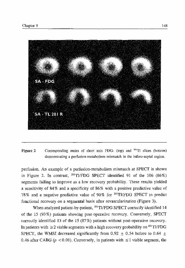
Chapter 9 148
Figure 2 Corresponding series of short axis FDG- (top) and 201Tl slices (bottom)
demonstrating a perfusion-metabolism mismatch in the infero-septal region.
perfusion. An example of a perfusion-metabolism mismatch at SPECT is shown
in Figure 2. In contrast, 201TlIFDG SPECT identified 91 of the 106 (86%)
segments failing to improve as a low recovery probability. These results yielded
a sensitivity of 84 % and a specificity of 86 % with a positive predictive value of
78 % and a negative predictive value of 90 % for 201TlIFDG SPECT to predict
functional recovery on a segmental basis after revascularization (Figure 3).
When analyzed patient-by-patient, 201TlIFDG SPECT correctly identified 14
of the 15 (93 %) patients showing post-operative recovery. Conversely, SPECT
correctly identified 13 of the 15 (87 %) patients without post-operative recovery.
In patients with ;;::: 2 viable segments with a high recovery probability on 201TI/FDG
SPECT, the WMSI decreased significantly from 0.92 ± 0.54 before to 0.64 ± 0.46 after CABG (p < 0.01). Conversely, in patients with::; 1 viable segment, the
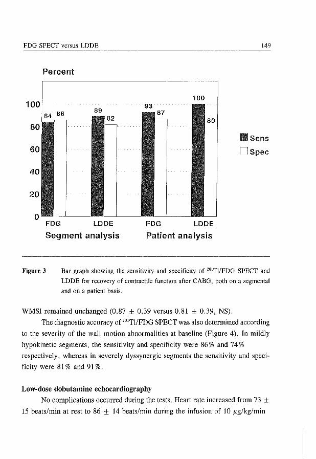
FDG SPECT versus LDDE 149
Percent
100 100 93
84 86 89
80 Sens
60 DSpec
40
20
0 FOG LODE FOG LODE
Segment analysis Patient analysis
Figure 3 Bar graph showing the sensitivity and specificity of 201TlIFDG SPECT and
LDDE for recovery of contractile function after CABG, both on a segmental
and on a patient basis.
WMSI remained unchanged (0.87 ± 0.39 versus 0.81 ± 0.39, NS).
The diagnostic accuracy of 201TI/FDG SPECT was also determined according
to the severity of the wall motion abnormalities at baseline (Figure 4). In mildly
hypo kinetic segments, the sensitivity and specificity were 86 % and 74%
respectively, whereas in severely dyssynergic segments the sensitivity and speci
ficity were 81 % and 91 %.
Low-dose dobutamine echo cardiography
No complications occurred during the tests. Heart rate increased from 73 ± 15 beats/min at rest to 86 ± 14 beats/min during the infusion of 10 ltg/kg/min
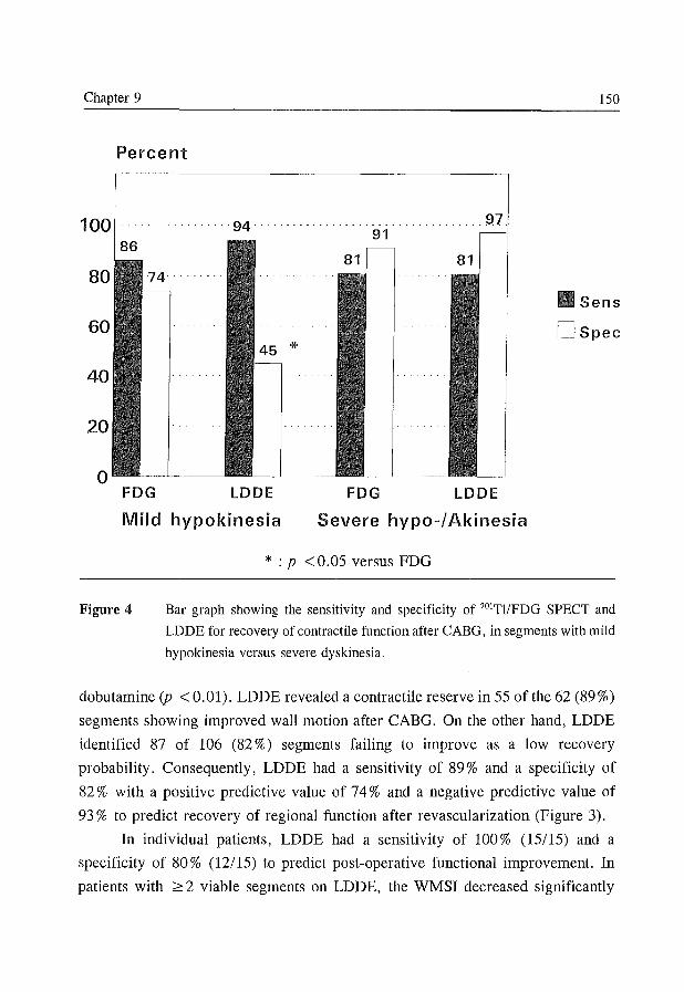
Chapter 9 150
Percent
100 86
80
60
40
20
o FDG
..... ·94·
*
LDDE
................ 97 91
FDG LDDE
Sens
DSpec
Mild hypokinesia Severe hypo-/ Akinesia
Figure 4
* : p < 0.05 versus FDG
Bar graph showing the sensitivity and specificity of 201Tl/FDG SPECT and
LDDE for recovery of contractile function after CABG, in segments with mild
hypokinesia versus severe dyskinesia.
dobutamine (p < 0.01). LDDE revealed a contractile reserve in 55 of the 62 (89%)
segments showing improved wall motion after CABG. On the other hand, LDDE
identified 87 of 106 (82 %) segments failing to improve as a low recovery
probability. Consequently, LDDE had a sensitivity of 89% and a specificity of
82 % with a positive predictive value of 74% and a negative predictive value of
93 % to predict recovery of regional function after revascularization (Figure 3).
In individual patients, LDDE had a sensitivity of 100% (15/15) and a
specificity of 80% (12/15) to predict post-operative functional improvement. In
patients with ;;::: 2 viable segments on LDDE, the WMSI decreased significantly
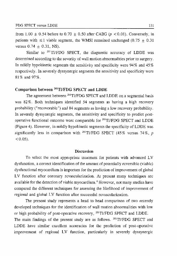
FDG SPEer versus LDDE 151
from 1.00 ± 0.54 before to 0.70 ± 0.50 after CABG (p <0.01). Conversely, in
patients with ~ 1 viable segment, the WMSI remained unchanged (0.75 ± 0.31
versus 0.74 ± 0.31, NS).
Similar to 201TlIFDG SPECT, the diagnostic accuracy of LDDE was
determined according to the severity of wall motion abnormalities prior to surgery.
In mildly hypokinetic segments the sensitivity and specificity were 94 % and 45 %
respectively. In severely dyssynergic segments the sensitivity and specificity were
81 % and 97%.
Comparison between 201TlIFDG SPECT and LDDE
The agreement between 201TlIFDG SPECT and LDDE on a segmental basis
was 82 %. Both techniques identified 54 segments as having a high recovery
probability ("recoverable") and 84 segments as having a low recovery probability.
In severely dyssynergic segments, the sensitivity and specificity to predict post
operative functional outcome were comparable for 201TlIFDG SPECT and LDDE
(Figure 4). However, in mildly hypokinetic segments the specificity of LDDE was
significantly less in comparison with 201TlIFDG SPECT (45 % versus 74 %, P
<0.05).
Discussion
To select the most appropriate treatment for patients with advanced LV
dysfunction, a correct identification of the amount of potentially reversible (viable)
dysfunctional myocardium is important for the prediction of improvement of global
LV function after coronary revascularization. At present many techniques are
available for the detection of viable myocardium. 4 However, not many studies have
compared the different techniques for assessing the likelihood of improvement of
regional and global LV function after successful revascularization.
The present study represents a head to head comparison of two recently
developed techniques for the identification of wall motion abnormalities with low
or high probability of post-operative recovery, 201TlIFDG SPECT and LDDE.
The main findings of the present study are as follows. 201TI/FDG SPECT and
LDDE have similar excellent accuracies for the prediction of post-operative
improvement of regional LV function, particularly in severely dyssynergic
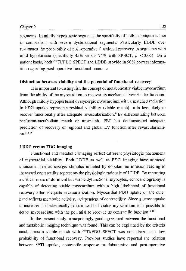
Chapter 9 152
segments. In mildly hypokinetic segments the specificity of both techniques is less
in comparison with severe dysfunctional segments. Particularly LDDE ove
restimates the probability of post-operative functional recovery in segments with
mild hypokinesia (specificity 45% versus 74% with SPECT, p <0.05). On a
patient basis, both 20lTlIFDG SPECT and LDDE provide in 90% correct informa
tion regarding post-operative functional outcome.
Distinction between viability and the potential of functional recovery
It is important to distinguish the concept of metabolically viable myocardium
from the ability of the myocardium to recover its mechanical ventricular function.
Although mildly hypoperfused dyssynergic myocardium with a matched reduction
in FDG uptake represents residual viablility (viable match), it is less likely to
recover functionally after adequate revascularization. 6 By differentiating between
perfusion-metabolism match or mismatch, PET has demonstrated adequate
prediction of recovery of regional and global LV function after revascularizati
on. 5•8-15
LDDE versus FDG imaging
Functional and metabolic imaging reflect different physiologic phenomena
of myocardial viability. Both LDDE as well as FDG imaging have attracted
clinicians. The adrenergic stimulus initiated by dobutamine infusion leading to
increased contractility represents the physiologic rationale of LDDE. By recruiting
a critical mass of dormient but viable dyfunctional myocytes, echocardiography is
capable of detecting viable myocardium with a high likelihood of functional
recovery after adequate revascularization. Myocardial FDG uptake on the other
hand reflects metabolic activity, independent of contractility. Since glucose uptake
is increased in ischemically jeopardized but viable myocardium it is possible to
detect myocardium with the potential to recover its contractile function. 5-15
In the present study, a surprisingly good agreement between the functional
and metabolic imaging technique was found. This can be explained by the criteria
used, since a viable match with 20lTlIFDG SPECT was considered as a low
probability of functional recovery. Previous studies have reported the relation
between 20lTI uptake, contractile response to dobutamine and post-operative
FDG SPECT versus LDDE 153
recovery.22.32 Although 201TI SPECT detected myocardial viability (defined as
> 50 % uptake) more frequently, 32 it also seemed to overestimate functional
recovery after CABG compared to LDDE.22 Our findings are supported by a recent
comparative study between LDDE and PET in similar patients. 33 The extent of
dobutamine-induced contractile reserve appeared to compare less closely with the
total extent of viable myocardium than with the extent of perfusion-metabolism
mismatch.
Mildly versus severely dyssynergic segments
In hypokinetic segments both techniques, but especially LDDE, seems to
overestimate functional recovery after revascularization. These segments are likely
to contain normal myocardium, subendocardial scar and/or viable but dysfunctional
myocardium. If relatively little viable tissue is present, dobutamine challenge may
stimulate normal myocardium to a hyperkinetic response simulating a positive test
for viability. It is conceivable that combining low with high dose dobutamine
infusion could have increased the specificity of stress echocardiography for
predicting functional recovery. 23 Using the entire dose- range some segments may
have exhibited a biphasic response (jeopardized but viable) with high probability
of recovery, whereas other segments may have shown continuous enhancement of
wall thickening (non-jeopardized). However our patients only underwent a low
dose dobutamine infusion. Furthermore, since predominance of normal myocytes
with normal 201TI uptake may obscure uptake defects of small islands of nonviable
cells, this may have led to some false positive SPECT results in mildly hypokinetic
segments.
FDG SPECT
So far the study of myocardial FDG uptake has mainly been confined to the
availability of PET. 5-15 Recently, the development of a special collimator made it
possible to study myocardial FDG uptake with SPECT as well. 16-20 For clinical
purposes, SPECT imaging may provide a widely available approach for the detecti
on of myocardial viability. Some technical features of FDG imaging, applied in our
protocol should be underscored. In order to optimize and standardize metabolic
conditions, we used the hyperinsulinemic euglycemic clamping technique. These
Chapter 9 154
metabolic conditions lead to enough exogenous glucose uptake for optimal target
to-background ratio resulting in good diagnostic image quality, 27 and furthermore
enhance a more homogenous FDG uptake. 34 Moreover, Hariharan et al have
recently demonstrated that variations in metabolic and hormonal circumstances can
affect myocardial FDG uptake. 35 The more widespread used alternative approach
employing oral glucose loading may result in a more variable metabolic milieu and
was therefore not chosen.
In this study we have compared regional FDG uptake with perfusion
assessed with 20ITl. Although these tracers have different photon energies, we have
recently compared regional FDG and 20lTI uptake in normal individuals. 17 In that
study, no significant differences between uptake of the 2 tracers could be demon
strated in any of the different myocardial regions. However, influence of photon
attenuation, particularly on the 20lTI images, cannot be ruled out. A future study
using our protocol in combination with attenuation correction is warranted.
Previous studies
In comparison to the PET literature, relying on comparable methodologies,
similar sensitivities and specificities in patients undergoing revascularization were
reached as compared to our FDG SPECT results. In the reported 194 patients
derived from 9 studies PET showed a mean sensitivity of 87 % and a mean
specificity of 77 % .5,8-15 Previous studies have shown that pre-operative inotropic
reserve predicts improvement of wall motion abnormalities after revascularizati
on,21-25 and have demonstrated a sensitivity ranging from 74 % to 95 % and a
specificity ranging from 73 % to 95 %. The data obtained in our study are in line
with these results.
Limitations
In the present study graft patency was not assessed after revascularization.
Reocc1usion may have accounted for the failure of some viable segments to recover
in contractile function, particularly in the segments that were viable both on LDDE
and FDG. Recent data suggest that jeopardized myocardium, a combination of
myocardial viability and inducible ischemia, is more likely to recover after
revascularization than viable myocardium which is not in jeopardy. However we
FDG SPECT versus LDDE 155
did not perform high dose dobutamine stress testing and cannot comment on the
so called "biphasic response". We have not noted this response during the LDDE
studies. Functional improvement was arbitrarely assessed 3 months after surgical
revascularization. It cannot be excluded that additional functional improvement may
occur later as was already suggested. 36
Conclusion
LDDE and 201T1/FDG SPECT imaging are both valuable methods for the
prediction of improvement of regional and global LV function after CABG. Both
methods are more and more available and may be useful techniques for the routine
detection of potentially reversibly dysfunctional viable myocardium.
References
1. Elefteriades JA, Tolis G, Levi E, Mills LK, Zaret BL. Coronary artery bypass
grafting in severe left ventricular dysfunction: excellent survival with improved
ejection fraction and functional state. J Am CoIl CardioI1993;22:1411-1417.
2. Alderman EL, Fisher LD, Litwin P, et al. Results of coronary artery surgery in
patients with poor left ventricular function (CASS). Circulation 1983;68:785-795.
3. Nesto RW, Cohn LH, Collins JJ, Wynne J, Holman L, Cohn PF. Inotropic contrac
tile reserve: a useful predictor of increased 5 year survival and improved postoperati
ve left ventricular function in patients with coronary artery disease and reduced
ejection fraction. Am J Cardiol 1982;50:39-44.
4. Dilsizian V, Bonow RO. Current diagnostic techniques of assessing viability in
patients with hibernating and stunned myocardium. Circulation 1993;87: 1-20.
5. Tillisch J, Brunken R, Marshall R et al. Reversibility of cardiac wall motion
abnormalities predicted by positron tomography. N Engl J Med 1986;314:884-888.
6. Vom Dahl J, Eitzman DT, AI-Aouar ZR, et al. Relation of regional function,
perfusion and metabolism in patients with advanced coronary artery disease
undergoing surgical revascularization. Circulation 1994;90:2356-2366.
7. Maes A, Flameng W, Nuyts J, et al. Histological alterations in chronically hypoperfu
sed myocardium. Correlation with PET findings. Circulation 1994;90: 735-745.
8. Tamaki N, Yonekura Y, Yamashita K, et al. Positron emission tomography using
fluorine-18 deoxyglucose in evaluation of coronary artery bypass grafting. Am J
Cardiol 1989;64:860-865.
Chapter 9 156
9. Tamaki N, Ohtani H, Yamashita K, et al. Metabolic activity in the areas of new fill
in after thallium-201 reinjection: Comparison with positron emission tomography
using fluorine-18-deoxyg1ucose. J Nuc1 Med 1991;32:673-678.
10. Lucignani G, Paolini G, Landoni C, et al. Presurgical identification of hibernating
myocardium by combined use of technetium-99m hexakis 2-methoxyisobutylisonitrile
SPECT and fluorine-18 fluoro-2-deoxy-D-glucose positron emission tomography in
patients with coronary artery disease. Eur J Nuc1 Med 1992;19:874-881.
11. Carrel T, Jenni R, Haubold-Reuter S, Von Schulthess G, Pasic M, Turina M.
Improvement of severely reduced left ventricular function after surgical revascula
rization in patients with preoperative myocardial infarction. Eur J Cardiothorac Surg
1992;6:479-484.
12. Marwick TH, MacIntyre WJ, Lafont A, Nemec JJ, Salcedo EE. Metabolic responses
of hibernating and infarcted myocardium to revascularization. Circulation 1992;85:
1347-1353.
13. Gropler RJ, Siegel B, Sampathkumaran K, et al. Dependence of recovery of con
tractile function on maintainance of oxidative metabolism after myocardial infarction.
J Am ColI Cardiol 1992;19:989-997.
14. Gropler RJ, Geltman EM, Sampathkumaran K, et al. Comparison of carbon-11-
acetate with fluorine-18-fluorodeoxyglucose for delineating viable myocardium by
positron emission tomography. J Am ColI CardioI1993;22:1587-1597.
15. Tamaki N, Kawamoto M, Tadamura E, et al. Prediction of reversible ischemia after
revascularization. Perfusion and metabolic studies with positron emission tomography.
Circulation 1995; 91: 1697-1705.
16. Bax JJ, Visser FC, Blanksma PK, et al. Comparison of myocardial uptake of 18F
fluorodeoxyglucose imaged with positron emission tomography and single photon
emission computed tomography in dyssynergic myocardium. J Nuc1 Med 1996; in
press.
17. Bax JJ, Visser FC, van Lingen A, et al. Relation between myocardial uptake of
thallium-201 chloride and fluorine-18 fluorodeoxyglucose imaged with single-photon
emission tomography in normal individuals. Eur J Nuc1 Med 1995;22:56-60.
18. Burt RW, Perkins OW, Oppenheim BE, et al. Direct comparison of fluorine-18-FDG
SPECT, fluorine-18-FDG PET and rest thallium-201 SPECT for detection of
myocardial viability. J Nuc1 Med 1995;36:176-179.
19. Delbeke D, Videlefsky S, Patton JA, et al. Rest myocardial perfusion/metabolism
imaging using simultaneous dual-isotope acquisition SPECT with technetium-99m
MIBIIfluorine-18-FDG. J Nuc1 Med 1995;36:2110-2119.
20. Stoll HP, Helwig N, Alexander C, Ozbek C, Schieffer H, Oberhausen E. Myocardial
FDG SPECT versus LDDE 157
metabolic imaging by means of fluorine-18 deoxyglucose/technetium-99m sestamibi
dual isotope single-photon emission tomography. Eur J Nucl Med 1994; 21: 1085-
1093.
21. Marzullo P, Parodi 0, Reisenhofer B, et al. Value of rest thallium-201ltechnetium-
99m sestamibi scans and dobutamine echocardiography for detecting myocardial
viability. Am J CardiolI993;71:166-172.
22. Arnese M, Cornel JH, Salustri A, et al. Prediction of improvement of regional left
ventricular function after surgical revascularization: a comparison of low-dose
dobutamine echocardiography with 201TI single-photon emission computed tomo
graphy. Circulation 1995;91 :2748-2752.
23. Afridi I, Kleiman NS, Raizner AE, Zoghbi WA. Dobutamine echocardiography in
myocardial hibernation. Optimal dose and accuracy in predicting recovery of
ventricular function after coronary angioplasty. Circulation 1995;91:663-670.
24. La Canna G, Alfieri 0, Giubbini R, Gargano M, Ferrari R, Visioli 0. Echocardio
graphy during infusion of dobutamine for identification of reversible dysfunction in
patients with chronic coronary artery disease. JAm Coll Cardiol 1994;23:617-626.
25. Perrone-Filardi P, Pace L, Prastaro M, et al. Dobutamine echocardiography predicts
improvement of hypoperfused dysfunctional myocardium after revascularization in
patients with coronary artery disease. Circulation 1995 ;91 :2556-2565.
26. Melin JA, Becker LC. Quantitative relationship between global left ventricular
thallium uptake and blood flow: effects of propranolol, ouabain, dipyridamole and
coronary artery occlusion. J Nucl Med 1986;27:641-652.
27. Knuuti MJ, Nuutila P, Ruotsalainen U, et al. The value of quantitative analysis of
glucose utilization in detection of myocardial viability by PET. J Nucl Med
1993; 34: 2068-2075.
28. Vom Dahl J, Herman WH, Hicks RJ, et al. Myocardial glucose uptake in patients
with insulin-dependent diabetes mellitus assessed quantitatively by dynamic positron
emission tomography. Circulation 1993;88:395-404.
29. Gallagher BM, Fowler JS, Gutterson NI, MacGregor RR, Wan C, Wolf AP. Meta
bolic trapping as a principle of radiopharmaceutical design: some factors responsible
for the biodistribution of [18F] 2-deoxy-2-fluoro-D-glucose. J Nucl Med 1978;19:
1154-1161.
30. Lingen A van, Huijgens PC, Visser FC, et al. Performance characteristics of a 511-
KeV collimator for imaging positron emitters with a standard gamma-camera. Eur J
Nucl Med 1992;19:315-321.
31. Bax JJ, Cornel JH, Visser FC, et al. Functional recovery after revascularization pre
dicted by quantitative FDG SPECT. Eur J Nucl Med 1995;22:798 [abstract].
Chapter 9 158
32. Panza JA, Dilsizian V, Laurienzo JM, Curiel RV, Katsiyiannis PT. Relation between
thallium uptake and contractile response to dobutamine. Implications regarding
myocardial viability in patients with chronic coronary artery disease and left ventri
cular dysfunction. Circulation 1995; 91: 990-998.
33. Chan RKM, Lee KJ, Calafiore P, Berlangieri SU, McKay WJ, Tonkin AM. Compa
rison of dobutamine echocardiography and positron emission tomography in patients
with chronic ischemic left ventricular dysfunction. J Am ColI Cardiol 1996;27: 1601-
1607.
34. Hicks RJ, Herman WH, Kalff V, et al. Quantitative evaluation of regional substrate
metabolism in the human heart by positron emission tomography. J Am ColI Cardiol
1991; 18: 101-111.
35. Hariharan R, Bray M, Ganim R, Doenst T, Goodwin GW, Taegtmeyer H.
Fundamental limitations of [18F]2-deoxy-2-fluoro-D-glucose for assessing myocardial
glucose uptake. Circulation 1995 ;91 :2435-2444.
36. Bashour TT, Mason DT. Myocardial hibernation and "embalmment". Am Heart J
1990; 119:706-708.
Chapter 10
Biphasic Response to Dobutamine Predicts Recovery of Global
Ventricular Function After Surgical Revascularization
Implications of Time Course of Recovery on Diagnostic Accuracy
JH Cornel, JJ Bax**, A Elhendy, APWM Maat*, GJ Kimman, ML Geleijnse,
E Boersma, PR Nierop, JRTC Roelandt, PM Fioretti.
From the Thoraxcenter, Departments of Cardiology and Cardiac Surgery * ,
University Hospital Rotterdam-Dijkzigt, Rotterdam, and the Department of
Cardiology**, Free University Hospital, Amsterdam, the Netherlands.
Submitted for publication
Chapter 10 160
Abstract
Background
This study sought to evaluate the time course of recovery of left ventricular
dysfunction in stable patients and its implications on the accuracy of dobutamine
echocardiography to predict recovery after surgical revascularization.
Methods
Consecutive patients with chronic ischemic left ventricular dysfunction
scheduled for elective surgical revascularization were prospectively selected. They
underwent dobutamine echocardiography (5 to 40 /-tg/kg/min) and radionuc1ide
ventriculography both preoperatively and at 3 month follow-up. At 14 months
another evaluation of ventricular function was obtained. To analyze echocardio
grams a 16 segment model and a 5-point scoring system was used. Dyssynergic
segments were considered likely to improve in presence of a biphasic contractile
response to dobutamine. Improvement of global function was defined as a 25%
increase in left ventricular ejection fraction.
Results
Of the 61 patients, ejection fraction improved in 12 at 3 months and in a
total of 19, (i.e. 7 more), at late (14 month) follow-up (from 32 ± 8% to 42 ± 9%, p <0.0001). A biphasic response was predictive for recovery in 118/186
(63%) segments at 3 months and in 140/186 (75%) at late follow-up. Other
responses were highly predictive for non-recovery (92 %). In mildly hypokinetic
segments the positive predictive value of the test was significantly lower compared
to severely dyssynergic segments (67% (95% CI, 59% to 75%) versus 90% (95%
CI, 83% to 97%) at late follow-up). The sensitivity and specificity for recovery of
global function on a patient basis (24 biphasic segments) were 89 % and 81 % at
late follow-up.
Conclusions
Serial post-operative follow-up studies demonstrate incomplete recovery of
regional and global contractile function at 3 months. The diagnostic accuracy of
dobutamine echocardiography to predict recovery depends on combining low and
Contractile response to dobutamine and recovery 161
high dobutamine dosages, severity of dyssynergy and the timing of evaluation.
Introduction
Coronary artery bypass grafting (CABG) can improve symptoms, prognosis
and left ventricular (LV) function in selected patients. 1 The non-invasive identifica
tion of myocardial regions with high and low probability of functional improve
ment after revascularization is crucial for the decision to perform revascularization
procedures in individual patients with multiple severe wall motion abnormalities. 2
Since the presence of viable myocardium favourably influences prognosis after
revas'cularization,3-6 these procedures may even serve as an attractive alternative
to cardiac transplantation.
The contractile response of dyssynergic regions to "low-dose" dobutamine
in conjunction with echocardiography has been proposed as a simple method for
the assessment of residual viable myocardium capable of recovering its contractile
function, both spontaneously in patients early after myocardial infarction7-
11 and
after revascularization in patients with stable chronic ischemic heart disease. 12-16
Recent data suggest that jeopardized myocardium, a combination of
myocardial viability and inducible ischemia, is more likely to recover after
revascularization than viable myocardium which is not in jeopardy. 16 However all
previous studies using dobutamine echocardiography have been limited by the lack
of an independent method to verify changes in ventricular function. 12-20
The accuracy of predicting recovery of contractile function after revasculari
zation depends on several factors such as the response to dobutamine both at low
and high dosel6 and severity of baseline segmental dysfunction. ll•15 Furthermore
it is unclear what the optimal timing is to evaluate recovery of contractile function
after revascularization. Given the severity of structural changes observed in
hibernating myocardium,21.22 it is likely that recovery of function after revasculari
zation is delayed for several months.
Therefore we designed a prospective study to evaluate 1) the accuracy of
dobutamine echocardiography to predict recovery of regional and global LV
dysfunction after successful CABG in patients with a wide range of chronic LV
dysfunction, and 2) the time course of functional improvement after CABG using
both serial echo cardiographic - and radionuc1ide ventriculographic studies, and so
Chapter 10 162
defining the optimal timing to determine the diagnostic value of dobutamine
echocardiography in this clinical setting.
Methods
Patient enrollment
From January 1993 to April 1995, all patients with coronary artery disease
and LV dysfunction at rest, who were scheduled to undergo CABG at the
Thoraxcenter, were screened for enrollment in the study. The study protocol was
approved by the Institutional Review Board. Inclusion criteria were symptoms of
stable coronary artery disease, prior acceptance for elective surgical revasculariza
tion, ejection fraction (EF) of < 50 % on contrast ventriculography, and one or
more abnormal contractile segments on preoperative ( < 3 weeks before operation)
resting echocardiogram (16-segment left ventricular model). Furthermore a
subsequent uneventful surgical revascularization procedure was required to proceed
with the protocol. Exclusion criteria were unstable angina, recent myocardial
infarction ( < 3 months), significant ( > 50 %) left main stem stenosis, (hemodyna
mically) significant valvular disease, poor echo quality, or the inability to obtain
informed consent. Of the 89 preoperative eligible patients 28 were excluded. The
reasons for exclusion were resection of infarcted areas in addition to myocardial
revascularization in 13 patients, peri operative death in 6 (7 %), perioperative
nonfatal myocardial infarction in 2, poor echo quality in 3, inability to obtain
informed consent in 2, death on the waiting list for surgery in 1 and death early
in the follow-up (at 10 weeks) in 1 patient. Of the patients who died perioperative
ly, 2 deaths were due to pump failure and 4 due to perioperative myocardial
infarction. Of the 61 patients finally included in the present study, 5 (8 %)
underwent CABG as an alternative to cardiac transplantation.
Study protocol
Each patient underwent a low and high dose dobutamine stress echocardio
gram and a radionuclide ventriculography within the 3 weeks prior to surgery. All
patients underwent uneventful isolated CABG (by definition). The decision to
revascularize was based on clinical criteria. The results of the dobutamine stress
echocardiographic and radionuclide studies were withheld from the physicians
Contractile response to dobutamine and recovery 163
managing the patients. Adequate revascularization of a dyssynergic segment was
considered achieved if upon review of the operative report and the preoperative
coronary arteriogram, bypass grafts were placed on the stenotic major branches
supplying the dyssynergic segments. After the operation, patients were followed
up to a maximum of 19 months. At 3 months follow-up both low and high dose
dobutamine stress echocardiography and radionuclide ventriculography at rest were
repeated. At 12 months follow-up a two-dimensional resting echocardiogram and
a third radionuclide ventriculography were performed.
Dobutamine stress echo cardiography
Before the test, patients were asked to discontinue B-blockers for 36 hours.
All other cardiac medication (e.g. calcium antagonists, nitrates and/or ACE
inhibitors) were continued. The dobutamine stress test was performed as follows.
A two-dimensional transthoracic echo cardiogram in standard views and a 12-lead
ECG were recorded with the patient at rest. Dobutamine was infused through an
antecubital vein at dosages of 5 and 10 p,g/kg/min, for 5 minutes at each dose
(these 2 steps were considered as "low-dose"). Subsequently, 3 other steps from
20 to 40 p,g/kg/min (3 minutes each) were added. Finally, atropine (up to 1 mg)
was injected when 85 % of the predicted maximal (men (220 - age) x 85 %, women
(200 - age) x 85 %) heart rate had not been reached. 23
A 3-lead ECG was monitored continuously, and a 12-lead ECG was
recorded every minute. Cuff blood pressure was measured at each stage. The test
was interrupted prematurely if 85 % of the predicted maximal heart rate was
reached or if severe chest pain, ST -segment deviation > 2 mm, significant
ventricular or supraventricular arrhythmia, systolic blood pressure fall of > 40 mm
Hg or any other intolerable side effect occurred during the test.
The echo cardiogram was monitored throughout the test, and the last minute
of each stage, including recovery, was recorded on video tape. The echocardio
graphic images were also digitized on optical disk [Vingmed CFM 800] or on
floppy disk [Esaote Biomedica SIM 7000] and displayed side-by-side in quadscreen
format to facilitate the comparison of images at rest and at various stages of the
test.
Chapter 10 164
Analysis of echocardiograms
The interpretation of echocardiograms was performed by 2 experienced
observers who were blinded to the clinical, radionuclide, angiographic, and
previous echo cardiographic results of the individual patients. In case of dis
agreement, a third observer reviewed the study and a majority decision was
achieved. The assessment was based on both the digitized images displayed in a
quadscreen format and a review of the images recorded on the videotape. For
analysis of wall motion, the left ventricle was divided into 16 segments as
recommended by the American Society of Echocardiography. 24 The wall motion,
including wall thickening, of every segment was semi-quantitated using a 5-point
scoring system where 1 = normal wall motion and thickening, 2 = mildly
hypokinetic, 3 = severely hypokinetic, 4 = akinetic and 5 = dyskinetic. We
defined a segment as severely hypokinetic in the presence of minimal wall
thickening with very limited inward motion (during first half of systole); as
akinetic in the absence of systolic wall motion and thickening and, whenever
possible, confirmed by M-mode tracing; and as dyskinetic in the presence of systo
lic outward motion with thinning. Wall thickening was primarily utilized for the
classification of wall motion, preventing the problem of postoperative paradoxical
septal motion. Also, to reduce the confounding effect of tethering from adjacent
segments, segmental wall thickening was analyzed only during the first half of
systole.
During dobutamine infusion, abnormally contracting segments at rest were
classified into 4 different patterns of contractile response: biphasic, defined as
improvement at low-dose and worsening at peak stress; sustained improvement,
defined as improvement at low-dose without further deterioration at peak stress;
worsening, defined as direct worsening without any improvement at any stage; and
no change, defined as unchanged wall motion abnormality throughout the test.
We previously reported a low level of inter- and intra-observer variability
for the classification of resting wall motion (agreement 84 % and 87 %) and the
response to low-dose dobutamine (agreement 92 % and 94 %) in a comparable
patient group. IS Myocardial ischemia was judged to be present when there was
worsening by ~ 1 of the segmental score. As previously reported, ischemia was
not considered when akinetic segments at baseline became dyskinetic at stress
Contractile response to dobutamine and recovery 165
without improvement during low-dose dobutamine infusion. 25 Follow-up
echo cardiograms were compared with the correspondent preoperative resting
images. The observers were blinded to the preoperative dobutamine results. For
each segment, improvement of function was defined as a decrease of one or more
grades. A change from dyskinetic to akinetic was not considered to be improved
contractile function.
Radionuclide ventriculography
Equilibrium radionuclide ventriculography was performed at rest with the
patient in supine position after intravenous administration of 555 MBq of
99mtechnetium. Images were acquired with a small-field-of-view gamma camera
(Orbiter, Siemens Corp., Iselin, NY, USA) oriented in the 45 degree left anterior
oblique position with a 5-10 degree caudal tilt. The L VEF was calculated by an
automated technique. Improvement of global LV function after revascularization
was defined as an increase in LVEF by at least 5 points (e.g. from 30% to 35%).
Statistical analysis
Age, number of stenotic coronary arteries, L VEF, heart rate and systolic
blood pressure are expressed as mean ± standard deviation. Differences within
continuous variables over time were evaluated by analysis of variance (ANOV A)
for repeated measures or by paired Student's t-test whenever appropriate.
Significance for all tests was stated at the 0.05 probability level. Sensitivity,
specificity, positive and negative predictive value rely on the standard definition
and are reported with 95 % confidence intervals (CI).
Results
Patient population
A total of 61 patients were included in the study. Mean age was 61 years
(range, 43 to 77 years), and 49 were men. All patients were symptomatic, 57 had
angina pectoris (29 in NYHA class II, 28 in NYHA class III) and 29 had dyspnea
on effort (26 in NYHA class II, 3 in NYHA class III). Fifty-nine patients had a
history of myocardial infarction (median of 24 months before study, range 4 to 210
months). The mean number of significantly stenosed coronary arteries was 2.7 ±
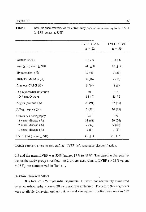
Chapter 10 166
Table 1 Baseline characteristics of the entire study population, according to the L VEF
(> 35 % versus::::; 35%)
LVEF >35% LVEF ::::;35%
n = 22 n = 39
Gender (M/F) 16 I 6 33 I 6
Age (yr) (mean ± SD) 61 ± 8 60 ± 9
Hypertension (%) 10 (45) 9 (23)
Diabetes Mellitus (%) 4 (18) 7 (18)
Previous CABG (%) 3 (14) 3 (8)
Old myocardial infarction 21 38
Q I non-Q wave 14 I 7 33 I 5
Angina pectoris (%) 20 (91) 37 (95)
Effort dyspnea (%) 5 (23) 24 (62)
Coronary arteriography 22 39
3 vessel disease (%) 14 (64) 29 (74)
2 vessel disease (%) 7 (32) 9 (23)
1 vessel disease (%) 1 (5) 1 (3)
LVEF (%) (mean ± SD) 41 ± 4 28 ± 5
CABG: coronary artery bypass grafting, LVEF: left ventricular ejection fraction.
0.5 and the mean LVEF was 33% (range, 17% to 49%). The baseline characteris
tics of the study group stratified into 2 groups according to L VEF (> 35 % versus
:::;; 35 %) are summarized in Table 1.
Baseline characteristics
Of a total of 976 myocardial segments, 19 were not adequately visualized
by echocardiography whereas 28 were not revascularized. Therefore 929 segments
were available for serial analysis. Abnormal resting wall motion was seen in 537
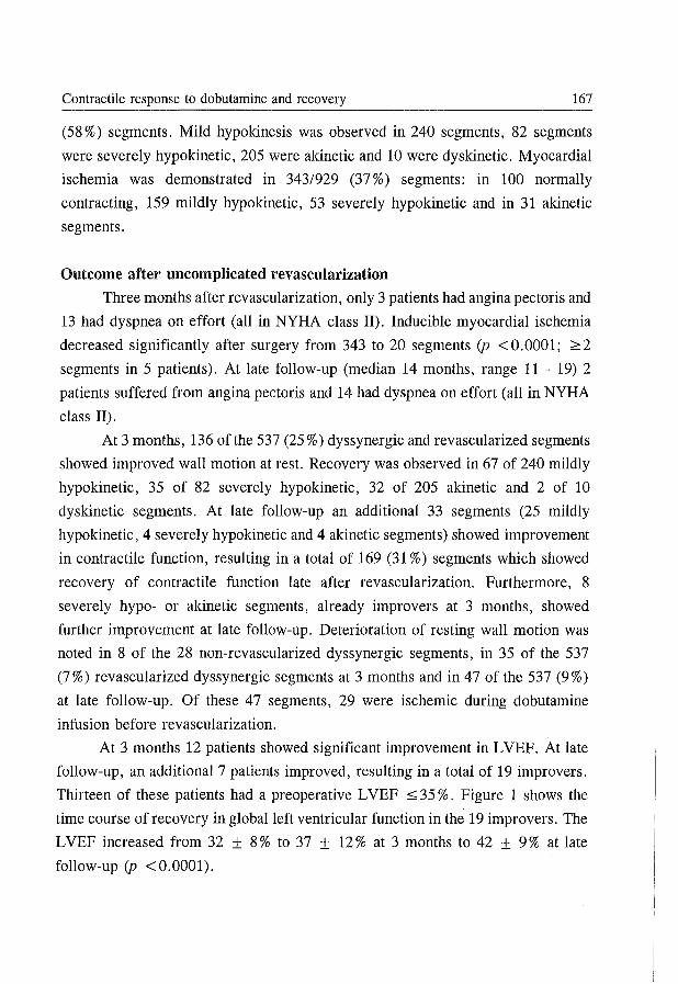
Contractile response to dobutamine and recovery 167
(58 %) segments. Mild hypokinesis was observed in 240 segments, 82 segments
were severely hypokinetic, 205 were akinetic and 10 were dyskinetic. Myocardial
ischemia was demonstrated in 343/929 (37%) segments: in 100 normally
contracting, 159 mildly hypokinetic, 53 severely hypokinetic and in 31 akinetic
segments.
Outcome after uncomplicated revascularization
Three months after revascularization, only 3 patients had angina pectoris and
13 had dyspnea on effort (all in NYHA class II). Inducible myocardial ischemia
decreased significantly after surgery from 343 to 20 segments (p < 0.0001; ~ 2
segments in 5 patients). At late follow-up (median 14 months, range 11 - 19) 2
patients suffered from angina pectoris and 14 had dyspnea on effort (all in NYHA
class II).
At 3 months, 136 of the 537 (25%) dyssynergic and revascularized segments
showed improved wall motion at rest. Recovery was observed in 67 of 240 mildly
hypokinetic, 35 of 82 severely hypokinetic, 32 of 205 akinetic and 2 of 10
dyskinetic segments. At late follow-up an additional 33 segments (25 mildly
hypokinetic, 4 severely hypokinetic and 4 akinetic segments) showed improvement
in contractile function, resulting in a total of 169 (31 %) segments which showed
recovery of contractile function late after revascularization. Furthermore, 8
severely hypo- or akinetic segments, already improvers at 3 months, showed
further improvement at late follow-up. Deterioration of resting wall motion was
noted in 8 of the 28 non-revascularized dyssynergic segments, in 35 of the 537
(7%) revascularized dyssynergic segments at 3 months and in 47 of the 537 (9%)
at late follow-up. Of these 47 segments, 29 were ischemic during dobutamine
infusion before revascularization.
At 3 months 12 patients showed significant improvement in LVEF. At late
follow-up, an additional 7 patients improved, resulting in a total of 19 improvers.
Thirteen of these patients had a preoperative L VEF :::;; 35 %. Figure 1 shows the
time course of recovery in global left ventricular function in the 19 improvers. The
LVEF increased from 32 ± 8 % to 37 ± 12 % at 3 months to 42 ± 9 % at late
follow-up (p <0.0001).
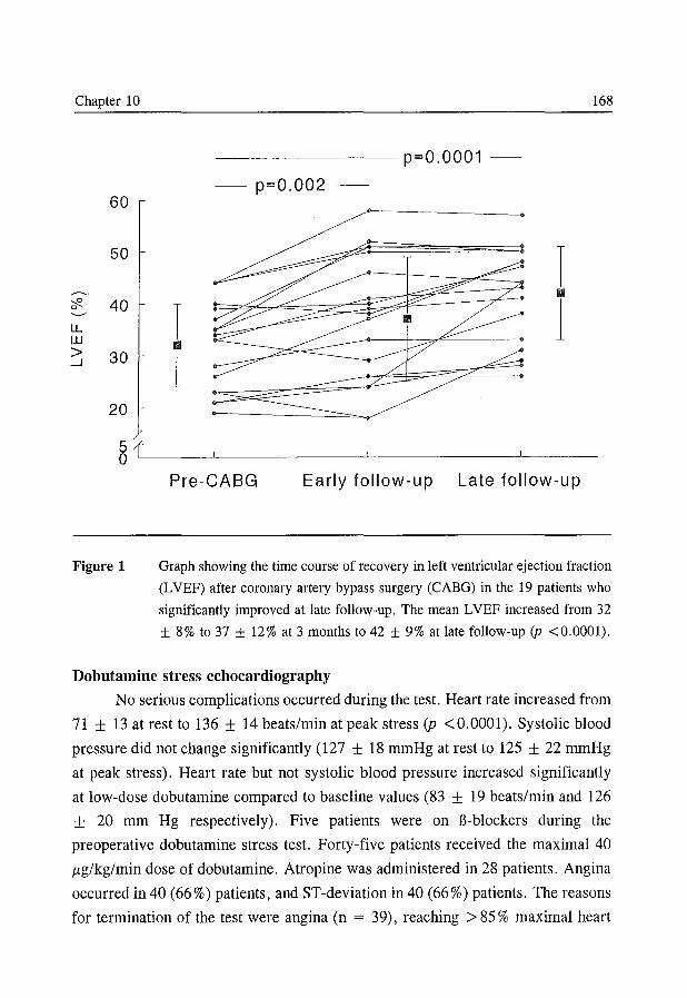
Chapter 10 168
-------- p=O.0001 -
- p=O.002 -60
50 I ,---.. II 'Cf2. 40-
LL W
~ 30
I II
1 1
20
8{~--~-----~------~---
Figure 1
Pre-CABG Early follow-up Late follow-up
Graph showing the time course of recovery in left ventricular ejection fraction
(LVEF) after coronary artery bypass surgery (CABG) in the 19 patients who
significantly improved at late follow-up. The mean LVEF increased from 32
± 8% to 37 ± 12% at 3 months to 42 ± 9% at late follow-up (p <0.0001).
Dobutamine stress ecbocardiograpby
No serious complications occurred during the test. Heart rate increased from
71 ± 13 at rest to 136 ± 14 beats/min at peak stress (p < 0.0001). Systolic blood
pressure did not change significantly (127 ± 18 mmHg at rest to 125 ± 22 mmHg
at peak stress). Heart rate but not systolic blood pressure increased significantly
at low-dose dobutamine compared to baseline values (83 ± 19 beats/min and 126
± 20 mm Hg respectively). Five patients were on J3-blockers during the
preoperative dobutamine stress test. Forty-five patients received the maximal 40
p,g/kg/min dose of dobutamine. Atropine was administered in 28 patients. Angina
occurred in 40 (66 %) patients, and ST -deviation in 40 (66 %) patients. The reasons
for termination of the test were angina (n = 39), reaching> 85 % maximal heart
Contractile response to dobutamine and recovery 169
rate (n = 20) with or without signs of myocardial ischemia, wall motion
abnormalities (n = 1) and hypotension (n = 1).
Of a total of 537 segments with abnormal wall motion that were successfully
revascularized, 186 (35 %) segments exhibited a biphasic response, 58 (11 %)
demonstrated sustained improvement, 68 (12 %) showed worsening (without
improvement) and 225 (42 %) showed no change in regional wall motion during
dobutamine challenge.
Functional recovery versus response during dobutamine echo cardiography
Analysis by segments
The different types of response to dobutamine infusion in relation to the
functional outcome of all dyssynergic segments after revascularization at 3 months
and at late follow-up is depicted in Figure 2. At 3 months, 118/186 (63%)
segments with a biphasic response improved in wall motion, whereas the other 3
patterns were not predictive for functional recovery. At late follow-up, 140/186
(75 %) segments with a biphasic response showed improvement of wall motion.
Also segments with sustained improvement showed a trend to recover at late
follow-up (22 % ). Assuming a biphasic response as indicative for recovery and the
other 3 predefined patterns as indicative for no recovery, the sensitivity,
specificity, positive - and negative predictive values for functional recovery are
87% (eI, 81 % to 93%),83% (eI, 79% to 87),63% (eI, 56% to 70%) and 95%
(eI, 93% to 97%) at 3 months follow-up, and at late follow-up 83% (eI, 77% to
89%), 88% (eI, 85% to 91 %), 75% (eI, 69% to 81 %) and 92% (eI, 89% to
95 %) respectively.
Figure 3 shows the influence of severity of segmental LV dysfunction at
baseline on the predictive value of dobutamine echocardiography and the timing
of the follow-up study. It clearly demonstrates a much less accurate prediction for
functional recovery in mildly hypokinetic segments compared to severely
hypokinetic/akinetic segments. Additionally it shows that the positive predictive
value of both mildly hypokinetic- and severely hypokinetic/akinetic segments
improves when late follow-up is considered (H: 67 % (eI, 59 % to 75 %) versus
53% (eI, 44% to 62%) and SH/AK: 90% (eI, 83% to 97%) versus 81 % (eI,
72% to 90%).
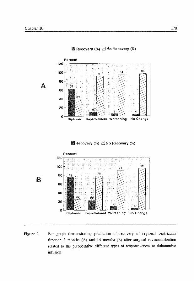
Chapter 10
A
B
Figure 2
Recovery (%) G No Recovery (%)
Percent 120~----------~------~--~----~--'
100 94 96 91
Biphasic Improvement Worsening No Change
Recovery (%) EJ No Recovery (%)
Biphasic Improvement Worsening No Change
170
Bar graph demonstrating prediction of recovery of regional ventricular
function 3 months (A) and 14 months (B) after surgical revascularization
related to the preoperative different types of responsiveness to dobutamine
infusion.
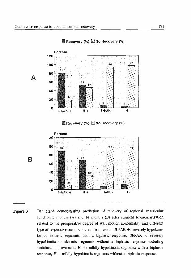
Contractile response to dobutamine and recovery 171
A
B
Figure 3
Recovery (%) G No Recovery (%)
Percent 120r-------~~--~----~----------,
100 94 97
81
3
SH/AK + H+ SH/AK - H -
Recovery (%) EJ No Recovery (%)
Percent 120.-~~--------------~~----------,
SH/AK + H+ SH/AK - H -
Bar graph demonstrating prediction of recovery of regional ventricular
function 3 months (A) and 14 months (B) after surgical revascularization
related to the preoperative degree of wall motion abnOlmality and different
type of responsiveness to dobutamine infusion. SRI AK +: severely hypo kine
tic or akinetic segments with a biphasic response, SRI AK -: severely
hypokinetic or akinetic segments without a biphasic response including
sustained improvement, R +: mildly hypokinetic segments with a biphasic
response, R -: mildly hypokinetic segments without a biphasic response.
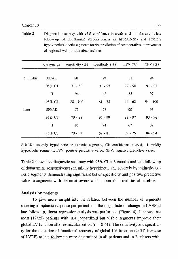
Chapter 10 172
Table 2 Diagnostic accuracy with 95 % confidence intervals at 3 months and at late
follow-up of dobutamine responsiveness in hypokinetic- and severely
hypokineticl akinetic segments for the prediction of postoperative improvement
of regional wall motion abnormalities
dyssynergy sensitivity (%) specificity (%) PPV (%) NPV (%)
3 months SH/AK 80 94 81 94
95% CI 71 - 89 91 - 97 72 - 90 91 - 97
H 94 68 53 97
95% CI 88 - 100 61 - 75 44 - 62 94 - 100
Late SH/AK 79 97 90 93
95% CI 70 - 88 95 - 99 83 - 97 90 - 96
H 86 74 67 89
95% CI 79 - 93 67 - 81 59 - 75 84 - 94
SH/AK: severely hypokinetic or akinetic segments, CI: confidence interval, H: mildly
hypokinetic segments, PPV: positive predictive value, NPV: negative predictive value.
Table 2 shows the diagnostic accuracy with 95 % CI at 3 months and late follow-up
of dobutamine responsiveness in mildly hypokinetic and severely hypokinetic/aki
netic segments demonstrating significant better specificity and positive predictive
value in segments with the most severe wall mati on abnormalities at baseline.
Analysis by patients
To give more insight into the relation between the number of segments
showing a biphasic response per patient and the magnitude of change in L VEF at
late follow-up, linear regression analysis was performed (Figure 4). It shows that
most (17/25) patients with 2::: 4 jeopardized but viable segments improve their
global LV function after revascularization (r = 0.61). The sensitivity and specifici
ty for the detection of functional recovery of global LV function ( 2::: 5 % increase
of L VEF) at late follow-up were determined in all patients and in 2 subsets with
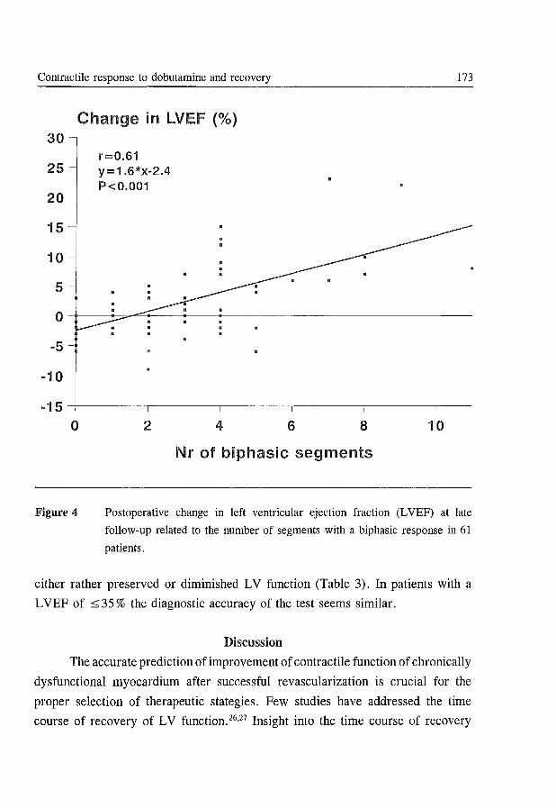
Contractile response to dobutamine and recovery
Change in LVEF (%) 30-
r=O.61 25 y=1.6*x-2.4
P<O.001 20
15
10
5
173
o-~--~~~--------~--------------------------------
-5 -
-10
-15~------~--------~------~------~--------~---
o
Figure 4
2 4 6 8 10
Nr of biphasic segments
Postoperative change in left ventricular ejection fraction (L VEF) at late
follow-up related to the number of segments with a biphasic response in 61
patients.
either rather preserved or diminished LV function (Table 3). In patients with a
L VEF of s 35 % the diagnostic accuracy of the test seems similar.
Discussion
The accurate prediction of improvement of contractile function of chronically
dysfunctional myocardium after successful revascularization is crucial for the
proper selection of therapeutic stategies. Few studies have addressed the time
course of recovery of LV function. 26,27 Insight into the time course of recovery
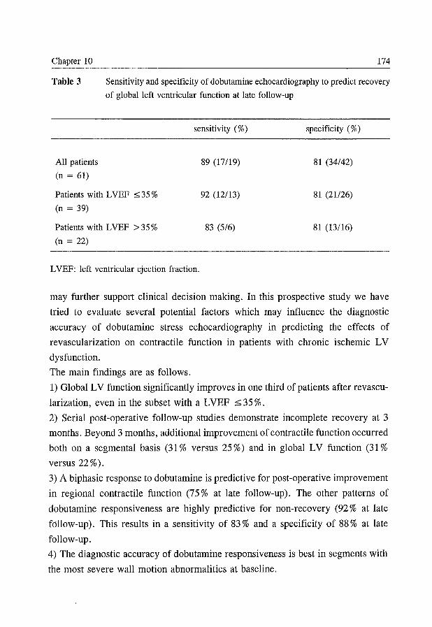
Chapter 10 174
Table 3 Sensitivity and specificity of dobutamine echocardiography to predict recovery
of global left ventricular function at late follow-up
sensitivity (%)
All patients
(n = 61)
Patients with L VEF ::.:; 35 % (n = 39)
Patients with LVEF > 35 % (n = 22)
L VEF: left ventricular ejection fraction.
89 (17/19)
92 (12/13)
83 (5/6)
specificity (%)
81 (34/42)
81 (21126)
81 (13/16)
may further support clinical decision making. In this prospective study we have
tried to evaluate several potential factors which may influence the diagnostic
accuracy of dobutamine stress echocardiography in predicting the effects of
revascularization on contractile function in patients with chronic ischemic LV
dysfunction.
The main findings are as follows.
1) Global LV function significantly improves in one third of patients after revascu
larization, even in the subset with a L VEF ::.:;; 35 % .
2) Serial post-operative follow-up studies demonstrate incomplete recovery at 3
months. Beyond 3 months, additional improvement of contractile function occurred
both on a segmental basis (31 % versus 25 %) and in global LV function (31 % versus 22 % ).
3) A biphasic response to dobutamine is predictive for post-operative improvement
in regional contractile function (75% at late follow-up). The other patterns of
dobutamine responsiveness are highly predictive for non-recovery (92 % at late
follow-up). This results in a sensitivity of 83 % and a specificity of 88 % at late
follow-up.
4) The diagnostic accuracy of dobutamine responsiveness is best in segments with
the most severe wall motion abnormalities at baseline.
Contractile response to dobutamine and recovery 175
5) Dobutamine stress echocardiography in the aforementioned clinical setting has
a sensitivity of 89% and a specificity of 81 % to predict functional recovery of
global LV function on a patient basis.
All previous studies with dobutamine echocardiography have used
echocardiography to assess improvement of wall motion, without an independent
technique to verify these data. 12-20 In the present study we have circumvented this
problem using serial radionuclide ventriculographic studies for the assessment of
changes in global LV function. Our study is also unique in reporting the effects of
sequential follow-up studies on the diagnostic accuracy of the test for post
revascularization improvement of both regional and global LV function. It provides
more insight into the clinical relevance of the test since it includes analysis on an
individual patient basis. In line with previous studies, a patient was expected to
improve in global LV function at late follow-up when at least 4 abnormal
contracting segments at baseline (25 % of the myocardium) demonstrated a biphasic
response to dobutamine. Furthermore this study adds to the accumulating evidence
that high dose dobtamine stress testing, even with atropine on top, can be
performed safely without serious complications also in patients with poor LV
function. 28
Biphasic response
Several studies, experimental or clinical using a head to head comparison
between dobutamine stress echo cardiography and myocardial perfusion scinti
graphy, describe a relation between a biphasic response to dobutamine and
myocardial scintigraphic ischemia. 29-31 This can be explained by the presence of
viable tissue in a segment subtended by a stenotic coronary artery, exhibiting a
contractile response to low-dose dobutamine and ischemia at high dose dobutamine
provoked by the increased rate pressure product and flow maldistribution. An
alternative explanation has been proposed by Schultz and coworkers. 32.33 In a swine
model of short-term hibernation, using a continuous intracoronary dobutamine
infusion, they showed that both myocardial ischemia and depletion of high-energy
phosphates explain the biphasic response. It remains unclear whether the increasing
dosage of dobutamine or the duration of dobutamine infusion is responsible for the
late decrease in myocardial thickening in segments exhibiting a biphasic response.
Chapter 10 176
In the present study, the majority of segments demonstrating such a biphasic
response recovered in contractile function after revascularization. These findings
are in line with data reported by Afridi et al. 16 It is likely that after successful
revascularization high-energy phosphate stores are ensured through adequate
myocardial perfusion at rest and at stress. Under these circumstances dyssynergic
myocardium may recover frequently.
Other types of dobutamine responsiveness
In segments demonstrating a sustained improvement during dobutamine, it
is likely that enough high-energy phosphates are present to maintain enhanced
contractility until the final stage of the test. Only ,22 % of the segments with
sustained improvement showed recovery after revascularization. Since these
segments are unlikelY to be exposed to repeated ischemic insults preoperatively,
they may recover less frequently after revascularization. An alternative explanation
for the low recovery rate of segments with sustained improvement may be the
coexistence of coronary artery disease with cardiomyopathy. Segments with direct
worsening in wall motion during dobutamine challenge rarely improved after
revascularization. It is conceivable that these segments contain a mixture of scar
tissue and normal myocardium becoming ischemic during dobutamine infusion.
Although Afridi et aP6 reported a higher recovery rate of these segments (35 %),
one should bear in mind that in absolute terms this represents only 6 segments.
Finally the lack of change in wall thickening during dobutamine infusion is a very
specific marker for the prediction of lack of functional improvement after surgical
revascularization. This echocardiographic pattern of non-responding to dobutamine
therefore is in agreement with the diagnosis of scar with no clinical relevant
amount of residual viable myocardium. This observation is quite relevant, since it
suggests that, in patients with chronic advanced ischemic LV dysfunction, further
tests for the assessment of potential reversibility of dysfunction in segments with
this specific echo cardiographic pattern are redundant.
Mildly versus severely dyssynergic segments
Previous studies with low-dose dobutamine echocardiography have
concentrated on the prediction of regional recovery after revascularization. They
Contractile response to dobutamine and recovery 177
show sensitivities ranging from 71 % to 97 % (weighted mean 85 %) and specifici
ties ranging from 63 % to 95 % (weighted mean 89 %).12.14-20 Our results add to the
accumulating evidence that the test is an accurate method for the evaluation of
myocardial viability in patients with chronic dysfunctional myocardium due to
coronary artery disease. In our study, both specificity and positive predictive value
improved when only severely dyssynergic segments were evaluated, similar to
previous studies.14.15.18 Mildly hypokinetic segments are likely to contain normal
myocardium, subendocardial scar and/or viable but dysfunctional myocardium. If
relatively little viable tissue is present, dobutamine challenge may stimulate normal
myocardium to a hyperkinetic response simulating a positive test for viability. If
on the other hand a substantial amount of viable myocardium is present, a similar
test result may appear. But only in the latter example one expects functional
recovery to occur after revascularization. False positive tests occur less often in
severe dyskinesis since the presence of normal myocardial tissue is minimal in
segments with severe contractile dysfunction.
Prediction of improvement in global LV function
In this study we showed that patients with ~ 4 jeopardized but viable
segments (25 % of the myocardium) are likely to improve global LV function. The
data confirm earlier studiesI7.34-38 employing different techniques to assess
myocardial viability, and indicate that a substantial amount of viable but
jeopardized myocardium needs to be present to result in improved global LV
function. This observation implies, if confirmed in larger series, that dobutamine
stress echocardiography is a reliable technique to predict functional outcome after
revascularization. An accurate prediction is of paramount importance for the
selection of the most appropriate treatment in individual patients with chronic
ischemic poor LV function, since L VEF is an important predictor for survival.
Timing of functional recovery
The optimal timing for the assessment of functional recovery after
revascularization is essential for the correct interpretation of the diagnostic
accuracy of a given test. Currently, functional follow-up studies in chronic
coronary artery disease are performed frequently within 3 months after the
Chapter 10 178
revascularization procedure. 14-20 ,38 However, in individual patients all types of
reversible and irreversible contractile dysfunction may coexist and in them it is
impossible to distinguish repetitive myocardial stunning from hibernation. In
stunned myocardium recovery of function may occur within days or weeks. Given
the severity of structural changes observed in hibernating myocardium,21,22 it is
likely that under these conditions recovery of function after revascularization is
delayed. Therefore, complete recovery of contractile function should not be
expected before 4 to 8 months after revascularization in patients with chronic
coronary artery disease. Our data support these concepts since 7 of the 19
improvers at late follow-up did not show improvement of global LV function at 3
months. Such delayed recovery after CABG has been described earlier26,27,39 and
underlines the importance of late follow-up studies after revascularization
procedures in this clinical setting.
Limitations of the study Incomplete revascularization may prohibit viable segments to recover,
thereby underestimating the diagnostic accuracy of a diagnostic technique. Since
we did not perform repeated angiography after surgery to assess graft patency, we
are not informed about the successfulness of coronary revascularization. However
repeated dobutamine stress echocardiographic studies performed 3 months after
CABG revealed a decrease in inducible myocardial ischemia from 343 to 20
segments. This study was performed in an experienced center dealing with patients
with LV dysfunction and thus it represents daily clinical practice.
Since we did not study patients at 6 months, we are not informed about the
completeness of recovery at 6 months. Therefore we cannot comment about this
issue regarding the optimal timing of follow-up studies for the detection of
complete recovery of contractile function after CABG in the aforementioned
clinical setting. Detailed and larger studies are needed to answer this question.
Regional wall thickness has not been measured. Normal end-diastolic
thickness has been described as marker of myocardial viability and a predictor of
recovery after revascularization. 4o ,41 On the other hand, dyssynergic segments with
severely reduced thickness rarely show improved contractile function after
revascularization. Further studies are needed to answer the question what the
Contractile response to dobutamine and recovery 179
additional value is of dobutamine challenge on top of wall thickness measurements.
Patients with poor LV function may potentially benefit most from
myocardial viability studies given the condition that viability leads to improvement
of global LV function. Since only a small number of patients with poor LV
function were studied we cannot draw firm conclusions about the diagnostic
accuracy of dobutamine stress echocardiography for the prediction of functional
recovery in these patients.
Conclusions
Serial post-operative follow-up studies demonstrate substantial incomplete
recovery of contractile function at 3 months. Dobutamine stress echocardiography
is an useful method to predict improvement of regional and global LV function
after CABG. Its diagnostic accuracy depends on combining low and high doses of
dobutamine infusion, severity of dyssynergy and the timing of evaluation. This
study contributes to the increasing clinical experience with dobutamine stress
echocardiography in patients with stable ischemic LV dysfunction.
References
1. Elefteriades JA, Tolis G, Levi E, Mills LK, Zaret BL. Coronary artery bypass
grafting in severe left ventricular dysfunction: excellent survival with improved
ejection fraction and functional state. J Am Coli Cardiol 1993;22:1411-1417.
2. Dilsizian V, Bonow RO. Current diagnostic techniques of assessing viability in
patients with hibernating and stunned myocardium. Circulation 1993;87:1-20.
3. Eitzman D, Al-Aouar ZR, Kanter HL, et al. Clinical outcome of patients with advan
ced coronary artery disease after viability studies with positron emission tomography.
J Am Coli Cardiol 1992;20:559-565.
4. Yoshida K, Gould KL. Quantitative relation of myocardial infarct size and myocardial
viability by positron emission tomography to left ventricular ejection fraction and 3-
year mortality with and without revascularization. J Am Coli Cardiol 1993;22:984-
987.
5. Di Carli MF, Davidson M, Little R, et al. Value of metabolic imaging with positron
emission tomography for evaluating prognosis in patients with coronary artery disease
and left ventricular dysfunction. Am J Cardiol 1994;73:527-533.
6. Lee KS, Marwick TH, Cook SA, et al. Prognosis of patients with left ventricular
Chapter 10 180
dysfunction, with and without viable myocardium after myocardial infarction. Relative
efficacy of medical therapy and revascularization. Circulation 1994;90:2687-2694.
7. Pierard LA, De Landsheere CM, Berthe C, Rigo P, Kulbertus HE. Identification of
viable myocardium by echocardiography during dobutamine infusion in patients with
myocardial infarction after thrombolytic therapy: comparison with positron emission
tomography. J Am ColI CardioI1990;15:1021-1031.
8. Barilla F, Gheorghiade M, Alam M, Khaja F, Goldstein S. Low-dose dobutamine in
patients with acute myocardial infarction identifies viable but not contractile
myocardium and predicts the magnitude of improvement in wall motion abnormalities
in response to coronary revascularization. Am Heart J 1991;122:1522-1531.
9. Smart SC, Sawada S, Ryan T, et al. Low-dose dobutamine echocardiography detects
reversible dysfunction after thrombolytic therapy of acute myocardial infarction.
Circulation 1993;88:405-415.
10. Watada H, Ito H, Oh H, et al. Dobutamine stress echocardiography predicts
reversible dysfunction and quantitates the extent of irreversibly damaged myocardium
after reperfusion of anterior myocardial infarction. J Am ColI Cardiol 1994;24:624-
630.
11. Salustri A, Elhendy A, Garyfallydis P, et al. Prediction of improvement of ventricular
function after first acute myocardial infarction using low-dose dobutamine stress
echocardiography. Am J Cardiol 1994;74:853-856.
12. Marzullo P, Parodi 0, Reisenhofer B, et al. Value of rest thallium-201ltechnetium-
99m sestamibi scans and dobutamine echocardiography for detecting myocardial
viability. Am J CardioI1993;71:166-172.
13. Cigarroa CG, de Filippi CR, Brickner ME, Alvarez LG, Wait MA, Grayburn PA.
Dobutamine stress echocardiography identifies hibernating myocardium and predicts
recovery of left ventricular function after coronary revascularization. Circulation
1993;88:430-436.
14. La Canna G, Alfieri 0, Giubbini R, Gargano M, Ferrari R, Visioli 0. Echocardio
graphy during infusion of dobutamine for identification of reversible dysfunction in
patients with chronic coronary artery disease. J Am ColI Cardiol 1994;23:617-626.
15. Arnese M, Cornel JH, Salustri A, et al. Prediction of improvement of regional left
ventricular function after surgical revascularization: a comparison of low-dose
dobutamine echocardiography with 20lTl single-photon emission computed tomograp
hy. Circulation 1995;91:2748-2752.
16. Afridi I, Kleiman NS, Raizner AE, Zoghbi WA. Dobutamine echocardiography in
myocardial hibernation: optimal dose and accuracy in predicting recovery of
ventricular function after coronary angioplasty. Circulation 1995;91:663-670.
Contractile response to dobutamine and recovery 181
17. Senior R, Glenville B, Basu S, et al. Dobutamine echocardiography and thallium-201
imaging predict functional improvement after revascularization in severe ischaemic
left ventricular dysfunction. Br Heart J 1995;74:358-364.
18. DeFilippi CR, Willett DWL, Irani WN, Eichhorn EJ, Velasco CE, Grayburn PA.
Comparison of myocardial contrast echocardiography and low-dose dobutamine stress
echocardiography in predicting recovery of left ventricular function after coronary
revascularization in chronic ischemic heart disease. Circulation 1995;92:2863-2868.
19. Voci P, Bilotta F, Caretta Q, Mercanti C, Marino B. Low-dose dobutamine echo car
diography predicts the early response of dysfunctioning myocardial segments to
coronary artery bypass grafting. Am Heart J 1995;129:521-526.
20. Perrone-Filardi P, Pace L, Prastaro M, et al. Dobutamine echocardiography predicts
improvement of hypoperfused dysfunctional myocardium after revascularization in
patients with coronary artery disease. Circulation 1995;91:2556-2565.
21. Maes A, Flameng W, Nuyts J, et al. Histological alterations in chronically
hypoperfused myocardium. Correlation with PET findings. Circulation 1994;90:735-
745.
22. Borgers M, Thone F, Wouters L, Ausma J, Shivalkar B, Flameng W. Structural
correlates of regional myocardial dysfunction in patients with critical coronary artery
stenosis: Chronic hibernation? Cardiovasc Pathol 1993;2;237-245.
23. Mc Neill AJ, Fioretti PM, EI-Said EM, Salustri A, Forster T, Roelandt JRTC.
Enhanced sensitivity for detection of coronary artery disease by addition of atropine
to dobutamine stress echocardiography. Am J CardioI1992;70:41-46.
24. Schiller NB, Shah PM, Crawford M, et al. Recommendations for quantitation of the
left ventricle by two-dimensional echocardiography. J Am Soc Echocardiogr
1989;2:358-367.
25. Arnese M, Fioretti PM, Cornel JR, Postma-Tjoa J, Reijs AEM, Roelandt JRTC.
Akinesis becoming dyskinesis during high-dose dobutamine stress echocardiography:
a marker of ischemia or a mechanical phenomenon? Am J CardioI1994;73:896-899.
26. Mintz U, Ingels NB Jr, Daughters GI II, Stinson EB, Alderman EL. Sequential
studies of left ventricular function and wall motion after coronary arterial bypass
surgery. Am J CardioI1980;45:210-216.
27. Vanoverschelde J-L, Melin JA, Depre C, Borgers M, Dion R, Wijns W. Time-course
of functional recovery of hibernating myocardium after coronary revascularization
(abstract). Circulation 1994;90 (part 2):1-378.
28. Cornel JR, Balk AHMM, Arnese M, et al. Safety and feasibility of dobutamine
atropine stress echocardiography in patients with ischemic left ventricular dysfunction.
J Am Soc Echocardiogr 1996;9:27-32.
Chapter 10 182
29. Chen C, Li L, Chen LL, et al. Incremental doses of dobutamine induce a biphasic
response in dysfunctional left ventricular regions subtending coronary stenoses.
Circulation 1995;92:756-766.
30. Senior R, Lahiri A. Enhanced detection of myocardial ischemia by stress dobutamine
echocardiography utilizing the "biphasic" response of wall thickening during low and
high dose dobutamine infusion. J Am ColI Cardiol 1995;26:26-32.
31. Elhendy A, Cornel JH, Roelandt JRTC, van Domburg RT, Fioretti PM. Relationship
between contractile response of akinetic segments during dobutamine stress
echocardiography and ischemia assessed by simultaneous 201 thallium SPECT. Am
J Cardiol 1996, in press.
32. Schultz R, Rose J, Martin C, Brodde OE, Heusch G. Development of short-term
myocardial hibernation: its limitation by the severity of ischemia and inotropic
stimulation. Circulation 1993;88:684-695.
33. Schultz R, Guth BD, Pieper K, Martin C, Heusch G. Recruitment of an inotropic
reserve in moderately ischemic myocardium at the expense of metabolic recovery:a
model of short-term hibernation. Circ Res 1992;70:1282-1295.
34. Tillisch J, Brunken R, Marshall R, et al. Reversibility of cardiac wall-motion abnor
malities predicted by positron tomography. N Engl J Med 1986;314:884-888.
35. Vanoverschelde J-U, Gerber BL, D'Hondt A-M, et al. Preoperative selection of
patients with severely impaired left ventricular function for coronary revasculariza
tion. Role of low-dose dobutamine echocardiography and exercise-redistribution
reinjection thallium SPECT. Circulation 1995;92 [Supplll]:37-44.
36. Iskandrian AS, Hakki A, Kane SA, et al. Rest and redistribution thallium-201
myocardial scintigraphy to predict improvement in left ventricular function after
coronary arterial bypass grafting. Am J CardioI1983;51:1312-1316.
37. Ragosta M, Beller GA, Watson DD, Kaul S, Gimple LW. Quantitative planar rest
redistribution Tl-201 imaging in detection of myocardial viability and prediction of
improvement in left ventricular function after coronary artery bypass surgery in
patients with severely depressed left ventricular function. Circulation 1993; 87: 1630-
1641.
38. Meluzin J, Cigarroa CG, Brickner ME, et al. Dobutamine echocardiography in
predicting improvement in global left ventricular systolic function after coronary
bypass or angioplasty in patients with healed myocardial infarcts. Am J Cardiol
1995;76:877-880.
39. Luu M, Warner Stevenson L, Brunken RC, Drinkwater DM, Schelbert HR, Tillisch
JH. Delayed recovery of revascularized myocardium after referral for cardiac
transplantation. Am Heart J 1990;119:668-670.

Contractile response to dobutamine and recovery 183
40. Baer FM, Voth E, Schneider CA, Theissen P, Schicha H, Sechtem U. Comparison
of low-dose dobutamine-gradient-echo magnetic resonance imaging and positron
emission tomography with [18FJf1uorodeoxyglucose in patients with chronic coronary
artery disease. A functional and morphological approach to the detection of residual
myocardial viability. Circulation 1995 ;91: 1006-1015.
41. Faletra F, Crivellaro W, Pirelli S, et al. Value of transthoracic two-dimensional
echocardiography in predicting viability in patients with healed Q-wave anterior wall
myocardial infarction. Am J Cardiol 1995;76:1002-1006.


Chapter 11
Summary and Future Perspectives
Chapter 11 186
Summary
Detection of ·myocardial viability has gained importance in clinical
cardiology. The increasing number of patients with left ventricular (LV) systolic
dysfunction due to ischemic injury and the awareness of the potential reversibility
of LV systolic dysfunction after revascularization has led to a search for accurate
and readily available diagnostic methods to detect residual viable myocardium.
Myocardial perfusion imaging may detect viable myocardium, since tracer uptake
requires adequate microvascular perfusion, cellular integrity and metabolic
function. Recruitment of contractile reserve in hypocontractile myocardial segments
by using inotropic stimuli also reflects residual viability but represents a different
cellular mechanism than perfusion. Indeed, the concept of metabolically viable
myocardium must be distinguished from the ability to recover mechanical
myocardial function. Dobutamine stress echocardiography may serve as an
alternative for myocardial perfusion and metabolic imaging techniques for the
recruitment of myocardial residual function. In this thesis, several different
diagnostic techniques are evaluated for their ability to identify patients who are
likely to improve resting LV function after myocardial infarction or after
revascularization procedures.
In Chapter 2, the value of resting sestamibi imaging to distinguish viable
myocardium from scar tissue and to predict functional recovery has been discussed.
Sestamibi is a technetium-99m labeled myocardial perfusion agent. The uptake of
sestamibi is dependent upon cell membrane integrity and mitochondrial function.
The results of all comparative studies between sestamibi imaging and other viability
tracers such as thallium-201 eOITl) or 18p_PDG using positron emission tomograp
hy suggest that sestamibi underestimates the presence of severely hypoperfused but
still viable myocardium in patients with chronic coronary artery disease. However,
recent data suggest that the differences between 20lTI and sestamibi are smaller
when nitrates are administered in combination with quantitative analysis and when
late imaging is used. Despite its lower sensitivity for detecting viable myocardium,
sestamibi tracer uptake may allow for adequate prediction of functional recovery
after successful revascularization. Indeed, failing to detect limited amounts of
viable myocardium has no major impact on the diagnostic accuracy in predicting
the outcome of revascularization. Only a small number of limited studies have
Summary and future perspectives 187
addressed the role of sestamibi in predicting functional recovery after revasculari
zation. Therefore, the exact value of sestamibi imaging has still to be defined.
In Chapter 3 we reported the results of a study addressing the concordance
between the two currently most advocated 201TI imaging protocols, post-stress 201TI
redistribution-reinjection and rest-redistribution 201TI SPECT, for the detection of
myocardial viability in patients with chronic coronary artery disease and depressed
LV function. An excellent agreement between the two protocols was found, even
after omission of the post-stress redistribution scan. The use of a semi-quantitative
analysis of the SPECT results was crucial to reach such high concordance. The
high level of agreement sustained when the analysis was limited to dyscontractile
myocardial segments. These results justify the use of dobutamine stress-reiIUection
201TI SPECT without the acquisition of redistribution images, in detecting residual
myocardial viability in patients with chronic coronary artery disease.
The safety and feasibility profile of high dose dobutamine-atropine stress
echocardiography in 318 patients with ischemic LV dysfunction is reported in
Chapter 4. In our study, no serious complications (myocardial infarction,
ventricular fibrillation or death) occurred. In patients with a LV ejection fraction
of 25 % or less, there was a higher incidence of significant tachyarrhythmias (14 %
versus 5 %), but the feasibility of the test remained high. Multivariate analysis
indicated that a history of tachyarrhythmias was the only predictor of stress
induced arrhythmias. Therefore, advanced LV dysfunction on itself does not
represent a risk factor for serious complications or adverse effects during
dobutamine-atropine stress testing.
The second part of this thesis represents the results of studies aimed at
evaluating various diagnostic methods for the identification of dyscontractile
myocardium likely to improve mechanical function.
In Chapter 5 the prevalence of spontaneous improvement of regional LV
dysfunction in 57 unselected patients after a first uncomplicated acute myocardial
infarction (thrombolysis, n = 27) was assessed. Approximately one fourth of the
dyscontractile segments showed functional recovery at 3 month follow-up.
Spontaneous recovery occurred more frequently in hypokinetic than in akinetic
segments (35 % versus 19 %). It was demonstrated that low-dose dobutamine stress
Chapter 11 188
echocardiography provides specific information for predicting lack of improvement
at follow-up in these patients. Also, the test has a high sensitivity for predicting
functional improvement in hypokinetic segments, but was not useful in identifying
akinetic segments which improve spontaneously.
Evaluation of ECG changes during a low-dose dobutamine stress test may
also be helpful for identifying dyssynergic but still viable myocardium in patients
after a recent Q-wave myocardial infarction (Chapter 6). In a group of 90
postinfarction patients with negative T waves in ~ 2 infarct-related ECG leads, we
tested the hypothesis that T -wave normalization during low-dose dobutamine
infusion represents residual viable myocardium. A good agreement was found
between low-dose dobutamine induced T-wave changes and wall motion changes.
Furthermore, the sensitivity of low-dose dobutamine stress echocardiography for
predicting late spontaneous recovery of segmental wall motion at 3 month follow
up increased, without loss of specificity, when the T-wave changes on the ECG
were added to the analysis. Thus T-wave normalization during low-dose
dobutamine stress testing provides additional information to identify viable
myocardium after acute myocardial infarction.
In patients with stable chronic coronary artery disease accepted for
myocardial revascularization, we have performed three studies describing the direct
comparison of two tests for the prediction of functional outcome of resting regional
and, or global LV function at 3 month follow-up (Chapters 7-9). In these patients
approximately 30 % of the dyscontractile segments showed functional recovery at
3 month follow-up. Most patients in whom residual viability in more than 25% of
the LV was demonstrated, showed post -operative improvement of global LV
function. Similarly to the spontaneous recovery observed after acute myocardial
infarction, recovery after revascularization occurred more frequently in hypokinetic
than in akinetic segments.
In Chapter 7 we report our study on low-dose dobutamine stress echocardio
graphy which was compared with dobutamine stress-reinjection 201Tl SPECT in 38
patients in predicting functional recovery of 170 severely hypokinetic or akinetic
segments. All patients had severe LV dysfunction. 201Tl SPECT demonstrated resi
dual viability more frequently than low-dose dobutamine stress echocardiography
(61 % versus 19 %). However , post-operative recovery was grossly overestimated
Summary and future perspectives 189
by 201TI SPECT resulting in a modest specificity of 48% as compared the 96%
specificity of low-dose dobutamine stress echocardiography. In contrast to the
results described in Chapter 5, the responsiveness of severely dysfunctional
segments to low-dose dobutamine stress was also a sensitive predictor of post
operative recovery, similar to 201TI SPECT. Thus, 201TI SPECT appears to be less
useful than low-dose dobutamine stress echocardiography to predict the post
operative improvement of regional wall motion in patients with severe LV
dysfunction. The data also suggest that the cellular mechanism responsible for a
positive inotropic response to dobutamine stimulation requires a higher degree of
myocyte functional integrity than the metabolic mechanism responsible for 201TI
uptake. For recovery of contractile function a "critioal mass" of viable myocytes
must be available.
Since it is feasible to image myocardial FDG uptake with a conventional
gamma camera using 511 keY collimators, we have evaluated the use of FDG
SPECT for its prediction of functional recovery in 55 patients prior to revasculari
zation and compared this technique with rest-redistribution 201TI SPECT in a
subgroup of 24 patients (Chapter 8). Dysfunctional segments with a resting
perfusion defect and increased FDG uptake, a perfusion-metabolism mismatch, had
a significant functional recovery. Considering both normal perfusion and a
mismatch as indicative for recovery, FDG SPECT reached a sensitivity of 85 %
and a specificity of 75 % to predict functional recovery. In hypokinetic segments
the specificity was lower in comparison to akinetic segments. Global LV function
improved significantly in patients with ~ 3 viable (of the 13) segments on FDG
SPECT. These results are similar to those reported using positron emission
tomography and suggest that FDG SPECT, in spite of its lower spatial resolution,
can be helpful in the clinical management of patients with chronic ischemic LV
dysfunction. In direct comparison FDG SPECT showed a better specificity as
compared to rest-redistribution 201TI SPECT (77 % versus 57 %). Stepwise logistic
regression analysis of the FDG and 201TI rest-redistribution data showed that a
perfusion-metabolism mismatch was the best predictor of functional recovery. All
.other parameters did not contribute independently. Thus FDG SPECT seems
superior to rest-redistribution 201TI SPECT in the identification of patients in whom
regional and global LV function can improve after revascularization.
Chapter 11 190
In Chapter 9 FDG SPECT was compared with low-dose dobutamine stress echo
cardiography. Both techniques demonstrate similar high diagnostic accuracies in
detecting post-operative improvement of regional and global LV function. However
in mildly hypokinetic segments both techniques, but especially low-dose
dobutamine stress echocardiography, overestimate the probability of post-operative
recovery (specificity 45 % versus 74% for SPECT). Since in clinical practice the
issue of myocardial viability is mainly relevant in severe contraction abnormalities,
this should not be considered an important limitation.
In Chapter lOwe evaluated the time course of functional improvement after
coronary artery bypass graft surgery (CABG). The follow-up period was 1 year.
We further explored the value of dobutamine stress echocardiography by
combining low- and high-dose dobutamine. Of the 61 patients, global LV function
improved in 12 at 3 months and in a total of 19 at late follow-up. A biphasic
response to dobutamine, defined as contractile improvement at low-dose and
worsening at peak stress, was predictive for segmental functional recovery in 63 %
at 3 months and in 75 % at late follow-up. Other responses were highly predictive
for non-recovery. Thus the combination of dysfunctioning myocardium which
shows improvement after dobutamine stimulation at low dose and inducible
ischemia more likely predicts recovery after revascularization than viable
myocardium which is not in jeopardy. Again, similar to our findings in Chapter
9, the positive predictive value was dependent on the severity of the contraction
abnormality at rest. The highest accuracy was reached in akinetic segments. The
test was also accurate in predicting improvement of global LV function at late
follow-up. In the presence of ~ 4 jeopardized segments prior to surgery, ejection
fraction improved with at least 5 points in 68% of the patients. Thus, serial post
operative follow-up studies demonstrate incomplete recovery of regional and global
contractile function at 3 months. Furthermore, the diagnostic accuracy of
dobutamine stress echocardiography to predict recovery seems to depend on
combining low and high dose dobutamine stimulation, severity of dyssynergy at
baseline and the timing of evaluation.
Conclusions and future perspectives
In patients with chronic ischemic LV dysfunction, both dobutamine stress
Summary and future perspectives 191
echocardiography and FDG SPECT are useful methods to detect dysfunctional but
viable myocardium likely to recover after adequate revascularization. Viable
myocardium which is in jeopardy seems more likely to recover than viable
myocardium which is not in jeopardy. Therefore both techniques, by detecting
either a biphasic contractile response or a perfusion-metabolism mismatch, have
high diagnostic accuracies. The exact value of scintigraphic perfusion techniques
remains unclear. It is likely that the traditional criteria of 20lTl SPECT used for the
identification of myocardial viability are not optimal for the prediction of functional
recovery. Furthermore the implications of an extended follow-up of 1 year on the
diagnostic accuracy of SPECT has not been addressed and several other questions
remain to be answered.
The studies described in this thesis concentrated on the potential of viable
myocardium to improve resting systolic LV function. Other potential aspects of
viable myocardium may have clinical importance such as the prevention of infarct
expansion, preservation of contractile reserve and, perhaps reversing remodeling.
This may improve the functional status of the patient, increase exercise capacity
(quality of life) and delay the development of heart failure. At present there are no
data available concerning these topics. Preliminary data using positron emission
tomography indicate that revascularization of areas with perfusion-metabolism
mismatch may favourably influence prognosis. Furthermore, larger studies are
needed in patients with severely impaired LV function to determine the value of
the detection of viable but jeopardized myocardium to predict recovery of global
LV function after successful revascularization. There are indications that both
contractile reserve and baseline end-diastolic volume (as a sign of severity of LV
remodeling) are independent predictors of post-operative improvement of global
LV function.


Samenvatting
Samenvatting 194
Sam en vatting De opsporing van vitaal myocard wordt steeds belangrijker in de dagelijkse
cardiologische praktijk. Niet aIleen neemt het aantal patienten toe met een
gestoorde linker ventrikel (LV) funktie door ischemische schade, tevens groeit het
besef dat de LV funktie kan herstellen na revascularisatie. Dit heeft geleid tot het
ontwikke1en van goede, relatief eenvoudige diagnostische methoden om vitaal
myocard op te sporen.
Het afbeelden van myocardia1e perfusie levert hieromtrent informatie op, daar
tracer opname athankelijk is van de mate van microvasculaire perfusie, van
cellulaire integriteit en metabole funktie. Hoewel het aantonen van contractiele
reserve van dysfunctioneel myocard door middel van ~n inotrope stimulus ook een
uiting is van vitaliteit, is er hierbij sprake van een ander cellulair mechanisme. Het
concept van metabool lev end myocard dient te worden onderscheiden van het
vermogen tot herste1 van de LV funktie. Dobutamine stress echocardiografie kan
een alternatief zijn voor afbeeldingstechnieken van myocardiale perfusie en
metabolisme om de mogelijkheden tot herstel van de LV funktie te voorspellen. In
dit proefschrift zullen verschillende diagnostische methoden worden onderzocht op
het vermogen om patienten te identificeren bij wie de LV funktie zal gaan
hersteIlen, zowel spontaan na een acuut hartinfarct als na een revascularisatie
procedure.
In hoofdstuk 2 wordt de waarde van sestamibi scintigrafie besproken.
Sestamibi is een aan technetium gekoppeld radiofarmacon. De myocardiale opname
van sestamibi is athankelijk van de integriteit van de celmembraan en van de
mitochondria1e funktie. Vergelijkende studies tussen sestamibi scintigrafie en
thallium-201 eOITl) scintigrafie of I8P_PDG positron emissie tomografie wijzen
erop, dat sestamibi de aanwezigheid van dysfunctionerend maar nog vitaal myocard
onderschat bij patienten met chronisch coronairlijden. De verschillen tussen 20ITl
en sestamibi blijven echter beperkt als een voorbehandeling met nitraten in
combinatie met kwantitatieve beeldanalyse plaats vindt, alsmede wanneer late
beeldacquisitie wordt toegepast. Ondanks de wat lagere sensitiviteit voor het
.opsporen van vitaal myocard kan sestamibi scintigrafie theoretisch nog steeds het
mogelijke herstel van pompfunktie na revascularisatie accuraat voorspellen.
Immers, de mate van onderschatting en niet de onderschatting zelve bepaald de
Samenvatting 195
nauwkeurigheid in het voorspellen hiervan. De waarde van sestamibi scintigrafie
voor het voorspellen 'van het herstel van pompfunktie na revascularisatie blijft
vooralsnog onduidelijk, daar tot op heden slechts een beperkt aantal kleine
onderzoeken hiernaar zijn verricht.
In hoofdstuk 3 wordt een vergelijkend onderzoek besproken naar de waarde
van 2 verschillende 20lTI protocoIlen, te weten dobutamine stress-redistributie
reinjectie en rust-redistributie, voor wat betreft de detectie van myocardiale
vitaliteit. Ze1fs zonder gebruik te maken van de post-stress redistributie scan werd
een uitstekende overeenkomst tussen beide protocollen gevonden. Om tot een
dergelijke goede overeenkomst te komen, was het toepassen van semi-kwantitatieve
gegevens verwerking essentieel. Aldus is het dobutamine stress-reinjectie 20lTl
SPECT protocol goed bruikbaar voor vitaliteitsonderzoek bij patienten met chro
nisch coronairlijden.
De veiligheids- en haalbaarheidsaspecten van dobutamine-atropine stress
echocardiografie bij 318 patienten met ischemisch bepaalde LV dysfunktie worden
belicht in Hoofdstuk 4. Bij dit onderzoek deden zich geen ernstige complicaties
voor (acuut myocardinfarkt, ventrikelfibrilleren of overlijden). Patienten met een
slechte LV funktie (ejectie fraktie 25 % of minder) kregen weI vaker tachyaritmieen
tijdens de test (14 % versus 5 %). Bij multivariate analyse bleek dat aIleen een
anamnese van tachyaritmieen een voorspellende waarde had voor het optreden van
aritmieen tijdens de stress test. Dus zelfs in patienten met een slechte LV funktie
kan de dobutamine-atropine stress test veilig en zonder al te veel problemen
worden uitgevoerd.
Het tweede deel van dit proefschrift beschrijft de resultaten van verschillende
onderzoeken naar de evaluatie van een aantal diagnostische methoden om herstel
van LV funktie te voorspellen.
In hoofdstuk 5 wordt spontaan herstel beschreven van regionale LV
dysfunktie in 57 ongese1ecteerde patienten na hun eerste acute hartinfarkt (27 van
hen waren met thrombolyse behandeld). Ongeveer een kwart van de dysfunctio
nerende segmenten vertoonde herstel na 3 maanden. Het spontane hers tel trad
vaker in hypokinetische segmenten dan in akinetische segmenten op (35 % versus
19%). Dobutamine stress echocardiografie met behulp van een lage dosis
Samenvatting 196
dobutamine (LDDE) gaf specifieke informatie over het voorspellen van de afwezig
heid van herstel na 3 maanden. Alhoewel de sensitiviteit voor het voorspellen van
herstel in hypokinetische segmenten hoog was, b1eek de techniek niet bruikbaar
voor het voorspellen van herstel in akinetische segmenten.
Hoofstuk 6 beschrijft de additione1e waarde van ECG veranderingen tijdens
LDDE voor het identificeren van vitaal myocard in patienten na een recent
transmuraal myocardinfarct. In 90 postinfarct patienten met negatieve T -toppen op
het ECG werd onderzocht, of T -top normalisatie tijdens dobutamine infusie een
teken is van residue1e myocardia1e vitaliteit. Er werd een goede overeenkomst
gevonden tussen het optreden van T-top normalisatie en het bestaan van
contractiele reserve. De sensitiviteit van LDDE voor het voorspellen van spontaan
hers tel na 3 maanden, nam toe indien de ECG bevindingen werden toegevoegd aan
de beoordeling van de echocardiografie.
In de hoofdstukken 7 tim 9 worden 3 studies beschreven waarbij 2 methoden
worden verge1eken voor het voorspellen van verbetering van regionale en glob ale
LV funktie na revascularisatie. Het betreft ditmaal stabie1e patienten met chronisch
coronairlijden. Na 3 maanden b1eek ongeveer 30% van de dysfunctionerende
segmenten herstel te vertonen. De meeste patienten met meer dan 25 % residuele
vitaliteit van de linker ventrikel vertoonden een verbetering van de glob ale LV
funktie. Het herstel na revascularisatie kwam vaker voor in hypokinetische
segmenten dan in akinetische segmenten.
In hoofdstuk 7 wordt een studie beschreven, waarin bij 38 patienten LDDE
werd vergeleken met dobutamine stress-reinjectie 201TI SPECT voor het voorspellen
van herstel van 170 ernstig dysfunctionerende segmenten. Alle patienten hadden
een sterk verminderde LV funktie. Residue1e vitaliteit werd vaker aangetoond met
201TI SPECT in vergelijking met LDDE (61 % versus 19%). Het postoperatieve
herstel werd echter in hoge mate overschat door 201TI SPECT in vergelijking met
LDDE (specificiteit 48% versus 96%). In tegenstelling tot de in hoofdstuk 5
beschreven resultaten, b1eek de door dobutamine gelnduceerde contractiele reserve
een goede voorspeller te zijn van hers tel van ernstig dysfunktionerende segmenten
en vergelijkbaar met 201TI SPECT. Derhalve lijkt 201TI SPECT minder bruikbaar
te zijn dan LDDE voor het voorspellen van postoperatief herstel van regionale
wandbeweging in patienten met een sterk verminderde LV funktie. Deze resultaten
Samenvatting 197
suggereren, dat voor een positief inotrope respons een grotere mate van myocyt
integriteit nodig is dan voor het verkrijgen van myocardiale 201Tl opname. Voor
het herstel van contractiliteit is een "kritisch" aantal vitale myocyten een conditio
sine qua non.
In hoofdstuk 8 wordt de toepassing beschreven van FDG SPECT voor het
voorspellen van funktioneel herstel van de LV funktie na revascularisatie. Aan dit
onderzoek namen 55 patienten deel; bij 24 van hen werd tevens een rust
redistributie 201Tl SPECT verricht. Door de ontwikkeling van 511 ke V collimatoren
is mogelijk geworden de opname van FDG af te beelden met een conventionele
gamma camera. Dysfunktionerende segmenten met een perfusiedefect en relatief
verhoogde FDG opname (perfusie-metabolisme mismatch) vertoonden in hoge mate
funktioneel hers tel. Indien zowel normale perfusie als een mismatch als aanwijzing
voor herstel werd beschouwd, bleek met FDG SPECT een sensitiviteit van 85 %
en een specificiteit van 75 % haalbaar. De specificiteit in hypokinetische segmenten
was lager vergeleken met akinetische segmenten. In patienten met 23 (van de 13)
vitale segmenten werd een significante verbetering van de globale LV funktie
aangetoond. Deze resultaten komen overeen met de beschreven resultaten van
positron emissie tomografie. Onze studie suggereert, dat FDG SPECT ondanks een
lager onderscheidend vermogen, nuttig kan zijn bij de zorg voor patienten met een
ischemische cardiomyopathie. Vergeleken met rust-redistributie 201Tl SPECT bleek
de specificiteit van FDG SPECT significant hoger te zijn (77% versus 57%). Na
logistische regressie analyse van de FDG en 201Tl rust-redistributie gegevens bleek
mismatch tussen perfusie en metabolisme de hoogste voorspellende waarde te
hebben voor herstel van funktie. Daarom lijkt FDG SPECT van grotere waarde te
zijn dan rust-redistributie 201Tl SPECT voor het herkennen van patienten bij wie
de regionale en globale LV funktie zal verbeteren na een revascularisatie
procedure.
Een vergelijkende studie tussen FDG SPECT en LDDE wordt beschreven
in hoofdstuk 9. Het voorspellen van een verbetering van de LV funktie na
revascularisatie procedure blijkt met beide technieken goed mogelijk te zijn.
Echter, in segmenten met een licht verminderde wandbeweging overschatten beide
technieken, maar met name LDDE, het postoperatieve herste1 (specificiteit 45 %
versus 74% voor SPECT). Dit vormt geen belangrijke beperking, gezien het feit
Samenvatting 198
dat de diagnostiek van myocardia1e vitaliteit met name van belang is bij patienten
met ernstige wandbewegingsstoornissen.
In hoofdstuk 10 wordt het verloop van herstel van de LV funktie na CABG
beschreven. De patienten werden tot 1 jaar na de operatie vervolgd. De
toegevoegde waarde van de combinatie van een hoge met een lage dosis
dobutamine tijdens stress echocardiografie werd tevens onderzocht. De globale LV
funktie was na 3 maanden in 12 van de 61 patienten verbeterd; na 1 jaar b1eken
zelfs 19 patienten herstel te vertonen. Ben bifasische reaktie op dobutamine
stimulatie, gedefinieerd als een verbetering van de wandbeweging tijdens een lage
dosis met vervolgens een verslechtering tijdens hogere dosering, bleek een goede
indicator te zijn voor het voorspellen van regionaal ftfnktie herstel. Na 3 maanden
bleek 63 % en na 1 jaar zelfs 75 % van de segmenten met een bifasisch patroon te
verbeteren. Alle andere patronen na dobutamine stimulatie waren in hoge mate
voorspellend voor het ontbreken van herstel. Dysfunktionerend myocard, met
zowel aanwijzingen voor vitaliteit als voor induceerbare ischemie, bleek een
grotere kans op postoperatief hers tel te hebben dan vitaal myocard zonder tekenen
van induceerbare ischemie. Zoals reeds in hoofdstuk 9 werd beschreven, blijkt de
positief voorspellende waarde van de test afhankelijk te zijn van de ernst van de
wandbewegingsstoornis. De beste diagnostische nauwkeurigheid wordt gevonden
in akinetische segmenten. Na 1 jaar follow-up bleek herstel van de globa1e LV
funktie goed te kunnen worden voorspeld met dobutamine stress echocardiografie.
Ben verbetering van de ejektie fraktie met meer dan 5 procentpunten werd
geconstateerd in 68 % van de patienten met ;;::: 4 segmenten met een bifasische
respons. Conc1uderend kan worden vastgesteld, dat het herste1 van de LV funktie
3 maanden na CABG van patienten met een chronische ischemische cardiomyo
pathie nog niet is voltooid. De nauwkeurigheid waarmee dobutamine stress
echocardiografie herstel van de LV funktie voorspeit, lijkt samen te hangen met
1) de combinatie van lage en hoge doses dobutamine stimulatie, 2) de ernst van de
wandbewegingsstoornis, en 3) het tijdstip van evaluatie.

Dankwoord
Dankwoord 200
Dankwoord
Dit proefschrift is het resultaat van samenwerking tussen de afdelingen Cardiolo
gie, Thoraxchirurgie, Nucleaire Geneeskunde van het Academisch Ziekenhuis
Rotterdam en de afdeling Cardiologie van het Academisch Ziekenhuis der Vrije
Universiteit te Amsterdam. Mijn dank gaat uit naar de vele medewerkenden van
de diverse afdelingen, die een bijdrage hebben geleverd aan de studies.
Met name wil ik de patienten bedanken, die belangeloos hun medewerldng hebben
verleend aan de diverse onderzoeken. Tevens gaat mijn dank uit naar de
"Rijnmond" cardiologen, zonder wiens medewerking en verwijzingen dit
onderzoek niet had kunnen plaatsvinden.
Ik ben Aggie Balk en mijn co-promotor Paolo Fioretti bijzonder erkentelijk voor
hun enthousiasme en begeleiding, met name in de "eerste uren" van dit promo tie
onderzoek.
Ik ben mijn opleider en promotor Jos Roelandt dank verschuldigd voor zijn visie,
steun en vertrouwen.
De collega's, verpleegkundigen en secretaresses van de afdeling Thoraxchirurgie
(hoofd: Prof.Dr. E. Bos) wil ik bedanken voor de bijzonder prettige samenwer
king.
Ik dank de medewerkers van de Computergroep van het Thoraxcentrum voor hun
ondersteuning en uitstekende service. Ad hoc problemen konden altijd snel worden
opgelost.
Medewerkers van de afdeling Nucleaire Geneeskunde (hoofd: Prof.Dr. E.P.
Krenning) en in het bijzonder Ambroos Reijs en Joyce Postma-Tjoa wil ik
bedanken voor hun geduld en inzet.
De afdeling Cardiologie van het VU ziekenhuis (hoofd: Prof.Dr. C.A.Visser), en
met name Frans Visser en Jeroen Bax wil ik bedanken voor hun gastvrijheid en

Dankwoord 201
voor de vruchtbare collegiale samenwerking. Hierdoor is het mogelijk geweest
kennis te nemen van nieuwe technieken en krachten te bundelen.
De medewerkers van de polikliniek Cardiologie en in het bijzonder van de echo
afdeling wil ik bedanken voor hun hulp bij het organiseren en uitvoeren van de
vele stress-echo studies.
Mijn collegae onder de "jonge onderzoekers" bedank ik voor hun more1e steun en
vriendschap: Erik Boersma, Abdou Elhendy, Marcel Ge1eijnse, Jarek Kasprzak,
Marcel Kofflard, Lex Maat, Peter Nierop, Don Poldermans en Alessandro Salustri.
I would like to thank my friend Tom Pierce for teaching me the subtleties of the
English language.
Ik dank mijn maatschapsleden Alf Arnold, Cees Burgersdijk, Joost Henneman,
Stan Reichert, Jaap Ruiter en Victor Umans voor de geboden ge1egenheid dit
proefschrift te voltooien.
Mijn grootste waardering gaat uit naar Joke. Joke, ik dank je voor het vele wat je
hebt bijgedragen. Ik weet, dat de laatste jaren voor jou zeker zo belastend zijn
geweest. Nu kunnen we eindelijk weer gaan zeilen.


Curriculum Vitae and List of Publications
Curriculum vitae
Date and place
of birth
Current position
Education
1972 - 1978
1978 - 1986
1982 - 1986
Postgraduate training
1987 - 1989
1989 - 1992
1993, January 1'(
1993 - 1995
Professional Societies
Curriculum Vitae
Jan Hein Cornel
23-05-1960, Harlingen, the Netherlands
Cardiologist, Medical Center Alkmaar
Alkmaar, the Netherlands
Rijks Scholen Gemeenschap, Harlingen
Atheneum B
University of Groningen, Groningen
Graduation School of Medicine
University of Groningen, Groningen
Bachelor of Law
204
Internal Medicine Residency, Stichting Deventer Zieken
huizen (Dr. JB Scholten), Deventer
Fellowship Cardiology, Department of Cardiology
(Prof.Dr. JRTC Roelandt), University Hospital Dijkzigt,
Rotterdam
Registration as cardiologist
Member of staff of the Department of Cardiology,
University Hospital Dijkzigt, Rotterdam
Since 1994 Member of the working group on "Nuc1eaire cardiologie en cardio
vasculaire MRI" of the Dutch Society of Cardiology.
Since 1996 Member of the working group on Coronary Circulation of the
European Society of Cardiology.
List of publications 205
List of Publications
Quantitative assessment of myocardial blood flow by contrast two-dimensional
echocardiography: initial clinical observations. FJ Ten Cate, JH Cornel, PW Serruys, WB
Vletter, J Roelandt, W Mittertreiner. Am J Physiol Imaging 1987;2:56-60.
Effect of papaverine administration on myocardial echocontrast distribution. FJ Ten Cate,
JH Cornel, P Widimsky, PW Serruys, WB Vletter, WH Mittertreiner. Am Heart J
1987;114: 1248-1249.
Myocardial perfusion imaging by contrast two-dimensional echocardiography. FJ Ten Cate,
JH Cornel. In: Echocardiography in coronary artery disease. Edited by C Visser, G Kan and
R. Meltzer. Kluwer Academic publishers 1988:87-94.
Evaluation of collateral blood flow by myocardial contrast enhanced echocardiography. P
Widimsky, JH Cornel, FJ Ten Cate. Br Heart J 1988;59:20-22.
Detection of left coronary artery stenosis by trans oesophageal echocardiography. MA Taams,
EJ Gussenhoven, JH Cornel, SHK The, JRTC Roelandt, CT Lancee, M vd Brand. Eur Heart
J 1988;9: 1162-1166.
Vervolgonderzoek van patienten met hypertrofische cardiomyopathie. MJM Kofflard, FJ Ten
Cate, JJJ Bucx, JH Cornel, SK Nugteren. Hart Bulletin 1990;21: 132-134.
Myocardial contrast echocardiography during hyperemia. FJ Ten Cate, PR Silverman, JH
Cornel, PW Serruys, JRTC Roelandt. Coronary Artery Disease 1990;1:573-578.
The effect of multiple-dose oral lomefloxacin on theophylline metabolism in man. GJA
Wijnands, JH Cornel, M Martea, TB Vree. Chest 1990;98: 1440-1444.
Clinical experience with Albunex: a standardized echocontrast agent for intravenous and
intracoronary use. FJ Ten Cate, JH Cornel, P Widimsky, W Vletter, P Serruys, A Waaler.
Am J Cardiac Imaging 1991;5:217-223.
Myocardial contrast echocardiography can depict thebesian vein outflow in humans. JH
Cornel, FJ Ten Cate, PW Serruys. Am Heart J 1992;123:1373-1374.
Exercise echocardiography versus thallium-201 SPECT for assessing patients before and after
List of publications 206
PTCA. PM Fioretti, MM Pozzoli, B Hmer, A Salustri, JH Cornel, AEM Reijs, EP
Krenning, JHC Reiber, PJ de Feyter, JRTC Roelandt. Eur Heart J 1992;13:213-219.
Relationship between exercise echocardiography and perfusion single-photon emission
computed tomography in patients with single-vessel coronary artery disease. A Salustri,
MMA Pozzoli, W Hermans, B Hmer, JH Cornel, AEM Reijs, JRTC Roelandt, PM Fioretti.
Am Heart J 1992;124:75-83.
Echocardiographic diagnosis of prosthetic valve dehiscence. JH Cornel, DT Linker.
Thoraxcentre Journal 1992;4:70-72.
Preliminary clinical experience with intracoronary AlbunexR: a standardized echo contrast
agent. FJ Ten Cate, JH Cornel, P Widimsky, PJ de Feyter, PW Serruys. In: Cardiovascular
Imaging by Ultrasound. Edited by P Hanrath, R Uebis, W Krebs. Kluwer Academic
Publishers 1993: 109-120.
Myocardial contrast echocardiography in heart transplant recipients. JH Cornel, MM vd
Linden, FJ Ten Cate. In: Echocardiography 1993. Edited by A Dagianti and H Feigenbaum.
Elsevier Science Publishers 1993:371-377.
Intracoronary Albunex: its effects on left ventricular hemodynamics, function and coronary
sinus flow in humans. FJ Ten Cate, P Widimsky, JH Cornel, DJ Waldstein, PW Serruys,
A Waaler. Circulation 1993;88:2123-2127.
Angiographic recognition of the anomalous course of the left coronary artery with an orifice
in the right sinus of Valsalva. JH Cornel, MJBM vd Brand, AJJC Bogers. Cardiologie
1994;1:95-101.
Akinesis becoming dyskinesis during high-dose dobutamine stress echocardiography: a
marker of myocardial ischemia or a mechanical phenomenon? M Arnese, PM Fioretti, JH
Cornel, J Postma-Tjoa, AEM Reijs, JRTC Roelandt. Am J Cardiol 1994;73:896-899.
Safety of dobutamine-atropine stress echocardiography in patients with suspected or proven
coronary artery disease. D Poldermans, PM Fioretti, E Boersma, T Forster, H van Urk, JH
Cornel, M Arnese, JRTC Roelandt. Am J Cardiol 1994;73:456-459.
Dobutamine-atropine stress echocardiography and clinical data for predicting late cardiac
List of publications 207
events in patients with suspected coronary artery disease. D Poldermans, PM Fioretti, E
Boersma, JH Cornel, F Borst, EGJ Vermeulen, M Arnese, A EI-Hendy, JRTC Roelandt. Am
J Med 1994;97:119-125.
Potential and limitations of Tc-99m sestamibi scintigraphy for the diagnosis of myocardial
viability. JH Cornel, M Arnese, T Forster, J Postma-Tjoa, AEM Reijs, PM Fioretti. Herz
1994; 19: 19-27.
Nuclear vs echocardiographic imaging in the diagnosis of coronary artery disease. A Salustri,
AEM Reijs, JH Cornel, M Arnese, A EI-Hendy, PM Fioretti. In: Proceedings AMC
symposium 1994. Edited by GK David and JJ Piek. Rodopi bv 1994: 151-158.
Ecocardiografia da stress: quattro anni di esperienza al thoraxcenter. A Salustri, D
Poldermans, M Arnese,,IH Cornel, AJ McNeill, A EI-Hendy, T Forster, EM EI-Said, MMA
Pozzoli, AEM Reijs, JRTC Roelandt, PM Fioretti. G Ital Cardiol 1994;24:915-930.
Prediction of improvement of ventricular function after first acute myocardial infarction using
low-dose dobutamine stress echocardiography. A Salustri, A Elhendy, P Garyfallydis, M
Ciavatti, JH Cornel, FJ ten Cate, E Boersma, A Gemelli, JRTC Roelandt, PM Fioretti. Am
J CardioI1994;74:853-856.
Pleural effusion as an acoustic window for transthoracic two-dimensional echocardiography.
PR Nierop, M Schiks, JH Cornel, JRTC Roelandt. Thoraxcentre Journal 1994;2:17-18.
Definizione del rischio ischemico residuo dopo infarcto miocardico acuto: analisi costo
efficacia. PM Fioretti, M Arnese, A Salustri, JH Cornel, B Vanhout. In: II post-infarto:
attualita e controversie. Centro Congressi Ville Ponti Piazza Ie Litta, Varese, 1994: 18-36.
Oorsprong van de linker coronaire arterie uit de pulmonaal arterie, een zeldzame aangeboren
afwijking. J Postma-Tjoa, JH Cornel, S Spitaels, PM Fioretti. Vangnet 1994;5: 16-17.
Atrial septal aneurysm in young stroke; two case reports. WAJ Bruggeling, JH Cornel, FJ
Ten Cate. Thoraxcentre Journal 1994;4:26-27.
Dobutamine-atropine stress echocardiography in elderly patients unable to perform an
exercise test. Hemodynamic characteristics, safety, and prognostic value. D Poldermans, PM
Fioretti, E Boersma, IR Thomson, JH Cornel, FJ ten Cate, M Arnese, H van Urk, JRTC
List of publications 208
Roelandt. Arch Int Med 1994;2681-2686.
Cerebrale embolie op jonge leeftijd, let op het atriumseptum. W AJ Brnggeling, JH Cornel.
Cardiologie 1995;2:18-20.
Correlation of coronary stenosis by quantitative coronary arteriography with exercise
echocardiography. A Salustri, M Arnese, E Boersma, JH Cornel, J Baptista, A Elhendy, FJ
Ten Cate, PJ de Feyter, JRTC Roelandt, PM Fioretti. Am J Cardiol 1995;75:287-290.
Dobutamine-atropine stress echocardiography: clinical use and prognostic value. D
Poldermans, JH Cornel, A Salustri, H van Urk, FJ Ten Cate, JRTC Roelandt, PM Fioretti.
Cardiologie 1995;2: 140-148.
T-wave normalization during dobutamine echocardiography for the diagnosis of viable
myocardium. A Salustri, P Garyfallidis, A Elhendy, M Ciavatti, JH Cornel, A Gemelli, FJ
Ten Cate, JRTC Roelandt, PM Fioretti. Am J CardioI1995;75:505-507.
Prediction of improvement of regional left ventricular function after surgical
revascularization: a comparison of low-dose dobutamine echocardiography with 201Tl single
photon emission computed tomography. M Arnese, JH Cornel, A Salustri, APWM Maat, A
Elhendy, AEM Reijs, FJ Ten Cate, D Keane, AHMM Balk, JRTC Roelandt, PM Fioretti.
Circulation 1995;91 :2748-2752.
Assessment of myocardial viability by pharmacological stress echocardiography. JH Cornel,
PM Fioretti. In: Cardiac positron emission tomography. Edited by EE van de Wall, PK
Blanksma, MG Niemeyer and AMJ Paans. Kluwer Academic Publishers 1995:103-115.
The role of FDG SPECT in predicting reversibility of regional wall motion abnormalities
after revascularization. JJ Bax, JH Cornel, FC Visser, PM Fioretti, A van Lingen, JM
Huitink, 0 Kamp, GJJ Teule, CA Visser. In: Cardiac positron emission tomography. Edited
by EE van de Wall, PK Blanksma, MG Niemeyer and AMJ Paans. Kluwer Academic
Publishers 1995:75-85.
Quantitative angiographic measurements of isolated left anterior descending coronary artery
stenosis: correlation with exercise echocardiography and technetium-99m 2-methoxy isobutyl
isonitrile single-photon emission computed tomography. M Arnese, A Salustri, PM Fioretti,
JH Cornel, E Boersma, AEM Reijs, PJ de Feyter, JRTC Roelandt. J Am Coli Cardiol
List of publications 209
1995;25: 1486-1491.
Stress-induced left ventricular dysfunction in silent and symptomatic myocardial ischemia
during dobutamine stress test. A Elhendy, ML Geleijnse, JRTC Roelandt, JH Cornel, RT
van Domburg, PM Fioretti. Am J CardioI1995;75:1112-1115.
Improved identification of coronary artery disease in patients with left bundle branch block
by use of dobutamine stress echocardiography and comparison with myocardial perfusion
tomography. GH Mairesse, TH Marwick, M Arnese, JJ Vanoverschelde, JH Cornel, JR
Detry, JA Melin, PM Fioretti. Am J CardioI1995;76:321-325.
Evaluation by quantitative 99m-technetium MIBI SPECT and echocardiography of myocardial
perfusion and wall motion abnormalities in patients with dobutamine-induced ST-segment
elevation. A Elhendy, ML Geleijnse, JRTC Roelandt, RT van Domburg, JH Cornel, FJ Ten
Cate, J Postma-Tjoa, AEM Reijs, GM EI-Said, PM Fioretti. Am J CardioI1995;76:441-448.
Detectie van vitaal myocardweefsel met 18F-fluorodeoxyglucose. Deel I: FDG PET. JJ Bax,
FC Visser, A van Lingen, JH Cornel, GW Sloof, GJJ Teule, CA Visser. Tijdschr Nucl
Geneeskd 1995; 17: 141-147.
Detectie van vitaal myocardweefsel met 18F-fluorodeoxyglucose. Deel II: FDG SPECT. JJ
Bax, FC Visser, A van Lingen, JH Cornel, GW Sloof, GJJ Teule, CA Visser. Tijdschr Nucl
Geneeskd 1996;18:23-29.
Assessment of myocardial viability before revascularization: can sestamibi accurately predict
functional recovery? JH Cornel, AEM Reijs, J Postma-Tjoa, PM Fioretti. In: Imaging and
intervention in cardiology. Edited by CA Nienaber and U Sechtem. Kluwer Academic
Publishers 1996:249-258.
Safety and feasibility of dobutamine-atropine stress echocardiography in patients with
ischemic left ventricular dysfunction. JH Cornel, AHMM Balk, M Arnese, APWM Maat,
A Elhendy, E Boersma, A Salustri, JRTC Roelandt, PM Fioretti. J Am Soc Echocardiogr
1996;9:27-32.
Dobutamine-induced hypoperfusion without transient wall motion abnormalities: less severe
ischemia or less severe stress? A Elhendy, ML Geleijnse, JRTC Roelandt, RT van Domburg,
FJ TenCate, JH Cornel, AEM Reijs, GM EI-Said, PM Fioretti. J Am ColI Cardiol
List of publications 210
1996;27:323-329.
Response to a Letter to the Editor regarding "Prediction of improvement of regional left
ventricular function after surgical revascularization". JH Cornel, PM Fioretti. Circulation
1996;93:396-397.
Comprehensive analysis of aortic valve vegetation with anyplane, paraplane, and three
dimensional echocardiography. JD Kasprzak, A Salustri, JRTC Roelandt, JH Cornel. Eur
Heart J 1996;17:318-319.
Prediction of reversibility of wall motion abnormalities after revascularization using F18-
fluorodeoxyglucose single photon emission computed tomography. JJ Bax, JH Cornel, FC
Visser, MAJM Huybregts, A van Lingen. Eur Heart J 1996;17:480-481.
Functional assessment of ALCAPA syndrome by dobutamine stress 201-thallium SPECT and
echocardiography. A Elhendy, S Zoet-Nugteren, JH Cornel, PM Fioretti, AJJC Bogers,
JRTC Roelandt, E Krenning, J Postma-Tjoa, J McGhie, S Spitaels. J Nuc1 Med
1996;37:748-751.
Relation between contractile response of akinetic segments during dobutamine stress
echocardiography and myocardial ischemia assessed by simultaneous thallium-201 single
photon emission computed tomography. A Elhendy, JH Cornel, JRTC Roelandt, RT van
Domburg, PR Nierop, ML Geleijnse, GM EI-Said, PM Fioretti. Am J CardioI1996;77:955-
959.
T-wave normalization during dobutamine stress testing in patients with non-Q wave
myocardial infarction. A marker of myocardial ischemia? A Elhendy, ML Geleijnse, A
Salustri, RT van Domburg, JH Cornel, M Arnese, JRTC Roelandt, PM Fioretti. Eur Heart
J 1996;17:526-531.
Coronary bypass surgery with internal-thoracic-artery grafts. Letter to the editor. M
Koorevaar, AER Arnold, JH Cornel. N Engl J Med 1996;334:1609-1610.
Prognostic significance of normal dobutamine-atropine stress sestamibi scintigraphy in women
with chest pain. ML Geleijnse, A Elhendy, RT van Domburg, JH Cornel, AEM Reijs, PM
Fioretti. Am J Cardiol 1996;77: 1057-1061.
List of publications 211
Atrial septal aneurysms: prevalence and relation to stroke. WAJ Bruggeling, JH Cornel.
Cardiologie 1996; 3: 223-225.
Low-dose dobutamine echocardiography and rest-redistribution thallium-201 tomography in
the assessment of spontaneous recovery of left ventricular function after recent myocardial
infarction. A Elhendy, G Trocino, A Salustri, JH Cornel, JRTC Roelandt, E Boersma, RT
van Domburg, EP Krenning, GM EI-Said, PM Fioretti. Am Heart J 1996;131:1088-1096.
Akinesis becoming dyskinesis during dobutamine stress echocardiography. A predictor of
poor functional recovery after surgical revascularization. A Elhendy, JH Cornel, JRTC
Roelandt, RT van Domburg, PM Fioretti. Chest 1996; 110: 155-158.
Assessment of patients after coronary artery bypass grafting by dobutamine stress
echocardiography. A Elhendy, ML Geleijnse, JRTC Roelandt, JH Cornel, RT van Domburg,
M EI-Refaee, M Ibrahim, GM EI-Said, PM Fioretti. Am J Cardiol 1996;77: 1234-1236.
Prognostic value of dobutamine-atropine stress technetium-99m sestamibi perfusion
scintigraphy in patients with chest pain. ML Geleijnse, A Elhendy, RT van Domburg, JH
Cornel, AEM Reijs, JRTC Roelandt, EP Krenning, PM Fioretti. J Am ColI Cardiol
1996;28:447-454.
Prediction of recovery of myocardial dysfunction following revascularization; comparison of
F18-fluorodeoxyglucose/thallium-201 SPECT, thallium-201 stress-reinjection SPECT and
dobutamine echocardiography. JJ Bax, JH Cornel, FC Visser, PM Fioretti, A van Lingen,
AEM Reijs, E Boersma, GJJ Teule, CA Visser. J Am CoIl Cardiol 1996;28, in press.
Accuracy of dobutamine stress echocardiography for the diagnosis of coronary artery stenosis
in patients with myocardial infarction: the impact of extent and severity of left ventricular
dysfunction. A Elhendy, RT van Domburg, JRTC Roelandt, ML Geleijnse, JH Cornel, GM
EI-Said, PM Fioretti. Heart 1996;76: 123-128.
Altered myocardial perfusion during dobutamine stress test in silent versus symptomatic
ischemia assessed by quantitative SPECT imaging. A Elhendy, ML Geleijnse, JTRC
Roelandt, JH Cornel, RT van Domburg, AEM Reijs, PM Fioretti. Eur J Nuc1 Med 1996,
in press.
FDG SPECT in the assessment of myocardial viability. JJ Bax, R Valkema, FC Visser, A
List of publications 212
van Lingen, JH Cornel, D Poldermans, R Rambaldi, PM Fioretti. Eur Heart J, in press.
Metabolic imaging using Fl8-fluorodeoxyglucose to assess myocardial viability. JJ Bax, FC
Visser, JH Cornel, PM Fioretti, EE van der Wall. Int J Cardiac Imaging, in press.
Improved detection of viable myocardium with FDG SPECT in a patient with hibernating
myocardium. Comparison with rest-redistribution thallium-201 SPECT. JJ Bax, FC Visser,
JH Cornel, CA Visser. J Nucl Cardiol, in press.
Assessment of myocardial viability by dobutamine stress echocardiography. JH Cornel, JJ
Bax, PM Fioretti. Current Opinion in Cardiology 1996, in press.
Relationship between ST -segment elevation during dobutamine stress test and myocardial
viability after a recent myocardial infarction. A Elhendy, JH Cornel, JRTC Roelandt, RT van
Domburg, ML Geleijnse, A Sciarra, PM Fioretti. Heart, in press.
Dobutamine 201-thallium SPECT imaging for the assessment of peri-infarction and remote
myocardial ischemia in symptomatic patients with left ventricular dysfunction. A Elhendy,
JH Cornel, JRTC Roelandt, RT van Domburg, PM Fioretti. J Nucl Med, in press.
Optimal metabolic conditions during 18F-fluorodeoxyglucose imaging. A comparative study
using different protocols. JJ Bax, MA Veening, FC Visser, A van Lingen, RJ Heine, JH
Cornel, CA Visser. Eur J Nucl Med, in press.
Cardiac Fl8-fluorodeoxyglucose SPECT studies in patients with noninsulin-dependent
diabetes mellitus during hyperinsulinemic euglycemic clamp. JJ Bax, FC Visser, PGHM
Raymakers, A van Lingen, JH Cornel, JM Huitink, RJ Heine, CA Visser. Nucl Med Comm,
in press.
Dobutamine stress-redistribution-reinjection versus rest-redistribution thallium-201 SPECT
in the assessment of myocardial viability. JH Cornel, JJ Bax, A Elhendy, AEM Reijs, PM
Fioretti. Int J Cardiac Imaging, in press.
The effect of severity of coronary artery stenosis and the collateral circulation on the
functional outcome of dyssynergic myocardium after revascularization in patients with
chronic left ventricular dysfunction. A Elhendy, JH Cornel, G Trocino, PR Nierop, RT van
Domburg, JRTC Roelandt, PM Fioretti. Am J Cardiol, in press.

213

214
Additional financial support by the following sponsors for the publication of this
thesis is gratefully acknowledged:
Bristol-Myers Squibb, MSD, Parke Davis, Astra Pharmaceutica, Pfizer, Lorex
SyntMlabo, Byk Nederland, Sanofi Winthrop, Boehringer Mannheim, SmithKline
Beecham Parma, Servier Nederland, Leo Pharmaceutical Products.


![Review Myocardial Ischemic Syndromes, Heart Failure ...coronary artery disease [10, 11] or angiographically normal coronary arteries, which was referred to as “variant of the variant”](https://img.pdfslide.net/doc/110x75/60f68753b72e944ed55f7235/review-myocardial-ischemic-syndromes-heart-failure-coronary-artery-disease.jpg)