Embed Size (px)
Citation preview
biomolecules
Review
Myofibroblasts: Function, Formation, and Scope of MolecularTherapies for Skin Fibrosis
Yifan Tai 1,†, Emma L. Woods 2,3,†, Jordanna Dally 2,3 , Deling Kong 1, Robert Steadman 2, Ryan Moseley 3,*and Adam C. Midgley 1,*
�����������������
Citation: Tai, Y.; Woods, E.L.; Dally,
J.; Kong, D.; Steadman, R.; Moseley,
R.; Midgley, A.C. Myofibroblasts:
Function, Formation, and Scope of
Molecular Therapies for Skin Fibrosis.
Biomolecules 2021, 11, 1095. https://
doi.org/10.3390/biom11081095
Academic Editor: Michael Grusch
Received: 3 July 2021
Accepted: 20 July 2021
Published: 23 July 2021
Publisher’s Note: MDPI stays neutral
with regard to jurisdictional claims in
published maps and institutional affil-
iations.
Copyright: © 2021 by the authors.
Licensee MDPI, Basel, Switzerland.
This article is an open access article
distributed under the terms and
conditions of the Creative Commons
Attribution (CC BY) license (https://
creativecommons.org/licenses/by/
4.0/).
1 Key Laboratory of Bioactive Materials for the Ministry of Education, College of Life Sciences,Nankai University, Tianjin 300071, China; [email protected] (Y.T.); [email protected] (D.K.)
2 Welsh Kidney Research Unit, Division of Infection and Immunity, Cardiff Institute of Tissue Engineering andRepair (CITER), School of Medicine, Cardiff University, Heath Park, Cardiff CF14 4XN, UK;[email protected] (E.L.W.); [email protected] (J.D.); [email protected] (R.S.)
3 Regenerative Biology Group, Oral and Biomedical Sciences, Cardiff Institute of Tissue Engineering andRepair (CITER), School of Dentistry, Cardiff University, Heath Park, Cardiff CF14 4XY, UK
* Correspondence: [email protected] (R.M.); [email protected] (A.C.M.)† These authors contributed equally to this work.
Abstract: Myofibroblasts are contractile, α-smooth muscle actin-positive cells with multiple roles inpathophysiological processes. Myofibroblasts mediate wound contractions, but their persistent pres-ence in tissues is central to driving fibrosis, making them attractive cell targets for the developmentof therapeutic treatments. However, due to shared cellular markers with several other phenotypes,the specific targeting of myofibroblasts has long presented a scientific and clinical challenge. Inrecent years, myofibroblasts have drawn much attention among scientific research communitiesfrom multiple disciplines and specialisations. As further research uncovers the characterisationsof myofibroblast formation, function, and regulation, the realisation of novel interventional routesfor myofibroblasts within pathologies has emerged. The research community is approaching themeans to finally target these cells, to prevent fibrosis, accelerate scarless wound healing, and atten-uate associated disease-processes in clinical settings. This comprehensive review article describesthe myofibroblast cell phenotype, their origins, and their diverse physiological and pathologicalfunctionality. Special attention has been given to mechanisms and molecular pathways governingmyofibroblast differentiation, and updates in molecular interventions.
Keywords: myofibroblast; fibrosis; wound healing; anti-scarring therapy; transforming growthfactor-β1
1. Myofibroblasts
In 1971, Gabbiani et al. identified large fibroblast-like cells within granulationtissue that had 40–80 A◦ diameter filamentous fibres traversing their entire cytoplasm.Since similar features are typical of smooth-muscle cells, the term ‘myofibroblast’ wascoined (essentially, muscle-fibroblast intermediate cells) and it was proposed that thesecells were implicated in wound contraction [1,2]. Further characterisation identifiedthat the filamentous fibres were actin-based, with incorporated myosin and α-smoothmuscle actin (α-SMA) proteins. Therefore, the myofibroblast’s ability to exert con-tractile force and to contract the wound edge was explained [3–5]. Myofibroblastsare morphologically enlarged and irregular (star or web-shaped) fusiform cells withwell-developed cell–matrix focal interactions and intracellular gap junctions [6,7]. Theincorporation of α-SMA into actin stress fibres grants the myofibroblast contractilepower, approximately 2-fold that of the force of fibroblasts, when cultured on sub-strates with high elastomer stiffness [8–10]. Increased production of extracellularmatrix (ECM) components: type I and type III fibrillar collagens, hyaluronan (HA),
Biomolecules 2021, 11, 1095. https://doi.org/10.3390/biom11081095 https://www.mdpi.com/journal/biomolecules
Biomolecules 2021, 11, 1095 2 of 27
fibronectin (FN), and extra domain A fibronectin (EDA–FN) distinguish the hallmarksof myofibroblasts [11]. This elevated ECM content is not always causally linked, drivingthe myofibroblast differentiation process. Rather, there is a prominent interplay be-tween ECM composition/arrangement and myofibroblast formation/function [12,13].Following the delineation of the roles of myofibroblasts in wound contracture, theywere quickly established as key drivers of progressive organ fibrosis [14], and havesince been implicated in tumour development and metastasis [15,16]. More recently,studies describing an array of functions and regulatory factors exhibited by myofibrob-lasts have suggested roles beyond wound contraction and scar formation, includingmacrophage-like phagocytosis [17], immunomodulation [18,19], and autophagy [20].In this review, we focus on the best described roles of myofibroblasts during woundhealing and fibrosis. In addition, we summarise recent advancements towards targetingthese cells with therapeutic molecular interventions.
2. Myofibroblast Origins
Multiple cell types are suggested to give rise to myofibroblasts, seemingly dependenton the tissue type. The heterogenous origins of myofibroblasts implies that these cellscan form inside almost every tissue within the human body. In addition to residentfibroblasts and pericytes, their cellular origins include circulating bone-marrow-derivedfibrocytes, tissue-derived mesenchymal stem cells, local epithelial and endothelial cells,de-differentiated smooth muscle cells, other hepatic stellate cells, mesangial cells, Schwanncells, and even monocytes and macrophages [21–24].
Differentiation from resident fibroblasts, present within most organs and connectivetissues, is the best described process of myofibroblast development. Resting (inactivated)fibroblasts produce ECM and matrix proteases required for homeostatic turnover [25].Upon activation, fibroblasts become highly migratory, proliferative, and increase the pro-duction of ECM, enzymes, and cytokines [26,27]; this transitional, activated state is termedthe proto-myofibroblast. Despite limited information available in the literature regard-ing this phenotype, key features of the activated fibroblast, or proto-myofibroblast, havebeen described [28]. The rearrangement of the actin cytoskeleton from largely membrane-associated monomeric G-actin to polymerised cytoplasmic F-actin stress filaments, whichtraverse the length of the widened cell, is a hallmark feature [10]. These stress fibresallow junction formation with ECM components and other cells via integrin-containingcomplexes at the cell membrane and cadherin-type adherens junctions, respectively [7,29].The absence of α-SMA within these stress fibres allows for proto-myofibroblast distinc-tion from myofibroblasts [28]. Maturation into myofibroblasts can be determined by theneo-expression of α-SMA-positive stress fibres [30].
3. Myofibroblasts in Skin Fibrosis
The fibrotic process is characterised by chronic inflammation; altered epithelial–mesenchymal interactions; fibroblast proliferation, and fibroblast–myofibroblast differenti-ation. The latter feature (differentiation of fibroblasts into myofibroblasts) is central to thedysregulated and excessive production of collagen-rich ECM, otherwise called scar tissue.Myofibroblasts are thought to be terminally differentiated cells that typically undergoapoptosis [31–34] after wound contraction, as they are rarely found in non-pathologicalsituations. Upon tissue trauma, myofibroblasts contribute to excessive ECM production forrapid, albeit dysfunctional, tissue repair. The tissue defect is repaired, but at the cost offunction, as the organised spatial arrangement of specialised cells and ECM constituentsis replaced by disorganised, abundant fibrous ECM. In this regard, the myofibroblastcould be considered the primordial emergency repair cell. The aberrant persistence andchronic activation of myofibroblasts can lead to the development of pathological healingcalled fibrosis, which afflicts most tissues within the body. Progressive fibrosis leadingto organ failure is considered the end-stage pathology of multiple diseases affecting themajor organs, including but not limited to myocardial fibrosis [35], pulmonary fibrosis [36],
Biomolecules 2021, 11, 1095 3 of 27
liver cirrhosis [37], and chronic kidney disease [38]. Skin fibrosis (cicatrix) is an umbrellaterm for a large, heterogeneous spectrum of pathological conditions that affect the skin.Examples of skin conditions in which myofibroblast activity is central include hypertrophicand keloid scars, scleroderma, Dupuytren’s contracture, eosinophilic fasciitis, and chronicgraft-versus-host disease [39–44]. The shared features of the abnormal and excessive accu-mulation of ECM constituents, particularly collagens, HA, and FN, in these diseases aregoverned by the myofibroblast phenotype. Infiltration of immune cells into fibrotic tissuealso plays a key role in amplifying the fibrotic response, by secreting several cytokinesand chemokines responsible for fibroblast–myofibroblast differentiation, the stimulation ofECM deposition, and the further recruitment of immune cells [45].
4. Mechanisms of Myofibroblast Formation
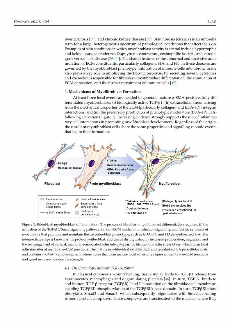
At least three local events are needed to generate mature α-SMA-positive, fully dif-ferentiated myofibroblasts: (i) biologically active TGF-β1; (ii) extracellular stress, arisingfrom the mechanical properties of the ECM (particularly collagen) and EDA–FN/integrininteractions; and (iii) the precursory production of phenotypic modulators (EDA–FN, HA)following activation (Figure 1). Increasing evidence strongly supports the role of inflamma-tory cell interactions in promoting myofibroblast development. Regardless of the origin,the resultant myofibroblast cells share the same properties and signalling cascade eventsthat led to their formation.
Biomolecules 2021, 11, x FOR PEER REVIEW 3 of 27
organ failure is considered the end-stage pathology of multiple diseases affecting the ma-jor organs, including but not limited to myocardial fibrosis [35], pulmonary fibrosis [36], liver cirrhosis [37], and chronic kidney disease [38]. Skin fibrosis (cicatrix) is an umbrella term for a large, heterogeneous spectrum of pathological conditions that affect the skin. Examples of skin conditions in which myofibroblast activity is central include hyper-trophic and keloid scars, scleroderma, Dupuytren’s contracture, eosinophilic fasciitis, and chronic graft-versus-host disease [39–44]. The shared features of the abnormal and exces-sive accumulation of ECM constituents, particularly collagens, HA, and FN, in these dis-eases are governed by the myofibroblast phenotype. Infiltration of immune cells into fi-brotic tissue also plays a key role in amplifying the fibrotic response, by secreting several cytokines and chemokines responsible for fibroblast–myofibroblast differentiation, the stimulation of ECM deposition, and the further recruitment of immune cells [45].
4. Mechanisms of Myofibroblast Formation At least three local events are needed to generate mature α-SMA-positive, fully dif-
ferentiated myofibroblasts: (i) biologically active TGF-β1; (ii) extracellular stress, arising from the mechanical properties of the ECM (particularly collagen) and EDA–FN/integrin interactions; and (iii) the precursory production of phenotypic modulators (EDA–FN, HA) following activation (Figure 1). Increasing evidence strongly supports the role of in-flammatory cell interactions in promoting myofibroblast development. Regardless of the origin, the resultant myofibroblast cells share the same properties and signalling cascade events that led to their formation.
Figure 1. Fibroblast–myofibroblast differentiation. The process of fibroblast–myofibroblast differentiation requires: (i) the activation of the TGF-β1/Smad signalling pathway; (ii) cell–ECM mechanotransduction signalling; and (iii) the synthesis of modulators that promote and maintain the myofibroblast phenotype, such as EDA–FN and HAS2-synthesised HA. The intermediate stage is known as the proto-myofibroblast, and can be distinguished by increased proliferation, migration, and the rearrangement of cortical, membrane-associated actin into cytoplasmic filamentous actin stress fibres, which form focal adhesion sites at membrane–ECM junctions. The mature myofibroblast exhibits thick and crosslinked HA pericellu-lar coats, and contains α-SMA+ cytoplasmic actin stress fibres that form mature focal adhesion plaques at membrane–ECM junctions and grant increased contractile strength.
4.1. The Canonical Pathway: TGF-β1/Smad In classical cutaneous wound healing, tissue injury leads to TGF-β1 release from
keratinocytes, macrophages and degranulating platelets [46]. In turn, TGF-β1 binds to and
Figure 1. Fibroblast–myofibroblast differentiation. The process of fibroblast–myofibroblast differentiation requires: (i) theactivation of the TGF-β1/Smad signalling pathway; (ii) cell–ECM mechanotransduction signalling; and (iii) the synthesis ofmodulators that promote and maintain the myofibroblast phenotype, such as EDA–FN and HAS2-synthesised HA. Theintermediate stage is known as the proto-myofibroblast, and can be distinguished by increased proliferation, migration, andthe rearrangement of cortical, membrane-associated actin into cytoplasmic filamentous actin stress fibres, which form focaladhesion sites at membrane–ECM junctions. The mature myofibroblast exhibits thick and crosslinked HA pericellular coats,and contains α-SMA+ cytoplasmic actin stress fibres that form mature focal adhesion plaques at membrane–ECM junctionsand grant increased contractile strength.
4.1. The Canonical Pathway: TGF-β1/Smad
In classical cutaneous wound healing, tissue injury leads to TGF-β1 release fromkeratinocytes, macrophages and degranulating platelets [46]. In turn, TGF-β1 binds toand induces TGF-β receptor (TGFβR) I and II association on the fibroblast cell membrane,enabling TGFβRII phosphorylation of the TGFβRI kinase domain. In-turn, TGFβRI phos-phorylates Smad2 and Smad3, which subsequently oligomerize with Smad4, formingtrimeric protein complexes. These complexes are translocated to the nucleus, where they
Biomolecules 2021, 11, 1095 4 of 27
act as the transcription or co-transcription factors in the induction or repression of gene ex-pression [47] (Figure 2A). Ultimately, TGF-β1/Smad pathway activation in fibroblasts leadsto their differentiation into myofibroblasts [48]. TGF-β1 can also induce Smad-independentand co-receptor signalling pathways, such as mitogen-activated protein kinase (MAPK),p42/p44 extracellular signal regulated kinase (ERK1/2), Rho/Rho-associated protein ki-nase (ROCK), phosphatidylinositol-3-kinase (PI3K)/AKT, protein phosphatase 2A (PP2A),p38/c-Jun N-terminal kinase (JNK), protein kinase C (PKC), and tumour necrosis factorreceptor associated factor (TRAF)-4/6 [49–53]. The tightly controlled TGF-β1/Smad-drivensignalling events have been extensively researched within the context of myofibroblastdifferentiation and fibrosis, as highlighted in an eloquent review by Frangogiannis [54].
Biomolecules 2021, 11, x FOR PEER REVIEW 6 of 27
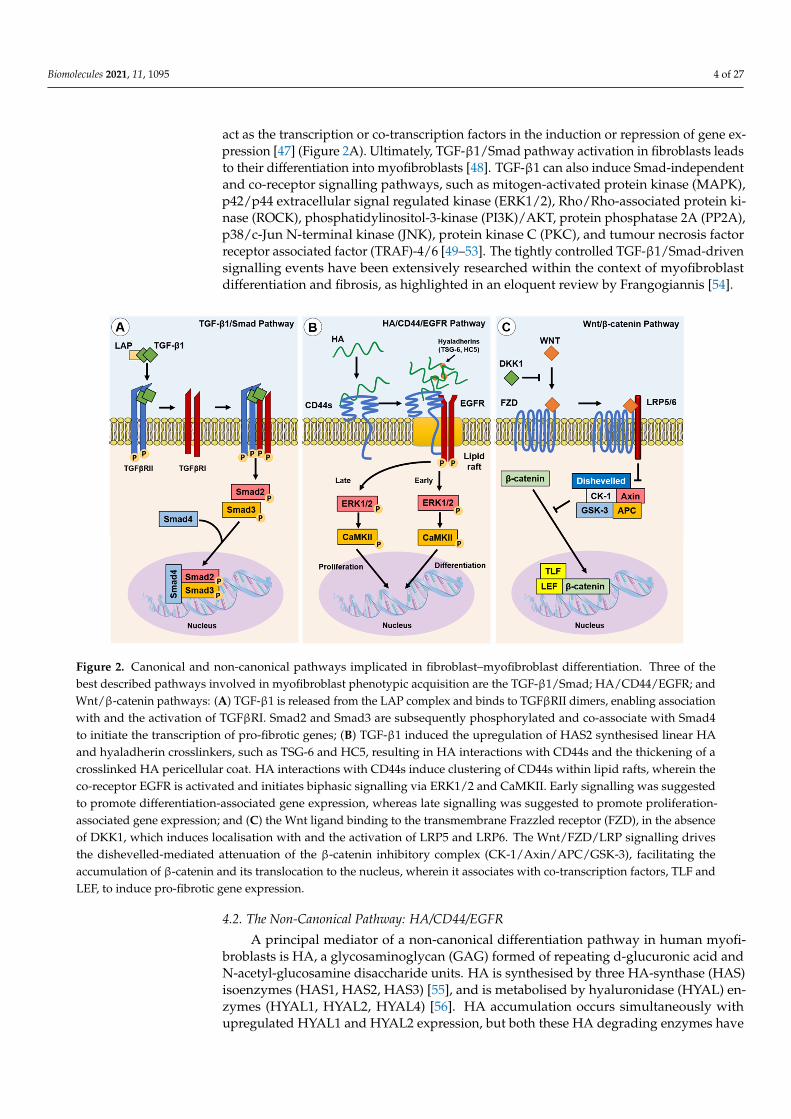
Figure 2. Canonical and non-canonical pathways implicated in fibroblast–myofibroblast differentiation. Three of the best described pathways involved in myofibroblast phenotypic acquisition are the TGF-β1/Smad; HA/CD44/EGFR; and Wnt/β-catenin pathways: (A) TGF-β1 is released from the LAP complex and binds to TGFβRII dimers, enabling association with and the activation of TGFβRI. Smad2 and Smad3 are subsequently phosphorylated and co-associate with Smad4 to initiate the transcription of pro-fibrotic genes; (B) TGF-β1 induced the upregulation of HAS2 synthesised linear HA and hya-ladherin crosslinkers, such as TSG-6 and HC5, resulting in HA interactions with CD44s and the thickening of a crosslinked HA pericellular coat. HA interactions with CD44s induce clustering of CD44s within lipid rafts, wherein the co-receptor EGFR is activated and initiates biphasic signalling via ERK1/2 and CaMKII. Early signalling was suggested to promote differentiation-associated gene expression, whereas late signalling was suggested to promote proliferation-associated gene expression; and (C) the Wnt ligand binding to the transmembrane Frazzled receptor (FZD), in the absence of DKK1, which induces localisation with and the activation of LRP5 and LRP6. The Wnt/FZD/LRP signalling drives the dishevelled-mediated attenuation of the β-catenin inhibitory complex (CK-1/Axin/APC/GSK-3), facilitating the accumulation of β-catenin and its translocation to the nucleus, wherein it associates with co-transcription factors, TLF and LEF, to induce pro-fibrotic gene expression.
4.4. Mechanotransduction Mechanotransduction is the ability of stress force to convert extracellular to intracel-
lular signalling. Fibroblasts can perceive external forces (mechanoperception) through their fibronexus structures in vivo or mature focal adhesion (FA) structures in vitro [86,87]. ECM rigidity determines the size of the cell’s FAs, or ‘anchors’, which in turn limit the level of tension generated within intracellular stress fibres. Only when substrate stiff-ness permits the formation of mature FAs (8–30 μm), and the generation of approximately four-fold greater stress compared with usual FAs (2–6 μm), does α-SMA become incorpo-rated into pre-existing cytoplasmic β-actin stress fibres [88]. Thus, the myofibroblast phe-notype is mechanosensitive. The transition of fibroblasts to the proto-myofibroblast state was suggested to be related to increased microenvironment stiffness [28,88]. TGF-β1 is locked within the ECM by latency-associated peptide (LAP) and latent TGF-β1-binding protein (LTBP), and is released by proteolysis or integrin-dependent mechanotransduc-tion [89]; resulting in a feedback loop of pro-fibrotic fibroblast activity and increased ECM stiffness [29,90–92]. In a recent study, the balance of ECM composition, elasticity, and TGF-β1 signalling was shown to govern fibroblast phenotypic heterogeneity and give rise to distinct, but overlapping, fibroblast subsets [93]. Indeed, Kollmannsberger and col-leagues showed that within 3D microtissues grown in vitro, fibroblasts transitioned to proliferative myofibroblasts at the growth front, where a high degree of tensile force and stretched FN fibres were present. As the tissue matrix matured into collagen-rich ECM
Figure 2. Canonical and non-canonical pathways implicated in fibroblast–myofibroblast differentiation. Three of thebest described pathways involved in myofibroblast phenotypic acquisition are the TGF-β1/Smad; HA/CD44/EGFR; andWnt/β-catenin pathways: (A) TGF-β1 is released from the LAP complex and binds to TGFβRII dimers, enabling associationwith and the activation of TGFβRI. Smad2 and Smad3 are subsequently phosphorylated and co-associate with Smad4to initiate the transcription of pro-fibrotic genes; (B) TGF-β1 induced the upregulation of HAS2 synthesised linear HAand hyaladherin crosslinkers, such as TSG-6 and HC5, resulting in HA interactions with CD44s and the thickening of acrosslinked HA pericellular coat. HA interactions with CD44s induce clustering of CD44s within lipid rafts, wherein theco-receptor EGFR is activated and initiates biphasic signalling via ERK1/2 and CaMKII. Early signalling was suggestedto promote differentiation-associated gene expression, whereas late signalling was suggested to promote proliferation-associated gene expression; and (C) the Wnt ligand binding to the transmembrane Frazzled receptor (FZD), in the absenceof DKK1, which induces localisation with and the activation of LRP5 and LRP6. The Wnt/FZD/LRP signalling drivesthe dishevelled-mediated attenuation of the β-catenin inhibitory complex (CK-1/Axin/APC/GSK-3), facilitating theaccumulation of β-catenin and its translocation to the nucleus, wherein it associates with co-transcription factors, TLF andLEF, to induce pro-fibrotic gene expression.
4.2. The Non-Canonical Pathway: HA/CD44/EGFR
A principal mediator of a non-canonical differentiation pathway in human myofi-broblasts is HA, a glycosaminoglycan (GAG) formed of repeating d-glucuronic acid andN-acetyl-glucosamine disaccharide units. HA is synthesised by three HA-synthase (HAS)isoenzymes (HAS1, HAS2, HAS3) [55], and is metabolised by hyaluronidase (HYAL) en-zymes (HYAL1, HYAL2, HYAL4) [56]. HA accumulation occurs simultaneously withupregulated HYAL1 and HYAL2 expression, but both these HA degrading enzymes have
Biomolecules 2021, 11, 1095 5 of 27
diminished expression in myofibroblasts. A subsequent decrease in HA degradation mayalso favour the accumulation of HA in fibrotic disease [57]. HAS2-synthesised HA is widelyconsidered to be an important mediator of fibroblast–myofibroblast differentiation andEMT processes [58–62]. HAS2-synthesised linear HA forms a thickened pericellular coatthat surrounds the activated fibroblast. This crosslinked and thickened HA pericellular coat,or glycocalyx, has been shown to be an essential structural element for human fibroblast–myofibroblast differentiation and phenotypic maintenance [59,63]. The HA pericellularcoat remains thin in fibroblast phenotypes resistant to TGF-β1-driven differentiation, suchas oral mucosal fibroblasts [64] and aged/senescent fibroblasts [58]. Interestingly, an en-riched pericellular HA microenvironment is synthesised by foetal fibroblasts during foetalwound healing.
The HA pericellular coat is anchored to the fibroblast cell surface by the CD44 recep-tor [65,66], and crosslinked by hyaladherins [59]. Association with CD44 and other hyalad-herins orchestrates the HA pericellular coat and results in the activation of downstreamextracellular–intracellular signal cascades, shown to be prominent in disease pathogenesisincluding fibrosis, cancers, and inflammatory diseases [67,68]. CD44 exists as multiplealternatively spliced variants and all CD44 variants (CD44v), as well as the standard form(CD44s), possess HA binding motifs within their extracellular head domain. CD44v havevariability within alternatively spliced exons of the extracellular stem region [69,70]. Stud-ies have shown that silencing CD44 using small interfering RNA (siRNA) prevented thedownstream intracellular signalling required for human fibroblast–myofibroblast differ-entiation and inhibited HA pericellular coat formation [71,72]. These studies concludedthat HA/CD44 association was essential for human myofibroblast differentiation, withrecent research showing that CD44s expressed on the surface of human fibroblasts wasthe dominant player in regulating HA coat-mediated myofibroblast differentiation [73].HA-crosslinking hyaladherins include inter-α-inhibitor (IαI) heavy chains (HC), which arelocalised by association with the tumour necrosis factor-stimulated gene (TSG)-6 [74,75].The increased presence of HA-associated HC and TSG-6 complexes have been associatedwith the pathology of multiple inflammatory diseases including arthritis, asthma, andsome cancers [76]. Martin et al. identified that IαI-HC5 was covalently bound to chon-droitin sulphate on bikunin and was essential for the formation of HA pericellular coatsin myofibroblasts. Increased expression of TSG-6 following TGF-β1 activation of fibrob-lasts facilitated the transfer of HC5 to HA by TSG-6 catalysis, in a metal ion-dependentmanner [77].
HA binding to CD44 plays a pivotal role in the intracellular signalling required forhuman fibroblast–myofibroblast differentiation [58,63,72]. CD44 was shown to be presentthroughout the membranes of fibroblasts but clustered into localised populations withinmyofibroblast membranes. These distinct areas were identified to be cholesterol-richlipid rafts, and ‘locked’ clusters of CD44 within lipid rafts were shown to be dependenton HA binding and crosslinking into pericellular coats [72]. HA coat removal releasedCD44 from lipid rafts and re-enabled their movement through the membrane, but alsoinhibited downstream signalling cascades [63]. Further investigation revealed that CD44was co-localised with epidermal growth factor (EGFR) within membrane lipid raft domains,which resulted in EGFR activation, and the phosphorylation of downstream effectors andtranscription factors, ERK1/2 and calcium calmodulin kinase (CaMK)-II (CaMKII). Thechemical inhibition of ERK1/2 or CaMKII prevented myofibroblast differentiation, and onlywhen TGF-βRII and CD44/EGFR signalling were active could myofibroblast differentiationoccur [72]. Both ERK1/2 and CaMKII were phosphorylated in a biphasic manner, withearly and late-phase activation. Early activation of ERK1/2 (<5 min) was thought to beassociated with eventual fibroblast–myofibroblast differentiation, whereas later activation(>30 min) was suggested to play a role mediating cellular proliferation responses [72](Figure 2B). However, signalling functions appear to be cell-dependent as early, but not late,ERK1/2 signalling peaks were observed in TGF-β1-stimulated non-scarring oral mucosal
Biomolecules 2021, 11, 1095 6 of 27
fibroblasts; these cells exhibited an anti-proliferative response in a HA/CD44-independentmanner [65].
4.3. The Wnt/β-Catenin Pathway
The Wnt signalling pathway is well described in its vital roles during embryogenesisand determination of cell fate, differentiation, proliferation, and apoptosis. We refer in-terested readers to a thorough review article on Wnt signalling functions [78]. Carefullyorchestrated Wnt signalling has been found to be essential for tissue homeostasis, whilstthe dysregulation of the Wnt pathway can result in pathogenesis [79,80]. Wnt signalssimultaneously through the co-association of Frazzled receptors and low-density lipidprotein co-receptors (LRP). The fate of β-catenin, an important regulator of Wnt signalling,is regulated by Dickkopf-related protein-1 (DKK1). DKK1 inhibits Wnt signalling by ac-tivating downstream signalling complexes, and results in β-catenin degradation. WhenDKK1 is not present, Wnt signalling leads to β-catenin translocation to the nucleus, itsassociation with co-transcription factors, and the subsequent activation of Wnt-associatedpro-fibrotic genes [78,79,81] (Figure 2C). The activation of Wnt signalling was implicatedin the fibrogenesis of multiple organs [82]. Wnt-1 and Wnt-10b were noted to be overex-pressed, whereas DKK1 was decreased, which led to the increased nuclear accumulationof β-catenin observed in human tissue samples from systemic scleroderma (SSc), idio-pathic pulmonary fibrosis (IPF), and liver cirrhosis [82]. Wnt signalling stimulated byTGF-β1-induced inhibition of DKK1 was shown to induce myofibroblast differentiation,up-regulate the release of ECM components (notably collagens) and induce fibrosis [82–84].In addition, crosstalk between the TGF-β/Smad pathway and the Wnt/β-catenin pathwayis suggested to be a prominent mechanism in the development of hypertrophic and keloidscars [85].
4.4. Mechanotransduction
Mechanotransduction is the ability of stress force to convert extracellular to intracel-lular signalling. Fibroblasts can perceive external forces (mechanoperception) throughtheir fibronexus structures in vivo or mature focal adhesion (FA) structures in vitro [86,87].ECM rigidity determines the size of the cell’s FAs, or ‘anchors’, which in turn limit thelevel of tension generated within intracellular stress fibres. Only when substrate stiffnesspermits the formation of mature FAs (8–30 µm), and the generation of approximatelyfour-fold greater stress compared with usual FAs (2–6 µm), does α-SMA become incor-porated into pre-existing cytoplasmic β-actin stress fibres [88]. Thus, the myofibroblastphenotype is mechanosensitive. The transition of fibroblasts to the proto-myofibroblaststate was suggested to be related to increased microenvironment stiffness [28,88]. TGF-β1is locked within the ECM by latency-associated peptide (LAP) and latent TGF-β1-bindingprotein (LTBP), and is released by proteolysis or integrin-dependent mechanotransduc-tion [89]; resulting in a feedback loop of pro-fibrotic fibroblast activity and increased ECMstiffness [29,90–92]. In a recent study, the balance of ECM composition, elasticity, andTGF-β1 signalling was shown to govern fibroblast phenotypic heterogeneity and giverise to distinct, but overlapping, fibroblast subsets [93]. Indeed, Kollmannsberger andcolleagues showed that within 3D microtissues grown in vitro, fibroblasts transitioned toproliferative myofibroblasts at the growth front, where a high degree of tensile force andstretched FN fibres were present. As the tissue matrix matured into collagen-rich ECMwith low FN fibre tension, more fibroblasts were present, suggesting that the myofibroblastphenotype stabilised by tensile forces could revert to a fibroblast phenotype by low tensileforce [94]. These studies suggest that substrate stiffness is a key determinant of fibroblastand myofibroblast plasticity, and that reducing excessive mechanotransduction may be anoption in controlling myofibroblast presence.
Myofibroblasts generate force through stress fibre contraction, and this is transmit-ted to the ECM via FAs containing transmembrane receptors, typically integrins [87,95],as summarised in Figure 3. Integrins mediate cell–cell, and especially, cell–ECM inter-
Biomolecules 2021, 11, 1095 7 of 27
actions, and are prominently involved in the initiation, maintenance, and resolution offibrosis. EDA–FN is synthesised by fibroblasts and various other cell types [96,97]. TheEDA–FN splice variant can only be detected during tissue repair [98,99], fibrosis [100],tumour development [101–103], and transiently during embryogenesis [104]. The accu-mulation of EDA–FN has been observed in multiple fibrotic disorders, including lung,liver, and skin [100,105–107]. EDA–FN promotes myofibroblast differentiation by orches-trating LAP/LTBP release and activation of TGF-β1 [108], increasing matrix stress–straintension [109,110], and by activating mechanotransducer-integrin signalling via FAK activa-tion [111–114]. Research by Shinde et al. [115] and Kohan et al. [112] demonstrated thatfibroblast-expressed integrins, α4β1 (VLA-4) and α4β7 (LPAM-1), mediate different roles inmyofibroblast formation, respectively. Integrin α4β1/EDA–FN promoted ECM synthesisand stiffening [115], whereas integrin α4β7/EDA–FN induced myofibroblast stress fibreformation and contractility [112]. The combined exclusivity of EDA–FN expression andthe prominent roles it plays in driving the myofibroblast phenotype have highlighted theprotein as an attractive target for anti-fibrotic interventional therapies.
Biomolecules 2021, 11, x FOR PEER REVIEW 8 of 27
matrices in vitro [129] and in fibrosis progression in vivo [130]. However, HYAL2 was shown to re-localize to the cytoplasm and align along the actin cytoskeleton in myofibro-blasts [128], which may contribute to increasing extracellular HA accumulation. The cyto-skeleton-aligned HYAL2 was associated with α-SMA, RhoA, and MLCK. The silencing of HYAL2 did not prevent but only delayed myofibroblast differentiation. The presence of HYAL2 was shown to accelerate RhoA/MLCK phosphorylation and cellular contractility, suggesting a role in the mechanotransduction and the orchestration of key cytoskeletal and FA-related proteins [128]. These early findings suggest that there may be complex orchestration roles that HYAL2 plays in cytoskeletal reassembly.
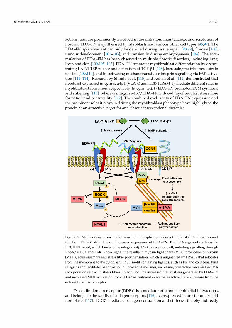
Figure 3. Mechanisms of mechanotransduction implicated in myofibroblast differentiation and function. TGF-β1 stimulates an increased expression of EDA–FN. The EDA segment contains the EDGIHEL motif, which binds to the integrin α4β1/α4β7 receptor cleft, initiating signalling through RhoA/MLCK and FAK. RhoA signalling results in myosin light chain (MLC) promotion of myosin (MYH)/actin assembly and stress fibre polymerisation, which is augmented by HYAL2 that relo-cates from the membrane to the cytoplasm. RGD motif containing ligands, such as FN and collagens, bind integrins and facilitate the formation of focal adhesion sites, increasing contractile force and α-SMA incorporation into actin stress fibres. In addition, the increased matrix stress generated by EDA–FN and increased MMP activation from CD147 recruitment exacerbates active TGF-β1 release from the extracellular LAP complex.
Other mediators of mechanotransduction and promising targets in mediating fibro-blast–myofibroblast differentiation include myocardin-related transcription factor (MRTF) [131,132], Yes-associated protein (YAP) [133–137], cadherins [138], and Notch [139]. In the case of these multifunctional proteins, further exploratory research will help establish the feasibility of targeting their activity to interfere with myofibroblast differen-tiation or function.
Figure 3. Mechanisms of mechanotransduction implicated in myofibroblast differentiation andfunction. TGF-β1 stimulates an increased expression of EDA–FN. The EDA segment contains theEDGIHEL motif, which binds to the integrin α4β1/α4β7 receptor cleft, initiating signalling throughRhoA/MLCK and FAK. RhoA signalling results in myosin light chain (MLC) promotion of myosin(MYH)/actin assembly and stress fibre polymerisation, which is augmented by HYAL2 that relocatesfrom the membrane to the cytoplasm. RGD motif containing ligands, such as FN and collagens, bindintegrins and facilitate the formation of focal adhesion sites, increasing contractile force and α-SMAincorporation into actin stress fibres. In addition, the increased matrix stress generated by EDA–FNand increased MMP activation from CD147 recruitment exacerbates active TGF-β1 release from theextracellular LAP complex.
Discoidin domain receptor (DDR)1 is a mediator of stromal–epithelial interactions,and belongs to the family of collagen receptors [116] overexpressed in pro-fibrotic keloidfibroblasts [117]. DDR1 mediates collagen contraction and stiffness, thereby indirectly
Biomolecules 2021, 11, 1095 8 of 27
mediating pro-fibrogenic responses that are largely independent from collagen binding tointegrins [118]. Additionally, the spatial and structural properties of the local 3D collagenmicroarchitecture can distinctly affect fibroblast and myofibroblast activity. Collagenfibril alignment and diameter were shown to affect fibroblast contractility and migrationvia alteration in integrin clustering and the stability of adhesion sites [119]. Seo andcolleagues suggested that collagen fibre thickness held more pertinence over dictatingmyofibroblast differentiation independently from collagen quantity [120]. Furthermore,collagen-rich ECM stiffening can also be potentiated by the myofibroblast and inflammatorycell production of collagen crosslinking enzymes, such as lysyl oxidases [11,90]. CD147, alsoknown as extracellular matrix metalloproteinase inducer (EMMPRIN), is a well-establishedmatrix metalloproteinase (MMP)-inducer that is rapidly becoming understood to mediatemultiple cellular responses. The linked association of CD147 with integrin α6β1 wasobserved during increased metastasis in human hepatoma cells [121], suggesting a rolefor CD147 in modulating integrin α6β1 associations, with matricellular protein CCN1,and mechano-sensitivity to senescence or apoptosis [122]. Interestingly, the nullification ofthe extracellular domain of CD147 reversed associations with integrin β subunits and FAsites, suggesting that the extracellular domain of CD147 binds to integrins to regulate themechanical tension of the ECM [121,123]. The roles of these mechanotransducer proteinsin stress relay, which is involved in myofibroblast differentiation, will become clearer withcontinued investigation.
Increasing evidence suggests that myofibroblasts have the capacity for classical CaMK-myosin light chain kinase (MLCK)-dependent smooth muscle cell contraction mecha-nisms [124] and contractile activation by the RhoA/ROCK/myosin light chain phosphatase(MLCP) pathway [28,125,126], highlighting a key difference between the smooth musclecell and myofibroblast contractile mechanisms. It was recently discovered that HYAL2,previously thought to only possess HA catalytic activity, was shown to have non-enzymaticfunctions [69,127,128]. Following TGF-β1 activation, enhanced HAS2 expression by fi-broblasts leads to increased HA accumulation within the ECM observed in 3D matricesin vitro [129] and in fibrosis progression in vivo [130]. However, HYAL2 was shown tore-localize to the cytoplasm and align along the actin cytoskeleton in myofibroblasts [128],which may contribute to increasing extracellular HA accumulation. The cytoskeleton-aligned HYAL2 was associated with α-SMA, RhoA, and MLCK. The silencing of HYAL2did not prevent but only delayed myofibroblast differentiation. The presence of HYAL2 wasshown to accelerate RhoA/MLCK phosphorylation and cellular contractility, suggesting arole in the mechanotransduction and the orchestration of key cytoskeletal and FA-relatedproteins [128]. These early findings suggest that there may be complex orchestration rolesthat HYAL2 plays in cytoskeletal reassembly.
Other mediators of mechanotransduction and promising targets in mediatingfibroblast–myofibroblast differentiation include myocardin-related transcription fac-tor (MRTF) [131,132], Yes-associated protein (YAP) [133–137], cadherins [138], andNotch [139]. In the case of these multifunctional proteins, further exploratory researchwill help establish the feasibility of targeting their activity to interfere with myofibrob-last differentiation or function.
5. Interventional Strategies to Target Myofibroblasts for Scarless Skin Healing
There are currently no clinically approved anti-scarring therapies specifically designedto limit or prevent dermal fibrosis. Several wound management strategies completed phaseII trials, but did not advance to phase III trials in recent years. The clinical studies database,https://clinicaltrials.gov (‘Fibrosis, Skin’ searched 25 June 2021), showed 10 registeredclinical trials that have either completed phase III or are currently in progress (Table 1).The proposed therapies include a range of treatment modes, including antibody and drugtherapies, scar resection or reduction, and tissue-engineering strategies.
Biomolecules 2021, 11, 1095 9 of 27
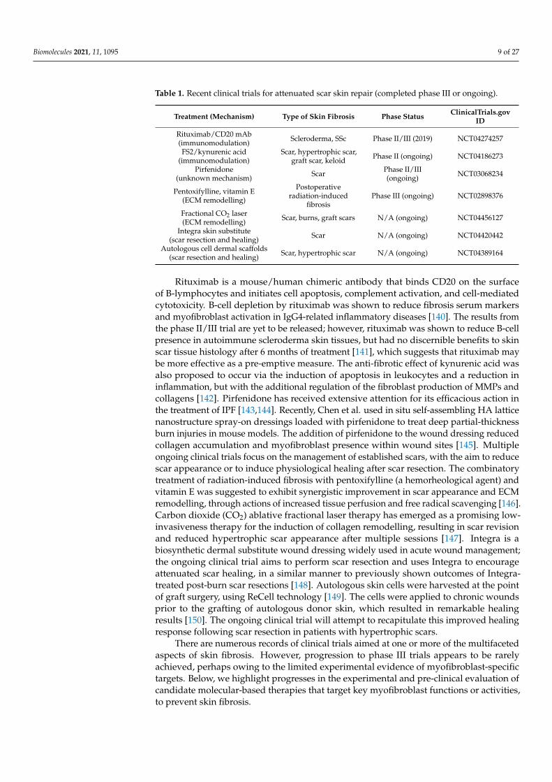
Table 1. Recent clinical trials for attenuated scar skin repair (completed phase III or ongoing).
Treatment (Mechanism) Type of Skin Fibrosis Phase Status ClinicalTrials.govID
Rituximab/CD20 mAb(immunomodulation) Scleroderma, SSc Phase II/III (2019) NCT04274257
FS2/kynurenic acid(immunomodulation)
Scar, hypertrophic scar,graft scar, keloid Phase II (ongoing) NCT04186273
Pirfenidone(unknown mechanism) Scar Phase II/III
(ongoing) NCT03068234
Pentoxifylline, vitamin E(ECM remodelling)
Postoperativeradiation-induced
fibrosisPhase III (ongoing) NCT02898376
Fractional CO2 laser(ECM remodelling) Scar, burns, graft scars N/A (ongoing) NCT04456127
Integra skin substitute(scar resection and healing) Scar N/A (ongoing) NCT04420442
Autologous cell dermal scaffolds(scar resection and healing) Scar, hypertrophic scar N/A (ongoing) NCT04389164
Rituximab is a mouse/human chimeric antibody that binds CD20 on the surfaceof B-lymphocytes and initiates cell apoptosis, complement activation, and cell-mediatedcytotoxicity. B-cell depletion by rituximab was shown to reduce fibrosis serum markersand myofibroblast activation in IgG4-related inflammatory diseases [140]. The results fromthe phase II/III trial are yet to be released; however, rituximab was shown to reduce B-cellpresence in autoimmune scleroderma skin tissues, but had no discernible benefits to skinscar tissue histology after 6 months of treatment [141], which suggests that rituximab maybe more effective as a pre-emptive measure. The anti-fibrotic effect of kynurenic acid wasalso proposed to occur via the induction of apoptosis in leukocytes and a reduction ininflammation, but with the additional regulation of the fibroblast production of MMPs andcollagens [142]. Pirfenidone has received extensive attention for its efficacious action inthe treatment of IPF [143,144]. Recently, Chen et al. used in situ self-assembling HA latticenanostructure spray-on dressings loaded with pirfenidone to treat deep partial-thicknessburn injuries in mouse models. The addition of pirfenidone to the wound dressing reducedcollagen accumulation and myofibroblast presence within wound sites [145]. Multipleongoing clinical trials focus on the management of established scars, with the aim to reducescar appearance or to induce physiological healing after scar resection. The combinatorytreatment of radiation-induced fibrosis with pentoxifylline (a hemorheological agent) andvitamin E was suggested to exhibit synergistic improvement in scar appearance and ECMremodelling, through actions of increased tissue perfusion and free radical scavenging [146].Carbon dioxide (CO2) ablative fractional laser therapy has emerged as a promising low-invasiveness therapy for the induction of collagen remodelling, resulting in scar revisionand reduced hypertrophic scar appearance after multiple sessions [147]. Integra is abiosynthetic dermal substitute wound dressing widely used in acute wound management;the ongoing clinical trial aims to perform scar resection and uses Integra to encourageattenuated scar healing, in a similar manner to previously shown outcomes of Integra-treated post-burn scar resections [148]. Autologous skin cells were harvested at the pointof graft surgery, using ReCell technology [149]. The cells were applied to chronic woundsprior to the grafting of autologous donor skin, which resulted in remarkable healingresults [150]. The ongoing clinical trial will attempt to recapitulate this improved healingresponse following scar resection in patients with hypertrophic scars.
There are numerous records of clinical trials aimed at one or more of the multifacetedaspects of skin fibrosis. However, progression to phase III trials appears to be rarelyachieved, perhaps owing to the limited experimental evidence of myofibroblast-specifictargets. Below, we highlight progresses in the experimental and pre-clinical evaluation ofcandidate molecular-based therapies that target key myofibroblast functions or activities,to prevent skin fibrosis.
Biomolecules 2021, 11, 1095 10 of 27
5.1. Experimental and Pre-Clinical Interventions for Myofibroblast Differentiation
Popularised pre-clinical strategies for preventing myofibroblast formation focus oninhibition of the canonical TGF-β1/Smad signalling pathway using cytokines, antibodies,and regulators of gene expression. Demonstrating the clinical efficacy of anti-TGF-β1therapies has been hindered by the cytokine’s pleiotropism and ubiquitous presence, aswell as the heterogenous processes of healing and fibrosis. Consequently, therapeutictargeting of TGF-β and its specific isoforms has proved challenging. Available clinical dataare limited to early phase trials and studies with conflicting outcomes [54]. Recombinanthuman TGF-β3 (Avotermin, Juvista) was developed by Renovo and hailed as a potentialnovel inhibitor of dermal scar formation, based on its elevated presence and roles in non-scarring tissues compared to TGF-β1, such as early gestational foetal skin and the oralmucosa [151–154]. Phase I/II clinical trials concluded that the intradermal application ofJuvista yielded both short- and long-term improvements in scar appearance, comparedto placebo and standard wound care management [155,156]. Unfortunately, Juvista didnot meet the necessary primary or secondary endpoints required by phase III trials, failingto demonstrate reduced scarring after excisional surgery. Fresolimumab is a neutralisingantibody that targets all TGF-β isoforms; the effects of Fresolimumab on cutaneous SScrevealed reduced expression of fibrotic biomarkers and diminished myofibroblasts [157].This suggested that a shotgun approach to TGF-β inhibition had anti-fibrotic effects, whilstneutralising TGF-β1 alone in SSc failed to demonstrate clinical efficacy [158]. An apparenttrend has emerged, wherein anti-scarring interventions that target an individual aspect orfactor of fibrotic conditions generally fail to realise clinical benefit. Moreover, the efficaciousindex of growth factors is limited by their low stability, short half-life in vivo, adverseeffects arising from elevated local and/or systemic concentrations, and their non-specificnature (multiple protein-binding partners) [159].
MicroRNAs (miRNA) are short non-coding RNAs (18–25 nucleotides) that bind the 3′
untranslated region of mRNA, thereby inhibiting mRNA translation or promoting mRNAdegradation. Multiple studies have shown the regulatory roles of miRNA in pathologicalwound healing and skin fibrosis. Notable miRNA targets upregulated in skin fibrosisinclude miRNA (miR)-21 [160] and miR-130b [161], whereas miR-29 [162], miR-129-5p [163],and miR-7 [164,165] were shown to be negative regulators of myofibroblast activity andfibrosis. The miRNAs, miR-21 and miR-17-5p, have been shown to be directly implicatedin TGF-β1/Smad pathway promotion. Smad2 activity was potentiated by these miRNAsthrough their inhibition of Smad7 [166,167]. Thus, inhibiting the activity of these miRNAshas arisen as a potential avenue in gene therapies to mitigate TGF-β1/Smad pathwayactivity and myofibroblast differentiation. We refer readers to an elegantly written reviewof miRNAs in various fibrotic diseases for a more in-depth discussion of the miRNAsinvolved in modulating TGF-β1 signalling [168].
Bone morphogenetic protein-7 (BMP7) exerted potent anti-fibrotic actions in vitro [169],and in pre-clinical fibrosis models [170–173]. BMP7 activation of Smad1/5/8 results in thecompetitive binding of Smad4, thereby inhibiting the TGF-β1/Smad2/3 pathway’s tran-scriptional activities [173]. Early reports indicated that BMP7 failed to prevent bleomycin-induced skin fibrosis [174]. However, a later study showed that BMP7 treatment at thepoint of thermal injury prevented hypertrophic scar formation [173]. Thus, BMP7 therapiesrequire further optimization and testing to determine their effectiveness in the preventionof dermal fibrosis. Interferon (IFN)-γ induced the expression of Smad7, which antagonisedinteractions between Smad2/3 and TGFβRI/II [175]. The effects of IFN-γ on fibroblastsin vitro are conflicting; exogenous IFN-γ was suggested to abrogate TGF-β1-induced prolif-eration, migration, and differentiation to myofibroblasts [176]; but T-cell secretion of IFN-γ,found to be prominent in SSc, aggravated fibrosis by promoting fibroblast proliferationand collagen synthesis [177]. Early pre-clinical and small clinical studies suggested thatIFN-γ was effective in the attenuation of lung, renal, and liver fibrosis [178–180], and oralsubmucosal fibrosis [178–181]. There is a current lack of studies describing definitive IFN-γ
Biomolecules 2021, 11, 1095 11 of 27
mechanisms involved in the regulation of myofibroblasts. Thus, whether IFN-γ deliverywould be effective in attenuating dermal fibrosis remains unclear.
Alternative approaches include molecular inhibitors of non-canonical and mechan-otransduction pathways to regulate ECM composition and disrupt the myofibroblast phe-notype. Hepatocyte growth factor (HGF) was first implicated in liver regeneration [182],but has since been shown to prevent fibrosis initiation and progression in animal mod-els [183,184]. HGF treatment was shown to attenuate collagen production by fibroblastsin multiple tissues [185–187]. Elevated HGF expression in fibrosis [188–191], but also indifferentiation-resistant fibroblasts [192,193], suggests a duality of HGF actions whichmay be a consequence of truncated isoforms possessing a variable number of kringledomains (HGF/NK1-4) with differential signalling activities. Indeed, human oral mucosalfibroblast resistance to TGF-β1-induced differentiation was dependent on heightened HGFand HGF/NK1 expression, whereas HGF/NK2 was preferentially expressed by dermalfibroblasts [194]. Recently, HGF/NK1 gene therapy was shown to exert potent anti-fibroticeffects through attenuated collagen types I, III, and IV deposition in mouse models of renalfibrosis [171]. In addition, the HGF inhibition of collagen synthesis and promotion of MMPproduction has shown promise for therapeutic applications in reducing the pro-fibroticactivity of keloid fibroblasts in vitro and in a keloid heterograft mouse model [195–197].Small molecule inhibitors of DDR1 have shown promise in reducing collagen types I and IVdeposition in bleomycin-induced renal and lung fibrosis [198–200]. Ongoing research intoDDR1-specific inhibitors have yielded promising results in models of renal fibrosis [199].
HA bioactivity is dependent on its molecular weight, enzymatic synthesis, and en-dogenous versus exogenous application [55,201,202]. The disruption or prevention of HApericellular coat synthesis using hyaluronidase enzymes [203], HAS inhibitors [204,205]or exogenous HA oligosaccharides [59] results in the failure of TGF-β1-stimulated my-ofibroblast phenotypic acquisition. The inhibition of HAS2 activity [206] or global HASsynthesis of HA [204,205] have demonstrated preventative effects in various models offibrosis. However, given the ubiquitous role of HAS-synthesised HA in tissues, and thelack of specific HAS isoenzyme inhibitors, it is unclear at present whether the inhibitionof HA synthesis would be beneficial in a clinical setting. An additional action of BMP7involved the induction of the nuclear translocation of HYAL2 and the subsequent splicingof CD44 mRNA, which resulted in the upregulated expression of the variant isoform,CD44v7/8. Fibroblasts with upregulated expression of CD44v7/8 exhibited ‘HA-phage’activity, wherein the HA pericellular coat was rapidly internalised and broken down,resulting in the destabilization of the myofibroblast and phenotypic reversion to fibrob-lasts [69,169]. Whether CD44v7/8-dependent actions convey anti-fibrotic effects in dermalfibrosis is currently unknown.
The most promising integrin targets that have demonstrated roles in the pathogen-esis of fibrosis are α4-containing integrins (α4β1/7) [112,115] and αv-containing inte-grins (αvβ1/3/5/6) [207–209]. Mechanotransduction by integrin αvβ6 promoted trac-tion from proliferating liver cholangiocytes to FN/LAP, which subsequently releasedactive TGF-β1 and initiated the differentiation of surrounding hepatic stellate cellsto myofibroblasts [207,210]. A small peptidomimetic, EMD527040, mimics the RGD-binding sites of αvβ6 and αvβ1; orally administered EMD527040 attenuated biliaryand non-biliary fibrogenesis [207]. The expression of integrin αvβ6 was elevated in ker-atinocytes during wound healing and fibrosis [211], but the detailed mechanistic rolesof integrin αvβ6 and epithelial–mesenchymal crosstalk during dermal fibrosis have yetto be elucidated. Research has identified that integrin interactions with the EDGIHELmotif of EDA–FN was causative of downstream profibrotic responses [109,113,115].The IST-9 [109,110], F8 [212,213], and vaccine-generated [214] antibodies that targetEDA–FN or integrin α4 have demonstrated prevention of fibroblast–myofibroblastdifferentiation [109,110]. Therapeutic applications of antibodies can be limited by thecost and complexity of production, off-target immune activation or unspecific proteinmasking of critical protein–protein interactions. To address these issues, Zhang et al.
Biomolecules 2021, 11, 1095 12 of 27
developed a small blocking polypeptide to bind and block EDA–FN interactions withthe α4β1 binding cleft, with high specificity [114]. The polypeptide, AF38Pep, wasdesigned to mimic the integrin α4β1 receptor site for the EDGIHEL motif of EDA–FNand was shown to interfere with TGF-β1-stimulated fibroblast–myofibroblast formationby the specific blockade of integrin α4β1 signalling, the inhibition of FAK activation,and the prevention of profibrotic gene transcription [114]. The aforementioned firstgeneration of small blocking polypeptides have revealed the renewed promise of thespecific interruption of integrin-mediated mechanotransduction and myofibroblast for-mation. The implementation of integrin receptor peptidomimetics for the prevention ofskin fibrosis will become more apparent in the coming years when the in vitro researchprogresses into pre-clinical models.
Another potential peptide therapy is the N-terminal amino acid sequence of α-SMA,Ac-EEED, which is important for the tropomyosin-1.6/7-stabilized incorporation of α-SMAinto cytoplasmic stress fibres [215]. Interestingly, the cytoplasmic delivery of the Ac-EEEDpeptide resulted in the loss of α-SMA from β-cytoplasmic actin stress fibres and inhibitedG-actin polymerization into F-actin [216,217], thereby reducing myofibroblast contractionin wound healing [218]. Ac-EEED peptide therapy has yet to be evaluated in skin fibrosismodels, which may be related to the current lack of available dermal myofibroblast-specific targeting moieties that operate by endosomal uptake and the cytoplasmic releaseof payloads.
5.2. Immunomodulating Biomolecules for Fibrosis Attenuation
Immunoregulatory interventions aim to control the inflammatory phase in postnatalskin healing to attenuate scar formation [219,220]. Studies in transgenic mice providedearly indicators that the absence of neutrophils and macrophages led to scar-free heal-ing [221]. Targeted repression of the gap junction and inflammatory mediator protein,connexin-43, supported these findings [220,222]. Additionally, connexin-43 was shownto mediate cardiac fibroblast–myofibroblast differentiation [223] and promote aberrantcardiomyocyte–myofibroblast functional coupling [224]. Certain interleukin (IL) cytokinesare implicated in the activation of inflammatory cascades. IL-8 production is a chemoat-tractant to neutrophils, whereas IL-6 secretion by fibroblasts activates macrophages andmonocyte chemotaxis. Both IL-6 and IL-8 exhibit the rapid induction of expression fol-lowing tissue injury, resulting in the recruitment of circulating inflammatory cells. Theexpression levels of IL-6 and IL-8 are elevated and maintained for longer in adult skin,compared to scarless scar-free foetal repair. The inhibition of phosphodiesterase 4 (PDE4)reduced scar formation in skin fibrosis models by interfering with the release of IL-6from M2 macrophages [225]. Thus, IL-6 and IL-8 are considered pro-fibrotic mediators,whereas IL-10 antagonised their activity [219,226]. IL-10 gene therapy resulted in reducedinflammation and the promotion of scarless healing in mouse wound healing studies [227].The exogenous addition and macrophage paracrine production of IL-10 were shown toinduce myofibroblast reversal to fibroblastic phenotypes in vitro [228,229]. More recently,IL-10 induced myofibroblast–fibroblast dedifferentiation was shown to alter dynamic inter-actions with the surrounding fibrillar matrix with a demonstratable loss of contractilityin IL-10 treated myofibroblasts [230]. Research into inflammation-induced fibrosis hasrevealed additional potential candidate ILs that may be targetable in skin fibrosis, includingIL-11 [35], IL-16 [231], and IL-33 [232]. Keratinocytes have suggested roles in the regulationof the myofibroblast phenotype and profibrotic ECM during wound healing [233–235].More recently, the keratinocyte secretion of IL-1α was demonstrated to restrain the myofi-broblast phenotype, dependent on fibroblast integrin α4β1 expression and Cox-2/Nrf2signalling [236,237].
The importance and therapeutic potential of macrophages in the wound healingprocess has been highlighted in recent years [6,238,239]. Crosstalk between myofibrob-lasts and macrophages during skin repair was reported [240]. Growth factors secretedby CD301b+ M2-type macrophages were shown to selectively stimulate the prolifera-
Biomolecules 2021, 11, 1095 13 of 27
tion of adipocyte precursor (AP)-derived myofibroblasts only. In aged mice wounds andexperimentally induced mouse skin fibrosis, AP-derived myofibroblasts and CD301b+
macrophages were reduced, and a CD29+ myofibroblast pool was increased. In keloids,CD301b+ macrophages and AP-derived myofibroblasts were also increased [241]. In fibroticlung tissues, cadherin-11 mediated the adhesion between macrophages and myofibrob-lasts, promoting pro-fibrotic myofibroblast activity via the paracrine release of TGF-β1 bythe macrophages [242]. Experimental research has suggested roles of mast cells in scarformation [243]; reduced numbers of activated mast cells were reported to improve healingand minimize scarring [244–246], whereas mast cell hyperplasia was causally linked tomyofibroblast hyperplasia [247–249]. Increased neuropeptide activity and the presenceof substance-P (SP) were found in hypertrophic scar samples [250,251]. Mast cells werepreviously suggested to be a major source of neuropeptide SP-stimulated inflammation andincreased myofibroblast activity [252]. These studies showed that myofibroblast activitycould be mediated through regulated leukocyte–myofibroblast interactions and suggestthat targeting certain leukocyte subpopulations may serve as anti-fibrotic strategies.
Targeting inflammatory meditators has shown promise in alleviating the magnitudeof fibrosis in various animal models. Follistatin-like-1 (FSTL1) was found to be elevatedin serum from patients with silicosis and in mouse lung fibrosis models. FSTL1-inducedIL-1β production by macrophages and positively regulated TGF-β1 signalling in fibrob-lasts. The inhibition of FSTL1 expression or activity protected against lung injury andfibrosis [253,254]. Recently, FSTL1 neutralizing antibodies were shown to exhibit potentanti-inflammatory actions, attenuate bleomycin-induced IPF and dermal fibrosis in vivo,and downregulate TGF-β1-driven fibrosis in human skin ex vivo [255].
The transmembrane serine protease and collagenase, fibroblast activation protein(FAP) is prominently expressed by activated fibroblasts and myofibroblasts during tissueremodelling and fibrosis [256]. FAP-cleaved collagen binds to the scavenger receptor (SR)-A, recruiting SR-A+ macrophages to sites of collagen turnover [257]. The liver expression ofFAP in cirrhosis was shown to correlate with the severity of fibrosis but was not exclusivelyexpressed by α-SMA+ myofibroblasts, suggesting that FAP marks a differentially activatedfibroblast state [258]. Lines of research have started to establish the mechanistic actions ofFAP in fibroblast heterogeneity and governance over the pro-fibrotic ECM [93]. Treatmentwith anti-FAP antibody reduced collagen type I production by fibro-stenotic intestinalmyofibroblasts [259]. The FAP and dipeptidyl peptidase IV inhibitor, talabostat mesylate(PT100), was used to treat bleomycin-induced IPF murine models. Treatment with PT100showed anti-fibro-proliferative activity but increased macrophage activation, with no effecton collagen expression [260]. Therefore, the present scope for specific FAP inhibition indermal fibrosis models remains unclear, until more mechanistic information is reported.
5.3. Targeted Myofibroblast Apoptosis
In physiological wound healing, myofibroblasts disappeared following wound closureand resolution [122,261], predominantly by apoptosis [33]. Despite the elevated productionof reactive oxygen species (ROS) by myofibroblasts during fibrosis, the persistence ofTGF-β1 expression, ECM deposition, and accumulative stress-induced FAK promotespro-survival and anti-apoptotic myofibroblast phenotypes [31,262]. The susceptibility ofmyofibroblasts to nitric oxide (NO)-induced apoptosis has been reported in vitro [263].Therefore, a combination of reduced profibrotic growth factor expression, increased ECMturnover, and increased NO generation may set the stage for triggering myofibroblastapoptosis during the resolution of tissue repair and remodelling [264,265].
A single chain antibody (C1-3) specifically targets synaptophysin+ liver myofibroblastswithout co-localising with liver monocytes or macrophages [266], thus demonstrating thatthe identification of unique markers of myofibroblasts in fibrosis could serve as targeting de-vices. The researchers showed that C1-3-gliotoxin conjugates induced non-parenchymal cellapoptosis and depleted liver myofibroblasts without affecting monocytes or macrophages,resulting in the reduced severity of fibrosis [266]. The anticancer drug, Elesclomol, was
Biomolecules 2021, 11, 1095 14 of 27
found to selectively induce apoptosis in activated fibroblasts and myofibroblasts isolatedfrom scar tissue samples. Elesclomol upregulated intracellular levels of ROS, caspase-3,and cytochrome-c proteins, resulting in reduced myofibroblast numbers and a lower scarelevation in vivo [267].
The mechanical tension-stimulated myofibroblast differentiation increased mitochon-drial priming and death signalling proteins, such as the pro-apoptotic BH3-only proteinBIM [268,269]. The anti-apoptotic protein BCL-XL sequesters BIM and ensures myofibrob-last survival. Lagares et al. showed that myofibroblasts were susceptible to apoptosisinduced by the BCL-2 inhibitor and the BH3 mimetic drug, ABT-263 (Navitoclax), whichinhibited BCL-XL and allowed BIM to activate myofibroblast apoptosis in mouse models ofscleroderma dermal fibrosis [32]. These results were recapitulated in rabbit ear hypertrophicscar models, wherein ABT-263 improved scar appearance and collagen arrangement [40].Future studies into the physiological triggers for time-appropriate myofibroblast apoptosiscould potentially lead to the identification of novel treatments with improved therapeuticindexes for scarless wound healing.
5.4. Antioxidant Therapeutics
Another proposed regulator of normal and pathological scarring in numerous tissuesis oxidative stress, referring to the overproduction of ROS via such mechanisms as NADPHoxidases (NOXs) and the mitochondria, at the expense of cellular and tissue antioxidantdefences [270–274]. The induction of the myofibroblast phenotype is accompanied bydepleted cellular antioxidants, leading to ROS generation and the implication of ROSin multiple signalling pathways associated with myofibroblast differentiation. Thus, anemergent area of research is focusing on evaluating the efficacious index of antioxidantsagainst fibrosis and in restoring cellular redox balance. The liposomal delivery of cop-per/zinc (Cu/Zn) superoxide dismutase (SOD)-attenuated TGF-β1, α-SMA and collagentype I expression in dermal fibroblasts, although myofibroblast apoptosis remained un-affected [275]. Similarly, SOD1-containing fusion proteins alleviated oxidative stress incardiac myofibroblasts via the reduced expression of TGF-β1, α-SMA, and collagen types Iand III—whilst restoring MMP-1 and attenuating MMP inhibitor (TIMP-1) secretion [276].Small molecule inhibitors of NOXs, such as GKT136901 and GKT137831, also exhibitedtherapeutic potential by reducing murine liver fibrosis [277,278]. In addition to the progressmade with such promising findings, the development of nanoparticles with inherent an-tioxidant activities, such as cerium oxide, fullerene, and mesoporous silica, have also beenexplored as potential therapeutic options for fibrosis, which may also be combined withpayloads of pharmaceuticals, genes, or proteins [279].
Natural compounds have a long history of use in wound healing, and the cellularmechanisms of action are beginning to be delineated as active components are extractedand assessed [280–282]. The exploitation of the aromatic nature and antioxidant capabili-ties of various naturally sourced polyphenolic compounds and their extracts have beendemonstrated in numerous in vitro and in vivo systems, with desirable biocompatible andanti-fibrotic effects in models of pulmonary [283–285]; renal [286,287]; myocardial [288,289];and skin fibrosis [290,291]. The clinical potential of natural compound-based therapies isoften restricted by the lack of clarity in their multifaceted bioactivities. Hence, delineatingcellular responses to the more specific and potent actions of natural compound extracts hasbecome a popularised concept towards their clinical translation.
6. Future Perspectives for the Discovery of Novel Therapeutics
The complex multistage process of wound healing is vulnerable to dysregulation by aplethora of factors that can initiate and maintain myofibroblast differentiation. Failure oftimely wound resolution and myofibroblast apoptosis can inevitably result in the excessiveand disorganised deposition of the collagen-rich ECM that is characteristic of fibrosis. Thegrowing knowledge base surrounding myofibroblast cell formation, function, and profi-brotic regulators of differentiation has revealed promising candidate therapies that target
Biomolecules 2021, 11, 1095 15 of 27
the myofibroblast phenotype and functions at various stages of wound healing, with theaim to achieve attenuated scar formation. In the wake of unsatisfactory pre-clinical and clin-ical trial results from the inhibition of TGF-β1, researchers have sought alternative routesto preventing myofibroblast differentiation. Fibroblasts resistant to TGF-β1-stimulatedmyofibroblast differentiation, such as embryonic fibroblasts, oral mucosal fibroblasts, andaged/senescent fibroblasts, which continue to be extensively studied to uncover importanttargets, including cytokines, ECM components, receptors, and intracellular signallingcascades involved in resistance to myofibroblast phenotypic acquisition. Fundamentalresearch into molecular-based interventions have given rise to an abundance of candidatetreatment modalities, but a key limitation remains: namely a deficit of cell-specific targetedtherapies due to the current lack of identified markers unique to myofibroblasts, perhapsas a result of their heterogenous origins. Biomaterials and drug carrier technologies mayoffer circumvention by facilitating the localised release of bioactive molecules within themicroenvironment and the vicinity of myofibroblasts and are likely to take precedence inthe sophisticated and controlled spatiotemporal delivery of therapeutic agents to woundsites. The progress and increased accessibility of single cell analytics and multi-omics mayhelp to further identify subsets of dermal fibroblasts (e.g., fibroblasts from papillary orreticular dermis) or myofibroblasts that have more prominent roles in driving fibrosis, andthis may facilitate the discovery of uniquely expressed cell surface receptors, proteins, orother targetable moieties. An alternative strategy only briefly touched upon here includesthe regulation of cell–cell dynamics. Research into direct heterogenous cell binding, cellparacrine activity, and influence on surrounding cells may provide further clues towardsmyofibroblast regulation (e.g., leukocyte, stem cell, keratinocyte/epithelial cell, or endothe-lial cell regulation of fibroblasts and vice versa). Regardless of the afflicted tissue, theunderlying cellular aetiology of fibrosis is largely similar. This undoubtedly implies thatthe progress of research in organ fibrosis will also provide applicable therapies for scarlesswound healing. Here, we provided a comprehensive overview of promising candidatemolecular-based therapies, which have been directly inspired by the discovery of keyregulatory mechanisms implicated in myofibroblast formation and function. Certainly,continued research into myofibroblast biology will usher in a new era of novel therapeuticsthat will, in-turn, contribute to the knowledge pool. This review highlights the currentliterature and understanding of cellular mechanisms and interventions of myofibroblastsin the context of reduced scar skin repair, and our hope is that it serves as a reference guidefor ongoing and planned research.
Author Contributions: Conceptualization, A.C.M., R.M., and R.S.; writing—original draft prepara-tion, Y.T., E.L.W., J.D., and A.C.M.; editing, D.K., R.S., R.M., and A.C.M. All authors have read andagreed to the published version of the manuscript.
Funding: The authors thank the following funding bodies for supporting the writing of this review;National Natural Science Foundation of China (NSFC) (82050410449; 81921004; 8130060); QBioticsGroup (Australia); and The Dunhill Medical Trust (UK, RPGF2006\248).
Institutional Review Board Statement: Not applicable.
Informed Consent Statement: Not applicable.
Data Availability Statement: No new data were created or analysed in this study. Data sharing isnot applicable to this article.
Conflicts of Interest: The authors declare no conflict of interest.
References1. Gabbiani, G.; Ryan, G.B.; Majno, G. Presence of modified fibroblasts in granulation tissue and their possible role in wound
contraction. Experientia 1971, 27, 549–550. [CrossRef]2. Majno, G.; Gabbiani, G.; Hirschel, B.J.; Ryan, G.B.; Statkov, P.R. Contraction of granulation tissue in vitro: Similarity to smooth
muscle. Science 1971, 173, 548–550. [CrossRef] [PubMed]
Biomolecules 2021, 11, 1095 16 of 27
3. Skalli, O.; Ropraz, P.; Trzeciak, A.; Benzonana, G.; Gillessen, D.; Gabbiani, G. A monoclonal antibody against alpha-smoothmuscle actin: A new probe for smooth muscle differentiation. J. Cell Biol. 1986, 103, 2787–2796. [CrossRef]
4. Darby, I.; Skalli, O.; Gabbiani, G. Alpha-smooth muscle actin is transiently expressed by myofibroblasts during experimentalwound healing. Lab. Investig. 1990, 63, 21–29. [PubMed]
5. Serini, G.; Gabbiani, G. Mechanisms of myofibroblast activity and phenotypic modulation. Exp. Cell Res. 1999, 250, 273–283.[CrossRef] [PubMed]
6. Pakshir, P.; Hinz, B. The big five in fibrosis: Macrophages, myofibroblasts, matrix, mechanics, and miscommunication. MatrixBiol. 2018, 68–69, 81–93. [CrossRef] [PubMed]
7. Hinz, B.; Pittet, P.; Smith-Clerc, J.; Chaponnier, C.; Meister, J.J. Myofibroblast development is characterized by specific cell-celladherens junctions. Mol. Biol. Cell 2004, 15, 4310–4320. [CrossRef]
8. Wrobel, L.K.; Fray, T.R.; Molloy, J.E.; Adams, J.J.; Armitage, M.P.; Sparrow, J.C. Contractility of single human dermal myofibroblastsand fibroblasts. Cell Motil. Cytoskelet. 2002, 52, 82–90. [CrossRef]
9. Hinz, B.; Dugina, V.; Ballestrem, C.; Wehrle-Haller, B.; Chaponnier, C. Alpha-smooth muscle actin is crucial for focal adhesionmaturation in myofibroblasts. Mol. Biol. Cell 2003, 14, 2508–2519. [CrossRef]
10. Hinz, B.; Celetta, G.; Tomasek, J.J.; Gabbiani, G.; Chaponnier, C. Alpha-smooth muscle actin expression upregulates fibroblastcontractile activity. Mol. Biol. Cell 2001, 12, 2730–2741. [CrossRef]
11. Klingberg, F.; Hinz, B.; White, E.S. The myofibroblast matrix: Implications for tissue repair and fibrosis. J. Pathol. 2013, 229,298–309. [CrossRef]
12. Lampi, M.C.; Reinhart-King, C.A. Targeting extracellular matrix stiffness to attenuate disease: From molecular mechanisms toclinical trials. Sci. Transl. Med. 2018, 10. [CrossRef]
13. Sapudom, J.; Rubner, S.; Martin, S.; Thoenes, S.; Anderegg, U.; Pompe, T. The interplay of fibronectin functionalization andTGF-beta1 presence on fibroblast proliferation, differentiation and migration in 3D matrices. Biomater. Sci. 2015, 3, 1291–1301.[CrossRef]
14. Gabbiani, G. The myofibroblast: A key cell for wound healing and fibrocontractive diseases. Prog. Clin. Biol. Res. 1981, 54,183–194.
15. Lieubeau, B.; Garrigue, L.; Barbieux, I.; Meflah, K.; Gregoire, M. The role of transforming growth factor beta 1 in the fibroblasticreaction associated with rat colorectal tumor development. Cancer Res. 1994, 54, 6526–6532.
16. De Wever, O.; Demetter, P.; Mareel, M.; Bracke, M. Stromal myofibroblasts are drivers of invasive cancer growth. Int. J. Cancer2008, 123, 2229–2238. [CrossRef] [PubMed]
17. Nakaya, M.; Watari, K.; Tajima, M.; Nakaya, T.; Matsuda, S.; Ohara, H.; Nishihara, H.; Yamaguchi, H.; Hashimoto, A.; Nishida,M.; et al. Cardiac myofibroblast engulfment of dead cells facilitates recovery after myocardial infarction. J. Clin. Investig. 2017,127, 383–401. [CrossRef] [PubMed]
18. Gargus, M.; Niu, C.; Vallone, J.G.; Binkley, J.; Rubin, D.C.; Shaker, A. Human esophageal myofibroblasts secrete proinflammatorycytokines in response to acid and Toll-like receptor 4 ligands. Am. J. Physiol. Gastrointest. Liver Physiol. 2015, 308, G904–G923.[CrossRef] [PubMed]
19. Fernando, M.R.; Giembycz, M.A.; McKay, D.M. Bidirectional crosstalk via IL-6, PGE2 and PGD2 between murine myofibroblastsand alternatively activated macrophages enhances anti-inflammatory phenotype in both cells. Br. J. Pharmacol. 2016, 173, 899–912.[CrossRef] [PubMed]
20. Bernard, M.; Dieude, M.; Yang, B.; Hamelin, K.; Underwood, K.; Hebert, M.J. Autophagy fosters myofibroblast differentiationthrough MTORC2 activation and downstream upregulation of CTGF. Autophagy 2014, 10, 2193–2207. [CrossRef] [PubMed]
21. Vierhout, M.; Ayoub, A.; Naiel, S.; Yazdanshenas, P.; Revill, S.D.; Reihani, A.; Dvorkin-Gheva, A.; Shi, W.; Ask, K. Monocyte andmacrophage derived myofibroblasts: Is it fate? A review of the current evidence. Wound Repair Regen. 2021. [CrossRef]
22. Schuster, R.; Rockel, J.S.; Kapoor, M.; Hinz, B. The inflammatory speech of fibroblasts. Immunol. Rev. 2021. [CrossRef]23. Pakshir, P.; Noskovicova, N.; Lodyga, M.; Son, D.O.; Schuster, R.; Goodwin, A.; Karvonen, H.; Hinz, B. The myofibroblast at a
glance. J. Cell Sci. 2020, 133. [CrossRef]24. Hinz, B.; Phan, S.H.; Thannickal, V.J.; Galli, A.; Bochaton-Piallat, M.L.; Gabbiani, G. The myofibroblast: One function, multiple
origins. Am. J. Pathol. 2007, 170, 1807–1816. [CrossRef]25. Laurent, G.J.; Chambers, R.C.; Hill, M.R.; McAnulty, R.J. Regulation of matrix turnover: Fibroblasts, forces, factors and fibrosis.
Biochem. Soc. Trans. 2007, 35, 647–651. [CrossRef] [PubMed]26. Shaw, T.J.; Rognoni, E. Dissecting Fibroblast Heterogeneity in Health and Fibrotic Disease. Curr. Rheumatol. Rep. 2020, 22, 33.
[CrossRef] [PubMed]27. Shaw, T.J.; Martin, P. Wound repair: A showcase for cell plasticity and migration. Curr. Opin. Cell Biol. 2016, 42, 29–37. [CrossRef]28. Tomasek, J.J.; Gabbiani, G.; Hinz, B.; Chaponnier, C.; Brown, R.A. Myofibroblasts and mechano-regulation of connective tissue
remodelling. Nat. Rev. Mol. Cell Biol. 2002, 3, 349–363. [CrossRef]29. Duscher, D.; Maan, Z.N.; Wong, V.W.; Rennert, R.C.; Januszyk, M.; Rodrigues, M.; Hu, M.; Whitmore, A.J.; Whittam, A.J.;
Longaker, M.T.; et al. Mechanotransduction and fibrosis. J. Biomech. 2014, 47, 1997–2005. [CrossRef]30. Hinz, B. Masters and servants of the force: The role of matrix adhesions in myofibroblast force perception and transmission. Eur.
J. Cell Biol. 2006, 85, 175–181. [CrossRef]
Biomolecules 2021, 11, 1095 17 of 27
31. Hinz, B.; Lagares, D. Evasion of apoptosis by myofibroblasts: A hallmark of fibrotic diseases. Nat. Rev. Rheumatol. 2020, 16, 11–31.[CrossRef]
32. Lagares, D.; Santos, A.; Grasberger, P.E.; Liu, F.; Probst, C.K.; Rahimi, R.A.; Sakai, N.; Kuehl, T.; Ryan, J.; Bhola, P.; et al. Targetedapoptosis of myofibroblasts with the BH3 mimetic ABT-263 reverses established fibrosis. Sci. Transl. Med. 2017, 9. [CrossRef]
33. Desmouliere, A.; Redard, M.; Darby, I.; Gabbiani, G. Apoptosis mediates the decrease in cellularity during the transition betweengranulation tissue and scar. Am. J. Pathol. 1995, 146, 56–66. [PubMed]
34. Grinnell, F.; Zhu, M.; Carlson, M.A.; Abrams, J.M. Release of mechanical tension triggers apoptosis of human fibroblasts in amodel of regressing granulation tissue. Exp. Cell Res. 1999, 248, 608–619. [CrossRef]
35. Schafer, S.; Viswanathan, S.; Widjaja, A.A.; Lim, W.W.; Moreno-Moral, A.; DeLaughter, D.M.; Ng, B.; Patone, G.; Chow, K.; Khin,E.; et al. IL-11 is a crucial determinant of cardiovascular fibrosis. Nature 2017, 552, 110–115. [CrossRef] [PubMed]
36. Hettiarachchi, S.U.; Li, Y.H.; Roy, J.; Zhang, F.; Puchulu-Campanella, E.; Lindeman, S.D.; Srinivasarao, M.; Tsoyi, K.; Liang, X.;Ayaub, E.A.; et al. Targeted inhibition of PI3 kinase/mTOR specifically in fibrotic lung fibroblasts suppresses pulmonary fibrosisin experimental models. Sci. Transl. Med. 2020, 12. [CrossRef]
37. Xu, Y.; Sun, X.; Zhang, R.; Cao, T.; Cai, S.Y.; Boyer, J.L.; Zhang, X.; Li, D.; Huang, Y. A Positive Feedback Loop of TET3 andTGF-beta1 Promotes Liver Fibrosis. Cell Rep. 2020, 30, 1310–1318. [CrossRef] [PubMed]
38. Kuppe, C.; Ibrahim, M.M.; Kranz, J.; Zhang, X.; Ziegler, S.; Perales-Patón, J.; Jansen, J.; Reimer, K.C.; Smith, J.R.; Dobie, R.; et al.Decoding myofibroblast origins in human kidney fibrosis. Nature 2021, 589, 281–286. [CrossRef]
39. Nangole, F.W.; Agak, G.W. Keloid pathophysiology: Fibroblast or inflammatory disorders? JPRAS Open 2019, 22, 44–54. [CrossRef][PubMed]
40. Yang, X.; Xiao, Y.; Zhong, C.; Shu, F.; Xiao, S.; Zheng, Y.; Xia, Z. ABT-263 Reduces Hypertrophic Scars by Targeting Apoptosis ofMyofibroblasts. Front. Pharmacol. 2020, 11, 615505. [CrossRef] [PubMed]
41. Canady, J.; Karrer, S.; Fleck, M.; Bosserhoff, A.K. Fibrosing connective tissue disorders of the skin: Molecular similarities anddistinctions. J. Dermatol. Sci. 2013, 70, 151–158. [CrossRef] [PubMed]
42. Park, J.S.; Oh, Y.; Park, Y.J.; Park, O.; Yang, H.; Slania, S.; Hummers, L.K.; Shah, A.A.; An, H.T.; Jang, J.; et al. Targeting of dermalmyofibroblasts through death receptor 5 arrests fibrosis in mouse models of scleroderma. Nat. Commun. 2019, 10, 1128. [CrossRef]
43. Musumeci, M.; Vadala, G.; Russo, F.; Pelacchi, F.; Lanotte, A.; Denaro, V. Dupuytren’s disease therapy: Targeting the vicious cycleof myofibroblasts? Expert Opin. Ther. Targets 2015, 19, 1677–1687. [CrossRef] [PubMed]
44. Goussetis, E.; Spiropoulos, A.; Theodosaki, M.; Stefanaki, K.; Petrakou, E.; Graphakos, S. Myofibroblasts generated in culturefrom sclerotic skin lesions of a patient with extensive chronic graft-versus-host disease after allogeneic hematopoietic stem celltransplantation are of recipient origin. Stem Cells Dev. 2010, 19, 1285–1287. [CrossRef]
45. Mack, M. Inflammation and fibrosis. Matrix Biol. 2018, 68–69, 106–121. [CrossRef] [PubMed]46. Singer, A.J.; Clark, R.A. Cutaneous wound healing. N. Engl. J. Med. 1999, 341, 738–746. [CrossRef]47. Shi, Y.; Massague, J. Mechanisms of TGF-beta signaling from cell membrane to the nucleus. Cell 2003, 113, 685–700. [CrossRef]48. Evans, R.A.; Tian, Y.A.C.; Steadman, R.; Phillips, A.O. TGF-β1-mediated fibroblast–myofibroblast terminal differentiation—the
role of smad proteins. Exp. Cell Res. 2003, 282, 90–100. [CrossRef]49. Attisano, L.; Wrana, J.L. Signal transduction by the TGF-beta superfamily. Science 2002, 296, 1646–1647. [CrossRef] [PubMed]50. Yu, L.; Hebert, M.C.; Zhang, Y.E. TGF-beta receptor-activated p38 MAP kinase mediates Smad-independent TGF-beta responses.
EMBO J. 2002, 21, 3749–3759. [CrossRef]51. Rodriguez-Garcia, A.; Samso, P.; Fontova, P.; Simon-Molas, H.; Manzano, A.; Castano, E.; Rosa, J.L.; Martinez-Outshoorn, U.;
Ventura, F.; Navarro-Sabate, A.; et al. TGF-beta1 targets Smad, p38 MAPK, and PI3K/Akt signaling pathways to induce PFKFB3gene expression and glycolysis in glioblastoma cells. FEBS J. 2017, 284, 3437–3454. [CrossRef]
52. Samuel, G.H.; Bujor, A.M.; Nakerakanti, S.S.; Hant, F.N.; Trojanowska, M. Autocrine transforming growth factor beta signalingregulates extracellular signal-regulated kinase 1/2 phosphorylation via modulation of protein phosphatase 2A expression inscleroderma fibroblasts. Fibrogenesis Tissue Repair 2010, 3, 25. [CrossRef]
53. Vander Ark, A.; Cao, J.; Li, X. TGF-beta receptors: In and beyond TGF-beta signaling. Cell. Signal. 2018, 52, 112–120. [CrossRef]54. Frangogiannis, N. Transforming growth factor-beta in tissue fibrosis. J. Exp. Med. 2020, 217, e20190103. [CrossRef]55. Itano, N.; Sawai, T.; Yoshida, M.; Lenas, P.; Yamada, Y.; Imagawa, M.; Shinomura, T.; Hamaguchi, M.; Yoshida, Y.; Ohnuki, Y.; et al.
Three Isoforms of Mammalian Hyaluronan Synthases Have Distinct Enzymatic Properties. J. Biol. Chem. 1999, 274, 25085–25092.[CrossRef] [PubMed]
56. Sampson, P.M.; Rochester, C.L.; Freundlich, B.; Elias, J.A. Cytokine regulation of human lung fibroblast hyaluronan (hyaluronicacid) production. Evidence for cytokine-regulated hyaluronan (hyaluronic acid) degradation and human lung fibroblast-derivedhyaluronidase. J. Clin. Investig. 1992, 90, 1492–1503. [CrossRef] [PubMed]
57. Jenkins, R.H.; Thomas, G.J.; Williams, J.D.; Steadman, R. Myofibroblastic differentiation leads to hyaluronan accumulationthrough reduced hyaluronan turnover. J. Biol. Chem. 2004, 279, 41453–41460. [CrossRef]
58. Simpson, R.M.; Meran, S.; Thomas, D.; Stephens, P.; Bowen, T.; Steadman, R.; Phillips, A. Age-related changes in pericellularhyaluronan organization leads to impaired dermal fibroblast to myofibroblast differentiation. Am. J. Pathol. 2009, 175, 1915–1928.[CrossRef]
59. Webber, J.; Jenkins, R.H.; Meran, S.; Phillips, A.; Steadman, R. Modulation of TGF beta 1-Dependent Myofibroblast Differentiationby Hyaluronan. Am. J. Pathol. 2009, 175, 148–160. [CrossRef]
Biomolecules 2021, 11, 1095 18 of 27
60. Bommaya, G.; Meran, S.; Krupa, A.; Phillips, A.O.; Steadman, R. Tumour necrosis factor-stimulated gene (TSG)-6 controlsepithelial–mesenchymal transition of proximal tubular epithelial cells. Int. J. Biochem. Cell Biol. 2011, 43, 1739–1746. [CrossRef]
61. Porsch, H.; Bernert, B.; Mehic, M.; Theocharis, A.D.; Heldin, C.H.; Heldin, P. Efficient TGFβ-induced epithelial–mesenchymaltransition depends on hyaluronan synthase HAS2. Oncogene 2013, 32, 4355–4365. [CrossRef]
62. Preca, B.-T.; Bajdak, K.; Mock, K.; Lehmann, W.; Sundararajan, V.; Bronsert, P.; Matzge-Ogi, A.; Orian-Rousseau, V.; Brabletz, S.;Brabletz, T.; et al. A novel ZEB1/HAS2 positive feedback loop promotes EMT in breast cancer. Oncotarget 2017, 8, 11530–11543.[CrossRef]
63. Webber, J.; Meran, S.; Steadman, R.; Phillips, A. Hyaluronan Orchestrates Transforming Growth Factor-β1-dependent Maintenanceof Myofibroblast Phenotype*. J. Biol. Chem. 2009, 284, 9083–9092. [CrossRef]
64. Meran, S.; Thomas, D.W.; Stephens, P.; Enoch, S.; Martin, J.; Steadman, R.; Phillips, A.O. Hyaluronan Facilitates TransformingGrowth Factor-β1-mediated Fibroblast Proliferation*. J. Biol. Chem. 2008, 283, 6530–6545. [CrossRef]
65. Meran, S.; Thomas, D.; Stephens, P.; Martin, J.; Bowen, T.; Phillips, A.; Steadman, R. Involvement of hyaluronan in regulation offibroblast phenotype. J. Biol. Chem. 2007, 282, 25687–25697. [CrossRef] [PubMed]
66. Meran, S.; Martin, J.; Luo, D.D.; Steadman, R.; Phillips, A. Interleukin-1β Induces Hyaluronan and CD44-Dependent CellProtrusions That Facilitate Fibroblast-Monocyte Binding. Am. J. Pathol. 2013, 182, 2223–2240. [CrossRef] [PubMed]
67. Johnson, P.; Arif, A.A.; Lee-Sayer, S.S.M.; Dong, Y. Hyaluronan and Its Interactions with Immune Cells in the Healthy andInflamed Lung. Front. Immunol. 2018, 9. [CrossRef] [PubMed]
68. Misra, S.; Hascall, V.C.; Markwald, R.R.; Ghatak, S. Interactions between Hyaluronan and Its Receptors (CD44, RHAMM)Regulate the Activities of Inflammation and Cancer. Front. Immunol. 2015, 6. [CrossRef] [PubMed]
69. Midgley, A.C.; Oltean, S.; Hascall, V.; Woods, E.L.; Steadman, R.; Phillips, A.O.; Meran, S. Nuclear hyaluronidase 2 drivesalternative splicing of CD44 pre-mRNA to determine profibrotic or antifibrotic cell phenotype. Sci. Signal. 2017, 10. [CrossRef][PubMed]
70. Senbanjo, L.T.; Chellaiah, M.A. CD44: A Multifunctional Cell Surface Adhesion Receptor Is a Regulator of Progression andMetastasis of Cancer Cells. Front. Cell Dev. Biol. 2017, 5, 18. [CrossRef]
71. Meran, S.; Luo, D.D.; Simpson, R.; Martin, J.; Wells, A.; Steadman, R.; Phillips, A.O. Hyaluronan facilitates transforming growthfactor-beta1-dependent proliferation via CD44 and epidermal growth factor receptor interaction. J. Biol. Chem. 2011, 286,17618–17630. [CrossRef] [PubMed]
72. Midgley, A.C.; Rogers, M.; Hallett, M.B.; Clayton, A.; Bowen, T.; Phillips, A.O.; Steadman, R. Transforming growth factor-beta1(TGF-beta1)-stimulated fibroblast to myofibroblast differentiation is mediated by hyaluronan (HA)-facilitated epidermal growthfactor receptor (EGFR) and CD44 co-localization in lipid rafts. J. Biol. Chem. 2013, 288, 14824–14838. [CrossRef] [PubMed]
73. Woods, E.L.; Grigorieva, I.V.; Midgley, A.C.; Brown, C.V.M.; Lu, Y.; Phillips, A.O.; Bowen, T.; Meran, S.; Steadman, R. CD147mediates the CD44s-dependent differentiation of myofibroblasts driven by transforming growth factor-β1. J. Biol. Chem.2021, in press.
74. Selbi, W.; de la Motte, C.; Hascall, V.C.; Day, A.J.; Bowen, T.; Phillips, A.O. Characterization of hyaluronan cable structure andfunction in renal proximal tubular epithelial cells. Kidney Int. 2006, 70, 1287–1295. [CrossRef] [PubMed]
75. Zhuo, L.; Kimata, K. Structure and function of inter-alpha-trypsin inhibitor heavy chains. Connect Tissue Res. 2008, 49, 311–320.[CrossRef] [PubMed]
76. Lord, M.S.; Melrose, J.; Day, A.J.; Whitelock, J.M. The Inter-α-Trypsin Inhibitor Family: Versatile Molecules in Biology andPathology. J. Histochem. Cytochem. 2020, 68, 907–927. [CrossRef]
77. Martin, J.; Midgley, A.; Meran, S.; Woods, E.; Bowen, T.; Phillips, A.O.; Steadman, R. Tumor Necrosis Factor-stimulated Gene 6(TSG-6)-mediated Interactions with the Inter-α-inhibitor Heavy Chain 5 Facilitate Tumor Growth Factor β1 (TGFβ1)-dependentFibroblast to Myofibroblast Differentiation. J. Biol. Chem. 2016, 291, 13789–13801. [CrossRef]
78. Tepekoy, F.; Akkoyunlu, G.; Demir, R. The role of Wnt signaling members in the uterus and embryo during pre-implantation andimplantation. J. Assist. Reprod. Genet. 2015, 32, 337–346. [CrossRef]
79. Folke, J.; Pakkenberg, B.; Brudek, T. Impaired Wnt Signaling in the Prefrontal Cortex of Alzheimer’s Disease. Mol. Neurobiol.2019, 56, 873–891. [CrossRef]
80. Wanitsuwan, W.; Kanngurn, S.; Boonpipattanapong, T.; Sangthong, R.; Sangkhathat, S. Overall expression of beta-cateninoutperforms its nuclear accumulation in predicting outcomes of colorectal cancers. World J. Gastroenterol. 2008, 14, 6052–6059.[CrossRef]
81. MacDonald, B.T.; He, X. Frizzled and LRP5/6 receptors for Wnt/β-catenin signaling. Cold Spring Harb. Perspect. Biol. 2012, 4,a007880. [CrossRef] [PubMed]
82. Akhmetshina, A.; Palumbo, K.; Dees, C.; Bergmann, C.; Venalis, P.; Zerr, P.; Horn, A.; Kireva, T.; Beyer, C.; Zwerina, J.; et al.Activation of canonical Wnt signalling is required for TGF-beta-mediated fibrosis. Nat. Commun. 2012, 3, 735. [CrossRef]
83. Hamburg-Shields, E.; DiNuoscio, G.J.; Mullin, N.K.; Lafyatis, R.; Atit, R.P. Sustained beta-catenin activity in dermal fibroblastspromotes fibrosis by up-regulating expression of extracellular matrix protein-coding genes. J. Pathol. 2015, 235, 686–697.[CrossRef]
84. Tan, J.; Tong, B.-D.; Wu, Y.-J.; Xiong, W. MicroRNA-29 mediates TGFβ1-induced extracellular matrix synthesis by targetingwnt/β-catenin pathway in human orbital fibroblasts. Int. J. Clin. Exp. Pathol. 2014, 7, 7571–7577. [PubMed]
Biomolecules 2021, 11, 1095 19 of 27
85. Sun, Q.; Guo, S.; Wang, C.C.; Sun, X.; Wang, D.; Xu, N.; Jin, S.F.; Li, K.Z. Cross-talk between TGF-beta/Smad pathway andWnt/beta-catenin pathway in pathological scar formation. Int. J. Clin. Exp. Pathol. 2015, 8, 7631–7639. [PubMed]
86. Singer, I.I.; Kawka, D.W.; Kazazis, D.M.; Clark, R.A. In vivo co-distribution of fibronectin and actin fibers in granulation tissue:Immunofluorescence and electron microscope studies of the fibronexus at the myofibroblast surface. J. Cell Biol. 1984, 98,2091–2106. [CrossRef]
87. Dugina, V.; Fontao, L.; Chaponnier, C.; Vasiliev, J.; Gabbiani, G. Focal adhesion features during myofibroblastic differentiation arecontrolled by intracellular and extracellular factors. J. Cell Sci. 2001, 114, 3285–3296. [CrossRef] [PubMed]
88. Goffin, J.M.; Pittet, P.; Csucs, G.; Lussi, J.W.; Meister, J.J.; Hinz, B. Focal adhesion size controls tension-dependent recruitment ofalpha-smooth muscle actin to stress fibers. J. Cell Biol. 2006, 172, 259–268. [CrossRef] [PubMed]
89. Van De Water, L.; Varney, S.; Tomasek, J.J. Mechanoregulation of the Myofibroblast in Wound Contraction, Scarring, and Fibrosis:Opportunities for New Therapeutic Intervention. Adv. Wound Care 2013, 2, 122–141. [CrossRef]
90. Hinz, B. The extracellular matrix and transforming growth factor-beta1: Tale of a strained relationship. Matrix Biol. 2015, 47,54–65. [CrossRef] [PubMed]
91. Froese, A.R.; Shimbori, C.; Bellaye, P.S.; Inman, M.; Obex, S.; Fatima, S.; Jenkins, G.; Gauldie, J.; Ask, K.; Kolb, M. Stretch-inducedActivation of Transforming Growth Factor-beta1 in Pulmonary Fibrosis. Am. J. Respir. Crit. Care Med. 2016, 194, 84–96. [CrossRef]
92. Klingberg, F.; Chow, M.L.; Koehler, A.; Boo, S.; Buscemi, L.; Quinn, T.M.; Costell, M.; Alman, B.A.; Genot, E.; Hinz, B. Prestress inthe extracellular matrix sensitizes latent TGF-beta1 for activation. J. Cell Biol. 2014, 207, 283–297. [CrossRef] [PubMed]
93. Avery, D.; Govindaraju, P.; Jacob, M.; Todd, L.; Monslow, J.; Pure, E. Extracellular matrix directs phenotypic heterogeneity ofactivated fibroblasts. Matrix Biol. 2018, 67, 90–106. [CrossRef] [PubMed]
94. Kollmannsberger, P.; Bidan, C.M.; Dunlop, J.W.C.; Fratzl, P.; Vogel, V. Tensile forces drive a reversible fibroblast-to-myofibroblasttransition during tissue growth in engineered clefts. Sci. Adv. 2018, 4, eaao4881. [CrossRef] [PubMed]
95. Bochaton-Piallat, M.L.; Gabbiani, G.; Hinz, B. The myofibroblast in wound healing and fibrosis: Answered and unansweredquestions. F1000Research 2016, 5. [CrossRef] [PubMed]
96. Mao, Y.; Schwarzbauer, J.E. Fibronectin fibrillogenesis, a cell-mediated matrix assembly process. Matrix Biol. 2005, 24, 389–399.[CrossRef] [PubMed]
97. Sens, C.; Huck, K.; Pettera, S.; Uebel, S.; Wabnitz, G.; Moser, M.; Nakchbandi, I.A. Fibronectins containing extradomain A or Benhance osteoblast differentiation via distinct integrins. J. Biol. Chem. 2017, 292, 7745–7760. [CrossRef]
98. Jarnagin, W.R.; Rockey, D.C.; Koteliansky, V.E.; Wang, S.S.; Bissell, D.M. Expression of variant fibronectins in wound healing:Cellular source and biological activity of the EIIIA segment in rat hepatic fibrogenesis. J. Cell Biol. 1994, 127, 2037–2048. [CrossRef]
99. Muro, A.F.; Chauhan, A.K.; Gajovic, S.; Iaconcig, A.; Porro, F.; Stanta, G.; Baralle, F.E. Regulated splicing of the fibronectin EDAexon is essential for proper skin wound healing and normal lifespan. J. Cell Biol. 2003, 162, 149–160. [CrossRef]
100. Deng, N.; Sanchez, C.G.; Lasky, J.A.; Zhu, D. Detecting splicing variants in idiopathic pulmonary fibrosis from non-differentiallyexpressed genes. PLoS ONE 2013, 8, e68352. [CrossRef]
101. Xiang, L.; Xie, G.; Ou, J.; Wei, X.; Pan, F.; Liang, H. The extra domain A of fibronectin increases VEGF-C expression in colorectalcarcinoma involving the PI3K/AKT signaling pathway. PLoS ONE 2012, 7, e35378. [CrossRef] [PubMed]
102. Ou, J.; Deng, J.; Wei, X.; Xie, G.; Zhou, R.; Yu, L.; Liang, H. Fibronectin extra domain A (EDA) sustains CD133(+)/CD44(+)subpopulation of colorectal cancer cells. Stem Cell Res. 2013, 11, 820–833. [CrossRef] [PubMed]
103. Rossnagl, S.; Altrock, E.; Sens, C.; Kraft, S.; Rau, K.; Milsom, M.D.; Giese, T.; Samstag, Y.; Nakchbandi, I.A. EDA-FibronectinOriginating from Osteoblasts Inhibits the Immune Response against Cancer. PLoS Biol. 2016, 14, e1002562. [CrossRef] [PubMed]
104. Ffrench-Constant, C.; Van de Water, L.; Dvorak, H.F.; Hynes, R.O. Reappearance of an embryonic pattern of fibronectin splicingduring wound healing in the adult rat. J. Cell Biol. 1989, 109, 903–914. [CrossRef] [PubMed]
105. Moore, C.; Shen, X.D.; Gao, F.; Busuttil, R.W.; Coito, A.J. Fibronectin-alpha4beta1 integrin interactions regulate metalloproteinase-9expression in steatotic liver ischemia and reperfusion injury. Am. J. Pathol. 2007, 170, 567–577. [CrossRef] [PubMed]
106. Kelsh, R.M.; McKeown-Longo, P.J.; Clark, R.A.F. EDA Fibronectin in Keloids Create a Vicious Cycle of Fibrotic Tumor Formation.J. Investig. Derm. 2015, 135, 1714–1718. [CrossRef]
107. Bhattacharyya, S.; Tamaki, Z.; Wang, W.; Hinchcliff, M.; Hoover, P.; Getsios, S.; White, E.S.; Varga, J. FibronectinEDA promoteschronic cutaneous fibrosis through Toll-like receptor signaling. Sci. Transl. Med. 2014, 6, 232ra250. [CrossRef]
108. Klingberg, F.; Chau, G.; Walraven, M.; Boo, S.; Koehler, A.; Chow, M.L.; Olsen, A.L.; Im, M.; Lodyga, M.; Wells, R.G.; et al. Thefibronectin ED-A domain enhances recruitment of latent TGF-beta-binding protein-1 to the fibroblast matrix. J. Cell Sci. 2018, 131.[CrossRef]
109. Serini, G.; Bochaton-Piallat, M.L.; Ropraz, P.; Geinoz, A.; Borsi, L.; Zardi, L.; Gabbiani, G. The fibronectin domain ED-A is crucialfor myofibroblastic phenotype induction by transforming growth factor-beta1. J. Cell Biol. 1998, 142, 873–881. [CrossRef]
110. Hinz, B.; Mastrangelo, D.; Iselin, C.E.; Chaponnier, C.; Gabbiani, G. Mechanical tension controls granulation tissue contractileactivity and myofibroblast differentiation. Am. J. Pathol. 2001, 159, 1009–1020. [CrossRef]
111. Liao, Y.F.; Gotwals, P.J.; Koteliansky, V.E.; Sheppard, D.; Van De Water, L. The EIIIA segment of fibronectin is a ligand for integrinsalpha 9beta 1 and alpha 4beta 1 providing a novel mechanism for regulating cell adhesion by alternative splicing. J. Biol. Chem.2002, 277, 14467–14474. [CrossRef]
Biomolecules 2021, 11, 1095 20 of 27
112. Kohan, M.; Muro, A.F.; White, E.S.; Berkman, N. EDA-containing cellular fibronectin induces fibroblast differentiation throughbinding to alpha4beta7 integrin receptor and MAPK/Erk 1/2-dependent signaling. FASEB J. 2010, 24, 4503–4512. [CrossRef][PubMed]
113. Shinde, A.V.; Bystroff, C.; Wang, C.; Vogelezang, M.G.; Vincent, P.A.; Hynes, R.O.; Van De Water, L. Identification of the peptidesequences within the EIIIA (EDA) segment of fibronectin that mediate integrin alpha9beta1-dependent cellular activities. J. Biol.Chem. 2008, 283, 2858–2870. [CrossRef]
114. Zhang, L.; Yan, H.; Tai, Y.; Xue, Y.; Wei, Y.; Wang, K.; Zhao, Q.; Wang, S.; Kong, D.; Midgley, A.C. Design and Evaluation of aPolypeptide that Mimics the Integrin Binding Site for EDA Fibronectin to Block Profibrotic Cell Activity. Int. J. Mol. Sci.2021, 22, 1575. [CrossRef]
115. Shinde, A.V.; Kelsh, R.; Peters, J.H.; Sekiguchi, K.; Van De Water, L.; McKeown-Longo, P.J. The alpha4beta1 integrin and the EDAdomain of fibronectin regulate a profibrotic phenotype in dermal fibroblasts. Matrix Biol. 2015, 41, 26–35. [CrossRef]
116. Vogel, W.F.; Aszodi, A.; Alves, F.; Pawson, T. Discoidin domain receptor 1 tyrosine kinase has an essential role in mammary glanddevelopment. Mol. Cell Biol. 2001, 21, 2906–2917. [CrossRef]
117. Cario, M. DDR1 and DDR2 in skin. Cell Adh. Migr. 2018, 12, 386–393. [CrossRef]118. Coelho, N.M.; Wang, A.; McCulloch, C.A. Discoidin domain receptor 1 interactions with myosin motors contribute to collagen
remodeling and tissue fibrosis. Biochim. Biophys. Acta Mol. Cell Res. 2019, 1866, 118510. [CrossRef]119. Doyle, A.D.; Carvajal, N.; Jin, A.; Matsumoto, K.; Yamada, K.M. Local 3D matrix microenvironment regulates cell migration
through spatiotemporal dynamics of contractility-dependent adhesions. Nat. Commun. 2015, 6, 8720. [CrossRef]120. Seo, B.R.; Chen, X.; Ling, L.; Song, Y.H.; Shimpi, A.A.; Choi, S.; Gonzalez, J.; Sapudom, J.; Wang, K.; Andresen Eguiluz, R.C.; et al.
Collagen microarchitecture mechanically controls myofibroblast differentiation. Proc. Natl. Acad. Sci. USA 2020, 117, 11387–11398.[CrossRef]
121. Dai, J.-Y.; Dou, K.-F.; Wang, C.-H.; Zhao, P.; Lau, W.B.; Tao, L.; Wu, Y.-M.; Tang, J.; Jiang, J.-L.; Chen, Z.-N. The interaction ofHAb18G/CD147 with integrin α6β1 and its implications for the invasion potential of human hepatoma cells. BMC Cancer2009, 9, 337. [CrossRef]
122. Jun, J.I.; Lau, L.F. The matricellular protein CCN1 induces fibroblast senescence and restricts fibrosis in cutaneous wound healing.Nat. Cell Biol. 2010, 12, 676–685. [CrossRef] [PubMed]
123. Hunter, A.C.; Petley-Ragan, L.M.; Das, M.; Auld, V.J. Basigin Associates with Integrin in Order to Regulate Perineurial Glia andDrosophila Nervous System Morphology. J. Neurosci. 2020, 40, 3360. [CrossRef] [PubMed]
124. Arnoldi, R.; Chaponnier, C.; Gabbiani, G.; Hinz, B. Chapter 88—Heterogeneity of Smooth Muscle. In Muscle; Hill, J.A., Olson,E.N., Eds.; Academic Press: Cambridge, MA, USA, 2012; pp. 1183–1195. [CrossRef]
125. Tomasek, J.J.; Vaughan, M.B.; Kropp, B.P.; Gabbiani, G.; Martin, M.D.; Haaksma, C.J.; Hinz, B. Contraction of myofibroblastsin granulation tissue is dependent on Rho/Rho kinase/myosin light chain phosphatase activity. Wound Repair Regen. 2006, 14,313–320. [CrossRef] [PubMed]
126. Anderson, S.; DiCesare, L.; Tan, I.; Leung, T.; SundarRaj, N. Rho-mediated assembly of stress fibers is differentially regulated incorneal fibroblasts and myofibroblasts. Exp. Cell Res. 2004, 298, 574–583. [CrossRef]
127. Duterme, C.; Mertens-Strijthagen, J.; Tammi, M.; Flamion, B. Two novel functions of hyaluronidase-2 (Hyal2) are formation of theglycocalyx and control of CD44-ERM interactions. J. Biol. Chem. 2009, 284, 33495–33508. [CrossRef] [PubMed]
128. Midgley, A.C.; Woods, E.L.; Jenkins, R.H.; Brown, C.; Khalid, U.; Chavez, R.; Hascall, V.; Steadman, R.; Phillips, A.O.; Meran, S.Hyaluronidase-2 Regulates RhoA Signaling, Myofibroblast Contractility, and Other Key Profibrotic Myofibroblast Functions. Am.J. Pathol. 2020, 190, 1236–1255. [CrossRef] [PubMed]
129. Sapudom, J.; Muller, C.D.; Nguyen, K.T.; Martin, S.; Anderegg, U.; Pompe, T. Matrix Remodeling and Hyaluronan Production byMyofibroblasts and Cancer-Associated Fibroblasts in 3D Collagen Matrices. Gels 2020, 6, 33. [CrossRef]
130. Albeiroti, S.; Soroosh, A.; de la Motte, C.A. Hyaluronan’s Role in Fibrosis: A Pathogenic Factor or a Passive Player? BioMed Res.Int. 2015, 2015, 790203. [CrossRef]
131. Haak, A.J.; Tsou, P.S.; Amin, M.A.; Ruth, J.H.; Campbell, P.; Fox, D.A.; Khanna, D.; Larsen, S.D.; Neubig, R.R. Targetingthe myofibroblast genetic switch: Inhibitors of myocardin-related transcription factor/serum response factor-regulated genetranscription prevent fibrosis in a murine model of skin injury. J. Pharmacol. Exp. 2014, 349, 480–486. [CrossRef]
132. Johnson, L.A.; Rodansky, E.S.; Haak, A.J.; Larsen, S.D.; Neubig, R.R.; Higgins, P.D. Novel Rho/MRTF/SRF inhibitors blockmatrix-stiffness and TGF-beta-induced fibrogenesis in human colonic myofibroblasts. Inflamm. Bowel Dis. 2014, 20, 154–165.[CrossRef]
133. Halder, G.; Dupont, S.; Piccolo, S. Transduction of mechanical and cytoskeletal cues by YAP and TAZ. Nat. Rev. Mol. Cell Biol.2012, 13, 591–600. [CrossRef] [PubMed]
134. Ooki, T.; Murata-Kamiya, N.; Takahashi-Kanemitsu, A.; Wu, W.; Hatakeyama, M. High-Molecular-Weight Hyaluronan Is a HippoPathway Ligand Directing Cell Density-Dependent Growth Inhibition via PAR1b. Dev. Cell 2019, 49, 590–604. [CrossRef]
135. Liu, F.; Lagares, D.; Choi, K.M.; Stopfer, L.; Marinkovic, A.; Vrbanac, V.; Probst, C.K.; Hiemer, S.E.; Sisson, T.H.; Horowitz, J.C.;et al. Mechanosignaling through YAP and TAZ drives fibroblast activation and fibrosis. Am. J. Physiol. Lung Cell Mol. Physiol.2015, 308, L344–L357. [CrossRef]
136. Piersma, B.; de Rond, S.; Werker, P.M.; Boo, S.; Hinz, B.; van Beuge, M.M.; Bank, R.A. YAP1 Is a Driver of MyofibroblastDifferentiation in Normal and Diseased Fibroblasts. Am. J. Pathol. 2015, 185, 3326–3337. [CrossRef] [PubMed]
Biomolecules 2021, 11, 1095 21 of 27
137. Fan, Z.; Xia, H.; Xu, H.; Ma, J.; Zhou, S.; Hou, W.; Tang, Q.; Gong, Q.; Nie, Y.; Bi, F. Standard CD44 modulates YAP1 through apositive feedback loop in hepatocellular carcinoma. Biomed. Pharmacother. 2018, 103, 147–156. [CrossRef] [PubMed]
138. Black, M.; Milewski, D.; Le, T.; Ren, X.; Xu, Y.; Kalinichenko, V.V.; Kalin, T.V. FOXF1 Inhibits Pulmonary Fibrosis by PreventingCDH2-CDH11 Cadherin Switch in Myofibroblasts. Cell Rep. 2018, 23, 442–458. [CrossRef]
139. Condorelli, A.G.; El Hachem, M.; Zambruno, G.; Nystrom, A.; Candi, E.; Castiglia, D. Notch-ing up knowledge on molecularmechanisms of skin fibrosis: Focus on the multifaceted Notch signalling pathway. J. Biomed. Sci. 2021, 28, 36. [CrossRef]
140. Della-Torre, E.; Feeney, E.; Deshpande, V.; Mattoo, H.; Mahajan, V.; Kulikova, M.; Wallace, Z.S.; Carruthers, M.; Chung, R.T.; Pillai,S.; et al. B-cell depletion attenuates serological biomarkers of fibrosis and myofibroblast activation in IgG4-related disease. Ann.Rheum. Dis. 2015, 74, 2236–2243. [CrossRef]
141. Lafyatis, R.; Kissin, E.; York, M.; Farina, G.; Viger, K.; Fritzler, M.J.; Merkel, P.A.; Simms, R.W. B cell depletion with rituximab inpatients with diffuse cutaneous systemic sclerosis. Arthritis Rheum. 2009, 60, 578–583. [CrossRef]
142. Dolivo, D.M.; Larson, S.A.; Dominko, T. Tryptophan metabolites kynurenine and serotonin regulate fibroblast activation andfibrosis. Cell. Mol. Life Sci. 2018, 75, 3663–3681. [CrossRef]
143. Fang, C.; Huang, H.; Guo, J.; Ferianc, M.; Xu, Z. Real-world experiences: Efficacy and tolerability of pirfenidone in clinicalpractice. PLoS ONE 2020, 15, e0228390. [CrossRef] [PubMed]
144. Feng, H.; Zhao, Y.; Li, Z.; Kang, J. Real-life experiences in a single center: Efficacy of pirfenidone in idiopathic pulmonary fibrosisand fibrotic idiopathic non-specific interstitial pneumonia patients. Adv. Respir. Dis. 2020, 14. [CrossRef]
145. Chen, J.; Wang, H.; Mei, L.; Wang, B.; Huang, Y.; Quan, G.; Lu, C.; Peng, T.; Pan, X.; Wu, C. A pirfenidone loaded spray dressingbased on lyotropic liquid crystals for deep partial thickness burn treatment: Healing promotion and scar prophylaxis. J. Mater.Chem. B 2020, 8, 2573–2588. [CrossRef]
146. Kaidar-Person, O.; Marks, L.B.; Jones, E.L. Pentoxifylline and vitamin E for treatment or prevention of radiation-induced fibrosisin patients with breast cancer. Breast J. 2018, 24, 816–819. [CrossRef] [PubMed]
147. Patel, S.P.; Nguyen, H.V.; Mannschreck, D.; Redett, R.J.; Puttgen, K.B.; Stewart, F.D. Fractional CO2 Laser Treatment Outcomes forPediatric Hypertrophic Burn Scars. J. Burn Care Res. 2019, 40, 386–391. [CrossRef] [PubMed]
148. Stiefel, D.; Schiestl, C.; Meuli, M. Integra Artificial Skin for burn scar revision in adolescents and children. Burns 2010, 36, 114–120.[CrossRef] [PubMed]
149. Wood, F.M.; Giles, N.; Stevenson, A.; Rea, S.; Fear, M. Characterisation of the cell suspension harvested from the dermal epidermaljunction using a ReCell(R) kit. Burns 2012, 38, 44–51. [CrossRef]
150. Hu, Z.C.; Chen, D.; Guo, D.; Liang, Y.Y.; Zhang, J.; Zhu, J.Y.; Tang, B. Randomized clinical trial of autologous skin cell suspensioncombined with skin grafting for chronic wounds. Br. J. Surg. 2015, 102, e117–e123. [CrossRef]
151. Shah, M.; Foreman, D.M.; Ferguson, M.W. Neutralisation of TGF-beta 1 and TGF-beta 2 or exogenous addition of TGF-beta 3 tocutaneous rat wounds reduces scarring. J. Cell Sci. 1995, 108, 985–1002. [CrossRef]
152. Occleston, N.L.; O’Kane, S.; Laverty, H.G.; Cooper, M.; Fairlamb, D.; Mason, T.; Bush, J.A.; Ferguson, M.W. Discovery anddevelopment of avotermin (recombinant human transforming growth factor beta 3): A new class of prophylactic therapeutic forthe improvement of scarring. Wound Repair Regen. 2011, 19 (Suppl. 1), s38–s48. [CrossRef] [PubMed]
153. Ferguson, M.W.; O’Kane, S. Scar-free healing: From embryonic mechanisms to adult therapeutic intervention. Philos. Trans. R.Soc. Lond. B Biol. Sci. 2004, 359, 839–850. [CrossRef]
154. Eslami, A.; Gallant-Behm, C.L.; Hart, D.A.; Wiebe, C.; Honardoust, D.; Gardner, H.; Hakkinen, L.; Larjava, H.S. Expressionof integrin alphavbeta6 and TGF-beta in scarless vs. scar-forming wound healing. J. Histochem. Cytochem. 2009, 57, 543–557.[CrossRef]
155. Durani, P.; Occleston, N.; O’Kane, S.; Ferguson, M.W. Avotermin: A novel antiscarring agent. Int. J. Low Extrem. Wounds 2008, 7,160–168. [CrossRef] [PubMed]
156. Ferguson, M.W.; Duncan, J.; Bond, J.; Bush, J.; Durani, P.; So, K.; Taylor, L.; Chantrey, J.; Mason, T.; James, G.; et al. Prophylacticadministration of avotermin for improvement of skin scarring: Three double-blind, placebo-controlled, phase I/II studies. Lancet2009, 373, 1264–1274. [CrossRef]
157. Rice, L.M.; Padilla, C.M.; McLaughlin, S.R.; Mathes, A.; Ziemek, J.; Goummih, S.; Nakerakanti, S.; York, M.; Farina, G.; Whitfield,M.L.; et al. Fresolimumab treatment decreases biomarkers and improves clinical symptoms in systemic sclerosis patients. J. Clin.Investig. 2015, 125, 2795–2807. [CrossRef]
158. Denton, C.P.; Merkel, P.A.; Furst, D.E.; Khanna, D.; Emery, P.; Hsu, V.M.; Silliman, N.; Streisand, J.; Powell, J.; Akesson, A.; et al.Recombinant human anti-transforming growth factor beta1 antibody therapy in systemic sclerosis: A multicenter, randomized,placebo-controlled phase I/II trial of CAT-192. Arthritis Rheum. 2007, 56, 323–333. [CrossRef] [PubMed]
159. Park, J.W.; Hwang, S.R.; Yoon, I.S. Advanced Growth Factor Delivery Systems in Wound Management and Skin Regeneration.Molecules 2017, 22, 1259. [CrossRef] [PubMed]
160. Li, Y.; Zhang, J.; Lei, Y.; Lyu, L.; Zuo, R.; Chen, T. MicroRNA-21 in Skin Fibrosis: Potential for Diagnosis and Treatment. Mol.Diagn. 2017, 21, 633–642. [CrossRef]
161. Luo, H.; Zhu, H.; Zhou, B.; Xiao, X.; Zuo, X. MicroRNA-130b regulates scleroderma fibrosis by targeting peroxisome proliferator-activated receptor gamma. Mod. Rheumatol. 2015, 25, 595–602. [CrossRef]
Biomolecules 2021, 11, 1095 22 of 27
162. Gallant-Behm, C.L.; Piper, J.; Lynch, J.M.; Seto, A.G.; Hong, S.J.; Mustoe, T.A.; Maari, C.; Pestano, L.A.; Dalby, C.M.; Jackson,A.L.; et al. A MicroRNA-29 Mimic (Remlarsen) Represses Extracellular Matrix Expression and Fibroplasia in the Skin. J. Investig.Dermatol. 2019, 139, 1073–1081. [CrossRef] [PubMed]
163. Nakashima, T.; Jinnin, M.; Yamane, K.; Honda, N.; Kajihara, I.; Makino, T.; Masuguchi, S.; Fukushima, S.; Okamoto, Y.; Hasegawa,M.; et al. Impaired IL-17 signaling pathway contributes to the increased collagen expression in scleroderma fibroblasts. J. Immunol.2012, 188, 3573–3583. [CrossRef] [PubMed]
164. Midgley, A.C.; Bowen, T.; Phillips, A.O.; Steadman, R. MicroRNA-7 inhibition rescues age-associated loss of epidermal growthfactor receptor and hyaluronan-dependent differentiation in fibroblasts. Aging Cell 2014, 13, 235–244. [CrossRef] [PubMed]
165. Midgley, A.C.; Morris, G.; Phillips, A.O.; Steadman, R. 17beta-estradiol ameliorates age-associated loss of fibroblast function byattenuating IFN-gamma/STAT1-dependent miR-7 upregulation. Aging Cell 2016, 15, 531–541. [CrossRef] [PubMed]
166. Yuan, J.; Chen, H.; Ge, D.; Xu, Y.; Xu, H.; Yang, Y.; Gu, M.; Zhou, Y.; Zhu, J.; Ge, T.; et al. Mir-21 Promotes Cardiac Fibrosis AfterMyocardial Infarction Via Targeting Smad7. Cell Physiol. Biochem. 2017, 42, 2207–2219. [CrossRef] [PubMed]
167. Yu, F.; Guo, Y.; Chen, B.; Dong, P.; Zheng, J. MicroRNA-17-5p activates hepatic stellate cells through targeting of Smad7. Lab.Investig. 2015, 95, 781–789. [CrossRef]
168. O’Reilly, S. MicroRNAs in fibrosis: Opportunities and challenges. Arthritis Res. 2016, 18, 11. [CrossRef] [PubMed]169. Midgley, A.C.; Duggal, L.; Jenkins, R.; Hascall, V.; Steadman, R.; Phillips, A.O.; Meran, S. Hyaluronan regulates bone mor-
phogenetic protein-7-dependent prevention and reversal of myofibroblast phenotype. J. Biol. Chem. 2015, 290, 11218–11234.[CrossRef] [PubMed]
170. Higgins, D.F.; Ewart, L.M.; Masterson, E.; Tennant, S.; Grebnev, G.; Prunotto, M.; Pomposiello, S.; Conde-Knape, K.; Martin,F.M.; Godson, C. BMP7-induced-Pten inhibits Akt and prevents renal fibrosis. Biochim. Biophys. Acta Mol. Basis Dis. 2017, 1863,3095–3104. [CrossRef] [PubMed]
171. Midgley, A.C.; Wei, Y.; Zhu, D.; Gao, F.; Yan, H.; Khalique, A.; Luo, W.; Jiang, H.; Liu, X.; Guo, J.; et al. Multifunctional NaturalPolymer Nanoparticles as Antifibrotic Gene Carriers for CKD Therapy. J. Am. Soc. Nephrol. 2020, 31, 2292–2311. [CrossRef]
172. Tsujimura, T.; Idei, M.; Yoshikawa, M.; Takase, O.; Hishikawa, K. Roles and regulation of bone morphogenetic protein-7 in kidneydevelopment and diseases. World J. Stem Cells 2016, 8, 288–296. [CrossRef] [PubMed]
173. Guo, J.; Lin, Q.; Shao, Y.; Rong, L.; Zhang, D. BMP7 suppresses excessive scar formation by activating the BMP7/Smad1/5/8signaling pathway. Mol. Med. Rep. 2017, 16, 1957–1963. [CrossRef] [PubMed]
174. Murray, L.A.; Hackett, T.L.; Warner, S.M.; Shaheen, F.; Argentieri, R.L.; Dudas, P.; Farrell, F.X.; Knight, D.A. BMP-7 does notprotect against bleomycin-induced lung or skin fibrosis. PLoS ONE 2008, 3, e4039. [CrossRef]
175. Ulloa, L.; Doody, J.; Massague, J. Inhibition of transforming growth factor-beta/SMAD signalling by the interferon-gamma/STATpathway. Nature 1999, 397, 710–713. [CrossRef]
176. Vu, T.N.; Chen, X.; Foda, H.D.; Smaldone, G.C.; Hasaneen, N.A. Interferon-gamma enhances the antifibrotic effects of pirfenidoneby attenuating IPF lung fibroblast activation and differentiation. Respir. Res. 2019, 20, 206. [CrossRef]
177. Xing, X.; Li, A.; Tan, H.; Zhou, Y. IFN-gamma(+) IL-17(+) Th17 cells regulate fibrosis through secreting IL-21 in systemicscleroderma. J. Cell Mol. Med. 2020, 24, 13600–13608. [CrossRef] [PubMed]
178. Oldroyd, S.D.; Thomas, G.L.; Gabbiani, G.; El Nahas, A.M. Interferon-gamma inhibits experimental renal fibrosis. Kidney Int.1999, 56, 2116–2127. [CrossRef] [PubMed]
179. Weng, H.L.; Feng, D.C.; Radaeva, S.; Kong, X.N.; Wang, L.; Liu, Y.; Li, Q.; Shen, H.; Gao, Y.P.; Mullenbach, R.; et al. IFN-gammainhibits liver progenitor cell proliferation in HBV-infected patients and in 3,5-diethoxycarbonyl-1,4-dihydrocollidine diet-fedmice. J. Hepatol. 2013, 59, 738–745. [CrossRef]
180. Fusiak, T.; Smaldone, G.C.; Condos, R. Pulmonary Fibrosis Treated with Inhaled Interferon-gamma (IFN-gamma). J. Aerosol Med.Pulm. Drug Deliv. 2015, 28, 406–410. [CrossRef]
181. Haque, M.F.; Meghji, S.; Nazir, R.; Harris, M. Interferon gamma (IFN-gamma) may reverse oral submucous fibrosis. J. Oral Pathol.Med. 2001, 30, 12–21. [CrossRef] [PubMed]
182. Nakamura, T.; Nishizawa, T.; Hagiya, M.; Seki, T.; Shimonishi, M.; Sugimura, A.; Tashiro, K.; Shimizu, S. Molecular cloning andexpression of human hepatocyte growth factor. Nature 1989, 342, 440–443. [CrossRef]
183. Liu, Y. Hepatocyte growth factor in kidney fibrosis: Therapeutic potential and mechanisms of action. Am. J. Physiol. Ren. Physiol.2004, 287, F7–F16. [CrossRef]
184. Okayama, K.; Azuma, J.; Dosaka, N.; Iekushi, K.; Sanada, F.; Kusunoki, H.; Iwabayashi, M.; Rakugi, H.; Taniyama, Y.; Morishita,R. Hepatocyte growth factor reduces cardiac fibrosis by inhibiting endothelial-mesenchymal transition. Hypertension 2012, 59,958–965. [CrossRef]
185. Mou, S.; Wang, Q.; Shi, B.; Gu, L.; Ni, Z. Hepatocyte growth factor suppresses transforming growth factor-beta-1 and type IIIcollagen in human primary renal fibroblasts. Kaohsiung J. Med. Sci. 2009, 25, 577–587. [CrossRef]
186. Cui, Q.; Wang, Z.; Jiang, D.; Qu, L.; Guo, J.; Li, Z. HGF inhibits TGF-beta1-induced myofibroblast differentiation and ECMdeposition via MMP-2 in Achilles tendon in rat. Eur. J. Appl. Physiol. 2011, 111, 1457–1463. [CrossRef] [PubMed]
187. Jiang, D.; Jiang, Z.; Han, F.; Zhang, Y.; Li, Z. HGF suppresses the production of collagen type III and alpha-SMA induced byTGF-beta1 in healing fibroblasts. Eur. J. Appl. Physiol. 2008, 103, 489–493. [CrossRef] [PubMed]
Biomolecules 2021, 11, 1095 23 of 27
188. De Wever, O.; Nguyen, Q.D.; Van Hoorde, L.; Bracke, M.; Bruyneel, E.; Gespach, C.; Mareel, M. Tenascin-C and SF/HGF producedby myofibroblasts in vitro provide convergent pro-invasive signals to human colon cancer cells through RhoA and Rac. FASEB J.2004, 18, 1016–1018. [CrossRef] [PubMed]
189. Atta, H.; El-Rehany, M.; Hammam, O.; Abdel-Ghany, H.; Ramzy, M.; Roderfeld, M.; Roeb, E.; Al-Hendy, A.; Raheim, S.A.; Allam,H.; et al. Mutant MMP-9 and HGF gene transfer enhance resolution of CCl4-induced liver fibrosis in rats: Role of ASH1 andEZH2 methyltransferases repression. PLoS ONE 2014, 9, e112384. [CrossRef]
190. Shao, J.; Sheng, G.G.; Mifflin, R.C.; Powell, D.W.; Sheng, H. Roles of myofibroblasts in prostaglandin E2-stimulated intestinalepithelial proliferation and angiogenesis. Cancer Res. 2006, 66, 846–855. [CrossRef]
191. Kajihara, I.; Jinnin, M.; Makino, T.; Masuguchi, S.; Sakai, K.; Fukushima, S.; Maruo, K.; Inoue, Y.; Ihn, H. Overexpression ofhepatocyte growth factor receptor in scleroderma dermal fibroblasts is caused by autocrine transforming growth factor betasignaling. Biosci. Trends 2012, 6, 136–142. [CrossRef] [PubMed]
192. Xiang, Y.; Qin, Z.; Yang, Y.; Fisher, G.J.; Quan, T. Age-related elevation of HGF is driven by the reduction of fibroblast size in aYAP/TAZ/CCN2 axis-dependent manner. J. Dermatol. Sci. 2021, 102, 36–46. [CrossRef]
193. Qin, Z.; Worthen, C.A.; Quan, T. Cell-size-dependent upregulation of HGF expression in dermal fibroblasts: Impact on humanskin connective tissue aging. J. Dermatol. Sci. 2017, 88, 289–297. [CrossRef] [PubMed]
194. Dally, J.; Khan, J.S.; Voisey, A.; Charalambous, C.; John, H.L.; Woods, E.L.; Steadman, R.; Moseley, R.; Midgley, A.C. HepatocyteGrowth Factor Mediates Enhanced Wound Healing Responses and Resistance to Transforming Growth Factor-beta(1)-DrivenMyofibroblast Differentiation in Oral Mucosal Fibroblasts. Int. J. Mol. Sci. 2017, 18, 1843. [CrossRef]
195. Lee, W.J.; Park, S.E.; Rah, D.K. Effects of hepatocyte growth factor on collagen synthesis and matrix metalloproteinase productionin keloids. J. Korean Med. Sci. 2011, 26, 1081–1086. [CrossRef] [PubMed]
196. Eto, H.; Suga, H.; Aoi, N.; Kato, H.; Doi, K.; Kuno, S.; Tabata, Y.; Yoshimura, K. Therapeutic potential of fibroblast growth factor-2for hypertrophic scars: Upregulation of MMP-1 and HGF expression. Lab. Investig. 2012, 92, 214–223. [CrossRef] [PubMed]
197. Jeon, Y.R.; Ahn, H.M.; Choi, I.K.; Yun, C.O.; Rah, D.K.; Lew, D.H.; Lee, W.J. Hepatocyte growth factor-expressing adenovirusupregulates matrix metalloproteinase-1 expression in keloid fibroblasts. Int. J. Dermatol. 2016, 55, 356–361. [CrossRef] [PubMed]
198. Guerrot, D.; Kerroch, M.; Placier, S.; Vandermeersch, S.; Trivin, C.; Mael-Ainin, M.; Chatziantoniou, C.; Dussaule, J.C. Discoidindomain receptor 1 is a major mediator of inflammation and fibrosis in obstructive nephropathy. Am. J. Pathol. 2011, 179, 83–91.[CrossRef]
199. Moll, S.; Desmouliere, A.; Moeller, M.J.; Pache, J.C.; Badi, L.; Arcadu, F.; Richter, H.; Satz, A.; Uhles, S.; Cavalli, A.; et al. DDR1role in fibrosis and its pharmacological targeting. Biochim. Biophys. Acta Mol. Cell Res. 2019, 1866, 118474. [CrossRef]
200. Wang, Z.; Zhang, Y.; Bartual, S.G.; Luo, J.; Xu, T.; Du, W.; Xun, Q.; Tu, Z.; Brekken, R.A.; Ren, X.; et al. Tetrahydroisoquinoline-7-carboxamide Derivatives as New Selective Discoidin Domain Receptor 1 (DDR1) Inhibitors. ACS Med. Chem. Lett. 2017, 8,327–332. [CrossRef] [PubMed]
201. Cyphert, J.M.; Trempus, C.S.; Garantziotis, S. Size Matters: Molecular Weight Specificity of Hyaluronan Effects in Cell Biology.Int. J. Cell Biol. 2015, 2015, 563818. [CrossRef]
202. Qin, K.; Wang, F.; Simpson, R.M.L.; Zheng, X.; Wang, H.; Hu, Y.; Gao, Z.; Xu, Q.; Zhao, Q. Hyaluronan promotes theregeneration of vascular smooth muscle with potent contractile function in rapidly biodegradable vascular grafts. Biomaterials2020, 257, 120226. [CrossRef]
203. Skurikhin, E.G.; Pershina, O.V.; Reztsova, A.M.; Ermakova, N.N.; Khmelevskaya, E.S.; Krupin, V.A.; Stepanova, I.E.; Artamonov,A.V.; Bekarev, A.A.; Madonov, P.G.; et al. Modulation of bleomycin-induced lung fibrosis by pegylated hyaluronidase anddopamine receptor antagonist in mice. PLoS ONE 2015, 10, e0125065. [CrossRef]
204. Andreichenko, I.N.; Tsitrina, A.A.; Fokin, A.V.; Gabdulkhakova, A.I.; Maltsev, D.I.; Perelman, G.S.; Bulgakova, E.V.; Kulikov,A.M.; Mikaelyan, A.S.; Kotelevtsev, Y.V. 4-methylumbelliferone Prevents Liver Fibrosis by Affecting Hyaluronan Deposition,FSTL1 Expression and Cell Localization. Int. J. Mol. Sci. 2019, 20, 6301. [CrossRef]
205. Collum, S.D.; Chen, N.Y.; Hernandez, A.M.; Hanmandlu, A.; Sweeney, H.; Mertens, T.C.J.; Weng, T.; Luo, F.; Molina, J.G.; Davies,J.; et al. Inhibition of hyaluronan synthesis attenuates pulmonary hypertension associated with lung fibrosis. Br. J. Pharmacol.2017, 174, 3284–3301. [CrossRef] [PubMed]
206. Li, Y.; Liang, J.; Yang, T.; Monterrosa Mena, J.; Huan, C.; Xie, T.; Kurkciyan, A.; Liu, N.; Jiang, D.; Noble, P.W. Hyaluronan synthase2 regulates fibroblast senescence in pulmonary fibrosis. Matrix Biol. 2016, 55, 35–48. [CrossRef]
207. Patsenker, E.; Popov, Y.; Stickel, F.; Jonczyk, A.; Goodman, S.L.; Schuppan, D. Inhibition of integrin alphavbeta6 on cholangiocytesblocks transforming growth factor-beta activation and retards biliary fibrosis progression. Gastroenterology 2008, 135, 660–670.[CrossRef]
208. Bagnato, G.L.; Irrera, N.; Pizzino, G.; Santoro, D.; Roberts, W.N.; Bagnato, G.; Pallio, G.; Vaccaro, M.; Squadrito, F.; Saitta, A.; et al.Dual alphavbeta3 and alphavbeta5 blockade attenuates fibrotic and vascular alterations in a murine model of systemic sclerosis.Clin. Sci. 2018, 132, 231–242. [CrossRef] [PubMed]
209. Henderson, N.C.; Arnold, T.D.; Katamura, Y.; Giacomini, M.M.; Rodriguez, J.D.; McCarty, J.H.; Pellicoro, A.; Raschperger, E.;Betsholtz, C.; Ruminski, P.G.; et al. Targeting of alphav integrin identifies a core molecular pathway that regulates fibrosis inseveral organs. Nat. Med. 2013, 19, 1617–1624. [CrossRef] [PubMed]
Biomolecules 2021, 11, 1095 24 of 27
210. Peng, Z.W.; Ikenaga, N.; Liu, S.B.; Sverdlov, D.Y.; Vaid, K.A.; Dixit, R.; Weinreb, P.H.; Violette, S.; Sheppard, D.; Schuppan, D.;et al. Integrin alphavbeta6 critically regulates hepatic progenitor cell function and promotes ductular reaction, fibrosis, andtumorigenesis. Hepatology 2016, 63, 217–232. [CrossRef] [PubMed]
211. Koivisto, L.; Bi, J.; Hakkinen, L.; Larjava, H. Integrin alphavbeta6: Structure, function and role in health and disease. Int. J.Biochem. Cell Biol. 2018, 99, 186–196. [CrossRef]
212. Villa, A.; Trachsel, E.; Kaspar, M.; Schliemann, C.; Sommavilla, R.; Rybak, J.N.; Rosli, C.; Borsi, L.; Neri, D. A high-affinity humanmonoclonal antibody specific to the alternatively spliced EDA domain of fibronectin efficiently targets tumor neo-vasculaturein vivo. Int. J. Cancer 2008, 122, 2405–2413. [CrossRef]
213. Ziffels, B.; Grotsch, A.; Al-Bayati, L.; Neri, D. Targeted delivery of calreticulin to ED-A fibronectin leads to tumor-growthretardation. J. Biotechnol. 2019, 290, 53–58. [CrossRef]
214. Femel, J.; Huijbers, E.J.; Saupe, F.; Cedervall, J.; Zhang, L.; Roswall, P.; Larsson, E.; Olofsson, H.; Pietras, K.; Dimberg, A.;et al. Therapeutic vaccination against fibronectin ED-A attenuates progression of metastatic breast cancer. Oncotarget 2014, 5,12418–12427. [CrossRef]
215. Prunotto, M.; Bruschi, M.; Gunning, P.; Gabbiani, G.; Weibel, F.; Ghiggeri, G.M.; Petretto, A.; Scaloni, A.; Bonello, T.; Schevzov,G.; et al. Stable incorporation of alpha-smooth muscle actin into stress fibers is dependent on specific tropomyosin isoforms.Cytoskeleton 2015, 72, 257–267. [CrossRef] [PubMed]
216. Caballero, S.; Yang, R.; Grant, M.B.; Chaqour, B. Selective blockade of cytoskeletal actin remodeling reduces experimentalchoroidal neovascularization. Investig. Ophthalmol. Vis. Sci. 2011, 52, 2490–2496. [CrossRef] [PubMed]
217. Clement, S.; Hinz, B.; Dugina, V.; Gabbiani, G.; Chaponnier, C. The N-terminal Ac-EEED sequence plays a role in alpha-smooth-muscle actin incorporation into stress fibers. J. Cell Sci. 2005, 118, 1395–1404. [CrossRef] [PubMed]
218. Hinz, B.; Gabbiani, G.; Chaponnier, C. The NH2-terminal peptide of alpha-smooth muscle actin inhibits force generation by themyofibroblast in vitro and in vivo. J. Cell Biol. 2002, 157, 657–663. [CrossRef] [PubMed]
219. Liechty, K.W.; Crombleholme, T.M.; Cass, D.L.; Martin, B.; Adzick, N.S. Diminished interleukin-8 (IL-8) production in the fetalwound healing response. J. Surg. Res. 1998, 77, 80–84. [CrossRef]
220. Gawronska-Kozak, B.; Bogacki, M.; Rim, J.S.; Monroe, W.T.; Manuel, J.A. Scarless skin repair in immunodeficient mice. WoundRepair Regen. 2006, 14, 265–276. [CrossRef]
221. Redd, M.J.; Cooper, L.; Wood, W.; Stramer, B.; Martin, P. Wound healing and inflammation: Embryos reveal the way to perfectrepair. Philos. Trans. R. Soc. Lond. B Biol. Sci. 2004, 359, 777–784. [CrossRef]
222. Qiu, C.; Coutinho, P.; Frank, S.; Franke, S.; Law, L.Y.; Martin, P.; Green, C.R.; Becker, D.L. Targeting connexin43 expressionaccelerates the rate of wound repair. Curr. Biol. 2003, 13, 1697–1703. [CrossRef]
223. Cao, L.; Chen, Y.; Lu, L.; Liu, Y.; Wang, Y.; Fan, J.; Yin, Y. Angiotensin II upregulates fibroblast-myofibroblast transition throughCx43-dependent CaMKII and TGF-beta1 signaling in neonatal rat cardiac fibroblasts. Acta Biochim. Biophys. Sin. 2018, 50, 843–852.[CrossRef]
224. Schultz, F.; Swiatlowska, P.; Alvarez-Laviada, A.; Sanchez-Alonso, J.L.; Song, Q.; de Vries, A.A.F.; Pijnappels, D.A.; Ongstad, E.;Braga, V.M.M.; Entcheva, E.; et al. Cardiomyocyte-myofibroblast contact dynamism is modulated by connexin-43. FASEB J. 2019,33, 10453–10468. [CrossRef]
225. Maier, C.; Ramming, A.; Bergmann, C.; Weinkam, R.; Kittan, N.; Schett, G.; Distler, J.H.W.; Beyer, C. Inhibition of phosphodi-esterase 4 (PDE4) reduces dermal fibrosis by interfering with the release of interleukin-6 from M2 macrophages. Ann. Rheum. Dis.2017, 76, 1133–1141. [CrossRef] [PubMed]
226. Liechty, K.W.; Kim, H.B.; Adzick, N.S.; Crombleholme, T.M. Fetal wound repair results in scar formation in interleukin-10-deficientmice in a syngeneic murine model of scarless fetal wound repair. J. Pediatr. Surg. 2000, 35, 866–872. [CrossRef] [PubMed]
227. Gordon, A.; Kozin, E.D.; Keswani, S.G.; Vaikunth, S.S.; Katz, A.B.; Zoltick, P.W.; Favata, M.; Radu, A.P.; Soslowsky, L.J.; Herlyn,M.; et al. Permissive environment in postnatal wounds induced by adenoviral-mediated overexpression of the anti-inflammatorycytokine interleukin-10 prevents scar formation. Wound Repair Regen. 2008, 16, 70–79. [CrossRef] [PubMed]
228. Sapudom, J.; Wu, X.; Chkolnikov, M.; Ansorge, M.; Anderegg, U.; Pompe, T. Fibroblast fate regulation by time dependentTGF-beta1 and IL-10 stimulation in biomimetic 3D matrices. Biomater. Sci. 2017, 5, 1858–1867. [CrossRef]
229. Ullm, F.; Riedl, P.; Machado de Amorim, A.; Patzschke, A.; Weiss, R.; Hauschildt, S.; Franke, K.; Anderegg, U.; Pompe, T. 3DScaffold-Based Macrophage Fibroblast Coculture Model Reveals IL-10 Dependence of Wound Resolution Phase. Adv. Biosyst.2020, 4, e1900220. [CrossRef] [PubMed]
230. Riedl, P.; Pompe, T. Functional label-free assessment of fibroblast differentiation in 3D collagen-I-matrices using particle imagevelocimetry. Biomater. Sci. 2021. [CrossRef]
231. Tamaki, S.; Mano, T.; Sakata, Y.; Ohtani, T.; Takeda, Y.; Kamimura, D.; Omori, Y.; Tsukamoto, Y.; Ikeya, Y.; Kawai, M.; et al.Interleukin-16 promotes cardiac fibrosis and myocardial stiffening in heart failure with preserved ejection fraction. PLoS ONE2013, 8, e68893. [CrossRef]
232. Kotsiou, O.S.; Gourgoulianis, K.I.; Zarogiannis, S.G. IL-33/ST2 Axis in Organ Fibrosis. Front. Immunol. 2018, 9, 2432. [CrossRef][PubMed]
233. Russo, B.; Brembilla, N.C.; Chizzolini, C. Interplay between Keratinocytes and Fibroblasts: A Systematic Review Providing aNew Angle for Understanding Skin Fibrotic Disorders. Front. Immunol. 2020, 11, 648. [CrossRef] [PubMed]
Biomolecules 2021, 11, 1095 25 of 27
234. Koskela, A.; Engstrom, K.; Hakelius, M.; Nowinski, D.; Ivarsson, M. Regulation of fibroblast gene expression by keratinocytes inorganotypic skin culture provides possible mechanisms for the antifibrotic effect of reepithelialization. Wound Repair Regen. 2010,18, 452–459. [CrossRef] [PubMed]
235. Ghahary, A.; Ghaffari, A. Role of keratinocyte-fibroblast cross-talk in development of hypertrophic scar. Wound Repair Regen 2007,15 (Suppl. 1), S46–S53. [CrossRef]
236. Zheng, R.; Longmate, W.M.; DeFreest, L.; Varney, S.; Wu, L.; DiPersio, C.M.; Van De Water, L. Keratinocyte Integrin alpha3beta1Promotes Secretion of IL-1alpha to Effect Paracrine Regulation of Fibroblast Gene Expression and Differentiation. J. Investig.Dermatol. 2019, 139, 2029–2038. [CrossRef]
237. Zheng, R.; Varney, S.D.; Wu, L.; DiPersio, C.M.; Van De Water, L. Integrin alpha4beta1 is required for IL-1alpha- and Nrf2-dependent, Cox-2 induction in fibroblasts, supporting a mechanism that suppresses alpha-SMA expression. Wound Repair Regen.2021, 29, 597–601. [CrossRef]
238. Hesketh, M.; Sahin, K.B.; West, Z.E.; Murray, R.Z. Macrophage Phenotypes Regulate Scar Formation and Chronic Wound Healing.Int. J. Mol. Sci. 2017, 18, 1545. [CrossRef]
239. Rodrigues, M.; Kosaric, N.; Bonham, C.A.; Gurtner, G.C. Wound Healing: A Cellular Perspective. Physiol. Rev. 2019, 99, 665–706.[CrossRef]
240. Shook, B.A.; Wasko, R.R.; Rivera-Gonzalez, G.C.; Salazar-Gatzimas, E.; Lopez-Giraldez, F.; Dash, B.C.; Munoz-Rojas, A.R.;Aultman, K.D.; Zwick, R.K.; Lei, V.; et al. Myofibroblast proliferation and heterogeneity are supported by macrophages duringskin repair. Science 2018, 362. [CrossRef]
241. Xin, Y.; Wang, X.; Zhu, M.; Qu, M.; Bogari, M.; Lin, L.; Mar Aung, Z.; Chen, W.; Chen, X.; Chai, G.; et al. Expansion of CD26positive fibroblast population promotes keloid progression. Exp. Cell Res. 2017, 356, 104–113. [CrossRef]
242. Lodyga, M.; Cambridge, E.; Karvonen, H.M.; Pakshir, P.; Wu, B.; Boo, S.; Kiebalo, M.; Kaarteenaho, R.; Glogauer, M.; Kapoor, M.;et al. Cadherin-11-mediated adhesion of macrophages to myofibroblasts establishes a profibrotic niche of active TGF-beta. Sci.Signal. 2019, 12. [CrossRef]
243. Komi, D.E.A.; Khomtchouk, K.; Santa Maria, P.L. A Review of the Contribution of Mast Cells in Wound Healing: InvolvedMolecular and Cellular Mechanisms. Clin. Rev. Allergy Immunol. 2020, 58, 298–312. [CrossRef]
244. Wulff, B.C.; Parent, A.E.; Meleski, M.A.; DiPietro, L.A.; Schrementi, M.E.; Wilgus, T.A. Mast cells contribute to scar formationduring fetal wound healing. J. Investig. Dermatol. 2012, 132, 458–465. [CrossRef]
245. Glim, J.E.; Beelen, R.H.; Niessen, F.B.; Everts, V.; Ulrich, M.M. The number of immune cells is lower in healthy oral mucosacompared to skin and does not increase after scarring. Arch. Oral Biol. 2015, 60, 272–281. [CrossRef]
246. Tellechea, A.; Bai, S.; Dangwal, S.; Theocharidis, G.; Nagai, M.; Koerner, S.; Cheong, J.E.; Bhasin, S.; Shih, T.Y.; Zheng, Y.; et al.Topical Application of a Mast Cell Stabilizer Improves Impaired Diabetic Wound Healing. J. Investig. Dermatol. 2020, 140, 901–911.[CrossRef] [PubMed]
247. Ozbilgin, M.K.; Inan, S. The roles of transforming growth factor type beta3 (TGF-beta3) and mast cells in the pathogenesis ofscleroderma. Clin. Rheumatol. 2003, 22, 189–195. [CrossRef]
248. Irani, A.M.; Gruber, B.L.; Kaufman, L.D.; Kahaleh, M.B.; Schwartz, L.B. Mast cell changes in scleroderma. Presence of MCT cellsin the skin and evidence of mast cell activation. Arthritis Rheum. 1992, 35, 933–939. [CrossRef]
249. Monument, M.J.; Hart, D.A.; Salo, P.T.; Befus, A.D.; Hildebrand, K.A. Neuroinflammatory Mechanisms of Connective TissueFibrosis: Targeting Neurogenic and Mast Cell Contributions. Adv. Wound Care 2015, 4, 137–151. [CrossRef] [PubMed]
250. Kwak, I.S.; Choi, Y.H.; Jang, Y.C.; Lee, Y.K. Immunohistochemical analysis of neuropeptides (protein gene product 9.5, substanceP and calcitonin gene-related peptide) in hypertrophic burn scar with pain and itching. Burns 2014, 40, 1661–1667. [CrossRef][PubMed]
251. Lebonvallet, N.; Laverdet, B.; Misery, L.; Desmouliere, A.; Girard, D. New insights into the roles of myofibroblasts and innervationduring skin healing and innovative therapies to improve scar innervation. Exp. Dermatol. 2018, 27, 950–958. [CrossRef]
252. Hildebrand, K.A.; Zhang, M.; Befus, A.D.; Salo, P.T.; Hart, D.A. A myofibroblast-mast cell-neuropeptide axis of fibrosis inpost-traumatic joint contractures: An in vitro analysis of mechanistic components. J. Orthop. Res. 2014, 32, 1290–1296. [CrossRef]
253. Fang, Y.; Zhang, S.; Li, X.; Jiang, F.; Ye, Q.; Ning, W. Follistatin like-1 aggravates silica-induced mouse lung injury. Sci. Rep. 2017,7, 399. [CrossRef] [PubMed]
254. Chen, Z.; Fang, Y.; Zhang, S.; Li, L.; Wang, L.; Zhang, A.; Yuan, Z.; Wang, P.; Zhou, H.; Cui, W.; et al. Haplodeletion ofFollistatin-Like 1 Attenuates Radiation-Induced Pulmonary Fibrosis in Mice. Int. J. Radiat. Oncol. Biol. Phys. 2019, 103, 208–216.[CrossRef] [PubMed]
255. Li, X.; Fang, Y.; Jiang, D.; Dong, Y.; Liu, Y.; Zhang, S.; Guo, J.; Qi, C.; Zhao, C.; Jiang, F.; et al. Targeting FSTL1 for Multiple Fibroticand Systemic Autoimmune Diseases. Mol. Ther. 2021, 29, 347–364. [CrossRef]
256. Fitzgerald, A.A.; Weiner, L.M. The role of fibroblast activation protein in health and malignancy. Cancer Metastasis Rev. 2020, 39,783–803. [CrossRef]
257. Mazur, A.; Holthoff, E.; Vadali, S.; Kelly, T.; Post, S.R. Cleavage of Type I Collagen by Fibroblast Activation Protein-alpha EnhancesClass A Scavenger Receptor Mediated Macrophage Adhesion. PLoS ONE 2016, 11, e0150287. [CrossRef]
258. Levy, M.T.; McCaughan, G.W.; Abbott, C.A.; Park, J.E.; Cunningham, A.M.; Muller, E.; Rettig, W.J.; Gorrell, M.D. Fibroblastactivation protein: A cell surface dipeptidyl peptidase and gelatinase expressed by stellate cells at the tissue remodelling interfacein human cirrhosis. Hepatology 1999, 29, 1768–1778. [CrossRef]
Biomolecules 2021, 11, 1095 26 of 27
259. Truffi, M.; Sorrentino, L.; Monieri, M.; Fociani, P.; Mazzucchelli, S.; Bonzini, M.; Zerbi, P.; Sampietro, G.M.; Di Sabatino, A.; Corsi,F. Inhibition of Fibroblast Activation Protein Restores a Balanced Extracellular Matrix and Reduces Fibrosis in Crohn’s DiseaseStrictures Ex Vivo. Inflamm. Bowel Dis. 2018, 24, 332–345. [CrossRef] [PubMed]
260. Egger, C.; Cannet, C.; Gerard, C.; Suply, T.; Ksiazek, I.; Jarman, E.; Beckmann, N. Effects of the fibroblast activation proteininhibitor, PT100, in a murine model of pulmonary fibrosis. Eur. J. Pharmacol. 2017, 809, 64–72. [CrossRef]
261. Kisseleva, T.; Cong, M.; Paik, Y.; Scholten, D.; Jiang, C.; Benner, C.; Iwaisako, K.; Moore-Morris, T.; Scott, B.; Tsukamoto, H.; et al.Myofibroblasts revert to an inactive phenotype during regression of liver fibrosis. Proc. Natl. Acad. Sci. USA 2012, 109, 9448–9453.[CrossRef]
262. Varney, S.D.; Betts, C.B.; Zheng, R.; Wu, L.; Hinz, B.; Zhou, J.; Van De Water, L. Hic-5 is required for myofibroblast differentiationby regulating mechanically dependent MRTF-A nuclear accumulation. J. Cell Sci. 2016, 129, 774–787. [CrossRef]
263. Bae, H.; Kim, T.; Lim, I. Effects of nitric oxide on apoptosis and voltage-gated calcium channels in human cardiac myofibroblasts.Clin. Exp. Pharmacol. Physiol. 2020, 47, 16–26. [CrossRef] [PubMed]
264. Forte, A.; Della Corte, A.; De Feo, M.; Cerasuolo, F.; Cipollaro, M. Role of myofibroblasts in vascular remodelling: Focus onrestenosis and aneurysm. Cardiovasc. Res. 2010, 88, 395–405. [CrossRef]
265. Midgley, A.C.; Wei, Y.; Li, Z.; Kong, D.; Zhao, Q. Nitric-Oxide-Releasing Biomaterial Regulation of the Stem Cell Microenvironmentin Regenerative Medicine. Adv. Mater. 2020, 32, e1805818. [CrossRef] [PubMed]
266. Douglass, A.; Wallace, K.; Parr, R.; Park, J.; Durward, E.; Broadbent, I.; Barelle, C.; Porter, A.J.; Wright, M.C. Antibody-targetedmyofibroblast apoptosis reduces fibrosis during sustained liver injury. J. Hepatol. 2008, 49, 88–98. [CrossRef] [PubMed]
267. Feng, Y.; Wu, J.J.; Sun, Z.L.; Liu, S.Y.; Zou, M.L.; Yuan, Z.D.; Yu, S.; Lv, G.Z.; Yuan, F.L. Targeted apoptosis of myofibroblasts byelesclomol inhibits hypertrophic scar formation. EBioMedicine 2020, 54, 102715. [CrossRef]
268. Certo, M.; Del Gaizo Moore, V.; Nishino, M.; Wei, G.; Korsmeyer, S.; Armstrong, S.A.; Letai, A. Mitochondria primed by deathsignals determine cellular addiction to antiapoptotic BCL-2 family members. Cancer Cell 2006, 9, 351–365. [CrossRef]
269. Zhou, Y.; Huang, X.; Hecker, L.; Kurundkar, D.; Kurundkar, A.; Liu, H.; Jin, T.H.; Desai, L.; Bernard, K.; Thannickal, V.J. Inhibitionof mechanosensitive signaling in myofibroblasts ameliorates experimental pulmonary fibrosis. J. Clin. Investig. 2013, 123,1096–1108. [CrossRef]
270. Shroff, A.; Mamalis, A.; Jagdeo, J. Oxidative Stress and Skin Fibrosis. Curr. Pathobiol. Rep. 2014, 2, 257–267. [CrossRef]271. Siani, A.; Tirelli, N. Myofibroblast differentiation: Main features, biomedical relevance, and the role of reactive oxygen species.
Antioxid. Redox. Signal. 2014, 21, 768–785. [CrossRef]272. Gonzalez-Gonzalez, F.J.; Chandel, N.S.; Jain, M.; Budinger, G.R.S. Reactive oxygen species as signaling molecules in the
development of lung fibrosis. Transl. Res. 2017, 190, 61–68. [CrossRef]273. Su, H.; Wan, C.; Song, A.; Qiu, Y.; Xiong, W.; Zhang, C. Oxidative Stress and Renal Fibrosis: Mechanisms and Therapies. Adv. Exp.
Med. Biol. 2019, 1165, 585–604. [CrossRef]274. Ramos-Tovar, E.; Muriel, P. Molecular Mechanisms That Link Oxidative Stress, Inflammation, and Fibrosis in the Liver. Antioxi-
dants 2020, 9, 1279. [CrossRef] [PubMed]275. Vozenin-Brotons, M.C.; Sivan, V.; Gault, N.; Renard, C.; Geffrotin, C.; Delanian, S.; Lefaix, J.L.; Martin, M. Antifibrotic action of
Cu/Zn SOD is mediated by TGF-beta1 repression and phenotypic reversion of myofibroblasts. Free Radic. Biol. Med. 2001, 30,30–42. [CrossRef]
276. Tan, L.G.; Xiao, J.H.; Yu, D.L.; Zhang, L.; Zheng, F.; Guo, L.Y.; Yang, J.Y.; Tang, J.M.; Chen, S.Y.; Wang, J.N. PEP-1-SOD1 fusionproteins block cardiac myofibroblast activation and angiotensin II-induced collagen production. BMC Cardiovasc. Disord. 2015, 15,116. [CrossRef] [PubMed]
277. Lan, T.; Kisseleva, T.; Brenner, D.A. Deficiency of NOX1 or NOX4 Prevents Liver Inflammation and Fibrosis in Mice throughInhibition of Hepatic Stellate Cell Activation. PLoS ONE 2015, 10, e0129743. [CrossRef] [PubMed]
278. Nishio, T.; Hu, R.; Koyama, Y.; Liang, S.; Rosenthal, S.B.; Yamamoto, G.; Karin, D.; Baglieri, J.; Ma, H.Y.; Xu, J.; et al. Activatedhepatic stellate cells and portal fibroblasts contribute to cholestatic liver fibrosis in MDR2 knockout mice. J. Hepatol. 2019, 71,573–585. [CrossRef] [PubMed]
279. Morry, J.; Ngamcherdtrakul, W.; Yantasee, W. Oxidative stress in cancer and fibrosis: Opportunity for therapeutic interventionwith antioxidant compounds, enzymes, and nanoparticles. Redox Biol. 2017, 11, 240–253. [CrossRef]
280. Celiksoy, V.; Moses, R.L.; Sloan, A.J.; Moseley, R.; Heard, C.M. Evaluation of the In Vitro Oral Wound Healing Effects ofPomegranate (Punica granatum) Rind Extract and Punicalagin, in Combination with Zn (II). Biomolecules 2020, 10, 1234. [CrossRef]
281. Moses, R.L.; Dally, J.; Lundy, F.T.; Langat, M.; Kiapranis, R.; Tsolaki, A.G.; Moseley, R.; Prescott, T.A.K. Lepiniopsis ternatensis sapstimulates fibroblast proliferation and down regulates macrophage TNF-alpha secretion. Fitoterapia 2020, 141, 104478. [CrossRef]
282. Moses, R.L.; Fang, R.; Dally, J.; Briggs, M.; Lundy, F.T.; Kiapranis, R.; Moseley, R.; Prescott, T.A.K. Evaluation of Cypholophusmacrocephalus sap as a treatment for infected cutaneous ulcers in Papua New Guinea. Fitoterapia 2020, 143, 104554. [CrossRef][PubMed]
283. Liu, M.; Xu, H.; Zhang, L.; Zhang, C.; Yang, L.; Ma, E.; Liu, L.; Li, Y. Salvianolic acid B inhibits myofibroblast transdifferentiationin experimental pulmonary fibrosis via the up-regulation of Nrf2. Biochem. Biophys. Res. Commun. 2018, 495, 325–331. [CrossRef]
284. Zhang, Y.; Du, H.; Yu, X.; Zhu, J. Fucoidan attenuates hyperoxia-induced lung injury in newborn rats by mediating lung fibroblastsdifferentiate into myofibroblasts. Ann. Transl. Med. 2020, 8, 1501. [CrossRef]
Biomolecules 2021, 11, 1095 27 of 27
285. Tavares, L.A.; Rezende, A.A.; Santos, J.L.; Estevam, C.S.; Silva, A.M.O.; Schneider, J.K.; Cunha, J.L.S.; Droppa-Almeida, D.;Correia-Neto, I.J.; Cardoso, J.C.; et al. Cymbopogon winterianus Essential Oil Attenuates Bleomycin-Induced Pulmonary Fibrosisin a Murine Model. Pharmaceutics 2021, 13, 679. [CrossRef]
286. Wang, W.; Ma, B.L.; Xu, C.G.; Zhou, X.J. Dihydroquercetin protects against renal fibrosis by activating the Nrf2 pathway.Phytomedicine 2020, 69, 153185. [CrossRef] [PubMed]
287. Veeren, B.; Bringart, M.; Turpin, C.; Rondeau, P.; Planesse, C.; Ait-Arsa, I.; Gimie, F.; Marodon, C.; Meilhac, O.; Gonthier, M.P.;et al. Caffeic Acid, One of the Major Phenolic Acids of the Medicinal Plant Antirhea borbonica, Reduces Renal TubulointerstitialFibrosis. Biomedicines 2021, 9, 358. [CrossRef] [PubMed]
288. Wu, H.; Li, G.N.; Xie, J.; Li, R.; Chen, Q.H.; Chen, J.Z.; Wei, Z.H.; Kang, L.N.; Xu, B. Resveratrol ameliorates myocardial fibrosis byinhibiting ROS/ERK/TGF-beta/periostin pathway in STZ-induced diabetic mice. BMC Cardiovasc. Disord. 2016, 16, 5. [CrossRef]
289. Lyu, L.; Chen, J.; Wang, W.; Yan, T.; Lin, J.; Gao, H.; Li, H.; Lv, R.; Xu, F.; Fang, L.; et al. Scoparone alleviates Ang II-inducedpathological myocardial hypertrophy in mice by inhibiting oxidative stress. J. Cell Mol. Med. 2021, 25, 3136–3148. [CrossRef]
290. Sekiguchi, A.; Motegi, S.I.; Fujiwara, C.; Yamazaki, S.; Inoue, Y.; Uchiyama, A.; Akai, R.; Iwawaki, T.; Ishikawa, O. Inhibitoryeffect of kaempferol on skin fibrosis in systemic sclerosis by the suppression of oxidative stress. J. Dermatol. Sci. 2019, 96, 8–17.[CrossRef]
291. Jiang, L.; Deng, Y.; Li, W.; Lu, Y. Arctigenin suppresses fibroblast activity and extracellular matrix deposition in hypertrophicscarring by reducing inflammation and oxidative stress. Mol. Med. Rep. 2020, 22, 4783–4791. [CrossRef]