Embed Size (px)
Citation preview
ORIGINAL ARTICLE NON-SURGICAL AESTHETIC
Myomodulation with Injectable Fillers: An Innovative Approachto Addressing Facial Muscle Movement
Maurıcio de Maio1
Received: 30 November 2017 / Accepted: 4 March 2018 / Published online: 16 March 2018
� The Author(s) 2018
Abstract Consideration of facial muscle dynamics is
underappreciated among clinicians who provide
injectable filler treatment. Injectable fillers are customarily
used to fill static wrinkles, folds, and localized areas of
volume loss, whereas neuromodulators are used to address
excessive muscle movement. However, a more compre-
hensive understanding of the role of muscle function in
facial appearance, taking into account biomechanical con-
cepts such as the balance of activity among synergistic and
antagonistic muscle groups, is critical to restoring facial
appearance to that of a typical youthful individual with
facial esthetic treatments. Failure to fully understand the
effects of loss of support (due to aging or congenital
structural deficiency) on muscle stability and interaction
can result in inadequate or inappropriate treatment, pro-
ducing an unnatural appearance. This article outlines these
concepts to provide an innovative framework for an
understanding of the role of muscle movement on facial
appearance and presents cases that illustrate how modula-
tion of muscle movement with injectable fillers can address
structural deficiencies, rebalance abnormal muscle activity,
and restore facial appearance.
Level of Evidence V This journal requires that authors
assign a level of evidence to each article. For a full
description of these Evidence-Based Medicine ratings,
please refer to the Table of Contents or the online
Instructions to Authors www.springer.com/00266.
Keywords Myomodulation � Injectable fillers � Hyaluronicacid � Esthetic facial procedures � Palsy
Introduction
Theories of facial aging have largely focused on changes in
skin, underlying fat, and bone that result in sagging and
folds [1], while the role of muscle in aging has generally
been neglected [2]. The complementary and distinct ways
in which injectable fillers and neuromodulators have gen-
erally been used for rejuvenation and improvement of
facial esthetics [3] illustrate how skin and fat are consid-
ered separately from muscle action. Injectable fillers are
customarily used to fill static wrinkles, folds, and localized
areas of volume loss [4–7]. Neuromodulators (such as
onabotulinumtoxinA) are used to reduce muscle movement
in overacting muscles, for example, to diminish hyperdy-
namic lines or correct position or asymmetry by reducing
muscle activity [8–13]. However, long-term observations
of patients with certain structural deficiencies treated only
with injectable fillers suggest that fillers can also be used to
alter muscle movement (myomodulation) in facial esthetic
treatments and may provide another tool, in addition to
neurotoxins, in the armamentarium of facial muscle
modulation.
In the facial literature, consideration of the role of
functional muscle groups, including synergists and agonist/
antagonist pairs, is focused on the opposing actions of brow
levators and depressors, which has guided clinical practice
[14, 15]. However, it is clear that functional muscle groups
contribute to facial movement and appearance, not just in
the example of the brow, but throughout the face. Struc-
tural deficiency in either bone or fat pads can precipitate
abnormal movement patterns, and such an imbalance,
& Maurıcio de Maio
1 Clinica Dr. Maurıcio de Maio, Avenida Ibirapuera, 2907 cj
1202, Sao Paulo, SP Moema EP: 04029200, Brazil
123
Aesth Plast Surg (2018) 42:798–814
https://doi.org/10.1007/s00266-018-1116-z
whether resulting from congenital structural deficiencies or
changes in muscle action with aging, is reflected in both
movement and appearance. The aim of this article is to
introduce the concept of using injectable filler treatment to
modulate facial muscle action and to present cases that
illustrate the use of this approach. Filler treatment can be
used to address muscle imbalance that results from struc-
tural defects with or without volume loss, and therefore,
cases without substantial volume loss were selected to
illustrate this approach.
Understanding Muscle Movement in the Face
Mimetic muscles have several characteristics that differ-
entiate them from skeletal muscles. Generally, mimetic
muscles have their origin in bone and insert on the skin and
among the fibers of other muscles, with no tendons, except
for the sphincteric muscles [16]. In addition, mimetic
muscles appear to lack typical muscle spindles [17, 18],
which function in resetting resting tone. Together with
these characteristics, three key aspects underlie the role of
muscle movement in facial expression: length–tension
relationship, muscle pulley and lever systems, and the
action of functional muscle groups.
Length–Tension Relationship
The force that a muscle produces is described in part by the
length–tension relationship, which relates to two compo-
nents in a muscle model: a contractile component (active
tension, produced by contraction of the muscle) and an
elastic component (passive tension, resulting from the
elasticity of associated tendon and connective tissues).
Peak force is produced by the contractile component of the
muscle at resting length and is reduced if the muscle fiber is
either shortened or stretched. Passive tension increases
with increasing length in the elastic component: As the
connective tissue associated with the muscle is pulled, it
resists and pulls back when released.
For mimetic muscles that have no tendons and insert in
the skin [16], the elasticity of the skin and connective tissue
is the primary source of the elastic component of the
length–tension relationship. Loss of skin elasticity in aging
thus alters the length–tension relationship for mimetic
muscles: The muscle’s ability to return to rest after con-
traction and to maintain the resting position of the skin is
diminished. Moreover, loss of elasticity, together with the
decrease in fat compartment and bone bulk, leads to sag-
ging of the skin [19], which further stretches the facial
muscle in a domino effect.
Anchoring point
Levator
Depressor
Skin
Anchoring point
Aging
Sagging
Normal / youth
Anchoring point
Filler injection
Filler bolusFat
Levator
Fat
rosserpeDrosserpeD
LevatorSkin Skin
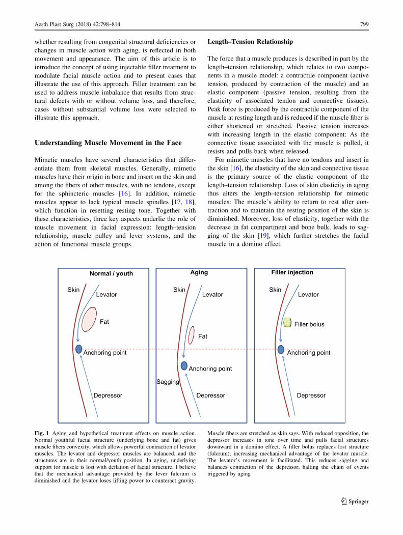
Fig. 1 Aging and hypothetical treatment effects on muscle action.
Normal youthful facial structure (underlying bone and fat) gives
muscle fibers convexity, which allows powerful contraction of levator
muscles. The levator and depressor muscles are balanced, and the
structures are in their normal/youth position. In aging, underlying
support for muscle is lost with deflation of facial structure. I believe
that the mechanical advantage provided by the lever fulcrum is
diminished and the levator loses lifting power to counteract gravity.
Muscle fibers are stretched as skin sags. With reduced opposition, the
depressor increases in tone over time and pulls facial structures
downward in a domino effect. A filler bolus replaces lost structure
(fulcrum), increasing mechanical advantage of the levator muscle.
The levator’s movement is facilitated. This reduces sagging and
balances contraction of the depressor, halting the chain of events
triggered by aging
Aesth Plast Surg (2018) 42:798–814 799
123
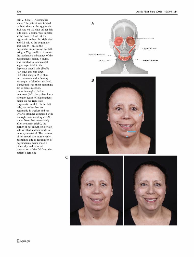
Fig. 2 Case 1: Asymmetric
smile. The patient was treated
on both sides at the zygomatic
arch and on the chin on her left
side only. Voluma was injected
at the bone, 0.1 mL at the
zygomatic arch on her right side
and 0.1 mL at the zygomatic
arch and 0.1 mL at the
zygomatic eminence on her left,
using a 27-g needle to increase
the mechanical advantage of the
zygomaticus major. Voluma
was injected in labiomental
angle superficial to the
depressor anguli oris (DAO)
(0.7 mL) and chin apex
(0.3 mL) using a 25-g blunt
microcannula and a fanning
technique. a Muscles involved.
b Injection sites (blue markings;
dot = bolus injection,
bar = fanning). c Before
treatment (left), the patient has a
stronger action of zygomaticus
major on her right side
(zygomatic smile). On her left
side, we notice that her
zygomatic is weaker and her
DAO is stronger compared with
her right side, creating a DAO
smile. Note that immediately
after treatment (right), the
corner of her mouth on her left
side is lifted and her smile is
more symmetrical. The corners
of her mouth are more evenly
positioned due to facilitation of
zygomaticus major muscle
bilaterally and reduced
contraction of the DAO on the
patient’s left side
800 Aesth Plast Surg (2018) 42:798–814
123
Muscle Pulley and Lever Systems
Biomechanical fixed pulley systems alter the angle of
action of muscles, and levers increase their mechanical
advantage, enhancing muscle force or displacement
[20, 21]. An example of such a biomechanical system in
the body is the patella [20]. The lateral suborbicularis oculi
fat pad (SOOF), located at the lateral/inferior orbital rim
and deep to the orbicularis oculi and zygomaticus major
[22, 23], acts as a pulley glide plane [1] and, together with
bone, as a lever fulcrum for the zygomaticus major muscle.
Acting over the SOOF appears to provide a mechanical
advantage to the zygomaticus major, which lifts the corners
of the mouth in a smile. In aging, the loss of structure
beneath the muscle, either from loss of bone or the loss
and/or ptosis of fat, might decrease that fulcrum effect,
reducing the muscle’s force and diminishing its ability to
lift the corner of the mouth.
Functional Muscle Groups
Groups of muscles (agonists and antagonists) working in
harmony contribute to a normal, youthful appearance in
facial expression. Levators and depressors work in oppo-
sition, and their interactions underlie facial appearance at
rest and in dynamic expression. In youth, levators are
usually stronger than depressors [24, 25]. The levator
muscle’s ability to maintain the position of soft tissue
structures of the face is counteracted by the downward pull
of gravity and the pull of its depressor antagonists. How-
ever, the balance between antagonist muscles may be dis-
rupted, due to structural deficiencies in youth or due to
bone and/or soft tissue loss in aging: If a levator muscle
lacks or loses lifting power, the depressor is freed to act
with reduced opposition.
Consider the synergistic levators of the upper lip: In
youth, the zygomaticus major and minor muscles play a
critical role in making the corner of the mouth tilt up in a
smile. If the zygomaticus major has reduced lifting power
due to a lack of underlying structural support, the relative
role of the risorius muscle increases and produces a more
horizontal smile. If zygomaticus major lifting capacity is
further diminished, the depressor anguli oris (DAO) muscle
predominates, and a ‘‘DAO smile,’’ with the corners of the
mouth downturned, is observed [11, 26]. The lack of
underlying structure leading to DAO smile may result from
changes over time in aging, or it may occur in youth due to
structural deficiency.
Addressing Muscle Movementwith Injectable Fillers
Each of these biomechanical concepts contributes to our
understanding of changes in muscle movement during
aging, as well as in cases of facial palsy. In the typical
youthful face, there is a clear convexity of the upper cheek
due to intact zygomatic arch, malar fat, and SOOF. Under
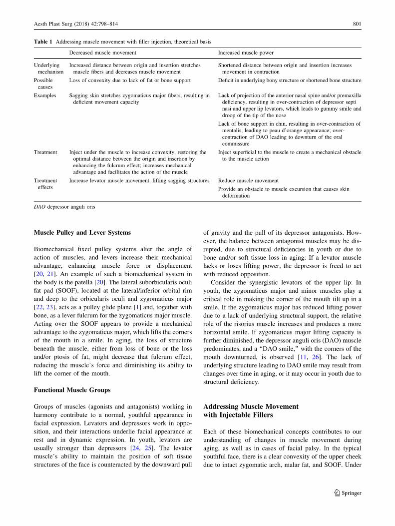
Table 1 Addressing muscle movement with filler injection, theoretical basis
Decreased muscle movement Increased muscle power
Underlying
mechanism
Increased distance between origin and insertion stretches
muscle fibers and decreases muscle movement
Shortened distance between origin and insertion increases
movement in contraction
Possible
causes
Loss of convexity due to lack of fat or bone support Deficit in underlying bony structure or shortened bone structure
Examples Sagging skin stretches zygomaticus major fibers, resulting in
deficient movement capacity
Lack of projection of the anterior nasal spine and/or premaxilla
deficiency, resulting in over-contraction of depressor septi
nasi and upper lip levators, which leads to gummy smile and
droop of the tip of the nose
Lack of bone support in chin, resulting in over-contraction of
mentalis, leading to peau d’orange appearance; over-
contraction of DAO leading to downturn of the oral
commissure
Treatment Inject under the muscle to increase convexity, restoring the
optimal distance between the origin and insertion by
enhancing the fulcrum effect; increases mechanical
advantage and facilitates the action of the muscle
Inject superficial to the muscle to create a mechanical obstacle
to the muscle action
Treatment
effects
Increase levator muscle movement, lifting sagging structures Reduce muscle movement
Provide an obstacle to muscle excursion that causes skin
deformation
DAO depressor anguli oris
Aesth Plast Surg (2018) 42:798–814 801
123
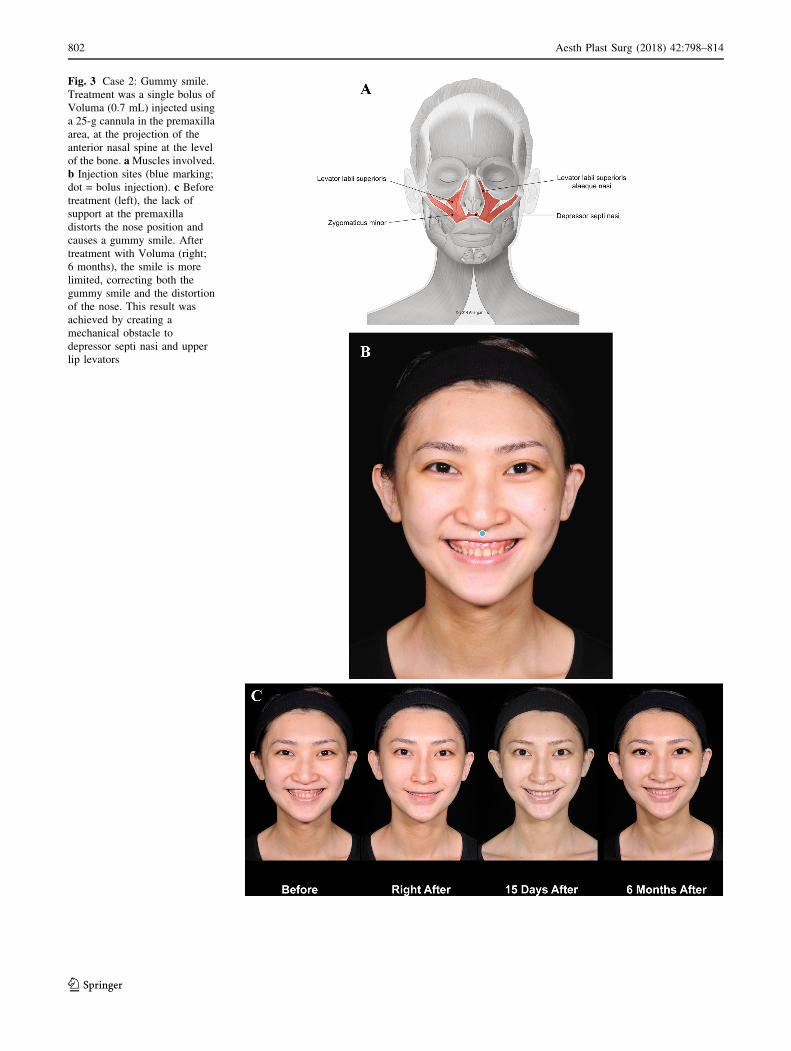
Fig. 3 Case 2: Gummy smile.
Treatment was a single bolus of
Voluma (0.7 mL) injected using
a 25-g cannula in the premaxilla
area, at the projection of the
anterior nasal spine at the level
of the bone. aMuscles involved.
b Injection sites (blue marking;
dot = bolus injection). c Before
treatment (left), the lack of
support at the premaxilla
distorts the nose position and
causes a gummy smile. After
treatment with Voluma (right;
6 months), the smile is more
limited, correcting both the
gummy smile and the distortion
of the nose. This result was
achieved by creating a
mechanical obstacle to
depressor septi nasi and upper
lip levators
802 Aesth Plast Surg (2018) 42:798–814
123
Aesth Plast Surg (2018) 42:798–814 803
123
these ideal conditions there is optimal pulling force of the
zygomaticus major muscle. In aging, midface volume loss,
displacement of fat pads, and loss of skin elasticity may
alter zygomaticus major muscle action. The distance
between the zygomaticus major origin in the zygomatic
bone and insertion in the modiolus area at the corner of the
mouth [27] increases when skin sags due to deflation of fat
pads, and as a result, the corner of the mouth falls (Fig. 1).
The stretching of the fibers of the zygomaticus major
results in a loss of resting tension and power in contraction.
At the same time, any mechanical advantage of the lever
effect over the lateral SOOF is reduced as SOOF volume is
depleted. Consequently, the zygomaticus major can no
longer adequately counterbalance the downward gravita-
tional pull and the contracting force of the DAO [11, 26].
Case 1, a mature woman with an asymmetrical smile,
illustrates some of these effects (Fig. 2). On her right side,
she presents a zygomatic smile, but on her left she shows a
DAO pattern (Fig. 2c, left). Before treatment, her left
cheek sags, yielding a more prominent nasolabial fold. The
zygomaticus major is less efficient on her left side, and the
balance between weakened zygomaticus major and its
antagonist, DAO, has been lost. DAO is now free to pull
down the corner of the mouth.
The presence of fillers can act mechanically to alter
muscle movement by either facilitating their action, via a
lever or pulley effect, or decreasing contracting by
blocking their movement (Table 1). A bolus of filler
injected under (deep to) a muscle increases its convexity,
acting as a fulcrum to increase mechanical advantage.
Conversely, injecting filler more superficially may reduce
contraction by impeding muscle movement. These are
simple mechanical effects.
The Case 1 patient was treated on both sides with
Juvederm Voluma� (Allergan plc, Dublin, Ireland). On her
right side, a bolus was injected at the level of the bone on
the zygomatic arch under zygomaticus major. On her left
side, two boluses were injected at the same plane (Fig. 2b).
The structural support increased the muscle’s lifting action,
and mechanical advantage on her left side was enhanced.
Voluma was injected superficial to mentalis, depressor labii
inferioris, and DAO along the labiomental angle on her left
side only. This mechanical obstacle to DAO movement
decreases its downward pulling effect. After treatment
(Fig. 2c, right), the action of zygomaticus major is facili-
tated on both sides and DAO is blocked on her left, so that
zygomaticus major now lifts that corner of her mouth.
A lack of structural support due to bone deficiency can
result in abnormal muscular contraction and surface
deformation. Case 2 (Fig. 3) illustrates the contribution of
a deficiency of the anterior nasal spine and premaxilla to
gummy smile, and correction using injectable filler treat-
ment. The young Asian patient presented with a lack of
projection of the anterior nasal spine, retruded underde-
veloped columella, and a deficit in the projection of the
upper maxilla. The lack of structural and mechanical sup-
port results in excessive movement of the upper lip levators
(levator labii superioris alaeque nasi [LLSAN], levator
labii superioris, and zygomaticus minor), which produces
gummy smile (Fig. 3c, left). The lack of support also leads
to collapse of the tip of the nose and widening of the nasal
flare. Voluma injected along the premaxilla and at the
projection of the anterior nasal spine helped to compensate
for the bone deficiency. After treatment, there is a reduc-
tion in the movement of the upper lip levators during smile
and the upward retraction of the upper lip is decreased
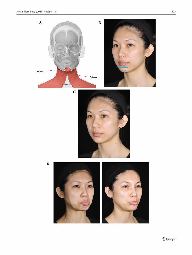
bFig. 4 Case 3: Lack of proper bone support in the chin. The patient
was treated with a total of 4 mL of Voluma (1 mL per side into the
labiomental angle and 2 mL in the chin apex using a 25-g cannula).
a Muscles involved. b Injection sites (blue markings; dot = bolus
injection, bar = fanning). c Before treatment, the patient has no
apparent structural deficiency at rest. d Pout. Before treatment (left),
lack of support in the chin causes mentalis over-contraction and peau
d’orange appearance. The platysma is also activated when pouting.
Six months after providing a mechanical obstacle to movement with
Voluma in her chin (right), the patient is able to protrude her lower lip
without skin wrinkling or recruitment of platysma. Blocking over-
contraction of the chin also reduces the development of hypertonic
platysmal bands
804 Aesth Plast Surg (2018) 42:798–814
123
(Fig. 3c, center and right panels). Injection behind the
columella has also helped to stabilize the nose.
The third case (Fig. 4) is a young woman who has no
apparent deficiency at rest (Fig. 4c). However, on anima-
tion, a lack of proper bone support in her chin becomes
evident (Fig. 4d, left). When she pouts, mentalis is acti-
vated and over-contracts. This results in upward rotation of
her chin and protrusion of the lower lip, with excessive skin
wrinkling and deformation. After treatment with Voluma,
the patient’s pout is normal (Fig. 4d, right). The presence
of a mechanical barrier at the labiomental angle and chin
apex prevents the upward rotation and consequent skin
wrinkling. Note that the extreme over-contraction of
mentalis is blocked while preserving proper mentalis action
and therefore, the patient is still able to protrude the lower
lip. When over-contraction of mentalis is treated with
onabotulinumtoxinA, the ability to evert the lower lip can
be reduced or lost depending on the dose.
In a second young woman with a lack of bone support
for mentalis (Case 4; Fig. 5), distortion of the chin is evi-
dent both at rest (Fig. 5c, left) and when she purses her lips
(Fig. 5d, left). Immediately after treatment, improvement
in her chin is observed at rest (Fig. 5c, left center) and on
animation (Fig. 5d, left center). Blocking the excessive
movement of mentalis eliminates the resulting distortion
and allows the patient to purse her lips with no skin
wrinkling.
Case 5 (Fig. 6) is a young woman with notable distor-
tion on animation (kissing, pouting). Normally, movement
of the upper and lower lips during pursing is governed by
nose and chin position, due to stability at the nasolabial
angle and the labiomental angle, respectively. With support
at these two sites, the direction of movement in a kiss is
horizontal. The patient presented here lacks support at the
level of the labiomental angle and chin. Instability at the
labiomental angle perturbs the contraction of orbicularis
oris. When she is asked to purse her lips (kiss; Fig. 6c,
left), this instability causes both upper and lower lips to
drop down, resulting in distortion. The normal action of
mentalis in pouting leads to protrusion of the lower lip. In
this case, however, the patient cannot properly protrude the
lip when asked to pout (Fig. 6d, left). Instead, her lower lip
everts toward the oral cavity and hides the upper lip.
Treatment with Voluma in the chin and Juvederm� Ultra
Plus injectable gel (Allergan plc) in the lip border allows
the patient to produce a natural appearance in a kiss
(Fig. 6c, right) and pout (Fig. 6d, right). With support in
the soft tissue of chin and lips, mentalis and orbicularis oris
contract in a more balanced, stable, and organized way.
The chin and lower lip are improved by support to mentalis
in the labiomental angle and chin apex. The upper lip is
improved both directly by Juvederm Ultra Plus injection
and indirectly by the more organized contraction in the
lower lip.
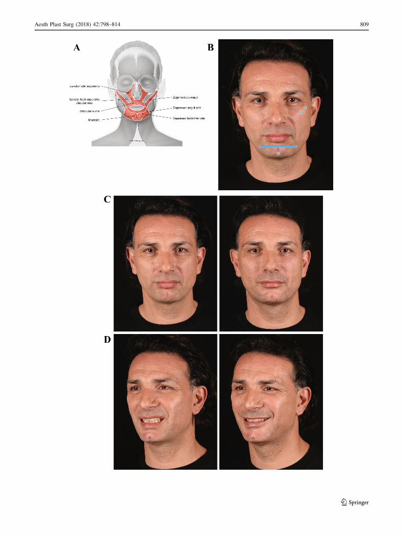
The sixth case is a man with mild facial asymmetry, but
normal facial nerve function observed at rest and on ani-
mation (Fig. 7). At rest, there is a more prominent naso-
labial fold on his left side due to contraction of LLSAN,
and his upper lip is slightly elevated (Fig. 7c, left). Upon
animation, both LLSAN and levator labii superioris are
over-contracted (Fig. 7d, left), and he presents lower teeth
show on his left side when smiling. The patient was
injected with two filler boluses under zygomaticus major at
the level of his zygomatic arch and zygomatic eminence,
respectively (Fig. 7b). He was also injected at the
labiomental angle on both sides, and at the chin apex. After
treatment, there is less lateral recruitment of the LLSAN
and the improvement of nasolabial fold can be observed at
rest (Fig. 7c, right). On animation, the upper lip is now
better aligned, and as observed in his oblique view, he
presents a stronger zygomatic smile due to facilitation of
the zygomaticus major muscle via the fulcrum effect
(Fig. 7d, right). He also presents less lower teeth show.
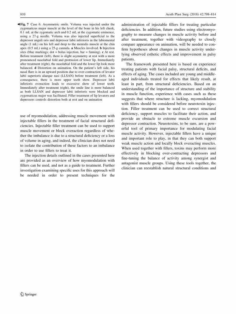
The final case provides a most compelling demonstra-
tion of the use of injectable fillers for myomodulation. Case
7 (Fig. 8) is a middle-aged man with facial palsy on the
right side resulting from acoustic neuroma surgery in 2011.
Prior to injectable filler injection, no treatment to improve
the facial asymmetry was performed. Before treatment
(Fig. 8c, left), the patient had classic signs of facial palsy
on his right side: a lack of function of orbicularis oculi and
zygomaticus major, with scleral show and skin laxity. A
more prominent nasolabial fold and lip deviation toward
his left side is observed. The patient’s smile (Fig. 8d, left)
shows even greater deviation of the oral commissure
toward his hyperkinetic side (left). On his facial palsy side
(right), the contraction of lateral platysmal band shows that
the facial nerve is not completely damaged. Slight con-
traction of the zygomaticus major, indicated by the pres-
ence of a dynamic line at the modiolus level and worsening
Aesth Plast Surg (2018) 42:798–814 805
123
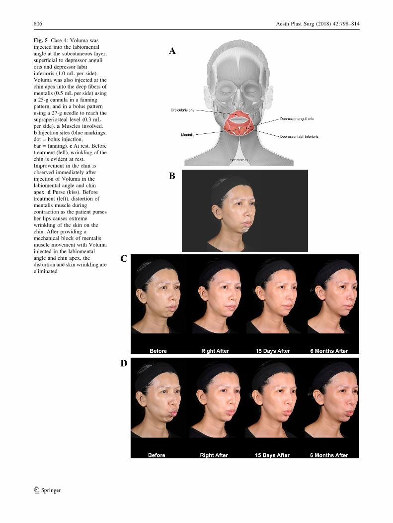
Fig. 5 Case 4: Voluma was
injected into the labiomental
angle at the subcutaneous layer,
superficial to depressor anguli
oris and depressor labii
inferioris (1.0 mL per side).
Voluma was also injected at the
chin apex into the deep fibers of
mentalis (0.5 mL per side) using
a 25-g cannula in a fanning
pattern, and in a bolus pattern
using a 27-g needle to reach the
supraperiosteal level (0.3 mL
per side). a Muscles involved.
b Injection sites (blue markings;
dot = bolus injection,
bar = fanning). c At rest. Beforetreatment (left), wrinkling of the
chin is evident at rest.
Improvement in the chin is
observed immediately after
injection of Voluma in the
labiomental angle and chin
apex. d Purse (kiss). Before
treatment (left), distortion of
mentalis muscle during
contraction as the patient purses
her lips causes extreme
wrinkling of the skin on the
chin. After providing a
mechanical block of mentalis
muscle movement with Voluma
injected in the labiomental
angle and chin apex, the
distortion and skin wrinkling are
eliminated
806 Aesth Plast Surg (2018) 42:798–814
123
Aesth Plast Surg (2018) 42:798–814 807
123
of the skin excess at the lower eyelid, suggests that there is
residual activity in the zygomatic branch. When the patient
tries to close his eyes tightly (Fig. 8e, left), abnormal
coordination of facial muscles is demonstrated by his
frown, excessive contraction of platysma, and activation of
orbicularis oris on his left with the eyebrow elevated.
When he tries to close his eyes without frowning (Fig. 8f,
left), scleral show and abnormal behavior of lateral muscles
are observed.
Mimetic muscles on both sides were treated with filler
injections (Fig. 8b), with depth of injection the major dif-
ference between the two sides. The treatment of the
patient’s right side focused on lifting the cheek and stabi-
lizing the lips. Filler was injected deep to muscle fibers, at
the level of the bone wherever possible, to create a lever
effect to increase muscle movement. On his left (hyperki-
netic) side, fillers were injected superficial to the muscle, at
the subcutaneous level, in order to reduce contraction by
providing a mechanical obstacle to muscle contraction.
Filler products and volumes used in this case are shown by
injection site in Table 2. Topographical muscle anatomy
and mimetic animation assessment during the injection
were used to guide the treatment. There was no need for the
use of electromyography.
Immediately after the injections, better symmetry and
muscle coordination were observed. On smiling, there is a
reduction in the upper lateral excursion of the zygomatic
major muscle on his left side and better positioning of the
oral commissure and upper and lower lips (Fig. 8d,
immediately after). When closing his eyes (with effort)
immediately after treatment, a deeper nasolabial fold is
apparent on his left, indicating a change in muscle behavior
between his right and his left side (Fig. 8e, immediately
after). When he tries to close his eyes without effort, there
is improvement in scleral show immediately after treatment
(Fig. 8f, immediately after).
Over the 6 months following treatment, the patient was
instructed to practice closing his eyes and smiling more
symmetrically using a mirror. Although improvement
resulting directly from filler treatment is evident in the
patient’s smile and eye closure in the photographs taken
immediately after treatment (Fig. 8d–f), the symmetry of
his smile continues to improve progressively with exercise
(Fig. 8d, 1–6 months). One month after treatment, con-
traction of zygomatic muscles on his right side appears
enhanced and there is less recruitment of platysma
(Fig. 8d, 1 month). By 4 months after treatment, contrac-
tion of orbicularis oculi and position of the eyebrows are
improved (Fig. 8e, 4 months), the patient closes his eye
more naturally, and enhanced zygomatic action is observed
(Fig. 8f, 4 months). At 6 months, he closes his eyes
effortlessly without recruitment of adjacent muscles
(Fig. 8e and f, 6 months).
Summary and Conclusions
Deficits in facial structure can yield abnormal muscle
action reflected at the skin and across the face. When
structural support is absent or lost, muscle action is altered,
affecting the balance in activity between muscles. Exam-
ining the interactions between facial structure and muscle
movement—and recognizing unbalanced action in muscle
synergists and antagonists—allows the clinician to under-
stand the effects on appearance both at rest and on ani-
mation. The cases presented here provide support for the
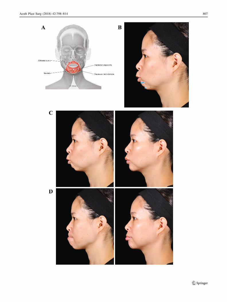
bFig. 6 Case 5: Lack of projection at the chin. Voluma was injected
into the labiomental angle, superficial to depressor anguli oris and
depressor labii inferioris (0.5 mL per side), and at the chin apex into
the deep fibers of mentalis (1 mL) using a 25-g cannula. Juvederm
Ultra Plus was injected in the lip border (1 mL each in cupid’s bow
and lip border) using a 27-g needle. a Muscles involved. b Injection
sites (blue markings; dot = bolus injection, bar = fanning). c Purse
(kiss). Abnormal pursing movement before treatment (left) is due to a
lack of support of chin and lips. Six months after treatment in the
labiomental angle and chin (right), the patient is able to correctly
contract orbicularis oris without deformation. d Pout. Filler injection
in the labiomental angle and in the chin also addresses abnormal
pouting movement observed before treatment (left). Six months after
the injection (right), the patient is able to pout correctly
808 Aesth Plast Surg (2018) 42:798–814
123
Aesth Plast Surg (2018) 42:798–814 809
123
use of myomodulation, addressing muscle movement with
injectable fillers in the treatment of facial structural defi-
ciencies. Injectable filler treatment can be used to support
muscle movement or block overaction regardless of whe-
ther the imbalance is due to a structural deficiency or a loss
of volume in aging, and indeed, the clinician does not need
to isolate the contribution of these factors to an imbalance
in order to use fillers to treat it.
The injection details outlined in the cases presented here
are provided as an overview of how myomodulation with
fillers can be used, and not as a guide to treatment. Further
investigation examining specific uses for this approach will
be needed in order to present techniques for the
administration of injectable fillers for treating particular
deficiencies. In addition, future studies using electromyo-
graphy to measure changes in muscle activity before and
after treatment, together with videography to closely
compare appearance on animation, will be needed to con-
firm hypotheses about changes in muscle activity under-
lying observed esthetic effects and improvement in palsy
patients.
The framework presented here is based on experience
treating patients with facial palsy, structural deficits, and
effects of aging. The cases included are young and middle-
aged individuals treated for effects that likely result, at
least in part, from structural deficiencies. Based on an
understanding of the importance of structure and stability
in muscle function, experience with cases such as these
suggests that where structure is lacking, myomodulation
with fillers should be considered before neurotoxin injec-
tion. Filler treatment can be used to correct structural
deficiency, support muscles to facilitate their action, and
provide an obstacle to extreme muscle excursion and
depressor contraction. Neurotoxins, to be sure, are a pow-
erful tool of primary importance for modulating facial
muscle activity. However, injectable fillers have a unique
and important role to play, in that they can both support
weak muscle action and locally block overacting muscles.
When used together with fillers, toxins may perform more
effectively in blocking over-contracting depressors and
fine-tuning the balance of activity among synergist and
antagonist muscle groups. Using these tools together, the
clinician can reestablish natural structural conditions and
bFig. 7 Case 6: Asymmetric smile. Voluma was injected under the
zygomaticus major muscle at the level of the bone in his left cheek,
0.1 mL at the zygomatic arch and 0.2 mL at the zygomatic eminence,
using a 27-g needle. Voluma was also injected superficial to the
depressor anguli oris and depressor labii inferioris in the labiomental
angle (1 mL) on his left and deep to the mentalis muscle at the chin
apex (0.5 mL) using a 25-g cannula. a Muscles involved. b Injection
sites (blue markings; dot = bolus injection, bar = fanning). c At rest.
Before treatment (left), there is slight asymmetry at rest with a more
pronounced nasolabial fold and protrusion of lower lip. Immediately
after treatment (right), the nasolabial fold and the lower lip look more
balanced. d Distortion on animation. On the patient’s left side, his
nasal flare is in an upward position due to over-contraction of levator
labii superioris alaeque nasi (LLSAN) before treatment (left). As a
consequence, there is more upper teeth show. Depressor labii
inferioris overaction leads to excessive show of lower teeth.
Immediately after treatment (right), the smile line is more balanced
as both LLSAN and depressor labii inferioris were blocked and
zygomaticus major was facilitated. Filler treatment of lip levators and
depressors controls distortion both at rest and on animation
810 Aesth Plast Surg (2018) 42:798–814
123
Aesth Plast Surg (2018) 42:798–814 811
123
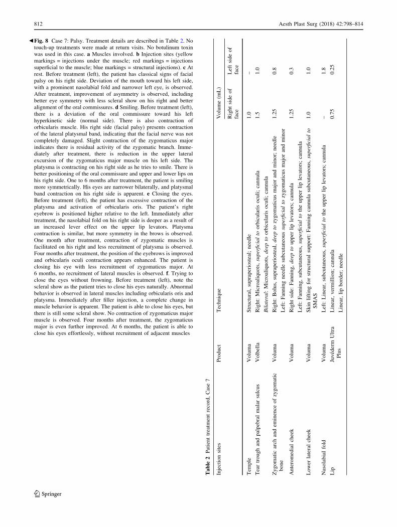
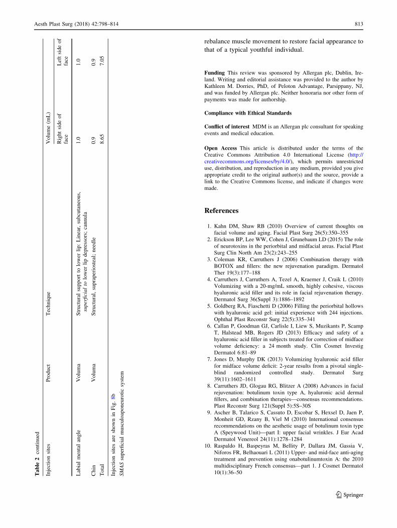
bFig. 8 Case 7: Palsy. Treatment details are described in Table 2. No
touch-up treatments were made at return visits. No botulinum toxin
was used in this case. a Muscles involved. b Injection sites (yellow
markings = injections under the muscle; red markings = injections
superficial to the muscle; blue markings = structural injections). c Atrest. Before treatment (left), the patient has classical signs of facial
palsy on his right side. Deviation of the mouth toward his left side,
with a prominent nasolabial fold and narrower left eye, is observed.
After treatment, improvement of asymmetry is observed, including
better eye symmetry with less scleral show on his right and better
alignment of the oral commissures. d Smiling. Before treatment (left),
there is a deviation of the oral commissure toward his left
hyperkinetic side (normal side). There is also contraction of
orbicularis muscle. His right side (facial palsy) presents contraction
of the lateral platysmal band, indicating that the facial nerve was not
completely damaged. Slight contraction of the zygomaticus major
indicates there is residual activity of the zygomatic branch. Imme-
diately after treatment, there is reduction in the upper lateral
excursion of the zygomaticus major muscle on his left side. The
platysma is contracting on his right side as he tries to smile. There is
better positioning of the oral commissure and upper and lower lips on
his right side. One to 6 months after treatment, the patient is smiling
more symmetrically. His eyes are narrower bilaterally, and platysmal
band contraction on his right side is apparent. e Closing the eyes.
Before treatment (left), the patient has excessive contraction of the
platysma and activation of orbicularis oris. The patient’s right
eyebrow is positioned higher relative to the left. Immediately after
treatment, the nasolabial fold on his right side is deeper as a result of
an increased lever effect on the upper lip levators. Platysma
contraction is similar, but more symmetry in the brows is observed.
One month after treatment, contraction of zygomatic muscles is
facilitated on his right and less recruitment of platysma is observed.
Four months after treatment, the position of the eyebrows is improved
and orbicularis oculi contraction appears enhanced. The patient is
closing his eye with less recruitment of zygomaticus major. At
6 months, no recruitment of lateral muscles is observed. f. Trying to
close the eyes without frowning. Before treatment (left), note the
scleral show as the patient tries to close his eyes naturally. Abnormal
behavior is observed in lateral muscles including orbicularis oris and
platysma. Immediately after filler injection, a complete change in
muscle behavior is apparent. The patient is able to close his eyes, but
there is still some scleral show. No contraction of zygomaticus major
muscle is observed. Four months after treatment, the zygomaticus
major is even further improved. At 6 months, the patient is able to
close his eyes effortlessly, without recruitment of adjacent muscles
Table
2Patienttreatm
entrecord,Case7
Injectionsites
Product
Technique
Volume(m
L)
Rightsideof
face
Leftsideof
face
Tem
ple
Voluma
Structural,supraperiosteal;needle
1.0
–
Teartroughandpalpebralmalar
sulcus
Volbella
Right:Microaliquots,superficialto
orbicularisoculi;cannula
Bilateral:Microaliquots,deepto
orbicularisoculi;cannula
1.5
1.0
Zygomatic
arch
andem
inence
ofzygomatic
bone
Voluma
Right:Bolus,supraperiosteal,deepto
zygomaticusmajorandminor;needle
Left:Fanningneedle
subcutaneoussuperficialto
zygomaticusmajorandminor
1.25
0.8
Anteromedialcheek
Voluma
Rightside:
Fanning,deepto
upper
liplevators;cannula
Left:Fanning,subcutaneous,superficialto
theupper
liplevators;cannula
1.25
0.3
Lower
lateralcheek
Voluma
Skin
liftingforstructuralsupport:Fanningcannula
subcutaneous,superficialto
SMAS
1.0
1.0
Nasolabialfold
Voluma
Left:Linear,subcutaneous,superficialto
theupper
liplevators;cannula
–1.8
Lip
Juvederm
Ultra
Plus
Linear,vermillion;cannula
Linear,lipborder;needle
0.75
0.25
812 Aesth Plast Surg (2018) 42:798–814
123
rebalance muscle movement to restore facial appearance to
that of a typical youthful individual.
Funding This review was sponsored by Allergan plc, Dublin, Ire-
land. Writing and editorial assistance was provided to the author by
Kathleen M. Dorries, PhD, of Peloton Advantage, Parsippany, NJ,
and was funded by Allergan plc. Neither honoraria nor other form of
payments was made for authorship.
Compliance with Ethical Standards
Conflict of interest MDM is an Allergan plc consultant for speaking
events and medical education.
Open Access This article is distributed under the terms of the
Creative Commons Attribution 4.0 International License (http://
creativecommons.org/licenses/by/4.0/), which permits unrestricted
use, distribution, and reproduction in any medium, provided you give
appropriate credit to the original author(s) and the source, provide a
link to the Creative Commons license, and indicate if changes were
made.
References
1. Kahn DM, Shaw RB (2010) Overview of current thoughts on
facial volume and aging. Facial Plast Surg 26(5):350–355
2. Erickson BP, Lee WW, Cohen J, Grunebaum LD (2015) The role
of neurotoxins in the periorbital and midfacial areas. Facial Plast
Surg Clin North Am 23(2):243–255
3. Coleman KR, Carruthers J (2006) Combination therapy with
BOTOX and fillers: the new rejuvenation paradigm. Dermatol
Ther 19(3):177–188
4. Carruthers J, Carruthers A, Tezel A, Kraemer J, Craik L (2010)
Volumizing with a 20-mg/mL smooth, highly cohesive, viscous
hyaluronic acid filler and its role in facial rejuvenation therapy.
Dermatol Surg 36(Suppl 3):1886–1892
5. Goldberg RA, Fiaschetti D (2006) Filling the periorbital hollows
with hyaluronic acid gel: initial experience with 244 injections.
Ophthal Plast Reconstr Surg 22(5):335–341
6. Callan P, Goodman GJ, Carlisle I, Liew S, Muzikants P, Scamp
T, Halstead MB, Rogers JD (2013) Efficacy and safety of a
hyaluronic acid filler in subjects treated for correction of midface
volume deficiency: a 24 month study. Clin Cosmet Investig
Dermatol 6:81–89
7. Jones D, Murphy DK (2013) Volumizing hyaluronic acid filler
for midface volume deficit: 2-year results from a pivotal single-
blind randomized controlled study. Dermatol Surg
39(11):1602–1611
8. Carruthers JD, Glogau RG, Blitzer A (2008) Advances in facial
rejuvenation: botulinum toxin type A, hyaluronic acid dermal
fillers, and combination therapies—consensus recommendations.
Plast Reconstr Surg 121(Suppl 5):5S–30S
9. Ascher B, Talarico S, Cassuto D, Escobar S, Hexsel D, Jaen P,
Monheit GD, Rzany B, Viel M (2010) International consensus
recommendations on the aesthetic usage of botulinum toxin type
A (Speywood Unit)—part I: upper facial wrinkles. J Eur Acad
Dermatol Venereol 24(11):1278–1284
10. Raspaldo H, Baspeyras M, Bellity P, Dallara JM, Gassia V,
Niforos FR, Belhaouari L (2011) Upper- and mid-face anti-aging
treatment and prevention using onabotulinumtoxin A: the 2010
multidisciplinary French consensus—part 1. J Cosmet Dermatol
10(1):36–50Table
2continued
Injectionsites
Product
Technique
Volume(m
L)
Rightsideof
face
Leftsideof
face
Labialmentalangle
Voluma
Structuralsupportto
lower
lip:Linear,subcutaneous,
superficialto
lower
lipdepressors;cannula
1.0
1.0
Chin
Voluma
Structural,supraperiosteal;needle
0.9
0.9
Total
8.65
7.05
Injectionsitesareshownin
Fig.8b
SMASsuperficial
musculoaponeuroticsystem
Aesth Plast Surg (2018) 42:798–814 813
123
11. Maas C, Kane MA, Bucay VW, Allen S, Applebaum DJ, Bau-
mann L, Cox SE, Few JW, Joseph JH, Lorenc ZP, Moradi A,
Nestor MS, Schlessinger J, Wortzman M, Lawrence I, Lin X,
Nelson D (2012) Current aesthetic use of abobotulinumtoxinA in
clinical practice: an evidence-based consensus review. Aesthet
Surg J 32(1 Suppl):8S–29S
12. de Maio M (2003) Use of botulinum toxin in facial paralysis.
J Cosmet Laser Ther 5(3–4):216–217
13. de Maio M, Bento RF (2007) Botulinum toxin in facial palsy: an
effective treatment for contralateral hyperkinesis. Plast Reconstr
Surg 120(4):917–927
14. Olson JJ (2007) Balanced botox chemodenervation of the upper
face: symmetry in motion. Semin Plast Surg 21(1):47–53
15. Lorenc ZP, Smith S, Nestor M, Nelson D, Moradi A (2013)
Understanding the functional anatomy of the frontalis and
glabellar complex for optimal aesthetic botulinum toxin type A
therapy. Aesthetic Plast Surg 37(5):975–983
16. Micheli-Pellegrini V (2011) About muscle insertions in man
(Proposal for a new nomenclature of striated muscle). Acta
Otorhinolaryngol Ital 31(3):167–176
17. Goodmurphy CW, Ovalle WK (1999) Morphological study of
two human facial muscles: orbicularis oculi and corrugator
supercilii. Clin Anat 12(1):1–11
18. Stal P, Eriksson PO, Eriksson A, Thornell LE (1987) Enzyme-
histochemical differences in fibre-type between the human major
and minor zygomatic and the first dorsal interosseus muscles.
Arch Oral Biol 32(11):833–841
19. Ezure T, Hosoi J, Amano S, Tsuchiya T (2009) Sagging of the
cheek is related to skin elasticity, fat mass and mimetic muscle
function. Skin Res Technol 15(3):299–305
20. Grelsamer RP, Klein JR (1998) The biomechanics of the patel-
lofemoral joint. J Orthop Sports Phys Ther 28(5):286–298
21. Doyle JR (2001) Palmar and digital flexor tendon pulleys. Clin
Orthop Relat Res 383:84–96
22. Aiache AE, Ramirez OH (1995) The suborbicularis oculi fat
pads: an anatomic and clinical study. Plast Reconstr Surg
95(1):37–42
23. Rohrich RJ, Arbique GM, Wong C, Brown S, Pessa JE (2009)
The anatomy of suborbicularis fat: implications for periorbital
rejuvenation. Plast Reconstr Surg 124(3):946–951
24. Gray H (1974) The muscles and fasciae. In: Pick TP, Howden R
(eds) Anatomy, descriptive and surgical. Running Press,
Philadelphia, pp 306–315
25. de Maio M, Rzany B (2009) Botulinum toxin in aesthetic med-
icine. Springer, Berlin
26. Ascher B, Talarico S, Cassuto D, Escobar S, Hexsel D, Jaen P,
Monheit GD, Rzany B, Viel M (2010) International consensus
recommendations on the aesthetic usage of botulinum toxin type
A (Speywood Unit)—part II: wrinkles on the middle and lower
face, neck and chest. J Eur Acad Dermatol Venereol
24(11):1285–1295
27. Pessa JE, Zadoo VP, Garza PA, Adrian EK Jr, DeWitt AI, Garza
JR (1998) Double or bifid zygomaticus major muscle: anatomy,
incidence, and clinical correlation. Clin Anat 11(5):310–313
814 Aesth Plast Surg (2018) 42:798–814
123