Embed Size (px)
Citation preview
A NOVEL LYSOPHOSPHATIDYLETHANOLAMINE DEHYDROGENASE FROM
MYXOCOCCUS XANTHUS
by
MADHAVI SITARAMA AVADHANI
(Under the Direction of LAWRENCE J. SHIMKETS)
ABSTRACT
Myxococcus xanthus serves as a prokaryotic paradigm to study multicellularity in
bacteria. Nutrient limitation initiates a developmental program in which the cells exhibit
spatially and temporally regulated, morphologically distinct behaviors. The
developmental program is coordinated by a series of intercellular signals. The most
significant among them is the contact-dependent signal termed the C-signal which is
absolutely essential for completion of development. The protein responsible for C-
signaling, CsgA, belongs to the short-chain alcohol dehydrogenase (SCAD) family of
enzymes. CsgA is proteolytically processed into a 17 kDa form. Much debate has
focused on whether intercellular signaling occurs because of the enzymatic activity of the
full length form or if the 17 kDa form is itself the signal. The genetic basis for the
enzymatic role of CsgA comes from isolation of overexpression suppressor mutants.
Overexpression of SocA, a SCAD with 28% identity to CsgA almost exclusively in the
enzyme active site, not only suppresses the C-signaling defect, but also restores C-
signaling to csgA mutants upon extracellular complementation. In addition, conserved
residues of CsgA are shown to be essential for C-signaling. The potential role of SocA in
generating the C-signal was exploited to determine the biochemical basis for C-signaling.
We developed a dehydrogenase assay based screen coupled with mass spectrometry to
identify SocA substrates. Our results indicate that SocA performs a novel biochemical
reaction in oxidizing lysophoaphatidylethanolamine. Oxidized
lysophosphatidylethanolamine does not function as an intercellular signal in M. xanthus.
INDEX WORDS: Myxococcus xanthus, Intercellular signaling, Short chain alcohol dehydrogenase, Lysophosphatidylethanolamine
A NOVEL LYSOPHOSPHATIDYLETHANOLAMINE DEHYDROGENASE FROM
MYXOCOCCUS XANTHUS
by
MADHAVI SITARAMA AVADHANI
M. Sc., Bangalore University, India, 1997
A Dissertation Submitted to the Graduate Faculty of The University of Georgia in Partial
Fulfillment of the Requirements for the Degree
DOCTOR OF PHILOSOPHY
ATHENS, GEORGIA
2005
© 2005
MADHAVI SITARAMA AVADHANI
All Rights Reserved
A NOVEL LYSOPHOSPHATIDYLETHANOLAMINE DEHYDROGENASE FROM
MYXOCOCCUS XANTHUS
by
MADHAVI SITARAMA AVADHANI
Major Professor: Dr. Lawrence J.
Shimkets
Committee: Dr. Harry Dailey Dr. Claiborne Glover Dr. Timothy Hoover Dr. Robert Maier
Electronic Version Approved: Maureen Grasso Dean of the Graduate School The University of Georgia May 2005
ACKNOWLEDGEMENTS
I am grateful to my advisor Dr. Larry Shimkets for believing in me during the
course of this arduous journey. I completely owe my growth as a scientist to him. I
thank my doctoral advisory committee: Dr. Harry Dailey, Dr. Claiborne Glover, Dr. Tim
Hoover, and Dr. Rob Maier for their expert counsel. I would like to acknowledge with
deep gratitude Dr. Roland Geyer, for putting a completely new spin on my research with
his insightful mass spectrometric expertise. I would also like to thank Dr. Christian Heiss
for helpful discussions on the structure of oxidized lysophosphatidylethanolamine. I
consider myself fortunate to have had the friends and colleagues who made it possible for
me to look beyond the frustrations of everyday research. I have a high regard for Minnie
in particular, who always found time to humor my questions, science or otherwise. I am
grateful to my extremely loving supportive family. I would certainly not have seen this
day without the love and sacrifice of Amma and Nanna. Shesh has been my greatest
source of strength and encouragement. I feel very fortunate to spend the rest of my life
with him. Finally, I would like to thank Priya, Ananth, and Geetha for their love and
support.
iv
TABLE OF CONTENTS
ACKNOWLEDGEMENTS........................................................................................................... iv
LIST OF TABLES......................................................................................................................... vi
LIST OF FIGURES ...................................................................................................................... vii
CHAPTER
1. INTRODUCTION AND LITERATURE REVIEW .....................................................1
Multicellularity in Myxococcus xanthus....................................................................2
Short chain alcohol dehydrogenase.........................................................................38
2. A NOVEL LYSOPHOSPHATIDYLETHANOLAMINE
DEHYDROGENASE FROM MYXOCOCCUS XANTHUS ...................................85
Introduction .............................................................................................................85
Materials and methods.............................................................................................89
Results .....................................................................................................................96
Discussion .............................................................................................................121
3. CONCLUSION..........................................................................................................129
APPENDICES
A. BIOASSAYS INVOLVING EXTRACELLULAR LIPID
COMPLEMENTATION ...........................................................................................137
B. RESCUE OF DEVELOPMENTAL AGGREGATION AND SPORULATION
BY PARTIALLY PURE E. COLI PROTEIN PREPARATION ..........................140
Materials and methods...........................................................................................140
Results ...................................................................................................................142
v
LIST OF TABLES
Table 1.1: Extracellular complementation groups in M. xanthus ....................................................9
Table 2.1: Fatty acid specificity of SocA.....................................................................................107
Table 2.2: Head group specificity of SocA..................................................................................111
Table 2.3: Lysophosphatidylethanolamine species (molecules/cell) in vegetative and
developing cells ...........................................................................................................118
vi
LIST OF FIGURES
Figure 1.1: Life cycle of M. xanthus................................................................................................4
Figure 1.2: Time scale indicating the various morphological stages and the earliest point
where gene expression is arrested by mutants in the different extracellular
complementation groups ...............................................................................................12
Figure 1.3: A model for C-signal transduction pathway................................................................40
Figure 1.4: Rossmann fold topology..............................................................................................44
Figure 1.5: Interaction of conserved residues from the central β-sheet.........................................47
Figure 1.6: The proposed proton relay chain in the binary complex (NAD+) of Drosophila
lebanonensis alcohol dehydrogenase ..........................................................................51
Figure 1.7: An alignment of two M. xanthus SCAD, CsgA and SocA, with 3α/20β-
hydroxysteroid dehydrogenase......................................................................................58
Figure 2.1: Silver-stained gel showing tSocAh (lane 1), hSocA (lane 2) purified by
affinity chromatography on a Ni column ......................................................................98
Figure 2.2: Total Wavelength Chromatogram of separation on C18 reverse phase column.......101
Figure 2.3: Mass spectrum of the fraction with highest enzyme activity ....................................103
Figure 2.4: Effect of pH on lysoPE 16:0 oxidation by SocA.......................................................106
Figure 2.5: Double reciprocal plot of initial velocity versus lysoPE 18:1 concentrations ..........109
Figure 2.6: Proposed keto-enol tautomerism and subsequent hydrolysis of oxidized
lysoPE..........................................................................................................................114
Figure 2.7: Thin layer chromatography of lysoPE 16:0 and oxidized lysoPE 16:0 ....................116
vii
Figure 2.8: Rescue of developmental aggregation by SocA-oxidized extract.............................120
Figure A.1: Silver-stained gel showing partially pure E. coli protein .........................................144
Figure A.2: Rescue of developmental aggregation by E. coli protein preparation......................146
Figure A.3: Sporulation rescue by E. coli protein .......................................................................148
viii
1
CHAPTER 1
INTRODUCTION AND LITERATURE REVIEW
A variety of environmental stimuli elicit collective or multicellular responses
from bacterial populations. The effects of multicellularity range from colony
morphogenesis, chemotaxis, and genetic exchange to formation of complex structures
like biofilms or fruiting bodies (118). For example, flagellated, free-living Pseudomonas
aeruginosa form surface-attached multicellular communities called biofilms within which
cells differentiate into non-flagellated, mushroom-shaped structures called microcolonies.
The cells forming the stalk of the microcolony are non-motile, while the mushroom cap is
formed by migration of cells via twitching motility (78). The microcolony also shows
remarkable resistance to antibiotics (25, 132). Thus, multicellularity enables cell survival
under adverse conditions because of structural and functional differentiation of individual
cells.
An essential feature of coordinated multicellular behavior is intercellular
signaling. The mechanism of signaling is not universal, and bacterial signals encompass
chemically diverse molecules. Acylated homoserine lactones (AHL) mediate cell density
(quorum) sensing in many gram-negative bacteria, while oligopeptides are the canonical
signaling molecules in gram positive bacteria. Signaling molecules that may not be as
widely represented across the phylogenetic tree have been identified in both gram
positive and gram-negative bacteria. Adenosine, β-lactam derivatives, and γ-
2
butyrolactones are produced by several species of Streptomyces and Norcardia.
Quinolone from P. aeruginosa and hydroxy ketone from Stigmatella aurantica have not
been reported to be produced by any other gram negative bacteria (117). Thus, bacterial
signals appear to follow a phylogenetic pattern of distribution with most signals, though
some are not as widely represented as others.
Intercellular signaling can also occur upon physical contact between adjacent
cells. For example, the invasion of human mucosal epithelia by pathogenic Neisseria
involves the interaction between the bacterial type IV pili and the human CD46 receptors
on the host cell surface that triggers an immune response (102). Although tactile
responses enable growth, differentiation, and movement during development of higher
organisms (38, 73), not much is known about contact-dependent communication between
bacteria.
Multicellularity in Myxococcus xanthus:
M. xanthus is a gram-negative soil bacterium that serves as a prokaryotic
paradigm to study social behavior and multicellularity in bacteria. The M. xanthus life
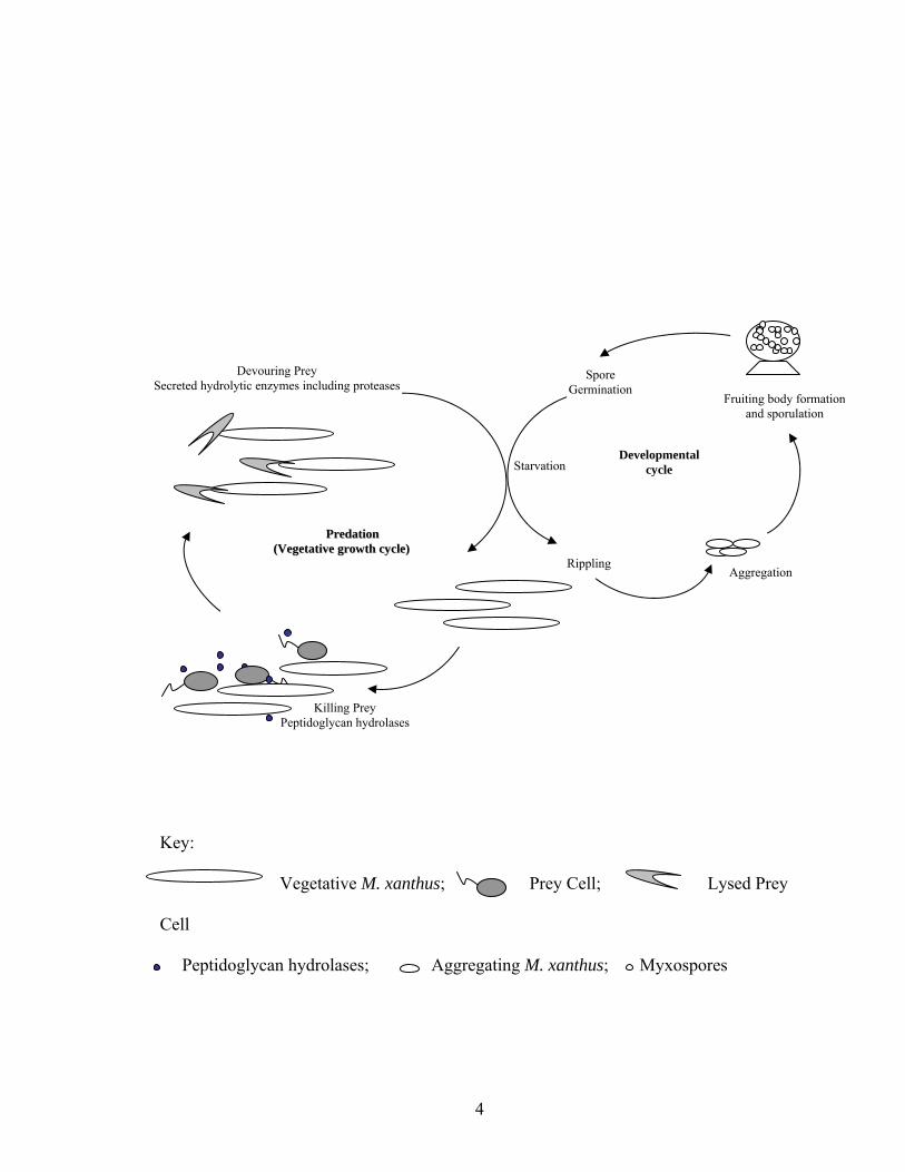
style (Figure 1.1) involves cooperative group behavior (34). During vegetative growth,
long slender rods glide over solid surfaces in large swarms, secreting hydrolytic enzymes
that kill prey bacteria. The hydrolyzed protein and lipid from the prey serves as the
primary source of carbon, nitrogen, and energy (33). The population feeds more
efficiently than individual cells. Rosenberg et al. showed that cells at a high density
hydrolyzed casein more efficiently than cells at a low density because of more efficient
use of extracellular proteases (113).
3
Figure 1.1. Life cycle of M. xanthus. Cells, enzymes, fruiting bodies, and spores are not
to scale.
4
Starvation
Killing Prey Peptidoglycan hydrolases
Devouring Prey Secreted hydrolytic enzymes including proteases
Developmental cycle
Spore Germination
PPrreeddaattiioonn ((VVeeggeettaattiivvee ggrroowwtthh ccyyccllee))
Rippling Aggregation
Fruiting body formationand sporulation
Key:
Vegetative M. xanthus; Prey Cell; Lysed Prey
Cell
Peptidoglycan hydrolases; Aggregating M. xanthus; Myxospores
5
Nutrient limitation triggers a developmental program during which the cells
exhibit a series of spatially and temporally regulated behaviors with a characteristic
multicellular dependence (34). Rippling, so called because of its resemblance to ripples
on the surface of water, is the first of the behavioral changes. Rippling is the coordinated
movement of ridges of cells that propagate as waves outward in a recurring pattern from
the center of a domain. Rippling is induced by peptidoglycan and some of its individual
components (126, 127). Sager and Kaiser observed two sets of wave patterns moving in
opposing directions with the same velocity and wavelength. They suggested that rippling
causes an end-to-end collision between waves of cells stimulating cell reversal (116).
Welch and Kaiser studied the properties of rippling cells using time-lapse microscopy
which showed that while the opposing crests appeared to pass right through each other, in
fact they reflected off of each other (138).
Welch and Kaiser studied the behavior of fluorescent rippling cells (138). Green
fluorescent protein (GFP) expressing cells were mixed with non-fluorescent cells at a
ratio of 1:500. The labeled cells in a wave crest were aligned perpendicular to the
direction of wave movement and moved in the direction of the crest. They reversed their
direction upon encountering the opposing crest head on. This led Igoshin et al to propose
the existence of a biochemical oscillator that controls the reversal of gliding direction
(59). The biochemical nature and mechanism of the proposed oscillator will be discussed
in a separate section. Rippling is proposed to play a role in the spacing of fruiting bodies.
However, equally spaced fruiting bodies have been seen in submerged culture
experiments where rippling has not been observed. Also, rippling is not essential for
completion of the developmental program. Hence the exact need for rippling has yet to
6
be deciphered.
Aggregation is the movement of cells into foci where they organize into fruiting
bodies. Cells exhibit gliding motility in the direction of their long axis with periodic
reversals in the gliding direction. Aggregation is proposed to be initiated and sustained
by contact-dependent C-signaling between individual cells (62, 63). The details of this
model will be discussed in a later section.
Kaiser (66) suggested a signal-independent mechanism of aggregate formation.
According to this model, the high cell density in the crests of rippling cells is the key to
aggregate formation. Collision between cells in countermigrating ripples is proposed to
inhibit cell movement, trapping the cells in an aggregation center. Time-lapse
microscopy reveals fusions between aggregates due to cell movement within and outside
the aggregates resulting in the formation of macroscopic fruiting bodies.
The fruiting bodies mature with the differentiation of 10-20% of the rod-shaped
vegetative cells into spherical structures called myxospores that are environmentally
resistant and metabolically dormant. Another 10% of the initial population differentiates
into specialized peripheral rods that show differential gene expression but never enter the
fruiting body or sporulate. Peripheral rods are proposed to play a protective role in
guarding the immature aggregates against consumption by other microbes. The majority
of the cells undergo developmental autolysis, which is proposed to provide nutrients
when cellular metabolism has slowed due to nutritional stress (34, 122).
The underlying feature of fruiting body development is intercellular signaling,
which is suggested by six extracellular complementation groups (A, B, C, D, E, and S).
The developmental defect in a mutant in one complementation group can be corrected by
7
codevelopment with wild-type cells or a mutant in another complementation group (50,
101). The vegetative cells recovered from the germinated spores of such mutants retain
their developmental defects and mutant genotype suggesting that complementation is not
due to the transfer of genetic material.
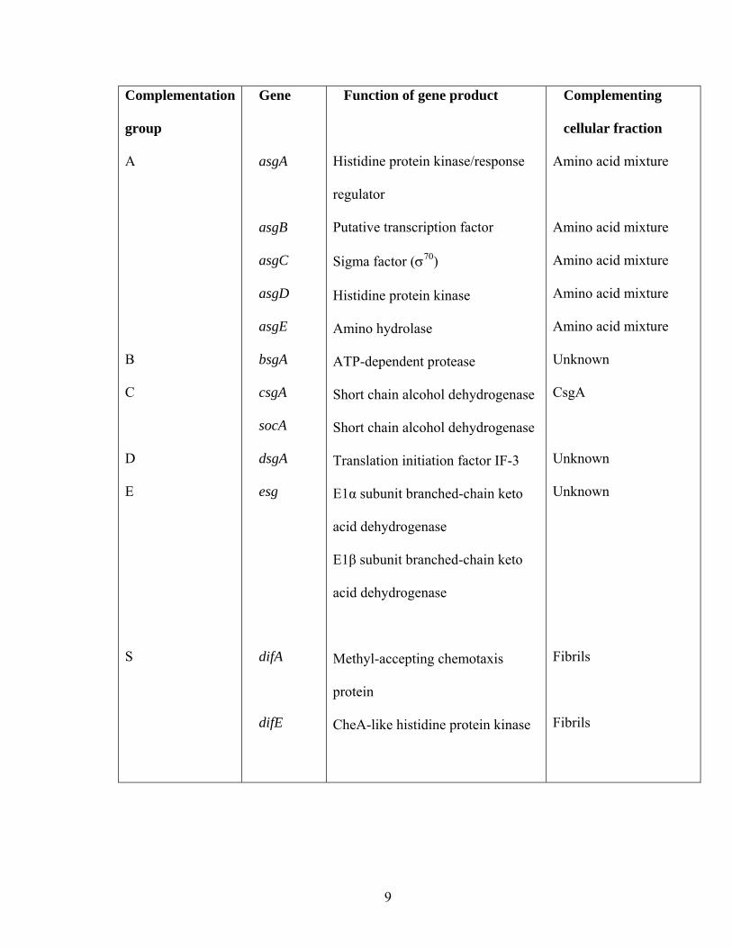
While the biochemical basis of extracellular complementation is at least partially
understood in the A, C, and S groups by addition of specific cellular fractions, the
mechanisms of B, D, and E signaling remain to be elucidated (122). Table 1.1
summarizes the different signaling systems along with what is known about their genetics
and biochemistry. The mutants in each class cease to develop at characteristic time
points. The signaling system defined by each complementation group directs the
expression of a distinct set of developmental genes in a temporal hierarchy (122). Kroos
and Kaiser (80) determined the time of developmental arrest in these mutants by studying
the expression pattern of a set of lacZ reporter fusions to development-specific
transcriptional units. If a particular mutation did not inhibit the expression of the reporter
gene, then it was presumed to interrupt the developmental process after the expression of
the reporter. On the other hand, if the expression of the reporter was inhibited by a
particular mutation, then it was assumed to interrupt the developmental process before
the expression of the reporter. The fact that each complementation group arrested
development at different time points suggested that each of these mutant classes was
responsible for generation of different signaling molecules and not part of a biochemical
pathway to generate a single signaling molecule. The A, B, C signaling systems act
within the first six hours after the initiation of development, while D and S (95) systems
act several hours later. The E signaling system seems to act after the A, B, and C systems
8
Table 1.1. Extracellular complementation groups in M. xanthus (Shimkets 1999).
The six extracellular complementation groups are listed with the genes defining them and
the respective complementing cellular fraction where known.
9
Complementation
group
A
B
C
D
E
S
Gene
asgA
asgB
asgC
asgD
asgE
bsgA
csgA
socA
dsgA
esg
difA
difE
Function of gene product
Histidine protein kinase/response
regulator
Putative transcription factor
Sigma factor (σ70)
Histidine protein kinase
Amino hydrolase
ATP-dependent protease
Short chain alcohol dehydrogenase
Short chain alcohol dehydrogenase
Translation initiation factor IF-3
E1α subunit branched-chain keto
acid dehydrogenase
E1β subunit branched-chain keto
acid dehydrogenase
Methyl-accepting chemotaxis
protein
CheA-like histidine protein kinase
Complementing
cellular fraction
Amino acid mixture
Amino acid mixture
Amino acid mixture
Amino acid mixture
Amino acid mixture
Unknown
CsgA
Unknown
Unknown
Fibrils
Fibrils
10
but before D and S (31, 134). Figure 1.2 depicts the time scale for various morphological
stages of development and the corresponding gene expression affected by the different
signaling groups.
The A-signal:
Mutations in five different genes define the A-complementation group (23, 39, 86,
99). All the mutants in this group show decreased A-signal production and reduction of
secreted proteins.
Genes required for A-signaling:
The asgA gene codes for an unusual member of a two-component regulatory
system with an N-terminal response regulator domain and a C-terminal histidine protein
kinase (HPK) domain (28, 111). The input and the output domains associated with ligand
binding and DNA binding, respectively, are missing in AsgA and are presumed to be
supplied by other unidentified proteins (111). Plamann et al. (111) suggest that AsgA
participates in a phosphor-relay essential for sensing starvation, and the subsequent
response results in A-signal generation.
asgB encodes a putative transcription factor with a C-terminal helix-turn-helix
motif (109). AsgB may have an essential role during growth as suggested by failed
attempts to create a null asgB allele (109). Hence, Plamann et al. (109) proposed that
AsgB acts as a transcriptional repressor of early developmental genes whose expression
during growth may have deleterious effects on vegetative cells.
asgC encodes the essential bacterial sigma factor σ70. The mutant asgC
phenotype is due to a single amino acid change in the highly conserved region 3, which is
11
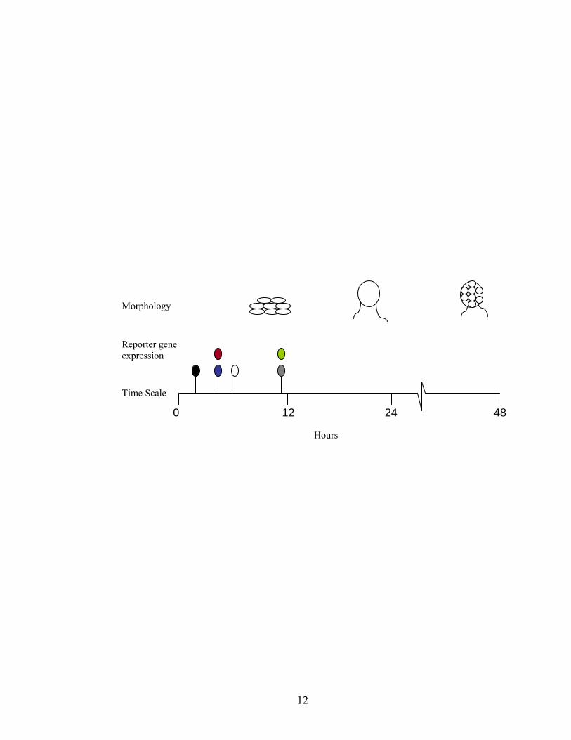
Figure 1.2. Time scale indicating the various morphological stages and the earliest point
where gene expression is arrested by mutants in the different extracellular
complementation groups (Shimkets 1999). Earliest representative genes whose
expression is completely dependent on the corresponding signaling group are indicated.
(A-group Black; B-group Blue; C-group Red; D-group Grey; E-group White; S-group
Green). The morphology of wild-type cells at various developmental times is represented
above the time scale. Aggregates start to appear about 8-12 hr of development.
Immature fruiting bodies without spores are formed by 24 hr, and sporulation is complete
by 48 hr. Cells, fruiting bodies, and spores are not to scale.
12
Hours
Time Scale
Morphology
Reporter gene expression
0 12 24 48
13
composed of 2 subregions. Subregion 3.2 is proposed to be involved in binding to core
RNA polymerase and subregion 3.1 has a weak helix-turn-helix (HTH) motif. The asgC
mutation is found immediately downstream of the putative HTH motif. This type of
mutation in sigma factors is known to affect the interaction between RNA polymerase
and transcription factors (56, 60, 85, 98). This led Davis et al. (28) to propose that the
inability of AsgC to interact with a transcriptional regulator required for A-signal
production results in the A-signaling defect. Alternately, the mutant sigma factor binds
the core polymerase with increased affinity preventing an alternate sigma factor
necessary for A-signal production to bind core polymerase.
asgD encodes an atypical HPK in which the receiver domain is at the N-terminus
and a kinase domain is at the C-terminus (23). The receiver and the sensor domains of
AsgD are separated by a 380-amino-acid-long intermediate region of unknown function.
The asgD mutant has an interesting phenotype. Wild-type cells complete development
upon starvation on low nutrient medium called CF (Clone Fruiting) medium that contains
10 mM MOPS (pH 7.6), 0.015% Casitone, 8 mM MgSO4, 1 mM KH2PO4, 2% sodium
citrate, 1% sodium pyruvate, and 1.5% Difco agar. The development of asgD mutant is
severely compromised on this low nutrient medium. However, on a more stringent
starvation medium with 10 mM MOPS, 4 mM MgSO4, 2 mM CaCl2 and 1.5% Difco
agar, asgD sporulates at 35% of wild-type levels. This led Cho and Zussman (23) to
propose that AsgD is involved in defining the nutrient threshold for sensing starvation
leading to the initiation of development.
asgE encodes a putative aminohydrolase (39). Aminohydrolases catalyze the
hydrolysis of substrates containing nitrogen-carbon heterocyclic rings. asgE mutants
14
produce only 4% of wild type spore levels. While asgE improved the sporulating
efficiency of asgA in extracellular complementation experiments to about 4% of the wild-
type, the converse is not true. asgA did not improve spore yields of asgE, suggesting
AsgE acts downstream of AsgA in the A-signal transduction pathway, and the asgE gene
could be one of the potential targets of AsgA (39).
Biochemical basis for A-signaling:
Plamann et al. (110) and Kuspa et al. (87) identified two sets of substances in the
medium conditioned by prior incubation with asg+ cells that restored developmental gene
expression in asg mutants. They used a bioassay to measure the reporter gene activity of
the A-signal-dependent transcriptional fusion of Ω4521 to lacZ in an asgB background.
One factor was heat-sensitive and the other heat-stable. Plamann et al. (110) identified
the heat-sensitive factor as two proteases of 27 kDa and 10 kDa. The 27 kDa protease
had trypsin-like substrate specificity, hydrolyzing peptides on the carboxy terminus of
arginine or lysine residues. In addition, the 27 kDa protease was able to rescue
aggregation and sporulation in asgB mutants upon extracellular complementation. The
substrate specificity of the 10 kDa protease has not been identified.
Kuspa et al. (87) determined that the heat-stable A-factor was composed of amino
acids and peptides. Pure amino acids with highest A-factor activity include tyrosine,
proline, tryptophan, phenylalanine, leucine, and isoleucine. These amino acids were
present at concentrations of 11-22 µM in starvation medium conditioned by asg+ cells.
Below the threshold concentration of 10 µM, these amino acids did not rescue
developmental gene expression individually or in mixtures. The specific activities of the
peptides were the sum of the specific activities of their individual components.
15
Kuspa et al. (88) used in vitro experiments to simulate conditions of in vivo A-
factor production. The crude A-factor was subjected to autodigestion followed by amino
acid analysis of the free amino acid content. The concentration of each of the six amino
acids with highest A-factor specific activity increased about 2-fold upon digestion. In
addition, the timing of A-factor release from asg+ cells coincided with the activation of
gene expression by the A-factor conditioned medium (87). Also, Plamann et al. (110)
found that proteases with substrate specificities different from trypsin also rescued the
developmental gene expression in asgB. Finally, inhibition of the endogenous trypsin
activity abolished A-factor production, strengthening the argument that amino acids are
the A-signal.
Role of A-signaling during development:
Kuspa et al. (88) proposed that A-signal amino acids serve in cell density sensing.
Transcription of the Ω4521 reporter decreased to 75-90% when wild-type cells were
diluted below the threshold levels required for development. Transcription increased
upon addition of amino acids added at the same levels required for Ω4521 expression in
the asgB mutant. While asgB mutants produced 5-10% of wild-type levels of A-factor,
increasing the cell density 10 to 20-fold restored Ω4521 expression and sporulation.
Since the concentration of A-signal amino acids is directly proportional to the cell
density, Kuspa et al. (88) proposed that A-signal determines whether an adequate density
of starved cells is present to complete development. Hence, the A-signal defines a novel
mechanism that regulates gene expression in response to changes in cell density.
16
A-signal transduction pathway:
Putative components of the A-signal sensory pathway have been identified based
on the isolation of suppressor mutations capable of A-signal independent expression of
genes that would otherwise require the A-signal. SasS, a putative histidine protein
kinase, is proposed to positively regulate Ω4521 expression during early development by
phosphorylating its cognate response regulator (143). A sasS null mutant shows
abnormal aggregation and sporulation and is severely compromised in the expression of
the Ω4521 reporter in spite of producing wild-type levels of the A-signal. sasS mutants
do not respond to extracellular A-signal.
Gorski and Kaiser (46) proposed that SasR, a sigma-54 activator protein, is
involved in sensing the A-signal. The sasR mutant is compromised in aggregation,
sporulation, and expression of the A-signal dependent reporter Ω4521. The fact that this
mutant makes wild-type levels of the A-signal was demonstrated by its ability to correct
the asgA phenotype upon extracellular complementation. However, the sasR phenotype
is not corrected by extracellular complementation with wild-type cells, suggesting its
potential role in the A-signal response pathway. SasS is proposed to phosphorylate SasR
in response to the A-signal, resulting in the expression of sigma-54-dependent genes
under A-signal control (46, 49).
The A-signal paradox and stringent response:
Starvation sensing in E. coli is characterized by the initiation of a stringent
response upon depletion of aminoacylated tRNA (18). When the ribosome pauses
because of lack of charged tRNA, RelA, the ribosome associated stringent response
protein, synthesizes the starvation signaling molecules guanosine pentaphosphate
17
(pppGpp) and guanosine tetraphosphate (ppGpp) (18). The reaction involves the transfer
of the β, γ -pyrophosphoryl group from ATP to the ribose 3’ hydroxyl group of GTP or
GDP, respectively, by RelA (18). These effector molecules regulate growth by positively
or negatively influencing gene expression and inhibit DNA replication initiation (18). A
M. xanthus relA mutation prevents the accumulation of (p)ppGpp and inhibits
development (52). Furthermore, ectopic expression of relA induces the untimely
expression of early developmental genes (129). The stringent response activates
transcription of many developmental genes (129) and inhibits transcription of a
developmental repressor socE (26, 27). The stringent response also activates the
extracellular A (52) and C signaling systems (26, 27). The extracellular amino acid pool
generated during A-signaling reaches a high enough concentration (25 µM) to sustain
slow growth. However, cells continue development using the limited carbon and energy
generated by the A-signal for growth (110). This paradox is explained by a novel
regulatory mechanism suggested by Crawford and Shimkets (26, 27). They have
demonstrated that ectopic depletion of SocE results in growth arrest even in the presence
of nutrients. Growth arrest is achieved by the action of CsgA and the loss of SocE, which
maintains RelA-dependent (p)pp(G)pp production in spite of the influx of amino acids
(26, 27). It is not known how the two proteins maintain the RelA-dependent starvation
response.
The B-signal:
The mutants in the B complementation group are aggregation- and sporulation-
defective and are compromised in the expression of early developmental genes (43, 81).
18
The bsgA gene encodes an ATP-dependent protease similar to the Lon protease of E. coli
(41, 42, 44). BsgA is localized in the cytoplasm (41), and the recombinant BsgA protein,
partially purified from E. coli, has an ATP-dependent protease activity (44). The E.coli
Lon protease degrades misfolded proteins and modulates the regulatory function of
several cellular proteins (47). For example, SulA, a physiological substrate of the Lon
protease, is induced by UV irradiation and aids in DNA repair by preventing premature
segregation of damaged DNA into daughter cells (57, 58). SulA inhibits cell division by
preventing self-assembly of FtsZ (12). Rapid degradation of SulA by Lon protease is
essential for normal cell cycle progression.
Hager et al. (51) described a bsgA suppressor that was localized to spdR. spdR
encodes a σ54-dependent transcriptional activator that belongs to the NtrC family of
transcriptional activators. The spdR mutants regained aggregation, sporulation, and
expression of many early developmental genes (51, 136). The spdR mutants showed
accelerated development, forming spore-filled fruiting bodies, about 8-12 hr earlier than
the wild-type. These mutants also showed development in the presence of casitone
concentrations (0.2%) high enough to suppress development. In addition, some of the
developmental genes were expressed in the mutant background in vegetative cells. This
led Hager et al. to propose that SpdR is a negative regulator of development. While their
data are consistent with the idea that SpdR is the cellular target of BsgA, immunoblot
analysis of wild-type and bsgA cells did not support this hypothesis. SpdR does not
accumulate in bsgA cells, and SpdR did not decrease upon initiation of wild-type
development. Furthermore, the spdR mutation does not exclusively suppress the bsgA
defect. It also suppresses the requirement for A-signaling (136). Although these data
19
rule out the possibility that SpdR is the physiological target of BsgA, the mechanism of
B-signaling could still be due to the proteolytic processing of a protein by BsgA. Since
the substrate of BsgA has not been identified yet, the basis for extracellular
complementation remains to be established.
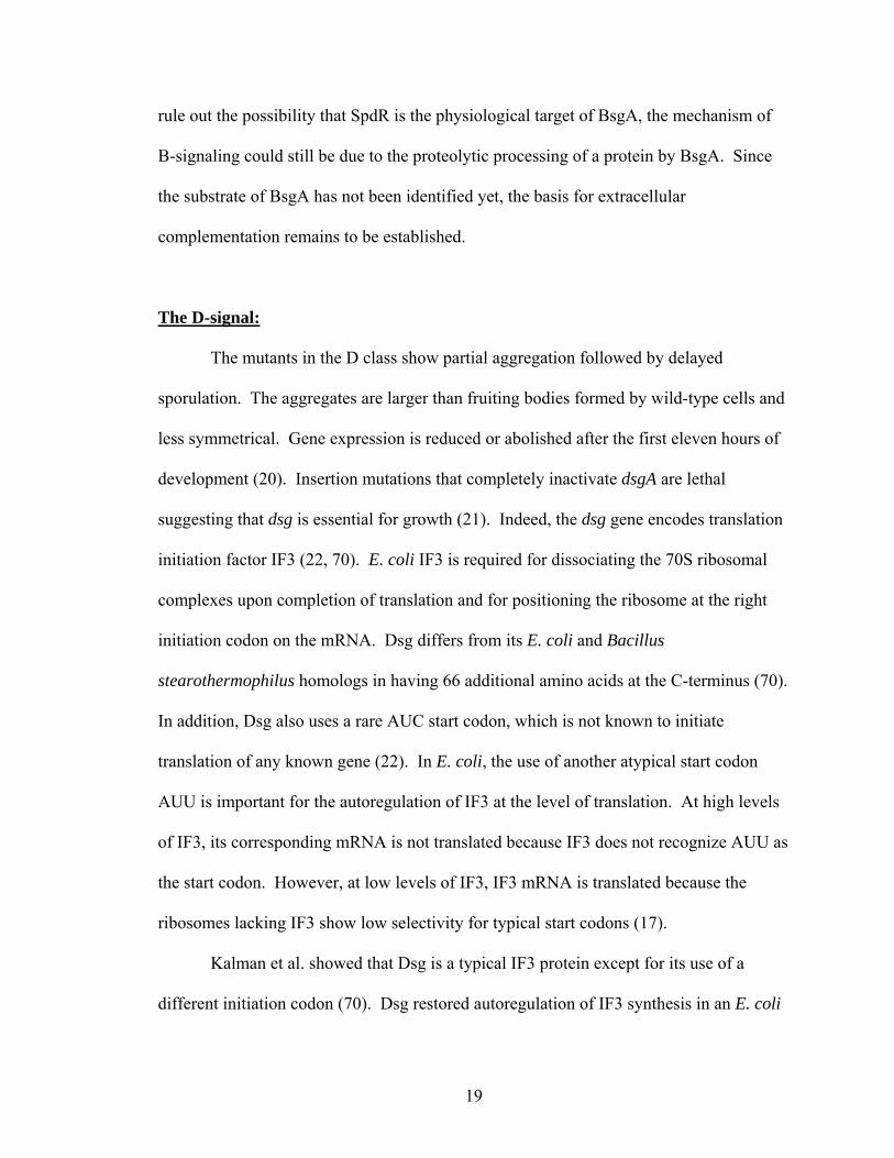
The D-signal:
The mutants in the D class show partial aggregation followed by delayed
sporulation. The aggregates are larger than fruiting bodies formed by wild-type cells and
less symmetrical. Gene expression is reduced or abolished after the first eleven hours of
development (20). Insertion mutations that completely inactivate dsgA are lethal
suggesting that dsg is essential for growth (21). Indeed, the dsg gene encodes translation
initiation factor IF3 (22, 70). E. coli IF3 is required for dissociating the 70S ribosomal
complexes upon completion of translation and for positioning the ribosome at the right
initiation codon on the mRNA. Dsg differs from its E. coli and Bacillus
stearothermophilus homologs in having 66 additional amino acids at the C-terminus (70).
In addition, Dsg also uses a rare AUC start codon, which is not known to initiate
translation of any known gene (22). In E. coli, the use of another atypical start codon
AUU is important for the autoregulation of IF3 at the level of translation. At high levels
of IF3, its corresponding mRNA is not translated because IF3 does not recognize AUU as
the start codon. However, at low levels of IF3, IF3 mRNA is translated because the
ribosomes lacking IF3 show low selectivity for typical start codons (17).
Kalman et al. showed that Dsg is a typical IF3 protein except for its use of a
different initiation codon (70). Dsg restored autoregulation of IF3 synthesis in an E. coli
20
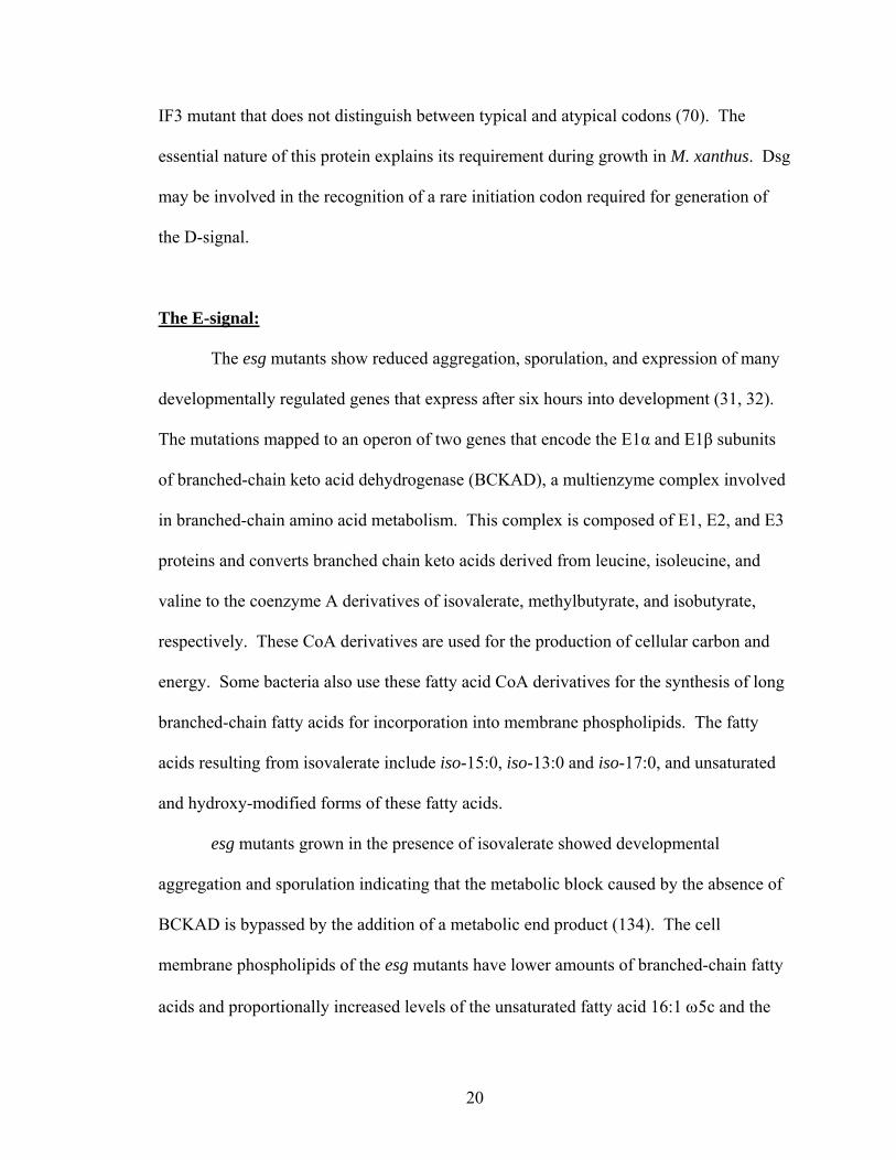
IF3 mutant that does not distinguish between typical and atypical codons (70). The
essential nature of this protein explains its requirement during growth in M. xanthus. Dsg
may be involved in the recognition of a rare initiation codon required for generation of
the D-signal.
The E-signal:
The esg mutants show reduced aggregation, sporulation, and expression of many
developmentally regulated genes that express after six hours into development (31, 32).
The mutations mapped to an operon of two genes that encode the E1α and E1β subunits
of branched-chain keto acid dehydrogenase (BCKAD), a multienzyme complex involved
in branched-chain amino acid metabolism. This complex is composed of E1, E2, and E3
proteins and converts branched chain keto acids derived from leucine, isoleucine, and
valine to the coenzyme A derivatives of isovalerate, methylbutyrate, and isobutyrate,
respectively. These CoA derivatives are used for the production of cellular carbon and
energy. Some bacteria also use these fatty acid CoA derivatives for the synthesis of long
branched-chain fatty acids for incorporation into membrane phospholipids. The fatty
acids resulting from isovalerate include iso-15:0, iso-13:0 and iso-17:0, and unsaturated
and hydroxy-modified forms of these fatty acids.
esg mutants grown in the presence of isovalerate showed developmental
aggregation and sporulation indicating that the metabolic block caused by the absence of
BCKAD is bypassed by the addition of a metabolic end product (134). The cell
membrane phospholipids of the esg mutants have lower amounts of branched-chain fatty
acids and proportionally increased levels of the unsaturated fatty acid 16:1 ω5c and the
21
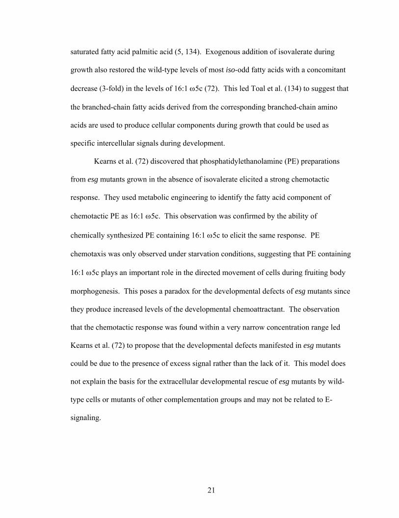
saturated fatty acid palmitic acid (5, 134). Exogenous addition of isovalerate during
growth also restored the wild-type levels of most iso-odd fatty acids with a concomitant
decrease (3-fold) in the levels of 16:1 ω5c (72). This led Toal et al. (134) to suggest that
the branched-chain fatty acids derived from the corresponding branched-chain amino
acids are used to produce cellular components during growth that could be used as
specific intercellular signals during development.
Kearns et al. (72) discovered that phosphatidylethanolamine (PE) preparations
from esg mutants grown in the absence of isovalerate elicited a strong chemotactic
response. They used metabolic engineering to identify the fatty acid component of
chemotactic PE as 16:1 ω5c. This observation was confirmed by the ability of
chemically synthesized PE containing 16:1 ω5c to elicit the same response. PE
chemotaxis was only observed under starvation conditions, suggesting that PE containing
16:1 ω5c plays an important role in the directed movement of cells during fruiting body
morphogenesis. This poses a paradox for the developmental defects of esg mutants since
they produce increased levels of the developmental chemoattractant. The observation
that the chemotactic response was found within a very narrow concentration range led
Kearns et al. (72) to propose that the developmental defects manifested in esg mutants
could be due to the presence of excess signal rather than the lack of it. This model does
not explain the basis for the extracellular developmental rescue of esg mutants by wild-
type cells or mutants of other complementation groups and may not be related to E-
signaling.
22
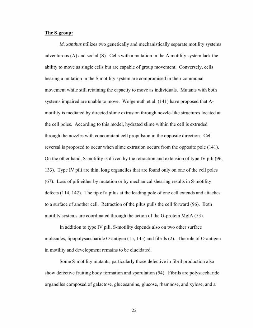
The S-group:
M. xanthus utilizes two genetically and mechanistically separate motility systems
adventurous (A) and social (S). Cells with a mutation in the A motility system lack the
ability to move as single cells but are capable of group movement. Conversely, cells
bearing a mutation in the S motility system are compromised in their communal
movement while still retaining the capacity to move as individuals. Mutants with both
systems impaired are unable to move. Wolgemuth et al. (141) have proposed that A-
motility is mediated by directed slime extrusion through nozzle-like structures located at
the cell poles. According to this model, hydrated slime within the cell is extruded
through the nozzles with concomitant cell propulsion in the opposite direction. Cell
reversal is proposed to occur when slime extrusion occurs from the opposite pole (141).
On the other hand, S-motility is driven by the retraction and extension of type IV pili (96,
133). Type IV pili are thin, long organelles that are found only on one of the cell poles
(67). Loss of pili either by mutation or by mechanical shearing results in S-motility
defects (114, 142). The tip of a pilus at the leading pole of one cell extends and attaches
to a surface of another cell. Retraction of the pilus pulls the cell forward (96). Both
motility systems are coordinated through the action of the G-protein MglA (53).
In addition to type IV pili, S-motility depends also on two other surface
molecules, lipopolysaccharide O-antigen (15, 145) and fibrils (2). The role of O-antigen
in motility and development remains to be elucidated.
Some S-motility mutants, particularly those defective in fibril production also
show defective fruiting body formation and sporulation (54). Fibrils are polysaccharide
organelles composed of galactose, glucosamine, glucose, rhamnose, and xylose, and a
23
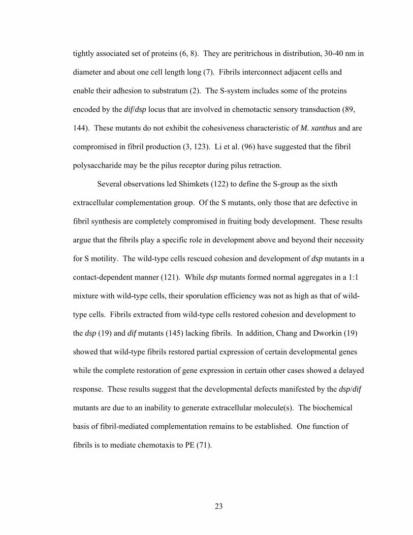
tightly associated set of proteins (6, 8). They are peritrichous in distribution, 30-40 nm in
diameter and about one cell length long (7). Fibrils interconnect adjacent cells and
enable their adhesion to substratum (2). The S-system includes some of the proteins
encoded by the dif/dsp locus that are involved in chemotactic sensory transduction (89,
144). These mutants do not exhibit the cohesiveness characteristic of M. xanthus and are
compromised in fibril production (3, 123). Li et al. (96) have suggested that the fibril
polysaccharide may be the pilus receptor during pilus retraction.
Several observations led Shimkets (122) to define the S-group as the sixth
extracellular complementation group. Of the S mutants, only those that are defective in
fibril synthesis are completely compromised in fruiting body development. These results
argue that the fibrils play a specific role in development above and beyond their necessity
for S motility. The wild-type cells rescued cohesion and development of dsp mutants in a
contact-dependent manner (121). While dsp mutants formed normal aggregates in a 1:1
mixture with wild-type cells, their sporulation efficiency was not as high as that of wild-
type cells. Fibrils extracted from wild-type cells restored cohesion and development to
the dsp (19) and dif mutants (145) lacking fibrils. In addition, Chang and Dworkin (19)
showed that wild-type fibrils restored partial expression of certain developmental genes
while the complete restoration of gene expression in certain other cases showed a delayed
response. These results suggest that the developmental defects manifested by the dsp/dif
mutants are due to an inability to generate extracellular molecule(s). The biochemical
basis of fibril-mediated complementation remains to be established. One function of
fibrils is to mediate chemotaxis to PE (71).
24
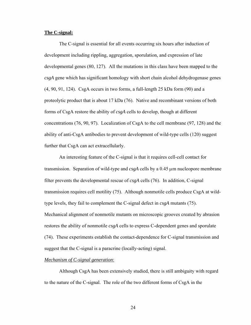
The C-signal:
The C-signal is essential for all events occurring six hours after induction of
development including rippling, aggregation, sporulation, and expression of late
developmental genes (80, 127). All the mutations in this class have been mapped to the
csgA gene which has significant homology with short chain alcohol dehydrogenase genes
(4, 90, 91, 124). CsgA occurs in two forms, a full-length 25 kDa form (90) and a
proteolytic product that is about 17 kDa (76). Native and recombinant versions of both
forms of CsgA restore the ability of csgA cells to develop, though at different
concentrations (76, 90, 97). Localization of CsgA to the cell membrane (97, 128) and the
ability of anti-CsgA antibodies to prevent development of wild-type cells (120) suggest
further that CsgA can act extracellularly.
An interesting feature of the C-signal is that it requires cell-cell contact for
transmission. Separation of wild-type and csgA cells by a 0.45 µm nucleopore membrane
filter prevents the developmental rescue of csgA cells (76). In addition, C-signal
transmission requires cell motility (75). Although nonmotile cells produce CsgA at wild-
type levels, they fail to complement the C-signal defect in csgA mutants (75).
Mechanical alignment of nonmotile mutants on microscopic grooves created by abrasion
restores the ability of nonmotile csgA cells to express C-dependent genes and sporulate
(74). These experiments establish the contact-dependence for C-signal transmission and
suggest that the C-signal is a paracrine (locally-acting) signal.
Mechanism of C-signal generation:
Although CsgA has been extensively studied, there is still ambiguity with regard
to the nature of the C-signal. The role of the two different forms of CsgA in the
25
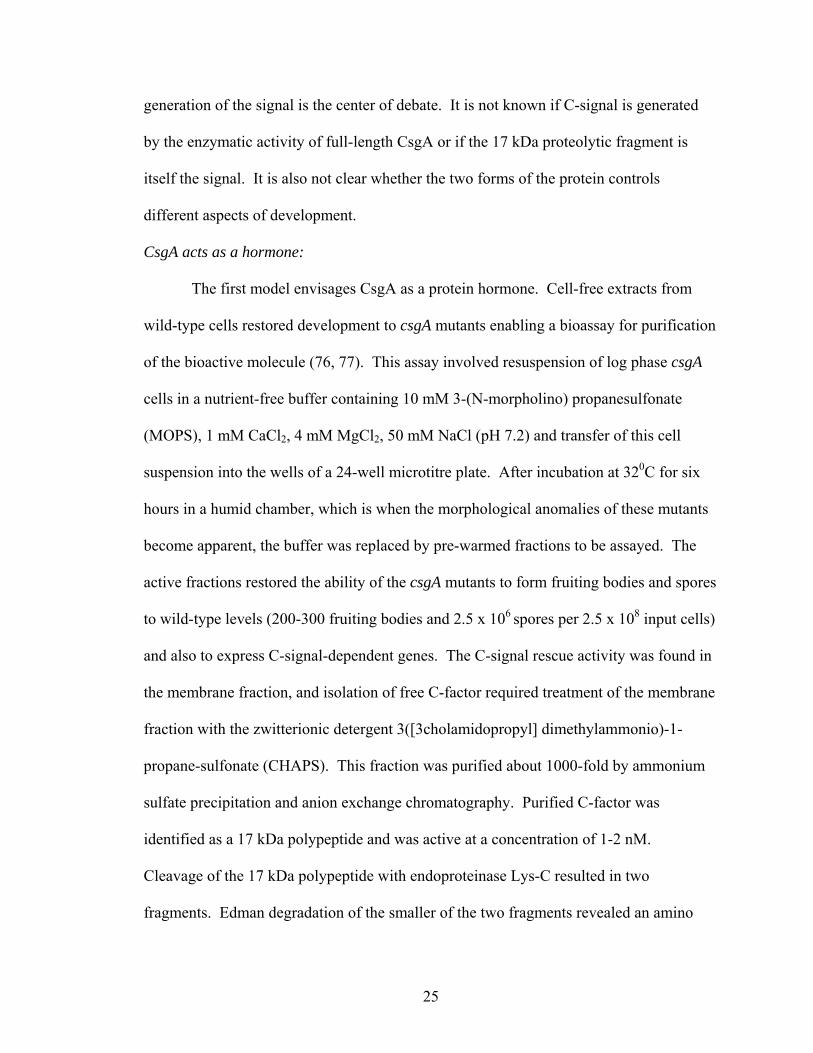
generation of the signal is the center of debate. It is not known if C-signal is generated
by the enzymatic activity of full-length CsgA or if the 17 kDa proteolytic fragment is
itself the signal. It is also not clear whether the two forms of the protein controls
different aspects of development.
CsgA acts as a hormone:
The first model envisages CsgA as a protein hormone. Cell-free extracts from
wild-type cells restored development to csgA mutants enabling a bioassay for purification
of the bioactive molecule (76, 77). This assay involved resuspension of log phase csgA
cells in a nutrient-free buffer containing 10 mM 3-(N-morpholino) propanesulfonate
(MOPS), 1 mM CaCl2, 4 mM MgCl2, 50 mM NaCl (pH 7.2) and transfer of this cell
suspension into the wells of a 24-well microtitre plate. After incubation at 320C for six
hours in a humid chamber, which is when the morphological anomalies of these mutants
become apparent, the buffer was replaced by pre-warmed fractions to be assayed. The
active fractions restored the ability of the csgA mutants to form fruiting bodies and spores
to wild-type levels (200-300 fruiting bodies and 2.5 x 10⁶ spores per 2.5 x 10⁸ input cells)
and also to express C-signal-dependent genes. The C-signal rescue activity was found in
the membrane fraction, and isolation of free C-factor required treatment of the membrane
fraction with the zwitterionic detergent 3([3cholamidopropyl] dimethylammonio)-1-
propane-sulfonate (CHAPS). This fraction was purified about 1000-fold by ammonium
sulfate precipitation and anion exchange chromatography. Purified C-factor was
identified as a 17 kDa polypeptide and was active at a concentration of 1-2 nM.
Cleavage of the 17 kDa polypeptide with endoproteinase Lys-C resulted in two
fragments. Edman degradation of the smaller of the two fragments revealed an amino
26
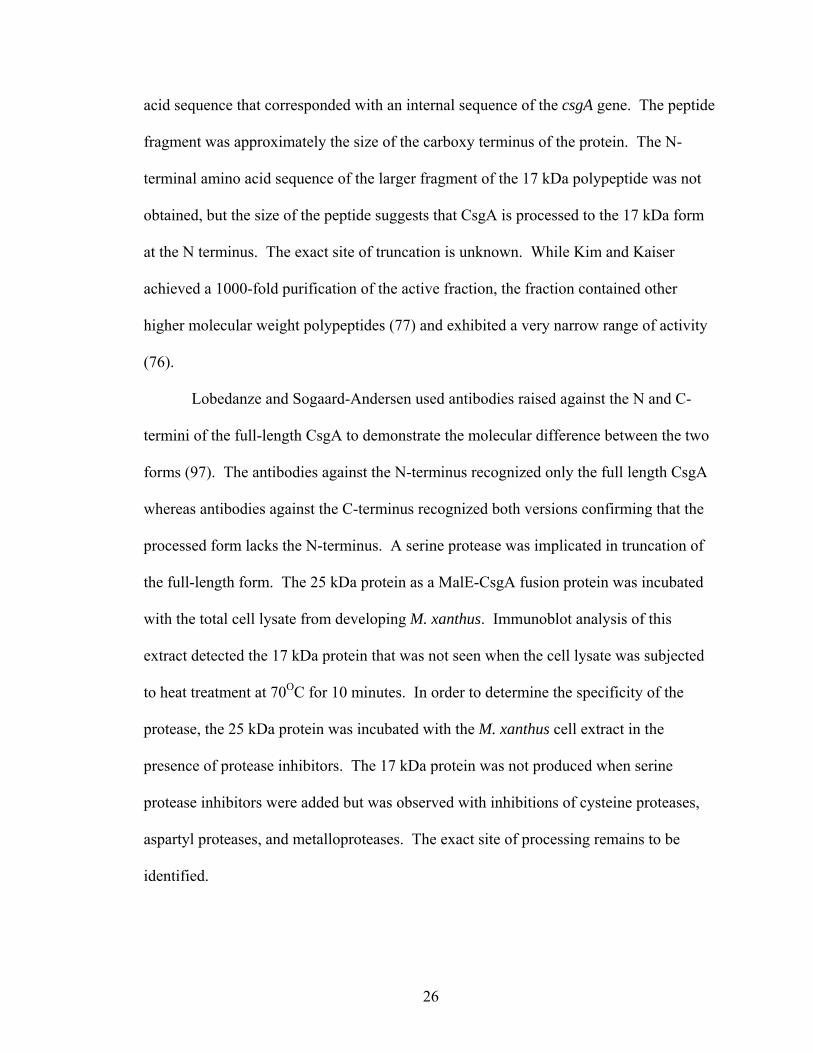
acid sequence that corresponded with an internal sequence of the csgA gene. The peptide
fragment was approximately the size of the carboxy terminus of the protein. The N-
terminal amino acid sequence of the larger fragment of the 17 kDa polypeptide was not
obtained, but the size of the peptide suggests that CsgA is processed to the 17 kDa form
at the N terminus. The exact site of truncation is unknown. While Kim and Kaiser
achieved a 1000-fold purification of the active fraction, the fraction contained other
higher molecular weight polypeptides (77) and exhibited a very narrow range of activity
(76).
Lobedanze and Sogaard-Andersen used antibodies raised against the N and C-
termini of the full-length CsgA to demonstrate the molecular difference between the two
forms (97). The antibodies against the N-terminus recognized only the full length CsgA
whereas antibodies against the C-terminus recognized both versions confirming that the
processed form lacks the N-terminus. A serine protease was implicated in truncation of
the full-length form. The 25 kDa protein as a MalE-CsgA fusion protein was incubated
with the total cell lysate from developing M. xanthus. Immunoblot analysis of this
extract detected the 17 kDa protein that was not seen when the cell lysate was subjected
to heat treatment at 70OC for 10 minutes. In order to determine the specificity of the
protease, the 25 kDa protein was incubated with the M. xanthus cell extract in the
presence of protease inhibitors. The 17 kDa protein was not produced when serine
protease inhibitors were added but was observed with inhibitions of cysteine proteases,
aspartyl proteases, and metalloproteases. The exact site of processing remains to be
identified.
27
Lobedanze and Sogaard-Andersen demonstrated that a C-terminal 18.1 kDa of
CsgA with an N-terminal MalE fusion was able to correct the developmental defects of
csgA mutants upon extracellular complementation. The recombinant 18.1 kDa form
purified from E. coli had a 2000–fold lower specific activity (97). The authors suggest
that the low specific activity is due to the lack of an unknown posttranslational
modification of the CsgA portion of the fusion protein. Expression of the truncated
protein in the csgA mutant did not restore the wild-type developmental phenotype.
Lobedanze and Sogaard-Andersen (97) used Triton X-114 phase separation to
obtain outer and inner membrane fractions of wild-type cells undergoing development.
Immunoblot analysis was performed to determine which, if any forms were found in the
outer membrane. They observed that both forms of CsgA were localized to the outer
membrane. This led them to suggest that the truncated form acts as a paracrine hormone
to signal the neighboring cell via a hypothetical receptor.
Simunovic and Shimkets (128) used a more refined approach to study the
localization of CsgA in vegetative cells. They separated the cellular membranes from the
spheroplast-enriched cells into inner, outer, and hybrid membrane fractions using a three-
step sucrose gradient followed by further separation on discontinuous sucrose gradients.
The purity of each fraction was assessed by unique protein markers. The succinate
dehydrogenase assay was used to confirm the purity of the inner membrane, and the
purity of the outer membrane fractions was determined from immunoblots performed
with monoclonal antibodies against the LPS core and O-antigen. This method detected
the full-length form of CsgA in the inner membrane and neither form in the outer
membrane. This discovery does not support the role of the processed form as a paracrine
28
signal. However, Simunovic and Shimkets used vegetative cells, and there could be
differential localization of CsgA during vegetative growth versus development. But then,
there is no direct evidence for this being the case.
Taken together, the experiments from the Kaiser and Sogaard-Andersen
laboratories appear to suggest that the processed form of CsgA acts as a protein hormone.
However, the evidence provided is not sufficient to establish this beyond a reasonable
doubt. The presence of some high molecular weight proteins in the initial preparation of
the 17 kDa form by Kim and Kaiser does not necessarily exclude the possibility of
developmental rescue by the full length form of CsgA. Although, the recombinant form
of the truncated protein had developmental activity, the specific activity was about 2000-
fold lower than the original preparation of Kim. In addition, expression of this protein in
a csgA mutant did not result in developmental rescue. These observations lead one to
question the validity of the 17 kDa form as the signal.
CsgA acts as an enzyme to generate the signal:
The second model suggests an enzymatic role for CsgA which shares significant
homology with the short-chain alcohol dehydrogenase (SCAD) family (90). The
members of the SCAD family utilize NAD(H) or NADP(H) to mediate the
interconversion of secondary alcohols and ketones (105). The first line of genetic
evidence for the enzymatic nature of CsgA comes from the isolation of the suppressor of
csgA (soc) mutants containing transposon insertions in the socABC operon (91, 92). The
first of the three genes in the socA operon, socA, encodes a short chain alcohol
dehydrogenase that bears 28% amino acid identity to CsgA, primarily in the enzyme
29
active site (91). The second gene in this operon encodes SocB, an integral membrane
protein with homology to FrdD which anchors fumarate reductase to the membrane in
Proteus vulgaris. The third gene in the socABC operon encodes SocC, a negative
regulator of the operon (92). Two transposon suppressor mutations were discovered, one
between socB and socC, and another in socC (92). These insertions are polar and
inactivate the negative regulator of the operon resulting in the overexpression of socA.
Lee and Shimkets studied the role of SocC in the regulation of the socABC operon using
the soc-lacZ fusions in socC+ and socC mutant backgrounds and found a 90-fold increase
in expression of socAB transcription in the absence of socC (92). Further, mRNA levels
also showed a 50-100 fold increase, confirming the role of SocC as the negative regulator
of the operon.
The ability of socA-overproducing mutants to rescue the C-signaling defect of
csgA mutants was examined by extracellular complementation of csgA mutants with cells
bearing mutations in the socA operon and csgA. Vegetative cells were mixed in a 1:1
ratio, and allowed to develop at 320C for 4 days. Spores were enumerated by plating as
each strain has a unique antibiotic resistance profile. The restoration of development to
csgA cells suggested that the socA csgA double mutant produces the C-signal (92). This
led Lee and Shimkets to propose that the overlapping substrate specificity between CsgA
and SocA may result in production of the C-signal.
Mutation of conserved CsgA residues provides further evidence in support of the
hypothesis that CsgA is an enzyme. The presumptive substrate binding site as well as the
coenzyme binding site are required for CsgA activity (90). Point mutations that cause
replacement of conserved active site residues, S135T and K155R, inactivated csgA (90).
30
A MalE-CsgA protein bearing the S135K mutation was unable to restore development to
csgA mutants when extracellularly complemented. A point mutation that caused
substitution of conserved arginine in the coenzyme binding region (R10A) with alanine
resulted in inactive CsgA (90). Deletion of the entire coenzyme binding region resulted
in an inactive protein that was unable to rescue development of csgA mutants (120). A
conserved threonine residue stabilizes coenzyme-binding through hydrogen bonding (40),
and the MalE-CsgA fusion protein bearing a threonine to alanine substitution (T6A) in
the coenzyme-binding domain failed to complement the C-signaling defect in csgA
mutants (90). This mutant protein showed reduced capacity to bind radiolabeled NAD+
(90). Furthermore, the addition of NAD+ and NADP+ along with MalE-CsgA stimulated
development while these nucleotides in their reduced forms delayed development (90)
highlighting the importance of the coenzyme-binding region for CsgA activity.
The experimental evidence described above suggests a role for the coenzyme
binding site as well as the substrate binding site of CsgA for the generation of the C-
signal during M. xanthus development. Both CsgA and SocA are members of a family of
short chain alcohol dehydrogenases that includes enzymes that catalyze the conversion of
a wide range of substrates including sugars, steroids, prostaglandins, and aromatic
hydrocarbons (105), making it difficult to predict putative substrates based on sequence
similarity alone.
Role of C-signaling during development:
Rippling:
Rippling is the first of the various morphologically distinct events controlled by
C-signaling. Shimkets and Kaiser (126) first observed that csgA null mutants are
31
completely compromised in their ability to ripple. The inability of csgA mutants to ripple
was restored by codevelopment with a 1:1 mixture of wild-type cells (116). Dilution of
wild-type cells with csgA mutants in different proportions led to an increased ripple
wavelength suggesting a reduced frequency of cell-cell signaling. Each cell type was
labeled with a different fluorescent dye, and fluorescence confocal microscopy revealed
that the csgA cells were arranged end-to-end as for wild-type cells. Addition of partially
purified CsgA resulted in increased reversal frequency of developing csgA cells. This
observation combined with the already established requirement for end-end orientation of
cells during C-signaling, led Sager and Kaiser (116) to propose that contact between cells
in waves migrating in opposing directions initiates C-signaling, triggering cell reversals.
Based on the above observations and those of Welch and Kaiser (138) who provided a
quantitative basis for cell-behavior in ripples as described earlier, Igoshin et al. (59)
developed a mathematical model of rippling in which C-signaling is proposed to be a
biochemical oscillator that controls reversal of cell gliding. This model also suggests that
the cell contact-dependent C-signaling increases the reversal probability by increasing the
cycle phase velocity, which is followed by a brief non-responsive phase. This prediction
has not been experimentally tested. The response to the C-signal is proposed to be
dependent on local cell density suggesting the cooperative nature of C-signaling.
Aggregation:
csgA mutants cannot form compact, hemispherical aggregates (125). csgA cells
can be induced to form multicellular aggregates by extracellular stimulation with csgA+
cells or by addition of either version of the C-signaling protein (50, 76, 90). Jelsbak and
Sogaard-Andersen (62, 63) defined the motility parameters controlled by C-signaling by
32
analyzing the behavior of isolated cells as well as those in a population. In the first set of
experiments, a high density of starving cells at the initiation of C-signaling was dispersed
on a solid surface to enable monitoring of isolated cells using time-lapse video
microscopy. The underlying assumption was that individual cells would already be C-
signal-stimulated to exhibit specific behavioral patterns while at high cell density. The
wild-type cells glided longer distances with an increased velocity and decreased stop
frequency. In contrast, the motility profiles of csgA cells showed no increase in the
gliding period or the velocity. However, csgA cells reacquired wild-type motility
parameters upon exposure to purified full-length form of MalE-CsgA for 30 minutes
before dispersal on starvation agar, suggesting that CsgA induces the changes in motility
parameters directly or indirectly (62). This approach however did not allow for
observation of cell behavior that depends on continuous cell-cell contact.
In order to overcome the limitation imposed by loss of contact, Jelsbak and
Sogaard-Andersen (63) used fluorescent tagging to study the behavior of individual cells
in a high density population. Green fluorescent protein (GFP)-expressing wild-type cells
were mixed 1:400 with non-fluorescing wild-type cells, and the cell mixture was
subjected to starvation-induced development at high cell density. The GFP-tagged cells
showed increased speed, extended gliding intervals, and a decreased stop and reversal
frequency. GFP-expressing csgA cells mixed with non-fluorescing csgA cells did not
exhibit the same behavior. The gliding velocity of csgA cells decreased slightly, and the
cells showed frequent periods of no cell movement. Also, the gliding interval, and the
reversal frequency did not change significantly. The net distance traveled by the cells
during the recording period was about 3-fold lower than that traveled by the wild-type
33
cells. GFP-expressing csgA cells mixed with non-fluorescing wild-type cells showed the
same behavior as wild-type cells. Conversely, GFP-expressing wild-type cells mixed
with non-fluorescing csgA cells exhibited the mutant motility parameters. These
observations, along with the already established evidence for contact dependence of C-
signaling transmission, led Jelsbak and Sogaard-Andersen (61) to propose that
aggregation begins with end-end contact between two cells enabling exchange of the C-
signal. This results in increased gliding velocity and reduced reversal frequency,
enabling formation of chains of cells. The direction of gliding is proposed to be
determined by the cell at the leading end of the chain. They suggest that the cells remain
in chains because of their inherent cohesive nature (123) and also because of the
continuing contact-mediated signaling. The primary chains of cells are proposed to give
rise to secondary chains, finally resulting in the formation of streams of cells.
Eventually, the streams of cells are proposed to be trapped in aggregation centers by an
unknown mechanism.
Timing of development:
C-signal is an intrinsic timer that M. xanthus uses to spatially and temporally
control rippling, aggregation, and sporulation. Kruse (84) have quantitatively
demonstrated that an ordered increase in CsgA levels is required for the separation of
these developmental processes. Overproduction of CsgA results in premature
aggregation and sporulation and the uncoupling of the two events in space and time.
Reduced CsgA levels caused delayed aggregation, and reduced sporulation (84). Li et al.
studied the genetic basis for this observation by using nested deletions upstream from the
csgA gene that resulted in reduced csgA expression. Successively larger deletions resulted
34
in cessation of development at stages prior to the onset of the next stage. 20%, 30%, and
82% csgA expression was required for rippling, aggregation, and sporulation,
respectively (94). This suggests that the accumulation of CsgA at different threshold
levels defines the corresponding morphological check-point.
Developmental rescue experiments involving addition of exogenous CsgA do not
show the concentration-dependence for the temporal separation of the developmental
events. Aggregation and sporulation occur with a 24 hour lag at 1 nM partially purified
17 kDa CsgA (76) or 100 nM 25 kDa MalE-CsgA (90). Uncoupling of these events
would not be seen if they were defined merely by the accumulation of extracellular
CsgA. This ambiguity might be explained if the short chain alcohol dehydrogenase
activity of CsgA could catalyze the conversion of different substrates at different times to
execute the different developmental events. Thus, CsgA could define a novel class of
timers that work based on enzyme catalysis.
Regulation of csgA expression:
Temporal regulation of developmental csgA expression is important during
fruiting body morphogenesis. csgA expression, which is relatively constant during
vegetative growth, increases gradually during the course of development, peaking at
about 72 hours (26, 94). Although the mechanism of csgA regulation has not been
completely deciphered, different regulatory elements have been identified. Optimal csgA
expression and sporulation requires an upstream region of about 400 bp from the
transcriptional start-site under stringent starvation conditions (94). However, an extended
region including another 530 bp further upstream is required for optimal C-signal activity
in the presence of low nutrient levels, suggesting that this region is important for sensing
35
carbon, nitrogen, and phosphorus (94).
Transcription of csgA increases in the presence of the C-signal and FruA, a
response regulator with a predicted HTH motif, suggesting that a potential positive
feedback loop controls csgA expression (35, 131). Indeed, the four genes of the act
operon control the timing and level of csgA expression. Mutations in actA and actB,
which encode a response regulator and a sigma-54 activator protein, respectively, lower
csgA transcription to about 25% of wild-type levels. Both these mutants show increased
periods of rippling, delayed aggregation, and no sporulation. Although, this suggests that
the sigma-54-dependent ActB response regulator affects csgA expression, Li et al. (94)
have reported that the promoter upstream of csgA is of the sigma-70 type. This could
suggest the existence of additional regulatory elements.
The other two genes in the operon, actC and actD, affect the temporal regulation
of csgA expression. The actC mutant shows a premature peak in maximal csgA
expression, while the actD mutant exhibits a 6 hour delay. Altered csgA expression is
evident in the developmental phenotype of these mutants. actC cells begin aggregation
earlier than wild-type cells, while actD cells show delayed aggregation relative to wild-
type (48). Since development of the act mutants cannot be rescued by codevelopment
with wild-type cells, it seems plausible that C-signal itself is the sensory input into the
operon (45). Thus, the act operon could constitute a positive feedback loop to control the
increase in csgA expression at appropriate times.
The C-signal transduction pathway:
Some components of the C-signal transduction pathway have been identified
based on the isolation and characterization of mutants that arrest development

36
prematurely in spite of being C-signal-proficient. FruA is the earliest known protein
required for C-signal transduction and is essential for rippling, aggregation, and
sporulation (131). FruA belongs to the FixJ family of transcriptional regulators whose
activity is modulated by phosphorylation of the conserved aspartate in the receiver
domain. Mutants with a substitution of the conserved aspartate with alanine, aspargine,
or glutamine were unable to aggregate or sporulate, suggesting the importance of this
residue for FruA activity (35). Expression of fruA is developmentally induced and is first
detected 6 hours after the onset of starvation (104). Expression of fruA is independent of
C-signaling, but requires input from A and E signaling (35, 104). Ellehauge et al. (35)
demonstrated that the fruA expression profiles of wild type cells and csgA mutants were
the same, while the FruA accumulation was severely reduced in the asg and esg
background as seen in immunoblot analysis. The cognate histidine kinase is unlinked and
unknown, which is unfortunate as it could be the receptor for the C-signal.
The C-signal transduction pathway branches downstream of FruA. One branch
regulates cell reversals while the other controls developmental gene expression, C-
signaling, and sporulation. An important component of the motility branch of the
pathway includes the cytoplasmic Frz proteins, which share homology with bacterial
chemotaxis proteins and control cell reversals (137). Both C-signal and FruA cause
increased methylation of FrzCD, a methyl-accepting chemotaxis protein (130). Increased
methylation of FrzCD in turn reduces reversal frequency. This observation forms the
basis of the model for aggregation proposed by Jelsbak and Sogaard-Andersen (61) but
does not exclude other possibilities.
37
Components of the C-signal transduction pathway downstream of C-signal and
FruA were identified by their reduced sporulation efficiency (35, 82, 83). The devRS
mutants showed wild-type levels of rippling and aggregation and about 300-fold reduced
sporulation efficiency compared to the wild-type cells (35). The expression of devRS in a
fruA or a csgA mutant background was about 5-fold reduced relative to wild-type (35).
Also, devRS expression was shown to be independent of frz (130). These observations
provide evidence for the role of devRS downstream of C-signaling and FruA, and
independent of frz.
DevT, which is also part of the dev operon, has no homology to any protein in the
database. It appears to participate in a positive feedback loop in the C-signal response
pathway. Phosphorylated FruA induces transcription of the dev operon (35) resulting in
DevT synthesis, which in turn enhances the expression of fruA (16). The devT mutant
ripples but exhibits delayed aggregation and reduced sporulation. DevT is required for
FrzCD methylation during development suggesting that this protein is active before the
FruA branch point. Although the devT mutant produced wild-type levels of the C-signal,
the level of FruA according to immunoblot analysis was about five-fold lower than in
corresponding wild-type cells. In addition, fruA transcription was about five-fold lower
in the devT mutant as measured by lacZ transcriptional fusion suggesting that DevT was
required for stimulating transcription of fruA. Apparently, FruA is made in sufficient
amounts to allow rippling but not to sustain aggregation and sporulation.
A model for C-signaling:
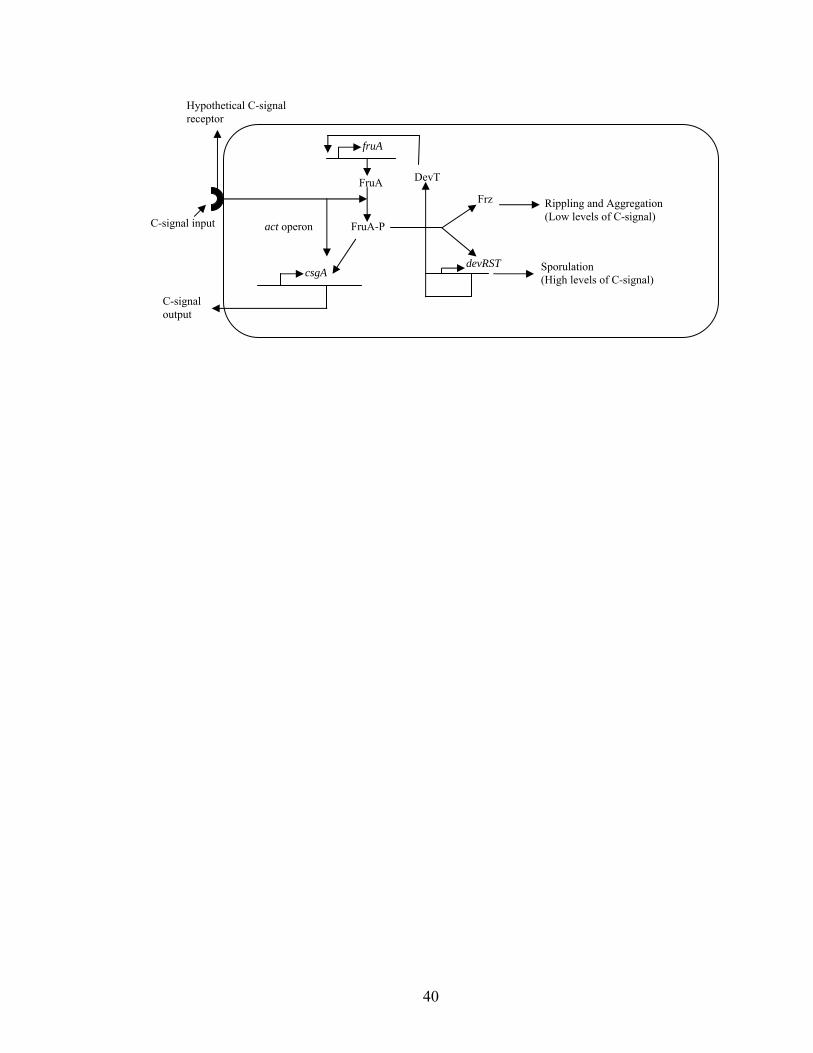
Figure 1.3 describes a model for the perception and transmission of the C-signal.
Briefly, the recognition of the C-signal by an unknown receptor, possibly the cognate
38
histidine kinase for FruA, triggers the cascade of events manifested by the signal. FruA,
which is phosphorylated by an unknown HPK interacts with the Frz signal transduction
system and initiates rippling and aggregation at low and intermediate levels of the C-
signal. The level of C-signal increases as aggregation progresses, and active FruA along
with the components of the act operon further increase csgA expression. At high levels
of C-signal and active FruA, devRST and the rest of the late developmental genes are
expressed, leading to sporulation.
Short chain alcohol dehydrogenase:
The short-chain alcohol dehydrogenase (SCAD) family is large, diverse, and
includes proteins from three EC classes, including oxidoreductases (EC 1.1),
dehydratases (EC 4.1) and epimerases (EC 5.1), with oxidoreductases forming the
majority of those that have been studied in vitro (105). The oxidoreductases catalyze the
interconversion of many different kinds of secondary alcohols and ketones using
NAD(P)(H) as the cofactor. For example, hydroxysteroid dehydrogenases catalyze the
reversible reduction of oxo/β-hydroxy groups at various positions of steroid hormones
and bile acids (107). SCAD dehydratases include the NADPH-dependent nucleotide
diphosphate (NDP)-sugar modifying enzymes that carry out an oxidation and subsequent
reduction reaction involving the removal of a molecule of water. For example, GDP-D-
mannose 4,6-dehydratase catalyzes the NADP+ dependent oxidation of GDP-D-mannose
to the ketomannose intermediate, which loses a molecule of water to result in the GDP-4-
keto-5,6-ene intermediate. This intermediate is then reduced using NADPH to GDP-4-
keto-6- deoxy-D-mannose (112). The SCAD epimerases catalyze the inversion of the
39
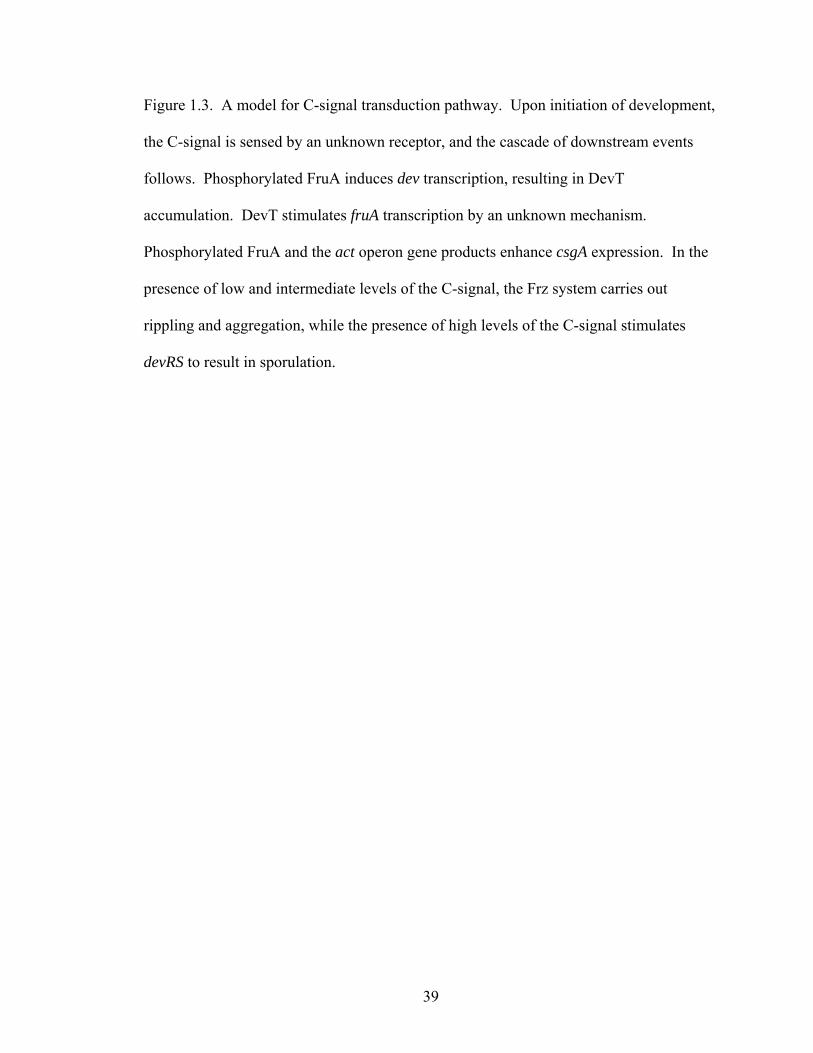
Figure 1.3. A model for C-signal transduction pathway. Upon initiation of development,
the C-signal is sensed by an unknown receptor, and the cascade of downstream events
follows. Phosphorylated FruA induces dev transcription, resulting in DevT
accumulation. DevT stimulates fruA transcription by an unknown mechanism.
Phosphorylated FruA and the act operon gene products enhance csgA expression. In the
presence of low and intermediate levels of the C-signal, the Frz system carries out
rippling and aggregation, while the presence of high levels of the C-signal stimulates
devRS to result in sporulation.
40
FruA
FruA-Pact operon
csgA
Frz
devRST
Rippling and Aggregation (Low levels of C-signal)
Sporulation (High levels of C-signal)
DevT
fruA
C-signal output
C-signal input
Hypothetical C-signal receptor
41
configuration around an asymmetric center of sugar substrates in a NAD+-dependent
manner. For example, UDP-galactose 4’-epimerase catalyzes the interconversion of
UDP-galactose and UDP-glucose through the transient reduction of NAD+. Catalysis
involves a tyrosine base that abstracts the 4’-hydroxyl hydrogen of the substrate and
mediates the subsequent hydride transfer to NAD+. The resulting 4’-ketopyranose
intermediate rotates 180O in the active site with the concomitant transfer of the hydride
from NADH back to the opposite face of the sugar substrate generating the epimerized
product (55). Thus, the SCAD family catalyzes reactions carried out by at least three of
the six known enzyme classes.
SCAD substrates include a wide variety of biomolecules like amino acids,
nucleotides, sugars, steroids, and xenobiotics. Because of their ability to act on many
different classes of compounds, SCADs exhibit a great degree of functional diversity.
They are involved in intermediary metabolism, biotransformation of xenobiotics, and
lipid hormone signaling during cellular differentiation in higher eukaryotes to produce
products such as steroids, prostaglandins and retinoids (105). Also, enzymes of this class
whose function is not yet understood are involved in developmental processes such as
Anabaena heterocyst differentiation (14), mouse adipocyte differentiation (139), and sex
determination in maize (30). The broad substrate specificity of these enzymes and the
lack of biochemical characterization of many of them have made it impossible to predict
the substrate based on amino acid sequence alone. In spite of the low primary sequence
identity between different enzymes in this family the tertiary structures display a similar
α/β folding pattern with the coenzyme-binding fold being strictly conserved. Also, the
critical residues involved in catalysis and coenzyme binding are conserved (69, 105).
42
With the exception of porcine and human carbonyl reductases, which are monomers (103,
140), SCAD enzymes are homodimers or homotetramers with 250-350 residues in each
subunit. Each subunit has a single domain with an N-terminal coenzyme-binding region
and a C-terminal substrate-binding region (105).
The architectures of SCAD active sites and coenzyme-binding regions have been
deduced from over thirty crystal structures. These crystals have been prepared in either
binary form, in which the enzyme is complexed with the coenzyme, or ternary form in
which the enzyme is bound to both the coenzyme and the substrate or inhibitor. The
proposed mechanism of catalysis is based on the structure of Drosophila lebanonensis
alcohol dehydrogenase (DADH) (9). The PDB accession codes for the crystals deposited
in Brookhaven Protein Data Bank are as follows: binary form (1B14), ternary form with
NAD-acetone (1B15), ternary form with NAD-3-pentanone (1B16), and ternary form
with NAD-cyclohexanone (1B2L). The indicated numbering is from DADH unless
otherwise specified.
Coenzyme binding in SCAD:
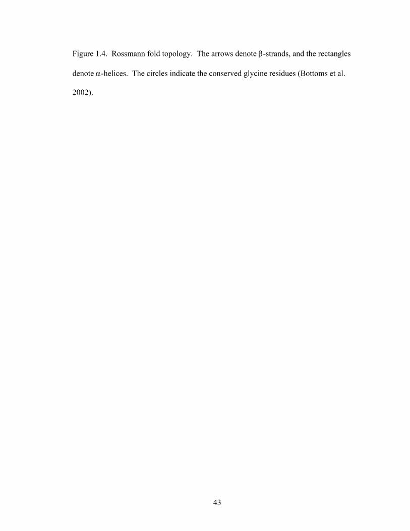
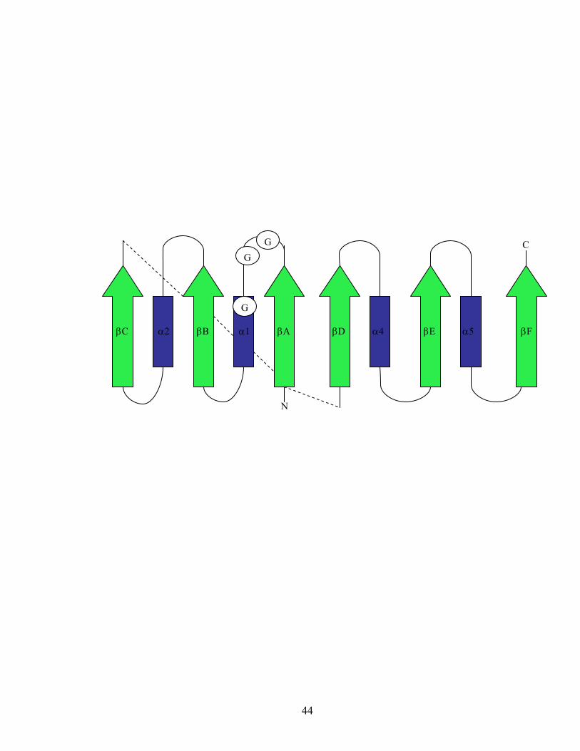
The N-terminal coenzyme-binding region is defined by a highly conserved
dinucleotide binding fold termed the “Rossmann fold” which consists of two βαβαβ
motifs that form a central β-sheet flanked by α helices (Figure 1.4). The coenzyme binds
to each of the two βαβαβ motifs (93, 115). Some enzymes show variation in the spacing
and distribution of the glycine residues in the first βαβαβ motif (69, 108). This motif
plays an essential role in maintaining the central β-sheet which is important for both
coenzyme positioning and binding.
43
Figure 1.4. Rossmann fold topology. The arrows denote β-strands, and the rectangles
denote α-helices. The circles indicate the conserved glycine residues (Bottoms et al.
2002).
44
βC α2 βB α1 βA βD α4 βE α5 βF
N
C
G
GG
45
Conserved residues that don’t interact directly with the coenzyme are preserved to
provide a structural framework for its binding. Thr12 interacts with strand βD to keep
the strands in the central β sheet for coenzyme positioning. A less conserved NNAG
motif or HxAA motif in some SCAD enzymes also contributes to the secondary structure
maintenance. The two aspargines [from 3β/17β-hydroxysteroid dehydrogenase (3β/17β-
HSD) (PDB accession code 2hsd)] in this motif form critical side chain and backbone
interactions through strands βD and βE respectively (Figure 1.5). The importance of
these motifs has been demonstrated by several chemical modification studies, site-
directed mutagenesis, and structure analysis. However, there is no evidence for the direct
interaction of these residues with the coenzyme (11, 36, 106). Some of the conserved
residues are thought to interact with the coenzyme by means of hydrogen bonding,
electrostatic, and hydrophobic interactions (Figure 1.5). Thr188 (from 3β/17β-HSD) is
proposed to stabilize the adenine-ribose end of the dinucleotide by hydrogen bonding
with the carboxamide group of the nicotinamide ring. Asp60 (from 3β/17β-HSD) in βB,
hydrogen bonds with the 2’- and 3’-hyroxyl groups of the adenine-ribose. However, the
phosphate group on the 2’-hydroxyl of the adenosine-ribose of NADP+ is repelled by this
aspartate, which is usually the sole determinant of the coenzyme preference of the
enzyme (36). There has also been a rare occurrence of glutamate as the critical acidic
residue (108). Another role of this residue is proposed to be stabilization of the turn
between βC and α4 in the adenine binding pocket. Finally, Ala88 (from 3β/17β-HSD) is
proposed to maintain hydrophobic contacts with the adenine ring (36).
NADP(H)-binding enzymes have two conserved basic residues (arginine or
lysine) that bind to the phosphate. The first of these is present in the glycine motif
46
Figure 1.5. Interaction of conserved residues from the central β-sheet with strands βA,
βD, βE of 3β/17β-HSD with NAD+. Hydrogen bond distances (in Å) are represented by
dotted lines (Filling et al. 2002).
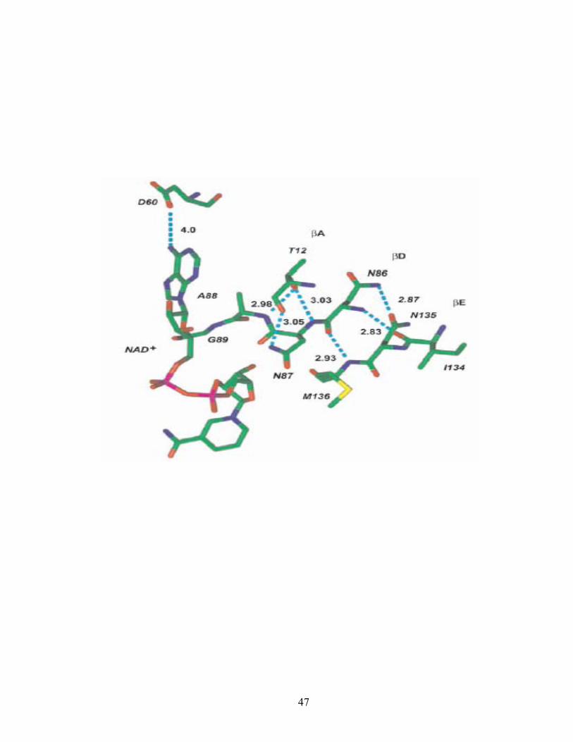
47
48
between βA and α1 immediately preceding the second glycine. The second basic residue
is present directly after the critical acidic residue (βC and α3) in the NAD(H) binding
enzymes. The requirement for these basic residues is not absolute, and there are
NADP(H)-binding enzymes that have only one of the basic residues. These positively
charged residues stabilize the bound NADPH via electrostatic interactions with the
negatively charged phosphate moiety of the coenzyme (36). About 10% of the sequences
analyzed do not have the typical pattern of charged residue distribution (108).
Active site structure and chemistry of catalysis:
The substrate binding site in the C-terminal region is the least conserved part of
the dehydrogenase. Three important structural features define the active site, the first of
which is a deep hydrophobic cavity. The residues in secondary structural elements βF to
α7 are responsible for the substrate specificity and are therefore quite diverse. The
hydrophobic cavity that constitutes the active site region consists of βD-βG, and is
enclosed by a flexible loop formed by residues 181-211. This cavity is closed by the C-
terminal residues 250-254 from the neighboring subunit upon coenzyme and substrate
binding (10). Deletion of the C-terminal residues 250-254 severely affects enzyme
activity (1).
The second important feature defining the active site is the presence of the
catalytic triad formed by Ser138, Tyr151, and Lys155. Recent studies with 3β/17β-HSD
have revealed that the conserved Asn111 also shows essential interactions with the
catalytic triad (36). Ser138 is proposed to orient and stabilize the substrate by hydrogen
bonding. Upon substrate binding, Tyr151 acts as a catalytic base and abstracts a proton
from the hydroxyl group of the alcohol substrate and transfers it to the ε-amino group of
49
Lys155 via the O2’ ribose hydroxyl of NAD+ (65). Asn111 stabilizes the active site
geometry by maintaining the position of Lys155 through an interaction with a water
molecule (W41 in Figure 1.6). This water molecule along with seven other water
molecules forms a hydrogen-bonded water chain which leads to the outside of the active
site. The proton abstracted from the substrate is finally conveyed to the bulk solvent
through this solvent channel. Thus, a proton relay is proposed to be formed at the active
site involving the substrate, Tyr151, the 2’OH of nicotinamide ribose, Lys155, and the
eight-membered water channel (Figure 1.6) (79).
Three well-ordered water molecules that interact with the catalytic residues
constitute the third structural feature defining the active site. One water molecule is
proposed to interact with Ser138 and Tyr151 until it becomes replaced by the substrate.
The second water molecule links Tyr151 and Lys155 and is proposed to be involved in
the deprotonation of the Tyr151 hydroxyl group and/or mimic the 2’O and 3’O atoms of
the NAD ribose in the absence of the coenzyme. The third water molecule is proposed to
play a structural role by linking Lys155 and the O atom of Asn107 (10).
Drosophila alcohol dehydrogenase follows the Theorell-Chance mechanism for
substrate conversion (9). This is an ordered pathway as illustrated below, in which the
coenzyme is the leading substrate and the dissociation of the binary enzyme-NADH
complex is the rate limiting step:
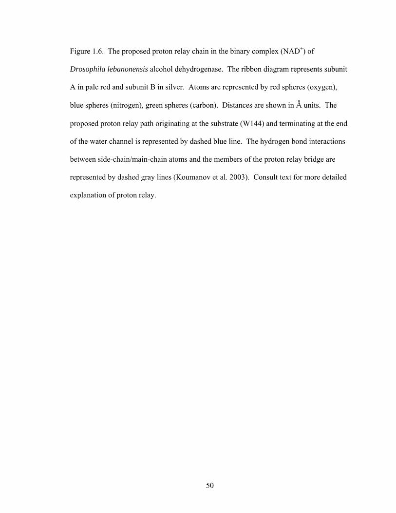
50
Figure 1.6. The proposed proton relay chain in the binary complex (NAD+) of
Drosophila lebanonensis alcohol dehydrogenase. The ribbon diagram represents subunit
A in pale red and subunit B in silver. Atoms are represented by red spheres (oxygen),
blue spheres (nitrogen), green spheres (carbon). Distances are shown in Å units. The
proposed proton relay path originating at the substrate (W144) and terminating at the end
of the water channel is represented by dashed blue line. The hydrogen bond interactions
between side-chain/main-chain atoms and the members of the proton relay bridge are
represented by dashed gray lines (Koumanov et al. 2003). Consult text for more detailed
explanation of proton relay.
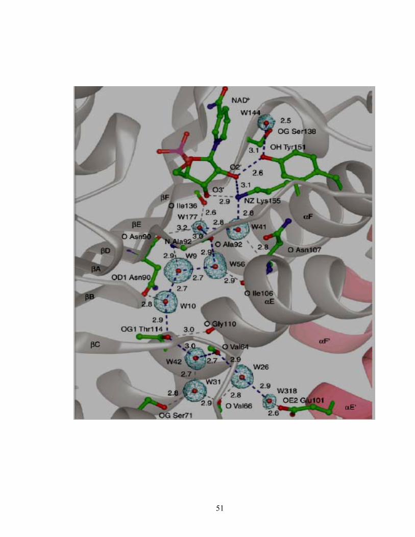
51
52
E + NAD+ <==> E•NAD+ + R-OH
<==> E•NAD+ •R-OH
<==> E•NADH•R=O
<==> R=O + E•NADH
<==> E + NADH
The coenzyme binding to the apo form (enzyme with unbound coenzyme and substrate)
results in a conformational change that affects the relative positions of α2, α3, α4, and
α7. This also results in a partially ordered structure of the entrance gate of the active site
cleft. The binding of the substrate replaces the water molecule between the side chains of
Ser138 and Tyr151. During the transition from the binary to the ternary form, the
enzyme undergoes a second conformational change. Once the substrate is positioned in
the active site cavity, the hydroxyl group of the substrate is placed between the oxygen
atoms of the side-chains of Ser138 and Tyr151. This facilitates proton transfer from the
alcohol group to the unprotonated Tyr151O- form, generating a partial positive charge on
the C1 atom of the alcohol enabling hydride transfer from Ser138 to the C4 position (with
a H at C4) of the coenzyme. Serine is proposed to orient the alcohol substrate, stabilize
the transition state intermediate, or both. Lysine is proposed to serve the dual roles of
orienting the coenzyme by formation of hydrogen bonds with the oxygen atoms of the
nicotinamide-ribose moiety and lowering the pKa of the tyrosine residue through ionic
interactions. The positively charged nicotinamide ring is also proposed to lower the pKa
of tyrosine.
53
Classification of SCAD superfamily members:
Hidden Markov model analysis was used to classify the SCAD superfamily
systematically into five families based on the chain length and the spacing and
distribution of glycine residues in the coenzyme binding region (69, 108). Each of the
five families is discussed in detail.
The classical family: The majority of SCADs fall into this family whose primary
sequence consists of about 250 amino acids. The coenzyme-binding domain exhibits a
typical glycine distribution pattern of TGxxxGxG in the coenzyme-binding region. There
is also a conserved aspartate about thirteen positions downstream from the active site
lysine residue that is involved in the stabilization of the adenine-binding pocket. The
other conserved sequence in this family is the NNAG motif in the coenzyme binding
region which is required for stabilizing the β-strands within the central β-sheet. Also, the
aspargine residue responsible for maintaining the active site geometry is highly
conserved throughout this family. This family includes oxidreductases like steroid
dehydrogenases and carbonyl reductases. Most of the enzymes use NADP(H) as the
preferred coenzyme.
The Extended family: Greater than 25% of the known SCADs are classified in this
family, which is characterized by a chain length of about 350 amino acids. The
coenzyme-binding motif TGxxGxxG differs from that of the classical enzymes in the
position and distribution of the conserved glycines. A motif DxxD in the loop between
βC and α3 contributes to the stability of the adenine-binding pocket. The coenzyme-
binding NNAG motif in βD is replaced by a HxAA motif. The catalytic domain is
characterized by the presence of a conserved proline immediately preceding the active
54
site tyrosine and a conserved negatively charged residue four residues downstream of the
lysine residue. This family includes isomerases like UDP-galactose 4’-epimerase and
lyases like UDP-D-mannose 4,6-dehydratase in addition to oxidoreductases like 3β-
hydroxysteroid dehydrogenase. Also, these enzymes prefer NAD(H) as the coenzyme.
The Intermediate family: This family accounts for 6-13% of the known sequences. The
glycines in the coenzyme binding region have been replaced by alanines. All the other
characteristics resemble the classical enzymes except that the NNAG motif for coenzyme
binding is replaced by NGAG. Drosophila alcohol dehydrogenase belongs to this family.
The Divergent family: These are NADH-dependent enzymes that exhibit certain
deviations from the typical SCAD pattern. The second and the third glycines in the
coenzyme binding fold have been replaced by serine and alanine, respectively. The
catalytic tyrosine and lysine in the active site are spaced differently. The recurring motif
in the active site of these enzymes is YxxMxxxK instead of YxxxK. This family is
exemplified by the enoyl reductases from bacteria and plants.
The complex family: This family includes some fatty acid synthases that are parts of
multifunctional enzyme complexes found in diverse living systems. The preferred
coenzyme of this family is NADP(H) and the signature motif in the active site is YxxxN
instead of YxxxK.
The divergent and the complex families together include fewer than 5% of all the
known enzymes. Three percent of the enzymes lack at least one of the signature motifs
to be classified under any family and hence remain unclassified.
The classical and extended families are further classified according to their
coenzyme specificity based on the pattern of charged residues at key positions in the
55
Rossmann fold. NAD(H)-preferring enzymes have an acidic residue at the C-terminal
end of βB while the NADP(H)-binding enzymes have two basic residues, the first of
which is found immediately preceding the second glycine. The second one is located
exactly after the crucial acidic residue of NAD(H)-binding enzymes. The former
enzymes are classified under the subfamily cP1, while the latter enzymes are classified
under the subfamily cP2. Enzymes with both of the basic residues are classified as
subfamily cP3.
NAD(H) binding classical SCADs with aspartate as the acidic residue that
determines coenzyme specificity are classified under the subfamily cD1d and those with
glutamate as the crucial acidic residue are found under the subfamily cD1e. The position
and the type of acidic residue are not strictly conserved and can be found one or two
positions downstream. Thus, enzymes with either an aspartate or a glutamate one
position downstream fall under cD2 and those that have the acidic residue two positions
downchain are classified as cD3.
Extended SCAD also have an acidic residue at the end of the C-terminal end of
βB as the sole determinant of the coenzyme preference of the enzyme. Enzymes with an
aspartate or a glutamate at position 33 are classified as eD1 and those with the acidic
residue two positions downstream are classified as eD2. NADP(H)-preferring enzymes
of the extended family do not have the first of the two characteristic basic residues in the
glycine motif. However, the second one is present in the defined position. These
enzymes are classified as belonging to the subfamily eP1.

56
CsgA and SocA as SCAD members:
CsgA has conserved motifs typical of a SCAD (Figure 1.7). The coenzyme
binding region is characterized by the motifs TGxxRGxG and the NNAG typical of
classical SCADs. It has a glutamate at the C-terminal end of the second β-strand that
could suggest a NAD(H) preference. CsgA could be classified under the classical
subfamily cD1e. However, the glycine motif in the coenzyme-binding region has an
arginine immediately preceding the second glycine, suggesting potential NADP(H)
binding and classification of CsgA in subfamily cP1. The 229 amino acid primary
structure of CsgA contains 34.7% identity with 11-cis retinol dehydrogenase from Bos
taurus (X82262). The other close relatives include enzymes whose substrate specificity
and biological function are unknown [Bradyrhizobium japonicum - P05406 (31.4);
Saccharomyces cereviceae - Z28071 (33.2); Maize - L20621 (29.5%)]. Molecular
modeling of CsgA with 3α/20β-HSD suggests that the overall shape of the protein is very
similar to that of hydroxysteroid dehydrogenase. The conserved residues in the catalytic
and coenzyme-binding regions are present, including all four catalytically important
residues. CsgA with T6A or R10A replacements was biologically inactive in C-signaling
(90). CsgA with a T6A replacement did not bind NAD+ (90). CsgA has an unusual AY
repeat in the YxxxK motif, the significance of which is not known. CsgA derivatives
containing a K155R or S135T replacement were inactive, highlighting the importance of
these active site residues for C-signal generation (90).
SocA has 28% identity with CsgA and exhibits all the primary and secondary
structural features that define a short-chain alcohol dehydrogenase (Figure 1.7). The
spacing and distribution of the glycine residues in the Rossmann fold, TGxxxGxG, is
57
Figure 1.7. An alignment of two M. xanthus SCAD CsgA and SocA with 3α/20β-
Hydroxysteroid dehydrogenase. The secondary structural elements are shown below the
sequence line based on the crystal structure of 3α/20β-Hydroxysteroid dehydrogenase.
Letters in red indicate the residues that are conserved in all the three proteins (Lee et al.
1995). The conserved residues in the coenzyme-binding region are indicated by a red
box and the conserved catalytic residues are indicated by a blue box. The predicted site
of processing of the 17 kDa form is indicated by a green arrow (Lobedanze and Sogaard-
Andersen, 2003).
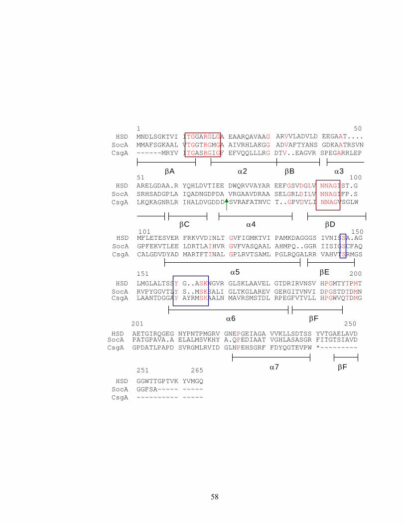
58
1 50 HSD MNDLSGKTVI ITGGARGLGA EAARQAVAAG ARVVLADVLD EEGAAT.... SocA MMAFSGKAAL VTGGTRGMGA AIVRHLAKGG ADVAFTYANS GDKAATRSVN CsgA ~~~~~~MRYV ITGASRGIGF EFVQQLLLRG DTV..EAGVR SPEGARRLEP
51 100
HSD ARELGDAA.R YQHLDVTIEE DWQRVVAYAR EEFGSVDGLV NNAGIST.G SocA SRHSADGPLA IQADNGDPDA VRGAAVDRAA SELGRLDILV NNAGIFP.S CsgA LKQKAGNRLR IHALDVGDDD SVRAFATNVC T..GPVDVLI NNAGVSGLW
101 150
151 200 HSD LMGLALTSSY G..ASKWGVR GLSKLAAVEL GTDRIRVNSV HPGMTYTPMT
SocA RVPYGGVTLY S..MSKSALI GLTKGLAREV GERGITVNVI DPGSTDTDMN
CsgA LAANTDGGAY AYRMSKAALN MAVRSMSTDL RPEGFVTVLL HPGWVQTDMG
201 250
251 265
HSD GGWTTGPTVK YVMGQ
SocA GGFSA~~~~~ ~~~~~ CsgA ~~~~~~~~~~ ~~~~~
βA α2 βB α3
βC α4 βD
α5 βE
α6 βF
α7 βF
HSD MFLETESVER FRKVVDINLT GVFIGMKTVI PAMKDAGGGS IVNISSA.AG SocA GPFEKVTLEE LDRTLAIHVR GVFVASQAAL AHMPQ..GGR IISIGSCFAQ CsgA CALGDVDYAD MARTFTINAL GPLRVTSAML PGLRQGALRR VAHVTSRMGS
HSD AETGIRQGEG NYPNTPMGRV GNEPGEIAGA VVKLLSDTSS YVTGAELAVD SocA PATGPAVA.A ELALMSVKHY A.QPEDIAAT VGHLASASGR FITGTSIAVD
CsgA GPDATLPAPD SVRGMLRVID GLNPEHSGRF FDYQGTEVPW *~~~~~~~~~
59
typical of a classical SCAD. The conserved aspartate between βC and α3 required for
stabilizing the adenine binding pocket is present in addition to the NNAG motif in βD
that provides the structural support for coenzyme binding. SocA lacks the crucial
aspartate that blocks NADP(H) binding, suggesting that it can utilize NADP(H). Also, it
has the first of the two basic residues immediately preceding the second glycine in the
glycine motif and could be classified in the subfamily cP1. However, the aspartate in α3
at position 42 could enable NAD(H) binding in an atypical manner. The substrate
binding region has the signature sequence YxxxK and the catalytically active serine. But
the aspargine or serine (located in α5) that interacts with Lys155 via a water molecule for
maintaining the active site geometry interaction is absent (92). None of the above
observations about the significance of conserved SocA residues have been confirmed by
mutational analysis because the gene appears to be essential for growth. The primary
structure of SocA contains 39% identity with 3-oxoacyl-[acyl carrier protein] reductase
from Brassica napus (P27582). The other close relatives include 3-oxoacyl-[acyl carrier
protein] reductases from E. coli - P25716 (37.8%), Cuphea lanceolata (34.3%),
acetoacetyl-CoA reductase from Zoogloea ramigera - P23238 (34.7%), 2, 3-Dihydro-2,3-
dihydroxy-benzoate dehydrogenase from E. coli - P15047 (35.6%).
Functional heterogeneity of SCADs:
Presently about 3,000 SCADs have been identified based on their primary amino
acid sequence, although the function(s) of the vast majority are unknown. Because of the
myriad of physiological processes these enzymes are involved in and also because of the
60
diverse substrate spectrum these enzymes exhibit, it is not possible to predict the potential
substrates based on amino acid sequence homology alone.
Analysis of completed genomes reveals the presence of SCAD-encoding genes in
significant numbers. The E. coli genome encodes about 24 SCADs out of about 4200
ORFs (0.6% of genes). Similarly, Mycobacterium tuberculosis and Bacillus subtilis
encode 57 and 32 SCADs, respectively (64). In the case of higher organisms, the human
genome has 58 SCAD sequences while Drosophila melanogaster, Caenorahbditis
elegans, and Arabidopsis thaliana genomes encode 62, 56, and 62, respectively (68). In
all the cases, classical SCADs form the majority.
Most SCAD genes are defined based only on the presence of the conserved
signature residues. For example, the E. coli genome encodes 17 classical SCADs and 7
extended SCADs out of which 11 sequences are annotated as hypothetical proteins
broadly classified under sugar-utilizing SCADs. Seven E. coli enzymes have defined
functions, including those that utilize hexose sugars or steroids, or are involved in fatty
acid biosynthesis. Although extended SCADs are not as common as classical SCADs in
Bacteria, they are the only SCADs in the Archaea Methanococcus jannaschii and
Pyrococcus horikoshii. Bacteria with smaller genomes like Mycoplasma sp. or Borrelia
burgdorferi also lack classical SCADs and encode at least one extended SCAD. The
presence of these extended enzymes in even the most primitive of life forms suggests
their evolutionary origin as sugar- or nucleotide-metabolizing enzymes that later evolved
to utilize a wide diversity of other substrates.
61
Examples of functional diversity of SCAD:
SCADs can be grouped into two main categories based on their functional
assignments as enzymes affecting intermediary metabolism or involved in intercellular
signaling. Enzymes in the former category act on substrates such as sugars, amino acids,
fatty acids, pteridines etc., while lipid substrates are the preferred choice for enzymes
involved in lipid hormone mediated-cell signaling in Eukarya (107).
SCADs affecting intermediary metabolism: This group of enzymes exhibits a wide range
of substrate specificity. Deficiency of many of these enzymes results in inherited
metabolic disorders in higher animals. In E. coli and other bacteria, UDP-galactose
epimerase is involved in galactose metabolism, catalyzing the interconversion of UDP-
glucose and UDP-galactose. In humans, this enzyme is involved in the biosynthesis of
glycoproteins and glycolipids by catalyzing the interconversion of UDP–N-
acetylglucosamine and UDP-N-acetylgalactosamine. An impairment in the functioning
of the human enzyme results in a severe metabolic disorder, epimerase-deficiency
galactosemia.
Dihydropteridin reductase and sepiapterin reductase are involved in pteridine
metabolism, the deficiency of which results in type II phenylketonuria. 3-hydroxy
butyrate dehydrogenase catalyzes the interconversion of 3-hydroxybutyrate and
acetoacetate during carbon metabolism in many bacteria that accumulate
polyhydroxybutyrate. The mammalian counterpart of this enzyme is involved in ketone
body metabolism during starvation (100). Many bacteria produce serine dehydrogenase
which is the only known SCAD that acts on amino acid substrates (24). The
physiological relevance of this enzyme is not known. SCADs also act on acylated
62
molecules like acyl carrier protein (ACP), coenzyme A, and cysteamine. β-Keto ACP
reductase catalyzes the reversible reduction of 3-oxoacyl ACP to its hydroxy product
during fatty acid biosynthesis (13, 119, 135).
SCADs involved in signaling and xenobiotic metabolism: Enzymes in this group act on
structurally similar substrates but have different implications for the cell physiology of
bacteria and higher animals. These SCADs mediate lipid hormone signaling during
development in higher animals while carrying out xenobiotic carbonyl metabolism in
bacteria. Some enzymes in this group have overlapping substrate specificity. These
enzymes mediate intercellular signaling in mammals by activating or inactivating various
bioactive molecules such as steroids and prostaglandins. Hydroxysteroid dehydrogenases
(HSD) play an essential role in the biosynthesis of all classes of steroid hormones and
bile acids. They catalyze the reversible reduction of oxo/β-hydroxy groups at various
positions of steroid hormones and bile acids. 11β-HSD controls the access of
glucocorticoids to their intracellular receptors (GR), thus preventing the nonspecific
activation of mineralocorticoid receptors (MR). The glucocorticoid hormones have a
high affinity for both GR and MR, while the mineralocorticoid hormone aldosterone
binds only to MR. 11β-HSD reduces the glucocorticoid hormones cortisol and
corticosterone to their corresponding inactive forms cortisone and 11-
dehydrocorticosterone which are not recognized by either GR or MR, enabling
recognition of MR by aldosterone (37). The 17β-HSD isoforms operate a similar
metabolic switch to regulate the activity of androgens and estrogens. The microbial
counterparts of these steroid hormone-metabolizing enzymes are involved in
biotransformation of polyaromatic hydrocarbons in contaminated soils. Comamonas
63
testosteroni, which is able to utilize testosterone as the sole source of carbon, produces
several enzymes such as 3α-HSD, 3β/17β-HSD, that are involved in the degradation of
steroids.
Retinoids are the key photosensitive elements involved in visual sensation.
Retinoic acids are chemical signals for cellular differentiation and proliferation. Retinol
dehydrogenases catalyze the oxidation of retinol to retinal which is further metabolized
into retinoic acid (107).
Unusual SCADs:
The SCAD family exhibits remarkable structural diversity, which contributes to
the broad range of substrates converted. Some of these enzymes have novel structural
features that lend themselves to novel functions. Haloalcohol dehalogenases catalyze the
cofactor-independent dehalogenation of environmental contaminants like 1,3-dichloro-2-
propanol to result in its corresponding epoxide, a halide ion, and a proton. These
enzymes have the otherwise highly conserved catalytic triad of a SCAD but with lysine
replaced by arginine. These enzymes also lack the ability to bind the coenzyme because
of the replacement of the glycine-rich motif at the N-terminus by a NVKHFGG motif that
provides a halide-binding site (29). The typical redox reaction mechanism of this SCAD
is replaced by a substitutive dehalogenation. The catalytic tyrosine deprotonates the
hydroxyl group of the substrate to generate an intramolecular nucleophile, which
substitutes the halogen (29). Thus, this enzyme provides a striking example of the
divergent evolution the SCAD family has undergone.
The SCAD fold is adapted for non-enzymatic functions such as transcriptional
regulation as well. For example, NmrA, a negative transcriptional regulator involved in
64
the control of nitrogen metabolite repression in many fungi, has a two-domain structure
including the Rossmann fold and shows significant homology with UDP-galactose 4-
epimerase. However, this protein lacks the catalytic tyrosine and hence is not
enzymatically active. The NAD+ binding is suggested to have a regulatory role. Thus,
the SCAD family exhibits diversity not just in terms of the range of substrates converted
but also in terms of the diverse roles these enzymes play in the cell.
Conclusion:
Short-chain alcohol dehydrogenases form a large and evolutionarily ancient
superfamily of enzymes that use NAD(P)(H) to catalyze redox reactions in addition to
epimerization and dehydration reactions. In spite of low sequence identity at the primary
amino acid level, they show a highly conserved secondary structure. The catalytically
important residues as well as those involved in the coenzyme binding and positioning are
conserved throughout the superfamily. They exhibit remarkable evolutionary divergence
which has contributed to their ability to act on a wide spectrum of biomolecules
implicating them in diverse cellular functions. Some of these functions are of biomedical
and pharmacological interests in humans including metabolic disorders and hormone-
dependent cancers. Then there are some proteins that deviate from the norm and perform
non-enzymatic functions as fungal transcription factors. Thus, this enzyme superfamily
exhibits extraordinary structural and functional diversity.
65
Statement of purpose:
The purpose of this dissertation is to examine the enzymatic role of CsgA in the
generation of the C-signal. It is important to resolve the debate about the chemical nature
of the C-signal. Genetic studies supporting the role of the full-length form as a short-
chain alcohol dehydrogenase are striking. However, establishing the biochemical basis is
complicated by the lack of significant sequence homology with enzymes of known
substrate specificity. We propose to employ an in vitro dehydrogenase assay to screen
for substrates prepared from whole cell extracts. The enzymatic activity of purified CsgA
will be confirmed by repeating the assay with purified SocA to determine the extent of
substrate overlap. Mass spectrometric methods will be employed to determine the
identity of the active molecule(s) in the extract. Finally, the biological significance of the
enzyme activity will be determined by the ability of the end product to reproduce the
biological effects of the purified protein.
66
Reference:
1. Albalat, R., M. Valls, J. Fibla, S. Atrian, and R. Gonzalez-Duarte. 1995.
Involvement of the C-terminal tail in the activity of Drosophila alcohol
dehydrogenase. Evaluation of truncated proteins constructed by site-directed
mutagenesis. Eur. J. Biochem. 233:498-505.
2. Arnold, J. W., and L. J. Shimkets. 1988. Cell surface properties correlated with
cohesion in Myxococcus xanthus. J. Bacteriol. 170:5771-5777.
3. Arnold, J. W., and L. J. Shimkets. 1988. Inhibition of cell-cell interactions in
Myxococcus xanthus by Congo red. J. Bacteriol. 170:5765-5770.
4. Baker, M. 1994. Myxococcus xanthus C-factor, a morphogenetic paracrine signal,
is similar to Escherichia coli 3-oxoacyl-[acyl-carrier-protein] reductase and
human 17ß-hydroxysteroid dehydrogenase. Biochem. J. 301:311-312.
5. Bartholomeusz, G., Y. Zhu, and J. Downard. 1998. Growth medium-dependent
regulation of Myxococcus xanthus fatty acid content is controlled by the esg
locus. J. Bacteriol. 180:5269-72.
6. Behmlander, R., and M. Dworkin. 1994. Biochemical and structural analyses of
the extracellular matrix fibrils of Myxococcus xanthus. J. Bacteriol. 176:6295-
6303.
7. Behmlander, R. M., and M. Dworkin. 1991. Extracellular fibrils and contact-
mediated cell interactions in Myxococcus xanthus. J. Bacteriol. 173:7810-7821.
8. Behmlander, R. M., and M. Dworkin. 1994. Integral proteins of the
extracellular matrix fibrils of Myxococcus xanthus. J. Bacteriol. 176:6304-6311.
67
9. Benach, J., S. Atrian, R. Gonzalez-Duarte, and R. Ladenstein. 1999. The
catalytic reaction and inhibition mechanism of Drosophila alcohol dehydrogenase:
Observation of an enzyme-bound NAD-ketone adduct at 1.4A resolution by X-ray
crystallography. J. Mol. Biol. 289:335-355.
10. Benach, J., S. Atrian, R. Gonzalez-Duarte, and R. Ladenstein. 1998. The
refined crystal structure of Drosophila lebanonensis alcohol dehydrogenase at 1.9
A resolution. J. Mol. Biol. 282:383-399.
11. Benach, J., C. Filling, U. C. Oppermann, P. Roversi, G. Bricogne, K. D.
Berndt, H. Jornvall, and R. Ladenstein. 2002. Structure of bacterial
3beta/17beta-hydroxysteroid dehydrogenase at 1.2 A resolution: a model for
multiple steroid recognition. Biochem 50:14659-14668.
12. Bi, E., and J. Lutkenhaus. 1993. Cell division inhibitors SulA and MinCD
prevent formation of the FtsZ ring. J. Bacteriol. 175:1118-1125.
13. Birge, C. H., and P. R. Vagelos. 1972. Acyl carrier protein. XVI. Intermediate
reactions of unsaturated fatty acid synthesis in Escherichia coli and studies of fab
B mutants. J. Biol. Chem. 16:4921-4929.
14. Black, T. A., and C. P. Wolk. 1994. Analysis of a Het- mutation in Anabaena sp.
strain PCC7120 implicates a secondary metabolite in the regulation of heterocyst
spacing. J. Bacteriol. 176:2282-2292.
15. Bowden, M. G., and H. B. Kaplan. 1998. The Myxococcus xanthus
lipopolysaccharide O-antigen is required for social motility and multicellular
development. Mol. Microbiol. 30:275-284.
68
16. Boysen, A., E. Ellehauge, B. Julien, and L. Sogaard-Andersen. 2002. The
DevT protein stimulates synthesis of FruA, a signal transduction protein required
for fruiting body morphogenesis in Myxococcus xanthus. J. Bacteriol. 184:1540-
1546.
17. Butler, J. S., M. Springer, and M. Grunberg-Manago. 1987. AUU-to-AUG
mutation in the initiator codon of the translation initiation factor IF3 abolishes
translational autocontrol of its own gene (infC) in vivo. Proc. Natl. Acad. Sci.
USA. 84:4022-4025.
18. Cashel, M., D. R. Gentry, V. J. Hernandez, and D. Vinella. 1996. The
Stringent Response, p. 1458-1496. In F. C. Neidhardt, R. I. Curtiss, J. L.
Ingraham, E. C. C. Lin, K. B. Low, B. Magasanik, W. S. Reznikoff, M. Riley, M.
Schaechter, and H. E. Umbarger (ed.), Escherichia coli and Salmonella: Cellular
and Molecular Biology., vol. I. Am. Soc. Microbiol., Washington, DC.
19. Chang, B.-Y., and M. Dworkin. 1994. Isolated fibrils rescue cohesion and
development in the dsp mutant of Myxococcus xanthus. J. Bacteriol. 176:7190-
7196.
20. Cheng, Y., and D. Kaiser. 1989. dsg, a gene required for cell-cell interaction
early in Myxococcus development. J. Bacteriol. 171:3719-3726.
21. Cheng, Y., and D. Kaiser. 1989. dsg, a gene required for Myxococcus
development, is necessary for cell viability. J. Bacteriol. 171:3727-3731.
22. Cheng, Y., L. V. Kalman, and D. Kaiser. 1994. The dsg gene of Myxococcus
xanthus encodes a protein similar to translation initiation factor IF3. J. Bacteriol.
176:1427-1433.
69
23. Cho, K., and D. R. Zusman. 1999. AsgD, a new two-component regulator
required for A-signaling and nutrient sensing during early development of
Myxococcus xanthus. Mol. Microbiol. 34:268-281.
24. Chowdhury, E. K., K. Higuchi, S. Nagata, and H. Misono. 1997. A novel
NADP(+)-dependent serine dehydrogenase from Agrobacterium tumefaciens.
Biosci. Biotechnol. Biochem. 61:152-157.
25. Costerton, J. W., P. S. Stewart, and E. P. Greenberg. 1999. Bacterial biofilms:
a common cause of persistent infections. Science. 284:1318-1322.
26. Crawford, E. W. J., and L. J. Shimkets. 2000. The Myxococcus xanthus socE
and csgA genes are regulated by the stringent response. Mol. Microbiol. 37:788-
799.
27. Crawford, E. W. J., and L. J. Shimkets. 2000. The stringent response in
Myxococcus xanthus is regulated by SocE and the CsgA C-signaling protein.
Genes Dev. 14:483-492.
28. Davis, J. M., J. Mayor, and L. Plamann. 1995. A missense mutation in rpoD
results in an A-signalling defect in Myxococcus xanthus. Mol. Microbiol. 18:943-
952.
29. de Jong, R. M., J. J. W. Tiesinga, H. J. Rozeboom, K. H. Kalk, L. Tang, D. B.
Janssen, and B. W. Dijkstra. 2003. Structure and mechanism of a bacterial
haloalcohol dehalogenase: a new variation of the short-chain
dehydrogenase/reductase fold without an NAD(P)H binding site. EMBO J.
22:4933-4944.
70
30. DeLong, A., A. Calderon-Urrea, and S. L. Dellaporta. 1993. Sex determination
gene TASSELSEED2 of maize encodes a short-chain alcohol dehydrogenase
required for stage-specific floral organ abortion. Cell. 74:757-768.
31. Downard, J., S. V. Ramaswamy, and K.-S. Kil. 1993. Identification of esg, a
genetic locus involved in cell-cell signaling during Myxococcus xanthus
development. J. Bacteriol. 175:7762-7770.
32. Downard, J., and D. Toal. 1995. Branched chain fatty acids: the case for a novel
from of cell-cell signaling during Myxococcus xanthus development. Mol.
Microbiol. 16:171-175.
33. Dworkin, M. 1962. Nutritional requirements for vegetative growth of
Myxococcus xanthus. J. Bacteriol. 84:250-257.
34. Dworkin, M. 1996. Recent advances in the social and developmental biology of
the myxobacteria. Microbiol. Rev. 60:70-102.
35. Ellehauge, E., M. Norregaard-Madsen, and L. Sogaard-Andersen. 1998. The
FruA signal transduction protein provides a checkpoint for the temporal co-
ordination of intercellular signals in Myxococcus xanthus development. Mol.
Microbiol. 30:807-817.
36. Filling, C., K. D. Berndt, J. Benach, S. Knapp, T. Prozorovski, E. Nordling,
R. Ladenstein, H. Jornvall, and U. Oppermann. 2002. Critical residues for
structure and catalysis in short-chain dehydrogenases/reductases. J. Biol. Sci.
277:25677-23684.
37. Funder, J. W. 1997. Glucocorticoid and mineralocorticoid receptors: biology and
clinical relevance. Annu. Rev. Med. 48:231-240.
71
38. Garrity, P. A., and S. L. Zipursky. 1995. Neuronal target recognition. Cell.
83:177-185.
39. Garza, A. G., B. Z. Harris, J. S. Pollack, and M. Singer. 2000. The asgE locus
is required for cell-cell signalling during Myxococcus xanthus development. Mol.
Microbiol. 35:812-824.
40. Ghosh, D., Z. Wawrzak, C. M. Weeks, W. L. Duax, and M. Erman. 1994. The
refined three-dimensional structure of 3 alpha,20 beta-hydroxysteroid
dehydrogenase and possible roles of the residues conserved in short-chain
dehydrogenases. Structure. 2:629-640.
41. Gill, R. E., and M. C. Bornemann. 1988. Identification and characterization of
the Myxococcus xanthus bsgA gene product. J. Bacteriol. 170:5289-5297.
42. Gill, R. E., M. Cull, and S. Fly. 1988. Genetic Identification and cloning of a
gene required for developmental cell interactions in Myxococcus xanthus. J.
Bacteriol. 170:5279-5288.
43. Gill, R. E., and M. G. Cull. 1986. Control of developmental gene expression by
cell-to-cell interactions in Myxococcus xanthus. J. Bacteriol. 168:341-347.
44. Gill, R. E., M. Karlok, and D. Benton. 1993. Myxococcus xanthus encodes an
ATP-dependent protease which is required for developmental gene transcription
and intercellular signaling. J. Bacteriol. 175:4538-4544.
45. Gorski, L., T. Gronewold, and D. Kaiser. 2000. A sigma(54) activator protein
necessary for spore differentiation within the fruiting body of Myxococcus
xanthus. J. Bacteriol. 180:2438-2444.
72
46. Gorski, L., and D. Kaiser. 1998. Targeted mutagenesis of sigma54 activator
proteins in Myxococcus xanthus. J. Bacteriol. 180:5896-5905.
47. Gottesman, S. 1996. Proteases and their targets in Escherichia coli. Annu. Rev.
Genet. 30:465-506.
48. Gronewold, T. M., and D. Kaiser. 2001. The act operon controls the level and
time of C-signal production for Myxococcus xanthus development. Mol.
Microbiol. 40:744-756.
49. Guo, D., Y. Wu, and H. B. Kaplan. 2000. Identification and characterization of
genes required for early Myxococcus xanthus developmental gene expression. J.
Bacteriol. 182:4564-4571.
50. Hagen, D. C., A. P. Bretscher, and D. Kaiser. 1978. Synergism between
morphogenetic mutants of Myxococcus xanthus. Dev. Biol. 64:284-296.
51. Hager, E., H. Tse, and R. E. Gill. 2001. Identification and characterization of
spdR mutations that bypass the BsgA protease-dependent regulation of
developmental gene expression in Myxococcus xanthus. Mol. Microbiol. 39:765-
780.
52. Harris, B. Z., D. Kaiser, and M. Singer. 1998. The guanosine nucleotide
(p)ppGpp initiates development and A-factor production in Myxococcus xanthus.
Genes Dev. 12:1022-1035.
53. Hartzell, P., and D. Kaiser. 1991. Function of MglA, a 22-kilodalton protein
essential for gliding in Myxococcus xanthus. J. Bacteriol. 173:7615-7624.
73
54. Hodgkin, J., and D. Kaiser. 1979. Genetics of gliding motility in Myxococcus
xanthus (Myxobacterales): Genes controlling movement of single cells. Mol. Gen.
Genet. 171:167-176.
55. Holden, H. M., I. Rayment, and J. B. Thoden. 2003. Structure and function of
enzymes of the Leloir pathway for galactose metabolism. J. Biol. Chem.
278:43885-43888.
56. Hu, J. C., and C. A. Gross. 1985. Mutations in the sigma subunit of E. coli RNA
polymerase which affect positive control of transcription. Mol. Gen. Genet.
199:7-13.
57. Huisman, O., and R. D'Ari. 1981. An inducible DNA replication-cell division
coupling mechanism in E. coli. Nature. 290:797-799.
58. Huisman, O., R. D'Ari, and S. Gottesman. 1984. Cell-division control in
Escherichia coli: specific induction of the SOS function SfiA protein is sufficient
to block septation. Proc. Natl. Acad. Sci. USA. 81:4490-4494.
59. Igoshin, O. A., A. Mogilner, R. D. Welch, D. Kaiser, and G. Oster. 2001.
Pattern formation and traveling waves in myxobacteria: theory and modeling.
Proc. Natl. Acad. Sci. USA. 98:14913-14918.
60. Ishihama, A. 1993. Protein-protein communication within the transcription
apparatus. J. Bacteriol. 175:2483-2489.
61. Jelsbak, L., and L. Sogaard-Andersen. 2003. Cell behavior and cell-cell
communication during fruiting body morphogenesis in Myxococcus xanthus. J.
Microbiol. Methods. 55:829-839.
74
62. Jelsbak, L., and L. Sogaard-Andersen. 1999. The cell surface-associated
intercellular C-signal induces behavioral changes in individual Myxococcus
xanthus cells during fruiting body morphogenesis. Proc. Natl. Acad. Sci. USA.
96:5031-5036.
63. Jelsbak, L., and L. Sogaard-Andersen. 2002. Pattern formation by a cell
surface-associated morphogen in Myxococcus xanthus. Proc. Natl. Acad. Sci.
USA. 99:2032-2037.
64. Jornvall, H., J. O. Hoog, and B. Persson. 1999. SDR and MDR: completed
genome sequences show these protein families to be large, of old origin, and of
complex nature. FEBS Lett. 445:261-264.
65. Jornvall, H., B. Persson, M. Krook, S. Atrian, R. Gonzalez-Duarte, J. Jeffery,
and D. Ghosh. 1995. Short-chain dehydrogenases/reductases (SDR). Biochem.
18:6003-6013.
66. Kaiser, D. 2004. Signaling in myxobacteria. Annu. Rev. Microbiol. 58:75-98.
67. Kaiser, D. 1979. Social gliding is correlated with the presence of pili in
Myxococcus xanthus. Proc. Natl. Acad. Sci. USA. 76:5952-5956.
68. Kallberg, Y., U. Oppermann, H. Jornvall, and B. Persson. 2002. Short-chain
dehydrogenase/reductase (SDR) relationships: A large family with eight clusters
common to human, animal, and plant genomes. Protein Sci. 11:636-641.
69. Kallberg, Y., U. Oppermann, H. Jornvall, and B. Persson. 2002. Short-chain
dehydrogenases/reductases (SDRs): Coenzyme-based functional assignments in
completed genomes. Eur. J. Biochem. 269:4409-4414.
75
70. Kalman, L. V., Y. L. Cheng, and D. Kaiser. 1994. The Myxococcus xanthus dsg
gene product performs functions of translation initiation factor IF3 in vivo. J.
Bacteriol. 176:1434-1442.
71. Kearns, D. B., B. D. Campbell, and L. J. Shimkets. 2000. Myxococcus xanthus
fibril appendages are essential for excitation by a phospholipid attractant. Proc.
Natl. Acad. Sci. USA. 97:11505-11510.
72. Kearns, D. B., A. Venot, P. J. Bonner, B. Stevens, G. J. Boons, and L. J.
Shimkets. 2001. Identification of a developmental chemoattractant in
Myxococcus xanthus through metabolic engineering. Proc. Natl. Acad. Sci. USA.
98:13990-13994.
73. Keynes, R., and G. M. Cook. 1995. Axon guidance molecules. Cell. 83:161-169.
74. Kim, S. K., and D. Kaiser. 1990. Cell alignment required in differentiation of M.
xanthus. Science 249:926-928.
75. Kim, S. K., and D. Kaiser. 1990. Cell motility is required for the transmission of
C-factor, an intercellular signal that coordinates fruiting body morphogenesis of
Myxococcus xanthus. Genes Dev 4:896-905.
76. Kim, S. K., and D. Kaiser. 1990. C-factor: A cell-cell signaling protein required
for fruiting body morphogenesis of M. xanthus. Cell 61:19-26.
77. Kim, S. K., and D. Kaiser. 1990. Purification and properties of Myxococcus
xanthus C-factor, an intercellular signaling protein. Proc. Natl. Acad. Sci. USA.
87:3635-3639.
76
78. Klausen, M., A. Aaes-Jorgensen, S. Molin, and T. Tolker-Nielsen. 2003.
Involvement of bacterial migration in the development of complex multicellular
structures in Pseudomonas aeruginosa biofilms. Mol. Microbiol. 50:61-68.
79. Koumanov, A., J. Benach, S. Atrian, R. Gonzalez-Duarte, A. Karshikoff, and
R. Ladenstein. 2003. The catalytic mechanism of Drosophila alcohol
dehydrogenase: Evidence for a proton relay modulated by the coupled ionization
of the active site lysine/tyrosine pair and a NAD+ ribose OH switch. Proteins: Str,
func and gent. 51:289-298.
80. Kroos, L., and D. Kaiser. 1984. Construction of Tn5 lac, a transposon that fuses
lacZ expression to exogenous promotors, and its introduction into Myxococcus
xanthus. Proc. Natl. Acad. Sci. USA 81:5816-5820.
81. Kroos, L., and D. Kaiser. 1987. Expression of many developmentally regulated
genes in Myxococcus depends on a sequence of cell interactions. Genes Dev
1:840-854.
82. Kroos, L., A. Kuspa, and D. Kaiser. 1990. Defects in fruiting body development
caused by the Tn5 lac insertions in Myxococcus xanthus. J. Bacteriol. 172:484-
487.
83. Kroos, L., A. Kuspa, and D. Kaiser. 1986. A global analysis of developmentally
regulated genes in Myxococcus xanthus. Dev. Biol. 117:252-266.
84. Kruse, T., S. Lobedanz, N. M. Berthelsen, and L. Sogaard-Andersen. 2001.
C-signal: a cell surface-associated morphogen that induces and co-ordinates
multicellular fruiting body morphogenesis and sporulation in Myxococcus
xanthus. Mol. Microbiol. 40:156-168.
77
85. Kuldell, N., and A. Hochshild. 1994. Amino acid substitutions in the -35
recognition motif of sigma 70 that result in defects in phage lambda repressor-
stimulated transcription. J. Bacteriol. 176:2991-2998.
86. Kuspa, A., and D. Kaiser. 1989. Genes required for developmental signaling in
Myxococcus xanthus: Three asg loci. J. Bacteriol. 171:2762-2772.
87. Kuspa, A., L. Plamann, and D. Kaiser. 1992. Identification of heat-stable A-
factor from Myxococcus xanthus. J. Bacteriol. 174:3319-3326.
88. Kuspa, A., L. Plamann, and D. Kaiser. 1992. A-Signalling and the cell density
requirement for Myxococcus xanthus development. J. Bacteriol. 174:7360-7369.
89. Lancero, H., J. E. Brofft, J. Downard, B. W. Birren, C. Nusbaum, J. Naylor,
W. Shi, and L. J. Shimkets. 2002. Mapping of Myxococcus xanthus social
motility dsp mutations to the dif genes. J. Bacteriol. 184:1462-1465.
90. Lee, B.-U., K. Lee, J. Mendez, and L. J. Shimkets. 1995. A tactile sensory
system of Myxococcus xanthus involves an extracellular NAD(P)+-containing
protein. Genes Dev. 9:2964-2973.
91. Lee, K., and L. J. Shimkets. 1994. Cloning and characterization of the socA
locus which restores development to Myxococcus xanthus C-signaling mutants. J.
Bacteriol. 176:2200-2209.
92. Lee, K., and L. J. Shimkets. 1996. Suppression of a signaling defect during
Myxococcus xanthus development. J. Bacteriol. 178:977-984.
93. Lesk, M. 1995. NAD-binding domains of dehydrogenases. Curr. Opin. Struct.
Biol. 6:775-783.
78
94. Li, S., B. Lee, and L. J. Shimkets. 1992. csgA expression entrains Myxococcus
xanthus development. Genes Dev. 6:401-410.
95. Li, S. F., and L. J. Shimkets. 1993. Effect of dsp mutations on the cell-to-cell
transmission of CsgA in Myxococcus xanthus. J. Bacteriol. 175:3648-3652.
96. Li, Y., H. Sun, X. Ma, A. Lu, R. Lux, D. Zusman, and W. Shi. 2003.
Extracellular polysaccharides mediate pilus retraction during social motility of
Myxococcus xanthus. Proc. Natl. Acad. Sci. USA. 100:5443-5448.
97. Lobedanz, S., and L. Sogaard-Andersen. 2003. Identification of the C-signal, a
contact-dependent morphogen coordinating multiple developmental responses in
Myxococcus xanthus. Genes Dev. 17:2151-2161.
98. Makino, K., M. Amemura, S. K. Kim, A. Nakata, and H. Shinagawa. 1993.
Role of the sigma 70 subunit of RNA polymerase in transcriptional activation by
activator protein PhoB in Escherichia coli. Genes Dev. 7:149-160.
99. Mayo, K. A., and D. Kaiser. 1989. asgB, a gene required early for
developmental signalling aggregation, and sporulation of Myxococcus xanthus.
Mol. Gen. Genet. 218:409-418.
100. McIntyre, J. O., L. A. Holladay, M. Smigel, D. Puett, and S. Fleischer. 1978.
Hydrodynamic properties of D-beta-hydroxybutyrate dehydrogenase, a lipid-
requiring enzyme. Biochem. 20:4169-4177.
101. McVittie, A., F. Messik, and S. A. Zahler. 1962. Developmental biology of
Myxococcus. J. Bacteriol. 84:546-551.
102. Merz, A. J., and M. So. 2000. Interactions of pathogenic Neisseriae with
epithelial cell membranes. Annu. Rev. Cell Dev. Biol. 16:423-457.
79
103. Nakajin, S., S. Ohno, M. Aoki, and M. Shinoda. 1988. 20 beta-hydroxysteroid
dehydrogenase of neonatal pig testis: purification and some properties. J.
Biochem. (Tokyo) 104:565-569.
104. Ogawa, M., S. Fujitani, X. Mao, S. Inouye, and T. Komano. 1996. FruA, a
putative transcription factor essential for the development of Myxococcus xanthus.
Mol. Microbiol. 22:757-767.
105. Oppermann, U., C. Filling, M. Hult, N. Shafqat, X. Wu, M. Lindh, J.
Shafqat, E. Nordling, Y. Kallberg, B. Persson, and H. Jornvall. 2003. Short-
chain dehydrogenases/reductases (SDR): the 2002 update. Chemico-Biol. Int.
143-144:247-253.
106. Oppermann, U. C., C. Filling, K. D. Berndt, B. Persson, J. Benach, R.
Ladenstein, and H. Jornvall. 1997. Active site directed mutagenesis of 3 beta/17
beta-hydroxysteroid dehydrogenase establishes differential effects on short-chain
dehydrogenase/reductase reactions. Biochem. 1:34-40.
107. Oppermann, U. C. T., C. Filling, and H. Jornvall. 2001. Forms and functions
of human SDR enzymes. Chemico-Biol. Int. 130-132:699-705.
108. Persson, B., Y. Kallberg, U. Oppermann, and H. Jornvall. 2003. Coenzyme-
based functional assignments of short-chain dehydrogenases/reductases (SDRs).
Chemico-Biological Interactions 143-144:271-278.
109. Plamann, L., J. M. Davis, B. Cantwell, and J. Mayor. 1994. Evidence that
asgB encodes a DNA-binding protein essential for growth and development of
Myxococcus xanthus. J. Bacteriol. 176:2013-2020.
80
110. Plamann, L., A. Kuspa, and D. Kaiser. 1992. Proteins that rescue A-signal-
defective mutants of Myxococcus xanthus. J. Bacteriol. 174:3311-3318.
111. Plamann, L., Y. Li, B. Cantwell, and J. Mayor. 1995. The Myxococcus xanthus
asgA gene encodes a novel signal transduction protein required for multicellular
development. J. Bacteriol. 177:2014-2020.
112. Rocchetta, H. L., L. L. Burrows, and J. S. Lam. 1999. Genetics of O-antigen
biosynthesis in Pseudomonas aeruginosa. Microbiol. Mol. Biol. Rev. 63:523-553.
113. Rosenberg, E., K. H. Keller, and M. Dworkin. 1977. Cell density-dependent
growth of Myxococcus xanthus on casein. J. Bacteriol. 129:770-777.
114. Rosenbluh, A., and M. Eisenbach. 1992. Effect of mechanical removal of pili on
gliding motility of Myxococcus xanthus. J. Bacteriol. 174:5406-5413.
115. Rossmann, M. G., D. Moras, and K. W. Olsen. 1974. Chemical and biological
evolution of nucleotide-binding protein. Nature. 463:194-199.
116. Sager, B., and D. Kaiser. 1994. Intercellular C-signaling and the traveling waves
of Myxococcus. Genes Dev. 8:2793-2804.
117. Shapiro, J. A. 1998. Thinking about bacterial populations as multicellular
organisms. Annu. Rev. Microbiol. 52:81-104.
118. Shapiro, J. A., and M. Dworkin (ed.). 1997. Bacteria as Multicellular
Organisms. Oxford University Press, New York.
119. Sheldon, P. S., R. G. Kekwick, C. G. Smith, C. Sidebottom, and A. R. Slabas.
1992. 3-Oxoacyl-[ACP] reductase from oilseed rape (Brassica napus). Biochim.
Biophys. Acta. 2:151-159.
81
120. Shimkets, L., and H. Rafiee. 1990. CsgA, an extracellular protein essential for
M. xanthus development. J. Bacteriol. 172:5299-5306.
121. Shimkets, L. J. 1986. Correlation of energy-dependent cell cohesion with social
motility in Myxococcus xanthus. J. Bacteriol. 166:837-841.
122. Shimkets, L. J. 1999. Intercellular signaling during fruiting-body development of
Myxococcus xanthus. Annu. Rev. Microbiol. 53:525-549.
123. Shimkets, L. J. 1986. Role of cell cohesion in Myxococcus xanthus fruiting body
formation. J. Bacteriol. 166:842-848.
124. Shimkets, L. J., and S. J. Asher. 1988. Use of recombination techniques to
examine the structure of the csg locus of Myxococcus xanthus. Mol. Gen. Genet.
211:63-71.
125. Shimkets, L. J., R. E. Gill, and D. Kaiser. 1983. Developmental cell interactions
in Myxococcus xanthus and the spoC locus. Proc. Natl. Acad. Sci. USA 80:1406-
1410.
126. Shimkets, L. J., and D. Kaiser. 1982. Induction of coordinated movement of
Myxococcus xanthus cells. J. Bacteriol. 152:451-461.
127. Shimkets, L. J., and D. Kaiser. 1982. Murein components rescue developmental
sporulation of Myxococcus xanthus. J. Bacteriol. 152:462-470.
128. Simunovic, V., F. C. Gherardini, and L. J. Shimkets. 2003. Membrane
localization of motility, signaling, and polyketide synthetase proteins in
Myxococcus xanthus. J. Bacteriol. 185:5066-5075.
82
129. Singer, M., and D. Kaiser. 1995. Ectopic production of guanosine penta- and
tetraphosphate can initiate early developmental gene expression in Myxococcus
xanthus. Genes Dev. 9:1633-1644.
130. Sogaard-Anderson, L., and D. Kaiser. 1996. C factor, a cell-surface-associated
intercellular signaling protein, stimulates the cytoplasmic Frz signal transduction
system in Myxococcus xanthus. Proc. Natl. Acad. Sci. USA 93:2625-2679.
131. Sogaard-Anderson, L., F. J. Slack, H. Kimsey, and D. Kaiser. 1996.
Intercellular C-signaling in Myxococcus xanthus involves a branched signal
transduction pathway. Genes Dev. 10:740-754.
132. Stewart, P. S., and J. W. Costerton. 2001. Antibiotic resistance of bacteria in
biofilms. Lancet. 358:135-138.
133. Sun, H., D. R. Zusman, and W. Shi. 2000. Type IV pilus of Myxococcus
xanthus is a motility apparatus controlled by the frz chemosensory system. Curr.
Biol. 10:1143-1146.
134. Toal, D. R., S. W. Clifton, B. R. Roe, and J. Downard. 1995. The esg locus of
Myxococcus xanthus encodes the E1a and E1ß subunits of a branched-chain keto
acid dehydrogenase. Mol. Microbiol. 16:177-189.
135. Toomey, R. E., and S. J. Wakil. 1966. Studies on the mechanism of fatty acid
synthesis. XV. Preparation and general properties of beta-ketoacyl acyl carrier
protein reductase from Escherichia coli. Biochim. Biophys. Acta. 2:189-197.
136. Tse, H., and R. E. Gill. 2002. Bypass of A- and B-signaling requirements for
Myxococcus xanthus development by mutations in spdR. J. Bacteriol. 184:1455-
1457.
83
137. Ward, M. J., and D. R. Zusman. 1997. Regulation of directed motility in
Myxococcus xanthus. Mol. Microbiol. 24:885-893.
138. Welch, R., and D. Kaiser. 2001. Cell behavior in traveling wave patterns of
myxobacteria. Proc. Natl. Acad. Sci. USA. 98:14907-14912.
139. Wenz, H. M., L. Hinck, P. Cannon, M. Navre, and G. Ringold. 1992. Reduced
expression of AP27 protein, the product of a growth factor-repressible gene, is
associated with diminished adipocyte differentiation. Proc. Natl. Acad. Sci. USA.
89:1065-1069.
140. Wermuth, B. 1981. Purification and properties of an NADPH-dependent
carbonyl reductase from human brain. Relationship to prostaglandin 9-
ketoreductase and xenobiotic ketone reductase. J Biol Chem 256:1206-1213.
141. Wolgemuth, C., E. Hoiczyk, D. Kaiser, and G. Oster. 2002. How myxobacteria
glide. Curr. Biol. 12:369-377.
142. Wu, S. S., and D. Kaiser. 1995. Genetic and functional evidence that Type IV
pili are required for social gliding motility in Myxococcus xanthus. Mol.
Microbiol. 18:547-558.
143. Yang, C., and H. B. Kaplan. 1997. Myxococcus xanthus sasS encodes a sensor
histidine kinase required for early developmental gene expression. J. Bacteriol.
179:7759-67.
144. Yang, Z., Y. Geng, D. Xu, H. B. Kaplan, and W. Shi. 1998. A new set of
chemotaxis homologues is essential for Myxococcus xanthus social motility. Mol.
Microbiol. 30:1123-1130.
84
145. Yang, Z., D. Guo, M. G. Bowden, H. Sun, L. Tong, Z. Li, A. E. Brown, H. B.
Kaplan, and W. Shi. 2000. The Myxococcus xanthus wbgB gene encodes a
glycosyltransferase homologue required for lipopolysaccharide O-antigen
biosynthesis. Arch. Microbiol. 174:399-405.
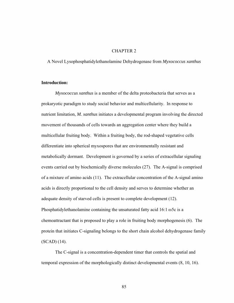
85
CHAPTER 2
A Novel Lysophosphatidylethanolamine Dehydrogenase from Myxococcus xanthus
Introduction:
Myxococcus xanthus is a member of the delta proteobacteria that serves as a
prokaryotic paradigm to study social behavior and multicellularity. In response to
nutrient limitation, M. xanthus initiates a developmental program involving the directed
movement of thousands of cells towards an aggregation center where they build a
multicellular fruiting body. Within a fruiting body, the rod-shaped vegetative cells
differentiate into spherical myxospores that are environmentally resistant and
metabolically dormant. Development is governed by a series of extracellular signaling
events carried out by biochemically diverse molecules (27). The A-signal is comprised
of a mixture of amino acids (11). The extracellular concentration of the A-signal amino
acids is directly proportional to the cell density and serves to determine whether an
adequate density of starved cells is present to complete development (12).
Phosphatidylethanolamine containing the unsaturated fatty acid 16:1 ω5c is a
chemoattractant that is proposed to play a role in fruiting body morphogenesis (6). The
protein that initiates C-signaling belongs to the short chain alcohol dehydrogenase family
(SCAD) (14).
The C-signal is a concentration-dependent timer that controls the spatial and
temporal expression of the morphologically distinct developmental events (8, 10, 16).
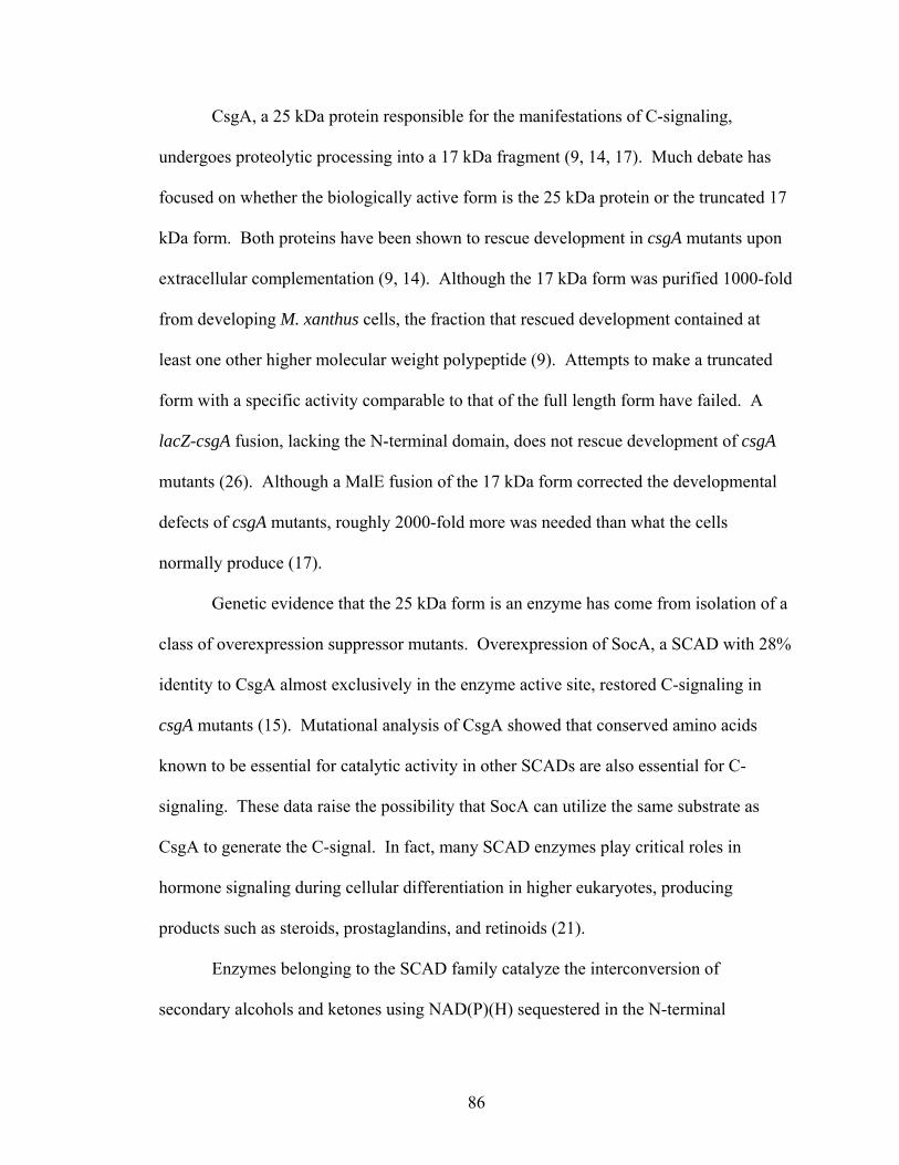
86
CsgA, a 25 kDa protein responsible for the manifestations of C-signaling,
undergoes proteolytic processing into a 17 kDa fragment (9, 14, 17). Much debate has
focused on whether the biologically active form is the 25 kDa protein or the truncated 17
kDa form. Both proteins have been shown to rescue development in csgA mutants upon
extracellular complementation (9, 14). Although the 17 kDa form was purified 1000-fold
from developing M. xanthus cells, the fraction that rescued development contained at
least one other higher molecular weight polypeptide (9). Attempts to make a truncated
form with a specific activity comparable to that of the full length form have failed. A
lacZ-csgA fusion, lacking the N-terminal domain, does not rescue development of csgA
mutants (26). Although a MalE fusion of the 17 kDa form corrected the developmental
defects of csgA mutants, roughly 2000-fold more was needed than what the cells
normally produce (17).
Genetic evidence that the 25 kDa form is an enzyme has come from isolation of a
class of overexpression suppressor mutants. Overexpression of SocA, a SCAD with 28%
identity to CsgA almost exclusively in the enzyme active site, restored C-signaling in
csgA mutants (15). Mutational analysis of CsgA showed that conserved amino acids
known to be essential for catalytic activity in other SCADs are also essential for C-
signaling. These data raise the possibility that SocA can utilize the same substrate as
CsgA to generate the C-signal. In fact, many SCAD enzymes play critical roles in
hormone signaling during cellular differentiation in higher eukaryotes, producing
products such as steroids, prostaglandins, and retinoids (21).
Enzymes belonging to the SCAD family catalyze the interconversion of
secondary alcohols and ketones using NAD(P)(H) sequestered in the N-terminal
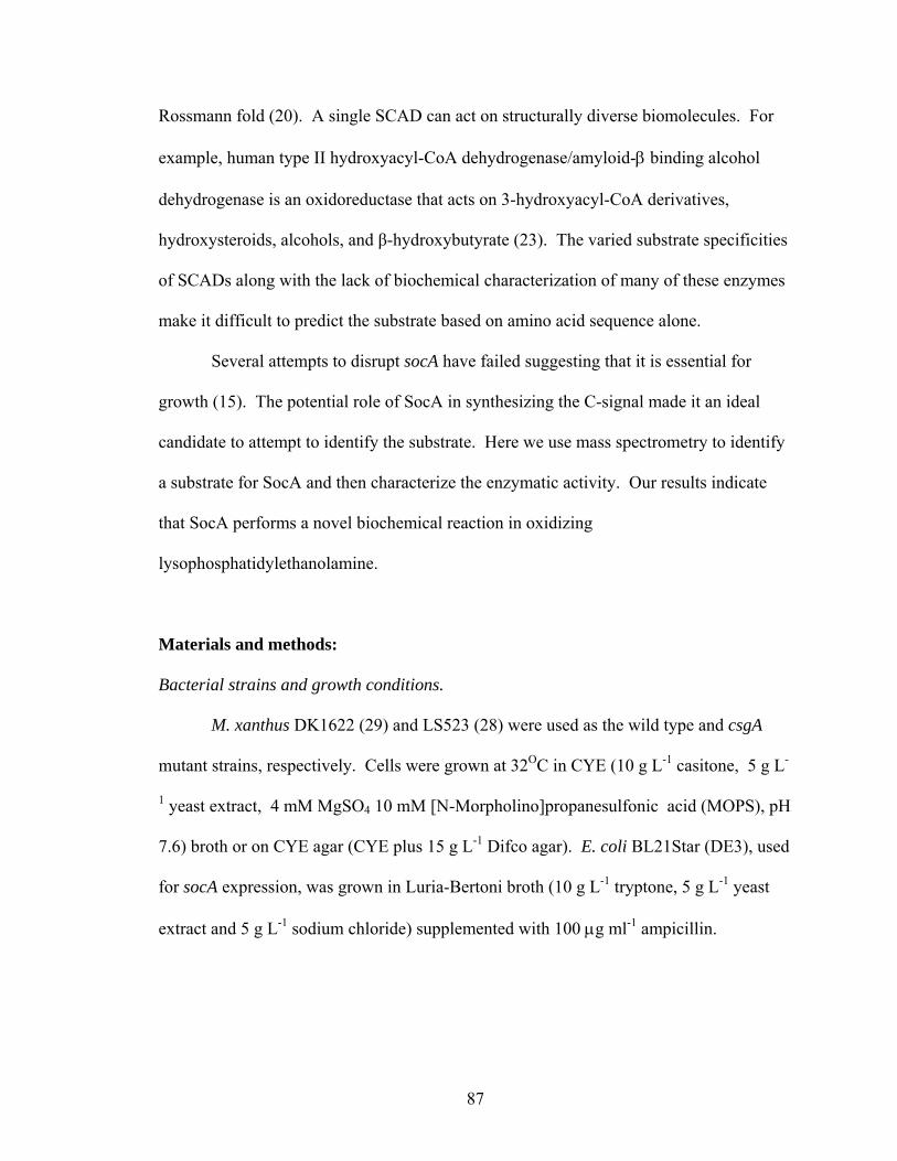
87
Rossmann fold (20). A single SCAD can act on structurally diverse biomolecules. For
example, human type II hydroxyacyl-CoA dehydrogenase/amyloid-β binding alcohol
dehydrogenase is an oxidoreductase that acts on 3-hydroxyacyl-CoA derivatives,
hydroxysteroids, alcohols, and β-hydroxybutyrate (23). The varied substrate specificities
of SCADs along with the lack of biochemical characterization of many of these enzymes
make it difficult to predict the substrate based on amino acid sequence alone.
Several attempts to disrupt socA have failed suggesting that it is essential for
growth (15). The potential role of SocA in synthesizing the C-signal made it an ideal
candidate to attempt to identify the substrate. Here we use mass spectrometry to identify
a substrate for SocA and then characterize the enzymatic activity. Our results indicate
that SocA performs a novel biochemical reaction in oxidizing
lysophosphatidylethanolamine.
Materials and methods:
Bacterial strains and growth conditions.
M. xanthus DK1622 (29) and LS523 (28) were used as the wild type and csgA
mutant strains, respectively. Cells were grown at 32OC in CYE (10 g L-1 casitone, 5 g L-
1 yeast extract, 4 mM MgSO4 10 mM [N-Morpholino]propanesulfonic acid (MOPS), pH
7.6) broth or on CYE agar (CYE plus 15 g L-1 Difco agar). E. coli BL21Star (DE3), used
for socA expression, was grown in Luria-Bertoni broth (10 g L-1 tryptone, 5 g L-1 yeast
extract and 5 g L-1 sodium chloride) supplemented with 100 µg ml-1 ampicillin.
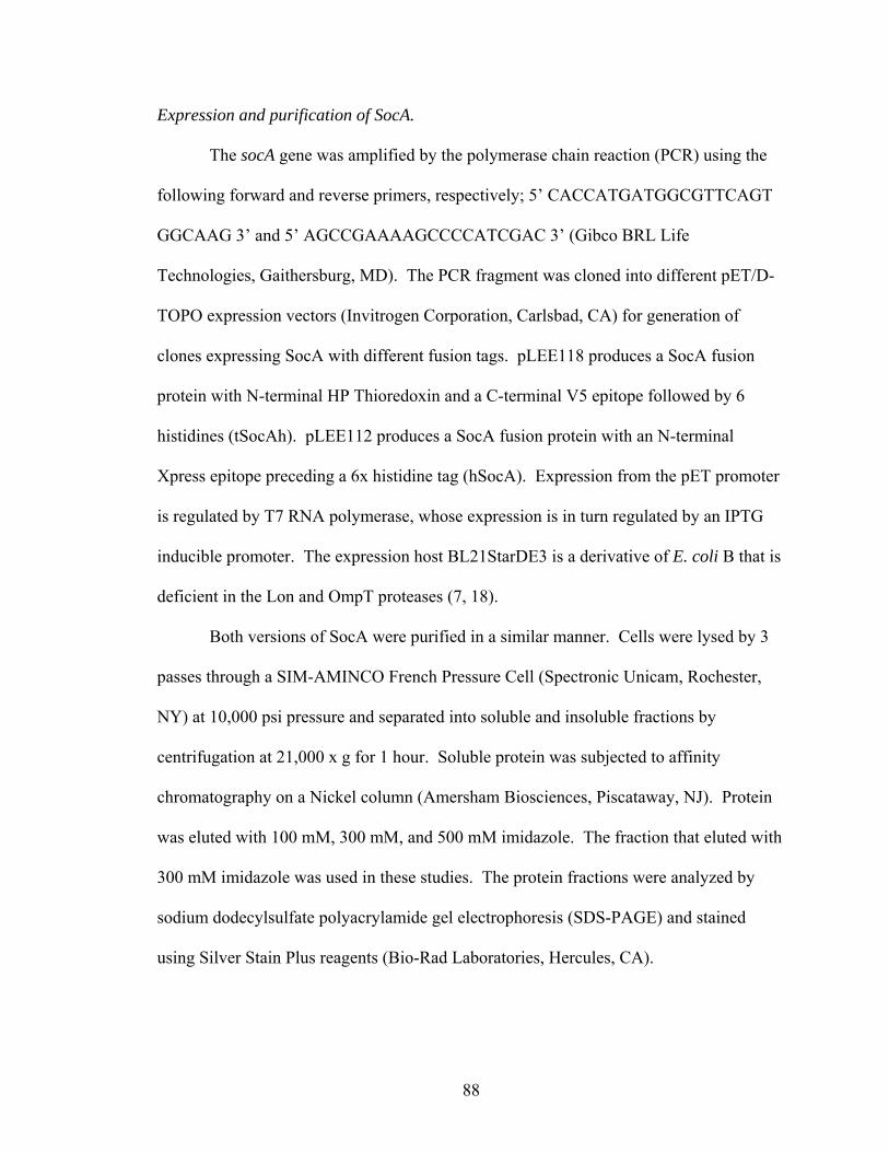
88
Expression and purification of SocA.
The socA gene was amplified by the polymerase chain reaction (PCR) using the
following forward and reverse primers, respectively; 5’ CACCATGATGGCGTTCAGT
GGCAAG 3’ and 5’ AGCCGAAAAGCCCCATCGAC 3’ (Gibco BRL Life
Technologies, Gaithersburg, MD). The PCR fragment was cloned into different pET/D-
TOPO expression vectors (Invitrogen Corporation, Carlsbad, CA) for generation of
clones expressing SocA with different fusion tags. pLEE118 produces a SocA fusion
protein with N-terminal HP Thioredoxin and a C-terminal V5 epitope followed by 6
histidines (tSocAh). pLEE112 produces a SocA fusion protein with an N-terminal
Xpress epitope preceding a 6x histidine tag (hSocA). Expression from the pET promoter
is regulated by T7 RNA polymerase, whose expression is in turn regulated by an IPTG
inducible promoter. The expression host BL21StarDE3 is a derivative of E. coli B that is
deficient in the Lon and OmpT proteases (7, 18).
Both versions of SocA were purified in a similar manner. Cells were lysed by 3
passes through a SIM-AMINCO French Pressure Cell (Spectronic Unicam, Rochester,
NY) at 10,000 psi pressure and separated into soluble and insoluble fractions by
centrifugation at 21,000 x g for 1 hour. Soluble protein was subjected to affinity
chromatography on a Nickel column (Amersham Biosciences, Piscataway, NJ). Protein
was eluted with 100 mM, 300 mM, and 500 mM imidazole. The fraction that eluted with
300 mM imidazole was used in these studies. The protein fractions were analyzed by
sodium dodecylsulfate polyacrylamide gel electrophoresis (SDS-PAGE) and stained
using Silver Stain Plus reagents (Bio-Rad Laboratories, Hercules, CA).
89
Protein concentration was determined using the biscinchoninic acid (BCA) assay
(Pierce Chemicals Co., Rockford, IL) with bovine serum albumin standards.
Dehydrogenase assay.
The ability of recombinant SocA to oxidize partially pure cellular extracts or
synthetic substrates was monitored at 340 nm using a spectrophotometer (DU 640B;
Beckman Instruments Inc., Fullerton, CA) at 25OC. The reaction mixture contained 100
mM sodium phosphate buffer, pH 6.8, 1 mM NAD+, and various amounts of substrate
and protein. The cellular extracts were solubilized in 100% methanol, and the final
concentration of methanol in the reaction mixture was maintained at less than 2.5 %. The
change in absorbance due to the production of NADH was monitored for at least 5
minutes or until the reaction plateaued. The specific activity was calculated from the
initial rates by using a molar extinction coefficient of 6220 M-1cm-1 for NADH at 340 nm.
Specific activity is defined as µmoles NADH formed per minute per mg protein. Specific
activity was routinely determined from three independent protein preparations ± the
standard deviation.
Synthetic lipids were purchased from Avanti Polar Lipids Inc. (Alabaster, Al).
All the other substrates and chemicals were purchased from Sigma Chemical Co. (St.
Louis, MO). Lysophosphatidylethanolamine (lysoPE) substrates with an unsaturated
fatty acid were solubilized in 100% methanol and diluted in the reaction mixture to a
final methanol concentration of 5%. Lysophospholipids with saturated fatty acids were
solubilized in chloroform:methanol:water (65:25:4), added to the assay mixture, and the
chloroform phase that separates from the aqueous assay medium was removed before

90
addition of the enzyme. Mixed micelles of the lysophospholipids and Trition X 100 were
prepared by equilibrating 2.5 mM lysophospholipid with 5 mM Triton X 100 in water at
room temperature overnight.
Preparation of 1-O-[11-(Z)-hexadecanoyl]-2-hydroxy-sn-glycero-3-
phosphoethanolamine.
1-O-[11-(Z)-hexadecanoyl]-2-hydroxy-sn-glycero-3-phosphoethanolamine
(lysoPE 16:1 ω5c) was generated by treating 1,2-O-Bis[11-(Z)-hexadecanoyl]-sn-
glycero-3-phosphoethanolamine (PE-16:1 ω5c/16:1 ω5c )[Courtesy of A. Venot and G. J.
Boons] with phospholipase A2. 600 µg PE-16:1 ω5c/16:1 ω5c was incubated with 20 µg
phospholipase A2 from Naja mossambica mossambica (Sigma Chemical Co., St Louis,
MO) in the presence of 10 mM CaCl2 and 200 mM Tris, pH 8.0 at 37OC for 2 hours. The
lipid was extracted with chloroform:methanol:acetic acid (50:50:1) and purified by thin
layer chromatography on silica gel 60 (20 x 20 cm; layer thickness 250 µm; EM Science,
Gibbstown, NJ ) with chloroform:methanol:water (65:25:4) as the mobile phase. The
same solvent was used for extraction of the lysoPE from the silica gel for subsequent use
in the dehydrogenase assay.
Extraction of oxidized lysophosphatidylethanolamine.
The end-product of lysoPE oxidation was extracted with chloroform for analysis
of its chemical structure and also to study the biological relevance of oxidized lysoPE. A
10 ml aqueous enzyme assay mixture was vortexed with 2 ml chloroform for 1 minute at
25OC and centrifuged at 10,000 x g for 1 minute to separate the chloroform phase from
the aqueous phase. The chloroform phase was dried under N2 and the extract was
91
resuspended in 100 µl methanol.
Extraction of substrate molecule(s) from DK1622.
DK1622 cells grown in 2.85 L CYE to stationary phase were harvested by
centrifugation at 10,000 x g for 10 minutes. The cell pellet was resuspended in 105 ml
MOPS buffer (10 mM MOPS, pH 7.2, 1 mM CaCl2, 4 mM MgCl2 and 50 mM NaCl).
Following 2 freeze thaw cycles (-20OC and room temperature for 4 hours each), the cell
suspension was subjected to sonication using ten 30 second pulses at 40 µW (W380, Heat
Systems Ultrasonics). Three extraction methods were compared: extraction with
acidified ethyl acetate (0.02% glacial acetic acid), extraction with ethyl acetate, and
extraction with methanol/chloroform.
For extraction with acidified ethyl acetate, a 35 ml cell lysate was extracted with
25 ml acidified ethyl acetate at 37OC for 30 minutes followed by centrifugation at room
temperature for 45 minutes at 10,000 x g to separate the phases. The aqueous phase was
reextracted with 25 ml acidified ethyl acetate, and the pooled ethyl acetate fractions were
dried under N2. The dried extract was dissolved in 1 ml methanol. Extraction with ethyl
acetate was the same except for the omission of glacial acetic acid.
Extraction with methanol/chloroform was performed using the method of Bligh
and Dyer as modified by Kearns and Shimkets (5). A 35 ml cell lysate was extracted
with 15 ml methanol:chloroform (2:1) at 37OC for 1 hour followed by centrifugation at
room temperature for 5 minutes at 10,000 x g to separate the aqueous phase from the
solvent phase. The pellet was reextracted with 5 ml methanol:chloroform:water (2:1:0.8,
v/v/v), vortexed, and centrifuged again. The chlorform phase was removed, dried under
N2, and dissolved in 1 ml methanol.
92
The amount of substrate in each sample was determined using the dehydrogenase
assay. The absorbance at 340 nm at the reaction plateau was used to calculate the amount
of substrate converted, assuming a 1:1 molar ratio with the amount of NADH produced.
Partial purification of the substrate.
The crude extract was fractionated on a 500 mg LCSi silica gel Sep-Pak column
(Supelco, Bellefonte, PA). The column was conditioned with 4 ml
hexane:methyltertiarybutylether (MTBE) (96:4) followed by 6 ml hexane. The sample
was loaded in hexane:MTBE (200:3). The column was then washed with solvents of
increasing polarity in the following order: 10 ml hexane:MTBE (200:3), 10 ml
hexane:MTBE (96:4), 10 ml hexane:acetic acid (100:0.2), 10 ml hexane:MTBE:acetic
acid (100:2:0.2), 10 ml MTBE:acetic acid (100:0.2), 6 ml MTBE:methanol:ammonium
acetate (25:4:1), 10 ml MTBE:methanol:ammonium acetate (10:4:1), 10 ml
MTBE:methanol:ammonium acetate (5:4:1), 10 ml MTBE:methanol:ammonium acetate
(5:8:2). The solvents were evaporated under N2 and the dry extract resuspended in 500
µl methanol. 20 µl of each of these fractions was used in the dehydrogenase assay to
quantify the amount of substrate recovered.
The active fraction from LCSi was subjected to reverse phase HPLC on a C18
column (3.9 x 150 mm; Waters, Milford, MA) equipped with a photodiode array detector.
Elution was carried out using a methanol:acetonitrile (65:35) gradient with 0.1%
triethylamine. Each fraction from the C18 column was assayed using the dehydrogenase
assay. The peak corresponding to the active fractions was further analyzed by
electrospray ionization mass spectrometry (ESIMS) in the negative mode.
93
Bioassay for developmental rescue.
A qualitative biological assay was employed to test the activity of the partially
pure extracts and the synthetic lipids. The partially pure extract was oxidized with
tSocAh and extracted by the modified method of Bligh and Dyer (5). Varying amounts
(250 nmol, 100 nmol, 50 nmol, 25 nmol, 10 nmol, 1 nmol) of the oxidized and the
reduced extracts (untreated with tSocAh) were dried on individual filter paper discs
(Whatman 1; 0.5 mm diameter).
csgA cells were grown to mid-log phase in CYE and harvested by centrifugation
at 10, 000 x g for 10 minutes at room temperature. The cell pellet was resuspended in
fresh CYE to a final density of 5 x 10⁹ ml-1. Development was induced by spreading 200
µl of the cell suspension on TPM agar plates (150 mm diameter) [10 mM Tris, pH 7.6, 1
mM KH2PO4, and 10 mM MgSO4, 1.5% (Difco) agar]. Plates were incubated at 32OC.
Whatman filter paper discs containing various amounts of the oxidized and reduced
extracts were placed on the csgA cell lawn 6 hours later. Rescue of developmental
aggregation and sporulation was monitored over the next 5 days using a dissection
microscope.
LysoPE 16:0 and lysoPE 16:1 were oxidized by SocA, and varying amounts (100
µg, 50 µg, 25 µg, 10 µg, 5 µg, and 1 µg) of the oxidized and reduced forms of lysoPE
16:0 and lysoPE 16:1 were tested for their ability to rescue the developmental defects of
csgA mutants.
94
Extraction and quantification of lysophosphatidylethanolamine.
M. xanthus cells in CYE were harvested at a density of 5 x 10⁹ ml-1 by
centrifugation at 10,000 x g for 10 minutes, resuspended in fresh CYE, and spread on
TPM agar at a final density of 2.8 x 10⁶cells (cm2)-1. After 24 hours at 32OC, the cells
were removed from the agar and used as the source of developmental lysophospholipids.
Vegetative M. xanthus cells grown in CYE to a density of 108 ml-1 were used as the
source of vegetative lysophospholipids. The lysophospholipids were extracted in
methanol:chloroform (5) and subjected to ESIMS. Synthetic lysoPE 16:0 and lysoPE
18:1 were used as standards for quantification of the saturated and unsaturated lysoPE,
respectively.
Thin layer chromatography.
Oxidized lysoPE 16:0 was subjected to thin layer chromatography (TLC) on silica
gel 60 (20 x 20 cm; layer thickness 250 µm; EM Science, Gibbstown, NJ) using
chloroform:methanol:water (65:25:4) as the mobile phase. The TLC plates were stained
with iodine vapors to observe molecules containing fatty acids. The TLC plates were
stained with ninhydrin (0.5% ninhydrin in 3% acetic acid in 1-butanol) and incubated at
100O C for 3 minutes to observe molecules containing amine group.
The partially pure extract from the silica column was further purified by thin layer
chromatography under the conditions mentioned above. Fifteen different fractions (1 cm
tall x 3.5 cm wide), extending from the origin to the solvent front were extracted from the
silica gel with 500 µl methanol. The silica gel and methanol mixture was vortexed for 15
seconds followed by centrifugation at 14,000 x g for 1 minute to separate the methanolic
95
supernatant, which was used as the source of substrate in the enzyme assay. The active
fractions were subjected to mass spectrometry.
Nuclear magnetic resonance (NMR) spectroscopy of oxidized and reduced lysoPE 16:0.
LysoPE (reduced and oxidized) was dissolved in deuterated chloroform:methanol
(2:1). 1-D Proton and 2-D gradient COSY spectra were recorded on a Varian Inova 500
MHz instrument at 25OC.
Gas chromatography-Mass spectrometric (GC/MS) analysis of oxidized lysoPE 16:0.
Oxidized lysoPE 16:0 was pertrimethylsilylated by treatment with Tri-Sil (Pierce
Chemical Co, Rockford, IL) at 80OC for 20 minutes. Trimethylsilyalate (73 amu) reacts
with the hydroxyl groups on the analyte enabling identification of the molecules based on
a shift in the molecular mass. The analysis was performed by chemical ionization (CI) -
GC/MS using an Agilent 6890N GC (Supelco, Bellefonte, PA; DB-1, 30 m × 0.25 mm
ID) interfaced with a 5973 MSD with ammonia as ionizing gas.
Matrix assisted laser desorption ionization (MALDI) Mass spectrometry of oxidized
lysoPE 16:0.
Oxidized lysoPE 16:0 was added to sinapinic acid matrix prepared in
acetronitrile:water (50:50) with 0.1% trifluoroaceticacid and dried before analysis. A
Bruker Autoflex mass spectrometer with a nitrogen laser was used to acquire the data.
The analysis was also repeated in the presence of cesium chloride, which forms an adduct
with the PE headgroup shifting the molecular mass of the analyte by 132 amu.
96
Results:
Purification of SocA:
SocA was overproduced in E. coli with an N- or C-terminal histidine (6x) fusion
for enzymatic characterization. tSocAh and hSocA were purified on a nickel affinity
column 8-fold and 5-fold respectively, to apparent homogeneity (Figure 2.1). The yields
of tSocAh and hSocA were 24 mg L-1 and 9 mg L-1 respectively. Although, both tSocAh
and hSocA had comparable specific activities, tSocAh was used in all the enzyme studies
due to higher yield of purified protein.
Purification of SocA substrate:
The M. xanthus cell lysate was subjected to three different organic extractions to
determine the most efficient extraction method. The amount of substrate in the solvent
was quantified using a spectrophotometric assay in which NAD+ is reduced to NADH by
tSocAh, with the assumption that one molecule of substrate is oxidized for each molecule
of NAD+ reduced. The acidified ethyl acetate extraction gave the highest yield, 4.1
µmoles 1010 cells-1. Extraction with ethyl acetate yielded 3.2 µmoles 1010 cells-1 (78% of
that of the acidified ethyl acetate), and a modified Bligh and Dyer extraction yielded 1.1
moles 1010 cells-1 (26%). Acidified ethyl acetate was used for all subsequent extractions.
The acidified ethyl acetate extract was applied to a solid phase LCSi silica gel
column and eluted stepwise with solvents of increasing polarity. Each fraction was
assayed for dehydrogenase activity and the substrate was quantified based on the final
yield of NADH. The substrate eluted with MTBE:acetic acid (100:0.2) in a single
fraction of intermediate polarity, with 83% recovery of the original activity.
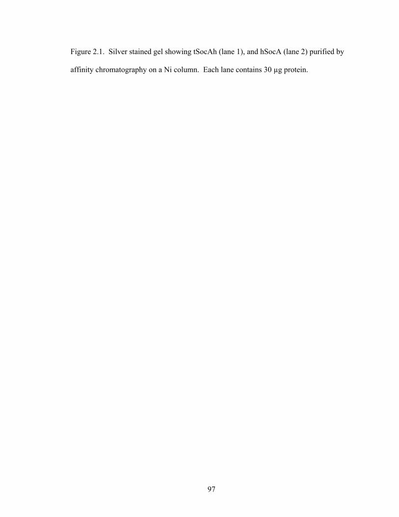
97
Figure 2.1. Silver stained gel showing tSocAh (lane 1), and hSocA (lane 2) purified by
affinity chromatography on a Ni column. Each lane contains 30 µg protein.
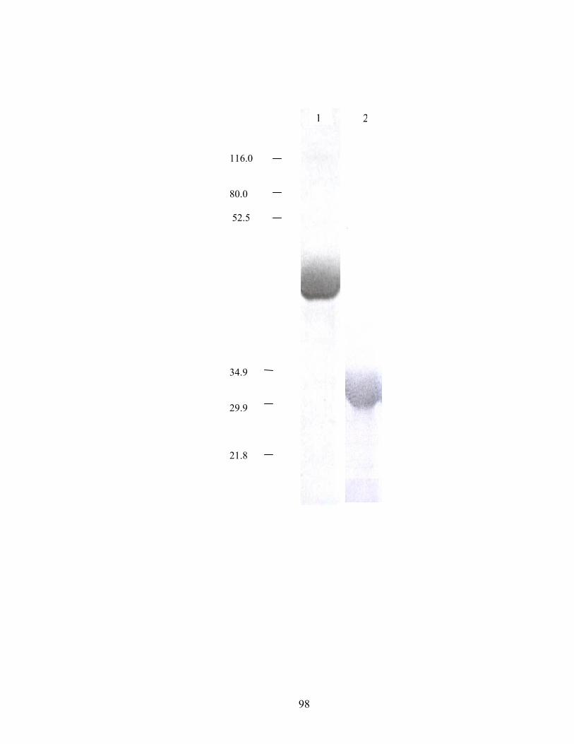
98
1 21 116.0 80.0 52.5 34.9 29.9 21.8
99
The partially pure extract from LCSi was subjected to reverse phase HPLC on a
C18 column, and the eluted fractions were used in the dehydrogenase assay. The
substrate had a retention time of 19-21 minutes (Figure 2.2A). There are at least two
sharp DAD spectral peaks that correspond to the broad substrate peak of SocA as
determined by the dehydrogenase assay (Figure 2.2B). One possible explanation is the
presence of multiple substrates. Attempts to devise another purification step with solid
phase separation on StrataX (Phenomenx, Torrance, CA) did not improve the
purification.
Identification of the SocA substrate.
Mass spectrometry of the active fractions was used to determine the masses of
compounds in the range of 140-500 amu (Figure 2.3). The most striking correlation was
with 1- acyl 2-hydroxy-sn-glycerophosphoethanolamine (lysoPE) containing a C16:1
fatty acid (m/z 450.7 amu). Previously it was found that 16:1 ω5c is one of the most
abundant fatty acids in M. xanthus (6).
SocA is a lysoPE dehydrogenase.
Since lysoPE 16:1 ω5c is not commercially available, biochemical analysis began
with lysoPE 16:0, which has a saturated fatty acid of identical length. Vesicles were
prepared by addition of chloroform:methanol:water (65:25:4)-solubilized lipid to the
aqueous assay mixture. The specific activities of tSocAh and hSocA with 100 µM
lysoPE 16:0 were 558 ± 69 and 461 ± 96 µmoles min-1 mg-1 respectively.
100
Figure 2.2. Total Wavelength Chromatogram of separation on C18 reverse phase
column. The fractions with a SocA substrate (retention time of 19-21 minutes) are
highlighted with vertical bars (A). Enzyme activity profile of corresponding fractions
using the dehydrogenase assay (B).
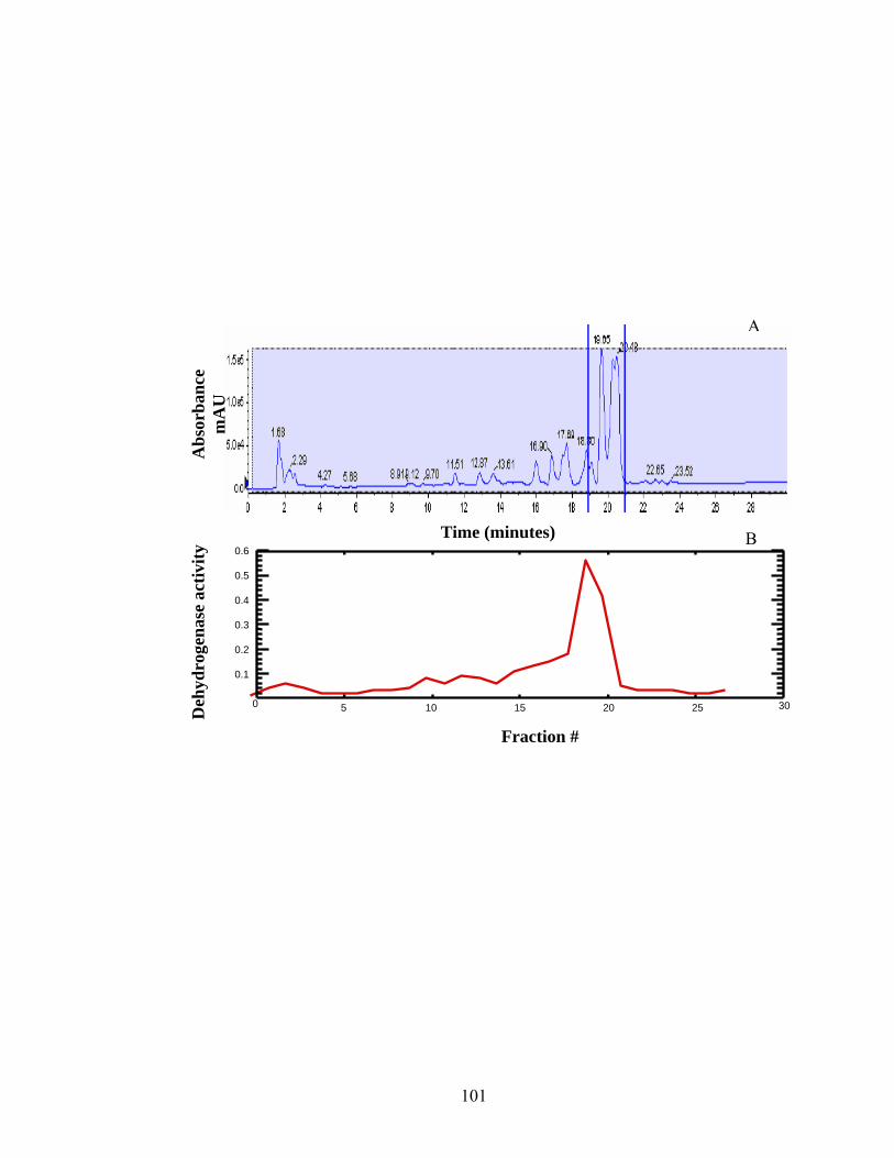
101
0.1
0.2
0.3
0.4
0.5
0.6
0 5 10 15 20 25 30
Time (minutes) B
Deh
ydro
gena
se a
ctiv
ity
Fraction #
A
Abs
orba
nce
mA
U
102
Figure 2.3. Mass spectrum of the fraction with highest enzyme activity (19-21 min).
Intensity of individual masses plotted against respective m/z. The m/z indicated with a
red arrow corresponds to the mass of lysoPE 16:1 in the negative mode.
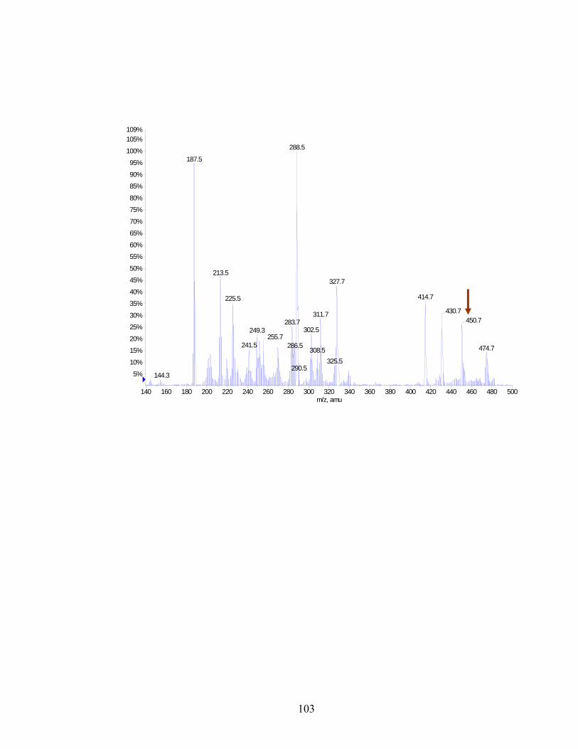
103
140 160 180 200 220 240 260 280 300 320 340 360 380 400 420 440 460 480 500m/z, amu
5%
10%
15%
20%
25%
30%
35%
40%
45%
50%
55%
60%
65%
70%
75%
80%
85%
90%
95%
100%
105%109%
288.5
187.5
414.7
327.7
311.7
213.5
302.5
225.5
430.7
290.5
450.7283.7
308.5
249.3255.7
241.5 286.5
325.5
474.7
144.3
104
SocA oxidized lysoPE16:0 over a greater than 100-fold pH range with maximal
activity at pH 7.0 (Figure 2.4). SocA utilized both NAD+ and NADP+ with comparable
efficiency (558 ± 69 and 652 ± 59 µmol min-1 mg-1, respectively).
Substrate specificity and kinetics.
The dehydrogenase assay was used to oxidize various synthetic lysoPE species of
varying acyl chain length and saturation in order to determine the fatty acid specificity of
SocA (Table 2.1). Although SocA oxidizes lysoPE 16:0 with highest efficiency, it
oxidizes other lysoPE substrates with less than half-fold difference in specific activity.
However, a sn-1 fatty acid appears to be essential for oxidation as SocA was unable to
oxidize glycerophosphorylethanolamine. Although, lysophospholipids may be acylated
at either sn-1 or sn-2, the acyl chain in 1-hydroxy 2-acyl lysophospholipid rapidly
migrates to sn-1 resulting in the formation of 1-acyl 2-hydroxy lysophospholipid (22).
Hence this was the only form of the molecule available for examination. Because it was
more soluble in methanol, initial rates were analyzed with various concentrations of
lysoPE 18:1. Initial rates were fitted to the Michaelis-Menten equation, V = Vmax[S]/[S]
+ Km using non-linear regression [SigmaPlot 8.0; SPSS Inc.] (Figure 2.5). V is the
velocity of the reaction, Vmax is the maximal rate, [S] is the concentration of substrate and
Km is the Michaelis constant. The apparent Km and Vmax were determined to be 116 µM
and 875 µmol min-1 mg-1, respectively. The catalytic efficiency (Kcat/Km) was calculated
to be 1 x 10⁸ (moles/L)-1sec-1. The range of concentrations tested was limited by the
solubility of lysoPE 18:1 in methanol. Methanol inhibited protein activity at higher
concentrations, and the detection limits of the spectrophotometer did
105
Figure 2.4. Effect of pH on lysoPE 16:0 oxidation by SocA. Enzyme was assayed with
100 µM lysoPE 16:0 in sodium phosphate buffer.
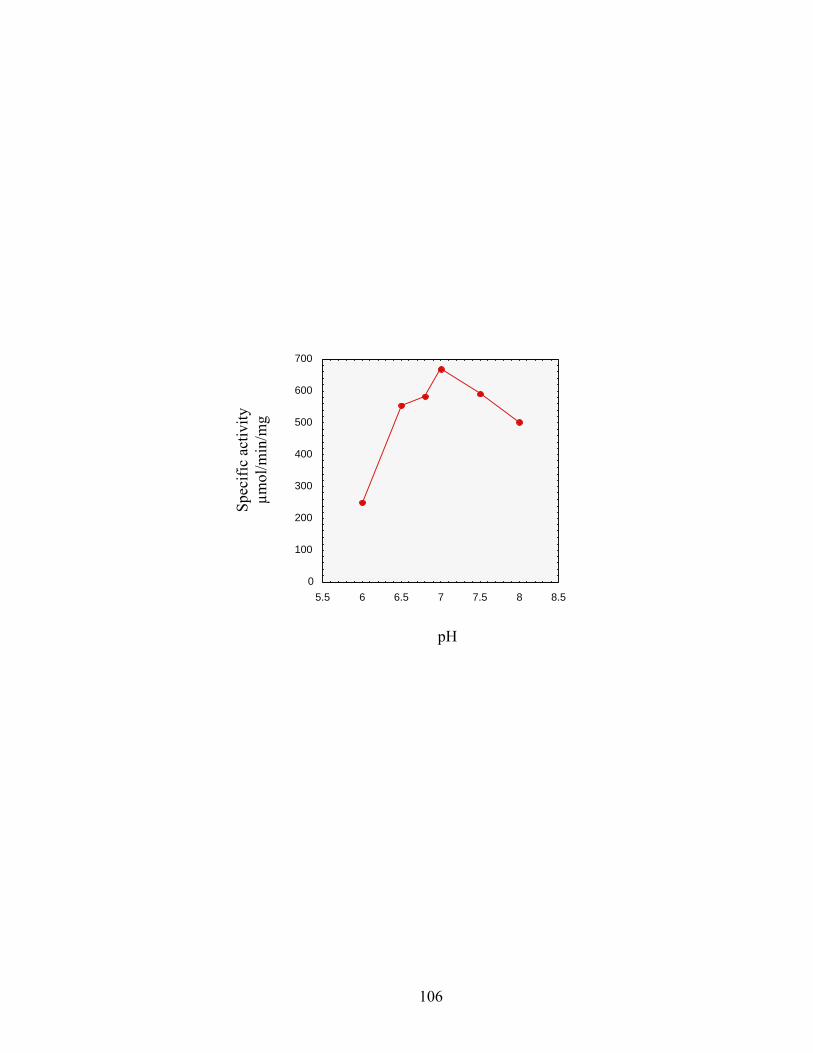
106
0
100
200
300
400
500
600
700
5.5 6 6.5 7 7.5 8 8.5
Spec
ific
activ
ity
µmol
/min
/mg
pH
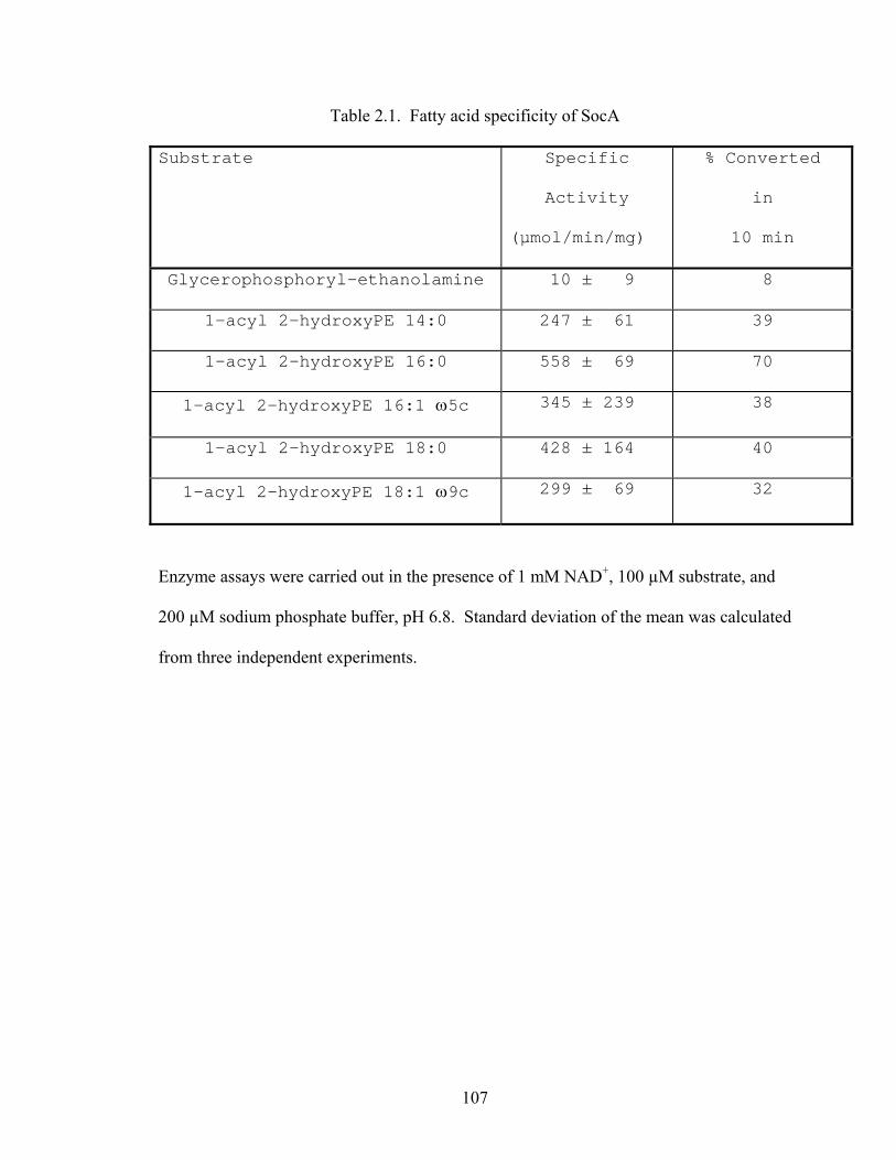
107
Table 2.1. Fatty acid specificity of SocA
Substrate Specific
Activity
(µmol/min/mg)
% Converted
in
10 min
Glycerophosphoryl-ethanolamine 10 ± 9 8
1-acyl 2-hydroxyPE 14:0 247 ± 61 39
1-acyl 2-hydroxyPE 16:0 558 ± 69 70
1-acyl 2-hydroxyPE 16:1 ω5c 345 ± 239 38
1-acyl 2-hydroxyPE 18:0 428 ± 164 40
1-acyl 2-hydroxyPE 18:1 ω9c 299 ± 69 32
Enzyme assays were carried out in the presence of 1 mM NAD+, 100 µM substrate, and
200 µM sodium phosphate buffer, pH 6.8. Standard deviation of the mean was calculated
from three independent experiments.
108
Figure 2.5. Double reciprocal plot of initial velocity versus lysoPE 18:1 concentrations.
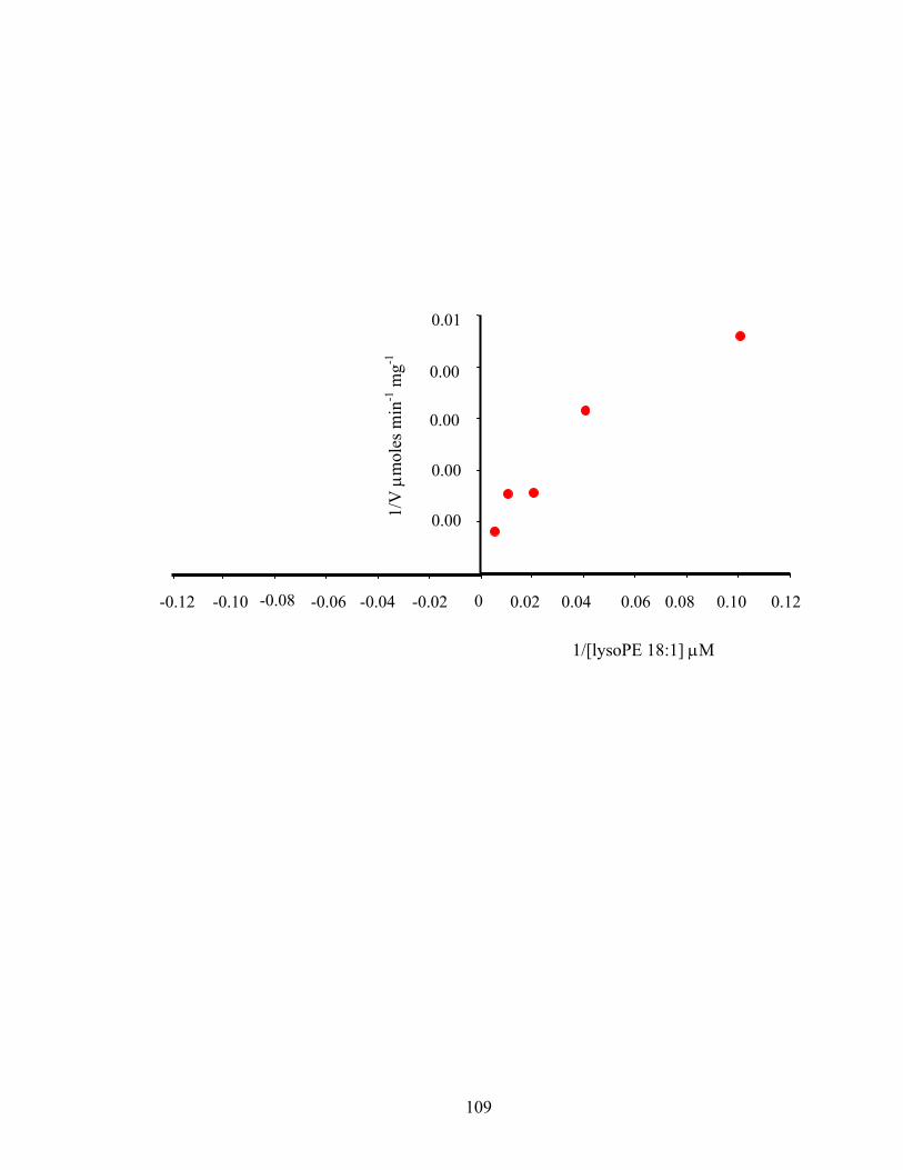
109
0
0.00
0.00
0.00
0.00
0.01
1/V
µm
oles
min
-1 m
g-1
1/[lysoPE 18:1] µM
-0.02 -0.04 -0.06 -0.10 -0.08 -0.12 0.02 0.04 0.06 0.08 0.10 0.12
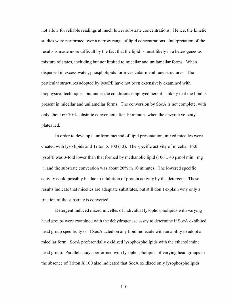
110
not allow for reliable readings at much lower substrate concentrations. Hence, the kinetic
studies were performed over a narrow range of lipid concentrations. Interpretation of the
results is made more difficult by the fact that the lipid is most likely in a heterogeneous
mixture of states, including but not limited to micellar and unilamellar forms. When
dispersed in excess water, phospholipids form vesicular membrane structures. The
particular structures adopted by lysoPE have not been extensively examined with
biophysical techniques, but under the conditions employed here it is likely that the lipid is
present in micellar and unilamellar forms. The conversion by SocA is not complete, with
only about 60-70% substrate conversion after 10 minutes when the enzyme velocity
plateaued.
In order to develop a uniform method of lipid presentation, mixed micelles were
created with lyso lipids and Triton X 100 (13). The specific activity of micellar 16:0
lysoPE was 3-fold lower than that formed by methanolic lipid (106 ± 43 µmol min-1 mg-
1), and the substrate conversion was about 20% in 10 minutes. The lowered specific
activity could possibly be due to inhibition of protein activity by the detergent. These
results indicate that micelles are adequate substrates, but still don’t explain why only a
fraction of the substrate is converted.
Detergent induced mixed micelles of individual lysophospholipids with varying
head groups were examined with the dehydrogenase assay to determine if SocA exhibited
head group specificity or if SocA acted on any lipid molecule with an ability to adopt a
micellar form. SocA preferentially oxidized lysophospholipids with the ethanolamine
head group. Parallel assays performed with lysophospholipids of varying head groups in
the absence of Triton X 100 also indicated that SocA oxidized only lysophospholipids
111
Table 2.2. Head group specificity of SocA
Substrate Specific
Activitya
(µmol/min/mg)
Specific
Activityb
(µmol/min/mg)
1-acyl 2-hydroxyPE 16:0 558 ± 69 106 ± 43
1-acyl 2-hydroxyPC 16:0 9 ± 4 3 ± 7.5
1-acyl 2-hydroxyPS 18:1 < 0.01 < 0.01
1-acyl 2-hydroxyPG 16:0 < 0.01 < 0.01
1-acyl 2-hydroxyPA 16:0 5 ± 8 < 0.01
Enzyme assays were carried out in the presence of 1 mM NAD+, 100 µM substrate, and
200 µM sodium phosphate buffer pH 6.8. Standard deviation of the mean was calculated
from three independent experiments. PE- Phosphatidylethanolamine; PC-
Phosphatidylcholine; PS- Phosphatidylserine; PG- Phosphatidylglycerol; PA-
Phosphatidic acid. aSpecific activity determined in the absence of Triton X. bSpecific
activity determined in the presence of Triton X 100.
112
with the ethanolamine head group (Table 2.2).
Analysis of the oxidized lysoPE 16:0.
The proposed product of lysoPE oxidation is 1-acyloxy-3-(2-
aminoethylphosphatyl) acetone. The NMR spectrum of the oxidation product indicated
the absence of the phosphoethanolamine moiety. The dihydroxyacetone core of this
molecule has the ability to rearrange via keto-enol tautomerism to glyceraldehyde, with
the concomitant loss of both the phosphoethanolamine and the fatty acyl groups (Figure
2.6). Furthermore, the oxidized molecule could also be susceptible to potential acidic
hydrolysis at the ester linkages, resulting in formation of glycerophosphorylethanolamine
and a free fatty acid or monoacylglycerol phosphate and ethanolamine. Consistently,
NMR spectroscopy and mass spectrometry indicated that oxidized lysoPE16:0 underwent
further degradation (data not shown). TLC analysis of the oxidized lysoPE 16:0 showed
two iodine stained spots with Rf values 0.35 and 0.93, indicating hydrolysis of the product
(Figure 2.7A). The Rf value of 0.93 corresponds to that of the free fatty acid and
monopalmitoylglycerol (data not shown) adding further evidence to the proposed
breakdown of oxidized lysoPE. The identity of the iodine stained spot with Rf value 0.35
remains unknown. Ninhydrin staining revealed that neither product contained the
ethanolamine group (Figure 2.7B). These results argue that the oxidized product is
unstable and fragments into multiple products.
The oxidation product of lysoPE 16:0 was reduced by SocA in an NADH-
dependent manner, with a specific activity of 733 ± 247 µmoles min-1 mg-1 suggesting
that SocA reduces one or more products of hydrolysis. The possible substrates include 1-
113
Figure 2.6. Proposed keto-enol tautomerism and subsequent hydrolysis of oxidized
lysoPE. Keto form of oxidized lysoPE (6A) is converted to its tautomeric enol form
(6B). The enol form is unstable in an aqueous environment and is hydrolyzed into 1-
acylglyceraldehyde and phosphoethanolamine (6C). 1-acylglyceraldehyde rearranges
into 1-acyl-2-keto glycerol (6D) which isomerizes into the enol form, 1-acylglycerol
(6E). 1-acylglycerol subsequently hydrolyzes into free fatty acid and glyceraldehyde
(6F).
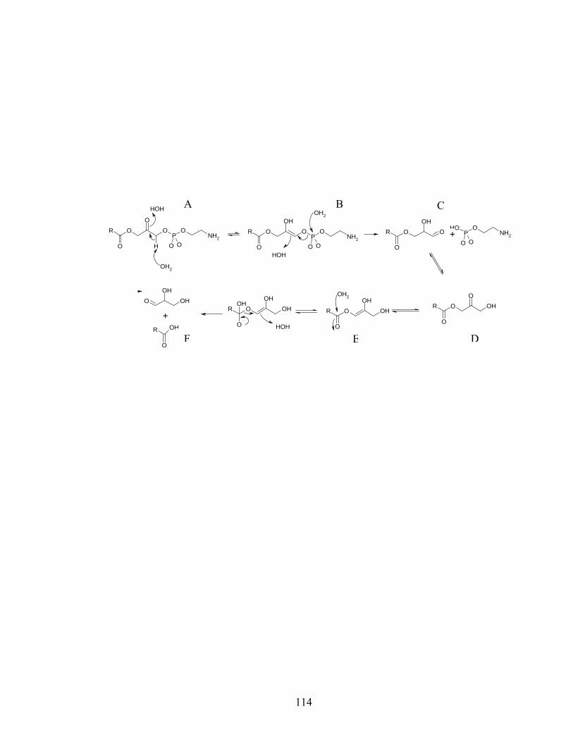
114
O OO
O
RP
O
O ONH2
H
O OOH
O
RP
O
O ONH2
HOH
OH2
OH2
HOH
O OOH
O
R PO
O ONH2
OH
O OHO
O
RO OH
OH
O
R
OH2
O OHOH
O
ROH
HOH
O OHOH
O
R OH
+
+
DEF
A B C
115
Figure 2.7. Thin layer chromatography of lysoPE 16:0 (lane 1) and the enzymatically
oxidized lysoPE 16:0 (lane 2) stained with iodine (A) and ninhydrin (B). The reduced form stains with both iodine and ninhydrin while the oxidized form stains only with iodine.
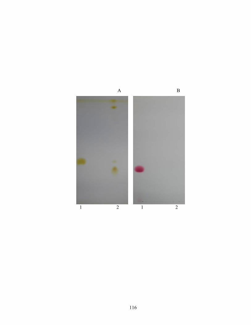
116
1 2 1 2
A B
117
acylglyceraldehyde, 1-acyl-2-keto glycerol, and glyceraldehyde. Since SocA did not
reduce the commercially available glyceraldehyde, the likely substrate for reduction is 1-
acyl-2-keto glycerol.
Bioassay for oxidized lysoPE 16:0 activity:
Bioassay involving the extracellular complementation of both the reduced and
enzymatically oxidized forms of lysoPE 16:0 and lysoPE 16:1 was performed to
determine the development rescue activity of these molecules. Both oxidized and
reduced lysoPE 16:0 and lysoPE 16:1 did not correct the developmental defects of csgA
mutants (data not shown).
SocA acts on multiple substrates.
The various lysoPE species were quantified during growth and development of
wild-type M. xanthus cells (Table 2.3). The levels of lysoPE 16:1 and lysoPE 18:0 show
a striking increase of 40-fold and 300-fold respectively during development.
Quantification after partial purification on the LCSi column, using the dehydrogenase
assay revealed the total number of substrate molecules per cell to be about 5 x 10⁷. This
is about 5 orders of magnitude higher than the total number of lysoPE 16:1 molecules
present per cell, providing evidence for the presence of multiple SocA substrates.
The biologically active molecule is not likely to be a lysoPE. TLC was used to
purify the biologically active substrate under conditions where all lysoPE species have a
similar mobility, regardless of the type of associated fatty acid. LysoPE 16:0 identified
by ninhydrin staining had an Rf value of 0.30, while the enzymatically active fractions
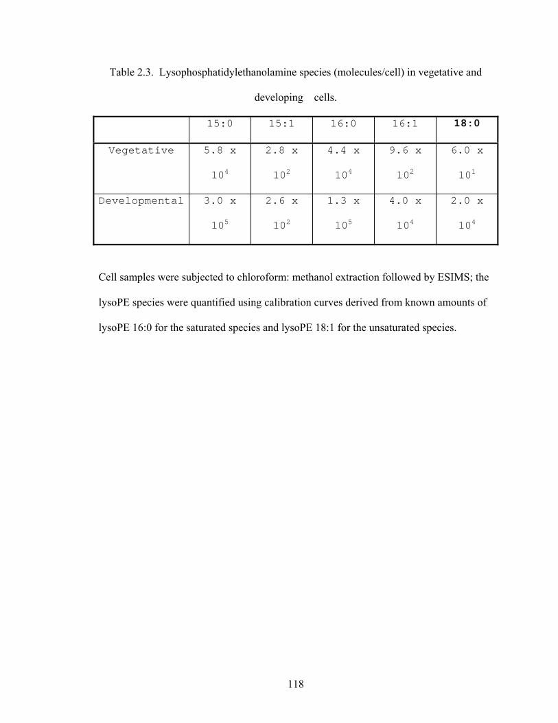
118
Table 2.3. Lysophosphatidylethanolamine species (molecules/cell) in vegetative and
developing cells.
15:0 15:1 16:0 16:1 18:0
Vegetative 5.8 x
104
2.8 x
102
4.4 x
104
9.6 x
102
6.0 x
101
Developmental 3.0 x
105
2.6 x
102
1.3 x
105
4.0 x
104
2.0 x
104
Cell samples were subjected to chloroform: methanol extraction followed by ESIMS; the
lysoPE species were quantified using calibration curves derived from known amounts of
lysoPE 16:0 for the saturated species and lysoPE 18:1 for the unsaturated species.
119
Figure 2.8. Rescue of developmental aggregation by the TLC fraction oxidized by SocA.
A single fruiting body (indicated by a box) is shown along the edge of the filter paper
disc. Bar is 50 µm.
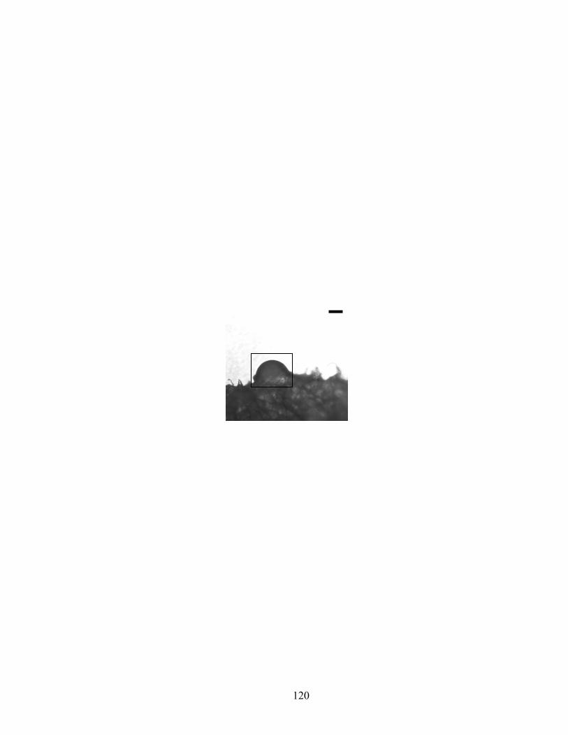
120
121
resolved at Rf 1.00, 0.93 and 0.80. Furthermore, 250 nmoles of SocA-oxidized fraction
with an Rf 1.00, induced aggregation of csgA mutants along the edge of the filter paper
disc (Figure 2.8). These results suggest that SocA oxidizes molecules other than lysoPE
in the extract.
Discussion:
SocA is the first lysophosphatidylethanolamine dehydrogenase to be described.
This enzyme acts on lysophospholipid substrates with fatty acids of different chain length
and saturation, showing preference for lysophospholipids with an ethanolamine
headgroup. The hydroxyl group at C2 on the glycerol backbone is proposed to be
oxidized to the corresponding keto form. The efficiency of catalysis is 9 x 10⁷ (moles/L)-
1 sec-1 and is comparable to other well characterized enzymes.
SocA has most of the sequence motifs that define the NADP(H) dependent
classical SCADs (4). An aspargine residue found at the end of β4 in classical SCADs is
absent in SocA. Also, SocA has a glutamate residue in place of the conserved aspartate,
13 positions downstream from the catalytic lysine. NAD(H)-binding SCADs have an
aspartate in β2 that repels the negatively charged phosphate group on NADP(H) and
hence confers NAD+ specificity to such enzymes by preferential exclusion of NADP+.
SocA lacks the crucial aspartate in β2 and is predicted to utilize NAD+ or NADP+. SocA
was able to utilize either coenzyme with comparable efficiency suggesting dual
coenzyme specificity.
Lysophospholipids are generated by phospholipases and transacylases during
phospholipid turnover in the cell membrane. E. coli phospholipase A, which is activated
122
upon damage to the cell membrane, hydrolyses the acyl chain at either sn-1 or sn-2 on the
glycerol backbone generating 1-hydroxy 2-acyl glycerophosphoethanolamine or 1-acyl 2-
hydroxy glycerophosphoethanolamine and a free fatty acid (30). Transacylases transfer
the acyl chain from sn-1 of phosphatidylethanolamine, phosphatidylglycerol, or
cardiolipin to lipoproteins generating the corresponding lysophosphatidylethanolamine,
lysophosphatidylglycerol, or lysophosphatidylcardiolipin, respectively. In addition,
palmitoyl transferase transfers the palmitate from sn-1 of a glycerophospholipid to the R-
3-hydroxymyristoyl moiety during lipopolysaccharide biosynthesis. Regardless of the
site of hydrolysis, the acyl chain migrates from sn-2 to sn-1 to generate 1-acyl 2-
hydroxyglycerophosphoethanolamine (22).
The M. xanthus genome encodes putative lipases and transacylases that could
potentially be involved in substrate generation for SocA (data not shown). At least nine
of these are acyl transferases and seven are phospholipases. The phospholipases include
five members of patatin-like (2), (25). These ORFs however have not been characterized.
Lysophospholipids serve as extracellular signaling molecules in mammals. In
their capacity as autocrine or paracrine signals, they mediate differentiation of cells of the
immune system as well as those involved in neurogenesis, angiogenesis, or
carcinogenesis. For example, lysophosphatidic acid (LPA) stimulates ionic conductance
changes in cortical neuroblasts during brain development (1). LPA is also produced by
autotaxin, a metastasis-promoting factor, suggesting a potential role for LPA in
carcinogenesis (19). Lysophospholipids exert their influence through specific G protein-
coupled receptors (3). The binding of the ligand to these receptors results in GDP/GTP
exchange on intracellular GTP-binding proteins, which then transmit the signal to
123
proteins involved in second messenger generation (24). While their role as mediators of
signal transduction has been studied extensively in eukaryotes, a similar role for
lysophospholipids in a bacterial system has not been reported.
Given a potential role for SocA in intercellular signaling during development of
M. xanthus, and the intercellular signaling by lysophospholipids in mammals, it is
tempting to speculate that SocA mediates lysoPE signaling in M. xanthus. Furthermore,
lysoPE shows a striking increase during development. However, SocA appears to be
substrate-limited as the physiological concentration of lysoPE is much lower than the
apparent Km of SocA, questioning the physiological relevance of lysoPE oxidation by
SocA. The discrepancy between the lysoPE concentrations and Km of SocA could be
explained if the membrane-bound lysoPE substrate is sequestered by the enzyme. At this
time, however, neither the distribution of lysoPE in the membrane nor the localization of
SocA is known. In addition, extracellular complementation of both reduced and
enzymatically oxidized lysoPE 16:0 and lysoPE 16:1 did not rescue the developmental
defects of the csgA mutant.
Based on the data discussed above, it is not likely that oxidized lysoPE has a role
in signaling during multicellular development of M. xanthus leading one to question the
biological significance of oxidized lysoPE. Understanding the complex pattern of socA
expression may illuminate the biological significance of SocA-mediated lysoPE
oxidation. Lee and Shimkets (15) found that socA was expressed at high levels in
vegetative cells during growth, and as development progressed, the expression levels
steadily dropped in the rod-shaped cells. Conversely, socA expression increased in
spores. This suggests that SocA may function during vegetative growth and also during
124
sporulation or spore germination. SocA could be part of the predatory machinery of M.
xanthus enabling prey cell degradation and fatty acid catabolism. SocA also has a
potential role in cell membrane remodeling during M. xanthus spore morphogenesis,
which involves extensive phospholipid turnover (Shimkets, unpublished data). These
possibilities appear to be attractive venues for exploring the significance of SocA-
mediated lysoPE oxidation in future experiments.
socA appears to be essential for M. xanthus as numerous attempts to delete it have
been unsuccessful. It is not clear that oxidized lysoPE has an essential function. Several
lines of evidence support the possibility of multiple SocA substrates. First, the partially
pure substrate extract that was oxidized by SocA, stimulated aggregation, while the
corresponding reduced substrate stimulated sporulation of csgA mutants. These
experiments seem to suggest that SocA controls different aspects of development by
acting on different substrates. Second, partial purification of the crude extract containing
the SocA substrate(s) yields about 5 x 10⁷ molecules per cell. This is about five orders of
magnitude higher than the number of lysoPE 16:1 molecules present per cell, suggesting
the existence of more than one SocA substrate. Finally, the active molecules from the
partially pure substrate extract migrate with Rf values of 1.00, 0.93 and 0.80, while
lysoPE migrates with an Rf value of 0.30 under the same conditions. Thus, SocA appears
to be acting on multiple substrates, which is not unusual for members of this family of
enzymes.
.
125
Reference:
1. Dubin, A. E., T. Bahnson, J. A. Weiner, N. Fukushima, and J. Chun. 1999.
Lysophosphatidic acid stimulates neurotransmitter-like conductance changes that precede
GABA and L-glutamate in early, presumptive cortical neuroblasts. J. Neurosci. 19:1371-
1381.
2. Hirschberg, H. J., J. W. Simons, N. Dekker, and M. R. Egmond. 2001. Cloning,
expression, purification and characterization of patatin, a novel phospholipase A. Eur. J.
Biochem. 268:5037-44.
3. Ishii, I., N. Fukushima, Y. X, and J. Chun. 2004. Lysophospholipid receptors:
signaling and biology. Annu. Rev. Biochem. 73:321-354.
4. Kallberg, Y., U. Oppermann, H. Jornvall, and B. Persson. 2002. Short-chain
dehydrogenases/reductases (SDRs): Coenzyme-based functional assignments in
completed genomes. Eur. J. Biochem. 269:4409-4414.
5. Kearns, D. B., and L. J. Shimkets. 2001. Directed movement and surface-borne motility
of Myxococcus and Pseudomonas. Methods Enzymol 9:126-129.
6. Kearns, D. B., A. Venot, P. J. Bonner, B. Stevens, G. J. Boons, and L. J. Shimkets.
2001. Identification of a developmental chemoattractant in Myxococcus xanthus through
metabolic engineering. Proc. Natl. Acad. Sci. USA. 98:13990-13994.
7. Kido, M., K. Yamanaka, T. Mitani, H. Niki, T. Ogura, and S. Hiraga. 1996. RNase E
polypeptides lacking a carboxy-terminal half suppress a mukB mutation in Escherichia
coli. J. Bacteriol. 178:3917-3925.
8. Kim, S., and D. Kaiser. 1991. C-Factor has distinct aggregation and sporulation
thresholds during Myxococcus development. J. Bacteriol. 173:1722-1728.
126
9. Kim, S. K., and D. Kaiser. 1990. C-factor: A cell-cell signaling protein required for
fruiting body morphogenesis of M. xanthus. Cell 61:19-26.
10. Kruse, T., S. Lobedanz, N. M. Berthelsen, and L. Sogaard-Andersen. 2001. C-signal:
a cell surface-associated morphogen that induces and co-ordinates multicellular fruiting
body morphogenesis and sporulation in Myxococcus xanthus. Mol. Microbiol. 40:156-
168.
11. Kuspa, A., L. Plamann, and D. Kaiser. 1992. Identification of heat-stable A-factor
from Myxococcus xanthus. J. Bacteriol. 174:3319-3326.
12. Kuspa, A., L. Plamann, and D. Kaiser. 1992. A-Signalling and the cell density
requirement for Myxococcus xanthus development. J. Bacteriol. 174:7360-7369.
13. Lambert, O., D. Levy, J. L. Ranck, G. Leblanc, and J. L. Rigaud. 1998. A new "gel-
like" phase in dodecyl maltoside-lipid mixtures: implications in solubilization and
reconstitution studies. Biophys J. 74:918-930.
14. Lee, B.-U., K. Lee, J. Mendez, and L. J. Shimkets. 1995. A tactile sensory system of
Myxococcus xanthus involves an extracellular NAD(P)+-containing protein. Genes Dev.
9:2964-2973.
15. Lee, K., and L. J. Shimkets. 1996. Suppression of a signaling defect during Myxococcus
xanthus development. J. Bacteriol. 178:977-984.
16. Li, S., B. Lee, and L. J. Shimkets. 1992. csgA expression entrains Myxococcus xanthus
development. Genes Dev. 6:401-410.
17. Lobedanz, S., and L. Sogaard-Andersen. 2003. Identification of the C-signal, a
contact-dependent morphogen coordinating multiple developmental responses in
Myxococcus xanthus. Genes Dev. 17:2151-2161.
127
18. Lopez, P. J., I. Marchand, S. A. Joyce, and M. Dreyfus. 1999. The C-terminal half of
RNase E, which organizes the Escherichia coli degradosome, participates in mRNA
degradation but not rRNA processing in vivo. Mol Microbiol 33:188-199.
19. Mills, G. B., and W. H. Moolenaar. 2003. The emerging role of lysophosphatidic acid
in cancer. Nat. Rev. Cancer. 3:582-591.
20. Oppermann, U., C. Filling, M. Hult, N. Shafqat, X. Wu, M. Lindh, J. Shafqat, E.
Nordling, Y. Kallberg, B. Persson, and H. Jornvall. 2003. Short-chain
dehydrogenases/reductases (SDR): the 2002 update. Chemico-Biol. Int. 143-144:247-
253.
21. Oppermann, U. C. T., C. Filling, and H. Jornvall. 2001. Forms and functions of
human SDR enzymes. Chemico-Biol. Int. 130-132:699-705.
22. Pluckthun, A., and E. A. Dennis. 1982. Acyl and phosphoryl migration in
lysophospholipids: importance in phospholipid synthesis and phospholipase specificity.
Biochem. 21:1743-1750.
23. Powell, A. J., J. A. Read, M. J. Banfield, F. Gunn-Moore, S. D. Yan, J. Lustbader,
A. R. Stern, D. M. Stern, and R. L. Brady. 2000. Recognition of structurally diverse
substrates by type II 3-hydroxyacyl-CoA dehydrogenase (HADH II)/amyloid-beta
binding alcohol dehydrogenase (ABAD). J. Mol.Biol 303:311-327.
24. Roberts, D. J., and M. Waelbroeck. 2004. G protein activation by G protein
coupled receptors: ternary complex formation or catalyzed reaction? Biochem.
Pharmacol. 68:799-806.
25. Rydel, T. J., J. M. Williams, E. Krieger, F. Moshiri, W. C. Stallings, S. M. Brown, J.
C. Pershing, J. P. Purcell, and M. F. Alibhai. 2003. The crystal structure, mutagenesis,
128
and activity studies reveal that patatin is a lipid acyl hydrolase with a Ser-Asp catalytic
dyad. Biochem. 42:6696-6708.
26. Shimkets, L., and H. Rafiee. 1990. CsgA, an extracellular protein essential for M.
xanthus development. J. Bacteriol. 172:5299-5306.
27. Shimkets, L. J. 1999. Intercellular signaling during fruiting-body development of
Myxococcus xanthus. Annu. Rev. Microbiol. 53:525-549.
28. Shimkets, L. J., and S. J. Asher. 1988. Use of recombination techniques to examine the
structure of the csg locus of Myxococcus xanthus. Mol. Gen. Genet. 211:63-71.
29. Shimkets, L. J., and D. Kaiser. 1982. Induction of coordinated movement of
Myxococcus xanthus cells. J. Bacteriol. 152:451-461.
30. Snidjer, H. J., and B. W. Dijkstra. 2000. Bacterial Phospholipase A: Structure and
function of an integral membrane phospholipase. Biochim. Biophys. Acta. 1488:91-101.
129
CHAPTER 3
CONCLUSION
Intercellular signaling systems consisting of diverse molecules coordinate the
formation of starvation-induced multicellular fruiting bodies in Myxococcus xanthus
(139). C-signaling is essential for the morphologically distinct events of rippling,
aggregation, and sporulation during the course of fruiting body formation. C-signaling is
initiated by CsgA, which has significant homology to short-chain alcohol dehydrogenases
(100). In addition to the full length form, a small proportion of CsgA also occurs as a
truncated 17 kDa form (83, 107). Much debate has centered on the role of these two
versions of CsgA in signal generation. The focus of this work initially was to establish
the biochemical basis for the mechanism of C-signal generation by identifying the
physiological substrate(s) of CsgA.
Previous work by Lee et al. (100) indicated that the full-length form of CsgA with
an N-terminal MalE fusion rescued development of csgA mutants at a concentration of
100 nM. The specific activity of the MalE fusion is roughly 1% of the native protein, and
the yields are low. In an attempt to obtain a protein with increased specific activity, we
expressed a non-fusion version of csgA under the control of the lambda promoter in E.
coli. However, the expression levels were low, compounding the problems associated
with purification of the recombinant protein. Varying the length of the induction period
and the concentration of the inducer did not improve expression levels. A 200-fold
purification of the protein was achieved by cation exchange followed by ADP-sepharose.
130
The partially pure protein from the ADP-sepharose column rescue development in csgA
mutants at physiological concentrations (4 nM). Several attempts to purify CsgA further
using various methods including anion exchange chromatography, hydrophobic
interaction chromatography, and hydroxyapatite column chromatography were not
successful, due ultimately to the low level of expression.
Attempts were made to explore other types of expression systems. Expression of
csgA under the control of promoters like pBAD, pTrc, and pT7 resulted in inhibition of E.
coli growth. One possible explanation for the csgA expression-induced toxicity is that
CsgA could oxidize or reduce a molecule required for cell viability. csgA expression was
not detected when coexpressed with hemN (with which CsgA showed a strong interaction
using the yeast two-hybrid assay). The idea behind this experiment was to see if CsgA-
induced toxicity could be over come, by virtue of its presumptive association with
HemN. Attempts to express csgA using an in vitro translation system were not
successful. A csgA construct is ready to be tested for expression in the eukaryotic host
Pichia pastoris. Transformation into the yeast is awaiting the availability of an anti-
CsgA antibody to assess csgA expression. In summary, in spite of a promising start,
biochemical characterization of CsgA had to be abandoned because of the lack of pure
protein.
The focus of the project then shifted to enzymatic characterization of SocA.
Complementation of developing csgA cells with csgA- socA over-expressing cells results
in developmental rescue of the csgA mutants (102). Lee and Shimkets (102) proposed
that the developmental rescue by SocA over-producing cells was caused by a substrate
overlap between CsgA and SocA. SocA was expressed as a His-tagged protein and
131
purified to apparent homogeneity. SocA was found to oxidize lysophosphoethanolamine
substrates with varying fatty acid chain length and saturation. The hydroxyl group at C2
on the glycerol backbone is proposed to be oxidized to 1-acyloxy-3-(2-
aminoethylphosphatyl)acetone, which is unstable and breaks, losing both the
phosphoethanolamine and fatty acyl groups. Attempts to identify the products of
oxidation and subsequent hydrolysis using NMR spectroscopy and mass spectrometry
were unsuccessful.
The identification of lysoPE as a substrate for SocA offers the attractive
possibility that SocA mediates lysoPE signaling in M. xanthus. A direct link between
developmental rescue by SocA complementation and its ability to oxidize lysoPE
substrates in vitro still needs to be established. Although recombinant SocA with an N or
C-terminal histidine (6x) fusion exhibited in vitro dehydrogenase activity, neither protein
rescued the developmental defects of csgA mutants upon extracellular complementation.
This could be due to the way SocA accesses the substrate under the conditions
encountered in the bioassay. It could also be due to inactivation of SocA by the many
proteases secreted during development. Also, the fusion tags used for purification could
interfere with the interaction of SocA with the membrane-bound substrate.
Several lines of evidence argue against oxidized lysoPE as the C-signal.
Although, the kinetic constants for SocA compare well with other well characterized
enzymes, the apparent Km is several orders of magnitude higher than the physiological
concentration of lysoPE. In addition, oxidized lysoPE appears to be highly unstable in an
aqueous environment. Cell-cell contact-dependence for C-signal transmission could be
explained by a potentially unstable signal with a very short half-life. Nevertheless,
132
extracellular complementation of lysoPE or oxidized lysoPE did not rescue the
developmental defects of the C-signaling mutant, and there is no evidence that it is a
signal in M. xanthus. Several pieces of data suggest the possibility of multiple SocA
substrates. Substrate(s) molecules in the partially purified extract were present at several
orders of magnitude higher concentrations than the total number of lysoPE molecules
present per cell. Extracellular complementation of the partially pure substrate extract
oxidized by SocA stimulated aggregation of csgA mutants, while the corresponding
reduced substrate stimulated sporulation. These experiments seem to suggest that
molecule(s) with properties consistent with those of the C signal are present in the
substrate extract.
In order to identify other SocA substrate(s), several potential targets were
systematically tested using the in vitro dehydrogenase assay. The rationale behind testing
each of the substrates differs for each set of compounds.
Crawford and Shimkets (26, 27) proposed that CsgA sustains the RelA-mediated
stringent response in the presence of an influx of A-signal amino acids during
development, possibly by oxidizing hydroxylated amino acids. SocA did not oxidize
serine and threonine.
Stigmolone (2,5,8-trimethyl-8-hydroxy-nonan-4-one) is a pheromone that
accelerates development in the closely related myxobacterium Stigmatella aurantiaca
(61, 121). SocA did not reduce stigmolone and deoxystigmolone (‘deoxy’ at C8). CsgA
has 34.7% identity to 11-cis retinol dehydrogenase from Bos taurus (X82262) (100). So
the ability of SocA to oxidize both cis and trans-retinol was tested without success.
socB, the second gene in the socABC operon, has 48% homology with frdD gene product
133
from Proteus vulgaris, which anchors fumarate reductase to the cell membrane.
Fumarate reductase provides electrons and protons for the electron transport chain. SocA
did not reduce menaquinone, an essential electron transport component. Furthermore, the
M. xanthus genome was analyzed to explore the possibility that SocA could be involved
in the TCA cycle or electron transport chain. The M. xanthus genome encodes a
complete complement of genes involved in the essential electron transport components,
leaving the perplexing question about the essential nature of SocA unanswered.
CsgA was found to have a strong interaction with HemN, a coproporphyrinogen-
III oxidase involved in heme O biosynthesis, using the yeast two hybrid assay. In order
to explore the possibility of metabolic channeling of substrates between HemN and
CsgA, the ability of SocA to oxidize the hydroxyl group on the pyrrole ring of different
hemes, Heme O and Heme A, (in the ferric state) was tested. SocA did not oxidize either
of the two hemes. HemN requires S-adenosyl-L-methionine, NAD(P)H, and other
unidentified components for conversion of coproporphyrinogen-III to
protoporphyrinogen-IX, and 5’-deoxyadenosine is a by-product formed during the
process (99). The ability of SocA to oxidize 5’-deoxyadenosine to possibly regenerate
the NAD(P)H was tested. SocA did not oxidize 5’-deoxyadenosine.
Future directions:
In addition to its role in development in the absence of CsgA, SocA also appears
to have essential vegetative function(s) as attempts to delete socA have been
unsuccessful. The closest relatives of SocA with known substrates include the 3-
oxoacyl-[acyl carrier protein] reductases (102). M. xanthus genome encodes 3-oxoacyl-
134
[acyl carrier protein] reductases in operon arrangements similar to E. coli, suggesting that
SocA performs other essential function(s).
The dehydrogenase assay-based screen coupled with NMR and mass
spectrometry will serve as a good tool for identification of the active molecule(s).
Development of chromatographic methods to improve the purification of the substrate(s)
with reduced background will enable identification of novel molecules with
morphogenetic properties and essential vegetative properties.
Since it has not been possible to obtain a socA deletion mutant, ectopic depletion
of SocA could be used to study the effect of the absence of SocA during growth and
development. The ectopic depletion experiments can be performed by cloning the socA
allele under the control of an inducible promoter in a plasmid vector capable of
undergoing site-specific recombination in the M. xanthus genome.
Finally, the fact that the extract treated with SocA confers developmental rescue
on csgA mutants suggests that the extract is enriched for a developmental morphogen,
possibly the substrate(s) of CsgA. Eukaryotic protein expression hosts like the Pichia
pastoris or baculovirus systems could be explored for achieving optimal expression of
csgA to confirm the biochemical basis for C-signaling.
135
References:
1. Crawford, E. W. J., and L. J. Shimkets. 2000. The Myxococcus xanthus socE
and csgA genes are regulated by the stringent response. Mol. Microbiol. 37:788-
799.
2. Crawford, E. W. J., and L. J. Shimkets. 2000. The stringent response in
Myxococcus xanthus is regulated by SocE and the CsgA C-signaling protein.
Genes Dev. 14:483-492.
3. Hull, W. E., A. Berkessel, and W. Plaga. 1998. Structure elucidation and
chemical synthesis of stigmolone, a novel type of prokaryotic pheromone. Proc.
Natl. Acad. Sci. USA. 95:11268-11273.
4. Kim, S. K., and D. Kaiser. 1990. C-factor: A cell-cell signaling protein required
for fruiting body morphogenesis of M. xanthus. Cell. 61:19-26.
5. Layer, G., K. Verfurth, E. Mahlitz, and D. Jahn. 2002. Oxygen-independent
coproporphyrinogen-III oxidase HemN from Escherichia coli. J. Biol. Chem.
277:34136-34142.
6. Lee, B.-U., K. Lee, J. Mendez, and L. J. Shimkets. 1995. A tactile sensory
system of Myxococcus xanthus involves an extracellular NAD(P)+-containing
protein. Genes Dev. 9:2964-2973.
7. Lee, K., and L. J. Shimkets. 1996. Suppression of a signaling defect during
Myxococcus xanthus development. J. Bacteriol. 178:977-984.
8. Lobedanz, S., and L. Sogaard-Andersen. 2003. Identification of the C-signal, a
contact-dependent morphogen coordinating multiple developmental responses in
Myxococcus xanthus. Genes Dev. 17:2151-2161.
136
9. Plaga, W., I. Stamm, and H. U. Schairer. 1998. Intercellular signaling in
Stigmatella aurantiaca: Purification and characterization of stigmolone, a
myxobacterial pheromone. Proc. Natl. Acad. Sci. USA. 95:11263-11267.
10. Shimkets, L. J. 1999. Intercellular signaling during fruiting-body development of
Myxococcus xanthus. Annu. Rev. Microbiol. 53:525-549.
137
Appendix A
Bioassays involving extracellular lipid complementation
Traditionally, bioassays involving extracellular complementation have been
employed to study and identify signaling molecules. In M. xanthus, extracellular
complementation experiments using reporter gene fusions or rescue of phenotypic
properties have been used to study the biochemical basis for A and C signaling. In the
case of A-signaling, a mixture of amino acids and proteases were partially purified using
traditional chromatographic methods from media conditioned by cells producing the A-
signal and added to cells compromised in A-signal production. The ability of individual
fractions to enable expression of the A-signal-dependent reporter gene was monitored
(95, 123). Kim and Kaiser (83) tested the ability of a partially purified protein fraction
enriched in CsgA to correct the aggregation and sporulation defects of C-signaling
mutants. Lee et al. (100) used a similar approach to study the developmental rescue
properties of a MalE-CsgA fusion protein. In both the A signal and C signal, the
molecules being tested were hydrophilic and were delivered to cells completing
development under submerged culture.
In the current dissertation work involving the study of SocA-mediated signaling,
the potential signal molecule(s) were contained in whole cell extracts prepared using
organic solvents. Several approaches for the delivery of the hydrophobic molecules were
tested as listed below:
138
Lipid delivery on polystyrene or agar surface under submerged culture:
Various amounts of the extract to be tested were spotted on the polystyrene surface of 24-
well microtitre plates (Corning Inc. Corning, NY). After the solvent completely
evaporated, the csgA cell suspension at a high density (2 x 10⁸) in 400 µl MOPS buffer
(10 mM MOPS, pH 7.2, 1 mM CaCl2, 4 mM MgCl2, and 50 mM NaCl) was added. The
plates were incubated at 32O C for 5 days and analyzed for the presence of spore-filled
fruiting bodies using a dissecting microscope. A similar approach involving lipid
complementation on a thin layer of agar on the polystyrene was also tested. Neither
method gave consistent results. The lipid was also delivered 6 hours into development
[which was optimal for CsgA delivery (83)], but the time of lipid delivery did not
improve the bioassay.
Exposure of the cells to the lipid before induction of development:
The lipid samples to be tested were mixed with csgA cells at a high density (2 x 10⁸) in
400 µl MOPS to ensure uniform exposure of all the cells to the lipid before addition into
the individual wells of a microtitre plate or before being plated at 1 x 10⁹ cells per 150
mm diameter TPM agar plate (10 mM Tris, pH 7.6, 1 mM KH2PO4, 10 mM MgSO4,
1.5% Difco agar). The plates were incubated at 32O C for 5 days and analyzed for the
presence of spore-filled fruiting bodies using a dissecting microscope. This method of
lipid complementation did not enable csgA development.
High density cell suspension spotted on lipid dried on agar surface:
This is a semi-quantitative method to study the effects of hydrophobic molecules on csgA
development. Various amounts of the lipid were spotted on TPM agar and dried. Five
microliters MOPS buffer containing 1 x 10⁷ cells were placed on the lipid spots. The
139
plates were incubated at 32O C for 5 days and analyzed for the presence of spore-filled
fruiting bodies using a dissecting microscope. This method did not enable developmental
aggregation and sporulation.
Lipid delivery via filter paper discs:
This is a qualitative assay in which csgA cells at mid-log phase of growth were harvested
by centrifugation at 10,000 x g for 10 minutes, resuspended in fresh CYE (casitone 1%,
yeast extract 0.5%, MgSO4 0.1%, 10 mM MOPS), and plated on TPM agar plates at a
density of 1 x 10⁹ cells per plate. After 6 hours, various amounts of the lipids were
spotted on sterile 0.5 mm diameter Whatman 1 filter paper discs. The dry discs were then
placed on a lawn of csgA cells undergoing development. This method has so far been the
only method to consistently allow for appropriate lipid presentation to the cells to enable
developmental aggregation and sporulation.
140
Appendix B
Rescue of developmental aggregation and sporulation by a partially pure E. coli protein
preparation
Escherichia coli is a common host for the expression of recombinant proteins. E.
coli G1724 was used as the host for the expression of a non-fusion csgA gene (Invitrogen
Co., Carlsbad, CA). The total soluble protein from the recombinant expression host
without the csgA expression vector was subjected to purification on the cation exchange
SP-sepharose column. The partially pure protein served as a control to monitor the
activity of the corresponding fraction with partially pure CsgA. Surprisingly, E. coli
protein from the SP-sepharose column, without CsgA rescued development of csgA
mutant. The activity of the E. coli protein preparation was found to peak at 100 nM,
which is at least a 100-fold lower specific activity than partially pure CsgA. This
suggests that E. coli makes a protein which has the potential to induce morphogenetic
changes in Myxococcus xanthus csgA mutants. One possibility is that an E. coli protein
shares substrate specificity with CsgA.
Materials and methods:
Bacterial strains and growth conditions.
M. xanthus DK1622 (143) and LS523 (141) were used as the wild-type and the
csgA mutant strain, respectively. Cells were grown at 32O C in CYE broth [casitone 1%,
yeast extract 0.5%, MgSO4 0.1%, 10 mM [N-Morpholino]propanesulfonic acid (MOPS),
141
pH7.6] or on CYE agar (CYE plus 1.5% Difco agar). E. coli G1724 was grown in
Luria-Bertoni broth (tryptone 1% yeast extract 0.5%, and sodium chloride 0.5%)
supplemented with 100 µg ml-1 ampicillin.
Partial purification of the E. coli protein:
E. coli G1724 was grown in 1 l LB broth to late log phase (O.D550- 1.14), and
the harvested cells were subjected to lysis by French press and separated into soluble
and insoluble fractions by centrifugation at 21,000 x g for 45 minutes. Soluble
protein was subjected to cation exchange chromatography on SP-sepharose
(Amersham Biosciences, Piscataway, NJ). Protein was eluted with a step-wise
gradient using 90 mM, 250 mM, and 500 mM KCl in 50 mM phosphate buffer, pH
6.8. The protein fraction that eluted with 250 mM KCl was used in the bioassay.
Protein concentration was determined using the biscinchoninic acid (BCA) assay
(Pierce Chemicals Co, Rockford, IL) with bovine serum albumin standards.
Biological assay for protein activity:
The biological activity of the protein preparation was tested for the rescue of
csgA development under submerged culture as described previously (84, 93). After 5
days, the assay plates were analyzed for the presence of spore-filled fruiting bodies
using a dissecting microscope. The spores were quantified using a Petroff-Hausser
chamber.
142
Results:
Partially pure E. coli protein (without CsgA) used in the bioassay contains at
least six proteins as seen on a silver-stained gel (Figure A.1). Two of the proteins are
about the size of classical short-chain alcohol dehydrogenases (SCAD) (25 kDa).
The E. coli genome encodes 17 classical SCADs, most of which have never been
characterized, and matrix-assisted laser desorption ionization mass spectrometry
(MALDI-MS) could possibly be used to identify these proteins.
E. coli protein restored wild-type levels of aggregation (Figure A.2) and 15%
of wild-type levels of sporulation in csgA cells (Figure A.3). The E. coli protein-
induced aggregates were much smaller than the wild-type aggregates. However, the
distribution and spacing of the aggregates resembled those of wild-type. The bell-
shaped curve for sporulation suggests that at protein concentrations 12.5 µg/ml and
higher, development was inhibited possibly because the protein concentration was
high enough to restore growth of M. xanthus. At protein concentrations 0.25 µg/ml
and lower, developmental aggregation and sporulation were not observed. The fact
that csgA cells aggregated but did not sporulate to wild-type levels suggests that the
potential SCAD in the protein preparation may not be as catalytically active as CsgA,
which restored aggregation and sporulation at 100-fold lower concentrations (with the
same level of purity).
143
Figure A.1. Silver-stained gel showing partially pure E. coli protein purified by cation exchange chromatography on SP-sepharose (20 µg protein).
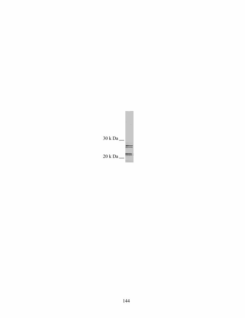
144
20 k Da
30 k Da
145
Figure A.2. Rescue of developmental aggregation by E. coli protein preparation. A. Wild-type M. xanthus; B. csgA; C. csgA complemented with 2.5 µg/ml E. coli protein. Bar 620 µm
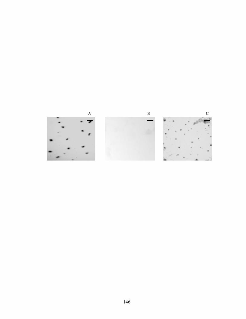
146
A B C
147
Figure A.3. Sporulation rescue by E. coli protein.
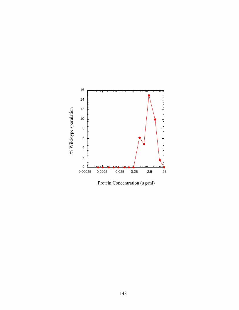
148
0 2 4 6 8
10 12 14 16
0.00025 0.0025 0.025 0.25 2.5 25
% W
ild-ty
pe sp
orul
atio
n
Protein Concentration (µg/ml)
149
References:
1. Kim, S. K., and D. Kaiser. 1990. C-factor: A cell-cell signaling protein
required for fruiting body morphogenesis of M. xanthus. Cell. 61:19-26.
2. Kim, S. K., and D. Kaiser. 1990. Purification and properties of Myxococcus
xanthus C-factor, an intercellular signaling protein. Proc. Natl. Acad. Sci.
USA 87:3635-3639.
3. Kuner, J. M., and D. Kaiser. 1982. Fruiting body morphogenesis in
submerged cultures of Myxococcus xanthus. J. Bacteriol. 151:458-461.
4. Kuspa, A., L. Plamann, and D. Kaiser. 1992. Identification of heat-stable
A-factor from Myxococcus xanthus. J. Bacteriol. 174:3319-3326.
5. Lee, B.-U., K. Lee, J. Mendez, and L. J. Shimkets. 1995. A tactile sensory
system of Myxococcus xanthus involves an extracellular NAD(P)+-containing
protein. Genes Dev. 9:2964-2973.
6. Plamann, L., A. Kuspa, and D. Kaiser. 1992. Proteins that rescue A-signal-
defective mutants of Myxococcus xanthus. J. Bacteriol. 174:3311-3318.
7. Shimkets, L. J., and S. J. Asher. 1988. Use of recombination techniques to
examine the structure of the csg locus of Myxococcus xanthus. Mol. Gen.
Genet. 211:63-71.
8. Shimkets, L. J., and D. Kaiser. 1982. Induction of coordinated movement of
Myxococcus xanthus cells. J. Bacteriol. 152:451-461.