Embed Size (px)
Citation preview
Research ArticleN-Acetyl-cysteine and Mechanisms Involved in Resolution ofChronic Wound Biofilm
Xin Li,1 Jane Kim,1 Jiabin Wu,2 Alaa’ I Ahamed,1 Yinsheng Wang,2
and Manuela Martins-Green 1
1Department of Molecular, Cell and Systems Biology, University of California, Riverside, CA, USA2Department of Chemistry, University of California, Riverside, CA, USA
Correspondence should be addressed to Manuela Martins-Green; [email protected]
Received 9 August 2019; Revised 18 November 2019; Accepted 7 December 2019; Published 28 January 2020
Academic Editor: Jonathan M. Peterson
Copyright © 2020 Xin Li et al. This is an open access article distributed under the Creative Commons Attribution License, whichpermits unrestricted use, distribution, and reproduction in any medium, provided the original work is properly cited.
Chronic wounds are a major global health problem with the presence of biofilm significantly contributing to wound chronicity.Current treatments are ineffective in resolving biofilm and simultaneously killing the bacteria; therefore, effective biofilm-resolving drugs are needed. We have previously shown that, together with α-tocopherol, N-acetyl-cysteine (NAC) significantlyimproves the healing of biofilm-containing chronic wounds, in a diabetic mouse model we developed, by causing disappearanceof the bacteria and breakdown of the extracellular polymeric substance (EPS). We hypothesize that NAC creates amicroenvironment that affects bacterial survival and EPS integrity. To test this hypothesis, we developed an in vitro biofilmsystem using microbiome taken directly from diabetic mouse chronic wounds. For these studies, we chose mice in whichchronic wound microbiome was rich in Pseudomonas aeruginosa (97%). We show that NAC at concentrations with pH< pKacauses bacterial cell death and breakdown of EPS. When used before biofilm is formed, NAC leads to bacterial cell deathwhereas treatment after the biofilm is established NAC causes biofilm dismantling accompanied by bacterial cell death.Mechanistically, we show that NAC can penetrate the bacterial membrane, increase oxidative stress, and halt protein synthesis.We also show that low pH is important for the actions of NAC and that bacterial death occurs independently of the presence ofbiofilm. In addition, we show that both the acetyl and carboxylic groups play key roles in NAC functions. The results presentedhere provide insight into the mechanisms by which NAC dismantles biofilm and how it could be used to treat chronic woundsafter debridement (NAC applied at the start of culture) or without debridement (NAC applied when biofilm is already formed).This approach can be taken to develop biofilm from microbiome taken directly from human chronic wounds to test moleculesthat could be effective for the treatment of specific biofilm compositions.
1. Introduction
Chronic wounds are a major global health problem. The carefor chronic wounds leads to a large financial and psycholog-ical problem to individuals and an economic burden to thesociety. In the United States alone, 6.5 million people sufferfrom chronic wounds, along with the cost of $25 billion peryear spent in wound care [1]. Chronic wounds are commonlyfound in situations involving obesity, vascular diseases, andaging [2]. In these wounds, reepithelialization does not occurin a timely and orderly manner; the wounds stall in theinflammatory phase with excessive levels of reactive oxygenspecies (ROS), cytokines, and proteases; increased cell death,
persistent infections, and lack of microvessel developmentoccur [3–6]. Moreover, defective mesenchymal stem cells[7–9], degradation of growth factors [10], diminishedcapacity of fibroblast proliferation and migration [11], anddownregulation of wound-associated keratin and its hetero-polymers [12] result in impairment of healing. These condi-tions promote pathogen colonization and strong biofilmformation in the wound bed, which then further delay thehealing [13, 14].
Basic steps in wound care include debridement to removebiofilm, topical and/or systematic antibiotic treatment, andapplication of various wound dressings [15]. However,wound biofilm is persistent and difficult to eradicate. Even
HindawiJournal of Diabetes ResearchVolume 2020, Article ID 9589507, 16 pageshttps://doi.org/10.1155/2020/9589507
though surgical debridement removes existing biofilm, freshbiofilm reappears within 2-3 days, given that bacteria persistwithin the margin of the wound tissue even after extensivedebridement [16]. Antibiofilm drugs that can either facilitatethe dispersion of preformed biofilm or inhibit the formationof new biofilm are an unmet need. There are numerouspotential antibiofilm treatments under investigation. Forexample, when topically applied, acetic acid, boric acid,ascorbic acid, alginic acid, and hyaluronic acid improve thewound healing process and decrease infections [17, 18].More recently, novel potential approaches, such as quorumsensing inhibitors that have unique structures compete withsignal molecules and interfere with biofilm formation. Also,peptidomimetics that disturb bacterial membrane structureand nanoparticles that transport drugs into bacterial cellshold great promise in dismantling existing biofilms orpreventing biofilm formation [19–23]. However, the field isin need of new treatments that not only are effective againstbacteria but also promote wound healing and are withoutsignificant side effects.
We have recently shown that NAC applied to chronicwounds in a diabetic mouse model we have developedimproved healing dramatically within 20 days of initiationof treatment [24].
However, the mechanisms by which NAC affects theextracellular polymeric substances (EPS) and interferes withbacterial function that lead to biofilm dismantling are notwell understood. To study the mechanisms of action ofNAC on biofilm initiation, development, and integrity, weestablished an in vitro system that leads to the developmentof biofilm by culturing microbiomes collected from chronicwounds. This system does not use laboratory-cultured bacte-ria; the bacteria are taken from biofilm collected directly fromchronic wounds that we create in our db/db-/- mouse model.In the studies presented here, we chose to study biofilmsestablished in vitro from microbiome taken from a chronicwound that contained primarily Pseudomonas aeruginosa.We hypothesize that NAC creates an environment that dis-rupts wound microbiome biofilm by interfering with bothbacterial function and survival and by disrupting biofilmEPS integrity. Our results show that NAC treatment of thebiofilm in vitro resulted in bacterial cell death and disruptionof the EPS in the biofilm. These effects were primarily accom-plished by an increase in oxidative stress in the bacteria, inhi-bition of protein synthesis, and a decrease in the amount ofDNA and proteins in the EPS. For these effects, the pH ofNAC and specific functional groups of this molecule are crit-ical. These results provide insight into the mechanismsinvolved in the ability of NAC to prevent chronic wound bio-film initiation and development as well as in disrupting exist-ing biofilm. Moreover, this approach can be taken to developbiofilm frommicrobiome taken directly from human chronicwounds to test molecules that could be effective for the treat-ment of specific biofilm composition.
2. Methods and Materials
2.1. Ethics Statement. All animal experimental protocols wereapproved by the University of California Riverside (UCR),
Institutional Animal Care and Use Committee (UCR-IACUC). All animal experiments took place in PI's (M.Martins-Green) lab at UCR. Mice were euthanized by carbondioxide (CO2) inhalation, which is the most commonmethod of euthanasia used by NIH. The amount and lengthof CO2 exposure were approved by UCR-IACUC. The ani-mals are cared every day, 7 days a week. UCR is fully accre-dited by AAALAC, complying with the Guide for the Careand Use of Laboratory Animals, The Public Health ServicePolicy, and the Humane Care and Use of Laboratory Ani-mals. The UCR campus veterinarian was available to assistus with any problems with the animals.
2.2. Collection and Storage of Wound Microbiome. Chronicwounds were made in db/db-/- mice as previously described[24]. Six to seven-month-old db/db-/- mice were treatedintraperitoneally with a catalase inhibitor, 3-amino-1,2,4-tri-azole, at 1 g/kg body weight before wounding. Immediatelyafter wounding, they were treated once topically with theglutathione peroxidase inhibitor, mercaptosuccinic acid, at150mg/kg body weight. Using our procedure, the woundsare fully chronic in 20 days and contain abundant biofilm.All bacteria forming the biofilm are from the mouse naturalskin with no additional manipulation or introduction oflaboratory-carried strains; therefore, the biofilm forms natu-rally from the microbiome in the skin. For these studies, thebiofilms were collected using the Levine method with sterilecotton swabs and stored for bacteria identification both dryat -80°C and in Luria-Bertani (LB) media (10 g tryptone, 5 gyeast extract, and 10 g NaCl and Milli-Q water to 1 L;Teknova) supplemented with 20% glycerol. Swabs in LBmedia with 20% glycerol were then cultured in fresh LBmediaovernight at 37°C (150 rpm) and aliquots stored at -80°C.
2.3. Bacteria Identification in the Wound Biofilm. Bacterialspecies in swabs and their abundance were analyzed byextracting bacterial DNA with the PowerSoil DNA IsolationKit (Cat. no.12888-50) as described by the manufacturer.For library construction, the ITS region was amplified usingbacterial ITS rRNA primers (ITS-1507F GGTGAAGTCGTAACAAGGTA, ITS-23SR GGTTBCCCCATTCRG) withspecific barcode sequences for each sample. Then, PCRproducts were purified with QIAGEN MinElute 96 UF PCRPurification Kit (Cat. no. 28053). The amounts of DNAs werenormalized for each sample and sent to the Illumina MiSeqsequencer. The sequences were identified and organized intoan OTU table generated via Qiime, an open-source bioinfor-matics pipeline, analyzing raw microbiome DNA sequencingdata [25].
2.4. In Vitro Chronic Wound Bacterial Biofilm Model.Although the mice are genetically identical, we found that, whenthe wounds are fully chronic, each mouse ends up with its ownbiofilm-forming bacteria mostly composed by 1 or 2 such bacte-rial species. The most predominant species are Pseudomonas,Staphylococcus, Acinetobacter, and Enterobacter. For thesestudies, we chose a biofilm from a wound that contained 97%P. aeruginosa. This biofilm was grown in LB at 37°C withshaking (150 rpm) overnight and then inoculated in media of
2 Journal of Diabetes Research
96-well flat microtiter plates (Cat. no. CLS3595, Corning Incor-porated, USA) or 35mm petri dishes (Cat. no. 353001, CorningFalcon, USA) to form a biofilm. An opaque film was formed onthe air-liquid interface and on solid-liquid interface attached tothe sides and bottom of the well. The air-liquid biofilm can beseen 12h after inoculation at 37°C and is fully developed by24h of incubation.
2.5. Bacteria Identification in the In Vitro Wound Biofilm. Todetermine whether this biofilm in vitro has the same compo-sition as the biofilm in the original wound biofilm, we usedthe API 20E identification kit (REF 20 100, bioMérieux,USA) which is a standardized identification system forEnterobacteriaceae and other nonfastidious, Gram-negativerods; it uses 21 miniaturized biochemical tests and a data-base. The kit has a table of bacteria that can be identified,which includes Enterobacter, Pseudomonas, and manygram-negative bacteria. 24 h biofilm inoculum, includingair-liquid interface and solid-liquid interface biofilm, wascollected and sonicated for 10 s with 15% amplitude. Bacte-rial cells were collected by centrifugation, plated on trypticsoy agar plates (Cat. no. 236950, BD Difco, Sparks, USA)containing 5.0% (v/v) defibrinated sheep blood (Cat. no.R54008, Thermo Fisher Scientific, USA) with serial dilutions,and cultured for 12-24 h at 37°C. Three morphologicallydifferent colonies were observed; we picked five colonies ofeach type from the plates and analyzed them with API 20Estrips consisting of 20 microtubes with dehydratedsubstrates. According to the manufacturer’s instructions, asingle colony was removed and resuspended in 0.85% NaClsolution. The suspension was distributed into the tubes ofthe strip and cultured at 37°C for 18-24 h, within an incuba-tion box with 5ml demineralized water to create a humidatmosphere. During the incubation, metabolism of the bacte-ria produces color changes spontaneously that can berevealed by the addition of reagents. The tests were separatedinto groups of 3, and a value of 1, 2, or 4 was assigned to each.After the incubation, the values were added together corre-sponding to positive reactions within each group. A 7-digitprofile number was obtained for the total 20 tests. The bacte-ria species identification can be determinate by referring tothe apiweb™. “API 20 E is a standardized identificationsystem for Enterobacteriaceae and other nonfastidious,Gram-negative rods which uses 21 miniaturized biochemicaltests and a database.”
2.6. NAC and Its Derivatives Used for This Study. N-Acetyl-cysteine (NAC, Cat. no. A7250-100G, Sigma-Aldrich), N-acetyl cysteine amide (NACA, Cat. no. A0737-5MG, Sigma-Aldrich), N-acetyl serine (NAS, Cat. no. A2638-1G, Sigma-Aldrich), cysteine (Cat. no. 168149-2.5G, Sigma-Aldrich),and glutathione (PHR1359-500MG, Sigma-Aldrich) wereused. These molecules were dissolved in LB broth, andfilter-sterilized. For the liquid culture in 96-well microtiterplates and 35mm petri dishes, chemical solution with a finalconcentration (1x) was added at specific time points againstthe side of the well by slowly pipetting without disturbingthe biofilm. Plates were continually cultured at 37°C. Thecondition of the surface biofilm was recorded digitally.
For chronic wound microbiome biofilm on solid surfaces,5μl diluted inoculum (ratio 1 : 100) was added on agar for7 h. NAC solutions (5μl) were added directly either on thetop of the colonies or on the bacterial colony patches(200μl) after the culture rings were placed. The effect ofNAC under these conditions was observed in 24h or 48 h.
2.7. Biofilm Quantification Assays. A modified microtiterplate test for the quantification of biofilm formation was usedbased on a previous assay [26]. Briefly, 0.1% of a crystal violet(CV; Cat. no. C0775-25G, Sigma-Aldrich, USA) stainingsolution was prepared in Milli-Q water and filtered througha 0.45μm filter (Cat. no. SLHA033SS, MF-Millipore). Themedium and unbounded cells were removed by pipetting.The remaining biofilm was staining in 225μl per well of0.1% CV solution for 5min. Staining solution was removedby pipetting. 300μl of 95% of ethanol was added to removethe staining from the biofilm. After 45min to 1 h of incubationat room temperature to fully distain the biofilm, the absor-bance was measured at 590nm using a microtiter plate reader(Epoch Microplate Spectrophotometer, BioTek). If the read-ing signals were overload, distaining solutions were dilutedaccordingly and the appropriate multiplier was applied.
2.8. Biofilm Protein and DNA Analysis. 24 h-biofilm culturedin 96-well microtiter plates is treated with various concentra-tions of NAC for 24 h. The same amount of culture for eachNAC treatment was collected and briefly sonicated for 10 swith 15% amplitude. This step is to loosen the EPS structure.Samples are saved as a whole biofilm fraction. Cultures werecentrifuged at 4°C (15,000 rpm) for 10min. The supernatantwas transferred to a new microcentrifuge tube and saved asthe soluble EPS fraction. 30μl of whole biofilm fractionsand EPS fractions was analyzed on 12% SDS-PAGE gels forprotein evaluation; 10μl of whole biofilm fractions wasanalyzed on 1% agarose gels for DNA evaluation.
2.9. NAC Intracellular Assay. 24 h-biofilm was treated withNAC for 24h. Biofilm culture was collected, briefly sonicatedfor 10s with 15% amplitude, and centrifuged at 4°C(15,000 rpm) for 5min. The supernatant was discarded, andthe cell pellets were washed with 1ml 1x PBS and resus-pended in 0.5ml 1x PBS. Cells were lysed by sonication andcentrifuged at 15,000 rpm for 10min. The supernatant wasfiltered (pore size: 0.22μm) and analyzed by HPLC. Theliquid chromatography method was performed on an Agilent1100 system with the Phenomenex Luna C18 column(Torrance, USA) (150 × 4:6mm; 5μm, 100Å). The mobilephase consisted of 0.05M KH2PO4 (solution A) and acetoni-trile (95 : 5) with 0.095% of phosphoric acid (solution B), with3% B phase isocratic elution at a flow rate of 1.3ml/min. Thesample volume injected was 100μl, and NAC was detected at214 nm. All analyses were performed at room temperature.
2.10. Measurement of Biofilm Bacteria NAD+/NADH Ratio.NAD+/NADH-Glo Assay Kit (Promega Co., Cat. no.G9072) was used to quantify NAD+ and NADH individually.The 24 h-biofilm was treated with NAC for 24 h. 600μl bio-film inoculum including air-liquid interface and solid-liquidinterface biofilm was collected, washed twice with 1ml 1x
3Journal of Diabetes Research
PBS, and resuspended with 600μl 1x PBS. To lyse the cellmembrane, 200μl of bacterial cells was mixed with 200μlbase solution (100mM sodium carbonate, 20mM sodiumbicarbonate, 0.05% TritonX-100) supplemented with 1%DTAB and vortexed for 15 s. To detect the amount ofNADH, 200μl of the 400μl mixture was directly incubatedat 60°C for 15min (A). The other 200μl of the mixture wasmixed with 100μl 0.4N HCl first and then incubated at60°C for 15min to measure the amount of NAD+ (B). BothA and B were equilibrated at room temperature for 10min.Then, 200μl HCl/Tris solution (prepared by mixing the samevolume of 0.4N HCl mixed with equal volume of 0.5M Trisbase) was added to A; 100μl 0.5M Tris base was added to B.For each NAC treatment, technical duplicates were appliedfor both NAD+ and NADH detection. 100μl of either solu-tion A or solution B was added to a 96-well microtiter plate,and 100μl NAD+/NADH-Glo detection reagent was addedto each well. After 45-50min incubation at room tempera-ture, luminescence was detected by a Promega Multi Reader.
2.11. Protein Synthesis Determination Using 35S Labeling.24 h-biofilm bacteria were collected by centrifugation at15,000g for 5min. The pellets were resuspended in LBcontaining either 5mg/ml or 20mg/ml NAC, together withthe exposure of 50μCi EXPRE35S35S Protein Labeling Mix(PerkinElmer, Cat. no. NEG072002MC). The mixtures wereincubated at 37°C (225 rpm) for 4 h and then at 99°C for5min to kill the bacteria and stop cellular protein synthesis.Cell pellets were centrifuged at 15,000g for 5min, washedwith 1ml 1x PBS, resuspended in 200μl 1x PBS, and analyzedon 10% SDS-PAGE gel. After staining and distaining, theSDS-PAGE gel was wrapped in a cling film and exposed toan X-ray film at 4°C for 72h.
2.12. Fluorescence Staining and Confocal Laser ScanningMicroscopy Analysis of the Biofilm. 24 h-biofilm was preparedin the air-liquid interface of 35mm diameter glass bottomdishes (WillCo Well, Cat. no. GWST-3522) and treated withNAC solutions for 24 h. The staining process was optimizedbased on a previous study [27]. Culture media were gentlyremoved, and the biofilm was rinsed with 1ml 1x PBS twice.All the staining processes were done at room temperature.Calcofluor White Stain solution in 1x PBS (Sigma-Aldrich,Cat. no. 18909-100ML-F) to stain polysaccharide in the bio-film was added to each plate and incubated for 20min. Thestaining solution was removed, and the biofilm was rinsedwith 1ml 1x PBS. Then, the biofilm was stained with FilmTra-cer SYPTO Ruby Biofilm Matrix Staining solution for 30minto label proteins in the biofilm matrix (Thermo Fisher Scien-tific, Cat. no. F10318). The staining solution was removed,and the biofilm was rinsed with 1ml 1x PBS. Lastly, the bio-film was stained with SYTO9 Green Fluorescent Nucleic AcidStain solution for 30min to stain biofilm DNA (2.5μl of SYTO9 Green Fluorescent Nucleic Acid Stain in 397.5μl 1x PBS).The staining solution was removed, and the biofilm was rinsedwith 1ml 1x PBS. The structure of the biofilm was observedusing a confocal laser scanning with the Zeiss880 InvertedUV Spectral Airyscan Microscope. Channel mode visualiza-tion was done using the 20x objective. During the image
collection, confocal tiles were obtained using channel modewith 3-track: Ex/Em 355nm/433 nm for Calcofluor WhiteStain, Ex/Em 280nm, 450nm/610nm for FilmTracerSYPTO Ruby, and Ex/Em 488nm/523nm for SYTO9 Green.
2.13. Statistical Analysis. For the statistical analysis of data,we used GraphPad Prism 6 software. The analysis of variance(ANOVA) was used to test the significance of group differ-ences between two or more groups. In the experiments withonly two groups, statistical analysis was conducted usingStudent’s t-test.
3. Results
In this study, we investigated the effects of NAC on biofilmdeveloped from the microbiome taken from chronic wounds.We have previously developed a db/db-/- mouse model forchronic wounds in which the wounds become chronic, pre-senting all the abnormalities found in human chronicwounds, including the presence of a biofilm with bacteriathat are found in chronic wounds in humans [24]. In ourmodel, bacteria are never introduced in the wounds fromexternal sources; the biofilm develops from the bacteria inthe skin in the presence of high levels of oxidative stress[24]. In this study, to make the biofilm in vitro, we chose touse wound biofilm that contains primarily P. aeruginosabecause P. aeruginosa colonizes about 52.2% of the patientswith chronic ulcers [28, 29]. It produces strong biofilm andquickly acquires antibiotic resistance leading the wounds tobecome chronic [30]. Although NAC has been used to helptreat mucus in cystic fibrosis, the mechanisms by whichNAC dissolves biofilm are not well known and NAC hasnot been used to treat human chronic wounds containingP. aeruginosa. Below, we present data describing how wedeveloped the in vitro biofilm system and how we tested theeffects of NAC on biofilm initiation and development andon resolution/dismantling of established biofilm. Mechanis-tically, in order to kill the bacteria, NAC needs to enter thebacteria itself and, for that, low pH is critical. When insidethe bacteria, NAC creates high levels of oxidative stress andinhibits protein synthesis. In addition, we also tested theimportance of the various functional groups of NAC in theseprocesses something that has never been shown previously.
3.1. Development of an In Vitro System to Study the Effects ofNAC on Biofilm. Microbiome was directly collected fromchronic wounds in our db/db-/- mouse model of chronicwounds, using the methods described in the Methods andMaterials section [24]. For these studies, we chose a swabtaken from a fully chronic wound (20 days after wounding)with a microbiome that contained ~97% of P. aeruginosa(Figure 1(a)). The microbiome composition in the fullychronic wounds varies from animal to animal much likelyin human subjects. To recreate a biofilm, this microbiomewas cultured in 96-well microtiter plates, 35mm petridishes, or culture tubes. The microbiome from chronicwounds formed strong biofilm overnight in both microtiterplates and culture tubes. Microbiome from nonchronicwounds did not develop biofilm in vitro under the same
4 Journal of Diabetes Research
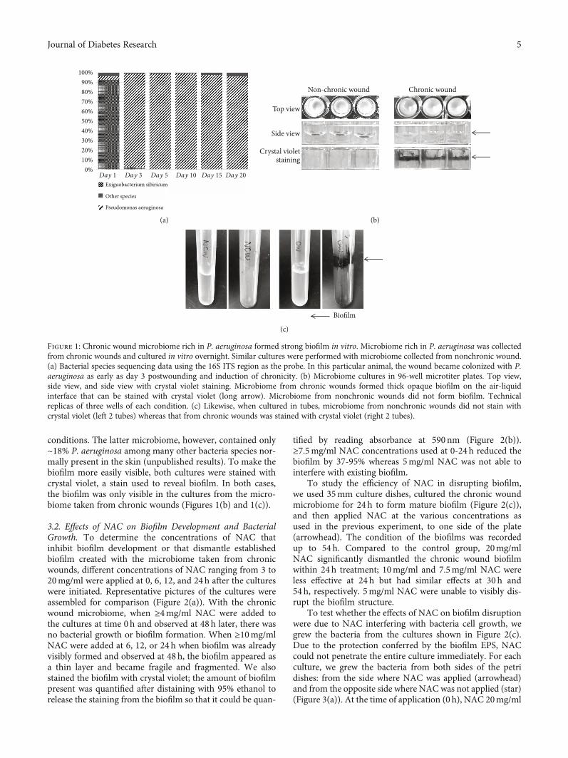
conditions. The latter microbiome, however, contained only~18% P. aeruginosa among many other bacteria species nor-mally present in the skin (unpublished results). To make thebiofilm more easily visible, both cultures were stained withcrystal violet, a stain used to reveal biofilm. In both cases,the biofilm was only visible in the cultures from the micro-biome taken from chronic wounds (Figures 1(b) and 1(c)).
3.2. Effects of NAC on Biofilm Development and BacterialGrowth. To determine the concentrations of NAC thatinhibit biofilm development or that dismantle establishedbiofilm created with the microbiome taken from chronicwounds, different concentrations of NAC ranging from 3 to20mg/ml were applied at 0, 6, 12, and 24h after the cultureswere initiated. Representative pictures of the cultures wereassembled for comparison (Figure 2(a)). With the chronicwound microbiome, when ≥4mg/ml NAC were added tothe cultures at time 0h and observed at 48 h later, there wasno bacterial growth or biofilm formation. When ≥10mg/mlNAC were added at 6, 12, or 24 h when biofilm was alreadyvisibly formed and observed at 48 h, the biofilm appeared asa thin layer and became fragile and fragmented. We alsostained the biofilm with crystal violet; the amount of biofilmpresent was quantified after distaining with 95% ethanol torelease the staining from the biofilm so that it could be quan-
tified by reading absorbance at 590nm (Figure 2(b)).≥7.5mg/ml NAC concentrations used at 0-24 h reduced thebiofilm by 37-95% whereas 5mg/ml NAC was not able tointerfere with existing biofilm.
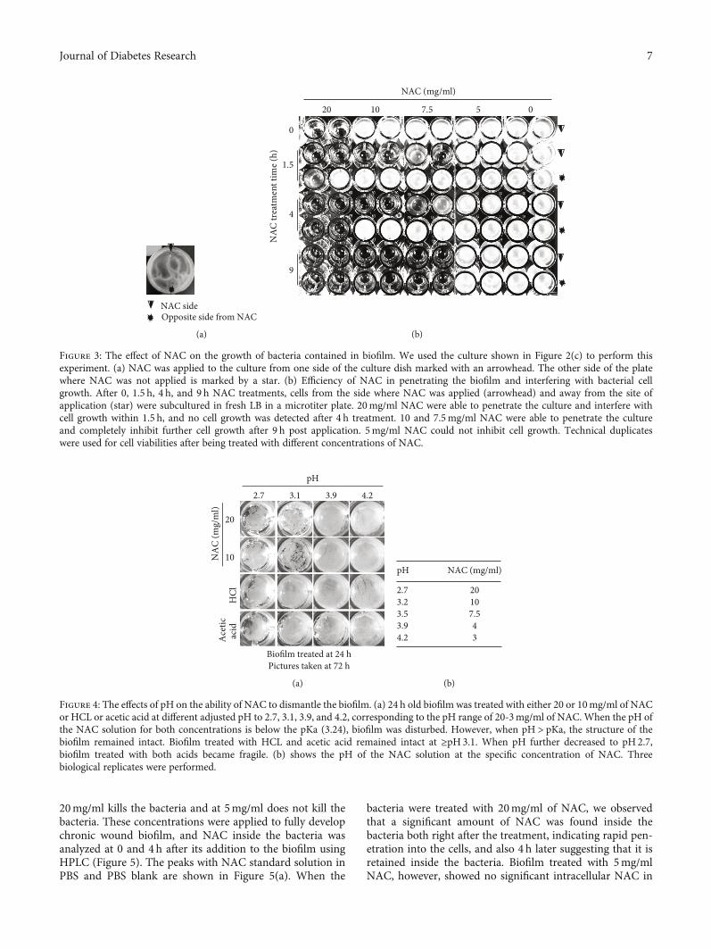
To study the efficiency of NAC in disrupting biofilm,we used 35mm culture dishes, cultured the chronic woundmicrobiome for 24 h to form mature biofilm (Figure 2(c)),and then applied NAC at the various concentrations asused in the previous experiment, to one side of the plate(arrowhead). The condition of the biofilms was recordedup to 54 h. Compared to the control group, 20mg/mlNAC significantly dismantled the chronic wound biofilmwithin 24 h treatment; 10mg/ml and 7.5mg/ml NAC wereless effective at 24 h but had similar effects at 30 h and54 h, respectively. 5mg/ml NAC were unable to visibly dis-rupt the biofilm structure.
To test whether the effects of NAC on biofilm disruptionwere due to NAC interfering with bacteria cell growth, wegrew the bacteria from the cultures shown in Figure 2(c).Due to the protection conferred by the biofilm EPS, NACcould not penetrate the entire culture immediately. For eachculture, we grew the bacteria from both sides of the petridishes: from the side where NAC was applied (arrowhead)and from the opposite side where NAC was not applied (star)(Figure 3(a)). At the time of application (0 h), NAC 20mg/ml
Day 10%
10%
20%
30%
40%
50%
60%
70%
80%
90%
100%
Exiguobacterium sibiricum
Other species
Pseudomonas aeruginosa
Day 3 Day 5 Day 10 Day 15 Day 20
(a)
Non-chronic wound Chronic wound
Top view
Side view
Crystal violetstaining
(b)
Biofilm
(c)
Figure 1: Chronic wound microbiome rich in P. aeruginosa formed strong biofilm in vitro. Microbiome rich in P. aeruginosa was collectedfrom chronic wounds and cultured in vitro overnight. Similar cultures were performed with microbiome collected from nonchronic wound.(a) Bacterial species sequencing data using the 16S ITS region as the probe. In this particular animal, the wound became colonized with P.aeruginosa as early as day 3 postwounding and induction of chronicity. (b) Microbiome cultures in 96-well microtiter plates. Top view,side view, and side view with crystal violet staining. Microbiome from chronic wounds formed thick opaque biofilm on the air-liquidinterface that can be stained with crystal violet (long arrow). Microbiome from nonchronic wounds did not form biofilm. Technicalreplicas of three wells of each condition. (c) Likewise, when cultured in tubes, microbiome from nonchronic wounds did not stain withcrystal violet (left 2 tubes) whereas that from chronic wounds was stained with crystal violet (right 2 tubes).
5Journal of Diabetes Research

affected the bacteria in a way that they could no longer growwhereas treatment with ≤10mg/ml was unable to affectbacterial growth. At 1.5 h posttreatment of the biofilm, cellstaken from the treated side (arrowhead) did not grow withtreatment of 7.5-20mg/ml whereas from the nontreated side(star) they did. At 4 h of treatment, the same results wereobserved for the treated side as at 1.5 h treatment. However,for the nontreated side, the 20mg/ml NAC was now affectingthe growth of the bacteria. By 9 h of treatment, the 7.5-20mg/ml NAC had spread throughout the entire petri dishleading to no growth of the bacteria. 5mg/ml NAC wereunable to stop bacterial growth on either side of the petri dish(Figure 3(b)). These results suggest that over time treatmentof the biofilm with 7.5-20mg/ml NAC stops bacterial growthand eventually dismantles the biofilm. We also investigatedthe effect of NAC on biofilm-free bacterial growth and foundthat NAC is also effective in the absence of biofilm (Supple-mentary material and Figures 1A–1C).
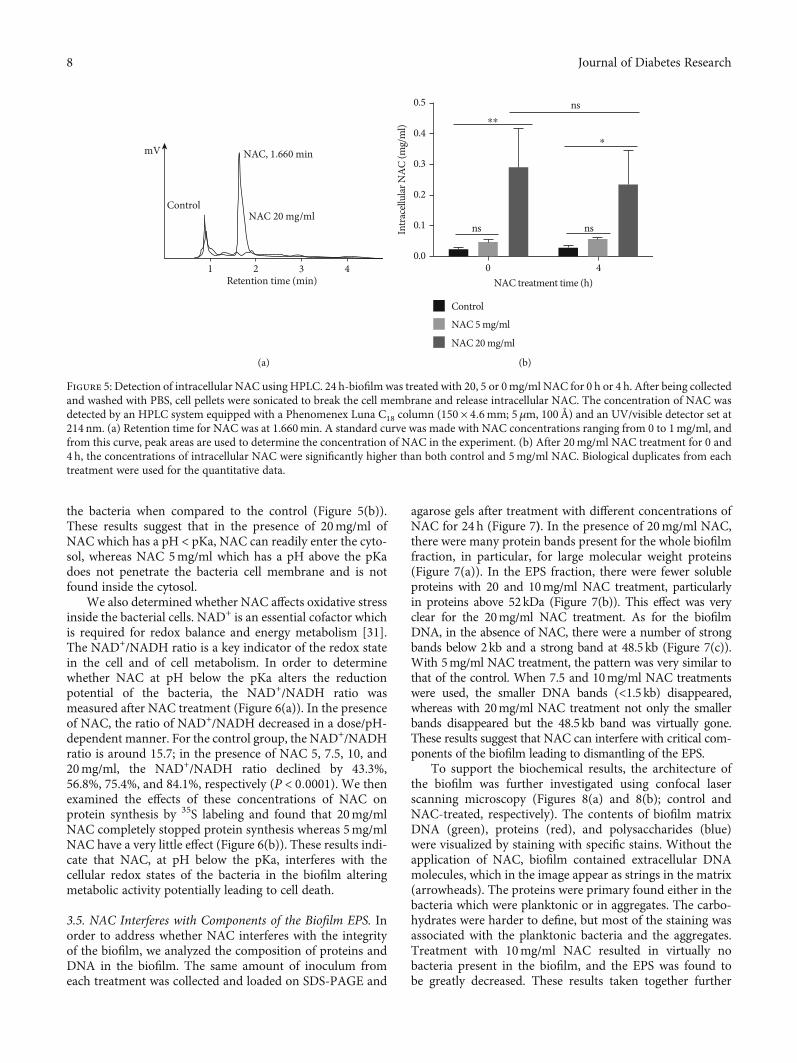
3.3. NAC-Induced pH Changes Are Important for ItsAntibiofilm Activity. NAC is a weak organic acid with apKa of 3.24. The pH of LB broth containing concentrationsof 3-20mg/ml of NAC varies from 4.2 to 2.7, respectively(Figures 4(a) and 4(b)). To identify whether the effect ofNAC in dismantling biofilm is associated with pH, we
replaced the LB in 24 h cultures, which contains fullydeveloped biofilm, with LB-NAC solutions at different pH.For 20mg/ml NAC, the pH was adjusted from 2.7 to 3.1,3.9, and 4.2 with sodium hydroxide, and for 10mg/ml NACthe pH was adjusted from 3.1 to 2.7 with HCl and to 3.9and 4.2 with sodium hydroxide. We also used a strong acid,HCl, and a weak acid, acetic acid, with adjusted pH as wedid with NAC. NAC 20 and 10mg/ml at pH2.7 and 3.1,which are below NAC pKa, disrupted the surface biofilmwhereas at pH3.9 and 4.2, which are above the pKa, didnot. Both acetic acid and HCl at pH2.7 disrupted the surfacebiofilm albeit to a lesser extent than NAC at the same pH,and they did not disrupt the biofilm at higher pH(Figure 4(a)). These results suggest that pH is not the onlyfactor involved in biofilm disruption by NAC and thatNAC has additional properties that affect the integrity ofthe biofilm either by affecting the bacteria or by disruptingthe EPS, or both, leading to dismantling of the biofilm.
3.4. At pH below the pKa, NAC Penetrates the Bacterial CellMembrane, Increases Oxidative Stress, and Inhibits ProteinSynthesis. To determine whether NAC affects the bacteriaby entering the cytosol and causing changes that potentiallykill the bacteria, we used HPLC to determine whether NACis found inside the bacteria after the treatments. NAC at
0 6 2412Treatment time (h)
20
10
7.5
4
3
NAC
(mg/
ml)
(a)
0 5 10 15 200
2
4
6
8
10
OD
590
NAC (mg/ml)0 h6 h
12 h24 h
(b)
NAC (mg/ml)
1.5
20 10
24h–
old
biofi
lm tr
eatm
ent t
ime (
h)
7.5 5 0
0
4
9
24
30
54
(c)
Figure 2: The effect of NAC on biofilm development and dismantling. (a) Microbiome taken from the chronic wounds and grown in 96-wellmicrotiter plates. Concentrations of NAC ranging from 3 to 20mg/ml were applied to the cultures at the times indicated (0, 6, 12, and 24 h).The pictures shown were taken at 48 h after initiation of the culture. When ≥4mg/ml NACwere applied to the culture at 0 h, the biofilm neverdeveloped. When ≥10mg/ml of NAC was applied at 6, 12, and 24 h after culture initiation, biofilm formation was visibly altered or becamefragile and disrupted. (b) At 48 h, biofilm was stained with crystal violet (CV) and the absorbance at 590 nm was measured to quantify biofilmbiomass. At 0 h, 5mg/ml application, 5mg/ml NAC significantly reduced the appearance of biofilm, and when applied at 6 and 12 h, it wasable to decrease biofilm production but not when applied at 24 h. ≥10mg/ml NAC or more significantly diminish biofilm formation. Threetechnical repeats with standard deviation as error bars for quantitative data. (c) The dose-dependent effect of NAC on 24 h old biofilmcultured in 35mm petri dishes was recorded over time. 20mg/ml of NAC dismantled biofilm by 24 h; 10 and 7.5mg/ml of NAC were ableto fully disrupt biofilm at 30 and 54 h, respectively. 5mg/ml NAC did not cause changes in existing biofilm.
6 Journal of Diabetes Research
20mg/ml kills the bacteria and at 5mg/ml does not kill thebacteria. These concentrations were applied to fully developchronic wound biofilm, and NAC inside the bacteria wasanalyzed at 0 and 4h after its addition to the biofilm usingHPLC (Figure 5). The peaks with NAC standard solution inPBS and PBS blank are shown in Figure 5(a). When the
bacteria were treated with 20mg/ml of NAC, we observedthat a significant amount of NAC was found inside thebacteria both right after the treatment, indicating rapid pen-etration into the cells, and also 4 h later suggesting that it isretained inside the bacteria. Biofilm treated with 5mg/mlNAC, however, showed no significant intracellular NAC in
NAC sideOpposite side from NAC
(a)
NAC (mg/ml)
057.51020
0
1.5
4
9N
AC tr
eatm
ent t
ime (
h)
(b)
Figure 3: The effect of NAC on the growth of bacteria contained in biofilm. We used the culture shown in Figure 2(c) to perform thisexperiment. (a) NAC was applied to the culture from one side of the culture dish marked with an arrowhead. The other side of the platewhere NAC was not applied is marked by a star. (b) Efficiency of NAC in penetrating the biofilm and interfering with bacterial cellgrowth. After 0, 1.5 h, 4 h, and 9 h NAC treatments, cells from the side where NAC was applied (arrowhead) and away from the site ofapplication (star) were subcultured in fresh LB in a microtiter plate. 20mg/ml NAC were able to penetrate the culture and interfere withcell growth within 1.5 h, and no cell growth was detected after 4 h treatment. 10 and 7.5mg/ml NAC were able to penetrate the cultureand completely inhibit further cell growth after 9 h post application. 5mg/ml NAC could not inhibit cell growth. Technical duplicateswere used for cell viabilities after being treated with different concentrations of NAC.
Biofilm treated at 24 h Pictures taken at 72 h
20
10
2.7 3.1 3.9 4.2
pH
NA
C (m
g/m
l)A
cetic
acid
H
Cl
(a)
pH NAC (mg/ml)
2.73.23.53.94.2
20107.543
(b)
Figure 4: The effects of pH on the ability of NAC to dismantle the biofilm. (a) 24 h old biofilm was treated with either 20 or 10mg/ml of NACor HCL or acetic acid at different adjusted pH to 2.7, 3.1, 3.9, and 4.2, corresponding to the pH range of 20-3mg/ml of NAC. When the pH ofthe NAC solution for both concentrations is below the pKa (3.24), biofilm was disturbed. However, when pH > pKa, the structure of thebiofilm remained intact. Biofilm treated with HCL and acetic acid remained intact at ≥pH 3.1. When pH further decreased to pH 2.7,biofilm treated with both acids became fragile. (b) shows the pH of the NAC solution at the specific concentration of NAC. Threebiological replicates were performed.
7Journal of Diabetes Research
the bacteria when compared to the control (Figure 5(b)).These results suggest that in the presence of 20mg/ml ofNAC which has a pH < pKa, NAC can readily enter the cyto-sol, whereas NAC 5mg/ml which has a pH above the pKadoes not penetrate the bacteria cell membrane and is notfound inside the cytosol.
We also determined whether NAC affects oxidative stressinside the bacterial cells. NAD+ is an essential cofactor whichis required for redox balance and energy metabolism [31].The NAD+/NADH ratio is a key indicator of the redox statein the cell and of cell metabolism. In order to determinewhether NAC at pH below the pKa alters the reductionpotential of the bacteria, the NAD+/NADH ratio wasmeasured after NAC treatment (Figure 6(a)). In the presenceof NAC, the ratio of NAD+/NADH decreased in a dose/pH-dependent manner. For the control group, the NAD+/NADHratio is around 15.7; in the presence of NAC 5, 7.5, 10, and20mg/ml, the NAD+/NADH ratio declined by 43.3%,56.8%, 75.4%, and 84.1%, respectively (P < 0:0001). We thenexamined the effects of these concentrations of NAC onprotein synthesis by 35S labeling and found that 20mg/mlNAC completely stopped protein synthesis whereas 5mg/mlNAC have a very little effect (Figure 6(b)). These results indi-cate that NAC, at pH below the pKa, interferes with thecellular redox states of the bacteria in the biofilm alteringmetabolic activity potentially leading to cell death.
3.5. NAC Interferes with Components of the Biofilm EPS. Inorder to address whether NAC interferes with the integrityof the biofilm, we analyzed the composition of proteins andDNA in the biofilm. The same amount of inoculum fromeach treatment was collected and loaded on SDS-PAGE and
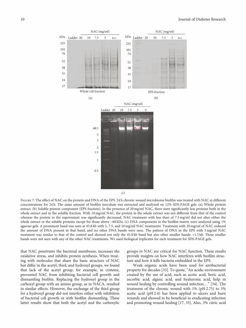
agarose gels after treatment with different concentrations ofNAC for 24 h (Figure 7). In the presence of 20mg/ml NAC,there were many protein bands present for the whole biofilmfraction, in particular, for large molecular weight proteins(Figure 7(a)). In the EPS fraction, there were fewer solubleproteins with 20 and 10mg/ml NAC treatment, particularlyin proteins above 52kDa (Figure 7(b)). This effect was veryclear for the 20mg/ml NAC treatment. As for the biofilmDNA, in the absence of NAC, there were a number of strongbands below 2kb and a strong band at 48.5kb (Figure 7(c)).With 5mg/ml NAC treatment, the pattern was very similar tothat of the control. When 7.5 and 10mg/ml NAC treatmentswere used, the smaller DNA bands (<1.5 kb) disappeared,whereas with 20mg/ml NAC treatment not only the smallerbands disappeared but the 48.5kb band was virtually gone.These results suggest that NAC can interfere with critical com-ponents of the biofilm leading to dismantling of the EPS.
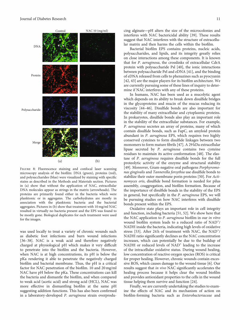
To support the biochemical results, the architecture ofthe biofilm was further investigated using confocal laserscanning microscopy (Figures 8(a) and 8(b); control andNAC-treated, respectively). The contents of biofilm matrixDNA (green), proteins (red), and polysaccharides (blue)were visualized by staining with specific stains. Without theapplication of NAC, biofilm contained extracellular DNAmolecules, which in the image appear as strings in the matrix(arrowheads). The proteins were primary found either in thebacteria which were planktonic or in aggregates. The carbo-hydrates were harder to define, but most of the staining wasassociated with the planktonic bacteria and the aggregates.Treatment with 10mg/ml NAC resulted in virtually nobacteria present in the biofilm, and the EPS was found tobe greatly decreased. These results taken together further
NAC, 1.660 minmV
1 2 3 4
NAC 20 mg/ml
Retention time (min)
Control
(a)
ns
nsns
00.0
0.1
0.2
0.3
0.4
0.5
4NAC treatment time (h)
Control
NAC 5 mg/ml
NAC 20 mg/ml
Intra
cellu
lar N
AC (m
g/m
l)
⁎⁎
⁎
(b)
Figure 5: Detection of intracellular NAC using HPLC. 24 h-biofilm was treated with 20, 5 or 0mg/ml NAC for 0 h or 4 h. After being collectedand washed with PBS, cell pellets were sonicated to break the cell membrane and release intracellular NAC. The concentration of NAC wasdetected by an HPLC system equipped with a Phenomenex Luna C18 column (150 × 4:6mm; 5μm, 100Å) and an UV/visible detector set at214 nm. (a) Retention time for NAC was at 1.660min. A standard curve was made with NAC concentrations ranging from 0 to 1mg/ml, andfrom this curve, peak areas are used to determine the concentration of NAC in the experiment. (b) After 20mg/ml NAC treatment for 0 and4 h, the concentrations of intracellular NAC were significantly higher than both control and 5mg/ml NAC. Biological duplicates from eachtreatment were used for the quantitative data.
8 Journal of Diabetes Research
support our hypothesis that NAC affects biofilm EPSmatrix and bacteria survival.
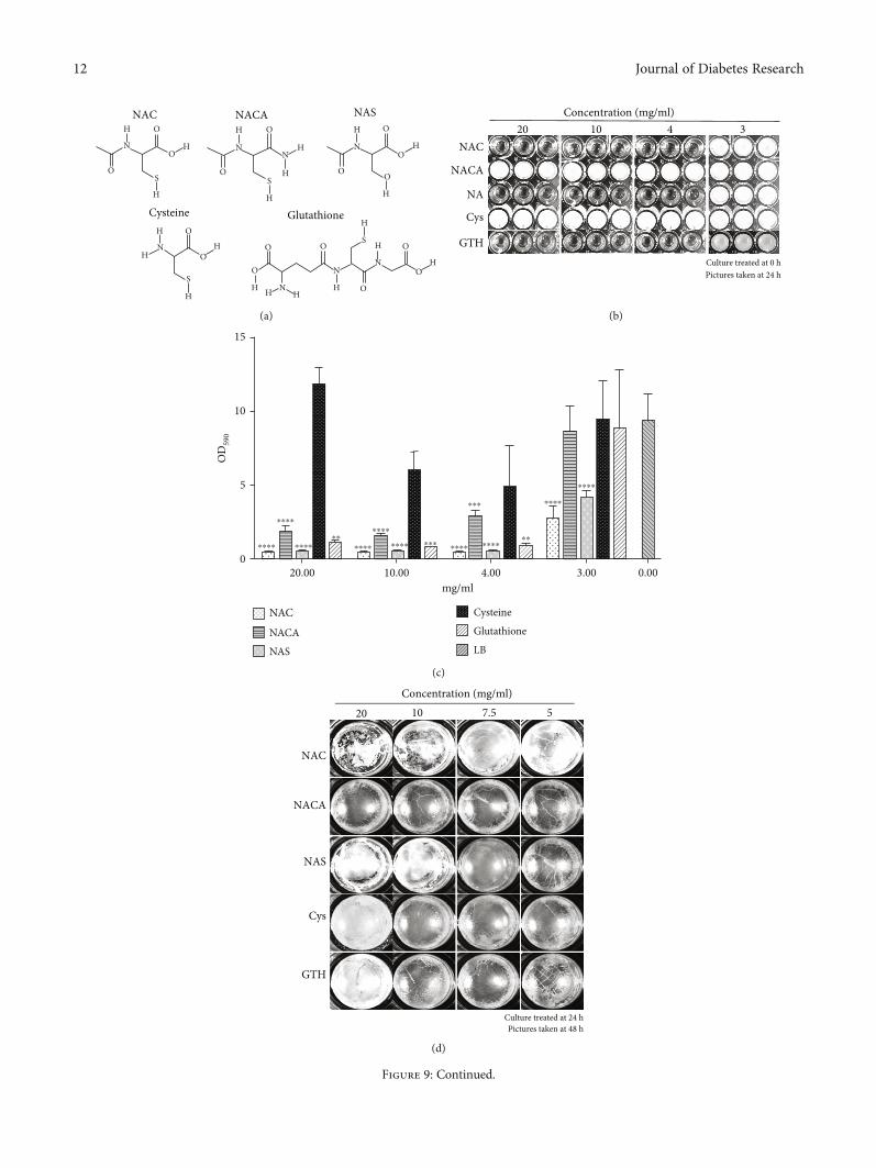
3.6. Importance of Chemical Groups for NAC Function. Inorder to determine whether the functional groups of NACare crucial to its biofilm-dismantling functions, we testedmolecules with analogous structure, includingN-acetyl cyste-ine amide (NACA), N-acetyl serine (NAS), cysteine (Cys),and glutathione (GTH) (Figure 9(a)). NACA has an aminogroup that neutralizes the carboxyl group; NAS contains ahydroxyl group in place of the thiol group; Cys lacks the ace-tyl group; GTH is a more complex molecule than NAC, but ithas a similar antioxidant effect as NAC. When ≥4mg/ml ofNAC, NAS, and GTH were applied at the beginning of theculture and observed 24 h later, there was no bacterial growth(Figure 9(b)). Similarly, there was virtually no significantcrystal violet staining confirming the absence of biofilmattached to the well (Figure 9(c)). However, bacteria grewwhen the same concentrations of NACA and cysteine wereapplied (Figure 9(b)), but the development of biofilm wasmuch less strong in the presence of ≥4mg/ml of NACA thanin the presence of Cys (Figure 9(c)).
When we tested the effects of these molecules on 24 hchronic wound biofilm and observed them 48h later(Figures 9(d) and 9(e)), we observed that ≥10mg/ml ofNAC and NAS were able to disrupt surface biofilm integrity,and CV staining showed that ≥7.5mg/ml of NAC decreasedthe amount of existing biofilm. Surface biofilm treated with≥10mg/ml NAS visibly became fragile, and CV stainingshowed similar results. NACA and Cys treatments did notdisrupt biofilm visually or by CV staining. 20mg/ml GTHreduced the existing biofilm by ~55%, but not with the lowerconcentrations (Figures 9(d) and 9(e)). These results suggest
that the acetyl and carboxyl groups play important roles inNAC biofilm dismantling ability.
4. Discussion
NAC has been used extensively to treat excess mucus forma-tion; in particular, it is very effective as a lung mucolytic incystic fibrosis patients [32]. Because P. aeruginosa is criticalin cystic fibrosis and also a very difficult bacteria to eradicatein the biofilms of human chronic wounds, we chose to studybiofilm derived from chronic wound microbiome that con-tained primarily P. aeruginosa. Using biofilm from chronicwounds in humans is not justifiable at this time, because weneed to first demonstrate that our in vitro system is reliableand reproducible. Indeed, our next step will be a proof-of-concept study with microbiome taken directly from humanchronic diabetic foot ulcers.
We have recently shown that the application of NAC tochronic wounds in a diabetic mouse model we developed,in conjunction with systemic application of α-tocopherol,results in the disappearance of the bacteria from the biofilmand leads to the loss of the EPS. This in turn results in thehealing of the wounds. This is the reason why we decidedto use the microbiomes from the chronic wounds in thismodel to establish the biofilms in vitro. When applied tothe biofilm in vitro, NAC killed bacteria, decreased biofilmformation, and dismantled existing biofilm. In the presenceof NAC at a pH below the pKa, both chronic wound bacteriaand the EPS in the biofilm were affected—bacteria did notsurvive, and the EPS began to fall apart and disperse. We alsodetermined that the DNA and protein content of EPS weresignificantly decreased after treatment with NAC at pHbelow the pKa. Moreover, under these conditions, we found
NAC (mg/ml)20.00
0
5
10
NA
D+ /N
AD
H15
20
10.00 7.50 5.00 0.00
⁎⁎⁎⁎⁎⁎⁎⁎
⁎⁎⁎⁎⁎⁎⁎⁎
(a)
225
102
76
52
38
31
24
17
NAC (mg/ml)
C520
(b)
Figure 6: The effects of NAC on bacterial oxidative stress and protein synthesis. (a) NAD+/NADH ratio for the 24 h biofilm treated withNAC. The ratio decreases with the increase of NAC concentration indicating that NAC treatment causes an increase in oxidative stress inthe cytosol of the bacteria. (b) 35S labeling of proteins shows that NAC at 20mg/ml inhibits protein synthesis whereas at 5mg/ml does not.
9Journal of Diabetes Research
that NAC penetrates the bacterial membrane, increases theoxidative stress, and inhibits protein synthesis. When treat-ing with molecules that share the basic structure of NACbut differ in the acetyl, thiol, and hydroxyl groups, we foundthat lack of the acetyl group, for example, in cysteine,prevented NAC from inhibiting bacterial cell growth anddismantling biofilm. Replacing the hydroxyl group in thecarboxyl group with an amino group, as in NACA, resultedin similar effects. However, the exchange of the thiol groupfor a hydroxyl group did not interfere either with inhibitionof bacterial cell growth or with biofilm dismantling. Theselatter results show that both the acetyl and the carboxylic
groups in NAC are critical for NAC function. These resultsprovide insights on how NAC interferes with biofilm struc-ture and how it kills bacteria embedded in the EPS.
Weak organic acids have been used for antibacterialproperty for decades [33]. To quote, “An acidic environmentcreated by the use of acid, such as acetic acid, boric acid,ascorbic acid, alginic acid, and hyaluronic acid, help inwound healing by controlling wound infection…” [34]. Thetreatment of the chronic wound with 1% (pH2.75) to 5%acetic acid (pH2.4) has been applied to ulcers and burnwounds and showed to be beneficial in eradicating infectionand promoting wound healing [17, 35]. Also, 3% citric acid
kDa
225
10276
52
38
31
24
17
20 10 7.5 n.c.Ladder 5
Whole cell fraction
NAC (mg/ml)
(a)
kDa
225
10276
52
38
31
24
17
20 10 7.5 n.c.Ladder 5
EPS fraction
NAC (mg/ml)
(b)
20 10 7.5 0Ladder 5
3
21.5
1
0.5
48.5
5
NAC (mg/ml)
(c)
Figure 7: The effect of NAC on the protein and DNA of the EPS. 24 h chronic wound microbiome biofilm was treated with NAC at differentconcentrations for 24 h. The same amount of biofilm inoculum was extracted and analyzed on 12% SDS-PAGE gels. (a) Whole proteinextract. (b) Soluble protein component (EPS fraction). In the presence of 20mg/ml NAC, there were significantly less proteins both in thewhole extract and in the soluble fraction. With 10mg/ml NAC, the protein in the whole extract was not different from that of the controlwhereas the protein in the supernatant was significantly decreased. NAC treatment with less than of 7.5mg/ml did not alter either thewhole extract or the soluble proteins except for those above ~60 kDa. (c) DNA components in the biofilm matrix were analyzed using 1%agarose gels. A prominent band was seen at 45.8 kb with 5, 7.5, and 10mg/ml NAC treatments. Treatment with 20mg/ml of NAC reducedthe amount of DNA present in that band, and no other DNA bands were seen. The pattern of DNA in the EPS with 5mg/ml NACtreatment was similar to that of the control and showed not only the 45.8 kb band but also other smaller bands: <1.5 kb. These smallerbands were not seen with any of the other NAC treatments. We used biological triplicates for each treatment for SDS-PAGE gels.
10 Journal of Diabetes Research
was used locally to treat a variety of chronic wounds suchas diabetic foot infections and burn wound infections[36–38]. NAC is a weak acid and therefore negativelycharged at physiological pH which makes it very difficultto penetrate into the biofilm and the bacteria. However,when NAC is at high concentrations, its pH is below thepKa rendering it able to penetrate the negatively chargedbiofilm and bacterial membrane. Thus, the pH is a criticalfactor for NAC penetration of the biofilm. 10 and 20mg/mlNAC have pH below the pKa. These concentrations can killthe bacteria and dismantle the biofilm, and when comparedto weak acid (acetic acid) and strong acid (HCL), NAC wasmore effective in dismantling biofilm at the same pHsuggesting addition functions. This has also been confirmedin a laboratory-developed P. aeruginosa strain overprodu-
cing alginate—pH alters the size of the microcolonies andinterferes with NAC bactericidal ability [39]. These resultssuggest that NAC interferes with the structure of extracellu-lar matrix and then harms the cells within the biofilm.
Bacterial biofilm EPS contains proteins, nucleic acids,polysaccharides, and lipids, and its integrity greatly relieson close interactions among these components. It is knownthat for P. aeruginosa, the crosslinks of extracellular CdrAprotein with polysaccharide Psl [40], the ionic interactionsbetween polysaccharide Pel and eDNA [41], and the bindingof eDNA released from cells to phenazines such as pyocyanin[42, 43] are the major players for its biofilm architecture. Weare currently pursuing some of these lines of inquiry to deter-mine if NAC interferes with any of these proteins.
In humans, NAC has been used as a mucolytic agentwhich depends on its ability to break down disulfide bridgesin the glycoproteins and mucin of the mucus reducing itsviscosity [44–46]. Disulfide bonds are also important forthe stability of many extracellular and cytoplasmic proteins.In prokaryotes, disulfide bonds also play an important rolein the stability of the extracellular substances. For example,P. aeruginosa secretes an array of proteins, many of whichcontain disulfide bonds, such as FapC, an amyloid proteinabundant in P. aeruginosa EPS, which requires two highlyconserved cysteines to form disulfide linkages between twomonomers to formmature fibrils [47]. A 29 kDa extracellularlipase secreted by P. aeruginosa contains two cysteineresidues to maintain its active conformation [48]. The elas-tase of P. aeruginosa requires disulfide bonds for the fullproteolytic activity of the enzyme and structural stability[49]. Moreover, Gram-negative oral pathogens Porphyromo-nas gingivalis and Tannerella forsythia use disulfide bonds tostabilize their outer membrane porin proteins [50]. For Acti-nomyces oris, disulfide bond formation is needed for pilusassembly, coaggregation, and biofilm formation. Because ofthe importance of disulfide bonds in the stability of the EPSin general, but specifically in the P. aeruginosa EPS, we willbe pursuing studies on how NAC interferes with disulfidebonds present within the EPS.
Oxidative state plays an important role in cell integrityand function, including bacteria [51, 52]. We show here thatthe NAC application to P. aeruginosa biofilm in our in vitrowound biofilm system leads to a reduced ratio of NAD+/NADH inside the bacteria, indicating high levels of oxidativestress [53]. After 24 h of treatment with NAC, the NAD+/NADH ratio significantly declines as the NAC concentrationincreases, which can potentially be due to the buildup ofNADH or reduced levels of NAD+ leading to the increaseof the intracellular oxidative status. During wound healing,low concentration of reactive oxygen species (ROS) is criticalfor proper healing. However, chronic wounds contain exces-sive ROS, which causes damage to the wound tissue [6]. Ourresults suggest that in vivo NAC significantly accelerates thehealing process because it helps clear the wound biofilmand provides antioxidant properties to the cells in the woundtissue helping them survive and function [24].
Finally, we are currently undertaking the studies to exam-ine the effects of NAC and its mechanism of action onbiofilm-forming bacteria such as Enterobacteriaceae and
DNA
Protein
Polysaccharide
Merge
(a) (b)
Control NAC 10 (mg/ml)
Figure 8: Fluorescence staining and confocal laser scanningmicroscopy analysis of the biofilm: DNA (green), proteins (red),and polysaccharides (blue) were visualized by staining with specificstains as described in the Methods and Materials section. Picturesin (a) show that without the application of NAC, extracellularDNA molecules appear as strings in the matrix (arrowheads). Theproteins are primarily found either in the bacteria which wereplanktonic or in aggregates. The carbohydrates are mostly inassociation with the planktonic bacteria and the bacterialaggregates. Pictures in (b) show that treatment with 10mg/ml NACresulted in virtually no bacteria present and the EPS was found tobe mostly gone. Biological duplicates for each treatment were usedfor the images.
11Journal of Diabetes Research
GlutathioneCysteine
NACANAC NAS
N NN
H
H
H H
H
H
HO
O
O
O
OO
O
O O
O
O
HS
NN
N
O
H H HH H
H
H
H
H
HN
S
O
OH
H
O
O
N
H
S SO
(a)
NAC
NACA
NA
Cys
20 10 4 3Concentration (mg/ml)
GTHCulture treated at 0 hPictures taken at 24 h
(b)
20.000
5
10
OD
590
15
10.00 4.00 3.00 0.00mg/ml
NAC
NACA
NAS
Cysteine
Glutathione
LB
⁎⁎⁎⁎
⁎⁎⁎⁎ ⁎⁎⁎⁎
⁎⁎⁎⁎
⁎⁎⁎⁎ ⁎⁎⁎⁎
⁎⁎⁎⁎
⁎⁎⁎⁎
⁎⁎⁎⁎ ⁎⁎⁎⁎ ⁎⁎⁎
⁎⁎⁎
⁎⁎⁎⁎
(c)
20 10 7.5 5Concentration (mg/ml)
NAC
NACA
NAS
Cys
GTH
Culture treated at 24 hPictures taken at 48 h
(d)
Figure 9: Continued.
12 Journal of Diabetes Research

Staphylococcus. These studies are designed to determinewhether NAC affects other biofilm-forming bacteria in thesame manner as it affects P. aeruginosa. In the end, whenwe understand better how NAC affects the various biofilmforming bacteria individually, we will be able to create com-plex biofilms in vitro to determine how to best use NAC tokill bacteria and dismantle wound biofilm and how to use it incombinationwith other drugs using high-throughput screening.
5. Summary
Chronic wounds cause a significant burden to individualsand the society. Using an in vitro biofilm system wedeveloped and microbiome taken from chronic wounds,we show here that NAC at pH < pKa significantly improvesthe healing of chronic wound-containing biofilm by killingthe bacteria and dismantling the EPS. We found that NACpenetrates the bacterial cell membrane, causes an increase inoxidative stress, and halts protein synthesis and that the acetyland carboxylic groups of NAC play an important role in theeffects of NAC on biofilm. Furthermore, NAC interfereswith the proteins and DNA in the EPS leading to the disman-tling of the biofilm.Using this system,we can perform aproof-of-concept study with biofilm taken directly from humanchronic wounds and then develop the system for clinical andpersonalizedmedicine. Our findings can provide insights into
the development of new therapeutics for the elimination ofwound microbiome.
Abbreviation
Cys: CysteineEPS: Extracellular polymeric substanceGTH: GlutathioneLB: Luria brothNAC: N-Acetyl-cysteineNACA: N-Acetyl cysteine amideNAS: N-Acetyl serineROS: Reactive oxygen species.
Data Availability
Access to the data will be made without restrictions after thedata is published.
Conflicts of Interest
The authors declare that there is no conflict of interest withthis work.
20.000
20
10
30
OD
590
40
10.00 4.00 3.00 0.00mg/ml
NACNACANAS
CysteineGlutathioneLB
⁎⁎⁎⁎⁎
⁎⁎
⁎⁎
⁎⁎⁎⁎
(e)
Figure 9: The effects of NAC and similar molecules in dismantling biofilm. (a) Structures of molecules similar to NAC used in thisexperiment: N-acetyl-cysteine amide (NACA), N-acetyl-serine (NAS), cysteine (Cys), and glutathione (GTH). (b) Chronic woundmicrobiome biofilm formation after the treatment at time 0 h with the different molecules with concentration ranging from 0 to20mg/ml. >4mg/ml of NAC, NAS, and GTH interfered with chronic wound microbiome cell growth. However, the same amount ofNACA and cysteine did not affect cell growth. (c) Quantification of the biofilm formation from (b) using crystal violet (CV)staining. Compared to LB media as the control, ≥4mg/ml NAC, NAS, and GTH were able to stop the formation of biofilm.≥4mg/ml NACA did not affect cell growth in (b), yet significantly reduced biofilm. The application of 3-20mg/ml Cys to chronicwound microbiome culture did not increase or reduce biofilm formation. (d) 24 h chronic wound microbiome biofilm was treatedwith various molecules and the effects recorded at 48 h. Biofilm was visually dismantled when treated with ≥10mg/ml NAC and20mg/ml NAS. NACA, cysteine, and GTH did not seem to affect the biofilm. (e) Quantification of existing biofilm formation from(d) using CV staining. ≥7.5mg/ml NAC and ≥10mg/ml NAS can significantly decrease existing biofilm. Technical triplicates foreach treatment for both cultured biofilm and quantitative data.
13Journal of Diabetes Research

Authors’ Contributions
Xin Li and Manuela Martins-Green conceived anddesigned the experiments. Xin Li, Jane Hannah Kim,and Jiabin Wu performed the experiments. Xin Li, JaneHannah Kim, Jiabin Wu, and Manuela Martins-Green ana-lyzed the data. Yinsheng Wang and Manuela Martins-Greencontributed reagents/materials/analysis tools. Xin Li andManuela Martins-Green wrote and edited the paper.
Acknowledgments
This work was supported by both NIH (1R21AI138188-01A1) and a generous gift provided by Osiris Inc. to MMG.The authors thank Dr. David Carter from the Institute forIntegrative Genome Biology at UC Riverside for his assis-tance in obtaining the data presented in some of the figures.We also thank Dr. George A. O’Toole Jr. from the Depart-ment of Microbiology and Immunology, Geisel School ofMedicine at Dartmouth, for providing advice on developingthe in vitro biofilm system assay.
Supplementary Materials
Effects of NAC on biofilm-free bacterial growth. To investi-gate the effect of NAC on biofilm-free bacterial growth,bacteria from the microbiome of chronic wounds werecultured and diluted so that individual colonies would formafter plating on LB agar plates for 7 h inoculation (Supple-mentary Figure 1A, top panel). 5-20mg/ml NAC wereapplied to the center of the colony patches. Colonies in themiddle of the patch treated with 20mg/ml remained to havethe same size or became smaller at 10 h and 24 h, respectively(Supplementary Figure 1A, bottom panel). However, the col-onies at the periphery of the circle where NAC was not addedcontinued to grow in all conditions. We then collected bacte-ria from the center and the periphery of the cultures at 24 h oftreatment with 20mg/ml NAC and subcultured them in LBto determine if the bacteria would grow. There was no furthergrowth of the colonies in the middle, which indicates that thebacteria were killed after NAC treatment (SupplementaryFigure 1B). However, those present on the edge of the circlegrew well. To determine whether a larger volume of NACsolution could kill the bacteria entirely, we applied moreNAC solution in culture rings placed over the patch cultureson the LB agar at 7 h after the start of the culture (Supple-mentary Figure 1C). 20mg/ml NAC were able to stop bacte-ria growth up to 48 h whereas 10mg/ml NAC inhibited cellgrowth up to 24 h. 7.5 and 5mg/ml NAC did not effectivelyprevent cell growth. These results suggest that NAC caninhibit bacterial cell growth independently of the presenceof biofilm. Supplementary Figure 1: the bactericidal abilityof NAC on chronic wound bacterial cells in the absence ofbiofilm. (A) 5μl of diluted chronic wound bacteria inoculumwas plated and cultured for 7 h when individual coloniesbecame visible. NAC at concentrations ranging from 5 to20mg/ml was applied by adding 5μl of the NAC solutionon the top of the colonies in the center of the patch. Colonygrowth was recorded at 10 and 24 h; treatment with 20mg/mlresulted in the disappearance of the colonies in the middle ofthe patch but not in the edges. (B) Technical duplicates for cell
viabilities of the middle/side of the colony patch. The middleand edge of 24 h cell patches treated with 20mg/ml of NACwere subcultured in fresh LB. 20mg/ml of NAC preventedfurther cell growth in the middle (left two tubes); 10 and5mg/ml of NAC were not able to stop the growth of thecolonies (right two tubes). (C) Culture rings were placed ontop of the patches to allow for the application of a largervolume of NAC solution. 200μl 20mg/ml NAC preventedfurther bacteria growth up to 48h. 10mg/ml of NACinhibited cell growth up to 24 h; however, it failed to inhibitcolony growth at the edge of the ring at 48 h. 7.5 and5mg/ml did not inhibit chronic wound bacteria growth.Biological triplicates for each treatment for culturedbiofilm. (Supplementary Materials)
References
[1] C. K. Sen, G. M. Gordillo, S. Roy et al., “Human skin wounds: amajor and snowballing threat to public health and the econ-omy,” Wound Repair and Regeneration, vol. 17, no. 6,pp. 763–771, 2009.
[2] S. R. Nussbaum, M. J. Carter, C. E. Fife et al., “An economicevaluation of the impact, cost, and medicare policy implica-tions of chronic nonhealing wounds,” Value in Health,vol. 21, no. 1, pp. 27–32, 2018.
[3] S. M. McCarty and S. L. Percival, “Proteases and delayedwound healing,” Advances in Wound Care, vol. 2, no. 8,pp. 438–447, 2013.
[4] S. Schreml, R. M. Szeimies, L. Prantl, S. Karrer, M. Landthaler,and P. Babilas, “Oxygen in acute and chronic wound healing,”The British Journal of Dermatology, vol. 163, no. 2, pp. 257–268, 2010.
[5] M. V. Mendez, A. Stanley, H. Y. Park, K. Shon, T. Phillips, andJ. O. Menzoian, “Fibroblasts cultured from venous ulcersdisplay cellular characteristics of senescence,” Journal of Vas-cular Surgery, vol. 28, no. 5, pp. 876–883, 1998.
[6] R. L. Zhao, H. L. N. Liang, E. Clarke, C. Jackson, andM. L. Xue, “Inflammation in chronic wounds,” Interna-tional Journal of Molecular Sciences, vol. 17, no. 12,p. 2085, 2016.
[7] W. J. Ennis, A. Sui, and A. Bartholomew, “Stem cells and heal-ing: impact on inflammation,” Advances in Wound Care,vol. 2, no. 7, pp. 369–378, 2013.
[8] F. Cianfarani, G. Toietta, G. Di Rocco, E. Cesareo,G. Zambruno, and T. Odorisio, “Diabetes impairs adiposetissue-derived stem cell function and efficiency in promotingwound healing,” Wound Repair and Regeneration, vol. 21,no. 4, pp. 545–553, 2013.
[9] L. Rodriguez-Menocal, M. Salgado, D. Ford, and E. VanBadiavas, “Stimulation of skin and wound fibroblast migra-tion by mesenchymal stem cells derived from normaldonors and chronic wound patients,” Stem Cells Transla-tional Medicine, vol. 1, no. 3, pp. 221–229, 2012.
[10] I. Pastar, O. Stojadinovic, A. Krzyzanowska et al., “Attenuationof the transforming growth factor beta-signaling pathway inchronic venous ulcers,” Molecular Medicine, vol. 16, no. 3-4,pp. 92–101, 2010.
[11] H. Brem, O. Stojadinovic, R. F. Diegelmann et al., “Molecularmarkers in patients with chronic wounds to guide surgicaldebridement,” Molecular Medicine, vol. 13, no. 1-2, pp. 30–39, 2007.
14 Journal of Diabetes Research

[12] C. A. Charles, M. Tomic-Canic, V. Vincek et al., “A gene signa-ture of nonhealing venous ulcers: potential diagnosticmarkers,” Journal of the American Academy of Dermatology,vol. 59, no. 5, pp. 758–771, 2008.
[13] K. F. Cutting and R. J. White, “Criteria for identifying woundinfection - revisited,” Ostomy Wound Management, vol. 51,no. 1, pp. 28–34, 2005.
[14] S. E. Gardner, R. A. Frantz, and B. N. Doebbeling, “The validityof the clinical signs and symptoms used to identify localizedchronic wound infection,” Wound Repair and Regeneration,vol. 9, no. 3, pp. 178–186, 2001.
[15] G. Han and R. Ceilley, “Chronic wound healing: a review ofcurrent management and treatments,” Advances in Therapy,vol. 34, no. 3, pp. 599–610, 2017.
[16] R. D. Wolcott, K. P. Rumbaugh, G. James et al., “Biofilmmaturity studies indicate sharp debridement opens a time-dependent therapeutic window,” Journal of Wound Care,vol. 19, no. 8, pp. 320–328, 2010.
[17] T. Bjarnsholt, M. Alhede, P. Ø. Jensen et al., “Antibiofilmproperties of acetic acid,” Advances in Wound Care, vol. 4,no. 7, pp. 363–372, 2015.
[18] B. S. Nagoba, S. P. Selkar, B. J. Wadher, and R. C. Gandhi,“Acetic acid treatment of pseudomonal wound infections - Areview,” Journal of Infection and Public Health, vol. 6, no. 6,pp. 410–415, 2013.
[19] M. Hentzer, K. Riedel, T. B. Rasmussen et al., “Inhibition ofquorum sensing in Pseudomonas aeruginosa biofilm bacteriaby a halogenated furanone compound,” Microbiology Society,vol. 148, no. 1, pp. 87–102, 2002.
[20] T. Bjarnsholt, P. Ø. Jensen, T. B. Rasmussen et al., “Garlicblocks quorum sensing and promotes rapid clearing of pulmo-nary Pseudomonas aeruginosa infections,” Microbiology,vol. 151, no. 12, pp. 3873–3880, 2005.
[21] H. Wu, B. Lee, L. Yang et al., “Effects of ginseng on Pseu-domonas aeruginosa motility and biofilm formation,” FEMSImmunology & Medical Microbiology, vol. 62, no. 1, pp. 49–56, 2011.
[22] B. E. Haug, W. Stensen, M. Kalaaji, O. Rekdal, and J. S. Svend-sen, “Synthetic antimicrobial peptidomimetics with therapeu-tic potential,” Journal of Medicinal Chemistry, vol. 51, no. 14,pp. 4306–4314, 2008.
[23] L. L. Wang, C. Hu, and L. Q. Shao, “The antimicrobial activityof nanoparticles: present situation and prospects for thefuture,” International Journal of Nanomedicine, vol. 12,pp. 1227–1249, 2017.
[24] S. Dhall, D. C. Do, M. Garcia et al., “Generating and reversingchronic wounds in diabetic mice by manipulating woundredox parameters,” Journal of Diabetes Research, vol. 2014,18 pages, 2014.
[25] J. G. Caporaso, J. Kuczynski, J. Stombaugh et al., “QIIMEallows analysis of high-throughput community sequencingdata,” Nature Methods, vol. 7, no. 5, pp. 335-336, 2010.
[26] G. A. O'Toole, “Microtiter dish biofilm formation assay,” Jour-nal of Visualized Experiments, no. 47, article e2437, 2011.
[27] F. J. Baird, M. P. Wadsworth, and J. E. Hill, “Evaluation andoptimization of multiple fluorophore analysis of a Pseudomo-nas aeruginosa biofilm,” Journal of Microbiological Methods,vol. 90, no. 3, pp. 192–196, 2012.
[28] F. Jockenhöfer, V. Chapot, M. Stoffels- Weindorf et al.,“Bacterial spectrum colonizing chronic leg ulcers: a 10-yearcomparison from a German wound care center,” Journal der
Deutschen Dermatologischen Gesellschaft, vol. 12, no. 12,pp. 1121–1127, 2014.
[29] K. Gjodsbol, J. J. Christensen, T. Karlsmark, B. Jorgensen,B. M. Klein, and K. A. Krogfelt, “Multiple bacterial speciesreside in chronic wounds: a longitudinal study,” InternationalWound Journal, vol. 3, no. 3, pp. 225–231, 2006.
[30] M. Wilson, R. Seymour, and B. Henderson, “Bacterial pertur-bation of cytokine networks,” Infection and Immunity,vol. 66, no. 6, pp. 2401–2409, 1998.
[31] K.-S. You and N. J. Oppenheimer, “Stereospecificity for nico-tinamide nucleotides in enzymatic and chemical hydrideTransfer Reaction,” Critical Reviews in Biochemistry, vol. 17,no. 4, pp. 313–451, 1985.
[32] I. M. Jackson, J. Barnes, and P. Cooksey, “Efficacy and tol-erability of oral acetylcysteine (Fabrol) in chronic-bronchitis - a double-blind placebo controlled-study,” Jour-nal of International Medical Research, vol. 12, no. 3,pp. 198–206, 1984.
[33] C. S. Johnston and C. A. Gaas, “Vinegar: medicinal uses andantiglycemic effect,” MedGenMed, vol. 8, no. 2, p. 61, 2006.
[34] B. S. Nagoba, N. M. Suryawanshi, B. Wadher, and S. Selkar,“Acidic environment and wound healing: a review,” Wounds,vol. 27, no. 1, pp. 5–11, 2015.
[35] K. S. Agrawal, A. V. Sarda, R. Shrotriya, M. Bachhav, V. Puri,and G. Nataraj, “Acetic acid dressings: finding the holy grailfor infected wound management,” Indian Journal of PlasticSurgery, vol. 50, no. 3, pp. 273–280, 2017.
[36] B. S. Nagoba, R. C. Gandhi, A. R. Hartalkar, B. J. Wadher, andS. P. Selkar, “Simple, effective and affordable approach for thetreatment of burns infections,” Burns, vol. 36, no. 8, pp. 1242–1247, 2010.
[37] B. S. Nagoba, R. C. Gandhi, B. J. Wadher, A. Rao, A. R.Hartalkar, and S. P. Selkar, “A simple and effectiveapproach for the treatment of diabetic foot ulcers withdifferent Wagner grades,” International Wound Journal,vol. 7, no. 3, pp. 153–158, 2010.
[38] B. S. Nagoba, B. J. Wadher, and A. G. Chandorkar, “Citric acidtreatment of non-healing ulcers in leprosy patients,” BritishJournal of Dermatology, vol. 146, no. 6, p. 1101, 2002.
[39] B. Kundukad, M. Schussman, K. Y. Yang et al., “Mechanisticaction of weak acid drugs on biofilms,” Scientific Reports,vol. 7, no. 1, p. 4783, 2017.
[40] P. M. Gallo, G. J. Rapsinski, R. P. Wilson et al., “Amyloid-DNAcomposites of bacterial biofilms stimulate autoimmunity,”Immunity, vol. 42, no. 6, pp. 1171–1184, 2015.
[41] L. K. Jennings, K. M. Storek, H. E. Ledvina et al., “Pel is acationic exopolysaccharide that cross-links extracellularDNA in the Pseudomonas aeruginosa biofilm matrix,” Pro-ceedings of the National Academy of Sciences of the UnitedStates of America, vol. 112, no. 36, pp. 11353–11358, 2015.
[42] T. Das, S. Sehar, and M. Manefield, “The roles of extracel-lular DNA in the structural integrity of extracellularpolymeric substance and bacterial biofilm development,”Environmental Microbiology Reports, vol. 5, no. 6,pp. 778–786, 2013.
[43] T. Das, S. K. Kutty, R. Tavallaie et al., “Phenazine virulencefactor binding to extracellular DNA is important for Pseudo-monas aeruginosa biofilm formation,” Scientific Reports,vol. 5, no. 1, 2015.
[44] G. A. Hurst, P. B. Shaw, and C. A. LeMaistre, “Laboratory andclinical evaluation of the mucolytic properties of
15Journal of Diabetes Research
acetylcysteine,” The American Review of Respiratory Disease,vol. 96, no. 5, pp. 962–970, 1967.
[45] L. F. Prescott, “New approaches in managing drug overdosageand poisoning,” British Medical Journal, vol. 287, no. 6387,pp. 274–276, 1983.
[46] R. Balsamo, L. Lanata, and C. G. Egan, “Mucoactive drugs,”European Respiratory Review, vol. 19, no. 116, pp. 127–133,2010.
[47] A. Bleem, G. Christiansen, D. J. Madsen et al., “Protein engi-neering reveals mechanisms of functional amyloid formationin Pseudomonas aeruginosa biofilms,” Journal of MolecularBiology, vol. 430, no. 20, pp. 3751–3763, 2018.
[48] K. Liebeton, A. Zacharias, and K. E. Jaeger, “Disulfide bond inPseudomonas aeruginosa lipase stabilizes the structure but isnot required for interaction with its foldase,” Journal of Bacte-riology, vol. 183, no. 2, pp. 597–603, 2001.
[49] P. Braun, C. Ockhuijsen, E. Eppens, M. Koster, W. Bitter, andJ. Tommassen, “Maturation of Pseudomonas aeruginosaelastase: Formation of the disulfide bonds,” The Journal of Bio-logical Chemistry, vol. 276, no. 28, pp. 26030–26035, 2001.
[50] S. F. Lee and L. Davey, “Disulfide bonds: a key modification inbacterial extracytoplasmic proteins,” Journal of DentalResearch, vol. 96, no. 13, pp. 1465–1473, 2017.
[51] S. Fulda, A. M. Gorman, O. Hori, and A. Samali, “Cellularstress responses: cell survival and cell death,” InternationalJournal of Cell Biology, vol. 2010, Article ID 214074, 23 pages,2010.
[52] Y. Guo, Y. Li, W. Zhan, T. K. Wood, and X. Wang, “Resistanceto oxidative stress by inner membrane protein ElaB isregulated by OxyR and RpoS,” Microbial Biotechnology,vol. 12, no. 2, pp. 392–404, 2019.
[53] M. R. de Graef, S. Alexeeva, J. L. Snoep, and M. J. Teixeira deMattos, “The steady-state internal redox state (NADH/NAD)reflects the external redox state and is correlated with catabolicadaptation in Escherichia coli,” Journal of Bacteriology,vol. 181, no. 8, pp. 2351–2357, 1999.
16 Journal of Diabetes Research